Embed Size (px)
Citation preview

J. Anat. (2000) 197, pp. 121–140, with 10 figures Printed in the United Kingdom 121
Review
Evolution of the human hand: approaches to acquiring,
analysing and interpreting the anatomical evidence
MARY W. MARZKE1 AND R. F. MARZKE2
Departments of "Anthropology and #Physics, Arizona State University, Tempe, AZ 85287, USA
(Accepted 11 January 2000)
The discovery of fossil hand bones from an early human ancestor at Olduvai Gorge in 1960, at the same
level as primitive stone tools, generated a debate about the role of tools in the evolution of the human hand
that has raged to the present day. Could the Olduvai hand have made the tools? Did the human hand
evolve as an adaptation to tool making and tool use? The debate has been fueled by anatomical studies
comparing living and fossil human and nonhuman primate hands, and by experimental observations. These
have assessed the relative abilities of apes and humans to manufacture the Oldowan tools, but consensus has
been hampered by disagreements about how to translate experimental data from living species into
quantitative models for predicting the performance of fossil hands. Such models are now beginning to take
shape as new techniques are applied to the capture, management and analysis of data on kinetic and
kinematic variables ranging from hand joint structure, muscle mechanics, and the distribution and density of
bone to joint movements and muscle recruitment during manipulative behaviour. The systematic
comparative studies are highlighting a functional complex of features in the human hand facilitating a
distinctive repertoire of grips that are apparently more effective for stone tool making than grips
characterising various nonhuman primate species. The new techniques are identifying skeletal variables
whose form may provide clues to the potential of fossil hominid hands for one-handed firm precision grips
and fine precision manoeuvering movements, both of which are essential for habitual and effective tool
making and tool use.
Key words : Paleoanthropology; biomechanics ; manual function.
In 1960 an extraordinary set of hand bones was found
at Olduvai Gorge in Tanzania (Fig. 1), at the same
level as primitive Oldowan stone tools, dating to
about 1±75 mYr ago (Leakey et al. 1964). Parts of a
skull vault and mandible were associated with the
hand, and from their morphology it was concluded
that this specimen, OH7, was an early member of our
human genus, Homo. Although some of the hand
bones appeared to have an ape-like pattern suggesting
a capacity for strong flexion of the fingers, there was
evidence that the thumb was quite similar to the
modern human thumb, in its range of motion and in
Correspondence to Dr Mary W. Marzke, Department of Anthropology, Arizona State University, PO Box 872402, Tempe, AZ 85287-2402,
USA.
its ability to flex strongly at the tip (Napier, 1962). The
species was named Homo habilis (‘handy man’),
communicating the view of its discoverers that the
hand was capable of making the associated tools. The
find set off a debate about the role of tools in the
evolution of the human hand, and specifically about
morphological correlates of human tool making and
tool using, that has persisted to the present day.
Napier published a description of the hand bones in
1962, and his publication set the direction for future
research on the evolution of the human hand. Ever
since Darwin, there has been a discussion of whether
the evolution of tools played an important role in the
evolution of human morphology. Napier used 3
Fig. 1. Fossil hand bones from Olduvai Gorge, drawn from
National Museums of Kenya casts.
Fig. 2. Human hand skeleton with selected bones and muscles
identified.
approaches to address the question. First, he com-
pared the fossil hand with the hand bones of living
human and nonhuman primate species to see whether
it shared features with humans that might shed light
on its capabilities for Oldowan stone tool making. He
found that the fossil hand resembled ours in having a
broad saddle-shaped surface on the trapezium (Fig. 2)
for the thumb metacarpal, a marked cavity on the
distal phalanx of the thumb in the region of the flexor
pollicis longus muscle insertion, and broad distal
phalanges. The saddle surface would have allowed full
opposition of the thumb to the fingers. The cavity on
the distal pollical phalanx is associated with the
insertion of the flexor pollicis longus tendon in
humans and, when considered along with the broad
distal phalanges, it suggests that H. habilis possessed
human-like capabilities for grasping objects. How-
ever, there were not enough bones to determine how
long the thumb was relative to the length of the
fingers. This was a major problem, because Napier’s
second approach had been to watch apes manipulate
objects. He saw immediately that the manipulative
capabilities of the apes seemed to be limited by the
great distance between the fingertips and the tips of
their relatively short, weak, thumbs. They were not
able to control objects by the palmar pads of the
thumb and fingers. He therefore came to the view that
a proportionately long thumb and short fingers,
facilitating precision grips by the thumb and finger
pads, might have been an essential element of hand
morphology for the hominids that manufactured the
Oldowan tools and that, without evidence for this
element, the tool-making capabilities of the fossil
species could not be assessed. This motivated him to
consider yet a third approach, in which he replicated
the Oldowan tools using an ape-like grip by the fingers
without the thumb. His reasoning was that since the
Oldowan tools were quite primitive, a proportionately
long thumb and short fingers might not have
precluded the ability of the fossil species to make
them. He succeeded in replicating the tools, and
concluded that the fossil hand could have made the
primitive associated tools, even if it turned out that it
lacked a long thumb and a human thumb}index finger
pad grip.
Since 1962 we have learned more about the
morphology of human and nonhuman primate hands
and about how the hands are used during locomotor
and manipulative behaviour. Until quite recently,
most of this knowledge has been qualitative, but it has
been important in generating functional models that
have attempted to link aspects of modern human
manipulative behaviour with distinctive patterns of
hand morphology. These models now need to be
tested. They have generated new questions and have
stimulated the collection of new data with recently-
developed techniques that quantify features pre-
viously described qualitatively, as well as much
additional information relating to muscle mechanics
and the 3-dimensional (3-D) topography of bones and
joints. Models using these new quantitative data
should enhance our ability to glean evidence for
manipulative capabilities from patterns of skeletal
morphology in fossil hominid hands. A detailed
122 M. W. Marzke and R. F. Marzke
review of the field at this qualitative stage, and
suggested explanations for the poor performance of
current models in predicting fossil hominid tool-
making capabilities, can be found in Marzke (1997).
This contribution reviews advances made since
1962 in our knowledge of hand morphology and
functions associated with manipulative behaviour in
humans, apes and monkeys, including the application
of this knowledge to the functional analysis of fossil
hominid hands. The earlier qualitative and more
recent quantitative findings are presented in sequence,
so that the reader may understand the current state of
the debate and appreciate the potential of the new
analytical techniques for reconstructing the functional
capabilities of fossil hominid hands.
Carpometacarpal and metacarpophalangeal
morphology
Anatomical studies of fossil hominid hands have built
upon a solid foundation of descriptive comparative
hand morphology of living species, laid down by
Lewis in publications beginning in 1965 and culmin-
ating in a book (Lewis, 1989). His work drew attention
to features in the carpometacarpal and metacarpo-
phalangeal joint regions of the human hand (Fig. 2)
that together contribute to our apparently distinctive
ability to cup the hand and accommodate it to the
varying shapes of objects (Lewis, 1977, 1989). This
ability also enhances the amount of information
about a manipulated object that can be conveyed by
the sensory system from the palm and fingers to the
brain (Landsmeer, 1993). The features of the hand
that allow it to be cupped include (in addition to the
opposable thumb, which is shared with apes) : (1) a
complex of 3 articular surfaces at the base of the 2nd
metacarpal for 3 carpal (wrist) bones, the trapezium,
trapezoid and capitate, facilitating pronation of the
metacarpal ; (2) marked asymmetry of the 2nd and 5th
metacarpal heads, in which protrusions of the ar-
ticular surface on the outer margins cause the index
finger to rotate toward the 5th finger with flexion and
abduction, and reciprocal rotation of the 5th finger;
and (3) a saddle joint between the base of the 5th
metacarpal and hamate that contributes to 5th finger
rotation toward the index finger and thumb. Lewis
also noted the characteristically broad human trap-
ezoid, which articulates with a broad capitate an-
teriorly, in a position to accommodate the large
stresses associated with opposition of the well-
developed thumb to the fingers in human manipulative
activities. These features have been considered in
qualitative assessments of manipulative capabilities of
fossil hominid hands by Lewis (1977, 1989) and
Marzke (1983, 1997) but quantification of the 3-D
joint surface topographical features Lewis describes is
needed to establish whether they do, and how they do
in fact distinguish humans significantly from non-
human primate species and how they differ. Tech-
niques for obtaining images of joint surfaces such as
these, with which to reconstruct 3-D models and
measure joint surface areas and curvature, have
become available in recent years. They include
stereophotogrammetry (Ateshian et al. 1992) and
laser scanning (Aiello et al. 1998), and they will be
described below in the section on modeling of bone
and joint surfaces.
Trinkaus (1989) found that the Neanderthal and
Olduvai H. habilis trapezium (Fig. 2) has a signifi-
cantly flatter surface for the pollical metacarpal than
in modern humans (based upon his caliper measure-
ments of arcs and chords). The difference may reflect
differences in loading of the joint (Trinkaus, 1989) and
also in the range of thumb opposition to the fingers
(Marzke & Shackley, 1986). There are interesting
additional variations among species in the shape of
the trapeziometacarpal joint (Guthrie, 1991), indi-
cating that Australopithecus afarensis and chimp-
anzees may have less potential thumb excursion in
opposition to the ulnar fingers, but more stability for
firm thumb}index finger pinch grips that tend to
displace the metacarpal dorsally. The modern human
trapeziometacarpal joint pattern seems to be a
compromise, with a structure that allows full op-
position to the fingers, but which retains enough
mutual curvature of the trapezium and metacarpal
base to stabilise thumb}index finger pinch grips
(Marzke, 1992). The new techniques for 3-D analysis
of surfaces will allow more precise comparisons of
species in the overall topography of the joint, and
therefore more insight into whether emphasis in fossil
species was placed on grips requiring thumb}finger
stability, or on grips requiring wide thumb excursion
in opposition to all 4 fingers.
A report by Bush et al. (1982), that the distinctive
styloid process on the proximal end of the 3rd
metacarpal of humans (Fig. 2) is absent from the
hands of A. afarensis, led to a comparative survey of
primate hands and a biomechanical analysis investi-
gating the possible functions of the process in the
manipulative behaviour of later hominids (Marzke &
Marzke, 1987). The 3rd metacarpal styloid process is
absent in other hominoids, and the analysis indicates
Evolution of the human hand 123
that its position at the dorsal base of the metacarpal,
together with a distinctive ligament from the pisiform
to the anterior base of the metacarpal (the pisometa-
carpal 3 ligament), may stabilise the bone against
large external forces in the palmar region of the 3rd
metacarpal head. The use of hammerstones for tool
manufacture by later hominid species was suggested
as a possible source of these kinds of forces, and
therefore as one possible explanation for the origin of
the feature in human ancestors (ibid). Ricklan (1987)
explored other possible explanations for the feature,
in connection with his description of a 3rd metacarpal
from Sterkfontein (Stw 64) dating to 2±5 mYr ago.
This is the earliest known fossil hominin specimen
with the feature.
Lewis (1989) and Sarmiento (1994) have argued
that a feature homologous with the 3rd metacarpal
styloid process may be found in African apes, but as
Marzke & Marzke (1987) noted and illustrated, the
projection on the metacarpal to which they refer is
directed anteriorly and proximally into a cup on the
distal dorsal radial aspect of the capitate, rimmed
dorsally by a raised border that is continuous with the
dorsal ulnar border of the capitate surface. Like other
irregularities in the carpometacarpal region of these
apes, this capitometacarpal interlocking feature
stabilises the metacarpal against sliding on the capitate
as body weight is borne by the dorsal surfaces of the
middle phalanges during knuckle-walking loco-
motion. The human styloid process lies behind the
capitate and is accommodated by a distinctive
beveling of the dorsal radial corner of the capitate.
Distal phalangeal morphology
Complementing these features in the carpometacarpal
and metacarpophalangeal regions of the human hand
are functionally differentiated and compartmentalised
volar pads (‘ungual pulp’) on the distal phalanges,
which are stabilised distally but are flexible proximally
(Shrewsbury & Johnson, 1975, 1983), where they
accommodate varying deformation forces from the
shapes of objects held by our cup-like grips. This
compartmentalisation of the pads has been found in
100% of a sample of 15 human hands but not in hand
specimens of Cercocebus, Presbytis, Macaca, Papio,
and Gorilla (Shrewsbury, personal communication).
In the humans and some Old World monkey
specimens the proximal volar pads were tethered to
the distal phalanx by lateral ligaments whose distal
attachments leave marks (spines) on the broad apical
tufts of the distal phalanges. Comparable spines were
not found in the gorilla (ibid). Interestingly, similar
spines and attached lateral ligaments were found at
the same relative location in the halluces of human
and Old World monkey specimens, but not in the
distal phalanges of the remaining toes (ibid). Compart-
mentalisation of the pads was not found in any distal
phalanges from the feet. These findings indicate that
the spines may reflect tethering of the pads that is
perhaps compatible with functions shared by humans
and these Old World monkey genera. However, they
do not confirm the previous suggestion by Shrewsbury
& Sonek (1986) that the spines are associated
specifically with human-like soft tissue compartment-
alisation of the pads.
Susman (1998) reported similar spines in a baboon
skeletal specimen, but did not indicate how they
related to the ligaments and to the overlying volar pad
of the distal phalanx. An extension of the study by
Shrewsbury, involving a wider range of human and
nonhuman primate species (Shrewsbury & Marzke,
unpublished observations) could potentially identify a
nonhuman species with a homologous structural
complex. Its functions might illuminate the conditions
under which the human compartmentalised pattern of
the pad might have originated.
Perhaps the most widely used morphological fea-
ture for inferring tool-making and tool-using capa-
bilities from fossil hand morphology has been the
marking on the distal pollical phalanx associated with
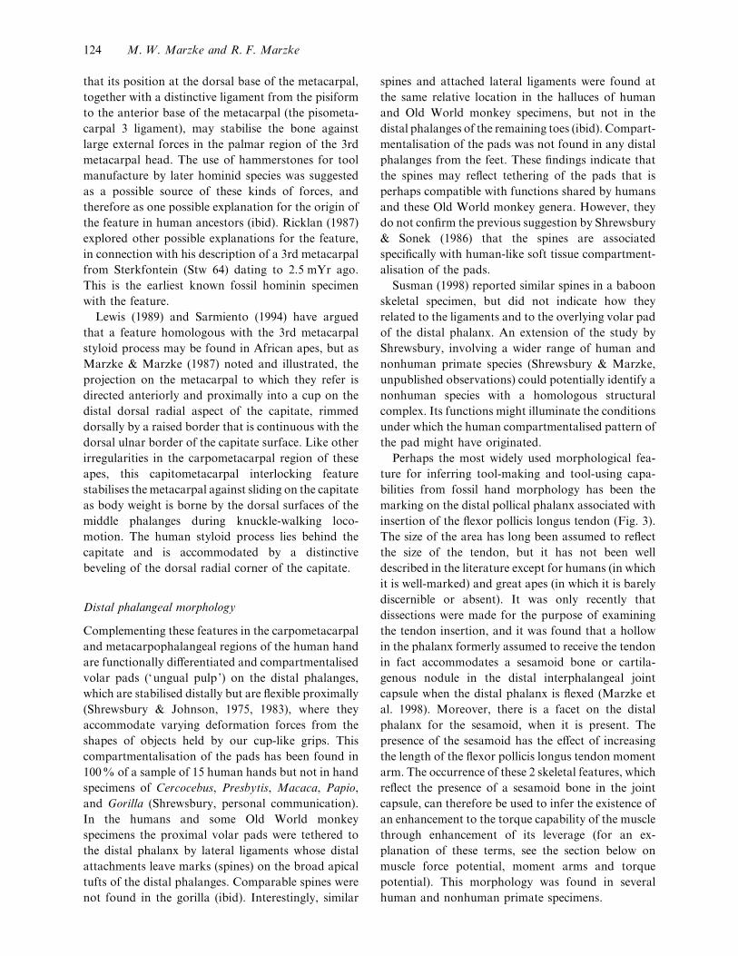
insertion of the flexor pollicis longus tendon (Fig. 3).
The size of the area has long been assumed to reflect
the size of the tendon, but it has not been well
described in the literature except for humans (in which
it is well-marked) and great apes (in which it is barely
discernible or absent). It was only recently that
dissections were made for the purpose of examining
the tendon insertion, and it was found that a hollow
in the phalanx formerly assumed to receive the tendon
in fact accommodates a sesamoid bone or cartila-
genous nodule in the distal interphalangeal joint
capsule when the distal phalanx is flexed (Marzke et
al. 1998). Moreover, there is a facet on the distal
phalanx for the sesamoid, when it is present. The
presence of the sesamoid has the effect of increasing
the length of the flexor pollicis longus tendon moment
arm. The occurrence of these 2 skeletal features, which
reflect the presence of a sesamoid bone in the joint
capsule, can therefore be used to infer the existence of
an enhancement to the torque capability of the muscle
through enhancement of its leverage (for an ex-
planation of these terms, see the section below on
muscle force potential, moment arms and torque
potential). This morphology was found in several
human and nonhuman primate specimens.
124 M. W. Marzke and R. F. Marzke
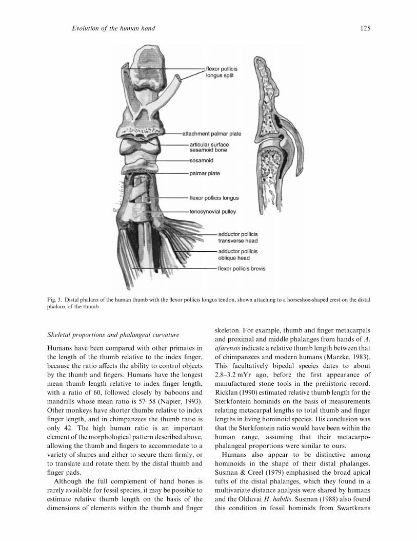
Fig. 3. Distal phalanx of the human thumb with the flexor pollicis longus tendon, shown attaching to a horseshoe-shaped crest on the distal
phalanx of the thumb.
Skeletal proportions and phalangeal curvature
Humans have been compared with other primates in
the length of the thumb relative to the index finger,
because the ratio affects the ability to control objects
by the thumb and fingers. Humans have the longest
mean thumb length relative to index finger length,
with a ratio of 60, followed closely by baboons and
mandrills whose mean ratio is 57–58 (Napier, 1993).
Other monkeys have shorter thumbs relative to index
finger length, and in chimpanzees the thumb ratio is
only 42. The high human ratio is an important
element of the morphological pattern described above,
allowing the thumb and fingers to accommodate to a
variety of shapes and either to secure them firmly, or
to translate and rotate them by the distal thumb and
finger pads.
Although the full complement of hand bones is
rarely available for fossil species, it may be possible to
estimate relative thumb length on the basis of the
dimensions of elements within the thumb and finger
skeleton. For example, thumb and finger metacarpals
and proximal and middle phalanges from hands of A.
afarensis indicate a relative thumb length between that
of chimpanzees and modern humans (Marzke, 1983).
This facultatively bipedal species dates to about
2±8–3±2 mYr ago, before the first appearance of
manufactured stone tools in the prehistoric record.
Ricklan (1990) estimated relative thumb length for the
Sterkfontein hominids on the basis of measurements
relating metacarpal lengths to total thumb and finger
lengths in living hominoid species. His conclusion was
that the Sterkfontein ratio would have been within the
human range, assuming that their metacarpo-
phalangeal proportions were similar to ours.
Humans also appear to be distinctive among
hominoids in the shape of their distal phalanges.
Susman & Creel (1979) emphasised the broad apical
tufts of the distal phalanges, which they found in a
multivariate distance analysis were shared by humans
and the Olduvai H. habilis. Susman (1988) also found
this condition in fossil hominids from Swartkrans
Evolution of the human hand 125
dating to about 1±8 mYr ago. The tufts would have
supported broad distal finger pads, which these
authors associated with human precision grip capa-
bilities. Smith (1995) measured radiographs of chim-
panzee and human hand bones and in a pattern profile
analysis found that modern human distal phalanges
are distinctive in their proportionately broader bases
relative to tuft breadth. She reported that, although
the Olduvai and Swartkrans hands have broad distal
phalangeal thumb tufts, the base width relative to tuft
width is small. This ratio is also characteristic of
chimpanzees, suggesting that different thumb loading
patterns may be indicated. She also suggested that
body size may be a factor in determining tuft width.
Phalangeal curvature is of particular interest in the
analysis of fossils, since the degree of curvature
probably reflects stresses to which the hand was
subjected during locomotor and}or manipulative
activities (Richmond, 1997). Stern & Susman (1983),
calculating an included angle to measure phalangeal
curvature (see also Stern et al. 1995), found that the
phalanges of A. afarensis were more curved than in
modern humans, resembling apes, indicating that this
early hominid species might have used its hands for
arboreal climbing. This ape-like curvature is part of
an interesting mosaic, incorporating features relating
to metacarpal rotation in the radial side of the hand
that could have enhanced the manipulation of natural
objects as tools (Marzke, 1983, 1997).
First metacarpal robusticity
Susman (1994) suggested the possibility of predicting
a capability for ‘refined, humanlike precision grasping
(and its correlate, tool behavior) ’ in fossil hominid
species using a ratio of thumb metacarpal head
breadth to length of the metacarpal. In a comparison
of humans, chimpanzees and bonobos he had found a
significantly larger relative head breadth in humans,
which he attributed to large muscle contraction forces
at the metacarpophalangeal joint. However, Hamrick
& Inouye (1995) and Ohman et al. (1995) found that
humans are not distinctive in having a large meta-
carpal head ratio; the gorilla ratio is within the
human range of variation. They also note the lack of
predictable links between tool behaviour and precision
gripping capabilities.
The identification of morphological predictors of
manipulative behavioural capabilities, even in this
small region of the hand, requires the quantification
of more variables and the identification of significant
correlations among variables. For example, measure-
ments should be made of muscle force potential,
tendon moment arms (whose length affects the
amount of muscle force required to rotate bones at
joints), joint surface areas and joint ranges of motion,
and compared among all hominoid species. In
addition, the species should be compared in the ranges
of thumb and finger movements associated with grips
elicited by their varied tool behaviours.
In summary, comparative anatomical studies of
hand skeletal morphology in living apes and humans
have identified numerous features that together
appear to distinguish the human hand from the hands
of apes, and which facilitate our ability to cup the
hand and accommodate it to the shapes of objects.
However, 3 factors should be taken into account
before applying this knowledge to the functional
interpretation of morphology in fossil hominid hands.
First, the effectiveness of this kind of grip depends
upon the presence of all the morphological com-
ponents set out above, yet rarely are sufficient fossil
hand bones recovered to establish the presence of the
total morphological pattern (Marzke, 1997). Sec-
ondly, few of the morphological features have been
screened for statistically significant differences among
species. Thirdly, experiments are needed to determine
how these morphological features contribute to
human manipulative behaviour. How can we test
these form-function hypotheses? Are these functions
essential in order to fashion early artefacts? Are there
alternative morphological patterns that could ac-
complish the same behaviours?
:
One source of new data relevant to understanding the
workings of the modern human hand is the dis-
tribution of osteoarthritis. Some regions of the hand
today sustain high frequencies of this degenerative
joint disease (DJD), a pathological process that has
been attributed to forceful and repetitive loading in
manipulative activities (Radin et al. 1972). Since the
locations of DJD indicate the location of manipulative
stresses, it is not unreasonable to assume that these
may be the most likely to have undergone mor-
phological change during human evolution in re-
sponse to stresses associated with the manufacture
and use of tools.
For example, DJD at the base of the thumb (in the
126 M. W. Marzke and R. F. Marzke
Fig. 4. Human trapeziometacarpal joint, drawn after Koebke
(1983). Arrow points to the anterior beak of the first metacarpal,
which rides up on the convex anterior surface of the trapezium in
full opposition of the thumb.
trapeziometacarpal joint) is a common and de-
bilitating condition in many human populations,
interfering with many activities of daily living. One
contributing factor appears to be the restricted area of
contact between the metacarpal and the convex
trapezial surface when the thumb is drawn into full
opposition to the fingers (Koebke, 1983; see Fig. 4),
and Koebke found that degeneration of the cartilage
begins in this region. Ateshian et al. (1992) and Xu et
al. (1998) have shown that female humans (known to
have a higher frequency of trapeziometacarpal os-
teoarthritis than males) have significantly greater
reciprocal curvature of the trapezial and metacarpal
surfaces than males, less congruence of the joint,
smaller contact areas, and thus an increased likelihood
of experiencing high contact stresses in small localised
areas for activities with comparable amounts of joint
loading. This finding suggests a possible explanation
for the relatively flat joint in the Olduvai H. habilis
and Neanderthals. Wide excursion of the thumb is
required for opposing the thumb and 4th and 5th
fingers in the manipulation of large stones during tool
manufacture. This activity requires strong, repeated
contraction of the thumb muscles, which would have
loaded the areas of contact between the proximal
anterior beak of the metacarpal and the surface of the
trapezium at their position of full opposition. The
more curved the trapezium and the more projecting
the anterior beak of the metacarpal, the more
restricted this contact area would have been, and thus
the more vulnerable it would have been to de-
generative disease. Thus the relatively flat joint is
consistent with tool-making behaviour (Marzke, 1990,
1992). But if this is the case, why do modern humans
have a more curved joint on the average than the
earlier hominins?
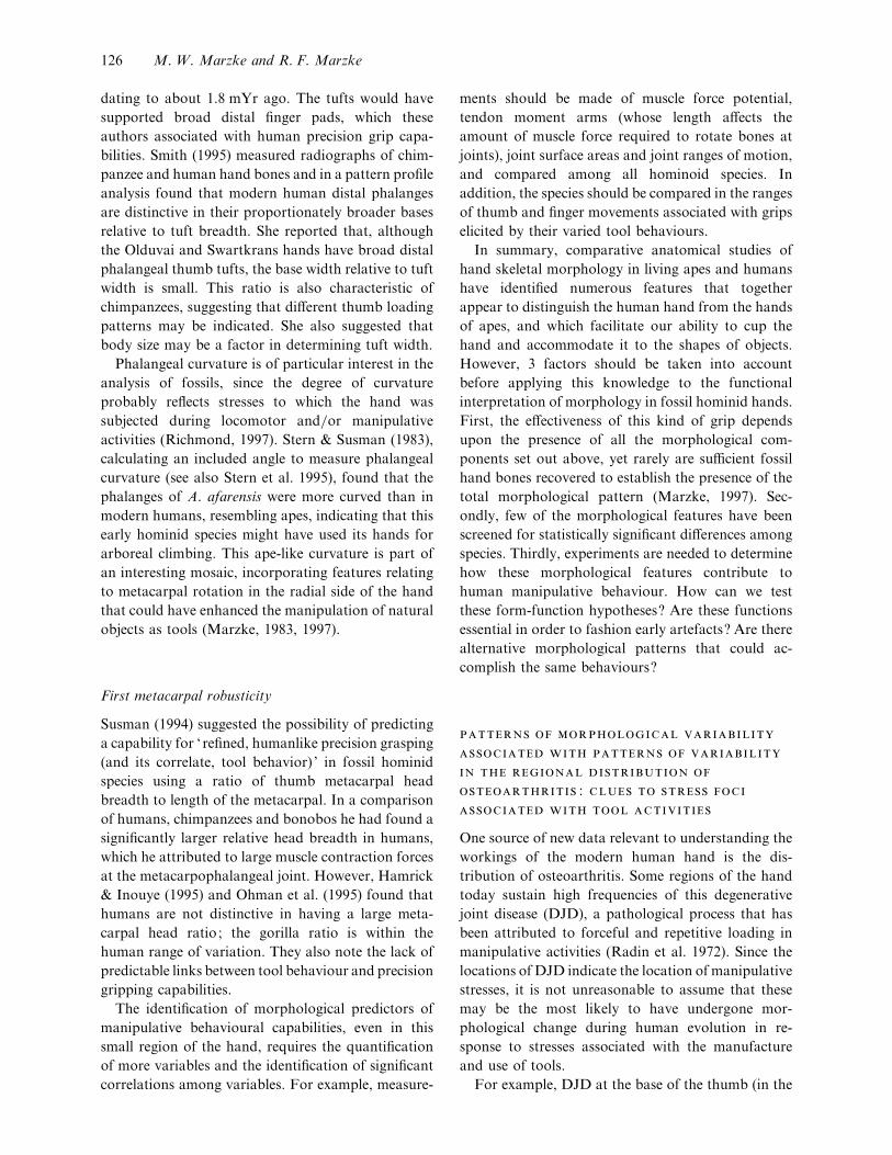
Fig. 5. Two midcarpal articular patterns. Left : type I, the hamate
(H) does not meet the lunate (L). Right : type II, the hamate and
lunate meet, creating a jog in the joint contour.
Another pathology involving the thumb emphasises
the functional interrelationships that may explain the
more curved trapeziometacarpal surfaces in modern
humans. Frequently the thumb metacarpal slips
dorsally and radially off the trapezium when objects
are pinched between the thumb and index finger
(Eaton & Dray, 1982). Mutually curved trapezio-
metacarpal surfaces have the advantage of stabilising
the joint against this kind of subluxation, by locking
of the anterior metacarpal beak (together with its
extension, the anterior oblique ligament) against the
convex surface of the trapezium. This stabilising
morphology appears to be emphasised in chimpanzees
and in A. afarensis, which, compared with modern
humans, have a proportionately longer proximal
anterior beak that restricts sliding and rotation of the
metacarpal on the trapezium (Guthrie, 1991). Thus to
some extent the requirements of human manipulative
activities for both wide excursion and stability of the
thumb metacarpal are contradictory. Comparative
quantitative 3-D data on trapeziometacarpal joint
surface curvature and congruence in humans and apes
are needed to develop models for reconstructing the
activities at joints involved in the manipulative
behaviour of fossil species.
The high frequency of arthritis at the midcarpal
joint (between the 1st and 2nd rows of carpals), and its
link with variations in modern human articular
patterns (Marzke et al. 1994) may also provide clues
to the evolution of the human hand. Viegas et al.
(1991) found that DJD at the proximal pole of the
hamate bone in the wrist occurs only in individuals
who have a blunt joint facet on this part of the hamate
for a corresponding facet on the lunate bone, which
lies proximal to it in the wrist. This pattern is present
in most African apes and is consistent with the
demands of their locomotor behaviour (Marzke et al.
1994; Fig. 5). This is the pattern seen in 65% of the
Evolution of the human hand 127

165 individuals in the Viegas et al. (1991) modern
human cadaver sample. In the remaining 35%, the
proximal hamate formed with the capitate a con-
tinuous curved surface for radioulnar deviation on the
lunate and triquetrum, without creating a jog at the
midcarpal joint. A functionally similar pattern is seen
in Asiatic apes and is more compatible with repeated
radioulnar movements of the wrist. The differential
occurrence of cartilage damage in humans suggests an
evolutionary explanation for midcarpal joint varia-
bility in humans. The pattern that appears in 65% of
humans, in spite of its vulnerability to damage
associated with activities requiring midcarpal radio-
ulnar deviation, may be the remnant of an earlier
adaptation for wrist stability, possibly relating to
locomotor behaviour before the hand was used
exclusively for the manipulation of tools and other
objects. The relatively high frequency (35%) of the
alternative pattern, which appears to be more com-
patible with human manipulative behaviour, may be
the result of selection that occurred as the hand
became used increasingly for manipulative activities
requiring repetitive, forceful midcarpal radioulnar
deviation.
It is too early to assess the effectiveness of localised
pathology as a proxy for biomechanical studies, but
the examination of fossil hominid hands for combin-
ations of morphological features with known DJD
correlates will throw interesting light on the sequence
and possible causes of changes in joint morphology
through time (Marzke et al. 1994).
,
A combination of Napier’s observational and ex-
perimental approaches to the analysis of the functions
of the hands of early fossil hominids is throwing new
light on possible links between hand morphology and
the manufacture and manipulation of tools. These
studies address the broad question of how form and
function relationships can be used to reconstruct the
manipulative skills of humans, apes and monkeys. In
our group we have investigated the following
questions. (1) What grips are required to control
tools, retrieve and process foods, groom other
animals, and build nests? (2) What ranges and kinds
of movement are associated with these manipulative
grips and activities? (3) What regions of the hand are
stressed by these grips and activities? By looking for
behavioural and environmental correlates for
between-species variation in hand morphology, and
then investigating possible functional links between
the morphological features, it should become possible
to draw some reasonable inferences about manipu-
lative and locomotor capabilities of the hand from
fossil evidence.
Experimental manufacture of prehistoric tools by
humans
This approach is based on the hypothesis that hand
morphology and prehistoric tool manufacture and use
evolved concomitantly during the Plio-Pleistocene.
Among activities that are likely to have required
morphological adjustments would have been the use
of unmodified stone, bone, horn and wood and the
manufacture of tools based on these raw materials.
Two experimental approaches have been used. First,
to what extent are ape-like hands capable of making
the earliest Oldowan stone tools? Secondly, at what
stage in the archaeological record do we see tools that
would have required the functional capabilities of a
modern human hand for their manufacture and use?
Napier (1962, 1964) and Krantz (1960) used the first
experimental approach when they attempted to
replicate Oldowan tools by mimicking an ape-like
power grip using the flexed fingers without opposition
of the thumb. They concluded that human-like
precision grips were not necessary for the manufacture
of these artefacts (Napier, 1964), although Krantz
(op. cit.) conceded that the lack of fine-tuned
thumb}finger movements affected precision, as did
the need to use large hammerstones. Marzke &
Shackley (1986) used the second approach, when they
examined the demands placed on modern human
hands during the manufacture and use of prehistoric
stone tools. They reasoned that if specific human grips
and hand movements were consistently elicited by
these activities, and if specific regions of the hand were
consistently stressed by the activities, it is reasonable
to assume that the complex of morphological features
that enables these grips would be most likely to have
evolved as adaptations to these tool-making and tool-
using activities as hominins became increasingly
dependent upon stone, wood and bone tools.
The initial findings using this approach, based upon
a qualitative analysis of films of the activities (Marzke
& Shackley, 1986), was that there is indeed a specific
group of grips elicited by the tool-making and tool-
using activities. During the use of hard hammer
percussion for the removal of flakes from cores in the
production of Oldowan-type tools, the core was
128 M. W. Marzke and R. F. Marzke

Fig. 6. Contrasts between: (a) a firm precision cradle grip of a stone
core; (b) a 3-jaw chuck precision pinch grip of a hammerstone; (c)
a delicate precision grip by the tips of the thumb and fingers and (d )
a spherical power grip. The delicate precision grip would not resist
displacement of the core by a hammerstone during the removal of
flakes. The spherical power grip encloses the stone, exposing the
fingers to damage by the hammerstone. The firm precision cradle
and 3-jaw chuck grips resist displacement of the core, but allow
exposure of the working edge of the core for safe flake removal.
generally held by a cradle grip (Fig. 6a), in which it is
supported by the pads of the fingers, and secured by
the opposing pressure of the thumb pad. A 3-jaw
chuck (baseball) grip of the hammerstone, involving
the thumb, index and third finger pads (Fig. 6b) was
used exclusively. These 2 grips combine elements of
the grip categories established by Napier (1956),
precision (Fig. 6c) and power (Fig. 6d ), and have
been referred to as ‘ forceful precision grips ’ (Marzke
& Wullstein, 1996). The element that forceful pre-
cision grips shares with the power grip is firmness,
but their advantage is that by securing the grip with
the thumb and finger pads (Fig. 6a, b) rather than
against the palm in a power grip (Fig. 6d ), the
working surfaces of smaller stones can remain exposed
for flaking, thus avoiding injury to the fingers. They
share with precision grips the ability to make precise
alterations to the orientation of the core and}or
hammerstone for each strike by moving the thumb
and finger joints. This important element of human
manipulative behaviour was called ‘precision hand-
ling’ by Landsmeer (1962). Both these grips require
convergence of the thumb and finger pads, and the
large stresses generated by each hammerstone strike
must be accommodated. It is hypothesised that these
requirements are met by the distinctive combination
of features in humans described above in the section
on skeletal morphology, that facilitate cupping of the
hand and the firm opposition of the thumb pad to the
pads of all the fingers.
The same experiments (Marzke & Shackley, 1986)
demonstrated that prehistoric stone tool use required
the same manipulative abilities and morphology as
stone tool manufacture. When the flakes produced by
the hard hammer technique were used for cutting and
scraping, a firm pad-to-side precision grip made by
the thumb and the side of the index finger was used.
This requires a strong thumb and rotation of the index
finger. The grasp of cylindrical objects (e.g. wood and
bone clubs) consistently prompted the use of the
squeeze form of power grip, in which the thenar and
hypothenar regions of the palm, as well as the flexed
fingers, squeeze the tool diagonally, so that it
functions as an extension of the forearm as it is
brought into contact with its target. The modern
human ability to rotate the 5th metacarpal towards
the opposed thumb metacarpal, to cup the palm,
together with thumb}finger proportions that allow
the thumb to stabilise objects at the level of the flexed
fingers, are the cardinal elements of the ability to
retain the core in the hand for one-handed clubbing
and pounding (Marzke et al. 1992).
The tool replication experiments suggest a possible
link between the evolution of human hand mor-
phology and the evolution of tools, but how do we test
the hypothesis that the human hand evolved in
adaptation to these activities? How can we justify
using the complex of morphological features outlined
above as a model for inferring tool-making and tool-
using capabilities from fossil hominid hands? Fol-
lowing the reasoning of Lauder (1995, 1996), an
attempt can be made to explain a unique mor-
phological pattern with experiments comparing per-
formance capabilities for the hypothesised activities in
other species with a different, but closely-related
morphology. As Lauder has made clear, there are
substantial weaknesses in this approach for predicting
how structures might have been recruited for use in
fossils, but he acknowledged that this biomechanical
approach may allow researchers to ‘define the realm
of the possible ’ for fossils (Lauder, 1995).
In the next section we compare the abilities of
modern humans with those of nonhuman primate
species to maintain a grip on objects of varying shapes
by each hand separately during activities similar to
prehistoric human tool making and tool use. In these
activities, it is imperative that the working edges of the
manipulated objects remain exposed, and the comp-
lementary grips must be maintained against strong
external forces.
Evolution of the human hand 129
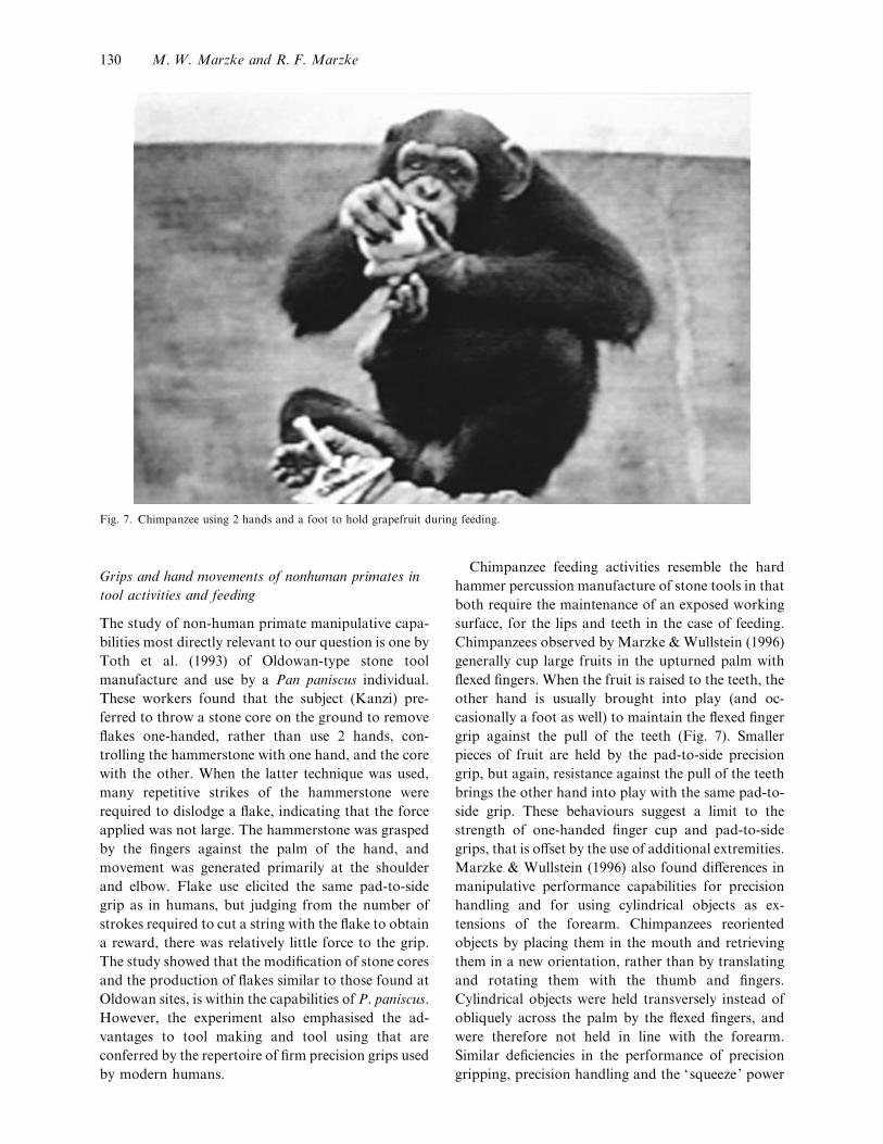
Fig. 7. Chimpanzee using 2 hands and a foot to hold grapefruit during feeding.
Grips and hand movements of nonhuman primates in
tool activities and feeding
The study of non-human primate manipulative capa-
bilities most directly relevant to our question is one by
Toth et al. (1993) of Oldowan-type stone tool
manufacture and use by a Pan paniscus individual.
These workers found that the subject (Kanzi) pre-
ferred to throw a stone core on the ground to remove
flakes one-handed, rather than use 2 hands, con-
trolling the hammerstone with one hand, and the core
with the other. When the latter technique was used,
many repetitive strikes of the hammerstone were
required to dislodge a flake, indicating that the force
applied was not large. The hammerstone was grasped
by the fingers against the palm of the hand, and
movement was generated primarily at the shoulder
and elbow. Flake use elicited the same pad-to-side
grip as in humans, but judging from the number of
strokes required to cut a string with the flake to obtain
a reward, there was relatively little force to the grip.
The study showed that the modification of stone cores
and the production of flakes similar to those found at
Oldowan sites, is within the capabilities of P. paniscus.
However, the experiment also emphasised the ad-
vantages to tool making and tool using that are
conferred by the repertoire of firm precision grips used
by modern humans.
Chimpanzee feeding activities resemble the hard
hammer percussion manufacture of stone tools in that
both require the maintenance of an exposed working
surface, for the lips and teeth in the case of feeding.
Chimpanzees observed by Marzke & Wullstein (1996)
generally cup large fruits in the upturned palm with
flexed fingers. When the fruit is raised to the teeth, the
other hand is usually brought into play (and oc-
casionally a foot as well) to maintain the flexed finger
grip against the pull of the teeth (Fig. 7). Smaller
pieces of fruit are held by the pad-to-side precision
grip, but again, resistance against the pull of the teeth
brings the other hand into play with the same pad-to-
side grip. These behaviours suggest a limit to the
strength of one-handed finger cup and pad-to-side
grips, that is offset by the use of additional extremities.
Marzke & Wullstein (1996) also found differences in
manipulative performance capabilities for precision
handling and for using cylindrical objects as ex-
tensions of the forearm. Chimpanzees reoriented
objects by placing them in the mouth and retrieving
them in a new orientation, rather than by translating
and rotating them with the thumb and fingers.
Cylindrical objects were held transversely instead of
obliquely across the palm by the flexed fingers, and
were therefore not held in line with the forearm.
Similar deficiencies in the performance of precision
gripping, precision handling and the ‘squeeze’ power
130 M. W. Marzke and R. F. Marzke

grip are reported for capuchin monkeys by Wester-
gaard & Suomi (1997), baboons (Guthrie, 1991; Jude,
1993), and orangutans (Marzke & Wullstein, 1996).
:
Comparative studies of hand morphology and ma-
nipulative behaviour focused attention on an inter-
esting pattern of capabilities and morphology that
appears to distinguish modern human manipulative
behaviour from that of other species. The distinctive-
ness is not confined to precision vs power grips, but is
based on the emphasis of modern humans on forceful
grips and precision handling, within the precision
category, and on the squeeze grip within the power
category. These grips, and related thumb}finger
movements, allow for the one-handed control of
stones, wood, bone and horn. Any models for modern
human manipulation must overcome the weaknesses
of earlier models that were based on isolated elements
of an overall pattern (Marzke, 1997).
Susman (1998) recommends ignoring the diversity
of grips within the precision and power categories in
discussions of hominid evolution and prehistoric tool
use and tool making. However, as Marzke & Shackley
(1986), Marzke & Wullstein (1996), Marzke (1997)
and Marzke et al. (1998) have shown, it is the
discovery of this diversity in the course of our research
that in fact has led to a much deeper understanding of
morphological variations among living anthropoid
primates than previously existed. Lewis (1989, pp.
114–115), concurred with the use of a more elaborate
classificatory scheme in the evolutionary analysis of
hominid morphology, noting that the power and
precision grips (as defined by Napier 1956) both
recruit features (in different combinations) that
together he considered to be apomorphic in humans.
A developing awareness of the behavioural and
morphophysiological complexity of manipulation is
indeed generating the interesting questions that will
guide us in the development of more detailed models
relevant to the functional analysis of fossil hominid
hands in the future.
Models being developed require data on structure
and functions that are only now becoming available.
These include information about the patterns of
muscle recruitment, and ranges of joint movement
collected from living subjects during the manufacture
and use of prehistoric tools, estimates of muscle
torque potential estimated from muscle fibre length
measurements and ranges of passive joint movement
and tendon excursion in cadaver limb specimens, free
body analysis of stresses associated with hard hammer
percussion manufacture of stone tools, and 3-D
imaging and measurement of joint surface areas and
curvatures. In addition to these, comparative studies
of bone density, trabecular patterns, cortical dis-
tribution, and area moments of inertia contribute
important kinetic data for biomechanical models.
Brief descriptions of the techniques used in these
experiments, with consideration of their potential as
well as their limitations for addressing our evol-
utionary questions, are given below.
It is important to keep in mind that models will be
more effective at predicting the performance of fossil
morphologies if they are based on activities that are
generic to the living species under study. The
musculoskeletal system and its neural control are very
flexible, and can be recruited for a much wider range
of activities than those for which they are regularly
used. Thus modern humans can swing from bars by
contracting their finger flexor muscles like apes, and
apes can remove flakes from tools by holding large
stones by the thumb and fingers like modern humans.
Functional morphologists must therefore identify and
focus upon the activities that elicit different musculo-
skeletal functional patterns, which in turn are facil-
itated by different patterns of associated mor-
phologies.
EMG analysis of muscle recruitment during
prehistoric tool making and tool use
In order to generate realistic models, we need to
know, for a given bone-ligament-tendon system,
which muscles are active during manipulation. One of
the traditional approaches to these questions is the use
of electromyography (EMG) to record the electrical
signal produced by muscle contraction during pre-
scribed activities. The approach is an important first
stage in testing hypotheses about possible functional
links between morphology and behaviours. It directs
attention to the muscles that are most likely to have
been consistently and strongly recruited in the
behaviour of interest, and thus targets for further
analysis skeletal regions that have the potential to
reveal evidence about the musculoskeletal adaptations
associated with the grips, ranges of motion and
stresses relating to these behaviours.
In the past, inferences about hand use have been
made from the size of muscle markings on fossil
hominid hand bones. For example, well-developed
Evolution of the human hand 131
attachments for flexor digitorum superificialis muscle
on the middle phalanges of the Olduvai H. habilis
hand have been interpreted as evidence for arboreal
climbing (Susman & Creel, 1979) ; a large opponens
pollicis insertion crest on a metacarpal from Swart-
krans is interpreted as evidence for tool making
(Napier, 1959) ; and a large excavation in the region of
the flexor pollicis longus tendon has suggested human-
like tool-making capabilities in the Olduvai H. habilis
(Napier, 1962) and the Swartkrans early hominid
(Susman, 1988). Forceful contraction, occurring re-
peatedly, does appear to cause some bone markings in
modern humans at muscle attachments (Welton et al.
1994). However, it should be emphasised that at the
present time there are no published data on the
correlation between muscle force generated and the
size of muscle markings, and it is not known whether
it is the maximum strength of contraction, or the
repetition of weaker contractions, or both, that
generate the muscle markings on bone.
An EMG study was undertaken to test these
inferences and to move beyond speculation to direct
examination of the patterns of muscle recruitment
associated with the manufacture and use of prehistoric
tools. Modified stones are virtually the only direct
evidence we have of early hominin tool making and
tool use, but hominin hands were probably used in a
much wider range of food-collecting and food-
processing activities requiring forceful use of the
hands alone for pulling and breaking fibrous
materials, and the use of bones, horn and wood. The
activities monitored in our experiments so far have
therefore included: (1) the manufacture and use of
stone tools, such as hard hammer percussion manu-
facture of Oldowan-type tools (Marzke et al. 1998),
side scraping of hides with a stone flake (Reece et
al.1997), end scraping, meat-cutting with flakes, bone-
chopping with stones to acquire marrow, nut-
pounding with stones, the manufacture of Acheulean
hand axes, and pressure flaking; (2) stone-throwing;
(3) digging with long sticks and with short bones and
horns; and (4) clubbing with bones and small tree
branches. The subjects were an archaeologist with
extensive experience in the replication of prehistoric
tools, and who makes them on a regular basis, a
second archaeologist also with considerable experi-
ence in tool replication, and a physical anthropologist
who had only recently begun experimenting with
techniques to replicate and use prehistoric tools. A
subsequent study (Marzke & He, unpublished) with 5
subjects incorporated a CyberGlove, which recorded
thumb and finger positions simultaneously with
muscle recruitment.
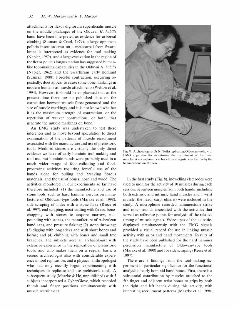
Fig. 8. Archaeologist (Dr N. Toth) replicating Oldowan tools, with
EMG apparatus for monitoring the recruitment of his hand
muscles. A microphone near his left hand registers each strike by the
hammerstone on the core.
In the first study (Fig. 8), indwelling electrodes were
used to monitor the activity of 10 muscles during each
session. Seventeen muscles from both hands (including
both extrinsic and intrinsic hand muscles and 1 wrist
muscle, the flexor carpi ulnaris) were included in the
study. A microphone recorded hammerstone strike
and other sounds associated with the activities that
served as reference points for analysis of the relative
timing of muscle signals. Videotapes of the activities
displayed simultaneously with the EMG signals
provided a visual record for use in linking muscle
activity with grips and hand movements. Results of
the study have been published for the hard hammer
percussion manufacture of Oldowan-type tools
(Marzke et al. 1998) and for side scraping (Reece et al.
1997).
There are 3 findings from the tool-making ex-
periment of particular significance for the functional
analysis of early hominid hand bones. First, there is a
substantial contribution by muscles attached to the
5th finger and adjacent wrist bones to grips by both
the right and left hands during this activity, with
interesting recruitment patterns (Marzke et al. 1998;
132 M. W. Marzke and R. F. Marzke
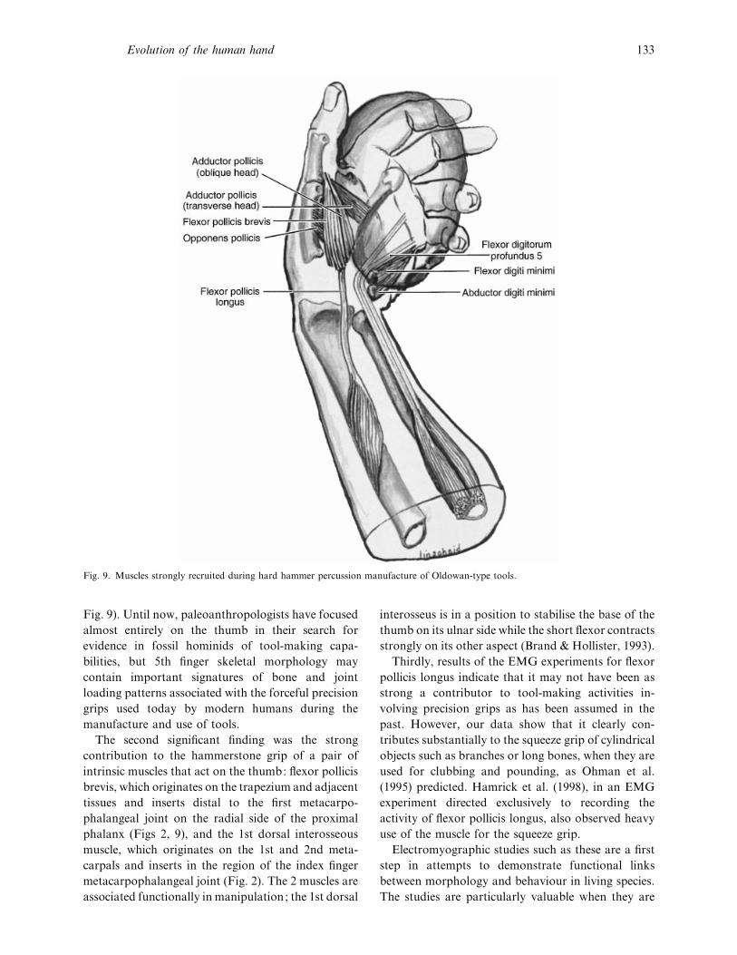
Fig. 9. Muscles strongly recruited during hard hammer percussion manufacture of Oldowan-type tools.
Fig. 9). Until now, paleoanthropologists have focused
almost entirely on the thumb in their search for
evidence in fossil hominids of tool-making capa-
bilities, but 5th finger skeletal morphology may
contain important signatures of bone and joint
loading patterns associated with the forceful precision
grips used today by modern humans during the
manufacture and use of tools.
The second significant finding was the strong
contribution to the hammerstone grip of a pair of
intrinsic muscles that act on the thumb: flexor pollicis
brevis, which originates on the trapezium and adjacent
tissues and inserts distal to the first metacarpo-
phalangeal joint on the radial side of the proximal
phalanx (Figs 2, 9), and the 1st dorsal interosseous
muscle, which originates on the 1st and 2nd meta-
carpals and inserts in the region of the index finger
metacarpophalangeal joint (Fig. 2). The 2 muscles are
associated functionally in manipulation; the 1st dorsal
interosseus is in a position to stabilise the base of the
thumb on its ulnar side while the short flexor contracts
strongly on its other aspect (Brand & Hollister, 1993).
Thirdly, results of the EMG experiments for flexor
pollicis longus indicate that it may not have been as
strong a contributor to tool-making activities in-
volving precision grips as has been assumed in the
past. However, our data show that it clearly con-
tributes substantially to the squeeze grip of cylindrical
objects such as branches or long bones, when they are
used for clubbing and pounding, as Ohman et al.
(1995) predicted. Hamrick et al. (1998), in an EMG
experiment directed exclusively to recording the
activity of flexor pollicis longus, also observed heavy
use of the muscle for the squeeze grip.
Electromyographic studies such as these are a first
step in attempts to demonstrate functional links
between morphology and behaviour in living species.
The studies are particularly valuable when they are
Evolution of the human hand 133
followed by biomechanical studies on cadaver hands,
described in the next section, which measure the cross
sectional areas and tendon moment arms of the
muscles whose functions are found to play an
important role in hypothesised prehistoric behaviours.
However, there are limitations to the kinds of
information that can be acquired with EMG. A few
cautionary comments are given below, along with a
discussion of what can be learned from these studies
that is relevant to functional analyses of fossil hominid
hand structure.
Size of electromyographic signals. It is well-known
that there is not a precise relationship between the
amount of muscle tension and the magnitude of the
voltage electrical signal recorded by an electrode.
Reasons for difficulties in finding the relationship, and
a discussion of approaches to investigating it may be
found in Herzog et al. (1994). The size of a signal also
does not reflect the relative importance of a muscle in
an activity. Muscles have multiple roles in joint
movements as prime movers (causing motion in the
desired direction), synergists (augmenting the move-
ment and stabilising joints against unwanted ancillary
movements) and agonists (helping to control motion
in the desired direction). The relative amount of
contraction will thus vary with any change in the role
of a muscle. It also will be affected by the length of its
moment arm, which affects its leverage. A muscle with
a long moment arm could make the same contribution
to torque at a joint as another muscle with more
fibres, but with a shorter moment arm.
Cross talk. Neighbouring muscles may convey
similar signals because of ‘cross talk’, in which an
electrode picks up a signal from a muscle nearby. We
address this potential for error by looking for evidence
of identity between muscle signals during the ex-
periment, and change the combinations of muscles
monitored if there is evidence for cross talk. The use
of indwelling rather than surface electrodes reduces
these artifacts, as well as those associated with
intervening tissues between surface electrodes and the
muscles they monitor.
Relative amounts of activity of a given muscle during
different behaviours. Two kinds of information may be
gleaned from the EMG that are relevant to functional
morphological studies. One is the relative amounts of
activity of a given muscle during different behaviours.
The other is the temporal pattern of recruitment for
the full set of muscles during each behaviour.
Since subjects vary in the total amount of force that
can be applied to functions such as gripping, their
signals must be compared using relative rather than
absolute scales. A standard baseline may be es-
tablished either by: (1) having a subject generate
maximum possible signals for each action, such as
each grip in the experiment described above, and
calculating a percentage level of each action during
the experimental sessions relative to the baseline; or
(2) using as a baseline the largest signal generated by
the action during the full experimental session.
Consistency among individuals in high recruitment
level for a given muscle in a specific action is the most
suggestive evidence we can obtain of a behaviour that
might explain a muscle attachment marking in a
fossil. High recruitment levels of functional muscle
groups in an activity are especially informative about
the kind of activity indicated by large muscle markings
for these groups.
Temporal pattern of muscle recruitment. Observing
the temporal pattern of muscle recruitment relative to
stages in an activity cycle can help to explain patterns
of markings on bone for muscles that do not
necessarily cocontract. For example, the use of
hammerstones elicits signals in the intrinsic thumb
muscles prior to strike and in the 5th finger muscles
after strike. Thus a single activity might explain the
presence of marks for both sets of muscles in a fossil
hand.
Hand muscle force potential, moment arms and
torque potential
Having targeted with EMG studies regions of the
hand whose skeletal morphology is most likely to
reflect torque capabilities of muscles important in tool
making and tool using, our research group proceeded
to a full comparative kinematic study of these regions
in a cadaver sample of hominoid hands. This work
expanded upon the classic study of hominoid hand
musculature by Tuttle (1967, 1969), who compared
genera in dry muscle weights. These weights are
proportional to the work potential of a muscle
(force¬distance through which the muscle contracts).
Our study (Marzke et al. 1999) took muscle cross-
sectional area and tendon moment arms into account
in comparing apes and humans in potential muscle
torque (i.e., in the ability of each muscle to rotate a
bone at a joint). The cross-sectional area is pro-
portional to the force a muscle can generate and the
moment arm lengths affect the leverage of a muscle.
For a muscle of a given force potential, its potential
torque increases in proportion to the length of its
moment arm. Marzke et al. (1999) estimated muscle
potential force using the approach of Brand et al.
(1981), in which the muscle fibre length is measured
with digital calipers, muscle volume is determined by
134 M. W. Marzke and R. F. Marzke
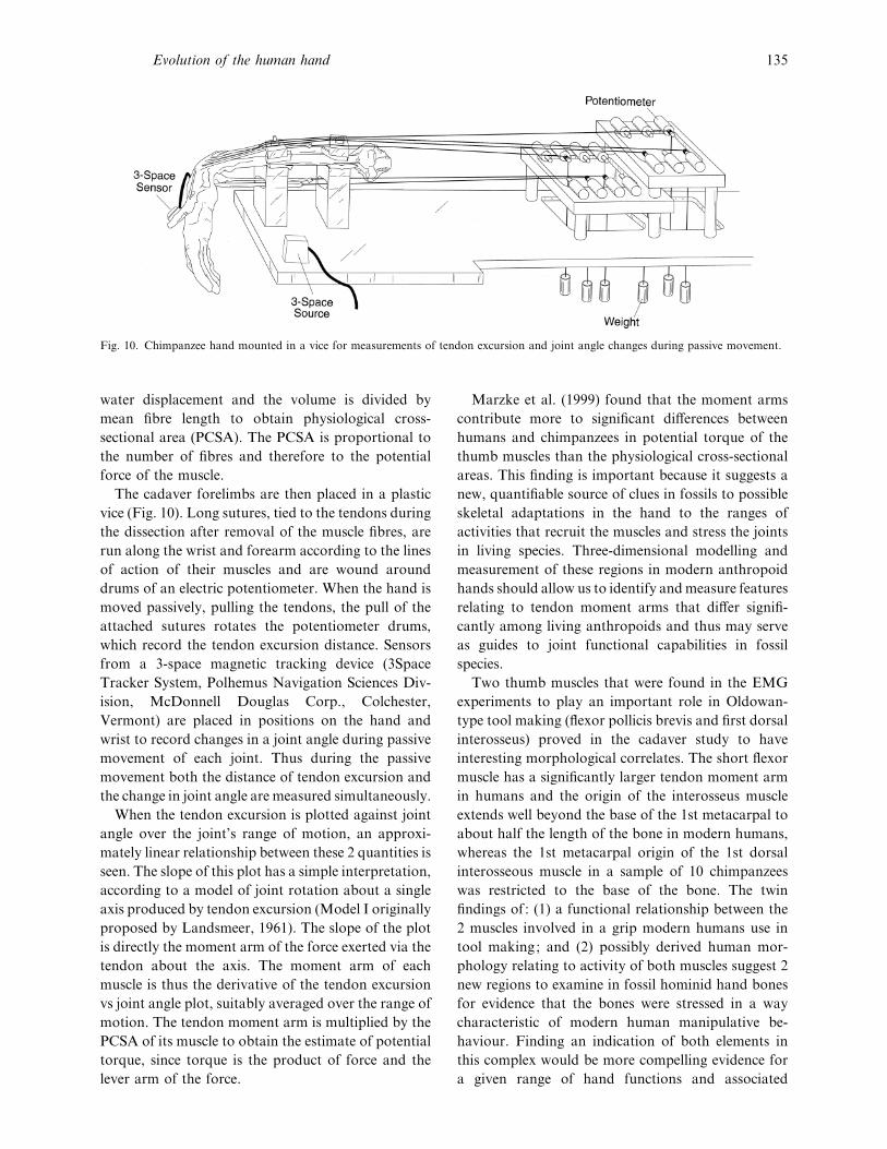
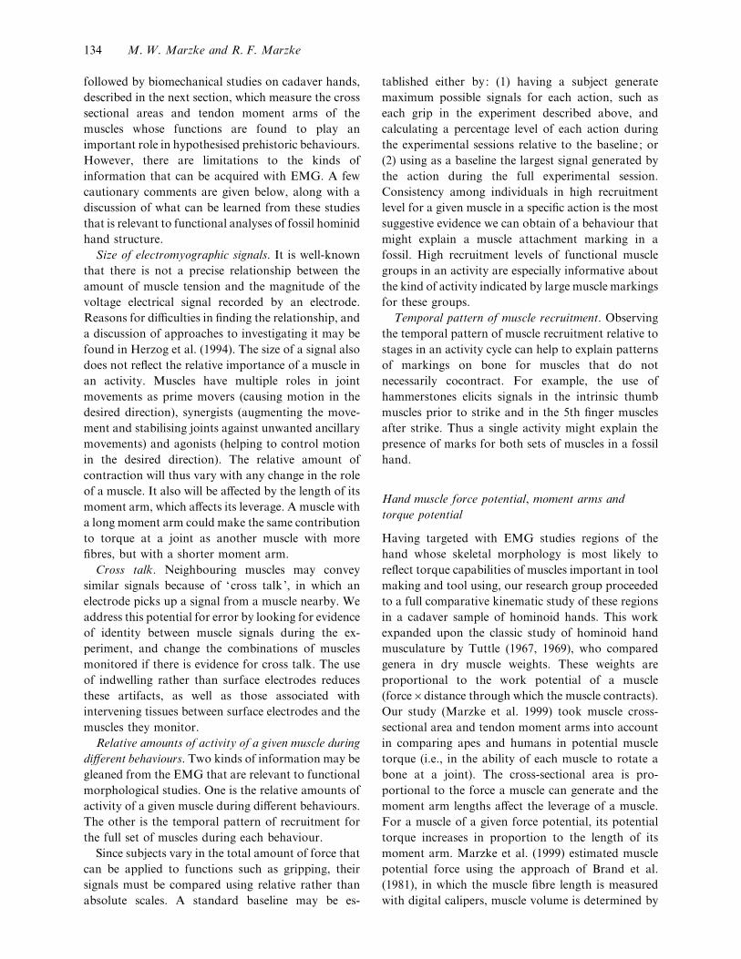
Fig. 10. Chimpanzee hand mounted in a vice for measurements of tendon excursion and joint angle changes during passive movement.
water displacement and the volume is divided by
mean fibre length to obtain physiological cross-
sectional area (PCSA). The PCSA is proportional to
the number of fibres and therefore to the potential
force of the muscle.
The cadaver forelimbs are then placed in a plastic
vice (Fig. 10). Long sutures, tied to the tendons during
the dissection after removal of the muscle fibres, are
run along the wrist and forearm according to the lines
of action of their muscles and are wound around
drums of an electric potentiometer. When the hand is
moved passively, pulling the tendons, the pull of the
attached sutures rotates the potentiometer drums,
which record the tendon excursion distance. Sensors
from a 3-space magnetic tracking device (3Space
Tracker System, Polhemus Navigation Sciences Div-
ision, McDonnell Douglas Corp., Colchester,
Vermont) are placed in positions on the hand and
wrist to record changes in a joint angle during passive
movement of each joint. Thus during the passive
movement both the distance of tendon excursion and
the change in joint angle are measured simultaneously.
When the tendon excursion is plotted against joint
angle over the joint’s range of motion, an approxi-
mately linear relationship between these 2 quantities is
seen. The slope of this plot has a simple interpretation,
according to a model of joint rotation about a single
axis produced by tendon excursion (Model I originally
proposed by Landsmeer, 1961). The slope of the plot
is directly the moment arm of the force exerted via the
tendon about the axis. The moment arm of each
muscle is thus the derivative of the tendon excursion
vs joint angle plot, suitably averaged over the range of
motion. The tendon moment arm is multiplied by the
PCSA of its muscle to obtain the estimate of potential
torque, since torque is the product of force and the
lever arm of the force.
Marzke et al. (1999) found that the moment arms
contribute more to significant differences between
humans and chimpanzees in potential torque of the
thumb muscles than the physiological cross-sectional
areas. This finding is important because it suggests a
new, quantifiable source of clues in fossils to possible
skeletal adaptations in the hand to the ranges of
activities that recruit the muscles and stress the joints
in living species. Three-dimensional modelling and
measurement of these regions in modern anthropoid
hands should allow us to identify and measure features
relating to tendon moment arms that differ signifi-
cantly among living anthropoids and thus may serve
as guides to joint functional capabilities in fossil
species.
Two thumb muscles that were found in the EMG
experiments to play an important role in Oldowan-
type tool making (flexor pollicis brevis and first dorsal
interosseus) proved in the cadaver study to have
interesting morphological correlates. The short flexor
muscle has a significantly larger tendon moment arm
in humans and the origin of the interosseus muscle
extends well beyond the base of the 1st metacarpal to
about half the length of the bone in modern humans,
whereas the 1st metacarpal origin of the 1st dorsal
interosseous muscle in a sample of 10 chimpanzees
was restricted to the base of the bone. The twin
findings of : (1) a functional relationship between the
2 muscles involved in a grip modern humans use in
tool making; and (2) possibly derived human mor-
phology relating to activity of both muscles suggest 2
new regions to examine in fossil hominid hand bones
for evidence that the bones were stressed in a way
characteristic of modern human manipulative be-
haviour. Finding an indication of both elements in
this complex would be more compelling evidence for
a given range of hand functions and associated
Evolution of the human hand 135

behaviours than finding an indication of one. In fact,
there is a potential third element in this behavioural}morphological pattern, the oblique portion of ad-
ductor pollicis, which originates in the region of the
base of the 2nd metacarpal and inserts distal to the
ulnar side of the 1st metacarpophalangeal joint. Both
the physiological cross-sectional area and the moment
arm of this muscle for flexion of the trapezio-
metacarpal joint were significantly larger in humans
compared with chimpanzees. This muscle functions
with the short flexor in stabilising the joint during firm
grips (Brand & Hollister, 1993). The moment arms of
both muscles would be affected by the orientation of
the thumb relative to the metacarpals, by the degree of
palmar projection of the metacarpal head and}or by
the size of the sesamoid bones, which tend to occur in
the joint capsule.
Modelling bone and joint surfaces
Stereophotogrammetry. In recent years several tech-
niques have become available for 3-D modelling of
bone and joint surfaces, thus enabling quantitative
comparisons of the joint surface of individuals, sexes
and species. This will enable the quantification of
skeletal contours that affect joint movement, load
transfer and tendon moment arms. The hypotheses
that the joint regions of modern human hands are
unique, based upon the qualitative observations and
simple measurements described at the beginning of
this review, can therefore be tested quantitatively.
Two techniques for obtaining 3-D images are stereo-
photogrammetry and laser scanning. Stereophoto-
grammetry involves photography of a surface from 2
or 3 directions. The bone is mounted in a calibrated
frame and a grid is projected onto the joint surface by
a projector, providing intersection points for sub-
sequent digitising. Borders of joint surfaces and other
skeletal features can be easily marked for identifi-
cation in the photographic images. Initially precision
format cameras were used (Ateshian et al. 1992), but
more recently digital cameras have been used to feed
the images directly into the computer for digitising
and subsequent analysis (Marzke et al. unpublished
observations). Resolution up to approximately 60 µm
is possible, allowing for comparison of small joint
surfaces, for example in the wrist and ankle. By
digitising the images it is possible to reconstruct 3-D
models of joint surfaces in the computer, which can
then be compared among individuals, sexes, popu-
lations or species in area and curvatures as Ateshian et
al. (1992) have done for the trapeziometacarpal joint.
This technique has the advantage of portability to
museums and laboratories for use in obtaining images
of large samples of joint surfaces from skeletons of
contemporary species and from fossil bones. All that
is required are a digital camera and tripod, laptop
computer, and a slide with the grid. Projectors are
usually available at the museum or laboratory site. A
disadvantage of the technique is that it currently
requires hand digitising of the images. This drawback
should be overcome in the near future with software
that can reference data points from the multiple
images.
Laser scanning and digitising. Laser scanning
systems (e.g. Cyberware Inc.) have the advantage of
both capturing images and digitising them with
currently available software. Scanners of varying sizes
are available for obtaining images of whole living
bodies or small joint surfaces with up to 200 µm
resolution. The current drawback of the technique is
that the margins of joint surfaces may not be clearly
recognisable in the images and may therefore need to
be defined prior to imaging with markers if com-
parative measurements are to be made (Aiello et al.
1998). Systems are near the limit for portability, which
is also constrained by the expense of having more than
one system to allow for data capture and analysis at
the home institution while the other is away from the
laboratory. Kappelman (1998) provided an excellent
review of the state of the art for this technique,
addressing some of the current problems in its
application to research in functional morphology and
paleoanthropology.
Software for measuring and comparing in detail the
3-D topography of joint surfaces imaged by the 2
techniques is not widely available at present, but has
been written for specific applications (e.g. Ateshian,
1993; Steinberg, 1999) and is becoming available to
the public. Aiello et al. (1998) devised a technique for
limited comparison of sections through the joint
surfaces in a study of joint congruency.
Application of cross-sectional, radiographic,
computerised tomographic (CT ) and magnetic
resonance imaging (MRI ) techniques to modelling of
hand biomechanics
Metacarpal shape and bone mass distribution measured
on metacarpal sections and biplanar radiographs. Data
from scans of metacarpal cross-sections from a large
modern human skeletal sample have been used by
Lazenby (1998) to model metacarpal shape and bone
mass distribution. His results point to differences
136 M. W. Marzke and R. F. Marzke
between right and left hands, and between sexes and
age groups, suggesting that there are likely to be
patterns of variation between groups of individuals
associated with occupations that differentially recruit
the hands. Any such patterns might serve as models
for the interpretation of fossil hominid metacarpal
shape and bone mass distribution. Coffing’s (1998)
finding of differences between A. afarensis and modern
humans in metacarpal cortical mass thickness and
distribution, using biplanar radiographs, has raised
interesting questions about the nature of habitual
hand recruitment patterns in this fossil species.
Angles between joint axes measured on CT images.
There are additional features of the hand whose
measurement and comparison may further illuminate
the morphological basis of the human facility for
cupping objects in the hand and maintaining control
of them against strong resistance. One of these is the
relative orientations of 2 sets of joint axes : (1) the
radioulnar (flexion}extension) axes of the distal carpal
bones relative to one another; and (2) the relative
orientations of the proximal and distal radioulnar
(flexion}extension) joint axes of each metacarpal.
Napier (1961), Day & Napier (1963), and later Lewis
(1989) and Sarmiento (1994) have noted an apparently
more marked angle between the trapeziometacarpal
joint axis and the axes of the joints at the bases of the
finger metacarpals in African apes compared with
humans. If this difference does indeed exist, it may
help to explain differences between chimpanzees and
humans in functions of the thumb muscles found by
Marzke et al. (1999), and thus the angle might serve as
a guide to possible muscle functions in fossil hands.
Peters & Koebke (1990) have developed a technique
for examining torsion of the finger metacarpals, using
CT images of human hands to locate the radioulnar
axes of the proximal and distal joint surfaces. They
found an interesting pattern of metacarpal torsion
that contributes to different orientations of meta-
carpal heads for the 2nd and 3rd fingers compared
with the 4th and 5th fingers. These differences affect
the orientations of the fingers when they are flexed,
and in turn affect the potential functions of these
fingers in power and precision grips. Comparable
images from nonhuman anthropoid species are cur-
rently being analysed (Marzke et al. unpublished
observations), to determine whether there are dif-
ferences among them and from humans that might be
applied to the functional interpretation of metacarpal
morphology in fossil hands.
Force generation capability measured with MRI and
force transducer. An innovative approach to esti-
mating functional capabilities from fossil hominid
hand bones was devised by Bimson et al. (1997) who
used magnetic resonance imaging together with a
force transducer to test a common assumption that
the dimensions of the human distal pollical phalanx
reflect relative force generation capability. High
resolution magnetic resonance images of the thumb
were obtained on a sample of living human subjects,
and measurements were made on the images of distal
pollical phalangeal length, breadth, tuft breadth and
joint depth. A force transducer measured peak
interphalangeal thumb joint flexion in the same
subjects. Multiple linear regression analysis showed
correlation coefficients of 0±73 and 0±67 for 2 different
combinations of dimension measurements vs force
capability. Formulae for force capability derived from
the study of the living sample were used to predict
potential distal pollical flexion force in several fossil
hominid hands. It was found that the force estimates
for earlier hominids were at the low end of the modern
human range, while Neanderthal force estimates were
higher than those found for modern humans. The
authors tested for potential error in measuring bone
dimensions on MR images and found no more than
5% error in a test using MRI and direct measurements
of chicken bones, indicating that this is a reasonable
approach to obtaining bone dimensions in living
subjects. However, the correlation coefficients are
low, and as the authors note, additional factors of
bone shape differences and body size differences
among these closely related hominids cannot be
considered in the model.
After all of these morphological and biomechanical
investigations, are we any closer to an answer to
Napier’s question about the Olduvai hand? Was it
capable of making the tools found at the same level at
Olduvai Gorge? The relatively flat metacarpal surface
on the trapezium would have allowed full opposition
of the thumb to the 4th and 5th fingers, which our
experiments indicate is essential for stabilising large
cores against the blows of hammerstones in Oldowan-
type tool manufacture. Control of the stones by
strong pinch and precision handling would have been
enhanced by the broad distal phalangeal pads. Clear
evidence of a sesamoid bone at the distal inter-
phalangeal joint of the thumb indicates good leverage
for the flexor pollicis longus muscle, which appears to
be essential for the use of cylindrical tools in activities
such as clubbing (but note the caveat of Susman &
Evolution of the human hand 137
Creel, 1979, that this may be the distal phalanx of the
hallux).
Reviews by Marzke et al. (1992) and Marzke (1997)
of existing evidence for morphological correlates to
prehistoric tool-making and tool-using activities led
to the disappointing conclusion that we currently did
not have enough regions of the hand from any fossil
species contemporary with manufactured stone tools
to be able to confidently determine whether any, or all
of them, were the makers of the tools and capable of
all modern human grips. Morphological patterns were
identified that are consistent with the human squeeze
form of power grip (Marzke et al. 1992) and with
human forceful precision grips and precision handling
(Marzke, 1997), but only isolated elements of the
patterns occur in the parts of the hand skeleton that
are preserved from hands discovered at Olduvai,
Sterkfontein, and Swartkrans, and these elements
occur in different combinations, rendering com-
parisons among the fossils difficult. Their occurrence
might cause us to surmise that the other comp-
lementary and necessary elements of the pattern were
also present, but without direct evidence for this the
possibility remains that the human-like elements were
part of other overall patterns.
However, we are now at a crossroad in the
functional interpretation of early hominid hand
morphology. The comparative studies of manipulative
behaviours, together with data on the relevant muscle
mechanics and associated skeletal topography, are
pointing to the existence of a distinctively modern
human pattern of features whose presence in fossil
hands will be compelling evidence for an ability to
effectively manufacture and use stone, wood, bone
and horn tools. Models of hands in living species
generated by these studies will provide us with an
increasingly extensive set of clues throughout the
hand skeleton to potential manipulative functions in
fossil species. With the wealth of new techniques
available to us for capturing and analysing data on
hand biomechanics, it should be possible to sub-
stantially improve our ability to predict these capa-
bilities from the hominin fossil record in the future.
We thank B. Wood for the invitation to participate in
the symposium, and for his valuable comments on the
manuscript. B. Richmond also kindly provided a
critical review. Our studies could not have been
conducted without the sustained support and com-
mitment of the staff at the Mayo Clinic Orthopedic
Biomechanics Laboratory, Rochester, Minnesota.
AIELLO L, WOOD B, KEY C, WOOD C (1998) Laser scanning
and paleoanthropology: an example from Olduvai Gorge,
Tanzania. In Primate Locomotion. Recent Advances (ed. Strasser
E, Fleagle J, Rosenberger A, McHenry H), pp. 223–236. New
York: Plenum Press.
ATESHIAN GA (1993) A B-spline least-squares surface-fitting
method for articular surfaces of diarthrodial joints. Journal of
Biomechanical Engineering 115, 366–373.
ATESHIAN GA, ROSENWASSER MP, MOW VC (1992)
Curvature characteristics and congruence of the thumb carpo-
metacarpal joint : differences between female and male joints.
Journal of Biomechanics 25, 591–607.
BIMSON B, OTTEVANGER J, ROBERTS N, MACHO G,
PERCY D, WHITEHOUSE GH (1997) Hominid thumb
strength predicted by high resolution magnetic resonance imaging
and force measurements in living subjects. Magnetic Resonance
Imaging 15, 899–908.
BRAND PW, BEACH RB, THOMPSON DE (1981) Relative
tension and potential excursion of muscles in the forearm and
hand. Journal of Hand Surgery 6, 209–219.
BRAND PW, HOLLISTER A (1993) Clinical Mechanics of the
Hand, 2nd edn. St Louis : Mosby Year Book.
BUSH ME, LOVEJOY CO, JOHANSON DC, COPPENS Y
(1982) Hominid carpal, metacarpal, and phalangeal bones
recovered from the Hadar formation: 1974–1977 collections.
American Journal of Physical Anthropology 57, 651–677.
COFFING KE (1998) The metacarpals of Australopithecus
afarensis : locomotor and behavioral implications of cross-sectional
geometry. Ph.D. Dissertation, The Johns Hopkins University,
Baltimore, Maryland.
DAY MH, NAPIER J (1963) The functional significance of the
deep head of flexor pollicis brevis in primates. Folia Primatologica
1, 122–134.
EATON RG, DRAY GJ (1982) Dislocations and ligament injuries
in the digits. In Operative Hand Surgery, vol. 1 (ed. Green DP),
pp. 637–668. New York: Churchill Livingstone.
GUTHRIE EA (1991) Variability of the primate trapeziometacarpal
articulation: description and functional evolution significance.
M.A. Thesis, Arizona State University, Tempe, Arizona.
HAMRICK MW, INOUYE SE (1995) Thumbs, tools, and early
humans. Science 286, 587.
HAMRICK MW, CHURCHILL SE, SCHMITT D,
HYLANDER WL (1998) EMG of the human flexor pollicis
longus muscle : implications for the evolution of hominid tool
use. Journal of Human Evolution 34, 123–136.
HERZOG W, GUIMARAES ACS, ZHANG YT (1994) EMG. In
Biomechanics of the Musculo-skeletal System (ed. Nigg BM,
Herzog W), pp. 308–336. Chichester : John Wiley and Sons.
JUDE J (1993) Manipulative behavior of hamadryas baboons. Senior
thesis, Arizona State University, Tempe, Arizona.
KAPPELMAN J (1998) Advances in three-dimensional data
acquisition and analysis. In Primate Locomotion. Recent
Advances, pp. 205–222. New York: Plenum Press.
KOEBKE J (1983) A biomechanical and morphological analysis of
human joints. Advances in Anatomy, Embryology and Cell Biology
80, 1–83.
KRANTZ G (1960) Evolution of the human hand and the great
hand-axe tradition. Kroeber Anthropological Society Papers 23,
114–125.
LANDSMEER JMF (1961) Studies in the anatomy of articulation.
I. The equilibrium of the ‘ intercalated’ bone. Acta Morphologica
Neerlando-Scandinavica 3, 287–303.
LANDSMEER JMF (1962) Power grip and precision handling.
Annals of the Rheumatic Diseases 21, 164–170.
LANDSMEER JMF (1993) Evolution and the hand. In Hands of
138 M. W. Marzke and R. F. Marzke
Primates (ed. Preuschoft H, Chivers DJ), pp. 91–108. New York:
Springer.
LAUDER GV (1995) On the inference of function from structure.
In Functional Morphology in Vertebrate Paleontology (ed.
Thomason JJ), pp. 1–18. Cambridge: Cambridge University
Press.
LAUDER GV (1996) The argument from design. In Adaptation
(ed. Rose MR, Lauder GV), pp. 55–91. San Diego: Academic
Press.
LAZENBY RA (1998) Second metacarpal midshaft geometry in an
historic cemetary sample. American Journal of Physical Anthro-
pology 106, 157–167.
LEAKEY LSB, TOBIAS PV, NAPIER JR (1964) A new species of
genus Homo from Olduvai Gorge. Nature 202, 7–9.
LEWIS OJ (1965) Evolutionary change in the primate wrist and
inferior radio-ulnar joints. Anatomical Record 151, 275–286.
LEWIS OJ (1977) Joint remodelling and the evolution of the
human hand. Journal of Anatomy 123, 157–201.
LEWIS OJ (1989) Functional Morphology of the Evolving Hand and
Foot. Oxford: Clarendon Press.
MARZKE MW (1983) Joint functions and grips of the Australo-
pithecus afarensis hand, with special reference to the region of the
capitate. Journal of Human Evolution 12, 197–211.
MARZKE MW (1990) Evolution of the human hand. President’s
Guest Lecture, presented at the 45th Annual Meeting of the
American Society for Surgery of the Hand, Toronto.
MARZKE MW (1992) Evolutionary development of the human
thumb. In Hand Clinics and Thumb Reconstructions, vol. 8 (ed.
Manske PR), pp. 1–8. Philadelphia: W.B. Saunders.
MARZKE MW (1997) Precision grips, hand morphology and
tools. American Journal of Physical Anthropology 102, 91–110.
MARZKE MW, SHACKLEY MS (1986) Hominid hand use in the
Pliocene and Pleistocene: evidence from experimental archae-
ology and comparative morphology. Journal of Human Evolution
15, 439–460.
MARZKE MW, MARZKE RF (1987) The third metacarpal
styloid process in humans: origin and functions. American
Journal of Physical Anthropology 73, 415–431.
MARZKE MW, WULLSTEIN KL, VIEGAS SF (1992) Evolution
of the power (squeeze) grip and its morphological correlates in
hominids. American Journal of Physical Anthropology 89,
283–298.
MARZKE MW, WULLSTEIN KL, VIEGAS SF (1994) Vari-
ability at the carpometacarpal and midcarpal joints involving the
fourth metacarpal, hamate, and lunate in Catarrhini. American
Journal of Physical Anthropology 93, 229–240.
MARZKE MW, WULLSTEIN KL (1996) Chimpanzee and human
grips : a new classification with a focus on evolutionary
morphology. International Journal of Primatology 17, 117–139.
MARZKE MW, TOTH N, SCHICK K, REECE S, STEINBERG
B, HUNT K et al. (1998) EMG study of hand muscle recruitment
during hard hammer percussion manufacture of Oldowan tools.
American Journal of Physical Anthropology 105, 315–332.
MARZKE MW, MARZKE RF, LINSCHEID RL, SMUTZ P,
STEINBERG B, REECE S et al. (1999) Chimpanzee muscle
cross sections, moment arms and potential torques, and com-
parisons with humans. American Journal of Physical Anthro-
pology 110, 163–178.
NAPIER JR (1956) The prehensile movements of the human hand.
Journal of Bone and Joint Surgery 38B, 902–913.
NAPIER JR (1959) Fossil metacarpals from Swartkrans. Fossil
Mammals of Africa, No. 17. London: British Museum (Natural
History).
NAPIER JR (1961) Prehensility and opposability in the hands of
primates. Symposia of the Zoological Society of London 5,
115–133.
NAPIER JR (1962) Fossil hand bones from Olduvai Gorge. Nature
196, 409–411.
NAPIER JR (1964) The locomotor functions of hominids. In
Classification and Human Evolution (ed. Washburn SL), pp.
178–189. New York: Wenner-Gren Foundation for Anthro-
pological Research.
NAPIER J (1993) Hands. Princeton, New Jersey: Princeton
University Press.
OHMAN JC, SLANINA M, BAKER G, MENSFORTH RP
(1995) Thumbs, tools and early humans. Science 268, 587–589.
PETERS VON D, KOEBKE J (1990) Torsion der metacarpalia II
bis V. Handchirurgie, Mikrochirurgie, Plastiche Chirurgie 22,
191–195.
RADIN EL, PAUL IL, ROSE RM (1972) Role of mechanical
factors in pathogenesis of primary osteoarthritis. Lancet 1,
519–522.
REECE S, STEINBERG B, MARZKE MW, TOTH N, SCHICK
K, HUNT K et al. (1997) Sidescraping, endscraping and the
hominid hand. Journal of Human Evolution 32, A17.
RICHMOND BG (1997) Ontogeny of phalangeal curvature and
locomotor behavior in lar gibbons. American Journal of Physical
Anthropology (Suppl.) 24, 197.
RICKLAN DE (1987) Functional anatomy of the hand of
Australopithecus africanus. Journal of Human Evolution 16,
643–664.
RICKLAN DE (1990) The precision grip in Australopithecus
africanus : Anatomical and behavioral correlates. In From Apes to
Angels: Essays in Anthropology in Honor of Phillip V. Tobias (ed.
Sperber GH), pp. 171–183. New York: Wiley-Liss.
SARMIENTO EE (1994) Terrestrial traits in the hands and feet of
gorillas. American Museum Novitates 3091, 1–56.
SHREWSBURY M, JOHNSON RK (1975) The fascia of the distal
phalanx. Journal of Bone and Joint Surgery 57A, 784–788.
SHREWSBURY MM, JOHNSON RK (1983) Form, function and
evolution of the distal phalanx. Journal of Hand Surgery 8,
475–479.
SHREWSBURY MM, SONEK A (1986) Precision holding in
humans, nonhuman primates, and Plio-Pleistocene hominids.
Human Evolution 1, 233–242.
SMITH SL (1995) Pattern profile analysis of hominid and
chimpanzee hand bones. American Journal of Physical Anthro-
pology 96, 283–300.
STEINBERG B (1999) The use of B-Spline surfaces for modeling
and analysis of bone shapes. M.S. Thesis, Arizona State
University, Tempe, Arizona.
STERN JT, JR, SUSMAN RL (1983) The locomotor anatomy of
Australopithecus afarensis. American Journal of Physical Anthro-
pology 60, 279–317.
STERN JT, JR, JUNGERS WL, SUSMAN RL (1995) Quantifying
phalangeal curvature: an empirical comparison of alternative
methods. American Journal of Physical Anthropology 97, 1–10.
SUSMAN RL (1988) New postcranial remains from Swartkrans
and their bearing on the functional morphology and behavior of
Paranthropus robustus. In Evolutionary History of the ‘Robust ’
Australopithecines (ed. Grine F), pp. 149–172. New York: Aldine
de Gruyter.
SUSMAN RL (1994) Fossil evidence for early hominid tool use.
Science 265, 1570–1573.
SUSMAN RL (1998) Hand function and tool behavior in early
hominids. Journal of Human Evolution 35, 23–46.
SUSMAN RL, CREEL N (1979) Functional and morphological
affinities of the subadult hand (OH7) from Olduvai Gorge.
American Journal of Physical Anthropology 51, 311–332.
TOTH N, SCHICK KD, SAVAGE-RUMBAUGH S, SEVCIK
RA, RUMBAUGH DM (1993) Pan the tool-maker : investi-
gations into the stone tool-making and tool-using capabilities of
a bonobo (Pan paniscus). Journal of Archaeological Science 20,
81–91.
TRINKAUS E (1989) Olduvai Hominid 7 trapezial metacarpal 1
Evolution of the human hand 139

articular morphology: contrasts with recent humans. American
Journal of Physical Anthropology 80, 411–416.
TUTTLE RH (1967) Knuckle-walking and the evolution of
hominoid hands. American Journal of Physical Anthropology 26,
171–206.
TUTTLE RH (1969) Quantitative and functional studies of the
hands of the Anthropoidea. Journal of Morphology 128, 309–364.
VIEGAS SF, CROSSLEY MM, MARZKE MW, WULLSTEIN
K (1991) The 4th carpometacarpal joint. Journal of Hand Surgery
16A, 525–533.
WELTEN DC, KEMPER HCG, POST GB, VAN MECHELEN
W, TWISK J, LIPS P et al. (1994) Weight-bearing activity
during youth is a more important factor for peak bone mass than
calcium intake. Journal of Bone Mineral Research 9, 1089–1096.
WESTERGAARD GC, SUOMI SJ (1997) Capuchin monkey
(Cebus apella) grips for the use of stone tools. American Journal
of Physical Anthropology 103, 131–135.
XU LF, STAUCH RJ, ATESHIAN GA, PAWLUK RJ, MOW
VC, ROSENWASSER MP (1998) Topography of the osteo-
arthritic thumb carpometacarpal joint and its variations with
regard to gender, age, site and osteoarthritic stage. Journal of
Hand Surgery 23A, 454–464.
140 M. W. Marzke and R. F. Marzke