Embed Size (px)
Citation preview

Evolutionary Transformation From Muscular to HydraulicMovements in Spider (Arachnida, Araneae) Genitalia: aStudy Based on Histological Serial SectionsBernhard A. Huber*
Zoological Research Institute and Museum Alexander Koenig, 53113 Bonn, Germany
ABSTRACT The male genitalia of 107 spider speciesrepresenting 73 families were serially sectioned and stud-ied with an emphasis on muscles moving the genital bulb.As a rule, most non-Entelegynae have two bulbal muscles,most Entelegyne have none, but many exceptions occur.Variation also occurs with regard to origin and attach-ment of bulbal muscles. There appears to be a trend to-wards a shift of the origin from proximal (Liphistius, Aty-pus) to more distal palpal segments (Haplogynae). In mostEntelegynae the muscular movement is replaced by hy-draulic movement caused by expanding membranes (he-matodochae). Hematodochae probably permit increasedbulbal rotation and movements of higher complexity. Newevidence is presented arguing against Palpimanidae beingrepresentatives of Entelegynae. Bulbal glands other thanthose discharging into the sperm duct (previously knownin Amaurobiidae and Dictynidae only) are described inseveral entelegyne families. J. Morphol. 261:364–376,2004. © 2004 Wiley-Liss, Inc.
KEY WORDS: spider genitalia; hydraulic movements;muscles; glands; Palpimanoidea
Many spider movements are heavily dependent onhydraulics. It has long been known that certain legjoints are not provided with extensor muscles andthat leg extension is accomplished by hydraulicpressure (Ellis, 1944; Frank, 1957; Parry andBrown, 1959). In many spiders, hydraulics are alsoinvolved in the movements of the male copulatoryorgans, the genital bulbs on the pedipalps (e.g., vanHelsdingen, 1965; Grasshoff, 1973; Lamoral, 1973;Loerbroks, 1984; Huber, 1993; Eberhard and Huber,1998). However, in contrast to leg extension, there isconsiderable variation in the way genital bulbs aremoved: muscular, hydraulic, or both (Cooke, 1970;Haupt, 1979; Huber, 1994). This provides an oppor-tunity to study the evolutionary transformation ofmuscular to hydraulic movement.
Male spider genital bulbs are ontogenetically de-rived from cells of the pedipalpal “claw fundament”(review: Coddington, 1990). These epidermal cellsalso secrete the tendons of two muscles (m29 andm30 following the terminology of Ruhland and Rath-mayer, 1978) that move the genital bulbs prior andduring intromission. In most “primitive” (non-Entelegynae) spiders, both muscles are present in
adult males. They play an important role both indirecting the genital bulb into the female copulatoryopening and in putative courtship during copulationby rhythmical movements (Harm, 1931; Cooke,1966; Haupt, 1979; Huber and Eberhard, 1997). In“higher” spiders (Entelegynae), these muscles havelong been thought to be absent (Cooke, 1970). Inthese spiders, bulbal movements result from theexpansion of folded membranes connecting the var-ious sclerites as well as from the elasticity of certaincomponents (van Helsdingen, 1965; Grasshoff, 1973;Loerbroks, 1984; Huber, 1993; Eberhard and Huber,1998). The membranes (hematodochae) are ex-panded by hydraulics, i.e., by pressure increase gen-erated primarily in the prosoma (Anderson andPrestwich, 1975). Different types of movements (ro-tation, expansion, tilt) are generated by the specificway in which the hematodochae are folded at rest.
The present study aims at providing an overviewof the morphological basis of genital bulb move-ments and attempts to answer when, how often, andwhy muscular movements were replaced by hydrau-lic movements in spider evolution.
MATERIALS AND METHODS
This study is based on histological serial sections of the malepedipalps of 107 species representing 73 spider families. TheAppendix lists the species studied. Most of the material was fixedand preserved in ethanol, 70–80%, which does not result inbrilliant sections but was both unavoidable and sufficient in thepresent case. The pedipalps were usually cut in the patella area,dehydrated, embedded in Spurr’s medium (ERL epoxy resin) af-ter vacuum impregnation, serially sectioned with a Microm HM350 rotation microtome (1 �m) using a diamond knife, andstained with a mixture of azur II (1%) and methylene blue (1%) inan aqueous borax solution (1%) at 70°C for about 20 sec. Photos
Contract grant sponsor: Deutsche Forschungsgemeinschaft (DFG);Contract grant number: HU 980/2-1.
*Correspondence to: B.A. Huber, Zoological Research Institute andMuseum Alexander Koenig, Adenauerallee 160, 53113 Bonn, Ger-many. E-mail: [email protected]
Published online inWiley InterScience (www.interscience.wiley.com)DOI: 10.1002/jmor.10255
JOURNAL OF MORPHOLOGY 261:364–376 (2004)
© 2004 WILEY-LISS, INC.

were made with a Nikon Coolpix 995 digital camera (2048 � 1536pixels) mounted on a Leitz Dialux 20 compound microscope. Intotal, about 40,000 sections were produced, of which about 10,000were saved and mounted on slides.
The term “hematodocha” has been used ambiguously (Huber,2002). I will emphasize the aspect of homology and use the termfor any membrane that allows or actively produces movement ofthe bulb or of parts of it. Most important in the present context isthe membrane connecting the genital bulb to the tarsus, the basalhematodocha. Pedipalps are shown with distal ends toward theleft and ventral sides up unless indicated otherwise.
RESULTS
There is a striking basic agreement between cer-tain taxonomic units and the morphology of the gen-ital bulb. Therefore, the results are presented in foursections, corresponding to successive branches of thephylogeny of spiders (Coddington and Levi, 1991): 1)Liphistius represents the most basal branch, theMesothelae, believed to show plesiomorphic condi-tions in many characters; 2) Mygalomorphae, thesister group of higher (araneomorph) spiders; 3)“non-entelegyne araneomorphs,” a paraphyleticgroup including the Palaeocribellatae, the Austro-chiloidea, and the Haplogynae; and finally, 4) theEntelegynae. Palpimanoids (including Palpimani-dae) are included in Entelegynae because cladisticanalysis has not yet suggested otherwise (Platnicket al., 1991; but see Discussion section).
Liphistiidae
Liphistius bristowei has the plesiomorphic pairof muscles, but these muscles originate more prox-imally than in any other spider studied. The m29originates in the patella and tibia, the m30 in thetibia (Fig. 1). A similar situation (with no muscleoriginating in the tarsus) was only found in Aty-pus (see below). Haupt (1983) reported a differentsituation for Heptathela kimurai, but it is notclear whether he specifically studied the points oforigin or simply assumed that they would be iden-tical to those reported for Aphonopelma hentzi(as Dugesiella h.) by Ruhland and Rathmayer(1978).
Both muscles attach to the bulb via tendons. Thetendon of m29 winds around the basal sclerite of thebulb (Fig. 1), while the tendon of m30 attaches tothis same sclerite without winding around it. At-tachment data thus agree with those reported byHaupt (1978, 1979, 1983) for Heptathela kimuraiand H. nishihirai.
The basal hematodocha of Liphistius bristowei isweakly folded (Fig. 1), comparable to the mem-branes of leg joints and between bulbal sclerites. InHeptathela nishihirai, the basal hematodocha sup-posedly aids the muscles in moving the genital bulb(Haupt, 1979).
Mygalomorphae
All mygalomorphs studied have both muscles, butthere is variation with respect to the origins. InAtypus affinis, both muscles originate in the tibia(Figs. 3, 4). In no other spider except Liphistius andAtypus was the origin of m30 restricted to the tibia.Two Atypus palps were sectioned and both show them30 to originate in the tibia and not in the tarsus asillustrated by Heimer (1990: fig. 21). Two species,Hadronyche sp. and Missulena bradleyi, are uniquein having a split m30: most of it originates in thetarsus, but a smaller part originates in the tibia(Fig. 7). In all other mygalomorphs (and araneo-morphs with muscles), the m30 is restricted to thetarsus (Figs. 2, 5; cf. Barrows, 1925 on “Eurypelmacalifornica Ausserer”).
A similar degree of variation occurs in the originof m29. In some mygalomorphs there is clearly apart that originates in the patella (Hadronyche,Chenistonia, Homostola), in others it is clearlyrestricted to the tibia (Masteria, Atypus; Barrows,1925: “Eurypelma c.”), in some it could not bedetermined if it reaches back to the patella or notbecause the patella was not sectioned (Bymain-iella, Australothele, Namirea, Aname, Seqocrypta,Missulena).
The m29 enters the tarsus as a tendon and at-taches to the basal sclerite of the bulb windingaround it (Fig. 2). The m30 may be provided with atendon (Australothele: �10% of total length; Atypus:very short) or attach to the distal rim of the basalbulbal sclerite without a tendon. As in Liphistius, itdoes not wind around the basal bulbal sclerite. Basalhematodochae were poorly developed in all cases,resembling the joint membranes of flexed legs (Figs.5, 6).
Non-Entelegyne Araneomorphae
In representatives of this large paraphyletic groupboth muscles are usually present. The m29 usuallyoriginates in the tibia, the m30 in the tarsus. Excep-tions occur in Hypochilus, in filistatids, and in oo-nopids. In Hypochilus (Fig. 9), the m29 partly orig-inates in the patella, comparable to Liphistius andsome mygalomorphs. In Filistatidae (Fig. 10) thereis no m30. In the unidentified oonopid, the bulb isfused to the tarsus and both muscles are absent. InOonops pulcher, the m29 is present but the m30 iseither absent or reduced to connective fibers withvery thin striated muscle fibers among them (Fig.12). If these fibers are correctly identified, the mus-cles are composed of no more than about four sarco-meres. Heimer (1989) reported both muscles presentin O. pulcher.
As in Liphistius and mygalomorphs, the m29 en-ters the tarsus as a tendon. Its mode of attachmentcould not be clearly resolved in all cases. Only inHypochilus and Caponia it appeared to wind around
365EVOLUTION OF MOVEMENTS IN SPIDER GENITALIA

the basal bulbal sclerite, much as in Liphistius andmygalomorphs (Fig. 9). In all other species the ten-don rather seems attached to the inside of the basalbulbal sclerite. Only in some species (Wiltonia, Oo-
nops, Dysdera), the tendon continues towards thesperm duct (Fig. 14; cf. Cooke, 1966 on Dysderacrocata; Harm, 1931 on Segestria bavarica; Heimer,1990, fig. 29 on Scytodes longipes).
Figs. 1–8. Mesothelae and Mygalomorphae. 1: Liphistius bristowei. 2: Homostola sp. 3–4: Atypus affinis. 5: Bymainiella terraere-ginae. 6–8: Hadonyche sp. b, genital bulb; bh, basal hematodocha; m29/30, genital bulb muscles m29 and m30; ta, tarsus; ti, tibia;tm29, tendon of m29. Scale lines: 0.2 mm (5, 6), 0.3 mm (3, 4), 0.5 mm (1, 2, 7, 8).
366 B.A. HUBER
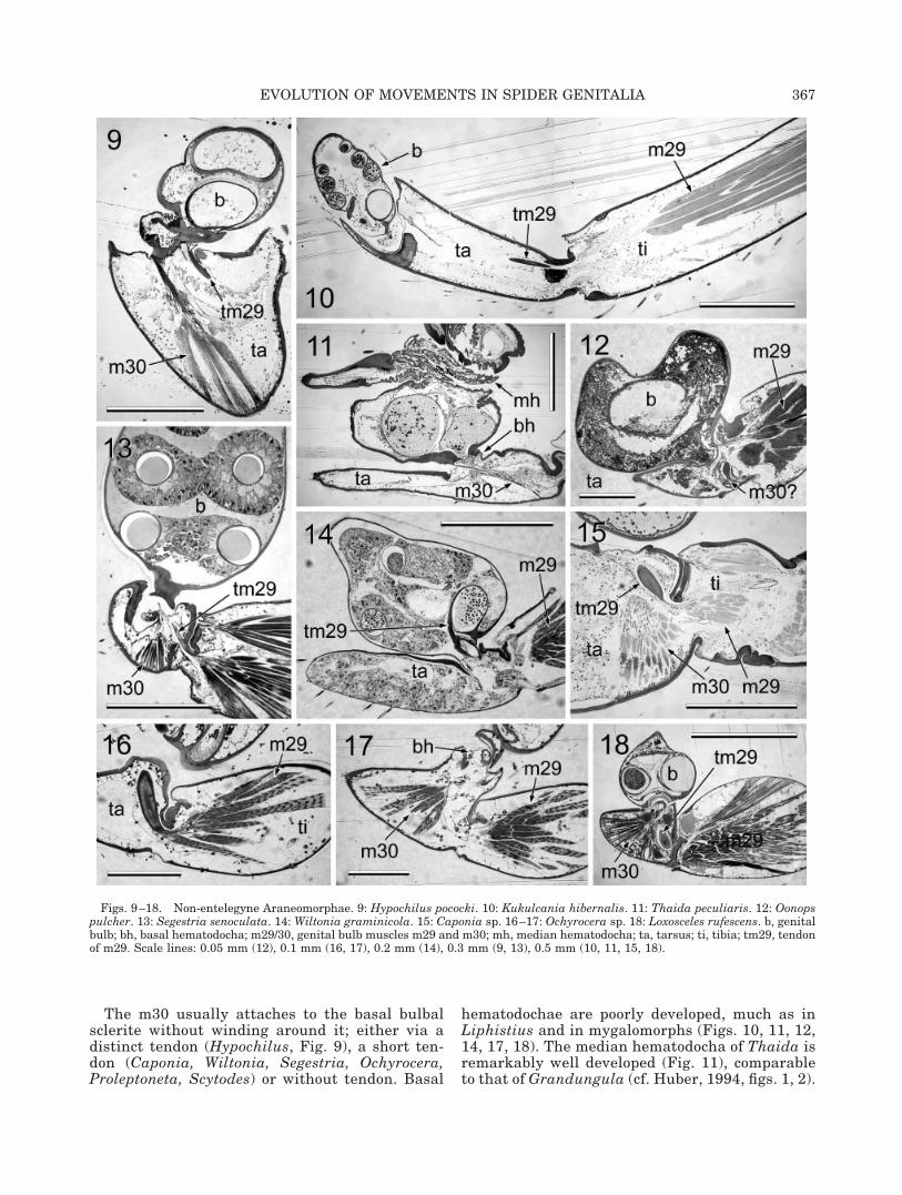
The m30 usually attaches to the basal bulbalsclerite without winding around it; either via adistinct tendon (Hypochilus, Fig. 9), a short ten-don (Caponia, Wiltonia, Segestria, Ochyrocera,Proleptoneta, Scytodes) or without tendon. Basal
hematodochae are poorly developed, much as inLiphistius and in mygalomorphs (Figs. 10, 11, 12,14, 17, 18). The median hematodocha of Thaida isremarkably well developed (Fig. 11), comparableto that of Grandungula (cf. Huber, 1994, figs. 1, 2).
Figs. 9–18. Non-entelegyne Araneomorphae. 9: Hypochilus pococki. 10: Kukulcania hibernalis. 11: Thaida peculiaris. 12: Oonopspulcher. 13: Segestria senoculata. 14: Wiltonia graminicola. 15: Caponia sp. 16–17: Ochyrocera sp. 18: Loxosceles rufescens. b, genitalbulb; bh, basal hematodocha; m29/30, genital bulb muscles m29 and m30; mh, median hematodocha; ta, tarsus; ti, tibia; tm29, tendonof m29. Scale lines: 0.05 mm (12), 0.1 mm (16, 17), 0.2 mm (14), 0.3 mm (9, 13), 0.5 mm (10, 11, 15, 18).
367EVOLUTION OF MOVEMENTS IN SPIDER GENITALIA

EntelegynaeMost entelegynes studied have no bulbal muscles
and are provided with a strongly developed basalhematodocha (Figs. 25–36). Exceptions occur as fol-lows: 1) In Tama and Hersilia (but not in Tamopsis)and in Uroecobius (but not in Oecobius and Uroctea)the m30 is present in the tarsus (Huber, 1994) andthe basal hematodocha is poorly (Uroecobius, Tama)
to well (Hersilia) developed. 2) In Palpimanidae,both muscles are present in Palpimanus and Ani-saedus (Figs. 19–21), while only m29 is present inOtiothops (Fig. 22). In all three species the basalhematodocha is very poorly developed, comparableto non-entelegynes. 3) In Mecysmaucheniidae (Figs.23, 24), Argyroneta aquatica, and Deinopis sp., them30 is present (in the tarsus), and the basal hema-
Figs. 19–24. Palpimanidae and Mecysmaucheniidae. 19–20: Palpimanus sp. 21: Anisaedus pellucidas. 22: Otiothops atlanticus. 23:Aotearoa sp. 24: Mecysmauchenius segmentatus. b, genital bulb; bh, basal hematodocha; m29, genital bulb muscle m29; ta, tarsus; ti,tibia; tm29, tendon of m29. All scale lines 0.2 mm.
368 B.A. HUBER

todocha is very well developed (slightly less inAotearoa: Fig. 23).
Some previous mentions of muscles in entelegynespider bulbs are clearly erroneous (Billaudelle,1957, on Dictyna; Haupt, 1983, fig. 1a on Nuctenea;Heimer, 1989, on various families).
A secondary result of this study is the discovery ofpresumably glandular tissue in addition to the usual
sperm duct glands in the bulbs of some entelegynespiders. Figures 27–30 show four examples (repre-sentatives of Cycloctenidae, Miturgidae, Stiphidi-idae, Amphinectidae), and similar tissue occurs inthe tegulum of Pisaura mirabilis. Such glands havepreviously been known from Amaurobius and Dic-tyna only (Suhm et al., 1995; Huber, 1995), but areapparently widespread.
Figs. 25–36. Entelegynae. 25: Eresus cinnaberinus (horizontal). 26: Megadictyna thilenii. 27: Cycloctenidae gen. sp. indet. 28:Uliodon tarantulinus. 29: Stiphidion facetum. 30: Maniho ngaitaho. 31: Mysmenopsis tengellacompa (horizontal). 32: Ero canionis. 33:Chilenodes australis. 34: Miagrammopes animotus. 35: Sofanapis antillanca. 36: Symphytognatha picta. b, genital bulb; bh, basalhematodocha; g, gland; ta, tarsus; ti, tibia. Scale lines: 0.05 mm (35, 36), 0.1 mm (31–34), 0.3 mm (25, 27, 28, 29), 0.5 mm (26).
369EVOLUTION OF MOVEMENTS IN SPIDER GENITALIA

DISCUSSIONHomologies: Muscles, EntelegyneHematodochae, and Sperm Ducts
The terminology above implies that the bulbalmuscles are homologs of the claw flexors (m29) andextensors (m30) in the walking legs (Ruhland andRathmayer, 1978). In walking legs, the m29 origi-nates either in patella and tibia (Aphonopelma: Ru-hland and Rathmayer, 1978), in the tibia (Zygiella:Frank, 1957), in tibia and basitarsus (Latrodectus:Whitehead and Rempel, 1959), or in the basitarsus(Gasteracantha: Emerit, 1972). This is a similar de-gree of variation as described herein for pedipalps(assuming that the thin patellar component has notbeen overlooked in some previous studies usinggross dissection). Much the same occurs with legmuscle m30: it may originate in tibia and basitarsus(Latrodectus: Whitehead and Rempel, 1959), or inthe basitarsus only (Aphonopelma, Zygiella, Gaste-racantha). In no spider leg is it known to originateentirely in the tibia, like in Liphistius and Atypuspedipalps, but it may well do so in these and otherunstudied taxa. The most parsimonious solution isthat the longer bulbal muscle is homologous to thelonger claw muscle, i.e., to the walking leg musclem29.
Another problem is the nomenclature in speciesthat have only one bulbal muscle. Considering thevariability in origins, it is the attachment that pro-vides confirmatory evidence. There is a significantdifference in the attachment mode between the twomuscles in most spiders. The m29 winds around thebasal sclerite of the bulb or enters the bulb, whilethe m30 attaches to the basal sclerite without wind-ing around it. This confirms that the single musclein filistatids and in Otiothops is m29, while thesingle muscle in certain hersiliids, Uroecobius,mecysmaucheniids, Argyroneta, and Deinopis is in-deed m30 (and not m29 whose origin has moved intothe tarsus).
The highly expandable basal hematodochae of en-telegynes are clearly derived from the simple mem-branes connecting tarsus and bulb in more primitivespiders. Interestingly, variation has been reported re-garding the direction of movement of the expandinghematodocha: in most entelegyne spiders studied, theleft bulb moves clockwise when seen in ventral view(Loerbroks, 1983, 1984: Heriaeus, Misumena; Sier-wald, 1987: Thalassius; Eberhard and Huber, 1998:Leucauge; Kropf, 1990: Comaroma; Lamoral, 1973:Palystes; Melchers, 1963: Cupiennius; Blest andPomeroy, 1978: Mynoglenes; van Helsdingen, 1969:Neriene; Osterloh, 1922: Linyphia, Agelena). This isthe same direction as observed for muscular move-ment in more primitive spiders (Kraus, 1984: Hep-tathela; Huber and Eberhard, 1997: Physocyclus).However, in some it moves counterclockwise (Dondale,1986: Lycosidae, some Pisauridae; van Helsdingen,1965: Lepthyphantes). The taxonomic distribution of
character states suggests that counterclockwise move-ment has repeatedly evolved from clockwise move-ment, rather than that highly expandable basal hema-todochae have originated more than once.
Cooke (1970) proposed the idea that sperm ducts ofHaplogynae and Mygalomorphae may not be homolo-gous, arguing with different muscular attachmentsites. Such differences do occur indeed. In Liphistius,Mygalomorphae and a few basal Araneomorphae (Hy-pochilus, Caponia), the tendon of m29 winds aroundthe basal sclerite of the bulb (Figs. 1, 2, 5, 9). In allother Araneomorphae with m29 present the tendoneither enters the bulb and continues to the sperm duct(Wiltonia, Fig. 14), attaches to the basal sclerite on theinside (Otiothops, Fig. 22), or attaches to the thickenedbasal hematodocha (Palpimanus, Anisaedus). Charac-ter mapping on the cladogram of Coddington and Levi(1991) suggests that the situation in Liphistius, Myga-lomorphae, and Hypochilus is plesiomorphic, and thatother attachment modes are derived. However, thedetails of attachment were often difficult to observe onthe sections and the topic warrants further investiga-tion.
Potential Selective Advantage of Hydraulics
Spiders evolved two major innovations concerningbulbal movement: 1) the transformation of the hingemovement of claws to a rotating movement of thebulb caused by changes in the attachment mode ofm29; and 2) the transformation of predominantlymuscular to predominantly hydraulic movement byexpansion of specifically folded membranes. Thefirst transformation occurred before the evolution ofthe most primitive extant spiders, the second at thebase of entelegyne spiders. I hypothesize that bothtransformations may have been driven by similarselective forces, i.e., to evolve, maintain, or improveon the mobility of the genital bulb. The question ofwhy complex movements are advantageous is a dif-ferent issue. It may be for the simple mechanicalreason that complex movements may improve thelock between male and female structures duringsperm transfer. It may also be that more complexmovements trigger female responses that are advan-tageous for the male in a sexual selection context.
Whatever the ultimate reason, muscular move-ment appears limited for several reasons: first, thelength of the muscle determines the degree of move-ment (maximum reported rotation in muscularbulbs is 300°: Huber and Eberhard, 1997); second,depending on the complexity of the joints, the result-ing movement is more or less limited to one plane,i.e., two-dimensional; third, structures cannot bemoved away by muscles unless a lever system beintegrated. All these limitations do not apply forhematodochae. Expanding hematodochae may ro-tate the bulb to considerable degrees (430° in Cupi-ennius [Melchers, 1963], 500° in Agelenopsis [Ger-ing, 1953], 540° in Tapinocyba [Martin, 1981]), and
370 B.A. HUBER

depending on how the hematodocha is folded at rest,it may unfold in a complex way, often includingrotation, tilt, and expansion that moves structuresaway.
In sum, hematodochae improve bulbal mobilityand produce movements of higher complexity, move-ments that are not easily generated with one pair ofmuscles.
Systematic Implications: the“Palpimanoidea”
Forster and Platnick (1984) created the superfam-ily Palpimanoidea to include both haplogyne (e.g.,Palpimanidae) and entelegyne (e.g., Mimetidae)families. The monophyly of Palpimanoidea was sup-ported by the cladistic analysis of Platnick et al.(1991) that placed the superfamily within the clade“Higher Entelegynes.” This view was adopted byCoddington and Levi (1991), but not by others (e.g.,Wunderlich, 1986; Lehtinen, 1996). It was Schutt(2000, 2003) who provided strong evidence againstit, moving several “palpimanoid” families to the Ara-neoidea. However, some “palpimanoid” families, likePalpimanidae and Mecysmaucheniidae, were nottreated by Schutt (2000, 2003) nor by Griswold et al.(1999), even though the latter were specifically con-cerned with Entelegynae. I propose that palpimanidbulbal muscles argue for a much more basal positionof palpimanids, not within “Higher Entelegynae”but at their base, or even outside Entelegynae.
Haplogyne female genitalia occur in several fam-ilies within Entelegynae (e.g., Uloboridae, Tetrag-nathidae, Anapidae), and are easily explained by thesecondary loss of the copulatory ducts. The occur-rence of “haplogyne” male genitalia (defined here asgenitalia with both bulbal muscles present) withinEntelegynae is not that easily explained. While itdoes not necessarily imply regaining of a lost struc-ture (the muscles may have been retained in juve-niles; cf. Szombathy, 1915; Barrows, 1925; Harm,1935; Bhatnagar and Rempel, 1962; Sadana, 1971;but see Gassmann, 1926), it is hard to imagine thepalpimanid bulb derived from an entelegyne bulbwithout muscles and well-developed basal hema-todocha. The apparently simultaneous reversal infemales also to a haplogyne condition makes it allthe more improbable. Therefore, I rather agree withPlatnick’s (1975, p. 2) former conclusion that “it isfar more likely that the palpimanids share a com-mon ancestor with one or more of the other haplo-gyne families.” Future data matrices for phyloge-netic reconstruction will have to include both bulbalmuscles and will show whether it is the male bulband female haplogyne genitalia or the canoe tape-tum (Homann, 1971) that is homoplastic and mis-leading.
Open Questions
The hemolymph pressure needed for leg extensionand hematodochal expansion is generated primarilyin the prosoma (Wilson, 1970; Anderson andPrestwich, 1975), resulting in a general increase ofpressure in the spider’s body rather than in a local-ized increase at specific joints or at the bulb in use.In legs, extension can be inhibited by the flexormuscles, but it is not clear how in genital bulbswithout muscles one hematodocha remains col-lapsed while the other is expanding. Rovner (1975)and Rovner and Wright (1975) hypothesized a valvein the palp that is opened by muscles but closedduring most of the spider’s life. This may explain theobservation that genital bulbs are often difficult toinfiltrate even with low viscosity resins. More spe-cifically, in the present study it usually was thetibia-tarsus joint that separated the well fixed andwell infiltrated tissue from the poorly fixed andpoorly infiltrated one. A similar position of the valveis suggested by Knoflach’s (2002) findings on Echi-notheridion gibberosum. In this species, the maleautotomizes the palp at the tibia-cymbium joint andthe expanded hematodocha remains expanded,probably due to the valve being closed. Whatever thedetails, some sort of valve must exist, but the con-structional details remain to be established. More-over, Rovner’s hypothesis raises the question of howthe area distal of the valve is supplied with hemo-lymph.
Kraus (1984) distinguished between hydraulicand glandular genital bulbs (admitting “intergrad-ing stages”), arguing with differences in the mode ofsperm ejaculation. However, with the exception ofLiphistiidae, and maybe a few other spiders (cf.Kraus, 1978; Lopez and Juberthie-Jupeau, 1985;Lopez, 1987), all genital bulbs studied herein seemto have glands that secrete into the sperm duct (cf.Lopez, 1977, 1987; Juberthie-Jupeau and Lopez,1981; Lopez and Juberthie-Jupeau, 1982, 1985;Suhm et al., 1995), and some hydraulic mechanismseems to be involved in most if not all bulbs. Thismakes a distinction of glandular vs. hydraulic prob-lematic. The temporal coincidence between hema-todochal expansion and ejaculation has lead to thebelief that the two are causally related, but experi-mental and morphological evidence argues againstit (Cooke, 1966; Huber and Eberhard, 1997). Obvi-ously, the exact mechanism of gland activation forsperm ejaculation (and induction) is still poorly un-derstood (Cooke, 1966; Lamoral, 1973). Therefore, Ipropose a distinction between hydraulic and muscu-lar genital bulbs, emphasizing the undisputed keyrole of hydraulics in bulbal mobility rather than insperm ejaculation.
ACKNOWLEDGMENTS
This study was made possible by many colleagueswho kindly provided material for sectioning: B.
371EVOLUTION OF MOVEMENTS IN SPIDER GENITALIA

Baehr, F. Coyle, A.S. Dippenaar-Schoeman, W.G.Eberhard, M. Filmer, R.R. Forster, F. Gasparo, C.Griswold, M. Gross, P. Jager, B. Opell, N.I. Platnick,M. Ramirez, R.L. Rodriguez-Sevilla, G. Schmidt, P.Schwendinger, K. Thaler, R. Raven, C. Vink, P.Zulka. Constructive comments by two anonymousreviewers helped improve the manuscript.
LITERATURE CITED
Anderson JF, Prestwich KN. 1975. The fluid pressure pumps ofspiders (Chelicerata, Araneae). Z Morph Tiere 81:257–277.
Barrows WM. 1925. Modification and development of the arach-nid palpal claw, with especial reference to spiders. Ann Ento-mol Soc Am 18:483–525.
Bhatnagar RDS, Rempel JG. 1962. The structure, function, andpostembryonic development of the male and female copulatoryorgans of the black widow spider Latrodectus curacaviensis(Muller). Can J Zool 40:465–510.
Billaudelle H. 1957. Zur Biologie der Mauerspinne Dictyna civica(H. Luc.) (Dictynidae, Araneida). Z Angew Entomol 41:474–512.
Blest AD, Pomeroy G. 1978. The sexual behaviour and genitalmechanics of three species of Mynoglenes (Araneae: Linyphi-idae). J Zool 185:319–340.
Coddington JA. 1990. Ontogeny and homology in the male palpusof orb-weaving spiders and their relatives, with comments onphylogeny (Araneoclada: Araneoidea, Deinopoidea). SmithsonContr Zool 496:1–52.
Coddington JA, Levi HW. 1991. Systematics and evolution ofspiders (Araneae). Annu Rev Ecol Syst 22:565–592.
Cooke JAL. 1966. Synopsis of the structure and function of thegenitalia of Dysdera crocata (Araneae, Dysderidae). Senck Biol47:35–43.
Cooke JAL. 1970. Spider genitalia and phylogeny. Bull Mus NatHist Natur, 2e ser, 41(Suppl 1):142–146.
Dondale CD. 1986. The subfamilies of wolf spiders (Araneae:Lycosidae). Actas X Congr Int Arachnol Jaca, Espana 1986:327–332.
Eberhard WG, Huber BA. 1998. Courtship, copulation, andsperm transfer in Leucauge mariana (Araneae, Tetragnathi-dae) with implications for higher classification. J Arachnol26:342–368.
Ellis CH. 1944. The mechanism of extension in the legs of spiders.Biol Bull 86:41–50.
Emerit M. 1972. Le developpement des gasteracanthes (Ara-neida, Argiopidae). Une contribution a l’etude de la morphologiede l’appendice araneidien. Ann Mus R Afr Centr Tervuren195:1–103, tables 1–4, plates 1–6.
Forster RR, Platnick NI. 1984. A review of the archaeid spidersand their relatives, with notes on the limits of the superfamilyPalpimanoidea (Arachnida, Araneae). Bull Am Mus Nat Hist178:1–106.
Frank H. 1957. Untersuchungen zur funktionellen Anatomie derlokomotorischen Extremitaten von Zygiella x-notata, einer Rad-netzspinne. Zool Jb Anat 76:423–460.
Gassmann F. 1926. Die Entwicklung des mannlichen Spinnen-tasters, dargestellt an Lepthyphantes nebulosus Sund. Z MorphOkol Tiere 5:98–118.
Gering RL. 1953. Structure and function of the genitalia in someAmerican agelenid spiders. Smithson Misc Coll 121:1–84.
Grasshoff M. 1973. Bau und Mechanik der Kopulationsorganeder Radnetzspinne Mangora acalypha (Arachnida, Araneae). ZMorph Tiere 74:241–252.
Gray M. 1987. Distribution of the funnel web spiders. In: Co-vacevich J, Davie P, Pearn J, editors. Toxic plants and animals:a guide for Australia. Brisbane: Queensland Museum. p 313–321.
Griswold CE, Coddington JA, Platnick NI, Forster RR. 1999.Towards a phylogeny of entelegyne spiders (Araneae, Araneo-morphae, Entelegynae). J Arachnol 27:53–63.
Harm M. 1931. Beitrage zur Kenntnis des Baues, der Funktionund der Entwicklung des akzessorischen Kopulationsorganesvon Segestria bavarica C. L. Koch. Z Morph Okol Tiere 22:629–670.
Harm M. 1935. Bau, Funktion und Entwicklung des akz-essorischen Kopulationsorganes von Evarcha marcgravi Sco-poli. Z Wiss Zool 146:123–134.
Haupt J. 1978. Funktionsmorphologie des Palpenorgans von Hep-tathela kimurai (Kishida) (Araneae: Liphistiidae). Verh DschZool Ges 1978:282.
Haupt J. 1979. Lebensweise und Sexualverhalten der mesothelenSpinne Heptathela nishihirai n. sp. (Araneae, Liphistiidae).Zool Anz 202:348–374.
Haupt J. 1983. Vergleichende Morphologie der Genitalorganeund Phylogenie der liphistiomorphen Webspinnen (Araneae:Mesothelae). Z Zool Syst Evol 21:275–293.
Heimer S. 1989. Untersuchungen zur Evolution der Kopulations-organe bei Spinnen (I) (Arachnida, Araneae). Ent Abh MusTierk Dresden 53:1–25.
Heimer S. 1990. Untersuchungen zur Evolution der Kopulations-organe bei Spinnen (II) (Arachnida, Araneae). Ent Abh MusTierk Dresden 53:97–123.
Homann H. 1971. Die Augen der Araneae. Anatomie, Ontogenieund Bedeutung fur die Systematik (Chelicerata, Arachnida). ZMorph Tiere 68:201–272.
Huber BA. 1993. Genital mechanics and sexual selection in thespider Nesticus cellulanus (Araneae: Nesticidae). Can J Zool71:2437–2447.
Huber BA. 1994. Genital bulb muscles in entelegyne spiders. JArachnol 22:75–76.
Huber BA. 1995. The retrolateral tibial apophysis in spiders —shaped by sexual selection? Zool J Linn Soc 113:151–163.
Huber BA. 2002. Functional morphology of the genitalia in thespider Spermophora senoculata (Pholcidae, Araneae). Zool Anz241:105–116.
Huber BA, Eberhard WG. 1997. Courtship, copulation, and gen-ital mechanics in Physocyclus globosus (Araneae, Pholcidae).Can J Zool 74:905–918.
Juberthie-Jupeau L, Lopez A. 1981. Ultrastructure du tube semi-nifere chez Leptoneta microphthalma Simon, 1872 (Araneae,Leptonetidae). Rev Arachnol 3:65–73.
Knoflach B. 2002. Copulation and emasculation in Echinoth-eridion gibberosum (Kulczynski, 1899) (Araneae, Theridiidae).In: Toft S, Scharff N, editors. European Arachnology 2000.Aarhus: Aarhus University Press. p 139–144.
Kraus O. 1978. Liphistius and the evolution of spider genitalia.Symp Zool Soc Lond 42:235–254.
Kraus O. 1984. Male spider genitalia: evolutionary changes instructure and function. Verh Naturwiss Ver Hamburg (NF)27:373–382.
Kropf C. 1990. Comaroma is an anapid spider (Arachnida, Ara-neae, Anapidae). Abh Naturwiss Ver Hamburg (NF) 31/32:185–203.
Lamoral BH. 1973. On the morphology, anatomy, histology andfunction of the tarsal organ on the pedipalpi of Palystescastaneus (Sparassidae, Araneida). Ann Natal Mus 21:609 –648.
Lehtinen P. 1996. The ultrastructure of leg skin in the phylogenyof spiders. Rev Suisse Zool, vol hors serie 2:399–422.
Loerbroks A. 1983. Revision der Krabbenspinnen-Gattung Heri-aeus Simon (Arachnida: Araneae: Thomisidae). Verh NaturwissVer Hamburg (NF) 26:85–139.
Loerbroks A. 1984. Mechanik der Kopulationsorgane von Mi-sumena vatia (Clerck, 1757) (Arachnida: Araneae: Thomisidae).Verh Naturwiss Ver Hamburg (NF) 27:383–403.
Lopez A. 1977. Le tube seminifere des araignees males: quelquesprecisions sur sa structure microscopique. Rev Arachnol 1:1–7.
Lopez A. 1987. Glandular aspects of sexual biology. In: NentwigW, editor. Ecophysiology of spiders. Berlin: Springer. p 121–132.
Lopez A, Juberthie-Jupeau L. 1982. Structure et ultrastructuredu bulbe copulateur chez la mygale Nemesia caementaria (La-
372 B.A. HUBER

treille, 1798) (Araneae, Ctenizidae). Bull Soc Sci Nat BeziersNS VIII(49):12–19.
Lopez A, Juberthie-Jupeau L. 1985. Ultrastructure comparee dutube seminifere chez les males d’araignees. Mem Biospeol 12:97–109.
Martin D. 1981. Bau und Funktion der Kopulationsorgane beiZwergspinnen: 1. Tapinocyba insecta (L. Koch) (Arachnida,Araneae, Erigonidae). Ent Abh Mus Tierk Dresden 44:81–86.
Melchers M. 1963. Zur Biologie und zum Verhalten von Cupien-nius salei (Keyserling), einer amerikanischen Ctenide. Zool JbSyst 91:1–90.
Osterloh A. 1922. Beitrage zur Kenntnis des Kopulationsap-parates einiger Spinnen. Z Wiss Zool 119:326–421.
Parry DA, Brown RHJ. 1959. The hydraulic mechanism of thespider leg. J Exp Biol 36:423–433.
Platnick NI. 1975. A revision of the palpimanid spiders of the newsubfamily Otiothopinae (Araneae, Palpimanidae). Am Mus No-vit 2562:1–32.
Platnick NI, Coddington JA, Forster RR, Griswold CE. 1991.Spinneret morphology of haplogyne spiders (Araneae, Araneo-morphae). Am Mus Novit 3016:1–73.
Rovner JS. 1975. Behavioral evidence for valvular regulation ofhematodochal expansion in the spider’s palp. Proc 6th IntArachn Congr (Amsterdam, 1974). p 99–101.
Rovner JS, Wright EE. 1975. Copulation in spiders: experimentalevidence for fatigue effects and bilateral control of palpal inser-tions. Anim Behav 23:233–236.
Ruhland M, Rathmayer W. 1978. Die Beinmuskulatur und ihreInnervation bei der Vogelspinne Dugesiella hentzi (Ch.) Ara-neae, Aviculariidae). Zoomorphology 89:33–46.
Sadana GL. 1971. Studies on the postembryonic development ofthe palpal organ of Lycosa chaperi Simon (Lycosidae: Ara-neida). Zool Anz 186:251–258.
Schutt K. 2000. The limits of the Araneoidea (Arachnida: Ara-neae). Aust J Zool 48:135–153.
Schutt K. 2003. Phylogeny of Symphytognathidae s.l. (Araneae,Araneoidea). Zool Scr 32:129–151.
Sierwald P. 1987. Revision der Gattung Thalassius (Arachnida,Araneae, Pisauridae). Verh Naturwiss Ver Hamburg (NF) 29:51–142.
Suhm M, Thaler K, Alberti G. 1995. Glands in the male palpalorgan and the origin of the mating plug in Amaurobius species(Araneae: Amaurobiidae). Zool Anz 234:191–199.
Szombathy K. 1915. Uber Bau und Funktion des Bulbus dermannlichen Kopulationsorgane bei Agelena und Mygale. AnnMus Nat Hung 13:252–276.
van Helsdingen PJ. 1965. Sexual behaviour of Lepthyphantesleprosus (Ohlert) (Araneida, Linyphiidae), with notes on thefunctions of the genital organs. Zool Mededel 41:15–42.
van Helsdingen PJ. 1969. A reclassification of the species ofLinyphia Latreille based on the functioning of the genitalia(Araneida, Linyphiidae). Part I. Linyphia Latreille and NerieneBlackwell. Zool Verh 105:1–303.
Whitehead WF, Rempel JG. 1959. A study of the musculature ofthe black widow spider, Latrodectus mactans (Fabr.). Can JZool 37:831–870.
Wilson RS. 1970. Some comments on the hydrostatic system ofspiders (Chelicerata, Araneae). Z Morph Tiere 68:308–322.
Wunderlich J. 1986. Spinnenfauna gestern und heute. FossileSpinnen in Bernstein und ihre heute lebenden Verwandten.Wiesbaden: Erich Bauer Verlag bei Quelle und Meyer.
APPENDIX
Taxa studied, listed according to Coddington and Levi (1991).Asterisks indicate specimens that were taken from the ViennaUniversity students’ collection and whose collection data wereunknown.
LiphistiidaeLiphistius bristowei Platnick and Sedgwick, 1984
Thailand: Doi Suthep, 1290 m, 9.x.1986 (P. Schwendinger)
HexathelidaeHadronyche sp. (sp. 19: Gray 1987)
Australia: SEQ, Mt. Superbus (28°14�S, 152°29�E), 9.ii.-12.iii.1990 (G. Monteith, G. Thompson, T. Churchill), QMS21246
Bymainiella terraereginae (Raven, 1976)Australia: SEQ, Lamington N.P., O’Reillys (28°14�S,153°08�E), iv.-vi.1982 (G. Monteith); QM S11080
DipluridaeMasteria toddae Raven, 1979
Australia: NEQ, Cape Tribulation, 2.5 km W, site 5 (16°05�S,145°27�E), 23.ix.-7.x.1982 (G. Monteith, D. Yeates, G.Thompson), QM S8141
Australothele jamiesoni Raven, 1984Australia: SEQ, Numinbah Valley (28°12�S, 153°13�E),x.1989 (R. Fleay-Thompson); QM S15925
Namirea planipes Raven, 1984Australia: SEQ, Brisbane, Mt. Coot-tha (27°29�S, 152°57�E),14.xii.1979 (R.J. Raven); QM S 9790
NemesiidaeChenistonia earthwatchorum (Raven, 1984)
Australia: NEQ, Upper Gayundah Creek, Hinchinbrook(18°22�S, 146°13�E), 9-11.xi.1984 (G. Monteith, D. Cook); QMS 21249
Aname pallida L. Koch, 1873Australia: SEQ, Biloela (24°24�S, 150°31�E), 14.x.1987(D.P.I.), inside house; QM S3170
BarychelidaeSeqocrypta jakara Raven, 1994
Australia: SEQ, Brisbane, Fig Tree Pocket (27°28�S,153°03�E), 22.vii.1973 (V.E. Davies); QM S6627
AtypidaeAtypus affinis Eichwald, 1830*
CyrtaucheniidaeHomostola sp.
Swaziland: Mlilwane Game Reserve, 31.iii.2001 (K. Schutt),in ZFMK
ActinopodidaeMissulena bradleyi Rainbow, 1914
Australia: New South Wales, Murwillumbah (28°20�S,153°24�E), 1.iii.1975 (D. Ball, in swimming pool); QM S21245
HypochilidaeHypochilus pococki Platnick, 1987
USA: North Carolina, Jackson Co., Caney Fork Rd., 1.2 mi Nintersec. with Johns Cr. Rd., 2350 ft elev., 8.ix.1994 (F.Coyle), on boulders; det. F. Coyle 1994
GradungulidaeGradungula sorenseni Forster, 1955
New Zealand: Fiordland, Secretary Is., pitfall, iii.1984 (C.F.Butcher)
AustrochilidaeThaida peculiaris Karsch, 1880
Chile: Llanquihue; Salto Petrohue, V. Perez N.P., mixedmoist forest, 23.xii.1984-4.ii.1985 (S. and J. Peck), in AMNH
FilistatidaeKukulcania hibernalis (Hentz, 1842)
Costa Rica: San Jose, on building, 1995 (B.A. Huber)Filistata insidiatrix (Forskål, 1775)
Italy: Sicily, Taormina, 29.viii.1993 (B.A. Huber)
CaponiidaeCaponia sp.
373EVOLUTION OF MOVEMENTS IN SPIDER GENITALIA

South Africa: Northern Prov., near Pietersburg, 26.viii.1974(E. Kullmann), in ZFMK
OrsolobidaeWiltonia graminicola Forster and Platnick, 1985
New Zealand: Otago, Flagstaff, in tussock, 9.iii.1982 (R.R.Forster)
OonopidaeGen. sp.
Costa Rica: Heredia, La Selva, near Pto. Viejo de Sarapiqui,20 m, 1995 (G. Delgado), in ZFMK
Oonops pulcher Templeton, 1835Germany: Bonn, Plittersdorf (�50°42�N, 7°09�E), in house,2.x.2002 (B.A. Huber)
DysderidaeDysdera ninnii Canestrini, 1868*Harpactea rubicunda (C.L. Koch, 1838)
Austria: Vienna, Prater (�48°13�N, 16°24�E), 28.iii.1992(B.A. Huber)
SegestriidaeSegestria senoculata (Linnaeus, 1758)
Austria: Upper Austria, Walding (�48°21�N, 14°12�E),8.iii.1992 (B.A. Huber)
PholcidaePholcus phalangioides (Fuesslin, 1775)
Germany: Denzlingen (near Freiburg), in house, 16.ii.1992(B.A. Huber)
Metagonia mariguitarensis (Gonzalez-Sponga, 1998)Venezuela: Estado Sucre, Mariguitar (10°26.5�N, 63°54.5�W),�30 m a.s.l., 29.xi.2002 (B.A. Huber)
DiguetidaeDiguetia sp.
USA: California, San Benito Co., Clear Creek, 10 mi S Idra,15.viii.1969 (M.M. Bentzien), in AMNH
PlectreuridaePlectreuris tristis Simon, 1893
USA: California, San Bernardino Co., Pisgah Lava Flow,station 24, 18.iv.1959 (B. Banta)
OchyroceratidaeOchyrocera sp.
Costa Rica: San Jose Prov., San Antonio de Escazu, xi.1995(B.A. Huber), in ZFMK
LeptonetidaeProleptoneta sp. [cf. italica (Simon, 1970)]
Italy: Udine Prov., Bordano, 260 m, 12.vi.1991 (F. Gasparo)
SicariidaeLoxosceles rufescens (Dufour, 1820)
Italy: Sicily, Taormina, 29.viii.1993 (B.A. Huber)
ScytodidaeScytodes thoracica (Latreille, 1802)
Austria: Vienna, in house, 1.vii.1992 (B.A. Huber)
OecobiidaeUroctea durandi (Latreille, 1809)*Oecobius cellariorum (Duges, 1836)
Italy: Trieste, in house, 3.iv.1993 (F. Gasparo)Uroecobius ecribellatus Kullmann and Zimmermann, 1976
South Africa: North West Prov., Broederstroom (25°45�S,27°52�E), 21.vii.1986 (A.S. Dippenaar-Schoeman)
HersiliidaeHersilia sp.
South Africa: KwaZulu Natal, farm Vergeval, districtNgotsche near Pongola (27°29�S, 31°40�E), 2.v.1967 (A.S.Dippenaar-Schoeman)
Tama sp.South Africa: KwaZulu Natal, farm Vergeval, district
Ngotsche near Pongola (27°29�S, 31°40�E), 27.vi.1968 (H. v.Ark)
Tamopsis parthensis Baehr and Baehr, 1987Collection data unknown (probably Perth, Australia; B.Baehr, pers. commun.)
EresidaeEresus cinnaberinus (Olivier, 1789)*
NicodamidaeMegadictyna thilenii Dahl, 1906
New Zealand: Orongoronga, 1.iii.1992 (M. Fitzgerald)
Cycloctenidaegen. sp.
Australia: Tasmania, Florentine Valley, 29.2 km WNW May-dena, on Eleven Rd., 460 m, 6.ii.1980 (A. Newton, M.Thayer), det. D. Silva, AMNH
ZodariidaeZodarion germanicum (C.L. Koch, 1837)*
MiturgidaeUliodon tarantulinus (L. Koch, 1873)
Australia: SCQ, Mt. Moffatt N.P., Dargonelly Rock Holes(25°01�S, 147°57�E), 20-27.ix.1986 (M. Bennie); QM S15981
HomalonychidaeHomalonychus theologus Chamberlin, 1924
USA: California, San Bernardino Co., Pisgah Lava Flow,station 11, 17.iv.1959 (B. Banta)
MimetidaeEro canionis Chamberlin and Ivie, 1935
USA: Idaho, NE Fruitland, 116.55�W, 44.01�N, 10-25.ix.1943(W. Ivie)
Gnolus cordiformis (Nicolet, 1849)Chile: Cautin: Chacamo, NW of Nueva Imperial and W ofTemuco, 16-24.ii.1981 (L.E. Pena)
MalkaridaeChilenodes australis Platnick and Forster, 1987
Chile: Cautin, 9 km S Pucon Volcan Villarrica Nat. Pk.,900 m elev., 15.xii.1984-10.ii.1985 (S. and J. Peck)
PalpimanidaePalpimanus sp.
Greece: S-Peloponnese, Areopoli-Vahos, near coast, �50-100m elev., under stones, 1995 (K. Thaler, B. Knoflach)
Otiothops atlanticus Platnick, Grismado and Ramirez, 1999Brazil: Bahia, Itabuna, abandoned cocoa plantation, 100 melev., 14.38�S, 39.18�W, 26.i.1995 (D. Agosti)
Anisaedus pellucidas Platnick, 1975Chile: Antofagasta, 6 km E Paposo, 480 m elev., 12.x.1992 (N.Platnick, K. Catley, P. Goloboff)
MicropholcommatidaeTeutoniella cekalovici Platnick and Forster, 1986
Chile: Concepcion: Parque Hualpen, 10.xii.1971 (T. Cek-alocic) “TC-36”
MecysmaucheniidaeMecysmauchenius segmentatus Simon, 1884
Argentina: Rıo Negro, El Bolson, Cerro Piltriquitron,3-4.ii.1985 (M. Ramirez)
Aotearoa sp.New Zealand: South Island, Fiordland, Eglinton Valley, TheDivide, 27.iii.1987 (N. Platnick, R. Forster), in AMNH
DictynidaeDictyna uncinata Thorell, 1856
Austria: Vienna, Donauinsel (�48°15�N, 16°24�E), 6.iv.1992(B.A. Huber)
DesidaeOtagoa nova Forster, 1970
374 B.A. HUBER

New Zealand: Canterbury, Kaikoura, under stones on beach,15.iii.1969 (R.R. Forster)
CybaeidaeArgyroneta aquatica (Clerck, 1757)*
HahniidaeHahnia pusilla C.L. Koch, 1841
Austria: Upper Austria, Walding (�48°21�N, 14°12�E),8.iii.1992 (B.A. Huber)
NeolanidaeNeolana dalmasi (Marples, 1959)
New Zealand: Vinegar Hill Reserve (39°57�S, 175°39�E),6.i.1967 (R.R. Forster)
CorinnidaeCetonana laticeps (Canestrini, 1868)
Austria: Upper Austria, Walding (�48°21�N, 14°12�E), for-est, 4.viii.2003 (B.A. Huber)
LiocranidaeAgroeca brunnea (Blackwall, 1833)*
LamponidaeLampona cylindrata (L. Koch, 1866)
New Zealand: Christchurch (43°33�S, 172°38�E), no date (C.Vink)
GnaphosidaeGnaphosa nigerrima L. Koch, 1877*
ClubionidaeClubiona pallidula (Clerck, 1757)
Austria: Vienna, Prater (�48°13�N, 16°24�E), 29.i.1992 (B.A.Huber)
Clubiona lutescens Westring, 1851Austria: Vienna, Donauinsel (�48°15�N, 16°24�E), Toter Gr-und, 4.i.1992 (B.A. Huber)
AnyphaenidaeAnyphaena accentuata (Walckenaer, 1802)
Austria: Upper Austria, Walding (�48°21�N, 14°12�E),iii.1993 (B.A. Huber)
Patrera procera (Keyserling, 1891)Argentina: Misiones, Santa Ana, 16.vii.1986 (M. Ramirez)
Wulfila albus (Mello-Leitao, 1945)Argentina: Misiones, San Antonio, vi.1970 (M.E. Galiano)
SalticidaePseudeuophrys lanigera (Simon, 1871)
Austria: Vienna, v.1992 (B.A. Huber)Myrmarachne formicaria (DeGeer, 1778)
Austria: Vienna, Donauinsel (�48°15�N, 16°24�E), 6.v.1992(B.A. Huber)
Synageles venator (Lucas, 1836)Austria: Vienna, Donauinsel (�48°15�N, 16°24�E), 6.v.1992(B.A. Huber)
ThomisidaeXysticus cristatus (Clerck, 1757)*Ozyptila simplex (O.P.-Cambridge, 1862)*Misumenops tricuspidatus (Fabricius, 1775)
Austria: Vienna, Donauinsel (�48°15�N, 16°24�E), 14.v.1993(B.A. Huber)
SparassidaeBarylestis montandoni (Lessert, 1929)
Uganda: Semliki Forest (0°48�N, 30°08�E), 5-12.ii.1997 (T.Wagner)
SelenopidaeAnyphops sp.
South Africa: KZN, Bonamanzi Reserve, 1.iv.2001 (B.A. Hu-ber), in ZFMK
ZoridaeZora spinimana (Sundevall, 1833)
Germany: Bonn, forest W Heiderhof (50°39,5�N, 7°09�E),29.ix.2002 (B.A. Huber)
PhilodromidaePhilodromus dispar Walckenaer, 1826
Austria: Upper Austria, Walding (�48°21�N, 14°12�E),xii.1991 (B.A. Huber)
AmaurobiidaeAmaurobius fenestralis (Strom, 1768)
Austria: Upper Austria, Walding (�48°21�N, 14°12�E), for-est, 6-9.iii.1992 (B.A. Huber)
AgelenidaeAgelena gracilens C.L. Koch, 1841
Austria: Upper Austria, Walding (�48°21�N, 14°12�E),25.vii.1993 (B.A. Huber)
Histopona torpida (C.L. Koch, 1837)Austria: Upper Austria, Walding (�48°21�N, 14°12�E),17.vi.1993 (B.A. Huber)
Textrix denticulata (Olivier, 1789)Austria: Vienna, Prater (�48°13�N, 16°24�E), 2.iv.1993 (B.A.Huber)
AmphinectidaeManiho ngaitaho Forster and Wilton, 1973
New Zealand: Canterbury, Kaituna Valley, 13.iv.1964 (R.R.Forster)
CtenidaeCupiennius salei (Keyserling, 1877)
Austria: Vienna University laboratory population, 1993
PisauridaePisaura mirabilis (Clerck, 1757)*
LycosidaeAulonia albimana (Walckenaer, 1805)*
StiphidiidaeStiphidion facetum Simon, 1902
New Zealand: Ohope Beach, 1.x.1969 (C.L. Wilton)
DeinopidaeDeinopis sp.
Australia: SEQ, Brisbane (27°28�S, 153°01�E), no furtherdata; QM S21247
UloboridaeHyptiotes paradoxus (C.L. Koch, 1834)
Austria: Upper Austria, Walding (�48°21�N, 14°12�E),vii.1992 (B.A. Huber)
Philoponella sp.Costa Rica: San Jose Prov., 1995 (R.L. Rodriguez-Sevilla)
Waitkera waitakerensis (Chamberlain, 1946)New Zealand: Whangarei, 5.ii.1991 (B. Opell)
Miagrammopes animotus Chickering, 1968Puerto Rico: Loquillo National Forest (B. Opell)
Miagrammopes sp.Costa Rica: La Selva, 27.vi.1985 (B. Opell)
AraneidaeNuctenea umbratica (Clerck, 1757)*
LinyphiidaeLinyphia triangularis (Clerck, 1757)
Austria: Upper Austria, Walding (�48°21�N, 14°12�E),vii.1993 (B.A. Huber)
Neriene montana (Clerck, 1757)Austria: Vienna, Prater (�48°13�N, 16°24�E), iv.1993 (B.A.Huber)
CyatholipidaeWanzia fako Griswold, 1998
Cameroon: NW Prov., Menchum Div., forest nr. Lake Oku
375EVOLUTION OF MOVEMENTS IN SPIDER GENITALIA
(6°12�N, 10°27�E), 2150 m, 7.-13.ii.1992 (C. Griswold and C.Wanzie)
SynotaxidaeChileotaxus sans Platnick, 1990
Chile: Chiloe, 15 km S Chepu, 3.ii.1991 (M. Ramirez)
NesticidaeNesticus cellulanus (Clerck, 1757)
Austria: Upper Austria, Walding (�48°21�N, 14°12�E),xi.1992 (B.A. Huber)
TheridiidaeEnoplognatha ovata (Clerck, 1757)
Austria: Upper Austria, Walding (�48°21�N, 14°12�E),7.vi.1992 (B.A. Huber)
Tidarren chevalieri (Berland, 1936)Cape Verde Islands: Santiago, “Tarratal,” 10.ii.1992 (G.Schmidt)
TetragnathidaeTetragnatha extensa (Linnaeus, 1758)
Austria: Vienna, Donauinsel (�48°15�N, 16°24�E), v.1992(B.A. Huber)
Metellina mengei (Blackwall, 1870)Austria: Upper Austria, Walding (�48°21�N, 14°12�E),xii.1991 (B.A. Huber)
Leucauge mariana (Taczanowski, 1881)Costa Rica: San Jose, Ciudad Universitaria, 1995 (B.A. Huber)
MysmenidaeMysmenopsis tengellacompa Platnick, 1993
Costa Rica: Heredia, La Selva, nr. Puerto Viejo, �100 m, onwebs of Tengella radiata, 1983 (W. Eberhard)
AnapidaeSofanapis antillanca Platnick and Forster, 1989
Chile: Llanquihue, Caleta, La Arena, 30.i.1991 (M. Ramirez)Minanapis talinay Platnick and Forster, 1989
Chile: Concepcion, Camino a Ramuntcho, 8.xii.1981 (T. Ce-calovic) “TC-107”
SymphytognathidaeSymphytognatha picta Harvey, 1992
Australia: WA: Tall Tingle Tree Path, Wqalpole-NornalupNat. Pk., 13.vi.1987 (N.I. Platnick, R.J. Raven)
376 B.A. HUBER