Embed Size (px)
Citation preview
1
(Proceedings of the IEEE – NON-FINAL DRAFT PREPRINT – Special 2012 100th Anniversary Issue)
Evolving Signal Processing for Brain-Computer Interfaces
Scott Makeig1,2*
, Christian Kothe1, Tim Mullen
1,3, Nima Bigdely-Shamlo
1,4, Zhilin Zhang
1,4,
Kenneth Kreutz-Delgado1,4
1Swartz Center for Computational Neuroscience, Institute for Neural Computation,
University of California San Diego (UCSD)
9500 Gilman Drive, La Jolla CA USA 92093-0559
2Department of Neurosciences, UCSD
3Department of Cognitive Science, UCSD
4Department of Electrical and Computer Engineering, UCSD
*Corresponding author: [email protected]
2
Abstract
Because of the increasing portability and wearability of noninvasive electrophysiological
systems that record and process electrical signals from the human brain, automated systems for
assessing changes in user cognitive state, intent, and response to events are of increasing interest.
Brain-computer interface (BCI) systems can make use of such knowledge to deliver relevant
feedback to the user or to an observer, or within a human-machine system to increase safety and
enhance overall performance. Building robust and useful BCI models from accumulated
biological knowledge and available data is a major challenge, as are technical problems
associated with incorporating multimodal physiological, behavioral, and contextual data that may
in future be increasingly ubiquitous. While performance of current BCI modeling methods is
slowly increasing, current performance levels do not yet support widespread uses. Here we
discuss the current neuroscientific questions and data processing challenges facing BCI designers
and outline some promising current and future directions to address them.
Keywords: Electroencephalogram, EEG, Brain-Computer Interface, BCI, Cognitive State Assessment,
Signal Processing, Machine Learning, Source-Space Modeling, Blind Source Separation, Effective
Connectivity, Transfer Learning, Multi-modal Signal Processing, Independent Component Analysis
3
I. Introduction
Electroencephalography (EEG) is the recording of electric potentials produced by the local
collective partial synchrony of electrical field activity in cortical neuropile, today most
commonly measured by an array of electrodes attached to the scalp using water-based gel [1][2].
EEG is today the most widely known and studied portable non-invasive brain imaging modality;
another, less developed and not considered here, is functional near-infrared spectroscopy (fNIR).
The first report of signals originating in the human brain and recorded non-invasively from the
scalp was that of Berger in 1924 [3]. Half a century later both engineers and artists begin to
seriously consider the possible use of EEG for active information exchange between humans and
machines (Vidal, 1973 -- "Toward direct brain-computer communication") [4]. It is now
generally accepted that the spatiotemporal EEG activity patterns correlate with changes in
cognitive arousal, attention, intention, evaluation, and the like, thereby providing a potential
“window on the mind.” However, the biological mechanisms that link EEG patterns to these or
other aspects of cognition are not understood in much detail [2].
A companion paper [5] describes how, unlike most other forms of functional brain imaging
available today (fMRI, MEG, PET), EEG sensor systems can be made comfortably wearable and
thus potentially usable in a wide range of settings. Another companion paper [6] further explores
how advances in brain signal processing and deeper understanding of the underlying neural
mechanisms may make important contributions to enhancing human performance and learning.
The main focus of this paper is the description of the current state and foreseeable trends in the
evolution of signal processing approaches that support design of successful brain-computer

4
interface (BCI) systems that deliver interactive cognitive and mental assessment and/or user
feedback or brain-actuated control based on non-invasive brain and behavioral measures. Brain-
computer interactions using invasive brain measures, while also of intense current research
interest and demonstrated utility for some applications [7][8][9], will here be discussed only
briefly.
We believe that in the coming decades adequate real-time signal processing for feature extraction
and state prediction or recognition combined with new, non-invasive and even wearable
electrophysiological sensing technologies can produce meaningful BCI applications in a wide
range of directions. Here, we begin with a brief primer on the neuroscientific basis of cognitive
state assessment, i.e. the nature of the EEG itself, followed by a review of the history and current
state of the use of signal processing in the relatively young BCI design field and then consider
avenues for its short-term and medium-term technical advancement. We conclude with some
thoughts on potential longer-term developments and perspectives.
What is EEG? Electrical activity among the estimated twenty billion neurons and equal or
larger number of non-neural cells that make up the human neocortex (the outer layer of the brain)
would have nearly no net projection to the scalp without the spontaneous appearance of
sufficiently robust and/or sizable areas of at least partial local field synchrony [10][1][2]. Within
such areas, the local fields surrounding pyramidal cells, aligned radially to the cortical surface,
sum to produce far-field potentials projecting by passive volume conduction to nearly all the
scalp EEG sensors. These effective cortical EEG sources also have vertical organization (one or
more net field sources and sinks within the six anatomic layers), though currently recovery of

5
their exact depth configuration may not be possible from scalp data alone. At a sufficient
electrical distance from the cortex, e.g. on the scalp surface, the projection of a single cortical
patch source strongly resembles the projection of a single cortical dipole termed its equivalent
dipole [11][12].
________________
Figure 1 about here
________________
The very broad EEG spatial point-spread function, simulated in Figure 1 using a realistic
electrical forward head model [13][14][15], means that locally synchronous activities emerging
within relatively small cortical areas are projected and summed at nearly all the widely
distributed scalp electrodes in an EEG recording [16]. Unfortunately, the naive viewpoint that
EEG potential differences between each scalp channel electrode and a reference electrode
represent a single EEG signal originating directly beneath the active scalp electrode continues to
color much EEG analysis and BCI design.
When working with EEG it is also important to bear in mind that the circumstances in which
local cortical field synchronies appear are not yet well understood. Nor are the many biological
factors and influences that determine the strongly varying time courses and spectral properties of
the EEG source signals. Our relative ignorance regarding the neurobiology of EEG signals is, in
part, a side effect of the fifty-year focus of the field of animal electrophysiology on neural spike
events in single-cell neural recordings [17]. During much of this period, studies of the concurrent

6
lower-frequency spatiotemporal field dynamics of the cortical neuropile were rare, though
Walter Freeman observed and modeled emergent, locally near-synchronous field patterns [18] he
terms ‘phase cones’ [19] and more recently Beggs and Plenz have modeled similar ‘avalanche’
events [20], both descriptions consistent with production of far-field potentials that might reach
scalp electrodes.
Non-brain EEG artifacts. In addition to a mixture of cortical EEG source signals, each scalp
EEG recording channel also sums potentials from non-brain sources (‘artifacts’) and channel
noise. Fortunately, in favorable recording circumstances (e.g., using modern recording
equipment well connected to a quiet subject in an electrically quiet laboratory) EEG sensor noise
is relatively small. However, the strength of contributions from non-brain sources (eye
movements, scalp muscles, line noise, scalp and cable movements, etc.) may be larger than the
contributions of the cortical sources. EEG recorded outside the laboratory using new wearable
EEG systems with variable conductance between the electrodes and scalp must also take into
account and handle possible large, non-stationary increases in EEG sensor and/or system noise
relative to laboratory recordings. Thus, for robust and maximally efficient cognitive state
assessment or other BCI applications, explicit or implicit identification and separation of brain
source signals of interest from non-brain and other, less relevant brain signals is important [21].
Multiscale recording and analysis. A major obstacle to understanding how the brain supports
our behavior and experience is that brain dynamics are inherently multi-scale. Thus, their more
complete understanding will likely require the development of extremely high-density, multi-
resolution electrical imaging methods [22]. Unfortunately, to date cortical field recordings

7
sufficiently dense to fully reveal the spatiotemporal dynamics of local cortical fields across
spatial scales are not yet available. We believe that the most effective real-world applications
using EEG signals will depend on (but may also contribute to) better understanding of the
biological relationships between neural electrical field dynamics and cognitive/behavioral state.
This knowledge is currently still largely inferred from observed correlations between EEG
measures and subject behavior or experience, although efforts are underway both to observe the
underlying biological phenomena with higher resolution [23][24] and to model the underlying
biological processes mathematically [25][26][27] in more detail.
The EEG inverse problem. Recovery of the cognitive state changes that give rise to changes in
observed EEG (or other) measures fundamentally amounts to an inverse problem and although at
least the broad mixing of source signals at the scalp is linear, recovery of the (latent) source
signals from given scalp data without additional geometric constraints on the form of the source
distributions is a highly underdetermined problem [28]. Even when given an accurate electric
forward head model [15] and a near-exact cortical source domain model constructed from the
subject’s magnetic resonance (MR) head image, finding the sources of an observed EEG scalp
pattern remains challenging. However, finding the source of a ‘simple’ EEG scalp map
representing the projection of a single compact cortical source domain allows for favorable
assumptions (as discussed below) and is thereby more tractable.
Response averaging. Most recent approaches to estimating EEG source spatial locations or
distributions have begun by averaging EEG data epochs time locked to some class of sensory or
behavioral events posited to produce a single mean transient scalp-projected potential pattern.

8
This average event-related potential (ERP) [29] sums projections of the (typically small) portions
of source activities in relevant brain areas that are both partially time-locked and phase-locked
(e.g., most often positive or negative) at some fixed latencies relative to the events of interest.
Average ERPs were arguably the first form of functional human brain imaging, and the study of
scalp channel ERP waveforms has long dominated cognitive EEG research.
ERP models have been the basis of many BCI designs as well. Unfortunately, ERP averaging is
not an efficient method for finding scalp projections of individual EEG source areas other than
those associated with the earliest sensory processing. Also, average ERPs capture only one
aspect of the EEG activity transformation following meaningful events [30]. BCI designs based
on an ERP model therefore ignore other information contained in EEG data about subjects’
cognitive responses to events, and also require knowing the times of occurrence of such events.
Opposed to these are BCI methods that continuously monitor the EEG data for signal changes in
the power spectrum and other higher-order statistics, often data features derived from latent
source representations of the collected signals.
Blind source separation. In the last twenty years, methods have been developed for estimating
the latent time-courses and spatial projections of sources of spontaneous or evoked EEG activity.
Independent component analysis (ICA) and other blind source separation (BSS) methods use
statistical information contained in the whole data to learn simple maps representing the
projections of individual EEG source areas to the scalp channels [31][32]. These can also aid
inverse source localization methods in spatially localizing the sources of both ongoing and
evoked EEG activity [33][34]. Recently we have demonstrated that measures of source signals

9
unmixed from the continuous EEG by ICA may also be used as features in BCI signal processing
pipelines, with two possible advantages. First, they allow more direct use of signals from cortical
areas supporting the brain processes of interest, unmixed from other brain and non-brain
activities [35]. Second, source-resolved BCI models allow for examination of the anatomically
distinct features contributing most information and thereby can inform neuroscientific inquiry
into the brain processes that support the cognitive process of interest [36].
To date, most BCI signal processing research has not concentrated on neurophysiological
interpretation. We argue, however, that treating the EEG and other data used to design and refine
a successful BCI as unknown signals from a biological ‘black box’ is unlikely to produce as
efficient algorithms as those operating on better neuroscientifically informed and interpretable
data models; in particular, informed models may have less susceptibility to overfitting their
training data by incorporating biologically relevant constraints. BCI research should remain,
therefore, an enterprise requiring, prompting, and benefiting from continuing advances in both
signal processing and neuroscience.
II. Early BCI Designs
BCI design is still a relatively young discipline whose first scientific formulation was in the early
1970s [4]. In its original definition, the term referred to systems that provide voluntary control
over external devices (or prostheses) using brain signals, bypassing the need for muscular
effectors [37], originally aimed at restoring communication for cognitively intact but completely
paralyzed (‘locked-in’) persons. This is a somewhat restrictive definition, thus various extensions
10
have been proposed in recent years as the field has grown. These include “hybrid BCIs” [38] that
relax the restriction of input signals to brain activity measures to possibly include other
biosignals and/or system state parameters, and “passive BCIs” [39][40] that produce passive
read-out of cognitive state variables for use in human-computer applications without requiring
the user to perform voluntary control that may restrict performance of and attention to concurrent
tasks. Over the last 3-5 years these developments have opened a steadily widening field of BCI
research and development with a broad range of possible applications [41].
Since BCI systems (under any definition) transduce brain signals into some form of control or
communication signals, they are fundamentally brain (or multimodal) signal processing systems.
Indeed, the earliest tested BCI systems were essentially built from single-channel bandpower
filters and other standard signal processing components such as the surface Laplacian defined a
priori [42][43]. These primitives were found to detect some features of brain signals relatively
well, such as the circa 11-Hz central mu rhythm associated with motor stasis [44] over which
many (but not all) subjects can gain voluntary control, or some wavelet-like ERP peak
complexes found to indicate enhanced cognitive evaluation of an event by the subject, such as
the ‘P300’ complex following anticipated events [45][30].
The purpose of applying these filtering methods was to emphasize relevant combinations of
cortical source activities associated with the subject’s movement intent or imagination. These
original designs typically had pre-selected parameters, for example frequency band(s) that were
at best only slightly adapted to individual users. Weeks to months of practice were typically
required for a user to acquire the skill of controlling a device (for example, a cursor) using these

11
early BCI systems, as subjects learned to adapt their brain waves to match the expectations of the
BCI designers [46]. Not surprisingly, such systems were widely considered to be of foreseeable
practical use only to a relatively few cognitively intact ‘locked-in’ users suffering near-complete
loss of muscular control [47][48].
Introduction of machine learning approaches. In the early ‘90s the BCI field saw a paradigm
shift with the influx of adaptive signal processing and adaptive learning ideas. One such thrust
was inspired by the understanding that neural networks are capable of adapting to the
information structure of a very wide range of source signals ‘blindly’ without foreknowledge of
the specific nature of the transformations needed to produce more informative representations.
This resulted in the first BCI research applications of ICA [21][49], anatomically focused
beamforming [50], and other neural network learning methods (unsupervised and supervised)
which have produced a series of novel insights and successful applications [51][52][53].
A concurrent second approach to subject adaptivity introduced classical statistical learning into
the BCI field, one of the simplest examples being Fisher’s discriminant analysis (FDA) and the
related linear discriminant analysis (LDA) [54], and its later regularized extensions [55][56], all
of which have been applied to EEG and other forms of electrophysiological data with distinct
success. Today, these are among the most frequently used statistical methods for BCI design
[51][57][58]. Under some conditions linear models like these can be shown to discover optimal
statistical models linking input patterns to output signals [58]. In themselves, however, off-the-
shelf machine learning tools cannot solve the statistical problems arising from rarely (if ever)
having access to enough model training data to completely avoid overfitting. This results in a

12
lack of generality and robustness to changes in the many aspects of the recorded signals that do
not contribute directly to the parameters of interest. In part this is because these methods require
information from both ends of the brain signal decoding pipeline to find the desired model
parameters: input data in some appropriate representation, and corresponding desired output
values. Information about the desired output is often irregularly and sparsely available, and must
usually be extracted from dedicated calibration measurements -- not unlike using contemporary
voice recognition software.
Machine learning (ML) is not yet a consolidated field, but rather a broad assortment of
techniques and algorithms from a variety of schools or conceptual frameworks such as neural
networks [59], statistical learning theory [60][61], decision theory [62] or graphical models [63].
Yet today machine learning plays a fundamental role in BCI design because the functional role
of any given brain source or the precise configuration of a source network may be specific to the
individual and, as a consequence, not identifiable in advance [64]. This is the case both at the
near cm2-scale of locally synchronous cortical source patches and at finer spatial scales [65]. For
this reason, the modeling is usually framed as a ‘supervised’ machine learning problem in which
the task is to learn a mapping from some input (feature) space onto an output (category) space
from a set of (input, output) training data examples extracted from a dedicated calibration
recording [66]. A noteworthy complementary approach is ‘unsupervised’ learning that captures
structure latent in the input space under certain assumptions without use of ‘ground truth’ target
values [60].

13
Gaussian assumptions. Several well-known assumptions underlie the majority of popular ML
approaches in BCI research. One that has strongly influenced the design of adaptive BCI systems
is Gaussianity of input-space distributions. This assumption tends to make a vast range of
statistical problems analytically tractable, including those modeling brain processes and their
functional associations via methods such as linear and quadratic discriminant analysis [58],
linear and ridge regression [67], Gaussian mixture models [68], kernel formulations such as
Gaussian process regression [69] as well as most BCI approaches built on signal covariance, like
Common Spatial Patterns [70] or the Dual-Augmented Lagrangian (DAL) approach [71].
However, the BCI field is increasingly running into the limits of this not quite justified
assumption.
For input features based on scalp EEG measurements, a Gaussian assumption might be defended
by application of the central limit theorem to the multitude of stochastic processes that contribute
to the signal. However, measurable EEG signals of interest are typically generated by field
activities of highly dependent neural processes. Scalp-recorded brain signals are also
contaminated by a variety of often large, sporadic rare non-brain source artifacts [72][73]. Both
these factors can render the probability density functions of the observed signal distributions
heavy-tailed, strongly distorting estimates made using Gaussian assumptions. Improving on these
assumptions, however, requires additional computational and theoretical machinery.
III. Current BCI Design Directions and Opportunities
Below we discuss a variety of emerging or foreseeable near-term directions and avenues for
improvement in developing models for online cognitive state assessment. We point out a variety

14
of possible advantages derivable from explicit or implicit source representations, such as the
ability to compute informative source network properties. Source representations also allow co-
registering large pools of empirical data whose shared statistical strength may improve
estimation accuracy, robustness, and specificity under real-world conditions. We then discuss the
important multi-modal data integration problem.
ICA and related latent source models. Advances in signal processing and machine learning
affect all aspects of EEG analysis [74]. Blind source separation, in particular independent
component analysis [75][31][76][77][32], while still far from being universally adapted, has had
a large effect on the EEG field in the past decade, playing a significant role in removal of
artifacts from EEG data [78], in analysis of EEG dynamics [32][79][80][81], for BCI design
[82][83][84][36], and in clinical research applications [85][33][86]. The basic ICA model
assumes the multi-channel sensor data are noiseless linear mixtures of a number of latent
spatially stationary, maximally independent and non-Gaussian distributed sources or source
subspaces. The objective is to learn an ‘unmixing’ matrix that separates the contributions of
these sources (or source subspaces) from the observed channel data based on minimizing some
measure of their temporal dependency.
While linear propagation to and summation of EEG signals at the scalp channels is a safe
assumption [1], the maximal independence and spatial stationarity assumptions used in temporal
ICA may hold less strictly in some cases. Thus, future directions in BCI research based on ICA
may exploit related multiple mixture [87][88], convolutive mixture [89], and adaptive mixture
[88] models that have been introduced to model spatiotemporal nonstationarity [90][91], or
15
independence within specific frequency bands [92] and other subspaces [93][94], or to integrate
other tractable assumptions [95]. Although the details of cortical geometry and hence, source
scalp projections, as well as source temporal dynamics vary across individuals, accumulating
some source model information through simultaneous processing of data from multiple subjects
might prove beneficial [96].
ICA does not use an explicit biophysical model of source and channel locations in an electrical
forward head model. While this might be seen as an insufficiency of the approach, it may also
avoid confounding effects of head and conductance modeling errors while making efficient use
of statistical information in the data. Despite the demonstrated utility of advanced ICA and
related algorithms, because of the lack of “ground truth” in typical EEG data sets its real-world
estimation errors are not easily quantified. Continued work on statistical modeling and validation
[97] are needed to assess the reliability of ICA separation [98][99][100][35] and to minimize
propagation of estimation errors through data modeling that follows ICA decomposition.
Comparing brain processes across individuals is another important problem both for EEG
analysis and for building BCI models using data from more than one subject. The variability of
folding of the cortical surface across individuals means it is not sufficient to simply identify
component processes common to two or more individuals by their scalp projection patterns.
Promising avenues for future methods development here include joint diagonalization [101] and
extracting equivalent component clusters or brain domains using relevant constraints including
their co-registered 3-D equivalent dipole positions [35].

16
Unsupervised learning and adaptive filtering. Unsupervised learning [60] and adaptive signal
processing [102] generally both perform adaptive modeling and transformation of data samples.
Among the original examples of their use for cognitive state assessment are ICA [49], adaptive
noise cancelling [103], and variants of the Kalman filter [104]. More recently, work has
expanded into the promising areas of dictionary learning [105] unsupervised deep learning [106]
and entirely new directions such as stationary subspace analysis (SSA) [107]. One of the
currently most popular BCI algorithms, Common Spatial Patterns (and its 20 or more extensions)
[70][108][109][110][111] can also be viewed as producing adaptive spatial filters, although
adapted using a supervised cost function involving a categorical target or label variable. Many of
these techniques serve either of two purposes. The first is to generate better (e.g., more
informative, interpretable, or statistically better behaved) signal features or latent variables based
on information readily available in the signal itself, ideally features that make subsequent
processing tractable (or trivial). A second goal is to alleviate (or account for, as in co-adaptive
calibration) the effects of non-stationarity in the underlying brain and/or non-brain processes, an
important avenue of development that could affect almost all BCI methodology [112][113].
Sparsity assumptions. Signal processing exploiting data or parameter sparsity is now emerging
as a central tool in BCI design as in many other disciplines and can serve to express assumptions
of compactness, non-redundancy, or mutual exclusivity across alternative representations. When
applied to suitable data, sparse signal processing and modeling approaches can achieve
dramatically better statistical power than methods that ignore sparsity, particularly when applied
to sparse but very high-dimension data [114][115][116][117]. Sparse representations may also be
regarded as a numerical application of Occam’s Razor (“Among equally likely models the
simplest should be favored”). For example, because of functional segregation in cortex,

17
constellations of brain EEG sources linked to a specific aspect of cognitive state of interest (for
example, imagining an action) may be assumed to be a sparse subset of the entire source activity
[118][119].
A useful application of sparse signal processing is to precisely estimate EEG source distributions
[120][121][122][123]. Potential real-time EEG applications could include online scalp and
intracranial EEG source imaging to guide neurosurgeons [124][50]. Sparse Bayesian Learning
(SBL) [125] is a particularly promising framework for source localization and modeling of
spatiotemporal correlations among sources [16][126][127]. Some other popular source
localization algorithms are special cases of SBL and can be strengthened within the SBL
framework [128]. While not designed for real-time implementation, SBL speed can be enhanced
[129][63].
Spatiotemporal (e.g., groupwise) sparsity has been applied successfully to source signal
connectivity [130][131][132] (discussed below), where it can lead to substantial reductions in the
number of observed data samples required to accurately model a high-dimensional, sparsely-
structured system. Well-established sparse regression methods such as Lasso [60] provide
improved estimates of high-dimensional multivariate connectivity over both unconstrained and
regularization approaches [133][134][130]. Sparse modeling may also use graph theoretic
metrics to extract simple topological features from complex brain networks represented as
directed or undirected graphs [135][132] [136].
18
A complementary and often reasonable assumption is that the biosignal data are smooth across
closely related parameters [137][138]. Several recent further extensions of the sparsity concept,
such as low-rank structure [71] or structured sparsity [139], can also be formulated as tractable
convex and Bayesian estimation problems.
Exploiting dynamic brain connectivity. Historically, nearly all BCI systems have been based
on composed univariate signal features. However, as the primary function of our brains is to
organize our behavior (or more particularly, its outcome), modeling brain activity as a set of
disjoint cortical processes clearly may ignore information in the EEG data about the complex,
precisely timed interactions that may be required to fulfill the brain’s primary role. Transient
patterns of cortical source synchrony (or other dynamics) that modulate information transmission
among non-contiguous brain areas are posited to play critical roles in cognitive state
maintenance, information processing and motor control [140][141][142][143]. Therefore, an
ability of BCI systems to monitor dynamic interactions between cortical source processes could
provide key information about unobserved cognitive states and responses that might not be
obtainable from composed univariate signal analyses [142].
Effective versus functional connectivity. Functional connectivity refers to symmetric, undirected
correlations among the activities of cortical sources [144]. The earliest functional connectivity
studies examined linear cross-correlation and coherence between measured EEG scalp signals
[145][146]. These techniques alone carry a serious risk of misidentification in systems involving
(closed loop) feedback, subject to correlated noise, or having strong process autocorrelation
[147][148][149]. Although neural systems typically exhibit one or more of these characteristics
19
[150], cross-correlation and coherence are still among the most commonly used methods for
connectivity analysis in the neurosciences [151][152].
A general deficit of functional connectivity methods is that, being correlative in nature, they
cannot be used to identify asymmetric information transfer or causal dependencies between
cortical sources. Thus, they cannot distinguish, for instance, between “bottom-up” (sensory
cognitive) and “top-down” (cognitive sensory) interactions between a set of sources. In
contrast, effective connectivity denotes directed or causal dependencies between brain regions
[144]. Currently popular effective connectivity methods include dynamic causal modeling
(DCM), structural equation modeling (SEM), transfer entropy (TE) and Wiener-Granger causal
(WGC) methods, plus related multivariate methods including the directed transfer function
(DTF) (reviewed in [151][153][154][152]). Because of the potentially better fidelity of source-
level multivariate effective connectivity models to the underlying cortical dynamics, we foresee
a shift in BCI design research in these directions.
Confirmatory versus exploratory modeling. Methods for effective connectivity analysis
generally fall into two categories: confirmatory and exploratory [155]. Confirmatory methods are
hypothesis and model driven, seeking to identify the most plausible model amongst a finite
(generally small) set of valid candidates. Conversely, exploratory methods are data-driven and
capable of searching a large model space without requiring a set of well-formed hypotheses.
Confirmatory methods, such as DCM have shown demonstrated utility in neurobiological system
identification, and may be preferable for confirming a specific hypothesis [156]. However, due to
the current paucity in accurate neurobiological models of networks underlying complex
20
cognitive states, and the computational complexity of exploring very large model spaces using
DCM, fast exploratory methods such as WGC [157][158] and extensions thereof may be of
greater utility for exploratory BCI research in the near future. As distributed neurobiological
interactions are better understood, it will be fruitful to incorporate this understanding explicitly
into BCI designs via model constraints or confirmatory model selection.
Bivariate versus multivariate connectivity. Recent preliminary BCI designs exploiting
connectivity have utilized bivariate functional connectivity estimates such as spectral coherence
and phase synchronization measures applied to scalp channel pairs [159][160][161] with mixed
performance benefits ([136][162][163]). While these studies have primarily focused on simple
motor imagery tasks, the most significant gains from dynamic connectivity modeling seem likely
to be achieved when the objective is to identify, in higher-density data, a more complex
cognitive state or event linked to a specific pattern of multi-source network dynamics. However,
for even moderately complex networks, bivariate connectivity methods suffer from a high false
positive rate due to a higher likelihood of excluding relevant causal variables [164][165][166].
This leads to a higher likelihood of incorrectly linking the same connectivity structure to two or
more fundamentally different cognitive states, potentially limiting BCI performance. As such,
the use of multivariate methods is an important consideration in efficient BCI design.
Source versus sensor connectivity. Recent and current advances in source separation and
localization of electrophysiological signals greatly expand possibilities for explicit modeling of
cortical dynamics including interactions between cortical processes themselves. Assessing
connectivity in the cortical source domain rather than between surface EEG channel signals has

21
the advantage of greatly reducing the risk of misidentifying network events because of brain and
non-brain source mixing by volume conduction [167][168]. Shifting to the source domain
furthermore allows accumulating knowledge from functional neuroimaging and neuroanatomy to
be used to constrain dynamic connectivity models. In particular, noninvasive diffusion-based
MR imaging methods are providing increasingly more accurate in vivo estimates of brain
anatomical connectivity that might also be used to constrain dynamic connectivity models based
on localized EEG source signals.
Adapting to non-linear and non-stationary dynamics. Electrophysiological data exhibit
significant spatiotemporal nonstationarity and nonlinear dynamics [150][142]. Some adaptive
filtering approaches that have been proposed to incorporate nonstationarity include
segmentation-based approaches [169][170] and factorization of spectral matrices obtained from
wavelet transforms [171]. However, these techniques typically rely on multiple realizations to
function effectively, hindering their application in BCI settings. Among the most promising
alternatives are state-space representations (SSR) that assume the observed signals are generated
by a partially observed (or even fully hidden) dynamical system that can be non-stationary and/or
non-linear [172][173][174]. A class of methods for identifying such systems includes the long-
established Kalman filter [175] and its extensions including the cubature Kalman filter [176],
which exhibits excellent performance in modeling high-dimensional non-stationary and/or non-
linear systems [177]. These methods have led in turn to extensions of the multivariate Granger-
causality concept that allow for nonlinearity and/or nonstationarity while (in part) controlling for
exogenous or unobserved variables [174][178][179]. SSRs may also flexibly incorporate
structural constraints [180], sparsity assumptions [181], and non-Gaussian, e.g., sparse (heavy-

22
tailed) process distribution priors [182]. A final advantage of the state-space framework is the
potential to jointly perform source separation and/or localization (as in ICA) together with
identification of source dynamics and their causal interactions, all within a single unified state-
space model [183][134].
The developments we briefly describe above suggest that robust and efficient exploratory causal
identification in high-dimensional, partially-observed, noisy, non-stationary, and nonlinearly
generated EEG and other electrophysiological signal sets may become a reality in the coming
decades. Leveraging the benefits of such approaches to maximize the range and robustness of
brain-based prosthetic control and cognitive state assessment capabilities has great potential to
become a key area of BCI research and development.
Unified modeling approaches. Sparsity and Gaussianity are examples of principles that cross
branches of machine learning and signal processing and demonstrate the wide-ranging low-level
compatibility of these disciplines despite differences in problem formulation. Other well-known
links are convex optimization and graphical models. Deep connections like these tend to seed or
enable the development of new methods and frameworks in inter-disciplinary domains such as
BCI design [125][71][184] and will likely be anchors for future BCI methodology. For instance,
while a common operating procedure in current BCI system design is to extract and pass
domain-specific features through a processing pipeline built of standard signal processing and
machine learning blocks, the resulting approach may be neither a principled nor an optimal
solution to the overall problem. For example it has been shown that several of the most
commonly used multi-stage BCI approaches (including CSP followed by LDA) can be replaced
23
by a single joint optimization solution (dual-spectral regularized logistic regression) that is
provably optimal under principled assumptions [71]. A similar unification can also be naturally
realized in hierarchical Bayesian formulations [184]. Unified domain-specific approaches like
these may however require mathematically sophisticated problem formulations and custom
implementations that cut across theoretical frameworks.
General purpose tools. However, it is now easier than ever for application-oriented scientists to
design, verify, and prototype new classes of methods, thanks to powerful tools like CVX for
convex optimization [185], BNT [186], BUGS [187], or Infer.NET [188] for graphical modeling,
and more specialized but fast and still generalizable numerical solvers like DAL [189], glm-ie
[190] or ADMM [191]. These and other state-of-the-art tools are transforming what would have
been major research projects only 3-5 years ago into Matlab (The Mathworks, Inc.) three-liners
that are already finding their way into graduate student homework in statistical estimation
courses. As unified modeling and estimation/inference frameworks become easier to use, more
powerful, and more pervasive, developing principled and more close-to-optimal solutions will
require far less heavy lifting than today, leading to our expectation that they will soon become
the norm rather than the exception.
Mining large data resources. Overcoming the moderate performance ceiling of the current
generation of BCI systems can be viewed as one of the strongest challenges for BCI technology
development in the 21st century. One possible answer may lie in “scaling up” the problem
significantly beyond currently routine data collection and signal processing limits. In future more
and more intensive computations may likely be performed to calibrate brain activity models for
24
individual users, and these may take advantage of increasing volumes of stored and/or online
data. The potential advantages of such dual approaches to difficult problems are common
concepts in the current era of “Big Data” but have not yet been much explored in BCI research.
For example, current recording channel numbers are orders of magnitude smaller than what will
soon be or are already possible using ever-advancing sensing, signal processing, and signal
transmission technology (see [5] this issue), though it is not known when diminishing returns
appear in the amount of useful information about brain processes that can be extracted from EEG
data as channel numbers increase. Also, adaptive signal processing methods might continue to
adapt and refine the model of brain processes used in working BCIs during prolonged use,
thereby enhancing their performance beyond current time-limited laboratory training and use
scenarios. In the majority of contemporary BCI systems the amount of training data used for
estimation of optimal model parameters amounts to not more than a one-hour single-subject
calibration session, though for stable machine learning using much more data would be
preferable.
Another major limiting factor in BCI performance lies in the trade-off between adapting a model
to the complex context in which calibration data is initially acquired (for example, subject
fatigue or stress level, noise environment, subject intent, etc.) and the desirable ability of the
model to continue to perform well in new operational conditions. Although most BCI training
methods attempt to maximize generalization of their performance across the available range of
training and testing data, the lack of truly adequate data that spans a sufficiently rich range of

25
situations that could be potentially or likely encountered in practice severely limits the
achievable generalization of BCI performance.
Collaborative filtering. For some applications “zero-training” [192] or “cross-subject” BCI
designs, which are usable without any need for individual calibration, are highly desirable or
even necessary. However, pure zero-training BCI methods sacrifice performance compared to
BCI designs using individualized models. A promising solution is to combine some training data
or information from a targeted subject with stored data and information from a large collection of
training sets collected from similar subjects [67][111][193]. While incorporating relatively small
amounts of such data might give only marginal improvement, the availability of massive
amounts of data can make tractable learning in previously unthinkably sized parameter spaces,
thereby gaining enough statistical power to tune predictive models to more complex and highly
specific brain and behavioral models.
The continued scaling of both availability and cost of computational resources and memory
resources makes possible the use of methods for production work that were infeasible only a few
years ago, particularly when it comes to mining massive data sets (for example brain and
behavioral recordings from hundreds to many thousands of people) or solving optimization
problems that involve hundreds of thousands of parameters (for example large-scale functional
connectivity estimates or full joint time/space/frequency modeling of brain dynamics).
As a consequence, it has become possible to replace usual ad hoc simplifying assumptions
(including the need for massive data dimensionality reduction) [194][195] by data-relevant

26
assumptions (such as source or model sparsity or smoothness) -- or to propose to entirely
displace those assumptions by processing vast samples of routine (or perhaps even high-quality)
data from a large group of subjects -- and thereby achieve asymptotic optimality under quite mild
conditions. Only after the first such projects are carefully explored will it be possible to
empirically estimate the ultimate BCI system performance bounds attainable for a given goal and
data type, thereby informing further future roadmaps for sensor and processing technology
development.
The potential effectiveness of such approaches is rooted in the genetic and social commonalities
across subjects that make it possible to find statistical support in the form of empirical priors or
constraints across populations (or sub-populations) of users. In practice, this direction requires
further generalization of the problem formulation and some dedicated assumptions as to the
commonalities that are to be exploited (for example, via co-registration or alignment of
individual signal representations [196]). This further increases the need for approaches that are
both highly adapted to the particular data domain yet principled and (ideally) provably optimal
under reasonable assumptions.
Transfer learning. A theoretical framework that extends machine learning in these directions
has recently been termed transfer learning [197]. Transfer learning approaches [198] have been
successfully applied in a variety of fields including computer vision [199] and natural language
processing. First similar approaches have recently been demonstrated for BCI training [67][200].
Only algorithms that are designed to exploit commonalities across similar users (for example,
users of similar age, gender, expertise, etc.), tasks (for example, detecting either self-induced or
27
machine errors) and operational context (e.g., while composing a letter or a piece of music) will
be able to leverage this soon-available auxiliary data to maximum effect.
Collaborative filtering and transfer learning methods [201] designed to take advantage of such
databases have been in development for several years in other areas, mostly fueled by needs of
companies with ever-growing data resources such as Amazon [202], NetFlix [203], and Google
[204]. These methods try to estimate some information about each user (e.g., their movie
preferences, or for BCI their brain activity patterns) from small amounts of information from that
user (text query, or brain signal) combined with incomplete stored information from many other
users (even concurrently arriving information as in crowd sourcing). These techniques have the
potential to elevate the performance and robustness of BCI systems in everyday environments,
likely bringing about a paradigm shift in prevailing attitudes toward BCI capabilities and
potential applications.
Multimodal BCI systems. Proliferation of inexpensive dry and wireless EEG acquisition
hardware, coupled with advances in wearable electronics and smart-phone processing
capabilities may soon result in a surge of available data from multi-modal recordings of many
thousands of ’untethered’ users of personal electronics. In addition to information about brain
activity, multimodal BCI systems may incorporate other concurrently collected physiological
measures (respiration, heart and muscle activities, skin conductance, biochemistry), measures of
user behavior (body and eye movements), and/or ongoing machine classification of user
environmental events and changes in subject task or challenge from audio, visual, and even
thermal scene recording. We have recently termed brain research using concurrent EEG,
28
behavioral, and contextual measures collected during ordinary motivated behavior (including
social interactions) mobile brain/body imaging (MoBI) [205]. Behavioral information may
include information regarding human body kinematics obtained from motion capture [206],
facial expression changes from video cameras or EMG sensors [207], and eye tracking [208].
This information will likely be partially tagged with high-level contextual information from
computer classification.
Progress in modeling each data domain, then capturing orderly mappings between each set of
data domains, and mapping from these to the target cognitive state or response presents
significant challenges. However, the potential benefits of effective multi-modal integration for a
much wider range of BCI applications to the general population might be large, possibly even
transformative [6]. The most effective approaches for comparing and combining disparate brain
and behavioral data are not yet known. Some progress has been made in integrating brain activity
with subject behavioral data in Hybrid BCI systems [38] and MoBI [209][210]. Joint recordings
of EEG and functional near-infrared brain imaging data are possible and could be near-equally
lightweight [211].
Through efforts to develop more accurate automated affect recognition systems, the field of
affective computing has shifted towards incorporating multimodal data -- multiple behavioral
(posture, facial expression, etc) as well as physiological measurements (galvanic skin response,
cardiac rhythmicity, etc.) [212]. EEG dynamics have recently been shown to contain information
about the emotional state of the subject [213] and even the affective quality of the music the
subject is listening to [214] or imagining [215], with different patterns of neck and scalp muscle

29
activity, recorded along with the EEG, contributing additional non-brain but also potentially
useful information.
To overcome the significant challenge of learning mappings between high-dimensional,
sometimes noisy, and disparate brain and behavior modalities, it should be fruitful to draw on
successful applications of multimodal analysis in established fields such as affective and context
aware computing [216], as well as more general advances in machine learning such as nonlinear
dimensionality reduction, non-negative matrix and tensor factorization [95], multi-view
clustering and canonical correlation analysis [217][218][219], meta-classification approaches
[220][221] and hierarchical Bayesian models and deep learning networks [222][184]. Further
incorporation of natural constraints and priors derived from cognitive and systems neuroscience
and neuroanatomy, as well as those obtained from multi-subject transfer learning on large data
sets, might allow design and demonstration of a new generation of powerful and robust BCI
systems for online cognitive state assessment and other applications. A particular type of
constraint or assumption that is still under-represented in BCI and cognitive state assessment
algorithms today is probabilistic cognitive modeling, which allows to incorporate finer-grained
and potentially very complex knowledge about statistical dependencies between cognitive state
variables as well as their relationship to behavioral dynamics.
IV. The Future Evolution of BCI Methods
BCI technology for cognitive and mental state assessment. For BCI technology to exploit
information provided from expert systems modeling brain and behavioral data (such as EEG and
30
motion capture and/or scene recording), in combination with rich if imperfect information about
subject environment and intent, some modality-independent representations of high-level
concepts including human cognitive and affective processing states could be highly useful. These
representations could act as common nodes connecting different aspects of each ‘state concept.’
As a simple example, an emotional tone might likely simultaneously affect EEG dynamics,
gestural dynamics (captured by body motion capture), and facial expression (captured by video
and/or EMG recording). Development of intermediate representations of cognitive state could
not only facilitate the performance and flexibility of BCI systems, but also make their results
more useful for other expert systems, again considering that both BCI systems and intelligent
computer systems will doubtlessly grow far beyond their current levels of detail and complexity.
Possibly such representations might not map simply onto current psychological terminology.
Availability of such representations and appropriate functional links to different types of
physiological and behavioral data would make it possible to combine and exploit information
from a variety of source modalities within a common computational framework. Cognitive state
and response assessment, evolved to this stage, may lead to the development of more efficient
and robust human-machine interfaces in a wide range of settings, from personal electronics and
communication to training, rehabilitation and entertainment programs and within large-scale
commercial and military systems in which the human may increasingly be both the guiding
intelligence and the most unreliable link.
BCI technology and the scientific process. Currently, we are witnessing massive growth in the
amount of data being recorded from the human brain and body, as well as from computational
31
systems with which humans typically interface. In future years and decades, mobile brain/body
recording could easily become near ubiquitous [5]. As the amount of such data continues to
increase, semi-automated data mining approaches will become increasingly relevant both for
mundane BCI purposes such as making information or entertainment resources available to the
user when they are most useful and also for identifying and testing novel hypotheses regarding
brain support for human individual or social cognition and behavior. Brain-computer interfaces,
considered as a form of data mining, will increasingly provide a means for identifying neural
features that predict or account for some observable behavior or cognitive state of interest. As
such, these methods can be of significant use for taking inductive/exploratory steps in scientific
research. In recent years, this concept has been considered by a growing number of researchers
[223][224][225][226]. The meaningful interpretability of the features or feature structures
learned by such methods is required to allow them make a useful contribution to scientific
reasoning.
Scientifically accepted hypotheses about brain and behavior may be incorporated into subsequent
generations of BCI technology, improving the robustness, accuracy, and applicability of such
systems -- both for BCI applications with immediate real-world utility as well as for data
exploration for scientific purposes. Thus, machine intelligence may increasingly be used ‘in the
loop’ of the scientific process, not just for testing scientific hypotheses, but also for suggesting
them -- with, we believe, potential for generally accelerating the pace of scientific progress in
many disciplines. Bayesian approaches are particularly well suited to hypothesis refinement
(e.g., taking current hypotheses as refinable priors) and thus may represent a particularly
32
appropriate framework for future generations of integrative BCI technology as continually
expanding computational power makes more and more complex Bayesian analyses feasible.
BCI systems present and future. Today the BCI field is clearly observing an asymptotic trend
in the accuracy of EEG-based BCI systems for cognitive state or intent estimation, a
performance trend that does not appear to be converging to near-perfect estimation performance
but rather to a still significant error rate (5-20% depending on the targeted cognitive variable).
For BCI technology based on wearable or even epidermal EEG sensor systems [227] to become
as useful for everyday activity as computer mice and touch screens are today, technological and
methodological breakthroughs will be required that are not likely to represent marginal
improvements to current information processing approaches. Some such breakthroughs may be
enabled by Moore’s (still healthy) law that should continue to allow extended scaling up, likely
also by orders of magnitude, both the amount of information integrated and the amount of offline
and online computation performed. However, these processing capabilities almost certainly will
need to be leveraged by new computational approaches that are not considered under today’s
resource constraints -- thus quite possibly beyond those outlined or imagined here.
Continued advances in electrophysiological sensor technology also have enormous potential to
allow BCI performance breakthroughs, possibly via extremely high-channel count (1000s) and
high-SNR (near physical limits) non-invasive electromagnetic sensing systems that, combined
with sufficient computational resources, could conceivably allow modeling of brain activity and
its nonstationarity at a range of spatiotemporal scales. Alternatively, to reach this level of

33
information density, perhaps safe, relatively high-acceptance medical procedures might be
developed and employed to allow closer-to-brain source measurements (see [5]).
Continuing advances in BCI technology may also increase imaginative interest, at least, in future
development of bi-directional, high-bandwidth modes of communication that bypass natural
human sensory pathways, with potential to affect -- positively and/or negatively -- many aspects
of human individual and social life and society. Meanwhile, the expanding exploration of
potential means and uses for brain-computer interface systems should continue to generate
excitement in the scientific and engineering communities as well as in popular culture.
34
Acknowledgments
The authors thank Zeynep Akalin Acar for use of her EEG simulation (Figure 1) and Jason
Palmer and Clemens Bruner for useful discussions. This work was supported by the Army
Research Laboratories and was accomplished under Cooperative Agreement Number W911NF-
10-2-0022, as well as by a gift from The Swartz Foundation (Old Field NY). The views and
conclusions contained in this document are those of the authors and should not be interpreted as
representing the official policies, either expressed or implied, of the Army Research Laboratory
or the U.S. Government. The U.S. Government is authorized to reproduce and distribute reprints
for Government purposes notwithstanding any copyright notation herein.

35
References
[1] P. L. Nunez and R. Srinivasan, Electric Fields of the Brain: The Neurophysics of EEG, 2nd edition,
Oxford University Press, 2005.
[2] P. L. Nunez and R. Srinivasan, “Electoencephalogram,” Scholarpedia, vol. 2, no. 2, pp. 1348, 2007.
http://www.scholarpedia.org/article/Electroencephalogram
[3] H. Berger, “On the Electroencephalogram of Man,” Electroencephalography and Clinical
Neurophysiology, p. Suppl 28:37+, 1969.
[4] J. J. Vidal, “Toward Direct Brain-Computer Communication,” Annual Review of Biophysics and
Bioengineering, vol. 2, no. 1, pp. 157,180, 1973.
[5] C.-T. Lin et al., “Neurotechnology for Augmented Brain-Computer Interface (ABCI) in the Next
Decades,” IEEE Proceedings: Centennial Celebration Special Issue , 2012.
[6] B. Lance et al., “Brain-Computer Interaction Technologies in the Coming Decades,” IEEE
Proceedings: Centennial Celebration Special Issue,, 2012.
[7] M. Lebedev and M. Nicolelis, “Brain-machine interfaces: past, present and future,” Trends in
Neurosciences, vol. 29, no. 9, pp. 536-46, Sep. 2006.
[8] J. del R Millan and J. Carmena, “Invasive or Noninvasive: Understanding Brain-Machine Interface
Technology [Conversations in BME],” IEEE Engineering in Medicine and Biology Magazine,
vol. 29, no. 1, pp. 16-22, Feb. 2010.
[9] S. I. Ryu and K. V. Shenoy, “Human cortical prostheses: lost in translation?,” Neurosurgical Focus,
vol. 27, no. 1, p. E5, Jul. 2009.
[10] B. Pakkenberg and H. J. Gundersen, “Neocortical neuron number in humans: effect of sex and age,”
The Journal of Comparative Neurology, vol. 384, no. 2, pp. 312-320, Jul. 1997.
[11] J. de Munck, B. Van Duk, H Spekreijse, “Mathematical dipoles are adequate to describe realistic
generators of human brain activity.” IEEE Trans. Biomed. Eng. vol. 35, pp. 960-966, 1988.
[12] M. Scherg, “Fundamentals of dipole source potential analysis,”. Adv. Audiolog. vol. 6, pp. 40-69,
1990.
[13] Z. A. Acar, S. Makeig, “Neuroelectromagnetic forward head modeling toolbox,” Journal of
Neuroscience Methods, vol. 190, pp. 258-270, 2010.
[14] A. M. Dale, B. Fischl, M. I. Sereno, “Cortical Surface-based analysis: Segmentation and surface
reconstruction,” NeuroImage, vol. 9, no. 2, pp. 179-194, 1999.
[15] H. Hallez, B. Vanrumste, R. G., J. Muscat, W. De Clercq ,A. Vergult, Y. D'Asseler, K. P Camilleri,
S. G. Fabri, S. Van Huffel and I. Lemahieu, “Review on solving the forward problem in EEG
source analysis,” J Neuroengin Rehab vol. 4:46, 2007.
[16] S. B. van den Broek, F. Reinders, M. Donderwinkel, M. J. Peters, “Volume conduction effects in
EEG and MEG,” Electroencephalogr Clin Neurophysiol., vol. 106, no. 6, pp. 522-34, 1998.
36
[17] T. H. Bullock, M. V. L. Bennett, D. Johnston, R. Josephson, E. Marder, and R. D. Fields,
“Neuroscience. The neuron doctrine, redux,” Science (New York, N.Y.), vol. 310, no. 5749, pp.
791-793, Nov. 2005.
[18] W. J. Freeman, “Mass Action in the Nervous System,” Academic Press, 1975.
[19] W. J. Freeman and J. M. Barrie, “Analysis of spatial patterns of phase in neocortical gamma EEGs in
rabbit,” Journal of Neurophysiology, vol. 84, no. 3, pp. 1266-1278, Sep. 2000.
[20] J. M. Beggs and D. Plenz, “Neuronal Avalanches in Neocortical Circuits,” The Journal of
Neuroscience, vol. 23, no. 35, pp. 11167-11177, Dec. 2003.
[21] S. Makeig and et al., “A Natural Basis for Efficient Brain-Actuated Control,” IEEE Transactions on
Rehabilitation Engineering, vol. 8, no. 2, pp. 208-211, Jun 2000, doi: 10.1109/86.847818
[22] M. Stead et al., “Microseizures and the spatiotemporal scales of human partial epilepsy,” Brain, vol.
133, no. 9, pp. 2789-2797, 2010.
[23] M. Vinck et al., “Gamma-Phase Shifting in Awake Monkey Visual Cortex,” The Journal of
Neuroscience, vol. 30, no. 4, pp. 1250-1257, Jan. 2010.
[24] H. Markram, “The blue brain project,” Nature Reviews. Neuroscience, vol. 7, no. 2, pp. 153-160,
Feb. 2006.
[25] N. Kopell, M. A. Kramer, P. Malerba, and M. A. Whittington, “Are Different Rhythms Good for
Different Functions?,” Frontiers in Human Neuroscience, vol. 4, no. 187, 2010.
[26] P. A. Robinson, “Visual gamma oscillations: waves, correlations, and other phenomena, including
comparison with experimental data,” Biological Cybernetics, vol. 97, no. 4, pp. 317-335, Oct.
2007.
[27] F. Freyer, J. A. Roberts, R. Becker, P. A. Robinson, P. Ritter, and M. Breakspear, “Biophysical
mechanisms of multistability in resting-state cortical rhythms,” The Journal of Neuroscience: The
Official Journal of the Society for Neuroscience, vol. 31, no. 17, pp. 6353-6361, Apr. 2011.
[28] R. R. Ramírez, “Source Localization,” Scholarpedia, vol. 3, no. 11, pp. 1733, 2008.
[29] S. J. Luck, An introduction to the event-related potential techniques, MIT Press, 2005.
[30] S. Makeig, S. Debener, J. Onton, A. Delorme, “Mining event-related brain dynamics,” Trends in
Cognitive Science, vol. 8, no. 5, pp.204-210, 2004.
[31] A. J. Bell and T. J. Sejnowski, “An information maximization approach to blind separation and
blind deconvolution,” Neural Computation, vol. 7, pp. 1129-1159, 1995.
[32] S. Makeig, A.J. Bell, T.P. Jung, T. J. Sejnowski, “Independent component analysis of
electroencephalographic data,” Advances in Neural Information Processing Systems vol. 8, pp.
145-151, MIT Press, Cambridge, MA 1996.
[33] Z. A. Acar, G. Worrell, and S. Makeig, “Patch-basis electrocortical source imaging in epilepsy,” in
Annual International Conference of the IEEE Engineering in Medicine and Biology Society, pp.
2930-2933, 2009.
[34] A. C. Tang, B. A. Pearlmutter, N. A. Malaszenko, D. B. Phung, and B. C. Reeb, “Independent
components of magnetoencephalography: localization,” Neural Computation, vol. 14, no. 8, pp.
1827-1858, Aug. 2002.
37
[35] A. Delorme, J. Palmer, J. Onton, R. Ostenveld, S. Makeig, “Independent electroencephalographic
source processes are dipolar,” PLoS One, in press.
[36] C. A. Kothe and S. Makeig, “Estimation of Task Workload from EEG Data: New and Current Tools
and Perspectives,” Annual International Conference of the IEEE Engineering in Medicine and
Biology Society, vol. 2011, pp. 6547-6551, 2011.
[37] J. R. Wolpaw, N. Birbaumer, D. J. McFarland, G. Pfurtscheller, and T. M. Vaughan, “Brain-
computer interfaces for communication and control,” Clinical Neurophysiology, vol. 113, no. 6,
pp. 767–791, 2002.
[38] G. Pfurtscheller et al., “The hybrid BCI,” Frontiers in Neuroprosthetics, vol. 4, p. 30, 2010.
[39] L. George and A. Lécuyer, “An overview of research on ‘passive’ brain-computer interfaces for
implicit human-computer interaction,” Current, 2010.
[40] T. O. Zander and C. Kothe, “Towards passive brain-computer interfaces: applying brain-computer
interface technology to human-machine systems in general.,” Journal of Neural Engineering, vol.
8, no. 2, pp. 025005, Apr. 2011.
[41] J. d R. Millán et al., “Combining Brain–Computer Interfaces and Assistive Technologies: State-of-
the-Art and Challenges,” Frontiers in Neuroscience, vol. 4, pp. 161, 2010.
[42] J. R. Wolpaw, D. J. Mcfarland, and T. M. Vaughan, “Brain-Computer Interface Research at the
Wadsworth Center,” IEEE Trans. Rehab. Eng., vol. 8, pp. 222-226, 2000.
[43] N. Birbaumer et al., “A spelling device for the paralysed,” Nature, vol. 398, pp. 297-298, 1999.
[44] G. Buzsaki, Rhythms of the Brain, 1st ed. Oxford University Press, USA, 2006.
[45] L. Farwell and E. Donchin, “Talking off the top of your head: toward a mental prosthesis utilizing
event-related brain potentials. Electroencephalogr,” Clin. Neurophysiol., vol. 70, no. 6, pp. 510-
523, 1988.
[46] A. Delorme and S. Makeig, “EEG changes accompanying learned regulation of 12-Hz EEG
activity,” IEEE Transactions on Neural Systems and Rehabilitation Engineering, vol. 11, no. 2,
pp. 133-137, Jun. 2003.
[47] A. Kübler, A. Furdea, S. Halder, E. M. Hammer, F. Nijboer, B. Kotchoubey, "A brain-computer
interface controlled auditory event-related potential (p300) spelling system for locked-in
patients," Ann N Y Acad Sci., vol. 1157, pp. 90-100, 2009.
[48] T. W. Berger, J. K. Chapin, G. A. Gerhardt, and D. J. McFarland, Brain-computer interfaces: an
international assessment of research and development trends. Springer, 2009.
[49] L. Qin, L. Ding, and B. He, “Motor imagery classification by means of source analysis for brain–
computer interface applications,” Journal of Neural Engineering, vol. 1, no. 3, pp. 135-141, 2004.
[50] M. Grosse-Wentrup, C. Liefhold, K. Gramann, and M. Buss, “Beamforming in Noninvasive Brain–
Computer Interfaces,” IEEE Transactions on Biomedical Engineering, vol. 56, no. 4, pp. 1209-
1219, Apr. 2009.
[51] A. Bashashati, M. Fatourechi, R. K. Ward, and G. E. Birch, “A survey of signal processing
algorithms in brain–computer interfaces based on electrical brain signals,” Journal of Neural
Engineering, vol. 4, no. 2, p. R32--R57, Jun. 2007.
38
[52] D. F. Wulsin, J. R. Gupta, R. Mani, J. A. Blanco, and B. Litt, “Modeling electroencephalography
waveforms with semi-supervised deep belief nets: fast classification and anomaly measurement,”
Journal of Neural Engineering, vol. 8, no. 3, p. 36015, Jun. 2011.
[53] C. L. Baldwin and B. N. Penaranda, “Adaptive training using an artificial neural network and EEG
metrics for within- and cross-task workload classification,” NeuroImage, vol. 59, no. 1, pp. 48-
56, Jan. 2012.
[54] R. A. Fisher, “The use of multiple measurements in taxonomic problems,” In Annals of Eugenics,
vol. 7, pp. 179-188, 1936.
[55] J. H. Friedman, “Regularized discriminant analysis,” Journal of the American Statistical Association,
vol. 84, no. 405, pp. 165-175, 1989.
[56] O. Ledoit and M. Wolf, “A well-conditioned estimator for large-dimensional covariance matrices,”
Journal of Multivariate Analysis, vol. 88, no. 2, pp. 365-411, 2004.
[57] F. Lotte, M. Congedo, A. Lécuyer, F. Lamarche, and B. Arnaldi, “A review of classification
algorithms for EEG-based brain-computer interfaces,” Journal of Neural Engineering, vol. 4, no.
2, pp. R1-R13, Jun. 2007.
[58] B. Blankertz, S. Lemm, M. Treder, S. Haufe, and K.-R. Müller, “Single-trial analysis and
classification of ERP components--a tutorial,” NeuroImage, vol. 56, no. 2, pp. 814-825, May.
2011.
[59] Y. Bengio, “Learning Deep Architectures for AI,” Foundations and Trends in Machine Learning,
vol. 2, pp. 1-127, 2009.
[60] T. Hastie, R. Tibshirani, and J. Friedman, The Elements of Statistical Learning: Data Mining,
Inference, and Prediction, Second Edition, 2nd ed. Springer, 2009.
[61] V. Vapnik, The Nature of Statistical Learning Theory, 2nd ed. Springer, 1999.
[62] D. J. C. MacKay, Information theory, Inference, and Learning Algorithms, Cambridge University
Press, 2003.
[63] D. Koller and N. Friedman, Probabilistic Graphical Models: Principles and Techniques, 1st ed. The
MIT Press, 2009.
[64] B. Blankertz, G. Dornhege, M. Krauledat, K.-R. Müller, and G. Curio, “The non-invasive Berlin
Brain–Computer Interface: Fast acquisition of effective performance in untrained subjects,”
NeuroImage, vol. 37, no. 2, pp. 539-550, Aug. 2007.
[65] N. Kriegeskorte and P. Bandettini, “Analyzing for information, not activation, to exploit high-
resolution fMRI,” NeuroImage, vol. 38, no. 4, pp. 649-662, Dec. 2007.
[66] C. M. Bishop, Pattern Recognition and Machine Learning, 1st ed. 2006. Corr. 2nd printing ed.
Springer, 2007.
[67] M. Alamgir, M. Grosse-Wentrup, and Y. Altun, “Multitask Learning for Brain-Computer
Interfaces,” in AISTATS’10: 13th International Conference on Articial Intelligence and Statistics,
pp. 17-24, 2010.
39
[68] G. Schalk, P. Brunner, L. A. Gerhardt, H. Bischof, and J. R. Wolpaw, “Brain-computer interfaces
(BCIs): detection instead of classification,” Journal of Neuroscience Methods, vol. 167, no. 1, pp.
51-62, Jan. 2008.
[69] M. Zhong, F. Lotte, M. Girolami, and A. Lecuyer, “Classifying EEG for brain computer interfaces
using Gaussian processes,” Pattern Recognition Letters, vol. 29, no. 3, pp. 354-359, 2008.
[70] H. Ramoser, J. Müller-Gerking, and G. Pfurtscheller, “Optimal spatial filtering of single trial EEG
during imagined hand movement,” IEEE Trans. Rehab. Eng., vol. 8, pp. 441-446, 1998.
[71] R. Tomioka and K.-R. Müller, “NeuroImage A regularized discriminative framework for EEG
analysis with application to brain – computer interface,” NeuroImage, vol. 49, no. 1, pp. 415-432,
2010.
[72] M. Fatourechi, A. Bashashati, R. Ward, and G. Birch, “EMG and EOG artifacts in brain computer
interface systems: A survey,” Clinical Neurophysiology, In Press, 2007.
[73] I. I. Goncharova, D. J. McFarland, T. M. Vaughan, and J. R. Wolpaw, “EMG contamination of EEG:
spectral and topographical characteristics,” Clinical Neurophysiology, vol. 114, no. 9, pp. 1580-
1593, 2003.
[74] F. H. Lopes da Silva, “The impact of EEG/MEG signal processing and modeling in the diagnostic
and management of epilepsy,” IEEE Reviews in Biomedical Engineering, vol. 1, pp. 143-156,
2008.
[75] J. F. Cardoso and P. Comon, “Tensor-Based Independent Component Analysis,” in Proc. European
Signal Processing Conf. EUSIPCO, pp. 673-676, 1990.
[76] A. Hyvarinen, J. Karhunen, E. Oja, Independent component analysis, Wiley-Interscience, 2001.
[77] A. Cichocki, S. Amari, Blind signal and image processing, Wiley, 2002.
[78] T.-P. Jung, S. Makeig, C. Humphries, T.-W. Lee, M. J. Mckeown, V. Iragui, T. J. Sejnowski,
“Removing electroencephalographic artifacts by blind source separation,” Psychophysiology, vol.
37, pp. 163-178, 2000.
[79] J. Karhunen, A. Hyvarinen, R. Vigario, J. Hurri, and E. Oja, “Applications of neural blind separation
to signal and image processing,” in IEEE International Conference on Acoustics, Speech, and
Signal Processing, 1997, vol. 1, pp. 131-134, 1997.
[80] S. Makeig, M. Westerfield, T.-P. Jung, S. Enghoff, J. Townsend, E. Courchesne, T. J. Sejnowski,
“Dynamic brain sources of visual evoked responses,” Science, vol. 295, pp. 690-694, 2002.
[81] A. Hyvärinen, P. Ramkumar, L. Parkkonen, and R. Hari, “Independent component analysis of short-
time Fourier transforms for spontaneous EEG/MEG analysis.,” NeuroImage, vol. 49, no. 1, pp.
257-71, Jan. 2010.
[82] N. Bigdely-Shamlo, A. Vankov, R. R. Ramirez, S. Makeig, "Brain Activity-Based Image
Classification From Rapid Serial visual Presentation," IEEE Transactions on Neural Systems and
Rehabilitation Engineering, vol. 16, no 4, 2008.
[83] A. Kachenoura, L. Albera, L. Senhadji, P. Comon, “ICA: A potential tool for BCI systems,” IEEE
Signal Processing Magazine, vol. 25, no.1, 2008.
40
[84] N. Xu, X. Gao, B. Hong, X. Miao, S. Gao, F. Yang, “BCI competition 2003-data set IIb:
enhancing P300 wave detection using ICA-based subspace projections for BCI applications,”
IEEE Trans. on Biomedical Engineering, vol. 51, no. 6, 2004.
[85] H. Nam, T.-G. Yim, S. K. Han, J.-B. Oh, and S. K. Lee, € ”Independent component analysis of ictal
EEG in medial temporal lobe epilepsy.,” Epilepsia, vol. 43, no. 2, pp. 160-4, Feb. 2002.
[86] T. Mullen, Z. A. Acar, G. Worrell, and S. Makeig, “Modeling Cortical Source Dynamics and
Interactions During Seizure,” Annual International Conference of the IEEE Engineering in
Medicine and Biology Society, 2011.
[87] T.-W. Lee, M. S. Lewicki, and T. J. Sejnowski, “ICA mixture models for unsupervised classification
of non-Gaussian classes and automatic context switching in blind signal separation,” IEEE
Transactions on Pattern Analysis and Machine Intelligence, vol. 22, no. 10, pp. 1078-1089, Oct.
2000.
[88] J. A. Palmer, S. Makeig, K. Kreutz-Delgado, and B. D. Rao, “Newton Method for the ICA Mixture
Model,” in Proceedings of the 33rd IEEE International Conference on Acoustics and Signal
Processing (ICASSP 2008), Las Vegas, NV, pp. 1805-1808, 2008.
[89] M. Dyrholm, S. Makeig, L.K. Hansen, Convolutive “ICA for spatiotemporal analysis of EEG,”
Neural Computation, vol. 19, pp. 934-955, 2007.
[90] J. Hirayama, S. Maeda and S. Ishii, “Markov and semi-Markov switching of source appearances for
nonstationary independent component analysis,” IEEE Trans. on Neural Networks, vol. 18, no. 5,
2007.
[91] S. Lee, H. Shen, Y. Truong, M. Lewis and X. Huang, “Independent component analysis involving
autocorrelated sources with an application to functional magnetic resonance imaging,” Journal of
the American Statistical Association, vol. 106, no. 495., pp. 1009-1024, 2011.
[92] K. Zhang, L. W. Chan, “An adaptive method for subband decomposition ICA,” Neural Computation,
vol. 18, no. 1, pp. 191-223, 2006.
[93] F. J. Theis, “Towards a general independent subspace analysis,” Advances in Neural Information
Processing Systems 19, MIT Press, Cambridge, MA, pp. 1361-1368, 2007.
[94] Z. Szabo, B. Poczos Lorincz, “Separation theorem for independent subspace analysis and its
consequences, Pattern Recognition,” available online 22 September 2011,
10.1016/j.patcog.2011.09.007
[95] A. Cichocki, R. Zdunek, A. H. Phan, and S.-I. Amari, Nonnegative Matrix and Tensor
Factorizations. Chichester, UK: John Wiley & Sons, Ltd, 2009.
[96] M. Congedo, R. E. John, D. De Ridder, and L. Prichep, “Group independent component analysis of
resting state EEG in large normative samples,” International Journal of Psychophysiology:
Official Journal of the International Organization of Psychophysiology, vol. 78, no. 2, pp. 89-99,
Nov. 2010.
[97] E. Vincent, S. Araki, F. Theis, et al, “The signal separation evaluation campaign (2007-2010):
Achievements and remaining challenges,” Signal Processing, accepted for publication,
DOI:10.1016/j.sigpro.2011.10.007
41
[98] J. Himberg, A. Hyvarinen, F. Esposito, “Validating the independent components of neuroimaging
time series via clustering and visualization,” NeuroImage, vol. 22, pp. 1214-1222, 2004.
[99] S. Harmeling, F. Meinecke, K.-R. Müller, “Injecting noise for analysing the stability of ICA
components,” Signal Processing, vol. 84, pp. 255-266, 2004.
[100] A. Hyvarinen, “Testing the ICA mixing matrix based on inter-subject or inter-session consistency,”
NeuroImage, vol. 58, pp. 122-136, 2011.
[101] M. Congedo, R. Phlypo, and D. T. Pham, “Approximate Joint Singular Value Decomposition of an
Asymmetric Rectangular Matrix Set,” IEEE Transactions on Signal Processing, vol. 59, no. 1,
pp. 415-424, 2011.
[102] A. H. Sayed, Fundamentals of Adaptive Filtering, Wiley, NJ, 2003.
[103] P. He, G. Wilson, and C. Russell, “Removal of ocular artifacts from electro-encephalogram by
adaptive filtering,” Medical & Biological Engineering & Computing, vol. 42, no. 3, pp. 407-412,
May. 2004.
[104] Z. Li, J. E. O’Doherty, T. L. Hanson, M. A. Lebedev, C. S. Henriquez, and M. A. L. Nicolelis,
“Unscented Kalman filter for brain-machine interfaces,” PloS One, vol. 4, no. 7, pp. 6243, 2009.
[105] B. Hamner, C. L. Ricardo, and J. d. R. Millán, “Learning dictionaries of spatial and temporal EEG
primitives for brain-computer interfaces,” in Workshop on Structured Sparsity: Learning and
Inference, ICML 2011, 2011.
[106] D. Balderas, T. Zander, F. Bachl, C. Neuper, and R. Scherer, “Restricted Boltzmann Machines as
useful tool for detecting oscillatory EEG components,” in Proc. of the 5th International Brain-
Computer Interface Conference, Graz, Austria, 2011.
[107] P. von Bünau, F. C. Meinecke, S. Scholler, and K.-R. Müller, “Finding stationary brain sources in
EEG data,” Annual International Conference of the IEEE Engineering in Medicine and Biology
Society, vol. 2010, pp. 2810-2813, 2010.
[108] G. Dornhege, B. Blankertz, M. Krauledat, F. Losch, G. Curio, and K.-R. Müller, “Optimizing
spatio-temporal filters for improving Brain-Computer Interfacing,” in Advances in Neural Inf.
Proc. Systems (NIPS) 05, vol. 18, pp. 315-322, 2005.
[109] R. Tomioka, G. Dornhege, G. Nolte, K. Aihara, and K.-R. Müller, “Optimizing Spectral Filters for
Single Trial EEG Classification,” Pattern Recognition, vol. 4174, pp. 414-423, 2006.
[110] K. K. Ang, Z. Y. Chin, H. Zhang, and C. Guan, “Filter Bank Common Spatial Pattern (FBCSP)
algorithm using online adaptive and semi-supervised learning,” in The 2011 International Joint
Conference on Neural Networks (IJCNN), pp. 392-396, 2011.
[111] F. Lotte and C. Guan, “Regularizing common spatial patterns to improve BCI designs: unified
theory and new algorithms,” IEEE Transactions on Biomedical Engineering, vol. 58, no. 2, pp.
355-362, Feb. 2011.
[112] P. Shenoy, M. Krauledat, B. Blankertz, R. P. N. Rao, and K.-R. Müller, “Towards Adaptive
Classification for BCI,” Journal of Neural Engineering, vol. 3, pp. R13-R23, 2006.
42
[113] C. Vidaurre, M. Kawanabe, P. von Bünau, B. Blankertz, and K.-R. Müller, “Toward Unsupervised
Adaptation of LDA for Brain–Computer Interfaces,” IEEE Transactions on Biomedical
Engineering, vol. 58, no. 3, pp. 587-597, Mar. 2011.
[114] D. L. Donoho, “Compressed sensing,” IEEE Transactions on Information Theory, vol. 52, no. 4,
pp. 1289-1306, Apr. 2006.
[115] P. Zhao and B. Yu, “On Model Selection Consistency of Lasso,” Journal of Machine Learning
Research, vol. 7 pp. 2541-2563, 2006.
[116] M. J. Wainwright, “Sharp thresholds for high-dimensional and noisy sparsity recovery using l1-
constrained quadratic programming (Lasso),” IEEE Trans. Inf. Theor., vol. 55, no. 5, pp. 2183–
2202, May 2009.
[117] R. G. Baraniuk, V. Cevher, M. F. Duarte, and C. Hegde, “Model-Based Compressive Sensing,”
IEEE Transactions on Information Theory, vol. 56, no. 4, pp. 1982-2001, Apr. 2010.
[118] A. W. Toga and J. C. Mazziotta, Brain Mapping: The Trilogy, Three-Volume Set: Brain Mapping:
The Systems, 1st ed. Academic Press, 2000.
[119] R. S. J. Frackowiak, J. T. Ashburner, W. D. Penny, S. Zeki, Human Brain Function, 2nd ed.,
Elsevier Academic Press, 2004.
[120] C. M. Michel, M. M. Murray, G. Lantz, S. Gonzalez, L. Spinelli, and R. de Peralta, “EEG source
imaging,” Clinical Neurophysiology: Official Journal of the International Federation of Clinical
Neurophysiology, vol. 115, no. 10, pp. 2195-2222, Oct. 2004.
[121] D. Wipf, R. R. Ramirez, J. Palmer, S. Makeig, and B. Rao, “Analysis of empirical Bayesian
methods for neuroelectromagnetic source localization,” Advances in Neural Information
Processing Systems 19, MIT Press, 2007.
[122] R. Grech, T. Cassar, J. Muscat, et al, “Review on solving the inverse problem in EEG source
analysis,” in Journal of NeuroEngineering and Rehabilitation, vol. 5, 2008.
[123] B. He, L. Yang, C. Wilke, and H. Yuan, “Electrophysiological imaging of brain activity and
connectivity-challenges and opportunities,” IEEE Transactions on Bio-Medical Engineering, vol.
58, no. 7, pp. 1918-1931, Jul. 2011.
[124] F. Lotte, A. Lecuyer, and B. Arnaldi, “FuRIA: An Inverse Solution Based Feature Extraction
Algorithm Using Fuzzy Set Theory for Brain–Computer Interfaces,” IEEE Transactions on
Signal Processing, vol. 57, no. 8, pp. 3253-3263, Aug. 2009.
[125] D.P. Wipf and S. Nagarajan, “A Unified Bayesian Framework for MEG/EEG Source Imaging,”
NeuroImage, vol. 44, no. 3, February 2009.
[126] Z. Zhang, B. D. Rao, :Sparse Signal Recovery with Temporally Correlated Source Vectors Using
Sparse Bayesian Learning,” IEEE Journal of Selected Topics in Signal Processing, vol. 5, no. 5,
pp. 912-926, 2011.
[127] D. P. Wipf, J. P. Owen, H. T. Attias, K. Sekihara, and S. S. Nagarajan, “Robust Bayesian
estimation of the location, orientation, and time course of multiple correlated neural sources using
MEG.,” NeuroImage, vol. 49, no. 1, pp. 641-655, 2010.
43
[128] Z. Zhang, B. D. Rao, “Iterative Reweighted Algorithms for Sparse Signal Recovery with
Temporally Correlated Source Vectors,” in Proc. of the 36th International Conference on
Acoustics, Speech, and Signal Processing (ICASSP 2011), Prague, the Czech Republic, 2011.
[129] M.W. Seeger, D.P. Wipf, “Variational Bayesian inference techniques,” in IEEE Signal Processing
Magazine, vol. 27, no. 6, pp. 81-91, 2010.
[130] P. a Valdés-Sosa et al., “Estimating brain functional connectivity with sparse multivariate
autoregression.,” Philosophical transactions of the Royal Society of London. Series B, Biological
Sciences, vol. 360, no. 1457, pp. 969-81, May 2005.
[131] O. Sporns and C. J. Honey, “Small worlds inside big brains.,” Proceedings of the National
Academy of Sciences of the United States of America, vol. 103, no. 51, pp. 19219-20, Dec. 2006.
[132] O. Sporns, Networks of the Brain. Cambridge, MA: The MIT Press, 2010.
[133] S. Haufe, K. R. Mueller, G. Nolte, and N. Kramer, “Sparse causal discovery in multivariate time
series,” Journal of Machine Learning Research, pp. 1-16, 2009.
[134] S. Haufe, R. Tomioka, and G. Nolte, “Modeling sparse connectivity between underlying brain
sources for EEG/MEG,” Biomedical Engineering, no. c, pp. 1-10, 2010.
[135] O. Sporns and G. Tononi, “Structural Determinants of Functional Brain Dynamics,” Networks,
2005.
[136] I. Daly, S. J. Nasuto, and K. Warwick, “Brain computer interface control via functional connectivity
dynamics,” Pattern Recognition, pp. 1-14, May 2011.
[137] K. Friston, L. Harrison, J. Daunizeau, et al., “Multiple sparse priors for the M/EEG inverse
problem,” NeuroImage, vol. 39, no. 3, pp. 1104-1120, 2008.
[138] M. de Brecht and N. Yamagishi, “Combining sparseness and smoothness improves classification
accuracy and interpretability,” NeuroImage, 2012, to appear.
[139] J. Mairal, R. Jenatton, G. Obozinski, F. Bach, “Convex and Network Flow Optimization for
Structured Sparsity,” Journal of Machine Learning Research (JLMR), vol. 12, pp. 2681-2720,
2011.
[140] F. Varela, J.-P. Lachaux, E. Rodriguez, and J. Martinerie, “The Brainweb: Phase synchronization
and large-scale integration,” Nature Reviews Neuroscience, vol. 2, no. 4, pp. 229-239, 2001.
[141] G. Tononi and G. M. Edelman, “Consciousness and Complexity,” Science, vol. 282, no. 5395, pp.
1846-1851, Dec. 1998.
[142] K. Friston, “Beyond phrenology: what can neuroimaging tell us about distributed circuitry?,”
Annual Review of Neuroscience, vol. 25, pp. 221-50, Jan. 2002.
[143] J. F. Kalaska and D. J. Crammond, “Cerebral cortical mechanisms of reaching movements.,”
Science, vol. 255, no. 5051, pp. 1517-23, Mar. 1992.
[144] K. J. Friston, “Functional and effective connectivity in neuroimaging: A synthesis,” Human Brain
Mapping, vol. 2, no. 1-2, pp. 56-78, 1994.
[145] M. Brazier and J. Casby, “Crosscorrelation and autocorrelation studies of electroencephalographic
potentials,” Electroencephalography and clinical neurophysiology, vol. 4, no. 2, pp. 201-211,
1952.
44
[146] W. R. Adey, D. O. Walter, and C. E. Hendrix, “Computer techniques in correlation and spectral
analyses of cerebral slow waves during discriminative behavior.,” Experimental neurology, vol.
3, pp. 501-24, Jun. 1961.
[147] C. Chatfield, The Analysis of Time Series: An Introduction, 4th ed. Chapman & Hall, 1989.
[148] C. W. J. Granger and P. Newbold, “Spurious regressions in econometrics,” Journal of
econometrics, vol. 2, no. 2, pp. 111-120, 1974.
[149] G. E. P. Box and J. F. Macgregor, “The Analysis of Closed-Loop Dynamic-Stochastic Systems,”
Technometrics, vol. 16, no. 3, pp. 391-398, 1974.
[150] V. K. Jirsa and A. R. Mcintosh, Eds., Handbook of Brain Connectivity. Springer, 2007.
[151] E. Pereda, R. Q. Quiroga, and J. Bhattacharya, “Nonlinear multivariate analysis of
neurophysiological signals.,” Progress in Neurobiology, vol. 77, no. 1-2, pp. 1-37, 2005.
[152] V. Sakkalis, “Review of advanced techniques for the estimation of brain connectivity measured
with EEG/MEG,” Computers in Biology and Medicine, pp. 1-8, Jul. 2011.
[153] B. Schelter, M. Winterhalder, and J. Timmer, Eds., Handbook of Time Series Analysis: Recent
Theoretical Developments and Applications, 1st ed. Wiley, 2006.
[154] A. Marreiros, K. J. Friston, and K. E. Stephan, “Dynamic Causal Modeling,” Scholarpedia, vol. 5,
no. 7, p. 9568, 2010.
[155] K. J. Friston, “Models of brain function in neuroimaging.,” Annual Review of Psychology, vol. 56,
pp. 57-87, Jan. 2005.
[156] K. E. Stephan, L. M. Harrison, S. J. Kiebel, O. David, W. D. Penny, and K. J. Friston, “Dynamic
causal models of neural system dynamics: current state and future extensions.,” Journal of
Biosciences, vol. 32, no. 1, pp. 129-44, Jan. 2007.
[157] C. W. J. Granger, “Investigating causal relations by econometric models and cross-spectral
methods,” Econometrica, vol. 37, no. 3, pp. 424-438, 1969.
[158] M. Ding, Y. Chen, and S. L. Bressler, “Granger causality: Basic theory and application to
neuroscience,” in Handbook of Time Series Analysis, B. Schelter, M. Winterhalder, and J.
Timmer, Eds. Wiley, Wienheim, 2006.
[159] E. Gysels and P. Celka, “Phase synchronization for the recognition of mental tasks in a brain-
computer interface.,” IEEE Transactions on Neural Systems and Rehabilitation Engineering, vol.
12, no. 4, pp. 406-15, Dec. 2004.
[160] Q. Wei, Y. Wang, X. Gao, and S. Gao, “Amplitude and phase coupling measures for feature
extraction in an EEG-based brain-computer interface.,” Journal of Neural Engineering, vol. 4, no.
2, pp. 120-9, Jun. 2007.
[161] D.-M. Dobrea, M.-C. Dobrea, and M. Costin, “An EEG Coherence Based Method Used for Mental
Tasks Classification,” 2007 IEEE International Conference on Computational Cybernetics, pp.
185-190, Oct. 2007.
[162] C. Brunner, R. Scherer, B. Graimann, G. Supp, and G. Pfurtscheller, “Online control of a brain-
computer interface using phase synchronization.,” IEEE Transactions on Bio-Medical
Engineering, vol. 53, no. 12 Pt 1, pp. 2501-6, Dec. 2006.
45
[163] D. J. Krusienski, D. J. McFarland, and J. R. Wolpaw, “Value of amplitude, phase, and coherence
features for a sensorimotor rhythm-based brain-computer interface.,” Brain Research Bulletin,
Oct. 2011.
[164] J. Pearl, Causality: Models, Reasoning and Inference, 2nd ed. New York, New York, USA:
Cambridge University Press, 2009.
[165] R. Kus, M. Kaminski, and K. Blinowska, “Determination of EEG Activity Propagation: Pairwise
Versus Multichannel Estimates,” IEEE Transactions on Biomedical Engineering, vol. 51, no. 9,
pp. 1501-1510, 2004.
[166] M. Eichler, “A graphical approach for evaluating effective connectivity in neural systems.,”
Philosophical transactions of the Royal Society of London. Series B, Biological sciences, vol.
360, no. 1457, pp. 953-67, May 2005.
[167] P. L. Nunez et al., “EEG coherency. I: Statistics, reference electrode, volume conduction,
Laplacians, cortical imaging, and interpretation at multiple scales,” Electroencephalography and
clinical neurophysiology, vol. 103, no. 5, pp. 499-515, Nov. 1997.
[168] J. Gross, J. Kujala, M. Hamalainen, L. Timmermann, A. Schnitzler, and R. Salmelin, “Dynamic
imaging of coherent sources: Studying neural interactions in the human brain.,” Proceedings of
the National Academy of Sciences of the United States of America, vol. 98, no. 2, pp. 694-9, Jan.
2001.
[169] M. Z. Ding, S. L. Bressler, W. M. Yang, and H. L. Liang, “Short-window spectral analysis of
cortical event-related potentials by adaptive multivariate autoregressive modeling: Data
preprocessing, model validation, and variability assessment,” Biological Cybernetics, vol. 83, pp.
35-45, 2000.
[170] G. Florian and G. Pfurtscheller, “Dynamic spectral analysis of event-related EEG data,”
Electroencephalography and clinical neurophysiology, vol. 95, no. 5, pp. 393-396, Nov. 1995.
[171] M. Dhamala, G. Rangarajan, and M. Ding, “Estimating Granger causality from Fourier and wavelet
transforms of time series data,” Physical Review Letters, vol. 100, no. 1, p. 18701, 2008.
[172] W. Hesse, E. Moller, M. Arnold, and B. Schack, “The use of time-variant EEG Granger causality
for inspecting directed interdependencies of neural assemblies,” J. Neurosci. Methods, vol. 124,
pp. 27-44, 2003.
[173] L. Sommerlade et al., “Time-variant estimation of directed influences during Parkinsonian tremor,”
Journal of Physiology, Paris, 2009.
[174] T. Ge, K. M. Kendrick, and J. Feng, “A novel extended Granger Causal Model approach
demonstrates brain hemispheric differences during face recognition learning.,” PLoS
computational biology, vol. 5, no. 11, p. e1000570, Nov. 2009.
[175] S. Haykin, Simon, Kalman filtering and neural networks. New York, NY, USA: John Wiley &
Sons, Inc., 2001.
[176] I. Arasaratnam and S. Haykin, “Cubature Kalman Filters,” IEEE Transactions on Automatic
Control, vol. 54, no. 6, pp. 1254-1269, Jun. 2009.
46
[177] M. Havlicek, K. J. Friston, J. Jan, M. Brazdil, and V. D. Calhoun, “Dynamic modeling of neuronal
responses in fMRI using cubature Kalman filtering.,” NeuroImage, Mar. 2011.
[178] S. Guo, J. Wu, M. Ding, and J. Feng, “Uncovering interactions in the frequency domain,” PLoS
computational biology, vol. 4, no. 5, p. e1000087, May 2008.
[179] S. Guo, A. K. Seth, K. M. Kendrick, C. Zhou, and J. Feng, “Partial Granger causality--eliminating
exogenous inputs and latent variables.,” Journal of Neuroscience Methods, vol. 172, no. 1, pp.
79-93, Jul. 2008.
[180] D. Simon, “Kalman filtering with state constraints: a survey of linear and nonlinear algorithms,”
Control Theory & Applications, IET, vol. 4, no. 8, pp. 1303–1318, 2010.
[181] A. Carmi, P. Gurfil, and D. Kanevsky, “Methods for sparse signal recovery using kalman filtering
with embedded pseudo-measurement norms and quasi-norms,” IEEE Transactions on Signal
Processing, vol. 58, no. 4, pp. 2405–2409, 2010.
[182] C.-M. Ting, S.-H. Salleh, Z. M. Zainuddin, and A. Bahar, “Spectral estimation of nonstationary
EEG using particle filtering with application to event-related desynchronization (ERD).,” IEEE
transactions on Biomedical Engineering, vol. 58, no. 2, pp. 321-31, Feb. 2011.
[183] B. L. P. Cheung, B. A. Riedner, G. Tononi, and B. D. Van Veen, “Estimation of cortical
connectivity from EEG using state-space models.,” IEEE Transactions on Biomedical
engineering, vol. 57, no. 9, pp. 2122-34, Sep. 2010.
[184] R. N. Henson and D. G. Wakeman, “A parametric empirical Bayesian framework for the
EEG/MEG inverse problem: generative models for multi-subject and multi-modal integration,”
Frontiers in Human Neuroscience, vol. 5, p. 76, 2011.
[185] M. Grant and S. P. Boyd, “CVX: Matlab Software for Disciplined Convex Programming.” 2011.
[186] K. P. Murphy, “The Bayes Net Toolbox for MATLAB,” Computing Science and Statistics, vol. 33,
p. 2001, 2001.
[187] W. R. Gilks, A. Thomas, and D. J. Spiegelhalter, “A Language and Program for Complex Bayesian
Modelling,” Journal of the Royal Statistical Society. Series D (The Statistician), vol. 43, no. 1,
pp. 169-177, 1994.
[188] T. Minka, J. Winn, J. Guiver., and D. Knowles, “Infer.NET 2.4, Microsoft Research Cambridge.”
2010.
[189] R. Tomioka and M. Sugiyama, “Dual-Augmented Lagrangian Method for Efficient Sparse
Reconstruction,” IEEE Signal Processing Letters, vol. 16, no. 12, pp. 1067-1070, Dec. 2009.
[190] Hannes Nickisch, “The Generalised Linear Models Inference and Estimation Toolbox.” 2011.
[191] S. Boyd, N. Parikh, E. Chu, and J. Eckstein, “Distributed Optimization and Statistical Learning via
the Alternating Direction Method of Multipliers,” Information Systems Journal, vol. 3, no. 1, pp.
1-122, 2010.
[192] M. Krauledat, M. Tangermann, B. Blankertz, and K.-R. Müller, “Towards Zero Training for Brain-
Computer Interfacing,” PLOS One, vol. 3, no. 8, 2008.
47
[193] S. Fazli, C. Grozea, M. Danoczy, B. Blankertz, F. Popescu, and K.-R. Müller, “Subject independent
EEG-based BCI decoding,” Advances in Neural Information Processing Systems, vol. 22, pp. 1-9,
2009.
[194] J. L. M. Perez and A. B. Cruz, “Linear Discriminant Analysis on Brain Computer Interface,” in
IEEE International Symposium on Intelligent Signal Processing, 2007. WISP 2007, pp. 1-6, 2007.
[195] M. Naeem, C. Brunner, and G. Pfurtscheller, “Dimensionality reduction and channel selection of
motor imagery electroencephalographic data,” Intell. Neuroscience, vol. 2009, vol. 5, pp. 1-8,
Jan. 2009.
[196] A. Collignon, F. Maes, D. Delaere, D. Vandermeulen, P. Suetens, and G. Marchal, “Automated
multi-modality image registration based on information theory,” Information Processing in
Medical Imaging, pp. 263-274, 1995.
[197] S. J. Pan, Q. Yang, "A Survey on Transfer Learning," IEEE Transactions on Knowledge and Data
Engineering, pp. 1345-1359, October, 2010.
[198] D. L. Silver, K. P. Bennett. “Guest editor’s introduction: special issue on inductive transfer
learning.” Machine Learning 73, no. 3 (October 21, 2008): 215-220.
[199] A. Quattoni, M. Collins, T. Darrell, “Transfer learning for image classification with sparse
prototype representations,” In Proc. of CVPR, 2008.
[200] D. Devlaminck, B. Wyns, M. Grosse-Wentrup, G. Otte, P. Santens, “Multisubject Learning for
Common Spatial Patterns in Motor-Imagery BCI,” Computational Intelligence and Neuroscience,
vol. 2011, 2011. doi:10.1155/2011/217987
[201] S. Xiaoyuan and T. M. Khoshgoftaar, “A Survey of Collaborative Filtering Techniques,” Advances
in Artificial Intelligence, vol. 2009, Article ID 421425, 19 pages, 2009. doi:10.1155/2009/421425
[202] G. D. Linden, J. A. Jacobi, E. A. Benson, U.S. Patent No. 6,266,649. Washington, DC: U.S., 2001
[203] J. Bennett, S. Lanning, "The Netflix Prize," in Proceedings of KDD Cup and Workshop 2007.
[204] A. S. Das, M. Datar, A. Garg, S. Rajaram, “Google News Personalization: Scalable Online
Collaborative Filtering,” Proceedings of the 16th International Conference on World Wide Web,
2007.
[205] S. Makeig, M. Westerfield, T.-P. Jung, J. Covington, J. Townsend, T. J. Sejnowski, E. Courchesne,
“Functionally independent components of the late positive event-related potential during visual
spatial attention,” Journal of Neuroscience, vol. 19, pp. 2665-2680, 1999.
[206] A. Tavakkoli, R. Kelley, C. King, M. Nicolescu, M. Nicolescu, and G. Bebis, “A Visual Tracking
Framework for Intent Recognition in Videos,” in Proceedings of the 4th International Symposium
on Advances in Visual Computing, pp. 450–459, 2008.
[207] M. S. Bartlett, Face Image Analysis by Unsupervised Learning, 1st ed. Norwell, MA: Kluwer
Academic Publishers, p. 192, 2001.
[208] A. Bulling, D. Roggen, and G. Troster, “What’s in the Eyes for Context-Awareness?,” IEEE
Pervasive Computing, vol. 10, no. 2, pp. 48-57, Feb. 2011.
48
[209] J. T. Gwin, K. Gramann, S. Makeig and D. P. Ferris, “Removal of Movement Artifact From High-
Density EEG Recorded During Walking and Running,” J Neurophysiol. vol. 103, pp. 3526-3534,
2010.
[210] K. Gramann, J. T. Gwin, N. Bigdely-Shamlo, D. P. Ferris, S. Makeig, “Visual evoked responses
during standing and walking.,” Front. Hum. Neurosci., vol. 4, no. 202, 2010.
doi:10.3389/fnhum.2010.00202
[211] M. Izzetoglu, S.C. Bunce, K Izzetoglu, B Onaral, K Pourrezei, “Functional brain imaging using
near-infradred technology: Assessing cognitive activity in real-life situations,” IEEE Engineering
in Medicine and Biology Magazine, pp. 38-46, July/August, 2007.
[212] A. Kapoor and R. W. Picard, “Multimodal affect recognition in learning environments,”
Proceedings of the 13th annual ACM international conference on Multimedia - MULTIMEDIA
’05, p. 677, 2005.
[213] J. Onton, S. Makeig, “High-frequency broadband modulation of electroencephalographic spectra,”
Front. Hum. Neurosci., vol. 3, no. 61, 2009.
[214] Y.-P. Lin, J.-R. Duann, J.-H. Chen, T.-P. Jung, “EEG-Based emotion recognition in music
listening,” IEEE Transactions on Biomedical Engineering, vol. 57, no. 7, pp. 1798-1806, 2010.
[215] S. Makeig, G. Leslie, T. Mullen, D. Sarma, N. Bigdely-Shamlo, C. Kothe, “Analyzing Brain
Dynamics of Affective Engagement,” Lecture Notes in Computer Science 6975, Springer, in
press.
[216] A. Popescu-Belis and R. Stiefelhagen, Eds., Machine Learning for Multimodal Interaction:
Proceedings of the 5th International Workshop, MLMI 2008. Utrect, The Netherlands: Lecture
Notes in Computer Science vol. 5237, Springer, 2008.
[217] V. R. de Sa, “Spectral clustering with two views,” in ICML Workshop on Learning with Multiple
Views, pp. 20-27, 2005.
[218] S. Bickel and T. Scheffer, “Multi-View Clustering,” Proceedings of the Fourth IEEE International
Conference on Data Mining (ICDM’04), pp. 19-26, 2004.
[219] K. Chaudhuri, S. M. Kakade, K. Livescu, and K. Sridharan, “Multi-view clustering via canonical
correlation analysis,” in Proceedings of the 26th Annual International Conference on Machine
Learning (ICML’09), pp. 129-136, 2009.
[220] G. Dornhege, B. Blankertz, G. Curio, and K.-R. Müller, “Boosting bit rates in noninvasive EEG
single-trial classifications by feature combination and multiclass paradigms.,” IEEE Transactions
on Biomedical Engineering, vol. 51, no. 6, pp. 993-1002, Jun. 2004.
[221] P. S. Hammon and V. R. de Sa, “Preprocessing and meta-classification for brain-computer
interfaces.,” IEEE Transactions on Biomedical Engineering, vol. 54, no. 3, pp. 518-25, Mar.
2007.
[222] J. Ngiam, A. Khosla, M. Kim, J. Nam, H. Lee, and A. Ng, “Multimodal Deep Learning,” in
Proceedings of the 28th International Conference on Machine Learning, 2011.
[223] T. M. Mitchell et al., “Predicting human brain activity associated with the meanings of nouns,”
Science (New York, N.Y.), vol. 320, no. 5880, pp. 1191-1195, May. 2008.

49
[224] K. N. Kay, T. Naselaris, R. J. Prenger, and J. L. Gallant, “Identifying natural images from human
brain activity,” Nature, vol. 452, no. 7185, pp. 352-355, Mar. 2008.
[225] X. Pei, D. L. Barbour, E. C. Leuthardt, and G. Schalk, “Decoding vowels and consonants in spoken
and imagined words using electrocorticographic signals in humans,” Journal of Neural
Engineering, vol. 8, p. 46028, Aug. 2011.
[226] O. Jensen et al., “Using brain-computer interfaces and brain-state dependent stimulation as tools in
cognitive neuroscience,” Frontiers in Psychology, vol. 2, p. 100, 2011.
[227] D.-H. Kim1, N. Lu, R. Ma,. Y.-S. Kim, R.-H. Kim, S. Wang, J. Wu, S. M. Won, H. Tao, A. Islam,
K. J. Yu, T.-I. Kim, R. Chowdhury, M. Ying, L. Xu, M. Li, H.-J. Cung, H. Keum, M.
McCormick, P. Liu, Y.-W. Zhang, F. G. Omenetto, Y Huang, T. Coleman, J. A. Rogers,
“Epidermal electronics,” Science vol. 333, no. 6044, 838-843, 2011.

50
Figure legends
Figure 1. (left) Simulation of a cm2-scale cortical EEG source representing an area of locally
synchronized cortical surface-negative field activity, and (right) its broad projection to the scalp.
Animation by Zeynep Akalin Acar using a biologically realistic Boundary Element Method
(BEM) electrical head model built from an individual subject MR head image using the
Neuroelectromagnetic Forward Head Modeling Toolbox (NFT) [13] and Freesurfer [14].
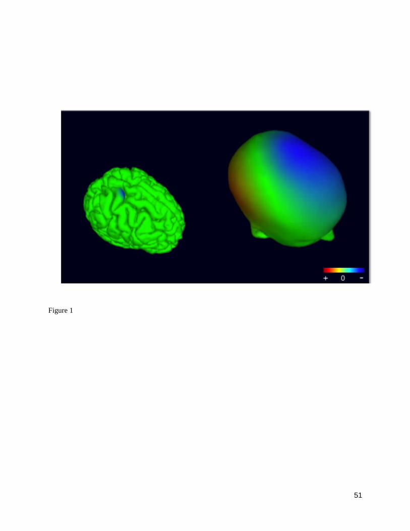
51
Figure 1