Embed Size (px)
Citation preview

Exam II Review(10 / 21 / 2008)
TOPICS
•Protein Structure
•Myoglobin/Hemoglobin
•Enzymes
•Enzyme Kinetics

Example of each level of protein structure

Fibrous Proteins
Nails, hair, horns and feathers or -forms
30 variants, tissue specific type I and type II
acidic negative charge basic positive charge
keratin• hair- 20 M diameter• macrofibril 2000 Å parallel to hair • microfibril 80 Å and high sulfur content protein• can break -S-S- with mercaptans and reconnect (i.e. can give hair a “permanent” wave)
Keratin - A Coiled Coil

keratin proteins are helical but spacing differs from a regular -helix
a 5.1 Å vs. 5.4 Å pitch. This change in pitch forms closely associated pairs of helices. Each pair consists of a type I and type II proteinLeft-handed coil coiled-coil310 AA residues 7-residue pseudo repeat.Helical wheel - 3.6 residues/turn 360 = 100 per residuea - b - c - d - e - f - g - a repeat on side of helix
Helical wheel diagram
a and d residues are nonpolar.
View down the coil axis

Collagen triple residue repeat:Gly-X-Y X often Pro Y often Hyp like a poly Gly or poly Pro helix
Left-handed 3.0 residues/turn pitch 9.4 extended conformation the prolines avoid each other.
3 left handed helices combine in a triple right handed coil.
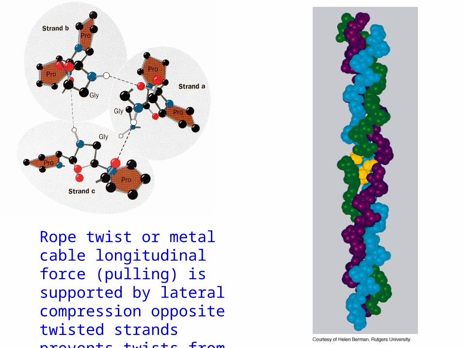
Rope twist or metal cable longitudinal force (pulling) is supported by lateral compression opposite twisted strands prevents twists from pulling out.

Hemoglobin switch T to R states

The Fe iron is about 0.6 Å out of the heme plane in the deoxy state. When oxygen binds it pulls the iron back into the heme plane. Since the proximal His F8 is attached to the Fe this pulls the complete F helix like a lever on a fulcrum.
The positive cooperativity of O2 binding to Hb The effect of the ligand-binding state of one heme on
the ligand-binding affinity of another.

a. Free energy changes with fractional saturation
b. Sigmoidal binding curve as a composite of the R state binding and the T state binding.

Origin of the Bohr Effect
The T R transition causes the changes in the pK’s of several groups. The N-terminal amino groups are responsible for 20-30% of the Bohr effect. His146 accounts for about 40% of the Bohr effect salt bridged with Asp 94. This interaction is lost in the R state.
• The T-state is shown above.
• TR transition causes breakage of terminal interactions and changes in ionization states of His146 and Val1 (part of Bohr effect)
Networks of H-bonds & ion pairs in T-state

D-2,3-bisphosphoglycerate (BPG)
BPG binds to Hb (deoxy state) and decreases the O2 affinity and keeps it in the deoxy form.
BPG binds 1:1 with a K=1x10⁻⁵ M to the deoxy form but weakly to the oxy form
Fetal Hb (22) has low BPG affinity-His143 to Ser in chain

BPG levels are partially responsible for High-Altitude adaptation
BPG restores the 37% release of O2 at higher elevations between arterial and venous blood

Sickle Cell Mutation
Glu 6 ---> Val 6 mutation on the hemoglobin B chain
Heterozygotes carrying only one copy of the sickle-cell gene are more resistant to malaria than those homozygous for for the normal gene.

106 rate enhancement requires a 106 higher affinity which is 34.2 kJ/mol
Enzymes: The more tightly an enzyme binds its reaction’s transition state (KT) relative to the substrate (KR) , the greater the rate of the catalyzed reaction (kE) relative to the uncatalyzed reaction (kN)
Catalysis results from the preferred binding and therefore the stabilization of the transition state (S ‡) relative to that of the substrate (S).
RTGG EN
‡‡
expk
k
N
E

Mechanism of lysozyme


Bovine Trypsin
Serine proteases


1. Conformational distortion forms the tetrahedral intermediate and causes the carboxyl to move close to the oxyanion hole
2. Now it forms two hydrogen bonds with the enzyme that cannot form when the carbonyl is in its normal conformation.
3. Distortion caused by the enzyme binding allows the hydrogen bonds to be maximal.

Enzyme Kinetics: The double reciprocal plot
maxmax
M
V
1
S
1
V
K1
ov

What is catalytic perfection?
When k2>>k-1 or the ratio 21
21
kk
kk
is maximum
Then1
MKk
kcat
Or when every substrate that hits the enzyme causes a reaction totake place. This is catalytic perfection. Note that for Michaelis -Menton kinetics k2= kcat
Diffusion-controlled limit- diffusion rate of a substrate is in the range of 108 to 109 M-1s-1. An enzyme lowers the transition state so there is no activation energy and the catalyzed rate is as fast as molecules collide.
Tmax
E
Vcatk

Reaction MechanismsA: Sequential Reactions
• All substrates must combine with enzyme before reaction can occur

Bisubstrate reactions

Random Bisubstrate Reactions

Ping-Pong Reactions
• Group transfer reactions
• One or more products released before all substrates added

Competitive Inhibition

Competitive Inhibition: Lineweaver-Burke Plot

Uncompetitive Inhibition

Uncompetitive Inhibition: Lineweaver-Burke Plot

Mixed inhibition


Mixed inhibition is when the inhibitor binds to the enzyme at a location distinct from the substrate binding site. The binding of the inhibitor will either alter the KM or Vmax or both.
EI
IEK I
ESI
IESK I
SK
SV
M
max
ov
IK
I1


Next Lecture 18 Exam II (10/23/08)
Lecture 19 10/28/08 Metabolism