Embed Size (px)
Citation preview
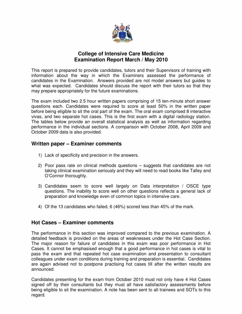
College of Intensive Care Medicine Examination Report March / May 2010
This report is prepared to provide candidates, tutors and their Supervisors of training with information about the way in which the Examiners assessed the performance of candidates in the Examination. Answers provided are not model answers but guides to what was expected. Candidates should discuss the report with their tutors so that they may prepare appropriately for the future examinations. The exam included two 2.5 hour written papers comprising of 15 ten-minute short answer questions each. Candidates were required to score at least 50% in the written paper before being eligible to sit the oral part of the exam. The oral exam comprised 8 interactive vivas, and two separate hot cases. This is the first exam with a digital radiology station. The tables below provide an overall statistical analysis as well as information regarding performance in the individual sections. A comparison with October 2008, April 2009 and October 2009 data is also provided.
Written paper – Examiner comments
1) Lack of specificity and precision in the answers.
2) Poor pass rate on clinical methods questions – suggests that candidates are not taking clinical examination seriously and they will need to read books like Talley and O’Connor thoroughly.
3) Candidates seem to score well largely on Data interpretation / OSCE type questions. The inability to score well on other questions reflects a general lack of preparation and knowledge even of common topics in intensive care.
4) Of the 13 candidates who failed, 6 (46%) scored less than 45% of the mark.
Hot Cases – Examiner comments The performance in this section was improved compared to the previous examination. A detailed feedback is provided on the areas of weaknesses under the Hot Case Section. The major reason for failure of candidates in this exam was poor performance in Hot Cases. It cannot be emphasised enough that a good performance in hot cases is vital to pass the exam and that repeated hot case examination and presentation to consultant colleagues under exam conditions during training and preparation is essential. Candidates are again advised not to postpone practising hot cases till after the written results are announced. Candidates presenting for the exam from October 2010 must not only have 4 Hot Cases signed off by their consultants but they must all have satisfactory assessments before being eligible to sit the examination. A note has been sent to all trainees and SOTs to this regard.
Vivas – Examiner comments Whilst the performance in the vivas was comparable to the October 2009 exam, the viva stations represent an opportunity to score marks and make up for any deficits in the clinical exam. Again, considerable deficiencies in knowledge were noted in mainstream topics. The pass rate in the radiology section continues to be low.
Overall Statistics
Written section March 2010
August 2009
March 2009
August 2008
Number of candidates appearing for the written 43 49 36 48
Number of candidates successful at written 30 36 23 35
Pass rate 70% 73% 64% 73%
Number of OTS (written required) 0 1 2 5
Number OTS successful at written 0 0 2 4
Pass rate 0% 0% 100% 80%
Total number successful at the written 30 36 25 39
Written pass rate (Including OTS) 70% 72% 66% 74%
Detailed statistics for the written paper 1) Highest aggregate mark in the written paper – 84% 2) In no question was there a 100% pass rate. 3) In 12 of the 30 questions, the pass rate was < 50%.
Pathway to oral section May 2010 Oct 2009 May 2009 Oct 2008
Number of candidates who scored >50% at written (Including OTS)
30 36 25 39
Number of candidates (written carry from previous attempt)
7 13 8 10
Number of OTS (written carry from previous attempt)
0 1 1 0
Number of OTS (exempt from the written) 2 4 2 3
Total number invited to the oral section 39 54 36 52
Oral section May 2010 Oct 2009 May 2009 Oct 2008
Number of candidates scoring > 50% who were successful at the orals
24 31 16 29
Pass rate 80% 86% 70% 83%
Total number of candidates carrying written who were successful at orals
3 11 1 8
Pass rate 43% 85% 13% 80%
Total number of OTS successful at orals (carry) 0 1 0 0
Pass rate 0% 100% 0% 0%
Total number of OTS candidates successful at orals (written required)
0 0 1 3
Pass rate 0% 0% 50% 75%
Total number of OTS candidates successful at orals (written exempt)
0 3 0 2
Pass rate 0% 75% 0% 67%
Total number of successful candidates 27 46 18 42
Overall Pass Rates May 2010 Oct
2009 May 2009
Oct 2008
Total number of candidates presenting 52 68 49 66
Total number invited to the oral section 39 54 36 52
Total number successful in the clinical section 20 34 15 31
Clinical section pass rate 51% 63% 42% 60%
Number of candidates passing both hot cases 13 18 8 21
Both hot case pass rate 33% 33% 22% 40%
Number of candidates passing viva section 32 47 28 47
Viva section pass rate 82% 87% 78% 90%
Oral candidate pass rate 69% 85% 50% 81%
Overall Pass rate 52% 68% 37% 64%
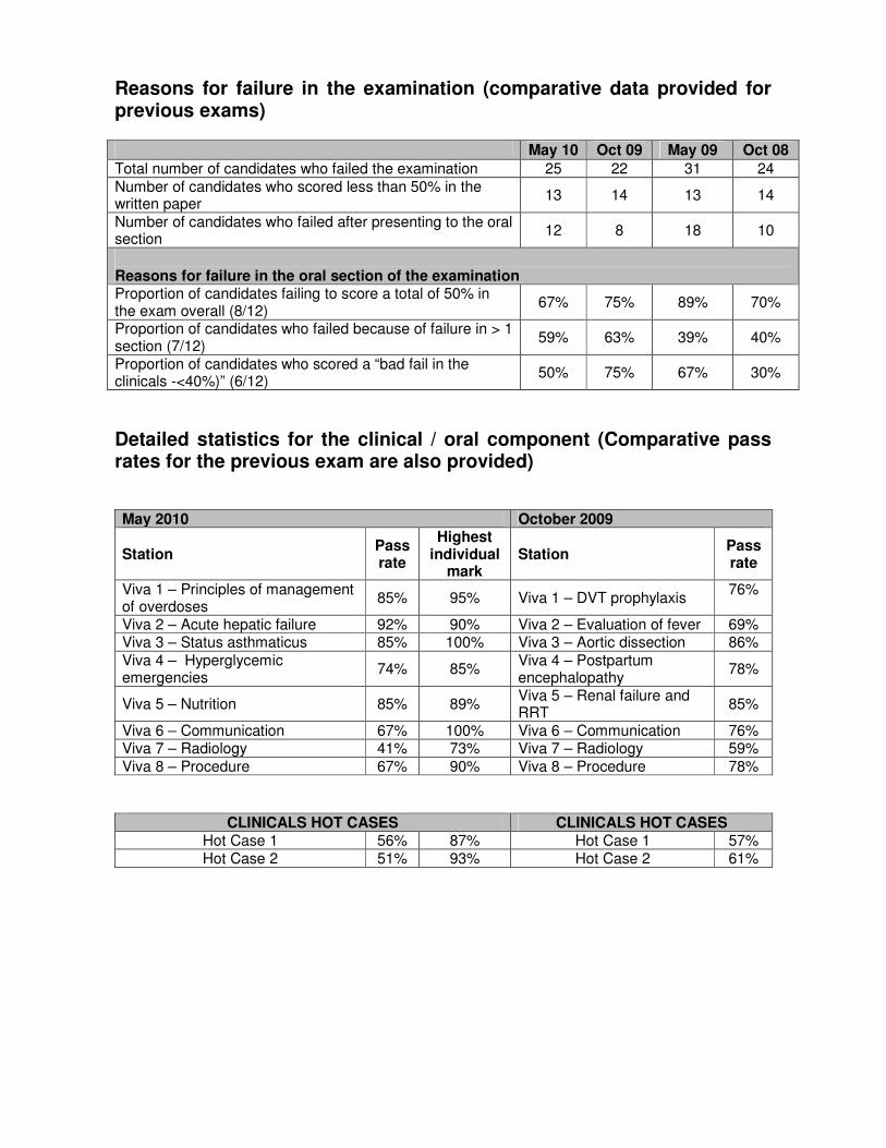
Reasons for failure in the examination (comparative data provided for previous exams) May 10 Oct 09 May 09 Oct 08 Total number of candidates who failed the examination 25 22 31 24 Number of candidates who scored less than 50% in the written paper
13 14 13 14
Number of candidates who failed after presenting to the oral section
12 8 18 10
Reasons for failure in the oral section of the examination Proportion of candidates failing to score a total of 50% in the exam overall (8/12)
67% 75% 89% 70%
Proportion of candidates who failed because of failure in > 1 section (7/12)
59% 63% 39% 40%
Proportion of candidates who scored a “bad fail in the clinicals -<40%)” (6/12)
50% 75% 67% 30%
Detailed statistics for the clinical / oral component (Comparative pass rates for the previous exam are also provided) May 2010 October 2009
Station Pass rate
Highest individual
mark Station
Pass rate
Viva 1 – Principles of management of overdoses
85% 95% Viva 1 – DVT prophylaxis 76%
Viva 2 – Acute hepatic failure 92% 90% Viva 2 – Evaluation of fever 69% Viva 3 – Status asthmaticus 85% 100% Viva 3 – Aortic dissection 86% Viva 4 – Hyperglycemic emergencies
74% 85% Viva 4 – Postpartum encephalopathy
78%
Viva 5 – Nutrition 85% 89% Viva 5 – Renal failure and RRT
85%
Viva 6 – Communication 67% 100% Viva 6 – Communication 76% Viva 7 – Radiology 41% 73% Viva 7 – Radiology 59% Viva 8 – Procedure 67% 90% Viva 8 – Procedure 78%
CLINICALS HOT CASES CLINICALS HOT CASES Hot Case 1 56% 87% Hot Case 1 57% Hot Case 2 51% 93% Hot Case 2 61%
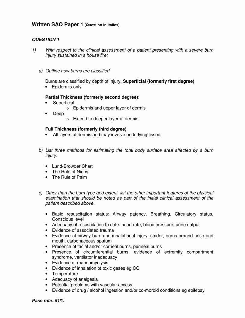
Written SAQ Paper 1 (Question in Italics)
QUESTION 1 1) With respect to the clinical assessment of a patient presenting with a severe burn
injury sustained in a house fire:
a) Outline how burns are classified.
Burns are classified by depth of injury. Superficial (formerly first degree):
• Epidermis only Partial Thickness (formerly second degree):
• Superficial o Epidermis and upper layer of dermis
• Deep o Extend to deeper layer of dermis
Full Thickness (formerly third degree)
• All layers of dermis and may involve underlying tissue
b) List three methods for estimating the total body surface area affected by a burn
injury.
• Lund-Browder Chart
• The Rule of Nines • The Rule of Palm
c) Other than the burn type and extent, list the other important features of the physical examination that should be noted as part of the initial clinical assessment of the patient described above.
• Basic resuscitation status: Airway patency, Breathing, Circulatory status, Conscious level
• Adequacy of resuscitation to date: heart rate, blood pressure, urine output
• Evidence of associated trauma • Evidence of airway burn and inhalational injury: stridor, burns around nose and
mouth, carbonaceous sputum
• Presence of facial and/or corneal burns, perineal burns • Presence of circumferential burns, evidence of extremity compartment
syndrome, ventilator inadequacy
• Evidence of rhabdomyolysis • Evidence of inhalation of toxic gases eg CO • Temperature
• Adequacy of analgesia
• Potential problems with vascular access • Evidence of drug / alcohol ingestion and/or co-morbid conditions eg epilepsy
Pass rate: 51%
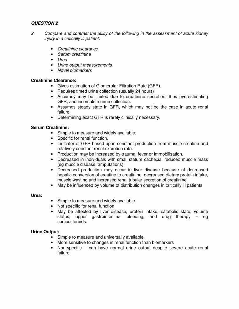
QUESTION 2 2. Compare and contrast the utility of the following in the assessment of acute kidney
injury in a critically ill patient:
• Creatinine clearance
• Serum creatinine
• Urea
• Urine output measurements
• Novel biomarkers Creatinine Clearance:
• Gives estimation of Glomerular Filtration Rate (GFR).
• Requires timed urine collection (usually 24 hours) • Accuracy may be limited due to creatinine secretion, thus overestimating
GFR, and incomplete urine collection.
• Assumes steady state in GFR, which may not be the case in acute renal failure.
• Determining exact GFR is rarely clinically necessary. Serum Creatinine:
• Simple to measure and widely available. • Specific for renal function. • Indicator of GFR based upon constant production from muscle creatine and
relatively constant renal excretion rate.
• Production may be increased by trauma, fever or immobilisation.
• Decreased in individuals with small stature cachexia, reduced muscle mass (eg muscle disease, amputations)
• Decreased production may occur in liver disease because of decreased hepatic conversion of creatine to creatinine, decreased dietary protein intake, muscle wasting and increased renal tubular secretion of creatinine.
• May be influenced by volume of distribution changes in critically ill patients
Urea:
• Simple to measure and widely available • Not specific for renal function
• May be affected by liver disease, protein intake, catabolic state, volume status, upper gastrointestinal bleeding, and drug therapy – eg corticosteroids.
Urine Output:
• Simple to measure and universally available. • More sensitive to changes in renal function than biomarkers • Non-specific – can have normal urine output despite severe acute renal
failure
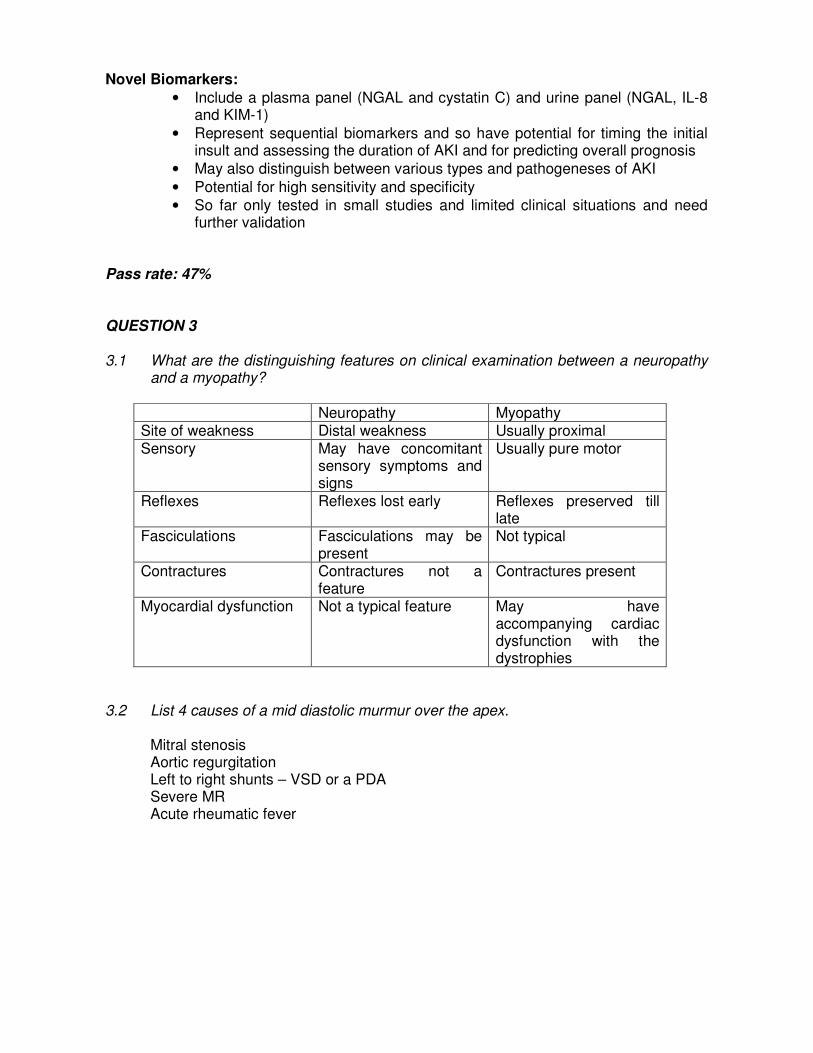
Novel Biomarkers:
• Include a plasma panel (NGAL and cystatin C) and urine panel (NGAL, IL-8 and KIM-1)
• Represent sequential biomarkers and so have potential for timing the initial insult and assessing the duration of AKI and for predicting overall prognosis
• May also distinguish between various types and pathogeneses of AKI
• Potential for high sensitivity and specificity • So far only tested in small studies and limited clinical situations and need
further validation Pass rate: 47% QUESTION 3 3.1 What are the distinguishing features on clinical examination between a neuropathy
and a myopathy?
Neuropathy Myopathy Site of weakness Distal weakness Usually proximal Sensory May have concomitant
sensory symptoms and signs
Usually pure motor
Reflexes Reflexes lost early Reflexes preserved till late
Fasciculations Fasciculations may be present
Not typical
Contractures Contractures not a feature
Contractures present
Myocardial dysfunction Not a typical feature May have accompanying cardiac dysfunction with the dystrophies
3.2 List 4 causes of a mid diastolic murmur over the apex.
Mitral stenosis Aortic regurgitation Left to right shunts – VSD or a PDA Severe MR Acute rheumatic fever
3.3 A previously fit 45 year old man was noted to be in respiratory distress 24 hours following maxillofacial surgery.
Clinical examination revealed the following:
• Respiratory rate of 22/min
• Decreased air entry left side
• Crackles left base
• HR-104/min, regular. JVP not raised. Apical impulse 5th left intercostal space anterior axillary line.
Investigations:
• ECG – normal
• Chest X-Ray – complete whiteout on the left side. (a) What is the likely cause of his respiratory distress?
• Left lung collapse from • Perioperative blood aspiration
• Aspiration of gastric contents • Sputum
Pass rate: 67% QUESTION 4 4. Stress induced hyperglycaemia (S.I.H) is common in critically ill patients.
a) Define S.I.H
Transient hyperglycaemia during acute illness –usually restricted to patients without prior evidence of diabetes with reversion to normal after discharge.
b) Outline the mechanisms thought important in the pathogenesis of S.I.H.
• S.I.H is thought to develop due to complex interplay between counter regulatory hormones such as catecholamines, GH, cortisol and cytokines.
• The underlying illness and treatments (TPN, enteral feed, steroids, and vasopressors) might affect the scale of these derangements.
• The key contributor would appear to be high hepatic glucose output via gluconeogenesis driven by glucagon, adrenaline and cortisol. Cytokines such as TNFα interact to enhance this response.
• Insulin resistance plays a role. • Underlying abnormalities in glucose regulation may be present.
c) Outline clinical implications and treatment of S.I.H.
• Recent data suggests that S.I.H and diabetic hyperglycaemia are two different phenomena with differing clinical outcomes.
• Patients with S.I.H have been shown in several studies to have increased risk of mortality, adverse events, and greater organ failure scores compared to those with diabetes.
• Whether S.I.H per se causes harm or instead is a marker of severity of counter regulatory response and degree of illness is unknown.
• Management of S.I.H cannot be distinguished from hyperglycaemia due to other causes. In most cases it is not generally predictable or preventable. Early recognition and interception might prevent persistence and exacerbation. Recommendations include insulin therapy with more conservative glucose targets.
• Candidates mentioning recent data from RCTs were given credit.
Pass rate: 56%
QUESTION 5
5. With respect to pathological conditions of the spinal cord, list 2 causes of and the clinical findings for each of the following syndromes: Complete cord transection Cord hemisection Central cord syndrome Anterior cord syndrome (anterior spinal artery syndrome) Cauda Equina syndrome You may tabulate your answer
Syndrome Aetiology Clinical Findings
Complete Transection
Trauma, Infarction, Transverse Myelitis, Abscess, Tumour
Complete loss of motor and sensory function below level of the lesion
Cord Hemisection Trauma, Multiple Sclerosis, Tumour, Abscess
Ipsilateral loss of motor and proprioception. Contralateral pain and temperature loss
Central Cord Neck hyperextension, syringomyelia, tumour
Motor impairment greater in upper limbs than lower Variable sensory loss, bladder dysfunction
Anterior Cord Hyperflexion, disc protusion, anterior spinal artery occlusion, Post AAA
Motor function impairment, Pain and temperature loss, proprioception spared.
Cauda Equina Disc protusion, tumour, infective
Bladder/bowel dysfunction Altered sensation in saddle area, sexual dysfunction.
Pass rate: 77%
QUESTION 6 6. A 20 year old primi-gravida presents at 37 weeks gestation with jaundice,
headache, blurred vision and hypertension (140/90mmHg). The antenatal period was otherwise unremarkable. She is febrile, drowsy, pale, icteric and has pedal oedema. The uterus is palpated as for a full term pregnancy with a normal CTG trace. Examination is otherwise normal.
The following are her early blood results:
Hb* 80 G/L (115-160)
Platelets* 52 x 109/L (140-400)
INR* 1.8 (0.9-1.3)
APTT* 55 seconds (25-38)
LDH* 654 U/L (110-250)
Fibrinogen* 1.0 G/L (1.5-4.0)
Total Bilirubin* 51µmol/L (<20)
Urea* 30 mmol/L (3-8)
Creatinine* 298 µmol/L (70-120)
Potassium* 5.1 mmol/L (3.2-4.5)
(a) List 4 likely differential diagnoses for this clinical presentation.
• Pre-eclampsia
• HELLP Syndrome • Sepsis with DIC
• HUS-TTP • Acute fatty liver of pregnancy
(b) What other investigations would you order for this patient and why?
• Transaminases (full liver function tests) Assessment of HELLP
• Peripheral blood film smear
Evidence of haemolysis or MAHA • Reticulocyte count, haptoglobins, conjugated/unconjugated bilirubin
Haemolysis screen
• Blood, sputum, urine and vaginal swab for MC&S Septic screen
• Urinalysis – protein, WBCs, RBCs, casts Evidence of infection or proteinuria (pre-eclampsia)
• Renal tract ultrasound Rule out obstruction

(c) List the important management interventions for each of your differential diagnoses.
a. Pre-eclampsia i. Deliver baby ii. Control BP iii. Hydralazine, beta blockers iv. SNP/GTN if intravenous agent required. v. Prevention of seizures vi. Magnesium sulphate
b. HELLP Syndrome
i. Deliver baby ii. Regular monitoring of platelet count and liver function iii. Supportive measures whilst observing in HDU for dangerous
complications – hepatic haemorrhage/rupture, progressive renal failure, pulmonary oedema.
c. Sepsis with DIC i. Timely delivery of baby in consultation with obstetrician. ii. Early broad spectrum antibiotics. iii. Cardiovascular support – adequate volume resuscitation and establish
a MAP > 65mmHg.
d. HUS-TTP i. Deliver the baby. ii. Fresh frozen plasma iii. Therapeutic plasma exchange iv. Corticosteroid therapy v. Monoclonal antibody therapy – Rituximab
e. Acute fatty liver of pregnancy
i. Timely delivery of baby once mother stabilised ii. Correction of DIC iii. Supportive therapy iv. Monitoring and treatment of complications post delivery eg
pancreatitis v. Consideration for liver transplantation in with irreversible severe liver
failure despite delivery and aggressive supportive care Pass rate: 63%
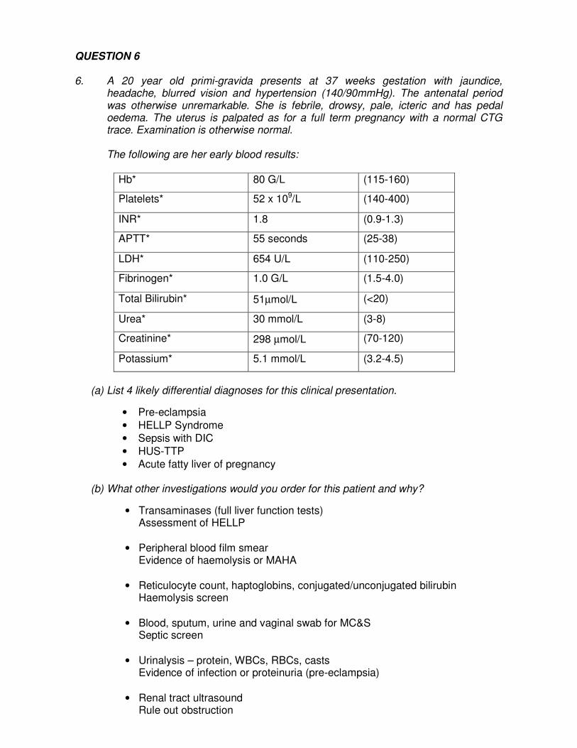
QUESTION 7 7.1. The following image is of the blood sample tubes into which a specimen of blood
from a critically ill patient had been drawn by the phlebotomist.
(a) What does this image show? A creamy supernatant in blood tubes (serum and plasma) due to severe hypertriglyceridaemia (lipaemic serum). (b) List three (3) causes for this appearance in blood samples from critically ill patients. Familial hyperlipedemia Propofol infusion TPN use Pancreatitis from hyperlipedemia (c) If the condition causing this appearance in the blood tubes were to be long
standing, what clinical signs specific to this condition may be found in this patient?
Eyes Lipaemia retinalis Corneal arcus senilis Xanthelasma Skin Xanthomata Tendon Eruptive
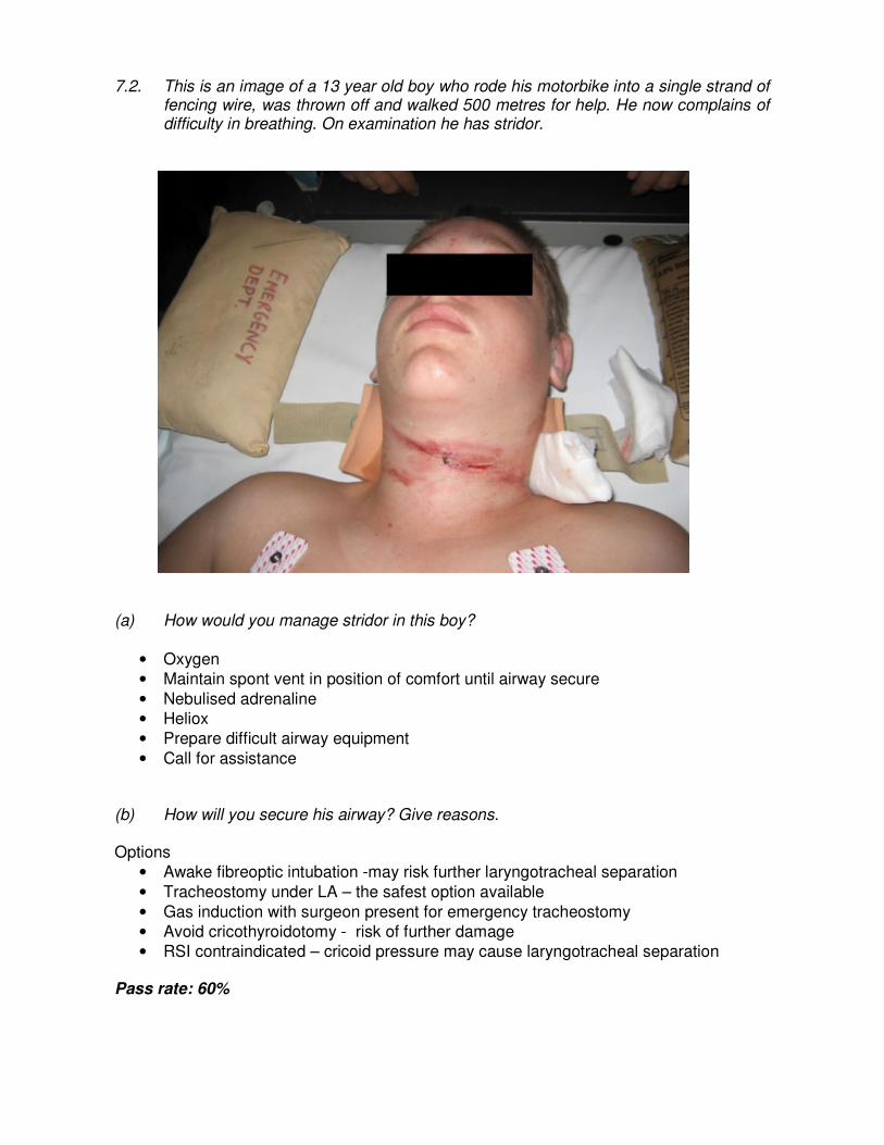
7.2. This is an image of a 13 year old boy who rode his motorbike into a single strand of fencing wire, was thrown off and walked 500 metres for help. He now complains of difficulty in breathing. On examination he has stridor.
(a) How would you manage stridor in this boy?
• Oxygen • Maintain spont vent in position of comfort until airway secure
• Nebulised adrenaline • Heliox • Prepare difficult airway equipment
• Call for assistance (b) How will you secure his airway? Give reasons. Options
• Awake fibreoptic intubation -may risk further laryngotracheal separation
• Tracheostomy under LA – the safest option available
• Gas induction with surgeon present for emergency tracheostomy • Avoid cricothyroidotomy - risk of further damage • RSI contraindicated – cricoid pressure may cause laryngotracheal separation
Pass rate: 60%
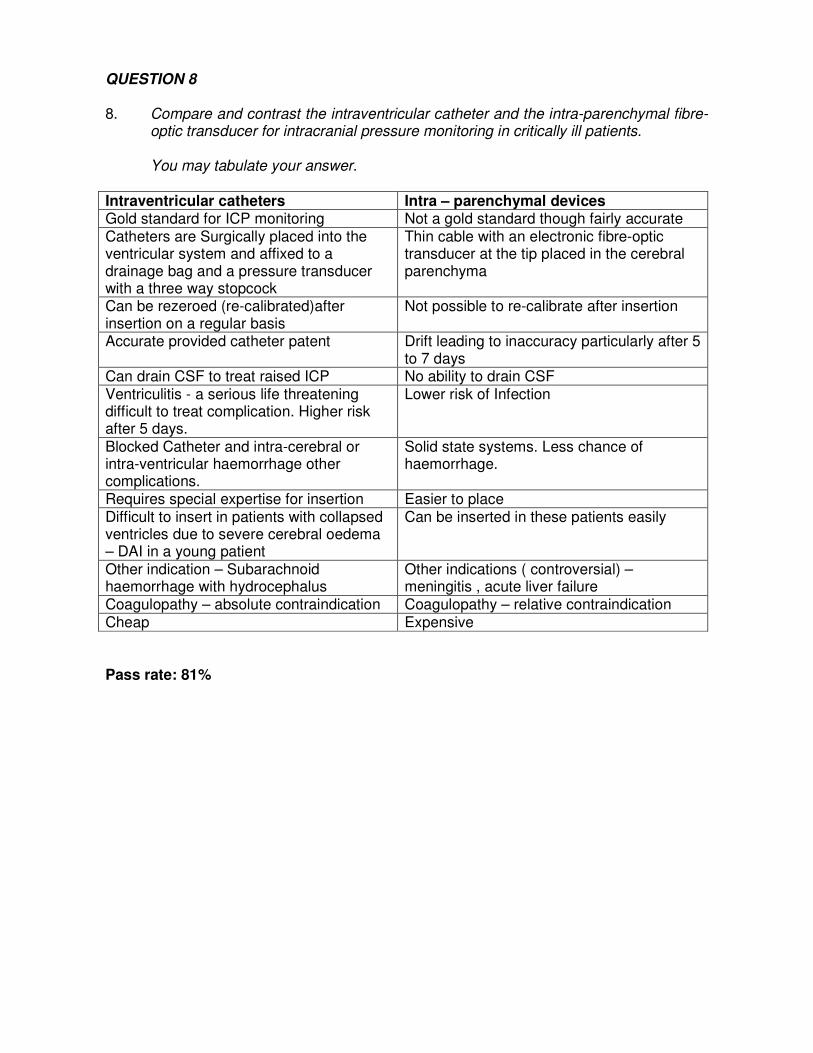
QUESTION 8 8. Compare and contrast the intraventricular catheter and the intra-parenchymal fibre-
optic transducer for intracranial pressure monitoring in critically ill patients.
You may tabulate your answer.
Intraventricular catheters Intra – parenchymal devices Gold standard for ICP monitoring Not a gold standard though fairly accurate Catheters are Surgically placed into the ventricular system and affixed to a drainage bag and a pressure transducer with a three way stopcock
Thin cable with an electronic fibre-optic transducer at the tip placed in the cerebral parenchyma
Can be rezeroed (re-calibrated)after insertion on a regular basis
Not possible to re-calibrate after insertion
Accurate provided catheter patent Drift leading to inaccuracy particularly after 5 to 7 days
Can drain CSF to treat raised ICP No ability to drain CSF Ventriculitis - a serious life threatening difficult to treat complication. Higher risk after 5 days.
Lower risk of Infection
Blocked Catheter and intra-cerebral or intra-ventricular haemorrhage other complications.
Solid state systems. Less chance of haemorrhage.
Requires special expertise for insertion Easier to place Difficult to insert in patients with collapsed ventricles due to severe cerebral oedema – DAI in a young patient
Can be inserted in these patients easily
Other indication – Subarachnoid haemorrhage with hydrocephalus
Other indications ( controversial) – meningitis , acute liver failure
Coagulopathy – absolute contraindication Coagulopathy – relative contraindication Cheap Expensive Pass rate: 81%
QUESTION 9 9. A two year old child presents with fever, stridor and a harsh cough. His condition
deteriorates and he requires intubation. Outline how you would do this. Call for help This should be in context –
a) If the child becomes hypoxic/has a respiratory arrest etc – proceed with attempt bag mask ventilation 100% oxygen immediately – attempt intubation.
b) If there is time – aim to have the person with the best paediatric airway management expertise – intubate child
Optimise medical management a) High flow oxygen b) if child hypoxic – can discuss avoiding distressing the child by holding mask away from face and with child on parents lap (unless really sick) c) IV steroids – adequate dose (` 0.6mg/kg dexamethasone d) NEB adrenaline 5mg (repeated doses) e) Oxygen/Helium mixture if tolerates Adequate discussion of preparation for intubation a) range of ETT’s (size 4.0, 4.5. 5.0, 5.5) b) two laryngoscopes with range of blade sizes – straight/curved c) small diam “bougie” d) cannula for percutaneous needle cricothyroidotomy + method for oxygen delivery e) suction Intubation: One of 2 approaches (1) Inhalational induction of anaesthesia with maintenance of spontaneous ventilation until adequate depth of anaesthesia achieved to allow intubation (or to assess ability to ventilate – then proceed to paralyse child) Or (2) IV induction – with paralysis There must be some discussion regarding risks of either technique. Mere mention of IV approach will not be enough to gain marks. There must be some discussion regarding risks of either technique However, if not trained in inhalational anaesthetic techniques – reasonable to proceed with IV induction of anaesthesia + muscle paralysis – with risk of being unable to ventilate Alternate strategies if unable to intubate Ventilate with LMA/face mask until help arrives Rarely need to proceed to needle cricothyroidotomy Pass rate: 47%
QUESTION 10 10. A 30 year old man has been admitted to hospital with severe multiple injuries
following a motor vehicle accident.
On day 2, his intracranial pressure has stabilised and his head CT shows scattered punctate haemorrhages with subarachnoid blood, with no mass lesion requiring evacuation. His pelvic fracture and right tibia / fibula fracture have been managed with external fixation and a left leg femoral fracture has undergone open reduction and internal fixation.
He has been in good health, but had a DVT 3 years ago and is not on any regular medication.
Outline your approach to prophylaxis for venous thrombo-embolism in this patient.
Risk of VTE is high based on:
• Major trauma with pelvic and lower limb injury and operative intervention • Possibility of a pro-thrombotic disorder
Therapy also has potential risks:
• Risk of intracranial haematoma expansion with unfractionated or LMW heparin Management options:
• Quantify potential pro-thrombotic disorder: ancillary history, previous investigations etc
• Unilateral mechanical prophylaxis
• Discuss timing of pharmacological prophylaxis • Clinical and imaging surveillance • IVC filter
Pass rate: 84%
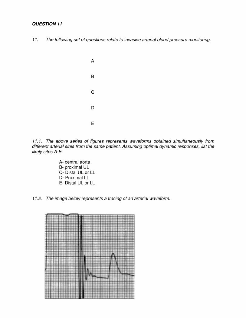
QUESTION 11 11. The following set of questions relate to invasive arterial blood pressure monitoring.
11.1. The above series of figures represents waveforms obtained simultaneously from different arterial sites from the same patient. Assuming optimal dynamic responses, list the likely sites A-E.
A- central aorta B- proximal UL C- Distal UL or LL D- Proximal LL E- Distal UL or LL
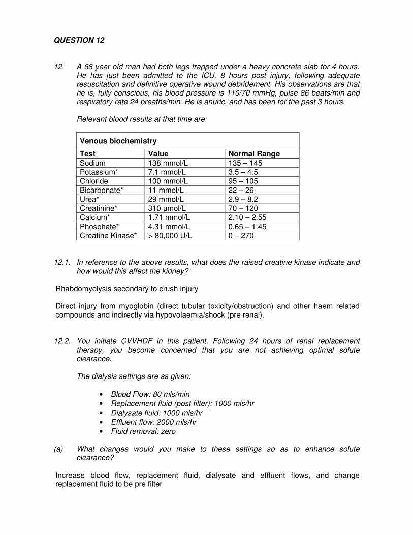
11.2. The image below represents a tracing of an arterial waveform.
A B C D E
The following set of questions relate to invasive arterial blood pressure monitoring.
The above series of figures represents waveforms obtained simultaneously from different arterial sites from the same patient. Assuming optimal dynamic responses, list the
The image below represents a tracing of an arterial waveform.
The following set of questions relate to invasive arterial blood pressure monitoring.
The above series of figures represents waveforms obtained simultaneously from different arterial sites from the same patient. Assuming optimal dynamic responses, list the

(a) What procedure has been performed? Fast flush test (b) What is your impression of the fidelity of the arterial system? Give two (2) reasons.
Underdamped trace – Multiple oscillations and systolic overshoot 11.3 List the important pieces of information that could be obtained from an arterial
waveform tracing.
1) Systolic, diastolic, mean and pulse pressures 2) Heart rate and rhythm 3) Effect of dysrhythmias on pefusion 4) ECG lead disconnect 5) Continuous cardiac output using pulse contour analysis 6) Specific waveform morphologies might be diagnostic – eg slow rising pulse –AS,
pulsus paradoxus in tamponade 7) Systolic pressure variation or pulse pressure variation may be useful in predicting
fluid responsiveness. Pass rate: 51%
QUESTION 12 12. A 68 year old man had both legs trapped under a heavy concrete slab for 4 hours.
He has just been admitted to the ICU, 8 hours post injury, following adequate resuscitation and definitive operative wound debridement. His observations are that he is, fully conscious, his blood pressure is 110/70 mmHg, pulse 86 beats/min and respiratory rate 24 breaths/min. He is anuric, and has been for the past 3 hours.
Relevant blood results at that time are:
Venous biochemistry
Test Value Normal Range
Sodium 138 mmol/L 135 – 145 Potassium* 7.1 mmol/L 3.5 – 4.5 Chloride 100 mmol/L 95 – 105 Bicarbonate* 11 mmol/L 22 – 26 Urea* 29 mmol/L 2.9 – 8.2 Creatinine* 310 µmol/L 70 – 120 Calcium* 1.71 mmol/L 2.10 – 2.55 Phosphate* 4.31 mmol/L 0.65 – 1.45 Creatine Kinase* > 80,000 U/L 0 – 270
12.1. In reference to the above results, what does the raised creatine kinase indicate and
how would this affect the kidney? Rhabdomyolysis secondary to crush injury Direct injury from myoglobin (direct tubular toxicity/obstruction) and other haem related compounds and indirectly via hypovolaemia/shock (pre renal). 12.2. You initiate CVVHDF in this patient. Following 24 hours of renal replacement
therapy, you become concerned that you are not achieving optimal solute clearance.
The dialysis settings are as given:
• Blood Flow: 80 mls/min
• Replacement fluid (post filter): 1000 mls/hr
• Dialysate fluid: 1000 mls/hr
• Effluent flow: 2000 mls/hr
• Fluid removal: zero (a) What changes would you make to these settings so as to enhance solute
clearance? Increase blood flow, replacement fluid, dialysate and effluent flows, and change replacement fluid to be pre filter

12.3. An alarm has sounded on the dialysis machine. Access pressures are high. How would you respond to this problem?
Check and manipulate vascular access
• Malposition (catheter tip, sucking against vessel wall)and kinking (subclavian )
• Change in patient position- side/supine/sitting
• Site of catheter- e.g. sitting up –femoral access problems • Type of catheter-geometry, length, diameter • Negative intra thoracic pressure - high intra abdominal pressures
• Hypovolemic patient –poor flow • Catheter occlusion / thrombosis
12.4. Briefly outline the relationship between dose of dialysis and outcome Candidates were not expected to list all of the literature but an understanding that this remains a controversial area- credit was given if they quoted relevant studies Although several clinical trials have suggested an improvement in survival with higher doses of CRRT results have not been consistent across all studies. To date five randomised trials have assessed the relationship between intensity of CRRT in terms of effluent flow rate and outcomes of acute kidney injury.
• Ronco (Lancet 2000) and Saudan (Kid Int 2006) found that lower doses around 20 -25ml kg hr were inferior in terms of survival to higher effluent flows of around 35 to 45 mls kg hr.
• Two other studies Bouman (Crit Care Med 2002) and Tolwani (J Am Soc Nephrol, 2008) however found no difference in survival with higher effluent rates.
• The latest study (NEJM 2008, VA/HIH acute renal failure Trial Network or ATN study) found that mortality at 60 days was no different between two intensity arms. In the less intensive arm both IHD and SLED were used as standard practice of thrice per week and CVVHDF effluent flow at 20 mls kg hr. In the more intensive arm IHD and or SLED were used six times per week and CVVHDF at an effluent flow rate of 35ml kg hr.
• The ANZICS CTG RENAL study just completed (25 v 40 ml kg hr). No difference in mortality between the two groups, a higher incidence of hypophosphatemia in the higher dose group.
Pass rate: 77%
QUESTION 13 13.1. Apart from vancomycin, list three antibiotics that have activity against hospital
acquired methicillin resistant staphylococcus aureus (MRSA).
• Linezolid
• Talavancin
• Streptogramins (not currently available in Australia) • tigecycline
13.2 List an example of each of the three main classes of systemic antifungal agents.
• Polyenes e.g. Amphotericin B • Azoles e.g. Fluconazole
• Echinocandins e.g. caspofungin, andulafungin, micafungin
13.3. Briefly outline the dosing adjustment and the monitoring necessary in patients with septic shock for each of the following drug groups in patients with moderate to severe renal dysfunction (without dialysis)
a) Aminoglycosides b) Fluoroquinolones c) Beta Lactams d) Carbapenems e) Glycopeptides
Aminoglycosides
High initial dose and monitor trough concentrations. Extend interval. May be necessary to decrease dose and monitor with MIC data
Fluoroqinolones
Reduce frequency but maintain dose. Monitor QT interval
Beta Lactams
Can reduce dose OR frequency Monitoring unnecessary
Carbapenems
As for Beta Lactams
Glycopeptides
High dosing on day one dose adjustments according to Cmin and dependent on degree of renal dysfunction
Pass rate: 86%
QUESTION 14 14. With respect to plasma exchange therapy: (a) What are the physical principles of plasma exchange therapy?
• Separation of plasma from blood cells by centrifugation or membrane filtration
• Reinfusion of cells plus autologous plasma or another replacement solution eg albumin
• Removes large molecular weight substances (b) What substances can plasma exchange effectively remove?
• Pathogenic auto-antibodies
• Immune complexes • Cryoglobulins
• Myeloma light chains
• Endotoxin • Cholesterol-containing lipoproteins /triglycerides
(c) List 5 acute conditions where therapeutic plasma exchange is indicated.
Myasthenic Crisis
• Goodpasture’s Syndrome with pulmonary haemorrhage • Hyperviscosity syndromes
o Cryoglobulinaemia o Paraproteinaemia o Waldenstrom’s Macroglobulinaemia
• Wegener’s Granulomatosis with pulmonary haemorrhage • Guillain-Barre Syndrome/Acute Inflammatory Demyelinating Polyradiculopathy
• Antiphospholipid Antibody Syndrome • HELLP syndrome
• Multiple sclerosis • HIV-related neuropathy • SLE
• Pemphigus • Paraneoplastic syndromes
• Rapidly progressive glomerulonephritis • Renal transplant rejection
• Coagulation inhibitors • Auto-immune haemolytic anaemia • DIC
• Overwhelming sepsis syndromes eg meningococcaemia
• Reye’s syndrome • Paraquat poisoning
(d) List 4 common complications of this therapy, excluding catheter-related complications
• Hypotension due to excess fluid removal +/ inadequate volume replacement
• Citrate-induced hypocalcaemia • Anaphylactic/transfusion reactions to fresh frozen plasma replacement solution
• Coagulation abnormalities due to removal of clotting factors not replaced when albumin replacement used.
• Removal of useful immunoglobulins and complement which can in theory lead to an immunodeficient state.
• Drug removal – especially drugs with high protein-binding and low volume of distribution. Potentials in the diseases in which therapeutic plasma exchange is used are cyclophosphamide and azathioprine.
• Hypothermia • Pyrogenic reactions
• Anaemia
• Thrombocytopenia
• Hepatitis • Vasovagal reactions
Pass rate: 74% QUESTION 15 15. Chest compression only CPR should replace the current guidelines on CPR.
Critically evaluate this statement. Reasons supporting the statement: Physiological: a) In cardiac arrest heart dilates acutely. Decompression of the heart occurs with good compressions b) Ventilation can lead to decreased venous return c) Passive ventilation still occurs with compression only CPR d) Gasping can provide adequate ventilation and in presence of a partial airway obstruction may lead to increased venous return Logistic reasons: a) Reluctance to perform mouth to mouth by rescuers therefore some people do not attempt CPR. b) Interruption to compressions therefore limiting their effectiveness c) Easier to teach compression only CPR. d) Out of hospital arrests it will minimise time to hospital. e) Useful particularly in the setting of a single rescuer
Studies: Mostly observational or animal. Some RCT No difference in outcome using compression only versus standard CPRs in most studies Evidence of value of good compressions Against: Most studies are observational. Reported survival is no better with compression only therefore why change. Data for most studies are prior to the change in recommendation to 30:2 RATIO Ventilation is important for many arrests EG drowning/children/in hospital arrests ARC not recommend as standard practice Present position: Not standard currently. Wait further studies. It can be used if rescuer is reluctant to use mouth to mouth Pass rate: 26%

Written SAQ Paper 2 (Question in Italics)
16. List the possible reasons why a patient with septic shock from infected pancreatitis
may have ongoing hypotension despite intravenous fluid therapy, antibiotics and escalating inotrope requirement.
Primary problem not fixed
• Untreated focus of infection/ inadequate primary source control eg pancreatic abscess, infected pseudocyst
• New sepstic site eg central line/ hospital acquired pneumonia /cholecystitis, urinary tract
Systematic Approach “hypovolaemic/ obstructive/ cardiac/ distributive +/- endocrine
• Hypovolaemia or hidden bleeding eg. From surgical site/ peptic ulcer, “third space” losses (eg ascites from peritonitis)
• Undiagnosed or new “obstructive shock” :Tension pneumothorax/ Pericardial
effusion/gas trapping (auto PEEP)/ pleural effusions/ pulmonary emboli
• Severe Intra abdominal hypertension
• Dysrhythmia eg SVT, junctional rhythm etc
• New myocardial ischaemia • New/ undiagnosed cardiac valve pathology • Severe adrenal/ pituitary/thyroid dysfunction.
• Drug reaction/ anaphylaxis
• Vitamin deficiency (B1) • Electrolyte abnormalities such as hypophosphataemia and hypocalcaemia (the
latter particularly with pancreatitis)
Technical
• CVL fallen out or not in a central vein / no pressors in the infusion bag
• Measurement error – eg arterial line not zeroed/under or over damped, transducer height, wrong NIBP cuff size etc
Miscellaneous
• Radial/ central arterial monitoring discrepancy with severe vasoconstriction • Upper limb vascular disease (radial arterial line) or obstruction (eg dissection or
aorto-occlusive disease: femoral arterial line)
• Anti hypertensive drugs taken as part of patients usual medications Pass rate: 28%

QUESTION 17 17.1. Outline four (4) causes for the capnograph trace (shown below) obtained from a
critically ill patient.
a) Ventilator disconnection b) Esophageal intubation c) Cardiac / respiratory arrest d) Apnoea test in a brain dead patient e) Capnograph obstruction
17.2. Examine the data provided from a co-oximeter and a simultaneous pulse oximeter
recording from patient A and B. List three (3) causes in each patient for the discrepancy between the two oximeters.
Patient A: Co-oximeter Oxy Hb 85% Pulse oximeter oxygen
saturation 95% Patient B: Co-oximeter Oxy Hb 98% Pulse oximeter oxygen
saturation 88% Patient A: CoHb Met Hb Radiofrequency interference Patient B: Tricuspid regurgitation Ambient light Poor peripheral perfusion Dyes- Methylene blue Poor probe contact
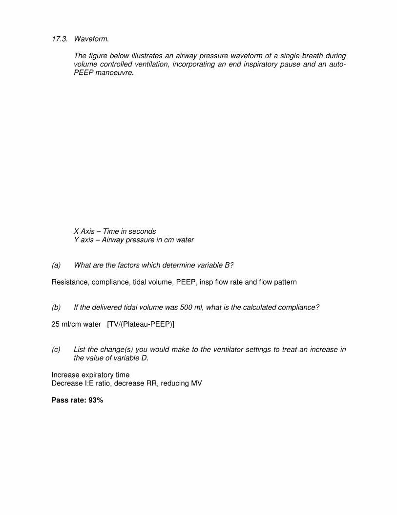
17.3. Waveform.
The figure below illustrates an airway pressure waveform of a single breath during volume controlled ventilation, incorporating an end inspiratory pause and an autoPEEP manoeuvre.
X Axis – Time in seconds Y axis – Airway pressure in cm water
(a) What are the factors which determine variable B? Resistance, compliance, tidal volume, PEEP, insp flow rate and flow pattern
(b) If the delivered tidal volume was 500 ml, what is the calculated compliance? 25 ml/cm water [TV/(Plateau-PEEP)] (c) List the change(s) you would make to the ventilator settings to treat an increase in
the value of variable D. Increase expiratory time Decrease I:E ratio, decrease RR, reducing MV Pass rate: 93%
The figure below illustrates an airway pressure waveform of a single breath during volume controlled ventilation, incorporating an end inspiratory pause and an auto
Airway pressure in cm water
What are the factors which determine variable B?
Resistance, compliance, tidal volume, PEEP, insp flow rate and flow pattern
If the delivered tidal volume was 500 ml, what is the calculated compliance?
PEEP)]
List the change(s) you would make to the ventilator settings to treat an increase in
Decrease I:E ratio, decrease RR, reducing MV
The figure below illustrates an airway pressure waveform of a single breath during volume controlled ventilation, incorporating an end inspiratory pause and an auto-
Resistance, compliance, tidal volume, PEEP, insp flow rate and flow pattern
If the delivered tidal volume was 500 ml, what is the calculated compliance?
List the change(s) you would make to the ventilator settings to treat an increase in
QUESTION 18 18. A previously fit and well 24 year old man sustained an isolated C5-C6 spinal injury
following a diving accident resulting in a tetraplegia. The spinal fracture was surgically fixed the following day and the patient was extubated on Day 6 of his ICU admission. Within 4 hours of extubation, the patient developed respiratory distress requiring urgent rapid sequence induction and reintubation. The patient sustained a cardiac arrest soon after intubation.
18.1. List five (5) likely causes of cardiac arrest in this patient.
• Oesophageal intubation
• Hypoxic cardiac arrest (unrelated to oesophageal intubation due to delayed or unanticipated difficulty with intubation)
• Suxamethonium induced hyperkalemia • Incidental PE • Autonomic dysfunction from the spinal injury.
• Tension pneumothorax • Anaphylaxis
Candidates presenting other reasonable causes were given credit 18.2. Outline how you would determine the cause of the cardiac arrest.
• Capnograph to check tube position and reintubate if not in the right position • Urgent serum K
• ECG
• CTPA • Echo
• CXray
18.3. List three (3) metabolic and three (3) gastrointestinal complications seen after spinal
cord transection.
Metabolic Hyponatremia (SIADH) Immobilisation hypercalcemia and Nitrogen wasting hypothermia GI Ileus acute gastric dilatation stress ulcerations
Pass rate: 81%

QUESTION 19 19.1. To evaluate a new biomarker as an early index of infected pancreatic necrosis, you
perform the measurement in a consecutive series of 200 critically ill patients with pancreatitis. You find that 100 of these patients had subsequently proven necrosis. Of these, 60 had a positive biomarker result. Of the remaining 100 patients without necrosis, 35 had a positive biomarker result.
Using the above data, show how you would calculate
a) Sensitivity = (TP/ {TP + FN}) = 60/100
b) Specificity = (TN/{TN + FP}) = 65/100
c) Positive predictive value = (TP/{TP+FP}) = 60/95
d) Negative predictive value = (TN({TN+FN}) = 65/105
19.2. A randomized controlled clinical trial was performed to evaluate the effect of a new
hormone called Rejuvenon on mortality in septic shock. 3400 patients with septic shock were studied (1700 placebo and 1700 in the Rejuvenon arms). The mortality rates in the placebo and the treatment arms were 30% and 25% respectively.
Calculate:
(a) The absolute risk reduction (b) The relative risk reduction (c) The number needed to treat
Using the above data, show how you would calculate:
a) The absolute risk reduction b) The relative risk reduction c) The number needed to treat ARR = 5% RRR = 5/30*100 =16.6% NNT =1/0.05 =20
Pass rate: 86%
QUESTION 20 20. (a) List the risk factors for and the clinical and laboratory findings of propofol infusion
syndrome.
Risk Factors Large doses (> 4mg/kg/hr for > 48 hours in adults): typically, but not always, large dose, long time Younger age Acute neurological injury Low carbohydrate intake Catecholamine and/or corticosteroid infusion
Clinical and laboratory findings Unexplained lactic acidosis Increasing inotrope support (Lipaemic serum, propofol levels / chromatography (if available??)) Brugada-like ECG abnormalities (Coved-type = convex-curved ST elevation in V1-3) (Green urine) Cardiovascular collapse, reflected in PICCO / PAC / ECHO Rhabdomyolysis, high CK, hyperkalaemia Arrhythmia / heart block Renal failure
(b) Outline your management of a patient with suspected propofol infusion syndrome.
Management: High index of suspicion Discontinue immediately Monitor for early warning signs: lactate, CK, Urine myoglobin, ECG Standard cardio-respiratory support Consider pacing (bradycardia often resistant to high dose CA and pacing) Adequate carbohydrate intake (6-8mg/kg/min) Carnitine supplementation: theoretical benefit Haemodialysis and haemoperfusion, used, unproven benefit ECMO: 2 case reports, readily reversible pathology
Pass rate: 47%
QUESTION 21 21. List the factors predisposing to medication error in ICU. How can these be
minimised? Note to examiners: This is a very broad question. The following is an example of a good answer to this question. It is expected that there will be a range of different answers by candidates. No breakdown has been provided for the marks. Examiners are urged to use their discretion and should award marks to all reasonable answers. Factors predisposing Patient factors
• Severity of illness
• Extremes of age • Prolonged hospitalisation
• Sedation, patient unable to tell nurse medication wrong. Medication errors Types of medications are infusions or weight based or programmed if an infusion pump is required. Number of medications, more than on the ward Number of interventions therefore increased risk of complications. ICU environment Complex environment – high stress, high turnover, high nursing turnover. Emergency admission Multiple care providers Minimisation of medication errors
• Optimise medication process
• Medication standardisation
• Computerised physician order entry • Barcode technology
• Computerised infusion device
• Medication reconciliation Eliminate situational factors
• Avoid excessive consecutive and cumulative working hours • Minimise interrupts and distractions
• Trainee supervision and graduated responsibility Oversight and error interception
• Primary doctor in charge of all drugs ( intensivist) • Adequate staffing • Pharmacist participation
• Quality assurance as part of education program. ( Evidence of adverse drug events dropping by 66% with pharmacist involvement, results in reducing length of stay, decreasing mortality and medication expenditure)
Nursing/Patient ratio
• If increased patient/ nurse ratio, increasing error.
• Mention AIMS ICU (Australian incident monitoring study in Intensive Care) has been developed with goal of balancing strengths with limitations of error reporting.
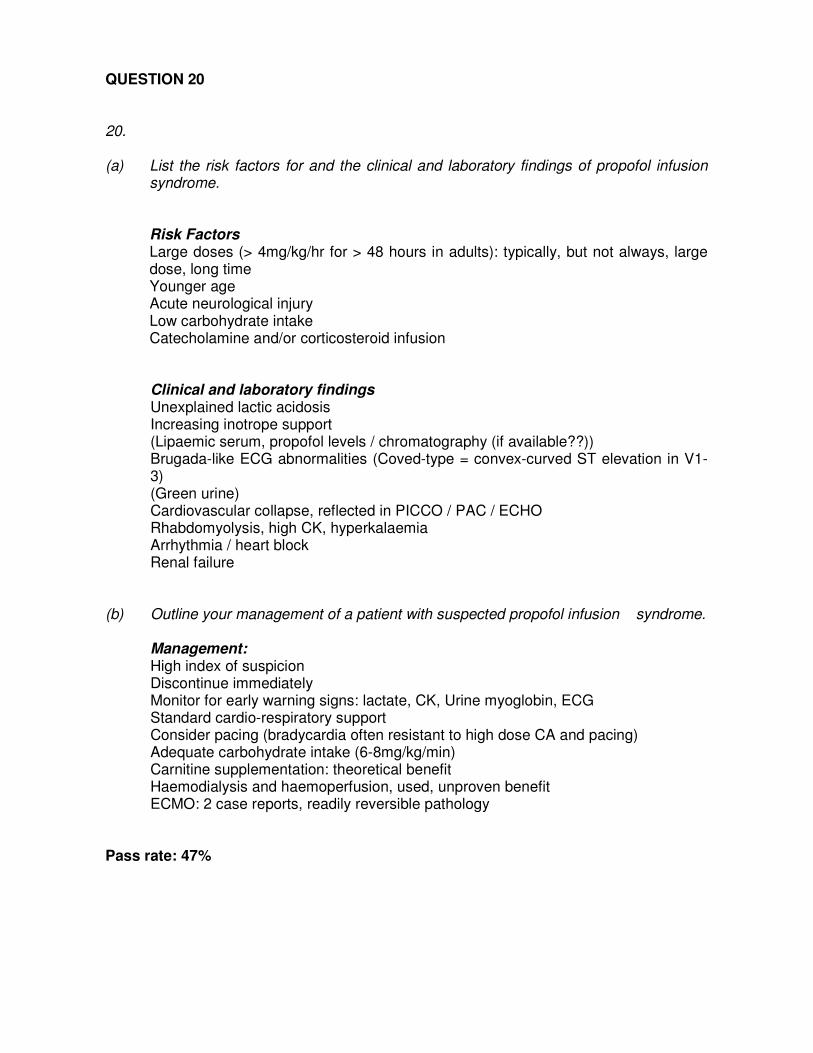
Pass rate: 33% QUESTION 22 22.1. What piece of equipment is shown below? Outline the principle of operation of this
equipment.
Non-rebreather or partial rebreather oxygen mask Reservoir bag attached to FGF One way valve between reservoir bag and patient which prevent expired gas entering the reservoir bag. Usual FiO2 reached is between 60-90%. Achieving 100% FiO2 is difficult because of valve inefficiency and lack of a tight fit around the face thus entraining room air.
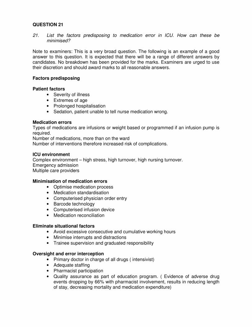
22.2. The following image shows the connector and a regulator for a Medical Wall Suction outlet.
(a) What design features of the above equipment prevent it from being connected to the oxygen outlet device?
1) Colour coding (oxygen is white, suction is yellow) 2) A unique sleeve index arrangement for each wall gas
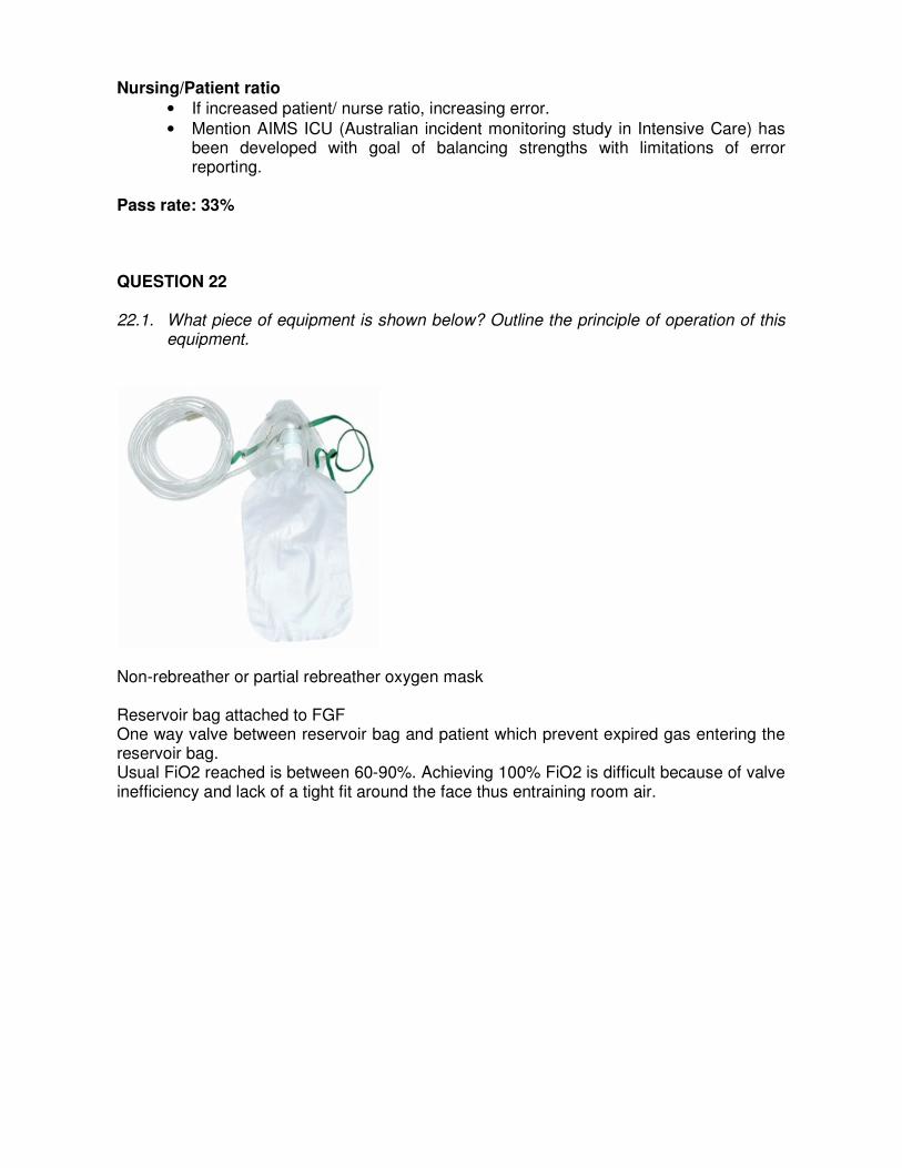
22.3. The image below is an example of a rapid volume infusion device.
(a) What are the major determinants of fluid flow through this device? Pressure gradient, radius of the catheter raised to the power of 4 (r4) and length of the catheter. As described by the simplified Poiseuilles formula for laminar flow through tube
Flow α ∆ρ.r4/l Pass rate: 86%

QUESTION 23 23. Critically evaluate the role of Procalcitonin (PCT) as a biomarker in the diagnosis
and management of sepsis. Diagnosis
• PCT is synthesized physiologically by thyroid C cells but in sepsis has extrathyroidal origin from the inflamed/infected tissue
• The biochemical and clinical profile well described
• It is easy to perform (Blood test), not too expensive and provides a quick answer in about 30 minutes. Blood cultures can take up to 24 hours.
• PCT is no gold standard for infection. There number of reports of PCT elevation in non-septic SIRS, immediately after surgery and trauma.
• Data from meta-analyisis are conflicting, some suggesting it is superior to CRP, whilst others have concluded it is a weak biomarker in critical illness.
• PCT is not elevated in viral infection, autoimmune disorders and immunocompromised patients – hence empiric therapy still the way in these patients.
• PCT does not tell you the site of infection/inflammation. History, clinical examination and other investigations like CT scan can.
• PCT is a biomarker and cannot replace good history taking, systematic clinical examination, appropriate investigations for the source of sepsis.
Management Few prospective randomised studies using,PCT as a guide to antibiotic therapy, have showed that prescription rate and the cost of antibiotics was reduced significantly with similar outcomes compared to the conventional approach (Mention of the recent Lancet paper (Jan2010 – ProRata study and its conclusions is worthy of extra credit). Pass rate: 28%
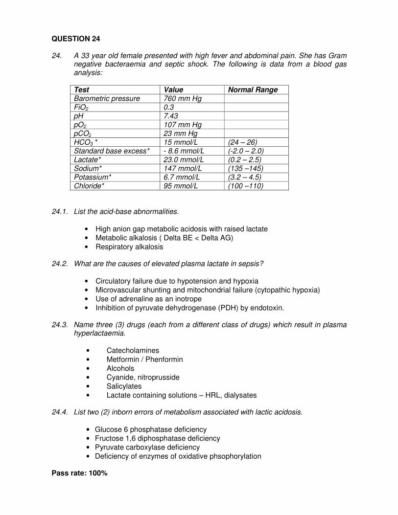
QUESTION 24 24. A 33 year old female presented with high fever and abdominal pain. She has Gram
negative bacteraemia and septic shock. The following is data from a blood gas analysis:
Test Value Normal Range Barometric pressure 760 mm Hg FiO2 0.3 pH 7.43 pO2 107 mm Hg pCO2 23 mm Hg
HCO3 * 15 mmol/L (24 – 26) Standard base excess* - 8.6 mmol/L (-2.0 – 2.0) Lactate* 23.0 mmol/L (0.2 – 2.5) Sodium* 147 mmol/L (135 –145) Potassium* 6.7 mmol/L (3.2 – 4.5) Chloride* 95 mmol/L (100 –110)
24.1. List the acid-base abnormalities.
• High anion gap metabolic acidosis with raised lactate • Metabolic alkalosis ( Delta BE < Delta AG)
• Respiratory alkalosis 24.2. What are the causes of elevated plasma lactate in sepsis?
• Circulatory failure due to hypotension and hypoxia • Microvascular shunting and mitochondrial failure (cytopathic hypoxia) • Use of adrenaline as an inotrope
• Inhibition of pyruvate dehydrogenase (PDH) by endotoxin.
24.3. Name three (3) drugs (each from a different class of drugs) which result in plasma hyperlactaemia.
• Catecholamines
• Metformin / Phenformin
• Alcohols • Cyanide, nitroprusside • Salicylates
• Lactate containing solutions – HRL, dialysates 24.4. List two (2) inborn errors of metabolism associated with lactic acidosis.
• Glucose 6 phosphatase deficiency • Fructose 1,6 diphosphatase deficiency • Pyruvate carboxylase deficiency
• Deficiency of enzymes of oxidative phsophorylation Pass rate: 100%
QUESTION 25 25. Critically evaluate the use of albumin-containing solutions in critically ill patients. Albumin solutions are frequently used in critically ill patients for a variety of indications. a) Volume replacement: The SAFE study showed that using colloids was equivalent in efficacy and safety to crystalloids. b) Hypoalbuminaemia: Clinical conditions that may benefit from albumin replacement for hypoalbuminaemia include:- Patients with decompensated liver cirrhosis and spontaneous bacterial peritonitis. The administration of albumin results in a reduced incidence of renal failure and reduction in mortality. Patients with Acute Lung Injury or ARDS.The study by Martin CCM 2005 shows that in patients who are hypoproteinaemic with ARDS, adding albumin to frusemide resulted in a significant improvement in oxygenation compared to frusemide alone. There was also a greater net negative fluid balance achieved and better haemodynamic stability in patients receiving albumin. Head injury: The clinical conditions in which you would avoid Albumin replacement is cerebral trauma where the SAFE subgroup analysis reported increased mortality at 28 days and 2 years. Sepsis: In the SAFE subgroup, a trend towards an improved outcome with albumin was noted as compared to saline In Australia, albumin is cheap (free). It is also risk free, not associated with serious complications such as coagulation abnormalities and renal failure as seen with other studies Pass rate: 70%
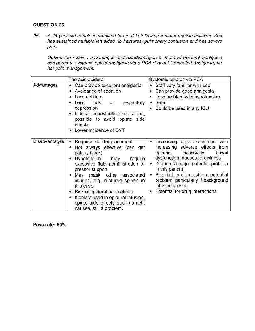
QUESTION 26 26. A 78 year old female is admitted to the ICU following a motor vehicle collision. She
has sustained multiple left sided rib fractures, pulmonary contusion and has severe pain.
Outline the relative advantages and disadvantages of thoracic epidural analgesia compared to systemic opioid analgesia via a PCA (Patient Controlled Analgesia) for her pain management.
Thoracic epidural Systemic opiates via PCA Advantages • Can provide excellent analgesia
• Avoidance of sedation
• Less delirium • Less risk of respiratory
depression • If local anaesthetic used alone,
possible to avoid opiate side effects
• Lower incidence of DVT
• Staff very familiar with use • Can provide good analgesia
• Less problem with hypotension • Safe
• Could be used in any ICU
Disadvantages • Requires skill for placement • Not always effective (can get
patchy block)
• Hypotension may require excessive fluid administration or pressor support
• May mask other associated injuries, e.g. ruptured spleen in this case
• Risk of epidural haematoma
• If opiate used in epidural infusion, opiate side effects such as itch, nausea, still a problem.
• Increasing age associated with increasing adverse effects from opiates, especially bowel dysfunction, nausea, drowiness
• Delirium a major potential problem in this patient
• Respiratory depression a potential problem, particularly if background infusion utilised
• Potential for drug interactions
Pass rate: 60%
QUESTION 27 27. A 26 year old lady presents from home confused with a low-grade fever. Her blood
pressure is 160/100 mm Hg. She has no gross motor deficits. Ten days ago she had an emergency termination of pregnancy for an intrauterine death that was complicated by disseminated intravascular coagulation. She was 32 weeks gestation and had been on labetalol for a pregnancy-induced hypertension.
Her discharge medications included paracetamol, tramadol and a selective serotonin reuptake inhibitor. She has a 6-year history of uncomplicated Hepatitis C.
27.1. List the differential diagnoses for her confusion and temperature. Pregnancy related: Eclampsia / preeclampsia / HELLP, Retained products with sepsis, Sheehan’s syndrome / pituitary apoplexy, Posterior reversible encephalopathy syndrome (PRES), Hypertensive encephalopathy Primary neurological: Infection (meningitis / encephalitis), cerebral venous thrombosis, seizure disorder, other cerebro-vascular
Metabolic: Sodium (hypo/hyper), Glucose (hypo/hyper), Renal failure, Liver failure (HCV / Paracetamol / Antidepressants), Drugs: Accidental / intentional overdose, drug reactions (serotonin syndrome) Infection: Uterine, intracranial, other (renal, chest etc) 27.2. Outline your approach to establishing the diagnosis. History: Collateral, Pregnancy issues, Ongoing blood loss, bleeding / bruising, drug ingestions, mood / affect, headaches Examination: BP, uterine size / discharge, oedema, meningism, neurological (tone, reflexes, symmetry), chronic liver disease Investigations: FBC: Bleeding, platelets, WCC UEC: urea / creatinine, Na, Ca, glucose Coagulation: DIC, INR for CLD LFT / Ammonia: hepatic encephalopathy, drug reactions ABG: hypoxia / hypercardia Urinary drug screen / paracetamol level Sepsis Screen, CT head +/- LP Pass rate: 51%
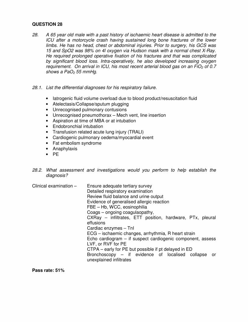
QUESTION 28 28. A 65 year old male with a past history of ischaemic heart disease is admitted to the
ICU after a motorcycle crash having sustained long bone fractures of the lower limbs. He has no head, chest or abdominal injuries. Prior to surgery, his GCS was 15 and SpO2 was 98% on 4l oxygen via Hudson mask with a normal chest X-Ray. He required prolonged operative fixation of his fractures and that was complicated by significant blood loss. Intra-operatively, he also developed increasing oxygen requirement. On arrival in ICU, his most recent arterial blood gas on an FiO2 of 0.7 shows a PaO2 55 mmHg.
28.1. List the differential diagnoses for his respiratory failure.
• Iatrogenic fluid volume overload due to blood product/resuscitation fluid
• Atelectasis/Collapse/sputum plugging
• Unrecognised pulmonary contusions • Unrecognised pneumothorax – Mech vent, line insertion • Aspiration at time of MBA or at intubation
• Endobronchial intubation • Transfusion related acute lung injury (TRALI) • Cardiogenic pulmonary oedema/myocardial event
• Fat embolism syndrome • Anaphylaxis
• PE 28.2. What assessment and investigations would you perform to help establish the
diagnosis? Clinical examination – Ensure adequate tertiary survey Detailed respiratory examination Review fluid balance and urine output Evidence of generalised allergic reaction
FBE – Hb, WCC, eosinophilia Coags – ongoing coagulaopathy, CXRay – infiltrates, ETT position, hardware, PTx, pleural effusions Cardiac enzymes – TnI ECG – ischaemic changes, arrhythmia, R heart strain Echo cardiogram – if suspect cardiogenic component, assess LVF, or RVF for PE CTPA – early for PE but possible if pt delayed in ED Bronchoscopy – if evidence of localised collapse or unexplained infiltrates
Pass rate: 51%
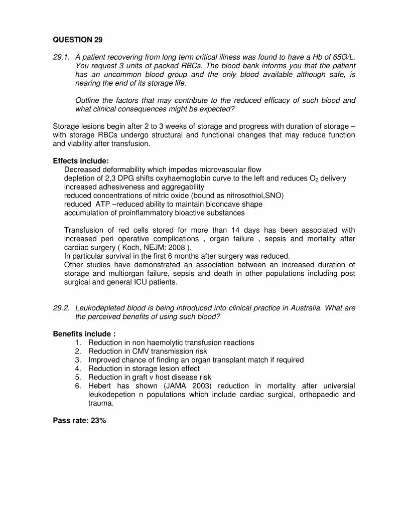
QUESTION 29 29.1. A patient recovering from long term critical illness was found to have a Hb of 65G/L.
You request 3 units of packed RBCs. The blood bank informs you that the patient has an uncommon blood group and the only blood available although safe, is nearing the end of its storage life.
Outline the factors that may contribute to the reduced efficacy of such blood and
what clinical consequences might be expected?
Storage lesions begin after 2 to 3 weeks of storage and progress with duration of storage –with storage RBCs undergo structural and functional changes that may reduce function and viability after transfusion. Effects include:
Decreased deformability which impedes microvascular flow depletion of 2,3 DPG shifts oxyhaemoglobin curve to the left and reduces O2 delivery increased adhesiveness and aggregability reduced concentrations of nitric oxide (bound as nitrosothiol,SNO) reduced ATP –reduced ability to maintain biconcave shape accumulation of proinflammatory bioactive substances Transfusion of red cells stored for more than 14 days has been associated with increased peri operative complications , organ failure , sepsis and mortality after cardiac surgery ( Koch, NEJM: 2008 ). In particular survival in the first 6 months after surgery was reduced. Other studies have demonstrated an association between an increased duration of storage and multiorgan failure, sepsis and death in other populations including post surgical and general ICU patients.
29.2. Leukodepleted blood is being introduced into clinical practice in Australia. What are
the perceived benefits of using such blood? Benefits include :
1. Reduction in non haemolytic transfusion reactions 2. Reduction in CMV transmission risk 3. Improved chance of finding an organ transplant match if required 4. Reduction in storage lesion effect 5. Reduction in graft v host disease risk 6. Hebert has shown (JAMA 2003) reduction in mortality after universial
leukodepetion n populations which include cardiac surgical, orthopaedic and trauma.
Pass rate: 23%
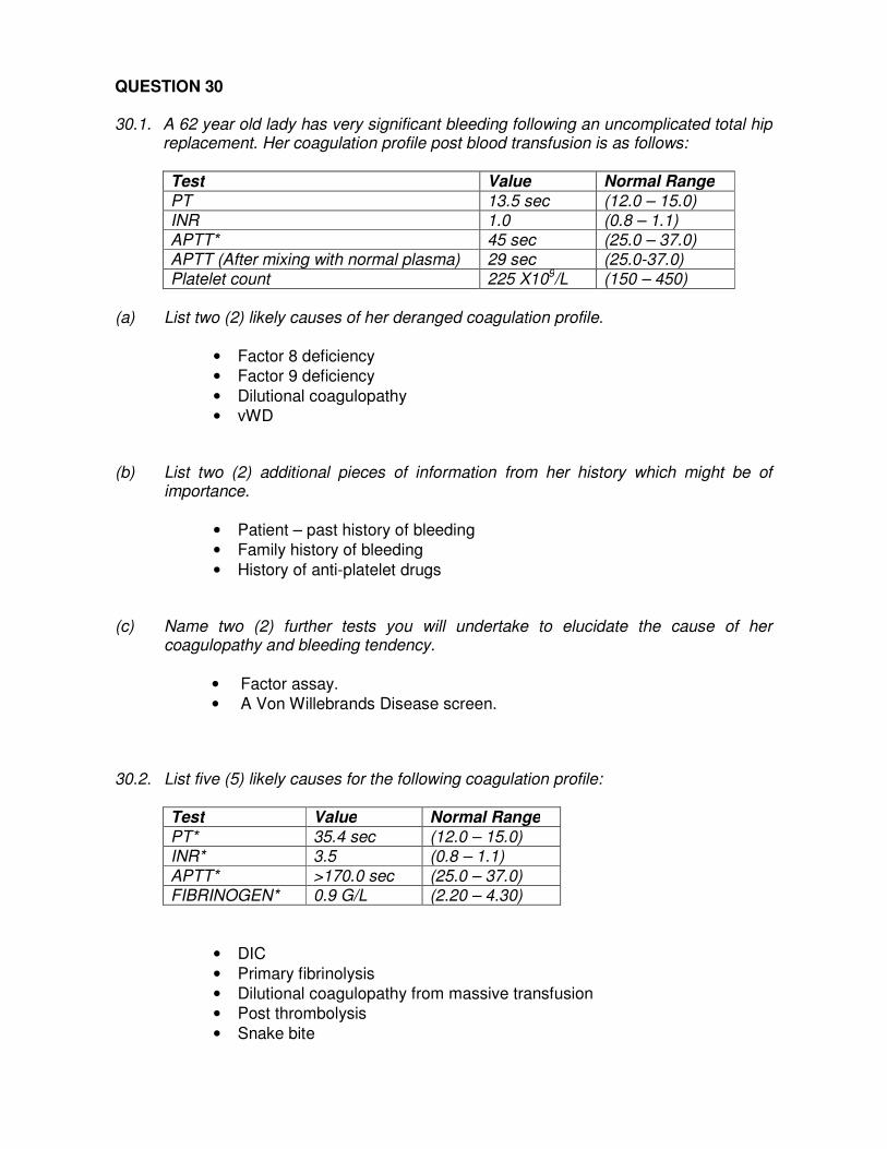
QUESTION 30 30.1. A 62 year old lady has very significant bleeding following an uncomplicated total hip
replacement. Her coagulation profile post blood transfusion is as follows:
Test Value Normal Range
PT 13.5 sec (12.0 – 15.0) INR 1.0 (0.8 – 1.1) APTT* 45 sec (25.0 – 37.0) APTT (After mixing with normal plasma) 29 sec (25.0-37.0) Platelet count 225 X109/L (150 – 450)
(a) List two (2) likely causes of her deranged coagulation profile.
• Factor 8 deficiency • Factor 9 deficiency
• Dilutional coagulopathy • vWD
(b) List two (2) additional pieces of information from her history which might be of importance.
• Patient – past history of bleeding • Family history of bleeding
• History of anti-platelet drugs (c) Name two (2) further tests you will undertake to elucidate the cause of her
coagulopathy and bleeding tendency.
• Factor assay. • A Von Willebrands Disease screen.
30.2. List five (5) likely causes for the following coagulation profile:
Test Value Normal Range
PT* 35.4 sec (12.0 – 15.0) INR* 3.5 (0.8 – 1.1)
APTT* >170.0 sec (25.0 – 37.0) FIBRINOGEN* 0.9 G/L (2.20 – 4.30)
• DIC
• Primary fibrinolysis • Dilutional coagulopathy from massive transfusion • Post thrombolysis
• Snake bite
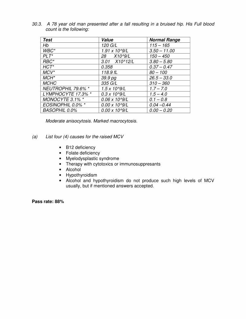
30.3. A 78 year old man presented after a fall resulting in a bruised hip. His Full blood count is the following:
Test Value Normal Range
Hb 120 G/L 115 – 165 WBC* 1.91 x 10^9/L 3.50 – 11.00 PLT* 28 X10^9/L 150 – 450 RBC* 3.01 X10^12/L 3.80 – 5.80 HCT* 0.358 0.37 – 0.47
MCV* 118.9 fL 80 – 100 MCH* 39.9 pg 26.5 – 33.0 MCHC 335 G/L 310 – 360 NEUTROPHIL 79.6% * 1.5 x 10^9/L 1.7 – 7.0 LYMPHOCYTE 17.3% * 0.3 x 10^9/L 1.5 – 4.0 MONOCYTE 3.1% * 0.06 x 10^9/L 0.1 – 0.8 EOSINOPHIL 0.0% * 0.00 x 10^9/L 0.04 –0.44 BASOPHIL 0.0% 0.00 x 10^9/L 0.00 – 0.20
Moderate anisocytosis. Marked macrocytosis.
(a) List four (4) causes for the raised MCV
• B12 deficiency
• Folate deficiency • Myelodysplastic syndrome • Therapy with cytotoxics or immunosuppresants
• Alcohol • Hypothyroidism • Alcohol and hypothyroidism do not produce such high levels of MCV
usually, but if mentioned answers accepted. Pass rate: 88%
CLINICAL SECTION Austin hospital 1. 58 year old male intubated following a late leak from a lobectomy stump. Septic with
marked acididosis. Lung not re-expanded on CXR, significant effusion. Still intubated. Candidates asked to formulate a management plan.
2. Deconditioned 69 year old male who had problems following CABG resulting in
tracheostomy and a VATS procedure. Patient awake and breathing spontaneously on a tracheostomy shield. Reduced air entry L lung and chest drain in situ. Candidates were told patient had had recent thoracic surgery and had had a near arrest after early extubation. They were asked to assess whether the patient was fit for transfer to the ward. Chest X ray and clinical examination showed poor aeration L lung.
3. 41 year old male in ICU following an emergency valve replacement for bacterial
endocarditis of a pulmonary valve (previous Ross procedure). Had clear signs of peripheral emboli with significant R lung consolidation and lung abscesses. The patient was fully conscious and extubated on 40% inspired oxygen. Candidates were told the patient had had emergency valve replacement and were required to evaluate his current clinical situation.
4. 61 year old lady with multiple surgical interventions following bariatric surgery years
earlier. Currently admitted with pneumonia for which she was ventilated. There were multiple potential causes for respiratory failure. Candidates were told she had been in the surgical ward for one month and had been admitted to ICU last night. Candidates were asked to do a general assessment with a focus on her respiratory status
5. 80 year old lady with cardio-pulmonary compromise (obstructed expiratory flow and
stigmata of steroid use, heart failure) who experienced respiratory failure following hip replacement under epidural. Significant post operative troponin rise. Candidates were asked to explain why they think this might have happened. X ray showed significant hiatus hernia (but otherwise clear lung fields) and flow trace on the ventilator showed marked obstruction, ECG showed LBBB.
6. 55 year old man with diabetic ESRF and peripheral vascular disease (a-v fistula
insitu). Day 1 post CAGS. Making good progress. Vascath in situ but not being used. Not intubated. Candidates told post CAGS and were asked to formulate a management plan.
7. 81 year old man with a brain stem haemorrhage and clear neurological signs
including sixth nerve palsy, weak gag (absent on soft palate) and right sided facial weakness. Breathing spontaneously through a tracheostomy on low insired oxygen and pressure support. Fine bore nasogastric tube in situ. Asked to evaluate why he was slow to wean from ventilation.
8 . 26 year old man 3 days post liver transplant for familial hypercholesterolaemia.
Fully awake and extubated. Widespread xanthalasma and arcus. Sternotomy scar and leg scars from vein harvesting (previous CAGS and MVR). Scar and dressing from liver transplant surgery. Prosthetic valve sounds. Inplanted plasmapheresis port in situ. Prostacyclin infusion in progress. Candidates were told that he had had a liver transplant 3 days earlier for familial hypercholesterolaemia and were asked to assess whether he could be discharged to the ward.

Alfred Hospital 1. A 70 year old lady who had residual abdominal fistula following a complicated
abdominal surgery. Had become deconditioned and was slow to wean. Globally weak, but had brisk reflexes. Problem was one of slow to wean
2. A diabetic with nephropathy who had collapsed at home. Had evidence of
brainstem signs and an encephalopathy. Also had a murmur of aortic sclerosis. Also had a temperature and an infected IV site. Candidates asked to assess neurology and formulate a plan.
3. A young man involved in a MVA, had severe TBI and had ongoing issues of sepsis,
MODS with slow neurological recovery. Candidates asked to examine patients and formulate a plan.
4. A young man in a MVA, severe TBI and also had a vascular injury to the lower
limbs and had ischemic rhabdomyolysis and associated pelvic injuries. Candidates asked to examine patients and formulate a plan.
5. A young man found collapsed at home following a SAH. Patient ventilated, EVD in
place, reduced GCS. Candidates were asked to assess neurology and discuss SAH and its complications and management.
6. A 60 year old lady readmitted to ICU post cardiac arrest due to a PE after an AVR
and a CABG and had been discharged to the ward. Candidates were asked to examine the patient and have an approach towards differential diagnosis of cardiac arrest post surgery and have management plan about PE post cardiac surgery.
7. A young man who had suffered a cardiac arrest during a rugby match and had
suffered hypoxic cerebral injury. Candidates were asked to assess neurology and discuss prognosis.
8. A middle aged man on V-A ECMO post MI who also had MODS and renal failure.
Candidates asked to assess patient and comment on methods of assessing global and tissue perfusion.
9. A young woman admitted with a reduced GCS due to a rebleed from an AVM.
Candidates were asked to comment on the neurological assessment and management of cerebral oedema.

Areas of weaknesses identified by examiners CNS examination
Incorrect interpretation of papillary responses Inability to perform a proper CNS examination Not being able to state a clear GCS Missing the presence of an EVD Inability to summarise the neurology and formulate a management plan including a realistic view of the prognosis for discussion with the family. Poor CT interpretation – inability to identify extradural haematoma
Respiratory:
Missed clear bronchial breathing Poor interpretation of CXR Failure to notice ICC Inablity to state what the arterial PO2 should be for a given FiO2 No coherent approach to a failure to wean case Missing scar of a bilateral lung transplant Candidates commented on tracheostomy decannulation without establishing clearly the presence of a cough reflex.
CVS
Missing Grade 5 murmurs Poor interpretation of ECG Limited understanding of mixed venous saturation
Abdomen:
Missing a large ascites General:
Disordered general examination Being rough with examination technique Limited examination and limited discussion Poor discussion of differential diagnoses Taking a long time to examine with little time for discussion. Failure to identify multiplicity of problems Lack of clear management plan Lack of clear antibiotic plan
VIVA SECTION VIVA 1 A 16 year old female is referred to the intensive care unit from another hospital’s emergency department. She was found unconscious in the toilet of a residential property during a party. She is reported to have had a GCS of 3 at the scene with reactive pupils. She is tachycardic, hypertensive and slightly febrile. She has been intubated and ventilated. What are the likely differential diagnoses? The rest of the questions focussed on the general principleso f management of overdose including lavage, alkaline diuresisi, hemoperfusion etc. VIVA 2 A 26 year old woman presents to the Emergency department having been found at home confused and jaundiced by her GP. Her GCS is E3V5M5 She has a Temp of 38 ˚C, BP 90/60, HR 90 and SpO2 94% on 4litres/min O2
She has a past history of alcoholism and intravenous drug abuse. Outline your initial assessment and management: The rest of the viva focussed on fulminant hepatic failure and itsassessment and management VIVA 3 A 36 year old female is brought into your ED department with acute shortness of breath. She is unable to provide any history due to her tachypnoea. She is sitting upright in bed grasping the bed sides. She has a respiratory rate of 30 breaths per minute, has a GCS of 15, is afebrile and has a BP of 90/60mmHg. She is using accessory muscles. On auscultation, she has widespread expiratory wheeze spread throughout both lung fields.
1) What are the differential diagnoses for her presentation?
The rest of the viva focussed on the assessment and management of bronchial asthma.

VIVA 4 A 56 year old, homeless man was admitted to the emergency department with clinical features suggestive of a bowel obstruction. As he is confused, it is not possible to elicit a clear history. The first set of blood tests show- Sodium 137 mmol/L (137-145) Potassium 4.0 mmol/L (3.1-4.2) Chloride 98 mmol/L (101-109) Bicarbonate 15mmol/L (22-32) Glucose 48mmol/L (3.0-6.0) Urea 18.0 mmol/L (3.0-8.0) Creatinine 0.2 mmol/L (0.05-0.12) What are the possible causes of his metabolic acidosis? The rest of the viva focussed on the management of hyperglycaemic emergencies VIVA 5 A 67 year old man with a history of heavy alcohol use was found collapsed, unresponsive, and smelling of alcohol. On admission he was noted to have abdominal distention with thin, gaunt facies and cachexia. In the Emergency Department he had a massive haematemesis requiring resuscitation. A gastroscopy was performed with banding of several oesophageal varices. He subsequently arrives in your ICU intubated and ventilated and haemodynamically stable. 1. Comment on this patient’s likely nutritional status. The rest of the viva focussed on assessment of nutritional status and enteral nutrition. VIVA 6 (Radiology): 6 radiographs were shown including chest X-rays, and CT scans of head, chest and abdomen. Areas of weakness identified by examiners:
• Failure to identifiy common pathologies, inability to point out to specific lesions on the X-rays.
• Candidates did not always use the information given to them about each Xray eg CT abdomen post rectal contrast
• Candidates did not recognise significant psoas abscess on abdo CT.
VIVA 7 (Communication) Mr Smith is a 54 year old male who was admitted to the ICU 7 days ago. He presented with severe hypoxic respiratory failure and has a background of advanced motor neurone disease. He is currently unconscious, intubated and ventilated. Mr. Smith has a valid advanced care directive stating that he would not want life sustaining measures should he require invasive life support. There is consensus amongst you and your colleagues that extubation and palliation are appropriate. Mrs. Smith agrees that this is what her husband would want under these circumstances. Mr Smith has a nephew/niece named Pat, who has not seen him for some years, and s/he has just arrived back from overseas to see him. Mrs Smith has asked that you talk to Pat to discuss his/her Uncle’s condition and treatment plan and is happy for you to discuss all of his medical information with Pat. VIVA 8 A 65 year old woman is in your ICU post left pneumonectomy. She was admitted overnight for HDU care. You are called to see her by the nurse looking after her as she has become haemodynamically unstable during her shift. 1) Discuss the possible causes for this haemodynamic instability? Areas of weakness identified by examiners: Inability to clearly understand the significance of a chest drain on suction post pneumonectomy.
B. Venkatesh (Prof) Chair Fellowship Examination Committee


![Computer Tech 2016 Tom Browder … Software Downloads Computer Tech 2016 Tom Browder [tom.browder@gmail.com] Northwest Florida Linux User Group [nw ... Files and directories (folders)](https://img.pdfslide.net/doc/110x75/5abda77f7f8b9a3a428c05f7/computer-tech-2016-tom-browder-software-downloads-computer-tech-2016-tom-browder.jpg)


![TPC NA 2017 Tom Browder [tom.browder@gmail.com] · Getting Linux TPC NA 2017 Tom Browder [tom.browder@gmail.com] Northwest Florida Linux User Group [nw ug.org] ... Perl 6 is my programmimg](https://img.pdfslide.net/doc/110x75/5faf74c2ef98ea6e856c095d/tpc-na-2017-tom-browder-tombrowdergmailcom-getting-linux-tpc-na-2017-tom-browder.jpg)