Embed Size (px)
Citation preview

EXAM REPORT MARCH / MAY 2012
This report is prepared to provide candidates, tutors and Supervisors of Training with information regarding the assessment of candidates’ performance in the General Fellowship Examination. Answers provided are not necessarily model answers but guides as to what was expected. Candidates should discuss the report with their tutors so that they may prepare appropriately for future examinations. The exam comprises a written section and an oral section. The written exam consists of two 2.5 hour papers of 15 ten-minute short answer questions each. Candidates are required to score at least 50% in the written section to be eligible to sit the oral section. The oral exam consists of eight interactive vivas and two separate clinical “hot cases”. The tables below provide an overall statistical analysis as well as information regarding performance in the individual sections. A comparison with data from the three previous exams is provided.
Overall Performance May 2012
October 2011
May 2011
October 2010
Presenting for written (Including OTS) 41 55 36 48
Carrying a pass from a previous attempt 11 11 7 8
OTS Exempt 0 0 0 2
Total number presenting (written + carry + OTS) 52 66 43 58
Pathway to oral section
Invited to orals (>50% in written section)** 26 33 30 36
Total number invited to oral section 37 43 39 54
.../2

2.
Analysis of performance in individual sections May 2012
October 2011
May 2011
October 2010
Successful in the written section 26/41 45/55 23/36 33/48
63% 81% 64% 69%
Successful in the Hot Case section 15/37 39/56 15/29 32/43
40% 69% 52% 74%
Successful in both Hot Cases 7/37 22/56 5/29 22/43
19% 39% 17% 51%
Successful in the viva section 22/37 44/56 20/29 40/43
59% 78% 69% 93%
.../3
Sectional pass rates May 2012 October 2011
Pass rate Highest individual
mark Pass rate
Highest individual mark
Hot Case 1 32% 80% 59% 90%
Hot Case 2 43% 77% 57% 87%
Viva 1 57% 97% 66% 90%
Viva 2 57% 75% 75% 100%
Viva 3 81% 91% 65% 79%
Viva 4 30% 60% 79% 86%
Viva 5 43% 80% 93% 92%
Viva 6 (X ray) 49% 90% 54% 84%
Viva 7 (Communication) 84% 100% 68% 90%
Viva 8 (Procedure) 68% 85% 88% 90%
3.
Oral section pass rates May 2012
October 2011
May 2011
October 2010
Candidates who scored >50% in written section and passed the overall exam
19/26 36/45 17/22 27/33
73% 80% 77% 82%
All candidates invited to oral section and passed the overall exam (written + carry + OTS)
20/37 43/56 18/29 35/43
54% 76% 62% 81%
Overall Pass Rate 20/52 43/66 18/43 35/58
38% 65% 42% 60%
.../4
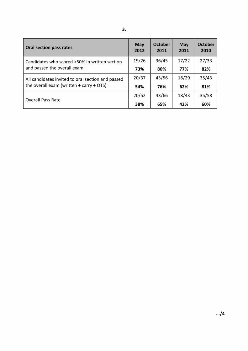
4.
EXAMINERS’ COMMENTS Written Paper Seven of the thirty questions had a pass rate of less than 50%. Topics covered by questions with a pass rate of less than 15% included the RIFLE classification of acute kidney injury, evaluation of the Glasgow Coma Score and obesity and nutritional status. There was an apparent lack of understanding of basic principles relating to commonly used tests, scoring systems and items of equipment. As in previous exams, candidates who failed did not answer the questions as asked; did not attempt to answer certain questions or parts of questions; and/or were too narrow in their answer. It appears that candidates do not always read the questions carefully and thoroughly. Candidates who failed the written section passed an average of 14/30 questions compared with candidates scoring > 50% and gaining an invitation to the oral section, passing an average of 23/30 questions. Hot Cases The overall pass rate was lower than previous exams. Concerns expressed by the examiners included:
Slow, hesitant and unsystematic examination of the patient. There is an impression that the standard of clinical examination of body systems has fallen now short cases are no longer included in the clinical exam.
A tendency to have a formulaic approach to examination of the patient rather than a flexible approach that is appropriately adapted to the given situation.
Causing potential harm or unnecessary discomfort to the patient, e.g. failing to ask about cervical spine clearance before moving the head in a patient with a neck injury; testing response to pain in a patient who has just obeyed commands.
Disorganised presentation of the clinical findings.
Inability to correctly interpret clinical signs especially neurological signs.
Inability to correctly interpret investigations such as ABGs, ECGs and CT scans of the head.
Lack of a definitive plan for patient management with decision-making at a registrar level rather than senior registrar/junior consultant
.../5

5.
Inability to synthesise the information available and demonstrate adequate decision-making ability
Candidates are reminded that they should not sit the General Fellowship Examination until they can confidently examine patients, present the clinical findings and discuss management issues at the appropriate level, i.e. senior registrar/junior consultant. Candidates are also encouraged to practice examination of individual systems. Vivas The Vivas tend to be the section in which candidates perform well and the pass rate in the Vivas for this exam was higher than that for the Hot Cases but lower overall than in previous exams. Three Vivas had pass rates less than 50% and the Viva with the lowest pass rate (30%) was Viva 4 (interpretation of biomarkers).
.../6

6.
GENERAL FELLOWSHIP WRITTEN EXAMINATION
(A) Write your answers in the blue book provided. (B) Start each answer on a new page and indicate the question number. It is not necessary to
rewrite the question in your answer book. (C) You should aim to answer each question in ten minutes. (D) The questions are worth equal marks. (E) Record your candidate number and each question number on the cover of each book and
hand in all books. GLOSSARY OF TERMS Critically evaluate: Evaluate the evidence available to support the hypothesis. Outline: Provide a summary of the important points. List: Provide a list. Compare and contrast: Provide a description of similarities and differences
(E.g. Table form). Management: Generic term that implies overall plan. Where appropriate, may
include diagnosis as well as treatment. NOTE Where laboratory values are provided, abnormal values are marked with an asterisk (*). Please note that in this report images from the SAQs have been removed.
.../7

7. Question 1 Outline the Intensive Care management of a 25-year-old male who has fulfilled brain death criteria and is awaiting surgery for organ donation. Temperature Maintenance:
Hypothermia is common due to: cold fluids, heat loss through exposure, inability to vasoconstrict or shiver, reduced metabolic rate.
Maintain normal core temperature o Cover patient o Warm room o Warming blanket o Warm fluids especially high volume o Humidification
Respiratory support:
Aim to avoid fluid overload
Aim for adequate Sp02 and normocarbia with lowest Fi02 and limit tidal volumes
Bronchoscopy for persisting collapse
Chest physiotherapy may be helpful
Circulatory Support: Immediately prior to brain death there is often a period of sympathetic hyperactivity with associated tachycardia and hypertension. This is lost following brain death commonly resulting in vasodilation and hypotension
Maintain adequate mean arterial pressure. Use judicious volume expansion and low dose inotropes (usually noradrenaline)
Monitor peripheral perfusion and urine output regularly
Continue maintenance fluids Metabolic haematology and biochemistry: Diabetes insipidus is common and if not recognized and treated can quickly lead to hypernatraemia and hyperosmolality
Measure electrolytes and creatinine regularly and treat as appropriate to maintain normal ranges
Treat Diabetes insipidus with desmopressin (DDAVP) 4-8µgrams intravenously and repeat if necessary, or low dose vasopressin
Start low dose insulin infusion if blood glucose persistently above 12mmol/L
Stop bleeding, correct coaguloapthy, thrombocytopaenia and anaemia
Avoid hypernatraemia
Other electrolyte abnormalities – K, PO4, Ca, Mg
Consider thyroxine replacement
.../8

8. Communication:
Family - counsel, explain, keep updated
Liaison with donor coordinator and surgical retrieval teams
Pass rate 56%
Question 2 You are asked to assist and help in the emergency management of a 69-year-old who presented to your emergency department with massive haemoptysis. a) List six major disease categories that cause massive haemoptysis and give one example of each.
b) Outline the emergency management of massive haemoptysis. a) Major disease categories 1. Infective – lung abscess, TB, Bronchiectasis, Fungal, Necrotising pneumonia 2. Neoplastic - primary or secondaries 3. Cardiac – Mitral Stenosis, Congenital heart Disease, Tricuspid Endocardidis 4. Vascular – Pulmonary embolism, Pulmonary infaction, Pulm Hxt, AVM 5. Systemic Disease – Wegeners, Goodpastures, SLE and other causes of vasculitis 6. Haematologial – Any severe coagulopathy including acquired causes – DIC, drugs etc 7. Latrogenic/Post Surgical – Swann Ganz, Pulmonary procedures 8. Trauma – blunt or penetrating injury, tracheo-innominate artery fistula, ruptured bronchus b) Emergency management
Manage ABC, volume resuscitate
Correct coagulopathy if relevant
Role of intubation- life- threatening i.e. airway compromise, desaturation or shock. Maybe
required if intervention planned
How to intubate- large size tube (>7.5) if possible so fibre optic bronchoscopy can be performed
if needed, single lumen if life threatening, consider double lumen if controlled circumstances and
unilateral pathology
Post intubation- can be nursed lateral decubitus with bleeding lung down to prevent soiling of
non-bleeding lung
Role of bronchoscopy – rigid or flexible – may be needed urgently to place balloon tipped
catheter endobronchially to tamponade bleeding
Bronchial artery embolization- effective non- surgical management
Surgery- lobectomy or pneumonectomy after resuscitation if other measures fail or not
available or for etiologies like A- V malformations, trauma, extensive fungal abscess
.../9

9.
Antimicrobial therapy for underlying infective causes
Immunosuppression therapy for underlying vasculitis
Pass rate 85%
Question 3 a) List the patient-related risk factors associated with the development of Clostridium difficile enterocolitis b) List two tests that can be used for diagnosis of Clostridium difficile enterocolitis. c) List four markers of severity of disease in Clostridium difficile enterocolitis d) What are other possible causes of infective diarrhoea in the critically ill? a) Patient-related risk factors
Broad spectrum antibiotics in particular clindamycin, quinolones, amoxycillin, cephalosporins
Immunosuppressive therapy /Cytotoxic chemotherapy
Gastric acid suppression
Age > 65
Prolonged hospitalisation
Renal impairment
Prior GI surgery b) Diagnostic tests
Faecal culture determination of the toxigenic status of the infecting C. difficile isolate
Screening EIA to detect C. difficile glutamate dehydrogenase (GDH)
EIAs to detect toxins A and/or B
Cell culture cytotoxicity assays that directly detect stool cytotoxic activity
PCR-based assays to detect conserved gene targets within the pathogenicity locus of C. difficile
c) Markers of severity Clinical
Fever (> 38.5°C), rigors Haemodynamic instability Peritonitis or evidence of bowel perforation Ileus or toxic megacolon
.../10

10.
Laboratory White blood cell count >15 × 109/L and < 20% neutrophils Elevated lactate level Rise in creatinine level (> 50% above baseline) Albumin level < 25 mg/L
Other investigations Large intestine distension, colonic wall thickening, fat stranding, unexplained ascites (imaging) Pseudomembranous colitis (colonoscopy)
d) Other infective causes of diarrhoea
Viruses –Norovirus, adenovirus, CMV (rotavirus in children) Bacterial pathogens – Campylobacter, E.Coli, cholera, salmonella Protozoa – Cryptosporidium, Giardia Parasitic -Strongyloides
Pass rate 66%
Question 4 A nine-month-old child is brought to your ED with a history of severe diarrhoea and vomiting over several days. On presentation the child is clearly dehydrated. a) Describe your approach to initial management in this situation b) How would you calculate the degree of dehydration in a child based on clinical assessment? a) Initial management
ABCDEFG approach.
Oxygen
Venous access (IO if required)
Secure airway if needed
Fluid bolus (20ml/kg early boluses ; aim for reversal of immediately life-threatening shock; post FEAST more controversial but remains key management)
Keep warm
Check BSL as an early priority
Early administration of empiric antibiotics
Investigations- blood gas essential mentioned essential for full marks; others may be mentioned as delayed (arterial, venous or capillary all reasonable; venous quick and useful)
Investigations: blood culture, FBC, ELFT, formal BSL as early keys, stool culture and viral screen, urine culture, nasopharyngeal aspirate for respiratory viruses, CXR
.../11

11.
b) Assessment of dehydration
Clinical assessment of dehydration can be difficult, especially in young infants, and rarely predicts the exact degree of dehydration accurately. The most useful individual signs for predicting 5% dehydration in children are an abnormal capillary refill time, abnormal skin turgor and abnormal respiratory pattern. Combinations of examination signs provide a much better method than any individual signs in assessing the degree of dehydration.
Clinical assessment therefore comprises some of the following indicators of dehydration:
Loss of body weight:
Normal: no loss of body weight.
Mild dehydration: 5-6% loss of body weight.
Moderate: 7-10% loss of body weight.
Severe: over 10% loss of body weight. Clinical features of mild-to-moderate dehydration; 2 or more of:
Restlessness or irritability.
Sunken eyes (also ask the parent).
Thirsty and drinks eagerly. Clinical features of severe dehydration; 2 or more of:
Abnormally sleepy or lethargic.
Sunken eyes.
Drinking poorly or not at all.
Pinch test (skin turgor): the sign is unreliable in obese or severely malnourished children.
Normal: skin fold retracts immediately.
Mild or moderate dehydration: slow; skin fold visible for less than 2 seconds.
Severe dehydration: very slow; skin fold visible for longer than 2 seconds.
Other features of dehydration include dry mucous membranes, reduced tears and decreased urine output.
Additional signs of severe dehydration include circulatory collapse (e.g. weak rapid pulse, cool or blue extremities, hypotension), rapid breathing, sunken anterior fontanelle.
Pass rate 75.6
.../12

12.
Question 5 a) Give a cause for the abnormality seen in the expiratory phase of the waveforms in figure 1. b) What has caused the fall in blood pressure seen in the latter half of figure 2? c) The recording depicted below was made from a patient on intermittent positive pressure ventilation. The upper trace represents the ECG, the middle trace the arterial pressure waveform and the lower trace respirations
i. What important haemodynamic abnormality is demonstrated? ii. List three causes of this abnormality.
d) The tracings in figure 4 are taken from a patient receiving intra-aortic balloon pumping. The upper trace is the ECG and the lower trace is the aortic pressure. What two abnormalities are shown in the pressure tracing? a) Either of the following answers acceptable
Water in circuit
Secretions in trachea or circuit
b) Causes
Loss of atrial contraction c) i) Haemodynamic Abnormality The important haemodynamic abnormality is pulse pressure variation (systolic pressure variation also acceptable). ii) Causes Causes of this abnormality include:
Hypovolaemia (fluid responsiveness acceptable)
Acute severe asthma
Cardiac tamponade
Excessive tidal volume
d) Abnormalities
Early balloon inflation
Early balloon deflation
Pass rate 88%
.../13

13. Question 6 A 33-year-old abattoir worker presented to the Emergency Department with a 2 week history of increasing shortness of breath and haemoptysis. He had previously been fit and well. On examination he is alert, normotensive but tachypnoeic (35 breaths per minute), centrally cyanosed (SaO2 85% on 10L/min O2) and tachycardic (120 beats per minute). Auscultation reveals a systolic murmur at the lower left sternal edge and coarse inspiratory crackles bibasally. The remainder of the examination was unremarkable. His chest radiograph demonstrates bibasal consolidation. List the most likely differential diagnoses, and for each diagnosis, list the specific investigations needed to confirm the diagnosis and the specific treatment required
Cause Investigation Treatment
Pneumonia Bacterial S. pneumonia, H. nfluenza K. pneumonia Atypical Viral- Influenza
Sputum MC&S Blood Culture Serology / PCR Viral PCR
3rd Generation cephalosporin (or similar) plus Azithromycin (or similar)
Oseltamivir / Ribavarin
Other infective Infective endocarditis Leptospirosis Q fever Anthrax
TTE/TOE, bld culture Serology, PCR Serology for C. burnetii Blood culture
Antibiotics surgery Doxycycline / cefotaxime / benzyl penicillin Doxycycline, ciprofloxacin Doxycycline, ciprofloxacin, benzyl penicillin
Vasculitis Goodpasture’s syndrome Wegener’s granulomatosis
Anti-GBM Abs cANCA
) Steroids ) Plasmapheresis ) Cyclophosphamide
Cardiovascular Acute mitral regurgitation Pulmonary infarction
TTE/TOE, screen for acute MI CTPA, V/Q scan
Surgery Anticoagulation
Pass rate 49%
.../14

14. Question 7 You have been asked by your director to assist in the planning and development of a new level II intensive care unit. a) Define what is meant by level I, level II and level III ICUs b) Briefly outline the principal considerations you should cover during the planning phase with specific attention to physical design of the unit and staffing This information is covered in the college document IC-1and candidates should be familiar with at least the broad areas that are covered when a new unit is planned. a) Definitions A Level I ICU should be capable of providing immediate resuscitation and short-term cardio-respiratory support for critically ill patients. It will also have a major role in monitoring and prevention of complications in “at risk” medical and surgical patients. It must be capable of providing mechanical ventilation and simple invasive cardiovascular monitoring for a period of at least several hours. A Level II ICU should be capable of providing a high standard of general intensive care, including complex multi-system life support, which supports the hospital’s delineated responsibilities. A Level III ICU is a tertiary referral unit for intensive care patients and should be capable of providing comprehensive critical care including complex multi-system life support for an indefinite period. Level III units should have a demonstrated commitment to academic education and research. All patients admitted to the unit must be referred for management to the attending intensive care specialist. b) Design and staffing Design A Level II ICU should have at least 6 beds. The unit needs to provide a suitable environment with adequate space for patient care delivery, storage, staff accommodation (including office space), education and research. (CICM IC-1 2011) Bed space There should be adequate space around each bed to allow easy access to the patient from all sides – with at least one hand basin for every 2 beds. Adequate lighting and service outlets need to be provided for each bed space and there should be provision for adequate privacy. At least one isolation room should be available. Environment – the unit should have appropriate air conditioning which allows control of temperature, humidity and air change.
.../15

15. Storage Pharmacy/drug preparation Equipment storage area. Dirty utility – area for cleaning appliances, urine testing, emptying and cleaning bed pans and urine bottles. Staff accommodation Including offices, tea room and education areas. Relatives Area Including a waiting room with basic facilities and separate room for interviewing or seeing distressed relatives. Staffing Medical Staff A dedicated director (should be a Fellow of the College) and team of specialists with junior medical cover. One specialist exclusively rostered to the unit at all times, structured bedside ward round. Nursing Staff A minimum of 1:1 for ventilated and other critically ill patients, and 1:2 nursing staff for lower acuity Consideration should also be given to ancillary staff Clerical, Wardsmen, Physiotherapists, social workers and cleaning staff
Pass rate 46%
Question 8 a) A 74-year-old man with known ischaemic heart disease was admitted to hospital for treatment of worsening heart failure (day 1). Despite treatment for heart failure he failed to improve and was referred for urgent intensive care assessment on day 5.
.../16

16.
Parameter Measured value Normal range
Day 1 Day 5
pH 7.52 7.06 7.35-7.45
PaCO2 45 (5.9) 109* (14.3*) 35-45 mmHg (4.6-6.0 kPa)
PaO2 141(18.5) 86.2 (11.3) 80-100 mmHg (10.5-13.0 kPa)
Oxygen Saturation 99.7 89.4 >95%
HCO3 36.5* 29.5* 22-27 mmol/l
Base Excess 12.6* -0.2 -2 to +2 mmol/L
Hb 94* 112* 130-150 g/L
Sodium 141 132* 135-145 mmol/l
Potassium 4.2 5.2* 3.2-4.5 mmol/l
Chloride 97* 92* 100-110 mmol/l
Glucose 6.0 19.4* 3.0-6.0 mmol/l
Lactate 1.8 6.8* < 2.0 mmol/l
Creatinine 83 73 50-100 μmol/l
i. Describe the acid-base abnormality on the Day 1 blood gas and give a clinical explanation that most likely caused this picture
ii. Describe the acid-base abnormality on the Day 5 blood gas and give three possible
clinical explanations a) i.
Chronic metabolic alkalosis with partial respiratory compensation
Background of chronic metabolic alkalosis with partial respiratory compensation could be due to diuretic therapy for heart failure (“contraction alkalosis")
ii.
Superimposed acute respiratory acidosis and metabolic acidosis (raised lactate)
Concomitant metabolic alkalosis given that the acidosis is not severe for the degree of PCO2 and lactate + BE is normal for the lactate + AG is also normal.
The acute superimposed pathology: o Cardiogenic shock o Sepsis o Respiratory depression from opiate or illness o PE Any other reasonable cause
b) A 67-year-old lady is transferred from a regional hospital to a tertiary referral centre with a diagnosis of septic shock from a urinary source. She has not improved despite 48 hours of treatment with antibiotics and supportive care.
.../17

17.
Parameter Measured value Normal range
Admission
pH 6.93* 7.35-7.45
PaCO2 48* (6.3*) 35-45 mmHg (4.6-6.0 kPa)
PaO2 160 (21.0*) 80-100 mmHg (10.5-13.0 kPa)
Oxygen Saturation 99.2 >95%
HCO3 10* 22-27 mmol/l
Base Excess -22* -2 to +2 mmol/L
Sodium 123* 135-145 mmol/l
Potassium 5.4* 3.2-4.5 mmol/l
Chloride 95* 100-110 mmol/l
Glucose 7.0* 3.0-6.0 mmol/l
Lactate 2.8* < 2.0 mmol/l
Urea 33.0* 3.5-7.2 mmol/l
Creatinine 541* 50-100 ol/l
i. Describe her biochemical profile on admission
ii. List the causes of a raised lactate in sepsis
b) i.
Mixed acidosis: Severe metabolic acidosis with a raised anion gap + additional respiratory acidosis
Renal failure with hyponatraemia and hyperkalaemia ii. 1) Adrenaline 2) Pyruvate dehydrogenase inhibtion by endotoxin 3) Liver dysfunction/failure 4) Tissue hypoxia c) A 75-year-old woman with a reduced level of consciousness is intubated and ventilated following a single grand mal convulsion. List the pathophysiological disturbances revealed by the following arterial blood gas and electrolyte profile taken 10 mins after intubation and give the likely explanation.
.../18

18.
c) Pathophysiological Disturbances
Parameter Measured value Normal range
FiO2 1.0
pH 7.05* 7.35-7.45
PaCO2 43 (5.6) 35-45 mmHg (4.6-6.0 kPa)
PaO2 280 (36.8) 80-100 mmHg (10.5-13.0 kPa)
HCO3 11.5* 22-27 mmol/l
Base Excess -16.8* -2 to +2 mmol/L
Sodium 128* 135-145 mmol/l
Potassium 3.1* 3.2-4.5 mmol/l
Chloride 82* 100-110 mmol/l
Glucose 79* 3.0-6.0 mmol/l
Lactate 9.2* < 2.0 mmol/l
Urea 22.0* 3.5-7.2 mmol/l
Creatinine 120* 50-100 μmol/l
Uncompensated metabolic acidosis (or metabolic + respiratory acidosis) with raised anion gap not solely due to elevated lactate Raised A-a gradient Sodium adjusted to normoglycaemia is about 153 Marked hyperglycaemia (candidates will say this but does not deserve a mark) Hyperosmolar hyperglycaemic syndrome with component of ketoacidosis and post-ictal lactic acidosis.
Pass rate 88%
Question 9 You are asked to review an 88-year-old man who has fallen from a ladder. He is in the ED with a large subdural haematoma (SDH) and significant mid-line shift on CT scan. His GCS is 6/15. He has a past medical history that includes atrial fibrillation (treated with warfarin and digoxin), chronic
renal impairment (creatinine 190 mol/L), non-insulin-dependent diabetes and mild cognitive impairment. a) List the factors in this patient’s history that suggest his outcome may be poor? b) Outline how age-related changes in:
i. Cardio-respiratory physiology and ii. Response to medications would impact on the management of this patient
.../19

19.
a) Factors predictive of poor outcome:
Severe TBI in an elderly patient
SDH increased risk of poor outcome
Warfarin therapy
Pre-existing co-morbidities – renal disease, diabetes, neurological dysfunction b) Age-related changes: i. Cardiovascular o Increased incidence of coronary artery disease o Systolic and diastolic dysfunction with CCF o Conduction disorders (SSS, AF, BBB) o Valvular disease o Decreased response to sympathetic stimulation
Respiratory
o Decreased respiratory muscle strength o Decreased respiratory centre sensitivity to hypoxia and hypercarbia o Reduced elastic recoil of lung o Increased chest wall stiffness o Reduced vital capacity and FEV1.
ii. Response to medications Age has been shown in multiple studies to be an independent risk factor for adverse drug reactions. Age related physiological changes affect absorption, distribution, metabolism and elimination of drugs. Poly-pharmacy is common, thus increased risk of adverse drug reaction. Cognitive impairment and drug errors – overdose, non-adherence, failure to disclose full medication list to often multiple medical practitioners involved in care. Reducing renal blood flow and GFR with age alters drug elimination potentially leading to drug accumulation.
Pass rate 61%
.../20

20. Question 10 Outline the potential mechanisms of ventilator associated lung injury in patients with Acute Respiratory Distress Syndrome and the steps that can be taken to minimise them. Injury Mechanism Minimisation Strategy
Volutrauma
Non-homogenous lung injury Over-distension of normal alveolar units to trans-pulmonary pressures above ~30 cm H2O (that corresponds to approximate total lung volume) causes basement membrane stretch and stress on intracellular junctions.
Avoid over-distending the “baby lung” of ARDS: (a) Maintain Plateau Airway pressure under 30 cm H20 (b) Use Tidal volumes 6ml/kg (4-8ml/kg) Good evidence to support this strategy (ARDSNet)
Barotrauma
Increasing the trans-pulmonary pressures above 50 cm H2O will cause disruption of the basement membranes with classical barotrauma
Biotrauma
Mechanotransduction and tissue disruption leads to upregulation and release of chemokines and cytokines with subsequent WBC attraction and activation resulting in pulmonary and systemic inflammatory response and multi-organ dysfunction
Protective lung ventilation strategies ?Use of neuromuscular blockers may ameliorate
Recruitment / Derecruitment Injury
The weight of the oedematous lung in ARDS contributes to collapse of the dependant portions of the lung Repetitive opening and closing of these alveoli with tidal ventilation will contribute to lung injury.
Consider recruiting collapsed lung +/- employing an open lung ventilation strategy. This may be achieved by: (a) Ventilation strategies: Sigh / APRV / “Higher PEEP” (b) A recruitment manoeuvres: e.g. CPAP 40/40, or stepwise PCV (c) Prone Positioning (gravitational recruitment manoeuvre) Good theoretical support and case series / few trials inconclusive outcomes
Shearing injury
This occurs at junction of the collapsed lung and ventilated lung. The ventilated alveoli move against the relatively fixed collapsed lung with high shearing force and subsequent injury.
Oxygen toxicity
Higher than necessary FiO2 overcomes the ability of the cells to deal with free oxygen free radicals and leads to oxygen related free radical related lung injury. High FiO2 may contribute to collapse through absorption atelectasis.
Limit FiO2 through the use of recruitment, higher PEEP and accepting SaO2 / PaO2 that correspond the the “shoulder” of the oxyhaemoglobin dissociation curve (SaO2 88-94)
.../21

21.
Pass rate 73%
Question 11 a) A 62-year-old woman has been admitted to hospital for investigation, giving a history of episodic facial flushing and diarrhoea, and fatigue. You are called to review her on the ward because she is hypotensive. Your examination shows features of right heart failure, with a tricuspid regurgitant murmur. ECHO REPORT: Normal LV size and systolic function. The right ventricle is dilated, with normal systolic function. Triscuspid valve leaflets are thickened, retracted, and relatively immobile. There is severe tricuspid regurgitation. Pulmonary valve leaflets are thickened. Mild pulmonary regurgitation. Other valves are normal.
i. What is the most likely diagnosis?
ii. What is the most useful investigation to confirm this diagnosis?
a) i. Diagnosis: Carcinoid syndrome with cardiac involvement ii. Investigation: 24 hour urinary HIAA (5-hydroxyindoleacetic acid) OR Serum chromogranin-A b) A 64-year-old woman presents with lethargy, shortness of breath on exertion and jaundice. Hb 64 g/L (115-155) MCV 102.4 fl (80.0-98.0) Platelets 114 X 109/L (150-400) White Cell Count 101 X 109/L (4.0-11.0) Neutrophils 0.22% 2.3 X 10⁹/L (1.8-7.5) Lymphocytes 97% 98.7 X10⁹/L (1.0-3.5) Monocytes 0.0% 0.0 X10⁹/L (0.20-0.80) Eosinophils 0.0% 0.0 X10⁹/L (0.02-0.50) Basophils 0.0% 0.0 X10⁹/L (0.0-0.10) Nucleated RBCs 3.6 per 100 WBC Reticulocyte count 280 X 109/L (20-150) Polychromasia. Poikilocytosis. Spherocytes. Smudge cells.
i. What is your interpretation of the leucocytosis?
.../22

22.
ii. What is your interpretation of the anaemia?
iii. What additional test would you perform to help determine the underlying cause of the anaemia?
b) i. Interpretation of the leucocytosis: CLL given lymphocytosis and smudge cells. ii. Interpretation of the anaemia: Jaundice, presence of spherocytes, and reticulocytosis suggest haemolysis. Autoimmune haemolytic anaemia associated with warm antibody. iii. Additional test: Direct Coomb’s test. c) A 70-year-old man presents with a seven-day history of recurrent epistaxis, bruising and increasing haemoptysis. He has no significant past medical history other than a TIA for which he takes Aspirin. He does not have any epistaxis at present. His INR and APTT are normal.
Hb 96 g/L * (135-175) RBC 3.32 x 10¹² /L* (4.50-6.00) PCV 0.29 * (0.40-0.50) MCV 86.7 fl (80.0-98.0) MCH 28.9 pg (27.0-33.0) MCHC 333g/L (315-355) Platelets 2 x 10⁹/L* (150-400) Immature Platelet Forms 17%* (1.1-6.1) Reticulocytes 0.6% (0.5-2.0) White Cell Count 8.82 x 10⁹/L (4.0-11.0)
i. List 6 potential aetiologies for the above blood picture in this patient.
c) Potential aetiologies:
Decreased platelet production o Marrow failure (eg aplastic anaemia, myelodysplasia) o Exposure to drugs (eg quinine) o Marrow infiltration (eg neoplastic) o Nutritional
Increased platelet destruction o Immune thrombocytopenic purpura (incl idiopathic, CT disease, lymphoproliferative disease, medications, infection (eg HIV, Hep C)). o Thrombotic microangiopathy o Drug-induced
.../23

23.
Increased sequestration of platelets o Hypersplenism
Pass rate 10%
Question 12 Describe the RIFLE classification system for Acute Kidney Injury and briefly discuss its implications and limitations. AKI can be defined as an abrupt (1 to 7 days) and sustained (more than 24 hours) decrease in kidney function. The ADQI formulated the RIFLE criteria to allow for AKI to be objectively and uniformly defined.
The implication of this classification is that a progression down the RIFLE criteria is associated with a higher length of stay in ICU and Hospital and is associated with a higher mortality. The limitations of this classification relate to it’s dependence on measuring urine output and creatinine which is confounded by the following:-
1. Accuracy of urine output measures.
2. Urine output affected by the use of diuretics.
3. Baseline creatinine may be affected by the patients concurrent health problem eg it may
be falsely high purely because the patient was dehydrated on admission.
4. It is uncertain how well balanced urine output and creatinine are even though they have
been given an equal weighting.
.../24 Pass rate 2%

24. Question 13 and Question 14 relate to the following clinical scenario: A 71-year-old man is transferred to your intensive care unit following a mechanical aortic valve replacement and coronary artery bypass surgery. The anaesthetist reports that he came off bypass readily, has not required any inotropic support, and has epicardial pacing wires in situ. However, shortly after arrival his blood pressure falls to 60/30.
Question 13
a) Outline your differential diagnosis for his hypotension
His blood pressure improves rapidly with a fluid bolus, and examination is otherwise unremarkable. However, he is noted to lose 250ml of blood from his mediastinal drains over the next 30 minutes.
b) List 4 likely causes of, or contributors to, excessive post-operative bleeding in this setting, and outline your immediate management. a) Differential diagnosis for hypotension
Artefactual
Preload – intravascular volume depletion (eg blood loss, rewarming), medication effect (eg propofol), anaphylaxis, vasoplegia
Contractility – arrhythmia, myocardial ischaemia, valvular dysfunction, hypoxia, pacing wire problem
Afterload – pericardial tamponade, tension pneumothorax, elevated intrathoracic pressure
Outflow tract obstruction b) Causes of post-operative bleeding and management Excessive bleeding is usually due to one or more of the following factors:
incomplete surgical hemostasis
residual heparin effect after cardiopulmonary bypass
platelet abnormalities (platelet dysfunction and thrombocytopenia – from bypass, antiplatelet agents etc)
hypothermia
postoperative hypertension
clotting factor depletion
hemodilution (dilutional thrombocytopenia and coagulopathy) Initial management
Assess airway, breathing, circulation.
PEEP 10
Look for potential underlying causes (as above). o Measure ACT and formal coagulation profile. If ACT/APTT raised, consider Protamine.
.../25

25. o Consider platelet transfusion early if antiplatelet agent in days prior to surgery or platelet dysfunction considered likely contributor. o Manage hypertension with titratable agent (eg GTN, SNIP) o Correct hypothermia
FFP if INR and/or APTT raised. Platelet transfusion if thrombocytopenia.
Transfusion of packed red blood cells may also be necessary to replace blood loss. Optimal transfusion strategy, including the level below which RBC transfusion clearly improves outcomes, is uncertain (eg TRACS trial).
Notify cardiothoracic surgeon early if concerned. Pre-existing protocol (including guidelines for notification) useful.
If haemodynamically unstable from blood loss, or if bleeding persists despite above measures, consider re-thoracotomy.
Use of DDAVP and Factor 7 controversial.
Pass rate 76%
Question 14 Twenty four hours later, he develops a new-onset tachycardia as shown in the ECG below. a) What is your interpretation of the ECG? b) Outline your initial management of the tachycardia b) List 3 primary non-cardiovascular causes of the above tachycardia.
a) Atrial fibrillation (vent rate approx. 170) LAD LVH Lateral T inversion. b)
Attention to airway, breathing and circulation.
Identify and rectify reversible factors as above o Fluid bolus if hypovolaemic o Correct electrolyte abnormalities o Check pacemaker o Treat pain
Consider MgSO4
.../26

26.
Assess for haemodynamic compromise. o If significant haemodynamic compromise, early mechanical cardioversion. o If tolerating arrhythmia haemodynamically, options are rate-control or pharmacological cardioversion.
Rate-control – IV Digoxin or beta-blocker Pharmacological cardioversion – Amiodarone, Sotalol, Class 1A or 1C
c)
Hyperthyroidism
Alcohol binge
Sepsis /Pneumonia
Carbon monoxide poisoning
Association with Friedrich’s ataxia although this is due to a cardiomyopathy.
Pass rate 42%
Question 15 a)
i. Identify the device depicted above
ii. List the indications for use of this device
iii. List the potential complications associated with the use of this device
a) i. Intra-osseous needle ii. Difficulty in establishing IV access
Need for rapid high-volume fluid infusion eg hypovolaemic shock or burns, and failure to establish IV access
Cardio-pulmonary arrest iii.
Infection – cellulitis and osteomyelitis
Extravasation of blood / infusion fluid / drugs
Compartment syndrome secondary to extravasation
Bone fracture or through and through penetration
Damage to surrounding structures
.../27

27.
b) i. List the indications for use of the items depicted above
ii. List five precautions for consideration when applying these items
b) i.
Defibrillation and monitoring in cardiac arrest
Continuous heart rhythm monitoring
Cardioversion
Transcutaneous pacing ii.
Avoid fluid – water, perspiration etc
Avoid excessive hair
Avoid metal (metal-backed patches, piercings, jewellery)
Do not place over bone
Roll on to the skin to avoid air pockets
Do not place over implanted pacemakers
Avoid wounds / broken skin c)
i. Identify the item of equipment depicted below
iii. Outline the principles of operation of this item c) i. Reservoir oxygen mask / non-rebreather or partial rebreather oxygen mask ii.
Fresh gas flow attached to reservoir bag and adjusted to ensure bag remains 2/3 full at all times
One-way valve between reservoir bag and patient preventing expired gas entering reservoir bag
One or two valves on side ports in mask close in inspiration reducing entrainment of room air and open in expiration to prevent rebreathing. (The presence of two valves requires close monitoring of the patient to ensure adequate fresh gas flow from the reservoir bag)
FiO2 varies from 60-80% depending on presence of valves on side ports and mask fit
Pass rate 80%
Question 16 Outline the advantages and limitations of various methods for induction of therapeutic hypothermia.
.../28

28.
Therapeutic hypothermia can be induced by a number of methods. These differ in their ease of use and the rapidity of onset of hypothermia. In all cases, irrespective of the methods used, core temperature should be monitored, as should be invasive arterial pressure, ECG etc. Shivering needs to be suppressed with sedation+/- muscle relaxants. The various methods are outlined below:
Method Advantages Limitations
1.Surface cooling
Circulating cold water blankets Forced cold air convective blankets Ice packs to axillae, groin etc
Readily available Easy to use Relatively cheap
Slow – takes up to 8 hours to reduce temp to 32-34oC Titration of temperature can be difficult Ice packs carry risk of burns
Alcohol and fans Cheap Use of fans not practical in ICU
Immersion in ice bath Effective for children Commercial devices under development
Limited practical use
Newer devices
Cooling garment / pads / suits / helmet
Increased efficiency Cooling up to 3oC / hr
Cost
2. Large volume ice cold IV fluid – 30 ml/kg crystalloid cooled to 4oC infused over 30 min
Easy Cheap Reduction in temp by 1.6oC Initial study by Bernard showed no adverse effects
Contra-indicated in pulmonary oedema Needs additional method to maintain hypothermia
3. Body cavity lavage Gastric 500 ml / 10 min Bladder 300 ml / 10 min Peritoneal
Cheap Gastric and bladder lavage use indwelling lines
Time-consuming Invasive
4. Extra-corporeal circuits May be part of CRRT Invasive
5. External heat exchange control devices via indwelling central line
Cool by 0.8oC / hr Will achieve and maintain target temp
Invasive Expensive
.../29
Pass rate 76%

29. Question 17 a) Briefly explain what is meant by “Evidence Based Medicine”? b) Give a classification for the levels of evidence used for therapeutic studies in EBM. c) Explain what is meant by the term “intention to treat analysis” a) EBM Evidence-based medicine is the process of systematically reviewing, appraising and using clinical research findings to aid the delivery of optimum clinical care to patients It involves considering research and other forms of evidence on a routine basis when making healthcare decisions. Such decisions include the clinical decisions about choice of treatment, test, or risk management for individual patients, as well as policy decisions for groups and populations. b) Levels of evidence
(Any recognised system acceptable) a) Level I - High-quality, multicentre or single-centre randomized controlled trial with
adequate power; or systematic review of these studies b) Level II - Lesser quality, randomized controlled trial; prospective cohort study; or
systematic review of these studies c) Level III - Retrospective comparative study; case-control study; or systematic review of
these studies d) Level IV - Case series e) Level V - Expert opinion; case report or clinical example; or evidence based on physiology,
bench research.
Levelsofevidence
Level Therapy/Prevention, Aetiology/Harm
1a Systematic review (with homogeneity) of RCTs
1b Individual RCT (with narrow Confidence Interval)
1c All or none (ie all patients died before the Rx became available, but some now survive on it; or when some patients died before the Rx
became available, but none now die on it)
2a Systematic review (with homogeneity ) of cohort studies
2b Individual cohort study (including low quality RCT; e.g., <80% follow-up)
2c "Outcomes" Research or ecologic studies (studies of group chics)
3a Systematic review (with homogeneity) of case-control studies
3b Individual Case-Control Study
4 Case-series (and poor quality cohort and case-control studies )
5 Expert opinion or based on physiology, bench research or "first principles"
Oxford Centre for Evidence Based Medicine .../30

30. Levelsofevidence–NH&MRC
NHMRCadditionallevelsofevidenceandgradesforrecommendationsfordevelopersofguidelinesPILOTPROGRAM2005-2007
Level
I Evidence from a systematic review of all relevant randomised controlled trials
II Evidence from at least one properly designed randomised controlled trial
III III.1 Evidence from well-designed pseudo-randomised controlled trials
III.2 Evidence obtained from comparative studies with concurrent controls and allocation not randomised (cohort studies) or case control studies
III.3 Evidence obtained from comparative studies with historical controls
IV Evidence from case series, opinions of respected authorities, descriptive studies, reports of expert (i.e. consensus) committees, case studies.
c) Intention to treat analysis Analysis based on the initial treatment intent not the treatment eventually administered. Everyone who begins treatment is considered to be part of the trial whether he/she completes the trial or not. ITT analysis avoids the effects of crossover and drop-out
Pass rate 71%
Question 18 a) List the clinical features that indicate a poor prognosis in a patient with community-acquired pneumonia? b) List 5 common organisms causing severe community acquired pneumonia in immunocompetent adults. c) What are the possible reasons for non-response to empiric treatment for patients treated for severe community acquired pneumonia?
d) Briefly outline your approach to stopping antibiotics given for CAP responding to empiric
treatment in ICU?
.../31

31.
a) Clinical features indicating poor prognosis
b) Common organisms
a. Streptococcus pneumonia b. Legionella spp c. Haemophilus influenza d. Klebsiella pneumonia e. Staphylococcus aureus f. Respiratory viruses g. Mycoplasma
c) Reasons for non-response
i. Wrong diagnosis a. Cardiac failure b. PE c. Pulmonary haemorrhage
ii. Wrong antibiotics a. Resistant organism e.g.: MRSA b. Wrong organism: e.g.: viral pneumonitis
iii. Wrong dose a. Under dosing (gentamicin, vancomycin) b. Wrong interval (vancomycin, cephalosporins)
.../32

32.
iv. Complication of the disease a. Empyema b. Lung abscess
v. Complication of treatment a. Antibiotic reaction b. Superinfection
vi. Underlying disease a. Cancer b. Airway obstruction c. Severe emphysema with bullae
d) Stopping antibiotics
a. Evidence in area is complicated, but in resolving CAP- 7-10 days most common in studies b. 5 days seems the minimum c. More than 8 days may be associated with super infection with resistant organisms. d. Pseudomonas- may need 15 days Legionella 3 weeks e. Biomarkers e.g. procalcitonin in some RCTS
Pass rate 85%
Question 19 a) Briefly, outline the concepts behind intra-aortic balloon counterpulsation (IABCP) b) Identify the points labelled A-F on the following intra-aortic balloon pump pressure-time trace? c) What methods can be used to check that the IABCP catheter is in the correct position, both
during and after insertion? d) What methods can be used to trigger the IABCP? e) Blood is seen in the tubing connected to the gas lumen of the IABCP catheter. What problem do you suspect, and what action should be taken? a) Concepts behind intra-aortic balloon counterpulsation (IABCP) Classic concept of intra-aortic balloon counter pulsation involves inflation in synchrony with aortic valve closure at the onset of isovolumic diastole and the appearance of the dicrotic notch This displaces blood comparable to the balloons volume into the peripheral circulation during diastole To accomplish further unloading and to prevent interference with left ventricular ejection, balloon deflation starts prior to opening of the aortic valve and the onset of LV ejection
.../33

33.
The classic response is thus is a lowering of the systolic pressure and augmentation of the diastolic pressure The main benefits are a decrease in afterload and increased coronary artery perfusion with secondary improvements in hemodynamics b) A = Assisted systole B = Diastolic augmentation C = Unassisted systole D = Unassisted aortic end-diastolic pressure E = Dicrotic notch F = Assisted aortic end-diastolic pressure c) Methods
Image intensifier screening during insertion
Length of catheter inserted should be distance from insertion point to umbilicus, plus distance from umbilicus to sternal angle
Position on TOE should be 2 cm distal to L subclavian
CXR to confirm position - Just above the level of the left main bronchus or 2nd or 3rd intercostal space e) Methods can be used to trigger the IABCP
ECG
BP
Pacing e) Problem
Balloon rupture should be suspected. IABCP catheter should be disconnected from console. It should then be removed (and replaced if it is still needed).
Pass rate 78%
Question 20 A 40-year-old woman who is 34 weeks pregnant, presents to hospital following a generalised tonic/clonic seizure lasting 5 minutes. a) List 6 differential diagnoses b) Briefly discuss the indications for an urgent CT scan in this patient.
.../34

34.
c) List the reasons why pregnant patients may experience worsening of seizure control d) List the consequences of seizures on perinatal morbidity and mortality a) Differential diagnoses
1) Eclampsia 2) Idiopathic epilepsy including non-compliance, or subtherapeutic anticonvulsant levels 3) Intracerebral haemorrhage – including, venous thrombosis 4) Infection – meningitis, encephalitis 5) Space occupying lesion – abscess, tumour 6) Metabolic disorders – hypoglycaemia, hyponatremia 7) Hepatic encephalopathy- fatty liver of pregnancy 8) Hypertensive encephalopathy 9) Cerebral vasculitis. 10) Use of elicit drugs – Amphetamines, Cocaine 11) Reversible posterior leukoencephalopathy syndrome
b) Indications for CT scan
It is probably unnecessary in those with a clear diagnosis (e.g. known epilepsy or being treated for pregnancy induced hypertension), and who wake quickly without focal neurological deficit.
In patients without a clear cause for the seizure, and in particular those who are at risk for focal intracranial pathology (ie. Persistent altered level of consciousness and focal neurological signs) a CT scan may be warranted with appropriate shielding of the baby
It may be indicated imaging is thought warranted and urgent MRI is unavailable or not feasible (patient unstable)
c) Seizure control in pregnancy
1) Psychological stress 2) Altered Vd 3) Increased hepatic metabolism 4) Poor compliance because of fears of teratogenecity 5) Sleep deprivation
d) Seizures and perinatal morbidity and mortality
1) Risk of fetal hypoxia and acidosis 2) Fetal intracranial haemorrhage 3) Stillbirth 4) Fetal bradycardia 5) Neonatal haemorrhagic disorder secondary to deficient Vit K dependent clotting factors
induced by AEDs 6) Maternal death 7) Maternal trauma leading to premature rupture of membranes, placental abruption, fetal
death
.../35

35.
Pass rate 78%
Question 21 Outline the initial management of a 62-year-old male presenting with haemorrhagic shock secondary to pelvic fractures following a fall from a ladder. Life-threatening situation and management involves a multi-disciplinary approach following EMST guidelines.
Obtain large-bore IV access (2 x 14G IV cannulae in ACFs) and send blood for cross-match and appropriate investigation
Resuscitation fluids – crystalloid / colloid / blood (group specific or cross-matched dependent on urgency) administered to resuscitation end-points (MAP 60-70) in ratio of packed cells to FFP and platelets 1:1
Avoid excessive movement of the pelvis and stabilize with sheet or commercial external pelvic stabilizer device
CXR and secondary survey to look for other sources of bleeding
Investigate for associated intra-abdominal or intra-pelvic injuries with FAST scan and/or CT scan if patient has stabilized with resuscitation
Urgent consultation with interventional radiologist for angiography and embolization if other sources of bleeding excluded and if interventional radiology service available
Urgent consultation with orthopaedic surgeon for external fixation
Urgent consultation with general surgeon if intra-abdominal blood or evidence of intestinal perforation
Aortic balloon occlusion also described as temporizing measure for patients in extremis from pelvic bleeding
Analgesia
Antibiotics if suspected / proven disruption of bowel or urinary tract
Pass rate 93%
Question 22 Discuss the use of the Glasgow Coma Scale (GCS) in patients with traumatic brain injury in your practice and outline its limitations. (There is no need to document the components of the GCS)
.../36

36. General introduction The GCS is a neurological scoring system used to assess conscious level after head injury. It is now usually scored out of 15 and is comprised of 3 categories, best eye response, best vocal response and best motor response. It has recently been used to categorise traumatic brain injury into mild, moderate and severe. Advantages It is the most widely recognised of all conscious level scoring systems in the world. It is quick and reproducible. It is skewed towards motor score, which is good since this is the most reliable measure of short-term prognosis in TBI. The distinction between a motor score of 2, 3 and 4 is a very useful clinical indicator of the severity of TBI, and the area of brain function that has been affected. Disadvantages
It fails to incorporate brain-stem reflexes
It is unreliable in patients in the middle range of 9-12
There is poor inter-observer reliability
It is difficult for untrained staff to apply properly, especially distinguishing between M= 3,4,5
Variation in scoring V in intubated patients
M score does not factor in unilateral pathology Controversy in the literature
There is little evidence demonstrating validity and reliability of the GCS
There are numerous other neurological scoring systems that have demonstrated greater validity and reliability e.g. the FOUR score
Debates within the literature as to when GCS can be first applied after TBI, i.e when is the first post-resuscitation GCS applicable How I use GCS in my practice.
A statement of when and how GCS is used in TBI
An appreciation of the need for all staff on the intensive Care Unit to be aware of the same criteria for its use and application
An appreciation that on-going education is needed to make sure that it is used correctly
Pass rate 15%
Question 23 a) A 45 year old woman is admitted with hyperosmolar hyperglycaemic non-ketotic coma (HONK). A routine ECG is performed.
.../37

37.
i. What does it show?
ii. What is your treatment of this problem?
b) A 72 year old male presents with a fractured neck of femur following a syncopal episode. He is now well and has an ECG prior to his surgical procedure.
i. What does it show?
ii. What could be the cause of his fall and what is the management of the findings you have identified in the ECG?
c) A 22 year-old-female presents following voluntary ingestion of 20 g of amisulpride (an atypical antipsychotic / anti-depressant). You are given the following ECG (ECG 1) and notice the changes seen on the second ECG tracing (ECG 2) whilst you are reviewing the patient.
i. What is the primary abnormality seen on the first ECG?
ii. What is the abnormality seen on the ECG tracing (ECG 2)? What is the most likely cause of this abnormality? What is the management of this problem?
a) i. Flattening of P waves & peaked T waves consistent with hyperkalaemia. ii. Treatment of hyperkalaemia:
b) Intravenous calcium based on K levels c) Bicarbonate d) Insulin/? Dextrose if she has HONK e) Resonium / beta 2 agonists f) Investigate for cause g) Renal function/electrolytes
b) i. Tri-fascicular block ii. Cause: Complete heart block. Management:
i) Correct electrolyte and endocrine abnormalities (e.g. K+, thyroid function tests) ii) Consider influence of drug therapies such as digoxin, calcium channel antagonists iii) Investigate for ischaemic heart disease iv) Referral to cardiology unit for further evaluation (?permanent pacemaker)
.../38

38. c) i. Prolonged QT.
ii.
a) Torsades de pointes. b) QT prolongation secondary to amisulpride intoxication. c) Resuscitation with respect to ABC
i) Correct abnormalities and administer electrolytes (1) Intravenous magnesium sulfate (2) Treatment of hypokalemia
ii) Consideration of acute cardiac pacing iii) Monitoring
Pass rate 83%
Question 24 A 46-year-old female patient with class 3 (BMI > 40kg/m2) obesity has been admitted to your ICU with community-acquired pneumonia. She is sedated and ventilated with no other organ dysfunction. You are considering starting nutritional therapy. a) Outline the metabolic derangements likely to be present in this patient. b) How would you make an assessment of this patient’s current nutritional status? c) Outline your nutritional regimen in particular your optimal target protein and energy delivery. a) A number of metabolic derangements affect fuel utilization:
Insulin resistance
Impaired glucose tolerance,
Increased fatty acid mobilization
Hyperlipidemia
Obese patients, compared to lean counterparts, may have accelerated protein degradation and depletion of lean body mass.
“Metabolic X syndrome” may exist: insulin resistance, hyperinsulinemia, hyperglycaemia, coronary artery disease, hypertension, and hyperlipidemia.
Obese patients are more likely to have a pre-existing pro inflammatory state.
Obese patients have increased resting energy expenditure secondary to increased BMI, with central adipose tissue being more metabolically active than peripheral adipose tissue.
.../39

39. b) Assessment
Assess patterns of weight change and nutrition intake prior to the admission
Anthropometrics –actual body weight, ideal body weight, usual body weight, height, BMI, and waist circumference should be determined
(Biomarkers of the metabolic syndrome; triglycerides, cholesterol, glucose serum albumin and pre-albumin) c) Nutritional Regimen
High protein (anabolic) hypocaloric feeding (reduced complications from overfeeding) should be provided to the obese critically ill patient regardless of whether the route of nutrition therapy is enteral or parenteral
o Most studies using this method give 11-14 kcal/kg/actual BW per day or 22-25 kcal/kg IBW per day- equates to about 60-70% of calorie requirement determined by indirect calorimetry or predictive equation.
o Protein requirements should be met to maximise protein synthesis and preserve lean body mass (> 2.0g/kg IBW/d for class 1 and 2 obesity and > 2.5g/kg IBW/d for class 3).
Pass rate 5%
Question 25 A 69-year-old man has been ventilated for an infective exacerbation of chronic obstructive pulmonary disease (COPD). Therapy has included steroids and an aminoglycoside antibiotic. His ICU course has been complicated by septic shock and acute kidney injury. Twelve days later neurological examination off sedation reveals moderate to severe weakness of his limbs with intact sensation, normal cognition and normal cranial nerves a) List the differential diagnoses for his weakness. b) Outline how you would determine the diagnosis. a) List of differential diagnoses for his weakness
Critical illness myopathy / Acute quadriplegic myopathy* (high dose steroids, with or without nondepolarising paralyzing agent use, Beta agonists)
Critical Illness polyneuropathy/myopathy* (SIRS, sepsis, MODS)
*Residual sedation and other drug influences (Renal [&/or liver] impairment contributing, aminoglycoside with NMJ effect)
*Electrolyte abnormalities (PO4, Ca++, K+)
Deconditioning with weakness exacerbated by subclinical sepsis
Underlying polyneuropathy/myopathy exacerbated by critical illness Alcohol, B12 deficiency, paraneoplastic, consider diabetic
.../40

40.
GBS
Rhabdomyolysis (drugs (e.g. statins), infections) should be thought of given renal failure
acute myelitis or watershed infarction (acute upper motor neuron lesions may not yet have developed hypertonia and hyper-reflexia)
Autoimmune neuropathy or myopathy (rare) b) Diagnosis Any relevant history. Clinical examination:
Normal higher functions and cranial nerves favour a critical illness neuromyopathy (CINM) or a purely peripheral nervous system. Spinal lesion possible although less likely + normal sensory examination
Symmetrical features on peripheral neurological exam also favours CINM. Cerebral watershed infarction is possible but less likely given distal power is usually relatively spared.
Critical illness myopathy often affects the diaphragm
Tendon reflexes are absent or profoundly decreased in neuropathies and decreased in myopathies. They should be essentially normal in de-conditioning or with residual sedative effect.
Plantars are unhelpful as they are equivocal
Lack of focal neurology or relative sparing of distal motor power (watershed infarction) tends to exclude central causes (eg CVA) or mononeuropathies.]
Distribution of weakness is symmetrical in all other causes and tends to be more profound proximally in myopathies, facial involvement tends to occur more in myopathies than in neuropathies (rare exception Miller-Fisher GBS variant).
Muscle wasting may be present with myopathies and when there exists a pre-existing myopathy or neuropathy. Muscle fasciculation is uncommon but if present supports a lower motor neuron lesion, severe neuropathy or myopathy. Investigations: a) Check biochem and ABGs (U&E’s, LFT’s – espy K, PO4, Ca, Mg, pH, PaCO2), FBE and inflammatory markers, Vit D and Gentamicin level b) Review cultures and sensitivities and abx used to determine the possibility of ongoing or resistant infection. c) CK – timing of test to be considered (Best done in first 7 days of illness – if not performed add test to previous blood samples - Elevated in myopathies d) Nerve conduction studies and Electromyographic studies help support diagnoses of neuropathy and myopathy (vs severe deconditioning or central causes) e) CSF analysis (GBS), may be required . f) Muscle Biopsy may be required. g) MRI if central cause suspected or NCS and EMG don’t support CINM
Pass rate 71%
.../41

41. Question 26 In patients suffering from major burns, outline the possible physiologic derangements and their underlying mechanisms that could contribute to problems of oxygenation and ventilation. Can affect 4 anatomic areas of the respiratory tract:
Supraglottal, tracheobronchial, and pulmonary parenchymal, and chest/abdominal wall. Derangements include: 1. Supraglottal
Loss of airway patency due to mucosal oedema Loss of airway reflexes due to coma (e.g. blast Traumatic brain injury, intoxications such as carbon monoxide,)
2. Tracheobronchial
Bronchospasm resulting from inhaled irritants Mucosal oedema and endobronchial sloughing causing small airway occlusion, leading to intrapulmonary shunting.
3. Pulmonary Parenchymal Pulmonary (alveolar) oedema and collapse leading to decreased compliance, and further intrapulmonary shunting. Loss of tracheobronchial epithelium and airway ciliary clearance contributing to tracheobronchitis and pneumonia. Barotrauma, ARDS, pleural effusions, Ventilator associated pneumonia, TRALI and tracheobronchitis may all result from Intensive Care resuscitation, and treatments of the above.
4. Mechanical
Circumferential full thickness burns of the chest and abdomen may cause reduced static compliance resulting in restrictive ventilator defect, made worse by large volumes of oedema with fluid resuscitation and capillary leak.
5. Other
Toxic inhalation of carbon monoxide (CO) resulting in a left shift of the ODC and oxygen transport capacity (Carboxy Hb) and decreased cellular oxidative processes. Other toxic gases NH3, HCL – pulmonary oedema,mucosal irritation and ALI CN- poisoning, cellular hypoxia Increased metabolic requirements may overwhelm a respiratory system already impaired by all the above.
Pass rate 83%
.../42

42. Question 27 A 40-year-old woman presents 7 days after a pan-colectomy for Crohn’s disease. She has a past history of antithrombin III deficiency. She has increasing abdominal pain and vomiting. There is marked tenderness in the right upper quadrant. An abdominal CT scan is performed. a) What does the CT scan show? b) What is the most likely cause for the appearances seen in the CT? c) What are the causes of anti-thrombin III deficiency? d) What further investigations would you order to help manage this lady’s condition? e) What treatment would you order for this lady’s condition? a) CT Scan:
Extensive hypodense areas in liver consistent with hepatic infarction
Splenomegaly
Hypodense areas in spleen consistent with splenic infarcts
Free fluid in abdomen b) Cause:
Portal venous thrombosis c) Causes of anti-thrombin III deficiency:
Hereditary
Acquired o Post-operative state o Liver disease o Disseminated intravascular coagulation o Nephrotic syndrome o Vasculitis
d) Further investigations:
Ultrasound of hepatic/abdominal vasculature.
ATIII activity on blood sample … Prothombotic screen also acceptable
e) Treatment:
Heparinisation
If there is heparin resistance or low ATIII activity, either antithrombin III concentrate or fresh frozen plasma.
Referral for advice regarding surgical (endovascular Vs open) options
Pass rate 56%
.../43

43. Question 28 a) With respect to the image depicted below:
i. What are the abnormal features and what is the diagnosis?
ii. List how this condition may impact on ICU management b)
i. List the clinical features of acute bowel ischaemia. ii. Briefly describe the distribution of the arterial blood supply to the large and small
bowel. a) i. Abnormal features and diagnosis: Tight, indurated skin with limited mouth opening and beak-like facies Telangiectasia
Scleroderma (or CREST syndrome if other features are present) ii. Management problems:
Difficult intubation
Aspiration risk
Risk of oesophageal perforation with instrumentation
Limited respiratory reserve
Pulmonary hypertension
Difficulty palpating peripheral pulses
Difficult vascular access and risk of digital gangrene with arterial lines and vasopressors
Increased risk of renal failure
Immunosuppression
Malabsorption and nutritional deficiencies
Skin breakdown / pressure areas b) i. Clinical features of acute bowel ischaemia:
Abdominal pain
Abdominal tenderness
Shock
Bloody diarrhoea or haematochezia
Evidence of ileus (vomiting, large NG aspirates, feed intolerance)
Absent/reduced bowel sounds
ii. Briefly describe the distribution of the arterial blood supply to the large and small bowel.
Extensive collateral circulation
Coeliac axis supplies the gut down to the opening of the bile duct (mid duodenum). .../44

44.
SMA supplies the bowel from the entrance of the bile duct (mid duodenum) to a level just short of the splenic flexure of the colon. (to splenic flexure or right two thirds of the transverse colon acceptable)
IMA supplies the bowel from just short of the splenic flexure of the colon, to the rectum and anus. The rectum and anus also receive blood via branches of the internal iliac artery.
Pass rate 73%
Question 29 A 25-year-old man presents to the Emergency Department following suspected snake bite. He has an effective pressure-immobilisation bandage in situ. a) List appropriate initial investigations specific to this presentation that should be performed in conjunction with clinical assessment b) List indications for the use of polyvalent antivenom in snake envenomation. c) Briefly discuss the role of pharmacological pretreatment prior to the administration of snake antivenom? d) List 3 parameters that would help you determine that adequate monovalent antivenom has been administered to a patient with snake bite envenomation. a) Initial Investigations:
CK
Coagulation
Venom detection (bite site if possible), if any clinical or investigation abnormalities are present
ELFTs … renal failure are a complication of rhabdomyolysis and a direct effect of brown snake bite.
Full blood count … measure platelets
b) Indications for the use of polyvalent antivenom in snake envenomation:
Unable to identify snake … could be due to no AVDK, or equivocal result.
Severe envenomation and can’t wait for SVDK result AND would need several monovalent snake antivenoms to cover the possible local snakes.
Unavailability of appropriate antivenom. o Rapid evolution of life-threatening clinical state (no time to wait for VDK) o Unavailability of appropriate monovalent antivenom o Equivocal VDK result o In setting that antivenom administration is justified
.../45

45.
c) Role of pharmacological pretreatment prior to the administration of snake antivenom:
Allergic phenomena are common with snake antivenoms and preparation for anaphylaxis is mandated when administering antivenom
No evidence for any pretreatment o Steroid, antihistamine, adrenaline- all no good evidence
Common practice in many centres though d) Parameters: Several possibilities here and many controversies:
Empiric dose administered – concordant with guidelines / CSL recommendations (that there is variability in these can be acknowledged, as can dose for children = dose for adults). Observation and assessment then required
Rise in fibrinogen/ resolution of coagulopathy. Takes time, role of FFP controversial
Resolution of neurotoxicity (if presynaptic effect)- if postsynaptic changes are established this will be unreliable
Resolution of nonspecific symptoms could also be mentioned, as could halt in CK rise
Pass rate 66%
Question 30 You are looking after a 54 year old man post cadaveric liver transplantation with impaired graft function and failure to progress. A large subhepatic bile collection was drained percutaneously on day 7 when he was started on piperacillin-tazobactam. Culture of the drain fluid reveals heavy growth of Enterococcus spp. a) What activity does piperacillin have against Enterococcus spp? b) The Provisional report is that the Enterococcus is resistant to Vancomycin. List 3 antibiotics would you consider in this situation. c) What are the main toxicities of each of the antibiotics you have listed in your answer to b)? d) Which antibiotic would you select, and why? a) Piperacillin: Piperacillin activity is similar to penicillin, and less than that of ampicillin. Enterococci are relatively penicillin resistant; E. faecium is more resistant than E. faecalis. Most VRE have high-level resistance to β-lactams (and aminoglycosides).
.../46

46. b) Antibiotics: The main options would be:
teicoplanin
linezolid
tigecycline
daptomycin
ceftaroline (E.faecalis not faecium) c) Main toxicities of each of the antibiotics:
Teicoplanin: relatively little toxicity, less than vancomycin; thrombocytopenia, anaemia, renal or hepatic dysfunction
Linezolid: mitochondrial toxin, hence thrombocytopenia, anaemia, peripheral or ocular neuropathy, lactic acidosis, serotonin syndrome
Tigecycline: nausea & vomiting; teratogenic; catabolic, FDA reports increased risk of death in HAP or VAP compared to alternative treatments
Daptomycin: myopathy
Ceftaroline: no significant toxicity d) Antibiotic: A reasoned answer is required. Linezolid may be preferred over teicoplanin due to its greater efficacy and better tissue penetration (it is poorly protein bound, so volume of distribution approximates to total body water). No dosage reduction is necessary in renal or hepatic failure. Van A resistance is common in Australia, so many VRE are teicoplanin resistant. Tigecycline and daptomycin generally regarded as third line drugs. Ceftaroline new to practice and limited experience to date.
Pass rate 63%
…/47

47.
HOT CASES Princess Alexandra Hospital
1. 70-year-old female day 10 with grade III sub-arachnoid haemorrhage complicated by hydrocephalus, seizures, severe vasospasm and extensive right MCA infarction, now febrile to 39oC. Candidates were asked to assess her neurological status, interpret the CT scan of her head and discuss management of her fever.
2. 26-year-old motor vehicle crash victim day 10 with fracture/dislocation C6-7 and recent
onset fevers and difficulty weaning from ventilation. Candidates were asked to discuss neurological findings and level of spinal injury, fever, ongoing management of spinal cord injury and interpret CXR.
3. 59-year-old female with ESKD day 5 post cardiac surgery complicated by cardiogenic shock,
dysrhythmias and ischaemic liver injury, with a cold foot and IABP still in situ. Candidates were asked to identify the major issues and discuss ongoing management.
4. 51-year-old male with MSSA endocarditis and ESKD, day 9 post MVR, AVR and aortic root
replacement, complicated by bleeding, persisting pneumothorax and ongoing septic shock. Candidates were asked to determine why post-operative progress had been slow, give a differential diagnosis for post-operative hypotension, give a differential diagnosis for the new onset fever and interpret the post-op CXR.
5. 53-year-old man who fell from a balcony 5 days earlier, sustaining a traumatic brain injury,
chest trauma and unstable fracture T12. Candidates were asked to assess him paying particular attention to his neurological state, interpret his CXR and CT chest and discuss which investigations would be helpful in evaluating his neurological state.
6. 18-year-old female day 5 with grade V subarachnoid haemorrhage complicated by
intracranial hypertension, management of this including therapeutic hypothermia, and aspiration pneumonia. Candidates were told she had been found unconscious and were directed to assess her neurological status. Discussion included differential diagnosis, aspects of therapeutic hypothermia and DVT prophylaxis.
Prince Charles Hospital
1. 42-year-old female day 10 post out of hospital cardiac arrest with hypoxic-ischaemic encephalopathy. Candidates were asked to assess her neurological state, formulate a management plan, discuss a differential diagnosis for the clinical signs and interpret the CT brain and CXR.
…/48

48.
2. 48-year-old male 6 weeks post bilateral sequential lung transplant for end-stage COPD complicated by ARDS secondary to acute rejection, Strep entilato infection and possible aspiration with multi-organ failure requiring intropes and renal replacement
3. therapy and critical illness weakness. Candidates were asked to identify the reasons tor failure to wean from entilator support.
4. 43-year-old male post attempted hanging with GCS 3, myoclonus, unreactive pupils and
absent oculocephalic reflexes. Candidates were asked to assess the patient and to formulate a management plan.
5. A male patient day 6 post CABG and AVR complicated in the post-operative period by
delirium and cardiorespiratory failure requiring re-intubation. Candidates were asked to assess the patient and discuss the main clinical issues and causes of respiratory failure following cardiac surgery.
6. 53-year-old man day 2 post MVR for Staph aureus endocarditis complicated by vasoplegic
shock, lactic acidosis and right heart dysfunction managed with vasopressors, inotropes, renal replacement therapy and nitric oxide. Candidates were asked to identify the clinical issues and discuss the causes of shock in this patient.
7. 28-year-old man 18 weeks+ in ICU with severe necrotizing pancreatitis requiring multiple
necrosectomies complicated by SIRS, MOF and a frozen abdomen. Candidates were asked to identify the main clinical issues.
Royal Brisbane Hospital
1. 29-year-old female day 23 ICU and 10 weeks post bone marrow transplantation for ALL complicated by engraftment syndrome and GVHD. Admitted to ICU following MET call for respiratory distress, managed with HFOV. Candidates were asked to identify causes for her deterioration and discuss the plan for the next 24-48 hours, her overall prognosis and rescue therapies for ARDS.
2. 43-year-old male day 4 ICU with septic shock and a background of pancreatitis, chronic
liver disease and COPD. Candidates were asked to assess the patient and identify the key issues and possible sources of sepsis.
3. 18-year-old male day 10 ICU with multi-trauma following pedestrian versus car motor
vehicle crash and presentation with haemorrhagic shock. Current issues included new onset fever and failed extubation. Candidates were asked to assess the patient and discuss potential causes of the fever.
4. 63-year-old man collapse at home with VF arrest. Candidates were asked to assess his
neurological status and discuss his prognosis, determination of brain death and what to say to his family.
…/49

49.
5. 61-year-old day 3 with sub-arachnoid haemorrhage. Candidates were asked to assess her and determine a cause for her presentation with GCS 9. Discussion topics included grading of SAH, underlying causes and prognosis and approach to weaning.
6. 65-year-old man post cardiac arrest on induction of anaesthesia for amputation of an
infected foot. Candidates were asked to assess him and determine his prognosis.
7. 60-year-old female 6 weeks in ICU with intra-abdominal sepsis and on-going haemodynamic instability, an open abdomen and a slow wean from ventilatory support. Candidates were asked to assess the patient and discuss approach to weaning and management of nutrition.
8. 63-year-old male day 6 ICU post emergency infrarenal AAA repair, presenting with
haemorrhagic shock. Candidates were asked to discuss management of intra-abdominal compartment syndrome and weaning.
VIVAS Viva 1 A 43 year old woman is retrieved to your hospital by helicopter following a motor vehicle crash. She was initially awake and complaining of severe abdominal and pelvic pain at the scene but deteriorated with shock and abdominal distention, and was intubated. She received 3 litres of crystalloid and 8 units of O negative packed red blood cells from the rural hospital and the retrieval physician prior to admission. On admission BP is 85/30 mmHg, HR is 140/min, and central capillary refill time is 8 seconds. What are the problems of giving O negative un-crossmatched blood to this patient? Viva 2 A 45 year old man had an out of hospital cardiac arrest secondary to a large anterior ST elevation myocardial infarction. He underwent early successful percutaneous coronary intervention to his proximal LAD artery but had a significant aspiration pneumonia. He is now day 14 with a tracheostomy in situ commencing weaning from ventilation. His nurse asks you to review him as he is communicating that he ‘can’t get enough air’. What is ‘dyspnoea’?
…/50

50. Viva 3
A 53 year old man presents to the Emergency Department with a sudden onset of severe occipital headache followed by a generalised tonic clonic seizure. His GCS is now E3V2M2.
Initial observations:
Temperature of 38.5°C PR 110/min BP 220/135 mmHg SpO2 91% on 15L O2 by Hudson mask He is previously well and is taking no current medications. What is your differential diagnosis for this man’s presentation?
Viva 4
A previously well, 19 year old man has been referred to you by the Emergency Department. He sustained a finger laceration that required debridement and suturing 3 days previously.
He has presented today with profound fatigue and lassitude and has been dizzy on standing today. He has also been complaining of a sore throat for the last few days.
The clinical findings include:
HR 137 BP 78/50 Temp 39.7 C RR 28 SpO2 100% on 2 litres via nasal prongs How would you proceed? Viva 5 You are called to the Emergency Department to review a 45 year old man with respiratory distress. His past medical history is of HIV, with prior Pneumocystis jiroveci infections currently treated with Dapsone.
.../51

51.
On examination: Temperature 38.80C Mucous membranes appear cyanotic Respiratory rate 35/min HR 125/min, BP 90/50 mmHg SpO2 82% on oxygen at 8L/min via Hudson mask
The initial arterial blood gases were as follows:
pH 7.32 PCO2 28 mmHg PO2 85 mmHg
Please comment on the Arterial Blood Gas results in relation to the clinical presentation. Viva 6 (Radiology viva) Consisted of 4 X-ray images and 3 CT scans for interpretation Viva 7 (Communication viva) You are the Intensivist in a tertiary metropolitan unit caring for Mr John Wilson, a previously well 45 year old man who has suffered a devastating neurological injury as a result of a subarachnoid haemorrhage. After detailed discussions with his family everyone has agreed that life sustaining measures should be withdrawn and Mr Wilson be allowed to die. During these discussions his family raised the possibility of organ donation. At present Mr Wilson has a GCS of 4 and has spontaneous respiratory activity. You do not envisage that he is likely to progress to brain death. You are about to enter a meeting with his partner to discuss the options. Viva 8 You have been asked to review a patient 4 hours post coronary artery bypass surgery who is increasingly inotrope dependent. What common techniques and measurements are available to assess the adequacy of the circulation and cardiac output?