Embed Size (px)
Citation preview

Examination of the lower back
Dr T Kruger
2012

History
NB in lower back examination
80% of people have lower back pain
Age
Disk – 15 – 40yr
Ankylosing Spondylitis 18 – 45yr
OA > 45 yr
Malignancy > 50 yr
Mechanism of injury

History
Pain – personality Radiating
Dermatomal distribution
Present at night
Worse in the morning
Increase with coughing,sneezing
Previous occurrences of pain
What makes it better/worse – posture Discogenic pain worse in flexion
Paresthesia - distribution

History
Bladder and bowel function
Peri-anal anesthesia
Any litigation/compensation
Other diseases - Diabetes
Previous surgery

Observation
Gait
Antalgic gait – caveat
Spinal posture
Look from behind
Scoliosis
Pelvic tilt
Leg length discrepancies
Look from the side
Saggital alignment
Kyphosis/lordosis
Observation
Skin markings
Scars
Tuft of hair
Dimple
Café au lait spots
Neurofibromas

Palpation
Important to identify the exact point
of tenderness
Spinous processes
SI joint
Muscles
Previous surgical scars
Level of tenderness in disk prolapse

Active movements
Remember to support patient
Flexion – ensure that movement is in
the lumbar spine

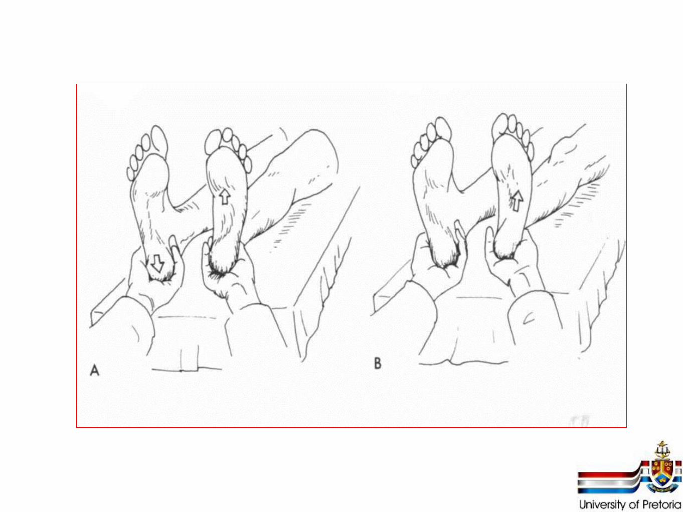
Macrae’s modification
of Schober’s test. The
lumbosacral junction is
identified between the
dimples of Venus, and
measurement made 5cm
below and 10cm above
10 cm
5 cm

Macrae’s modification of Schober’s test. The distraction of these marks is proportional to true lumbar flexion: in this case the patient has AS and skin distraction is limited.
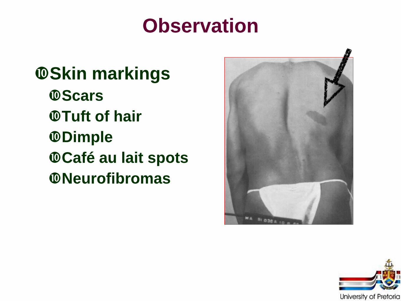
Active movements
Extension
Testing for mechanical symptoms
Lateral flexion Pain on the side being flexed to =
articular pain – other structures are relaxed
Lateral disk – root symptoms worse on that side
Pain with flexion away – muscular or articular
Disk protrusion medial to root

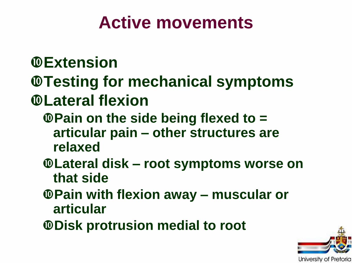
Standing
Squat test
Trendelenburg test

Standing
One-leg lumbar extension test

Table examination
Exclude other pathology
Scan upper limbs
Abdominal examination
Peripheral pulses
Skin,Hair
Other joints Hip
Knee
Ankle/Foot
SI joint
Faber test (Patrick's test)

Table examination
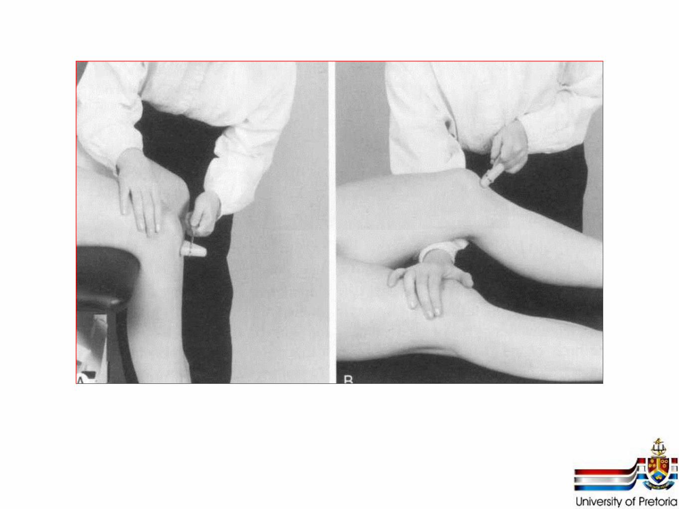
Active straight leg
raise
Passive straight leg
raise


Myotomes
Hip flexion L2
Knee extension L3
Foot dorsiflexion L4
Extension big toe L5
Ankle eversion and plantarflexion S1
Patient still on back

Sensation
Anterior dermatomes

Reflexes
Can also be done with patient sitting
Personal preference
Knee L3
Ankle S1
Medial Hamstring L5
Lateral Hamstring S1
Posterior Tibial L4,L5


If umbilicus moves:
indicative of myelopathy

Turn patient around
Hip extension S1
Knee flexion S2
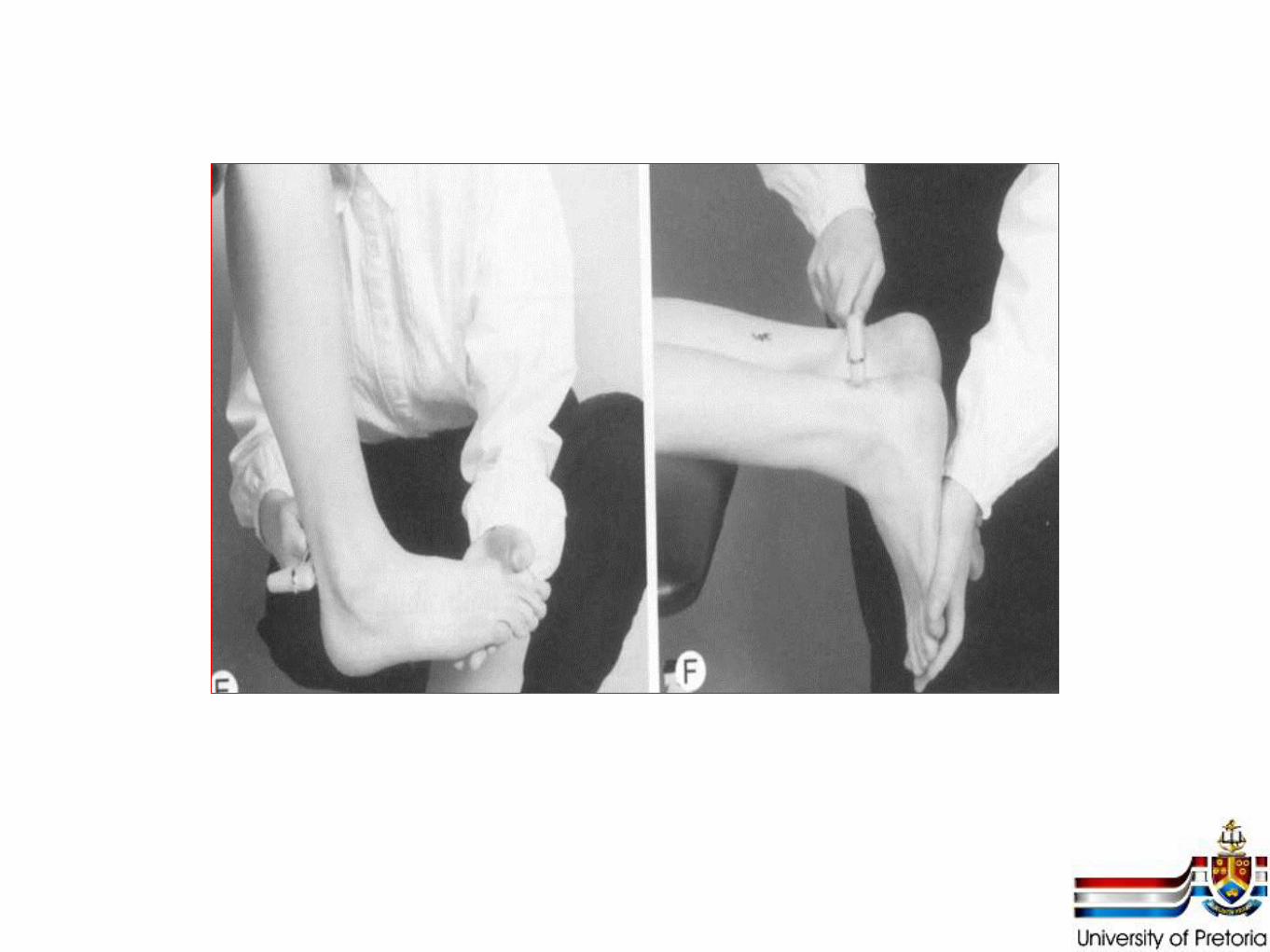
Femoral stretch test L2 or L3 root
Posterior dermatomes
Perianal sensation S4,S5
Anal wink
Anal tone


Femoral stretch test

Special tests
Intermittent claudication
Reflexes
Bicycle test of van Gelderen
Tests for malingering
Hoover test
Burns Test
Seated straight leg raise


Questions
Is the patient malingering?
Is there any secondary gain?
What is the pain generator? Mechanical Anatomic structure
Neurogenic Localizing of lesion/level
Inflammatory
Neurological status of patient Special investigations

Examination of the neck
Dr T Kruger
2012

History
Age
What is the complaint
Timing of symptoms
Other diseases
Severity of symptoms
Trauma? Mechanism
Remember “burners,stinger”
Sites and boundaries of pain Radiculopathy
Myelopathy
Sclerotomal pain

History
Radiation of pain
Lhermitte’s sign
Laughing,coughing,straining
Headaches
Occipital
Neck movement
Nuchal tenderness
Sensory abnormalities
Painful neck movements
Bakody’s sign – hand on head
Balance
Observation
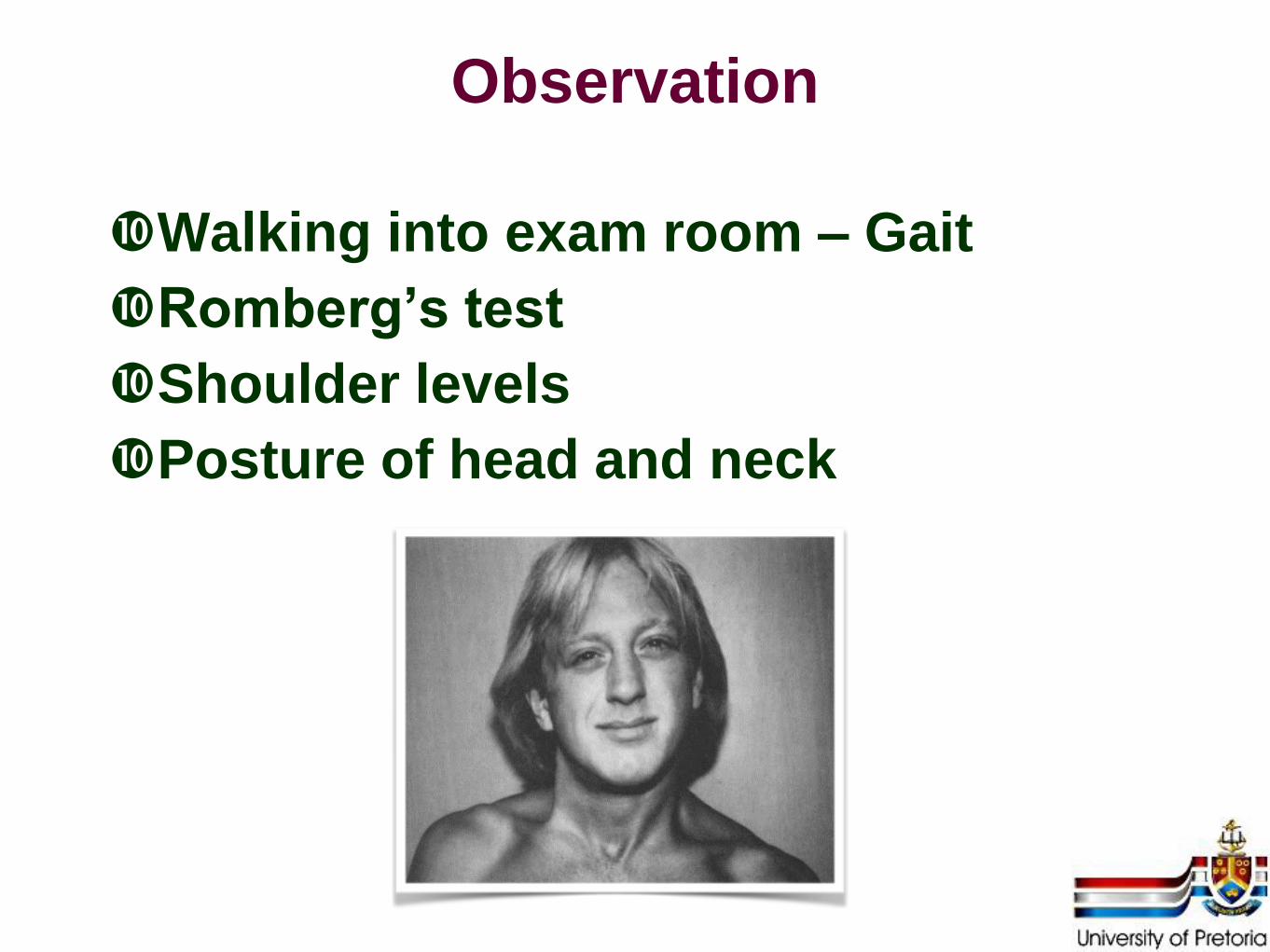
Walking into exam room – Gait
Romberg’s test
Shoulder levels
Posture of head and neck

Examination
Active movements
Nodding versus flexion
Remember the levels of movement

Palpation
Whole spine ant and posterior
Muscle atrophy
Paraspinal muscles
TM joint
Shoulder
AC joint
Rotator cuff
Brachial plexus
Percussion

Passive movements
Full Rom
Overpressure can be applied
Resisted isometric movements
Question?
mechanical pain
Neurogenic origin
Scanning of peripheral joints
Pre flight check

Myotomes
C1-C2 C3 C4

Myotomes
C6

Myotomes
C7

Myotomes
C8

Myotomes
T1
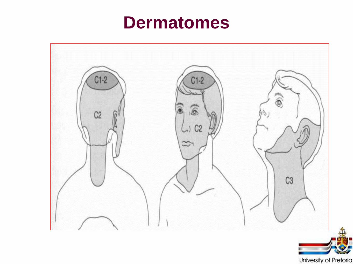
Dermatomes

Dermatomes
C5

Dermatomes
C6

Dermatomes
C7

Dermatomes
C8

Dermatomes
T1

Reflexes
Biceps C5,C6
Brachioradialis C5,C6
Triceps C7,C8
Babinsky – Plantar
Hoffman’s sign
Clonus
Abdominal reflex

Mindset at this point
Always examine the lower extremities
Is this a mechanical pain?
Is this pain of neurological origin?
Is there a vascular component?
Is there a radiculopathy?
Is there a myelopathy?

Special tests
Some used in most examinations
Some only done as confirmation
tests
Commonly performed tests
Spurling
Distraction test
Upper limb tension test
Shoulder abduction test
Vertebral artery test
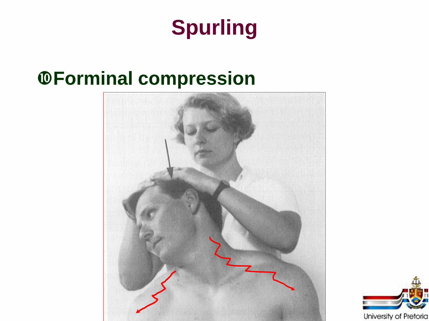
Spurling
Forminal compression

Distraction test
Distraction test

Upper limb tension tests
Like the straight leg raise test in the
lower limb
Also called Elvey’s test
Different positions put tension on
different nerve roots
Evans’s test – Bikele’s sign

Shoulder depression test
Positive = irritation, adhesion or
foraminal encroachment
Brachial plexus = more than one root

Shoulder abduction test
Relief of symptoms=cervical
exstradural compression
Bakody’s sign
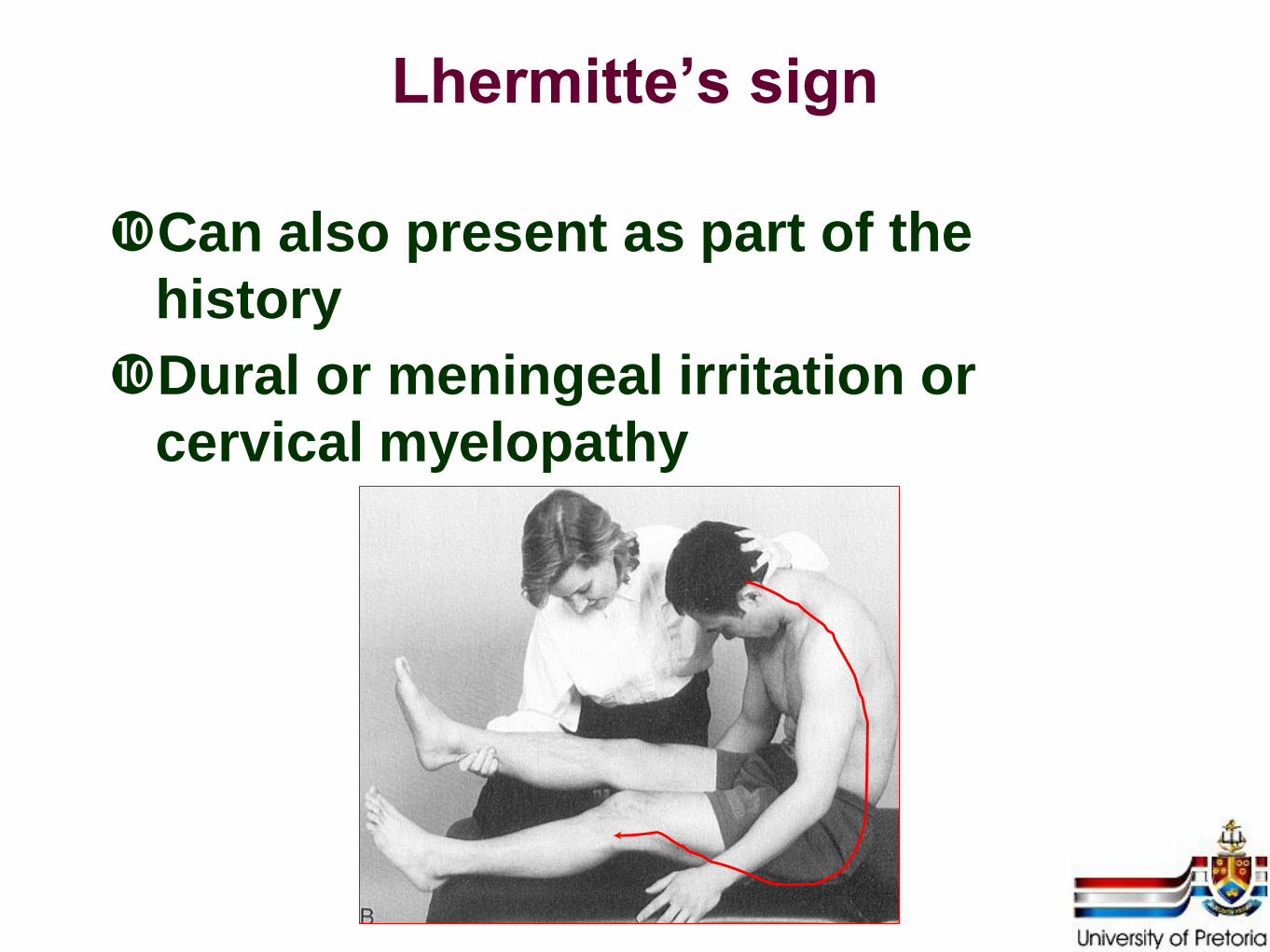
Lhermitte’s sign
Can also present as part of the
history
Dural or meningeal irritation or
cervical myelopathy

Vertebral artery test
Symptoms refer to the side that the
head is turned
Look for
Barre-Lieou sign

Sharp-Purser
Determine C1-C2 instability
Extreme caution

Valsalva tests
Express against a closed glottis
Nafzinger’s test

Thoracic outlet syndrome
Roos test
3 minutes

Answers
Secondary gain
Myelopathy
Radiculopathy
Mechanical symptoms
Pressure on the thecal sack
Level of the pathology
Special investigations