Embed Size (px)
Citation preview
STATISTICAL REPORT OF THE GENERAL FELLOWSHIP EXAMINATION
August / October 2010
This report is prepared to provide candidates, tutors and their Supervisors of training with information about the way in which the Examiners assessed the performance of candidates in the Examination. Answers provided are not model answers but guides to what was expected. Candidates should discuss the report with their tutors so that they may prepare appropriately for the future examinations The exam included two 2.5 hour written papers comprising of 15 ten-minute short answer questions each. Candidates were required to score at least 50% in the written paper before being eligible to sit the oral part of the exam. The oral exam comprised 8 interactive vivas (This is the second exam with a digital radiology station) and two separate hot cases. The tables below provide an overall statistical analysis as well as information regarding performance in the individual sections. A comparison with the previous three examinations is also provided.
2
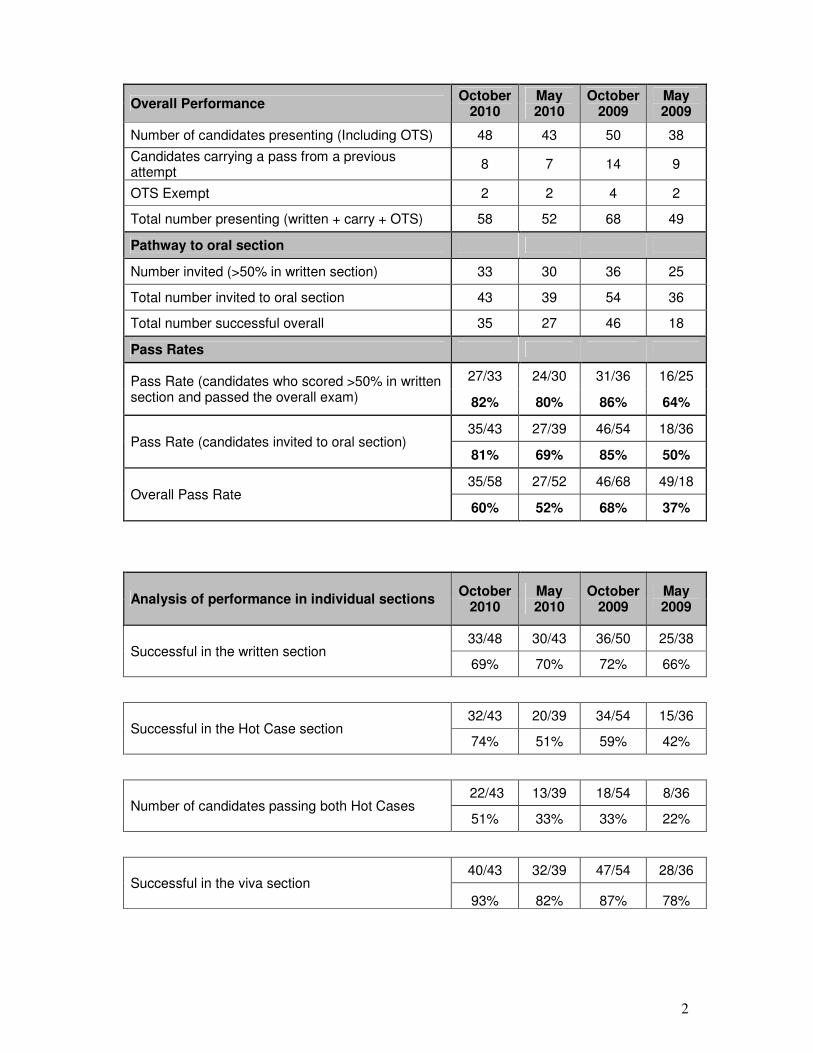
Overall Performance October
2010 May 2010
October 2009
May 2009
Number of candidates presenting (Including OTS) 48 43 50 38
Candidates carrying a pass from a previous attempt
8 7 14 9
OTS Exempt 2 2 4 2
Total number presenting (written + carry + OTS) 58 52 68 49
Pathway to oral section
Number invited (>50% in written section) 33 30 36 25
Total number invited to oral section 43 39 54 36
Total number successful overall 35 27 46 18
Pass Rates
Pass Rate (candidates who scored >50% in written section and passed the overall exam)
27/33 24/30 31/36 16/25
82% 80% 86% 64%
Pass Rate (candidates invited to oral section) 35/43 27/39 46/54 18/36
81% 69% 85% 50%
Overall Pass Rate 35/58 27/52 46/68 49/18
60% 52% 68% 37%
Analysis of performance in individual sections October
2010 May 2010
October 2009
May 2009
Successful in the written section 33/48 30/43 36/50 25/38
69% 70% 72% 66%
Successful in the Hot Case section 32/43 20/39 34/54 15/36
74% 51% 59% 42%
Number of candidates passing both Hot Cases 22/43 13/39 18/54 8/36
51% 33% 33% 22%
Successful in the viva section 40/43 32/39 47/54 28/36
93% 82% 87% 78%
3
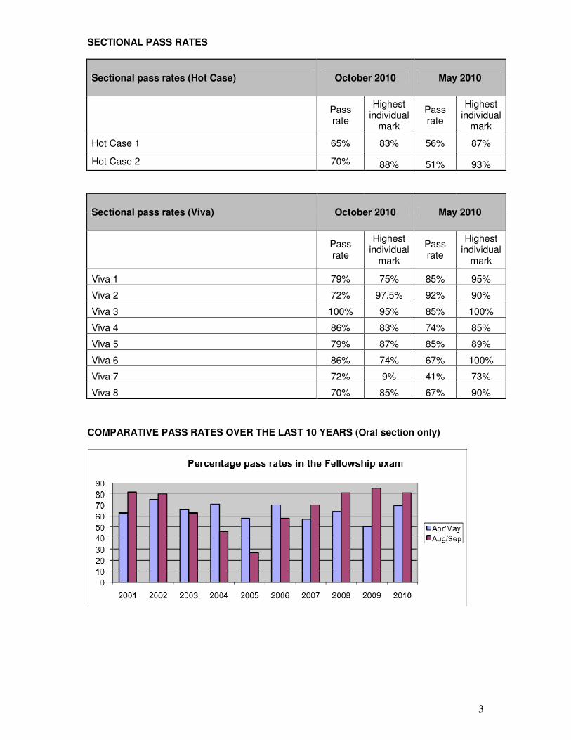
SECTIONAL PASS RATES
Sectional pass rates (Hot Case) October 2010 May 2010
Pass rate
Highest individual
mark
Pass rate
Highest individual
mark
Hot Case 1 65% 83% 56% 87%
Hot Case 2 70% 88% 51% 93%
Sectional pass rates (Viva) October 2010 May 2010
Pass rate
Highest individual
mark
Pass rate
Highest individual
mark
Viva 1 79% 75% 85% 95%
Viva 2 72% 97.5% 92% 90%
Viva 3 100% 95% 85% 100%
Viva 4 86% 83% 74% 85%
Viva 5 79% 87% 85% 89%
Viva 6 86% 74% 67% 100%
Viva 7 72% 9% 41% 73%
Viva 8 70% 85% 67% 90%
COMPARATIVE PASS RATES OVER THE LAST 10 YEARS (Oral section only)
4
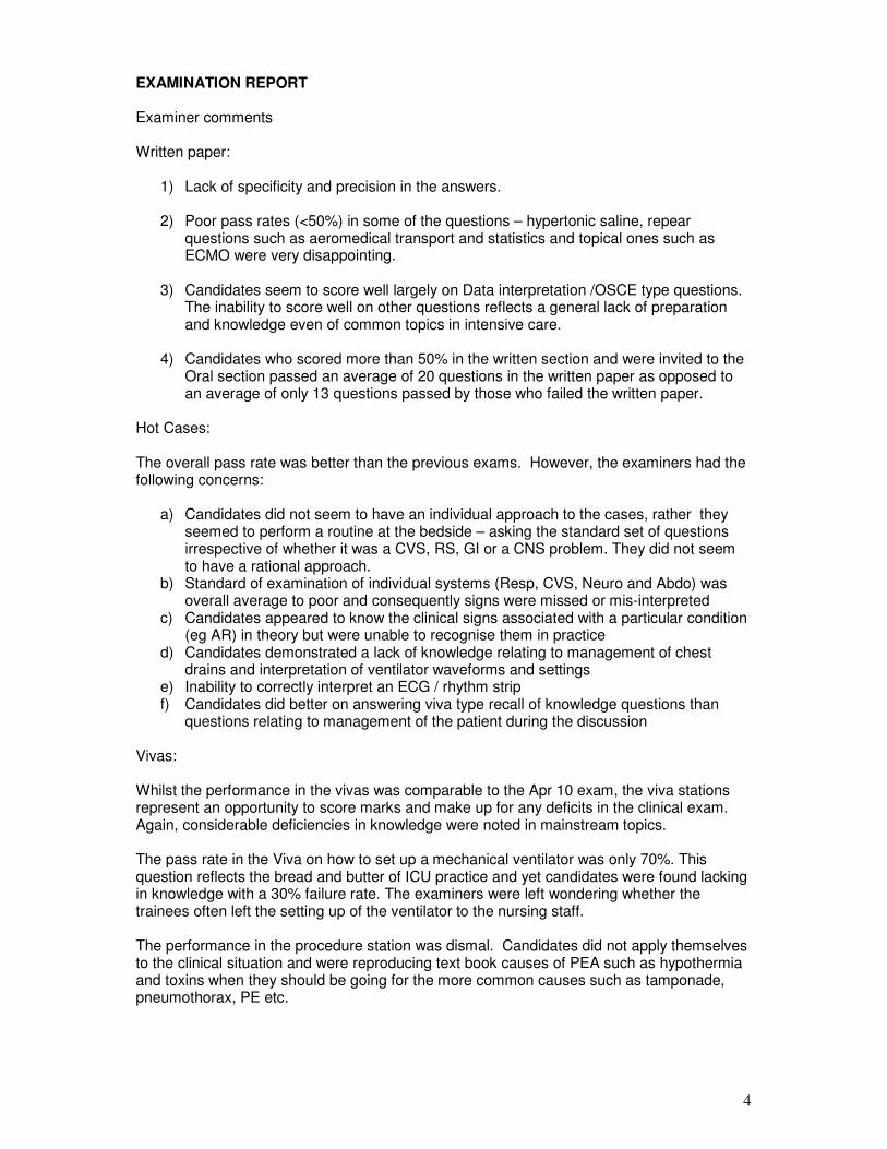
EXAMINATION REPORT
Examiner comments Written paper:
1) Lack of specificity and precision in the answers.
2) Poor pass rates (<50%) in some of the questions – hypertonic saline, repear questions such as aeromedical transport and statistics and topical ones such as ECMO were very disappointing.
3) Candidates seem to score well largely on Data interpretation /OSCE type questions. The inability to score well on other questions reflects a general lack of preparation and knowledge even of common topics in intensive care.
4) Candidates who scored more than 50% in the written section and were invited to the Oral section passed an average of 20 questions in the written paper as opposed to an average of only 13 questions passed by those who failed the written paper.
Hot Cases: The overall pass rate was better than the previous exams. However, the examiners had the following concerns:
a) Candidates did not seem to have an individual approach to the cases, rather they seemed to perform a routine at the bedside – asking the standard set of questions irrespective of whether it was a CVS, RS, GI or a CNS problem. They did not seem to have a rational approach.
b) Standard of examination of individual systems (Resp, CVS, Neuro and Abdo) was overall average to poor and consequently signs were missed or mis-interpreted
c) Candidates appeared to know the clinical signs associated with a particular condition (eg AR) in theory but were unable to recognise them in practice
d) Candidates demonstrated a lack of knowledge relating to management of chest drains and interpretation of ventilator waveforms and settings
e) Inability to correctly interpret an ECG / rhythm strip f) Candidates did better on answering viva type recall of knowledge questions than
questions relating to management of the patient during the discussion Vivas: Whilst the performance in the vivas was comparable to the Apr 10 exam, the viva stations represent an opportunity to score marks and make up for any deficits in the clinical exam. Again, considerable deficiencies in knowledge were noted in mainstream topics. The pass rate in the Viva on how to set up a mechanical ventilator was only 70%. This question reflects the bread and butter of ICU practice and yet candidates were found lacking in knowledge with a 30% failure rate. The examiners were left wondering whether the trainees often left the setting up of the ventilator to the nursing staff. The performance in the procedure station was dismal. Candidates did not apply themselves to the clinical situation and were reproducing text book causes of PEA such as hypothermia and toxins when they should be going for the more common causes such as tamponade, pneumothorax, PE etc.

5
SHORT ANSWER QUESTION PAPER 1 (Question in bold) 1 a) Briefly outline the rationale for the use of hypertonic saline in:
1) Hyponatremia
Hyponatremia
• Severe hyponatremia (<120 mEq/L) can cause significant and permanent neurologic injury or death. In the event of seizures or acute collapse relatively rapid initial correction may be required.
• There is evidence that the severity and duration of hyponatremia may be related to cerebro pontine myelinolysis, normal saline and fluid restriction may be inadequate to increase sodium levels appropriately.
• Some conditions such as cerebral salt wasting or large GIT losses may result in losses that may not be able to be replaced by other means.
2) Traumatic brain injury
Traumatic Brain Injury
• The rationale for hypertonic saline compared with normal saline
• Better compensates for blood loss
• Improved CPP
• Reduces harmful inflammatory responses
• May prevent cerebral edema.
• Can be used as a continuous infusion
• Obviates the need for osmolality testing Previous animal studies and smaller clinical trials suggested better outcomes in patients with TBI after use of hypertonic saline solution. The safety profile has been good, and some evidence suggests a potential survival benefit when hypertonic saline is given. However The National Heart, Lung, and Blood Institute (NHLBI) of the National Institutes of Health (NIH) has stopped enrollment of patients with severe traumatic brain injury (TBI) into a Resuscitation Outcomes Consortium (ROC) trial testing the effects of hypertonic saline solutions given before arrival at the emergency department. as early as possible after TBI. 1073 patients 6 month analysis – no difference. 1.2 List the possible complications of hypertonic saline administration.
• Hypernatremia
• Hyperchloraemic acidaemia
• Renal failure
• CCF/Pulmonary Oedema
• Neurological SAH
• rebound intracranial H/T
• Central Pontine Myelinolysis
Pass rate 48%
Highest mark 8.25 out of 10
6
2 A 36 year old female is brought into your Emergency Department with acute shortness of breath. She is unable to provide any history due to her tachypnoea. She is sitting upright in bed grasping the bed sides. She has a respiratory rate of 30 breaths per minute, has a GCS of 15, is afebrile and has a BP of 90/60mmHg. She is using accessory muscles. On auscultation, she has widespread expiratory wheeze spread throughout both lung fields.
a) In addition to acute severe asthma, what other differential diagnoses of her
clinical presentation should be considered?
Differential diagnoses
• anaphylaxis ( large % don’t have rash etc – just bronchospasm)
• Acute exacerbation COPD
• central foreign body
• acute pulmonary oedema
• Pneumothorax
• Hysterical hyperventilation
• acute Pulmonary embolus b) Assuming this patient has acute severe asthma, list your initial
management steps at this stage. • Resuscitation/ investigation and definitive management
• Initial salbutamol nebulisation – continuously. Consider IV infusion
• IV steroids - ? type and dose .
• Replace K/Mg
• Nebulised adrenalin if anaphylaxis still under consideration
• IV access, bloods including mast cell tryptase cultures/ +/- procalcitonin
• Portable CXR to exclude pnemothorax / localised consolidation and assess hyperinflation.
c) Despite optimal medical management, the patient tires. Briefly outline the
role of BiPAP in acute severe asthma. Intrinisic PEEP increases the negative intrathoracic pressure the patient must generate to trigger a breath and hence increases WOB. Application of extrinsic PEEP minimises this difference and reduces WOB. IPAP reduces the WOB associated with resistance. d) BiPAP fails and the patient is successfully intubated. Following intubation,
airway pressures rise and the chest becomes more silent. List other interventions may you consider.
• Another CXR to check no PTX post PPV
• Increasing salbutamol
• Deepen sedation
• Adding adrenalin/ aminophylline/ ketamine/ Mg ( no evidence) – doses required by candidate
• Volatile anaesthesia
• Paralysis- Train of four essential .
• ? bronchoscopy
• Measurement of iPEEP
Pass rate 85%
Highest mark 8.75
7
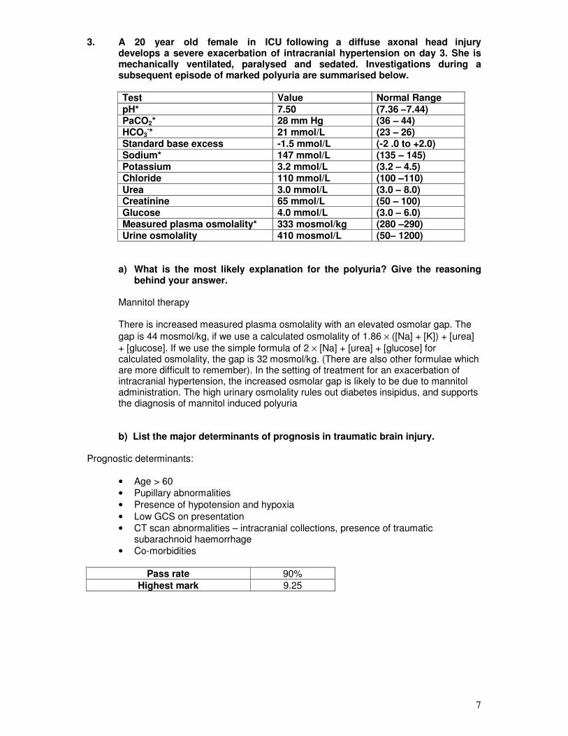
3. A 20 year old female in ICU following a diffuse axonal head injury develops a severe exacerbation of intracranial hypertension on day 3. She is mechanically ventilated, paralysed and sedated. Investigations during a subsequent episode of marked polyuria are summarised below.
Test Value Normal Range pH* 7.50 (7.36 –7.44) PaCO2* 28 mm Hg (36 – 44) HCO3
-* 21 mmol/L (23 – 26) Standard base excess -1.5 mmol/L (-2 .0 to +2.0) Sodium* 147 mmol/L (135 – 145) Potassium 3.2 mmol/L (3.2 – 4.5) Chloride 110 mmol/L (100 –110) Urea 3.0 mmol/L (3.0 – 8.0) Creatinine 65 mmol/L (50 – 100) Glucose 4.0 mmol/L (3.0 – 6.0) Measured plasma osmolality* 333 mosmol/kg (280 –290) Urine osmolality 410 mosmol/L (50– 1200)
a) What is the most likely explanation for the polyuria? Give the reasoning behind your answer.
Mannitol therapy
There is increased measured plasma osmolality with an elevated osmolar gap. The
gap is 44 mosmol/kg, if we use a calculated osmolality of 1.86 × ([Na] + [K]) + [urea]
+ [glucose]. If we use the simple formula of 2 × [Na] + [urea] + [glucose] for calculated osmolality, the gap is 32 mosmol/kg. (There are also other formulae which are more difficult to remember). In the setting of treatment for an exacerbation of intracranial hypertension, the increased osmolar gap is likely to be due to mannitol administration. The high urinary osmolality rules out diabetes insipidus, and supports the diagnosis of mannitol induced polyuria
b) List the major determinants of prognosis in traumatic brain injury. Prognostic determinants:
• Age > 60
• Pupillary abnormalities
• Presence of hypotension and hypoxia
• Low GCS on presentation
• CT scan abnormalities – intracranial collections, presence of traumatic subarachnoid haemorrhage
• Co-morbidities
Pass rate 90%
Highest mark 9.25
8
4. A 55 year old with severe sepsis develops Heparin Induced Thrombotic
Thrombocytopenia Syndrome (HITTS) while on continuous veno-venous haemodiafiltration (CVVHDF).
Outline the strategies available for prolonging the life of the CVVHDF circuit in
this patient, mentioning the advantages and disadvantages of each strategy. No Anticoagulant +/- Saline Flushes (50-100ml every hour) * Ensure good wide bore access, high flow rates, consider predilution Advantage:
Minimizes bleeding risk, but consumption of platelets and factors by membrane (theoretical)
Disadvantage: Shortened filter life / increased time off dialysis
Regional citrate Advantage:
Provides good regional anticoagulation Pre-mix solutions and protocols for use have simplified process
Disadvantage Labor intensive, Requires diligent monitoring of serum sodium, ionized calcium, and bicarbonate Requires infusion of calcium outside the circuit (access issues) Large sodium load occurs when trisodium citrate used May cause alkalosis Special diasylate required: hyponatraemic, without buffer, Ca free Not appropriate in liver failure
Prostacycline and Analogues Advantages
Reduced bleeding risk Disadvantages
Shorter filter life Hypotension
Direct thrombin Inhibitors: Hirudin / Lepirudin / Argatroban Advantages:
Linear relationship between levels and APTT (<100s) for Hirudin
Disadvantages
Renal clearance, accumulation in renal failure (Hirudin, Lepirudin) Hepatic metabolism, accumulation in liver disease (Argatroban) No antagonist Argatroban falsely raises INR / PT Expense
Other agents Danaparoid Limited availability Risk of cross-reactivity with heparin-induced antibodies Serine Protease inhibitors (nafamostat) limited experience, massive cost
9
Fondaparinux Not readily available Limited evidence supporting its use Warfarin /NSAIDS
Pass rate 71%
Highest mark 8.5 5.1 List 4 clinical signs of portal hypertension.
• Splenomegaly
• Caput medusae
• Ascites
• Haemorrhoids on rectal examination
• Haematemesis? Melaena
5.2 List 4 clinical signs of intracranial hypertension.
• Unilateral or bilateral papilledema
• Unilateral or bilateral papillary dilatation
• Impaired conscious state
• Bradycardia
5.3 On palpation of the arterial pulse, a double peak was noted with each cardiac
cycle. List 4 conditions/situations which can produce this phenomenon.
• AS + AR
• Severe AR
• HOCM
• IABP
5.4 List the classic clinical findings on praecordial examination in a patient with
Tetralogy of Fallot.
• ESM or PSM
• Right ventricular heave
• A loud single second sound
5.5 List 2 causes (apart from cardiovascular or respiratory) of cyanosis.
• Severe methemoglobinemia
• Sulfhemoglobinemia
• Hemoglobin mutation
• Polycythaemia
• Hypothermia / cold
• High altitude
Pass rate 94%
Highest mark 8.7
10
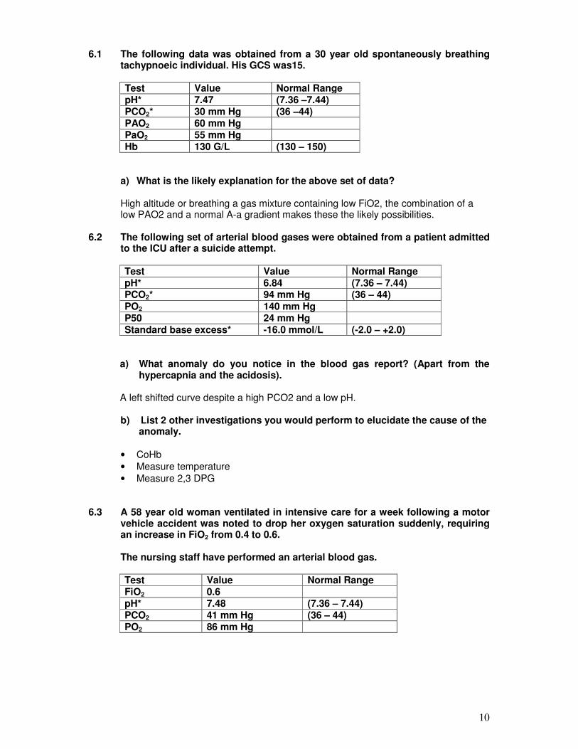
6.1 The following data was obtained from a 30 year old spontaneously breathing
tachypnoeic individual. His GCS was15.
Test Value Normal Range pH* 7.47 (7.36 –7.44) PCO2* 30 mm Hg (36 –44) PAO2 60 mm Hg PaO2 55 mm Hg Hb 130 G/L (130 – 150)
a) What is the likely explanation for the above set of data?
High altitude or breathing a gas mixture containing low FiO2, the combination of a low PAO2 and a normal A-a gradient makes these the likely possibilities.
6.2 The following set of arterial blood gases were obtained from a patient admitted
to the ICU after a suicide attempt.
Test Value Normal Range pH* 6.84 (7.36 – 7.44) PCO2* 94 mm Hg (36 – 44) PO2 140 mm Hg P50 24 mm Hg Standard base excess* -16.0 mmol/L (-2.0 – +2.0)
a) What anomaly do you notice in the blood gas report? (Apart from the hypercapnia and the acidosis).
A left shifted curve despite a high PCO2 and a low pH.
b) List 2 other investigations you would perform to elucidate the cause of the
anomaly.
• CoHb
• Measure temperature
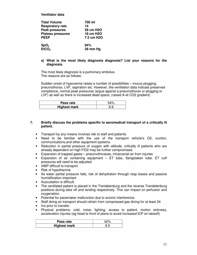
• Measure 2,3 DPG 6.3 A 58 year old woman ventilated in intensive care for a week following a motor
vehicle accident was noted to drop her oxygen saturation suddenly, requiring an increase in FiO2 from 0.4 to 0.6.
The nursing staff have performed an arterial blood gas.
Test Value Normal Range FiO2 0.6 pH* 7.48 (7.36 – 7.44) PCO2 41 mm Hg (36 – 44) PO2 86 mm Hg
11
Ventilator data Tidal Volume 700 ml Respiratory rate 14 Peak pressures 28 cm H2O Plateau pressures 18 cm H2O PEEP 7.5 cm H2O SpO2 94% EtCO2 28 mm Hg
a) What is the most likely diagnosis diagnosis? List your reasons for the diagnosis.
The most likely diagnosis is a pulmonary embolus. The reasons are as follows: Sudden onset of hypoxemia raises a number of possibilities – mucus plugging, pneumothorax, LVF, aspiration etc. However, the ventilation data indicate preserved compliance, normal peak pressures (argue against a pneumothorax or plugging or LVF) as well as there is increased dead space, (raised A-et CO2 gradient)
Pass rate 54%
Highest mark 8.9 7. Briefly discuss the problems specific to aeromedical transport of a critically ill
patient.
• Transport by any means involves risk to staff and patients
• Need to be familiar with the use of the transport vehicle’s O2, suction, communications,and other equipment systems.
• Reduction in partial pressure of oxygen with altitude, critically ill patients who are already dependent on high FiO2 may be further compromised.
• Expansion of trapped gases – pneumothoraces, intracranial air from injuries
• Expansion of air containing equipment – ET tube, Sengstaken tube. ET cuff pressures will need to be adjusted
• IABP difficult to transport
• Risk of hypothermia
• As water partial pressure falls, risk of dehydration through resp losses and passive humidification important
• Auscultation is difficult.
• The ventilated patient is placed in the Trendelenburg and the reverse Trendelenburg positions during take off and landing respectively. This can impact on perfusion and oxygenation.
• Potential for pacemaker malfunction due to avionic interference.
• Staff doing air transport should refrain from compressed gas diving for at least 24
• hrs prior to transfer.
• Physical problems: cold, noise, lighting, access to patient, motion sickness, acceleration injuries (eg head to front of plane to avoid increased ICP on takeoff)
Pass rate 42%
Highest mark 9.5
12
8.1) You are called to a cardiac arrest. The following rhythm was evident on your
arrival and the patient was pulseless.
Image provided in examination paper
a) List 5 causes of this presentation This is PEA
• Tension pneumothorax
• Tamponade
• PE
• Hypovolemia
• Hypothermia 8.2) Examine the ECG rhythm below. The patient was being paced AAI . What is the
problem with the pacing?
Image provided in examination paper
Oversensing. In this case myopotentials are sensed.
13
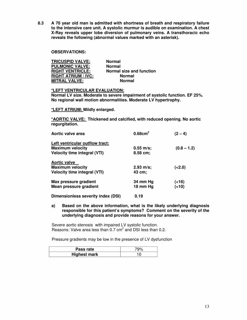
8.3 A 70 year old man is admitted with shortness of breath and respiratory failure
to the intensive care unit. A systolic murmur is audible on examination. A chest X-Ray reveals upper lobe diversion of pulmonary veins. A transthoracic echo reveals the following (abnormal values marked with an asterisk).
OBSERVATIONS: TRICUSPID VALVE: Normal PULMONIC VALVE: Normal RIGHT VENTRICLE: Normal size and function RIGHT ATRIUM / IVC: Normal MITRAL VALVE: Normal *LEFT VENTRICULAR EVALUATION: Normal LV size. Moderate to severe impairment of systolic function. EF 25%. No regional wall motion abnormalities. Moderate LV hypertrophy. *LEFT ATRIUM: Mildly enlarged. *AORTIC VALVE: Thickened and calcified, with reduced opening. No aortic regurgitation. Aortic valve area 0.68cm2 (2 – 4) Left ventricular outflow tract: Maximum velocity 0.55 m/s; (0.8 – 1.2) Velocity time integral (VTI) 8.58 cm;
Aortic valve Maximum velocity 2.93 m/s; (<2.0) Velocity time integral (VTI) 43 cm; Max pressure gradient 34 mm Hg (<16) Mean pressure gradient 18 mm Hg (<10) Dimensionless severity index (DSI) 0.19 a) Based on the above information, what is the likely underlying diagnosis
responsible for this patient’s symptoms? Comment on the severity of the underlying diagnosis and provide reasons for your answer.
Severe aortic stenosis with impaired LV systolic function. Reasons: Valve area less than 0.7 cm2 and DSI less than 0.2. Pressure gradients may be low in the presence of LV dysfunction
Pass rate 79%
Highest mark 10
14
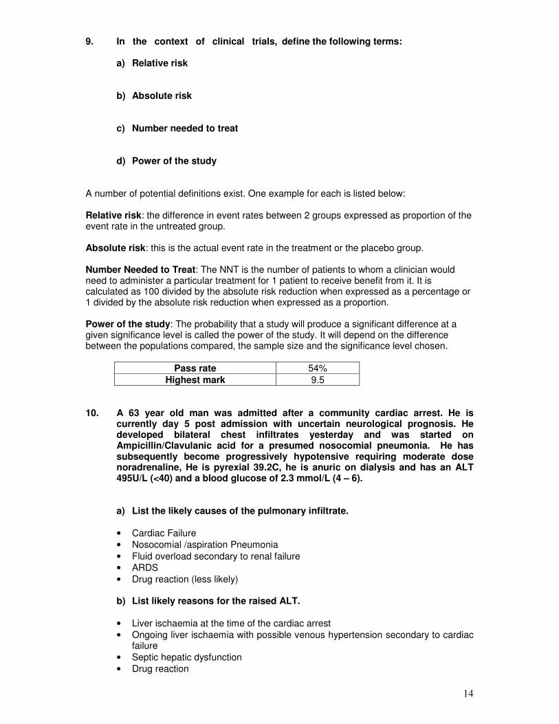
9. In the context of clinical trials, define the following terms:
a) Relative risk
b) Absolute risk
c) Number needed to treat
d) Power of the study
A number of potential definitions exist. One example for each is listed below: Relative risk: the difference in event rates between 2 groups expressed as proportion of the event rate in the untreated group. Absolute risk: this is the actual event rate in the treatment or the placebo group. Number Needed to Treat: The NNT is the number of patients to whom a clinician would need to administer a particular treatment for 1 patient to receive benefit from it. It is calculated as 100 divided by the absolute risk reduction when expressed as a percentage or 1 divided by the absolute risk reduction when expressed as a proportion. Power of the study: The probability that a study will produce a significant difference at a given significance level is called the power of the study. It will depend on the difference between the populations compared, the sample size and the significance level chosen.
Pass rate 54%
Highest mark 9.5 10. A 63 year old man was admitted after a community cardiac arrest. He is
currently day 5 post admission with uncertain neurological prognosis. He developed bilateral chest infiltrates yesterday and was started on Ampicillin/Clavulanic acid for a presumed nosocomial pneumonia. He has subsequently become progressively hypotensive requiring moderate dose noradrenaline, He is pyrexial 39.2C, he is anuric on dialysis and has an ALT 495U/L (<40) and a blood glucose of 2.3 mmol/L (4 – 6).
a) List the likely causes of the pulmonary infiltrate.
• Cardiac Failure
• Nosocomial /aspiration Pneumonia
• Fluid overload secondary to renal failure
• ARDS
• Drug reaction (less likely)
b) List likely reasons for the raised ALT.
• Liver ischaemia at the time of the cardiac arrest
• Ongoing liver ischaemia with possible venous hypertension secondary to cardiac failure
• Septic hepatic dysfunction
• Drug reaction
15
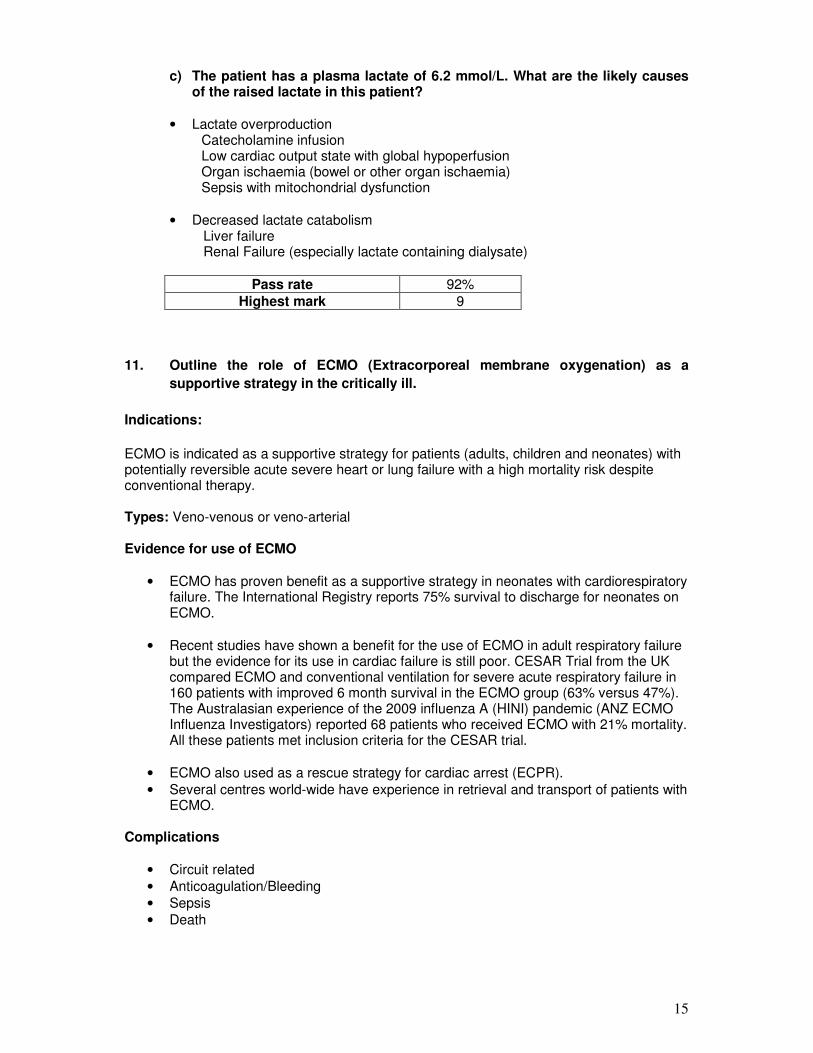
c) The patient has a plasma lactate of 6.2 mmol/L. What are the likely causes
of the raised lactate in this patient?
• Lactate overproduction Catecholamine infusion Low cardiac output state with global hypoperfusion Organ ischaemia (bowel or other organ ischaemia) Sepsis with mitochondrial dysfunction
• Decreased lactate catabolism Liver failure Renal Failure (especially lactate containing dialysate)
Pass rate 92%
Highest mark 9
11. Outline the role of ECMO (Extracorporeal membrane oxygenation) as a
supportive strategy in the critically ill.
Indications:
ECMO is indicated as a supportive strategy for patients (adults, children and neonates) with potentially reversible acute severe heart or lung failure with a high mortality risk despite conventional therapy. Types: Veno-venous or veno-arterial Evidence for use of ECMO
• ECMO has proven benefit as a supportive strategy in neonates with cardiorespiratory failure. The International Registry reports 75% survival to discharge for neonates on ECMO.
• Recent studies have shown a benefit for the use of ECMO in adult respiratory failure but the evidence for its use in cardiac failure is still poor. CESAR Trial from the UK compared ECMO and conventional ventilation for severe acute respiratory failure in 160 patients with improved 6 month survival in the ECMO group (63% versus 47%). The Australasian experience of the 2009 influenza A (HINI) pandemic (ANZ ECMO Influenza Investigators) reported 68 patients who received ECMO with 21% mortality. All these patients met inclusion criteria for the CESAR trial.
• ECMO also used as a rescue strategy for cardiac arrest (ECPR).
• Several centres world-wide have experience in retrieval and transport of patients with ECMO.
Complications
• Circuit related
• Anticoagulation/Bleeding
• Sepsis
• Death
16
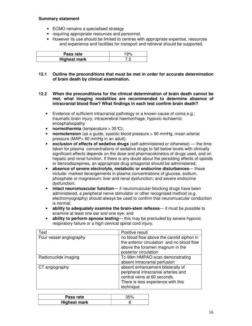
Summary statement
• ECMO remains a specialised strategy
• requiring appropriate resources and personnel.
• However its use should be limited to centres with appropriate expertise, resources and experience and facilities for transport and retrieval should be supported.
Pass rate 19%
Highest mark 7.5 12.1 Outline the preconditions that must be met in order for accurate determination
of brain death by clinical examination. 12.2 When the preconditions for the clinical determination of brain death cannot be
met, what imaging modalities are recommended to determine absence of intracranial blood flow? What findings in each test confirm brain death?
• Evidence of sufficient intracranial pathology or a known cause of coma e.g.;
traumatic brain injury, intracerebral haemorrhage, hypoxic-ischaemic encephaloopathy
• normothermia (temperature > 35°C);
• normotension (as a guide, systolic blood pressure > 90 mmHg, mean arterial pressure (MAP> 60 mmHg in an adult);
• exclusion of effects of sedative drugs (self-administered or otherwise) — the time taken for plasma concentrations of sedative drugs to fall below levels with clinically significant effects depends on the dose and pharmacokinetics of drugs used, and on hepatic and renal function. If there is any doubt about the persisting effects of opioids or benzodiazepines, an appropriate drug antagonist should be administered;
• absence of severe electrolyte, metabolic or endocrine disturbances— these include: marked derangements in plasma concentrations of glucose, sodium, phosphate or magnesium; liver and renal dysfunction; and severe endocrine dysfunction;
• intact neuromuscular function— if neuromuscular-blocking drugs have been administered, a peripheral nerve stimulator or other recognised method (e.g. electromyography) should always be used to confirm that neuromuscular conduction is normal;
• ability to adequately examine the brain-stem reflexes— it must be possible to examine at least one ear and one eye; and
• ability to perform apnoea testing— this may be precluded by severe hypoxic respiratory failure or a high cervical spinal cord injury.
Test Positive result Four vessel angiography no blood flow above the carotid siphon in
the anterior circulation and no blood flow above the foramen magnum in the posterior circulation
Radionuclide imaging Tc-99m HMPAO scan demonstrating absent intracranial perfusion
CT angiography absent enhancement bilaterally of peripheral intracranial arteries and central veins at 60 seconds. There is less experience with this technique
Pass rate 35%
Highest mark 8
17
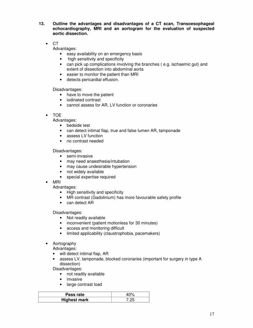
13. Outline the advantages and disadvantages of a CT scan, Transoesophageal
echocardiography, MRI and an aortogram for the evaluation of suspected aortic dissection.
• CT Advantages:
• easy availability on an emergency basis
• high sensitivity and specificity
• can pick up complications involving the branches ( e.g. ischaemic gut) and extent of dissection into abdominal aorta
• easier to monitor the patient than MRI
• detects pericardial effusion.
Disadvantages:
• have to move the patient
• iodinated contrast
• cannot assess for AR, LV function or coronaries
• TOE Advantages:
• bedside test
• can detect intimal flap, true and false lumen AR, tamponade
• assess LV function
• no contrast needed
Disadvantages:
• semi-invasive
• may need anaesthesia/intubation
• may cause undesirable hypertension
• not widely available
• special expertise required
• MRI Advantages:
• High sensitivity and specificity
• MR contrast (Gadolinium) has more favourable safety profile
• can detect AR Disadvantages:
• Not readily available
• inconvenient (patient motionless for 30 minutes)
• access and monitoring difficult
• limited applicability (claustrophobia, pacemakers)
• Aortography Advantages:
• will detect intimal flap, AR
• assess LV, tamponade, blocked coronaries (important for surgery in type A dissection)
Disadvantages:
• not readily available
• invasive
• large contrast load
Pass rate 40%
Highest mark 7.25
18
14.1 A 75 year old man was admitted 3 days post prostatectomy with a febrile
illness. Examination revealed lower abdominal tenderness and a clinical photograph of his groins and external genitalia is shown below.
a) What is shown in this picture below?
Fournier’s gangrene.
b) List 2 important management steps Urgent surgical debridement Use of meropenem or other reasonable combinations
A clinical photograph of Fournier’s gangrene was supplied.
14.2 a) What lesion is shown in the picture below in a patient presenting with
septic shock?
Purpura fulminans b) List 5 other causes of this lesion
• Meningococcemia
• Post-splenectomy pneumococcemia
• DIC
• Rickettsial infections
• High dose inotropes
• Endocarditis
A clinical photograph of purpura fulminans was supplied.
14.3 This clinical sign was noted in a patient involved in a motor vehicle accident.
a) What sign is shown below? A clinical photograph of Battle’s sign was supplied.
b) What does it indicate? Base of skull fracture
c) What associated signs support the diagnosis mentioned in Question 14.3 b?
• CSF otorrhoea
• Haemotympanum
• Racoon eyes
• CSF rhinorrhoea
• Cranial nerve abnormalities
Pass rate 79%
Highest mark 8.1
19
15. The following questions refer to implantable cardiac pacemakers and
implantable cardiac defibrillators.
a) What is the effect of applying a magnet to these devices? ICD: it turns off antiarrhythmic programme but has no affect on backup pacemaker Pacemaker: It defaults to asynchronous mode or a fixed rate. Rate depends on battery life.
b) What information can you gain from a chest X-Ray in a patient with an implantable cardiac device?
• Single v dual chamber
• Biventricular or left ventricular (cardiac resynchronisation)
• Lead displacement or injury
• Number of devices present
c) What are the advantages of DDD pacing compared to VVI pacing?
• AV synchronisation maintained
• Avoids pacemaker syndrome
• Reduced incidence of AF
• Possible decreased thrombotic events
d) List 4 benefits of cardiac resynchronisation therapy.
• improved LVEF,CO and haemodynamics
• improved exercise tolerance
• decreased NYHA class
• decreased hospitilisation
• improved quality of life
Pass rate 6%
Highest mark 5.5
20
SHORT ANSWER QUESTION PAPER 2 (Question in bold)
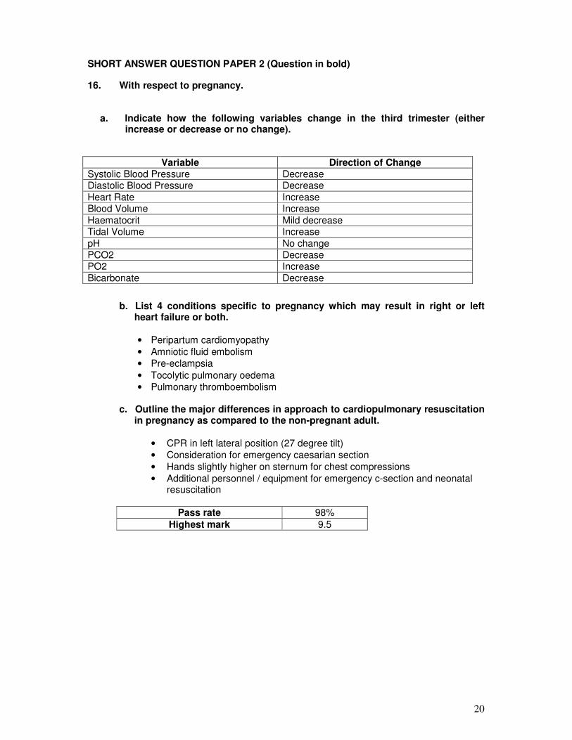
16. With respect to pregnancy.
a. Indicate how the following variables change in the third trimester (either increase or decrease or no change).
Variable Direction of Change Systolic Blood Pressure Decrease Diastolic Blood Pressure Decrease
Heart Rate Increase Blood Volume Increase
Haematocrit Mild decrease Tidal Volume Increase
pH No change PCO2 Decrease PO2 Increase
Bicarbonate Decrease
b. List 4 conditions specific to pregnancy which may result in right or left heart failure or both.
• Peripartum cardiomyopathy
• Amniotic fluid embolism
• Pre-eclampsia
• Tocolytic pulmonary oedema
• Pulmonary thromboembolism
c. Outline the major differences in approach to cardiopulmonary resuscitation in pregnancy as compared to the non-pregnant adult.
• CPR in left lateral position (27 degree tilt)
• Consideration for emergency caesarian section
• Hands slightly higher on sternum for chest compressions
• Additional personnel / equipment for emergency c-section and neonatal resuscitation
Pass rate 98%
Highest mark 9.5
21
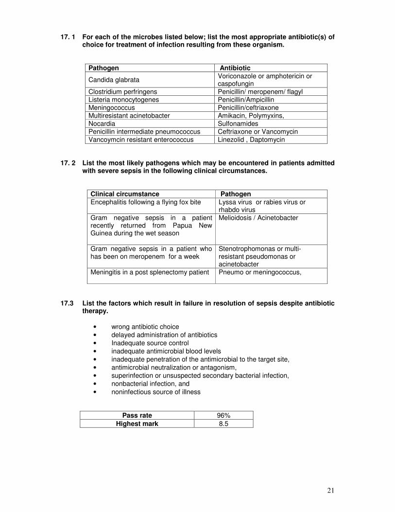
17. 1 For each of the microbes listed below; list the most appropriate antibiotic(s) of
choice for treatment of infection resulting from these organism.
Pathogen Antibiotic
Candida glabrata Voriconazole or amphotericin or caspofungin
Clostridium perfringens Penicillin/ meropenem/ flagyl Listeria monocytogenes Penicillin/Ampicillin Meningococcus Penicillin/ceftriaxone Multiresistant acinetobacter Amikacin, Polymyxins, Nocardia Sulfonamides Penicillin intermediate pneumococcus Ceftriaxone or Vancomycin Vancoymcin resistant enterococcus Linezolid , Daptomycin
17. 2 List the most likely pathogens which may be encountered in patients admitted
with severe sepsis in the following clinical circumstances.
Clinical circumstance Pathogen Encephalitis following a flying fox bite Lyssa virus or rabies virus or
rhabdo virus Gram negative sepsis in a patient recently returned from Papua New Guinea during the wet season
Melioidosis / Acinetobacter
Gram negative sepsis in a patient who has been on meropenem for a week
Stenotrophomonas or multi-resistant pseudomonas or acinetobacter
Meningitis in a post splenectomy patient
Pneumo or meningococcus,
17.3 List the factors which result in failure in resolution of sepsis despite antibiotic
therapy.
• wrong antibiotic choice
• delayed administration of antibiotics
• Inadequate source control
• inadequate antimicrobial blood levels
• inadequate penetration of the antimicrobial to the target site,
• antimicrobial neutralization or antagonism,
• superinfection or unsuspected secondary bacterial infection,
• nonbacterial infection, and
• noninfectious source of illness
Pass rate 96%
Highest mark 8.5
22
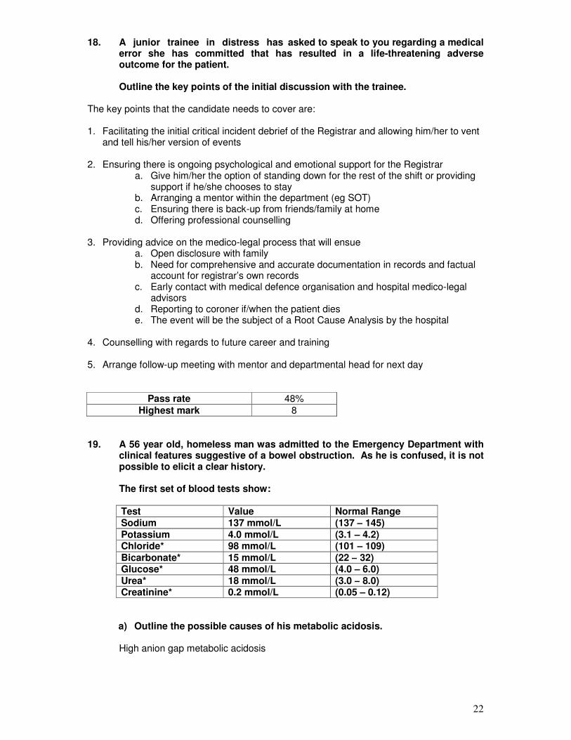
18. A junior trainee in distress has asked to speak to you regarding a medical error she has committed that has resulted in a life-threatening adverse outcome for the patient.
Outline the key points of the initial discussion with the trainee.
The key points that the candidate needs to cover are: 1. Facilitating the initial critical incident debrief of the Registrar and allowing him/her to vent
and tell his/her version of events
2. Ensuring there is ongoing psychological and emotional support for the Registrar a. Give him/her the option of standing down for the rest of the shift or providing
support if he/she chooses to stay b. Arranging a mentor within the department (eg SOT) c. Ensuring there is back-up from friends/family at home d. Offering professional counselling
3. Providing advice on the medico-legal process that will ensue
a. Open disclosure with family b. Need for comprehensive and accurate documentation in records and factual
account for registrar’s own records c. Early contact with medical defence organisation and hospital medico-legal
advisors d. Reporting to coroner if/when the patient dies e. The event will be the subject of a Root Cause Analysis by the hospital
4. Counselling with regards to future career and training
5. Arrange follow-up meeting with mentor and departmental head for next day
Pass rate 48%
Highest mark 8
19. A 56 year old, homeless man was admitted to the Emergency Department with
clinical features suggestive of a bowel obstruction. As he is confused, it is not possible to elicit a clear history.
The first set of blood tests show:
Test Value Normal Range Sodium 137 mmol/L (137 – 145) Potassium 4.0 mmol/L (3.1 – 4.2) Chloride* 98 mmol/L (101 – 109) Bicarbonate* 15 mmol/L (22 – 32) Glucose* 48 mmol/L (4.0 – 6.0) Urea* 18 mmol/L (3.0 – 8.0) Creatinine* 0.2 mmol/L (0.05 – 0.12)
a) Outline the possible causes of his metabolic acidosis.
High anion gap metabolic acidosis
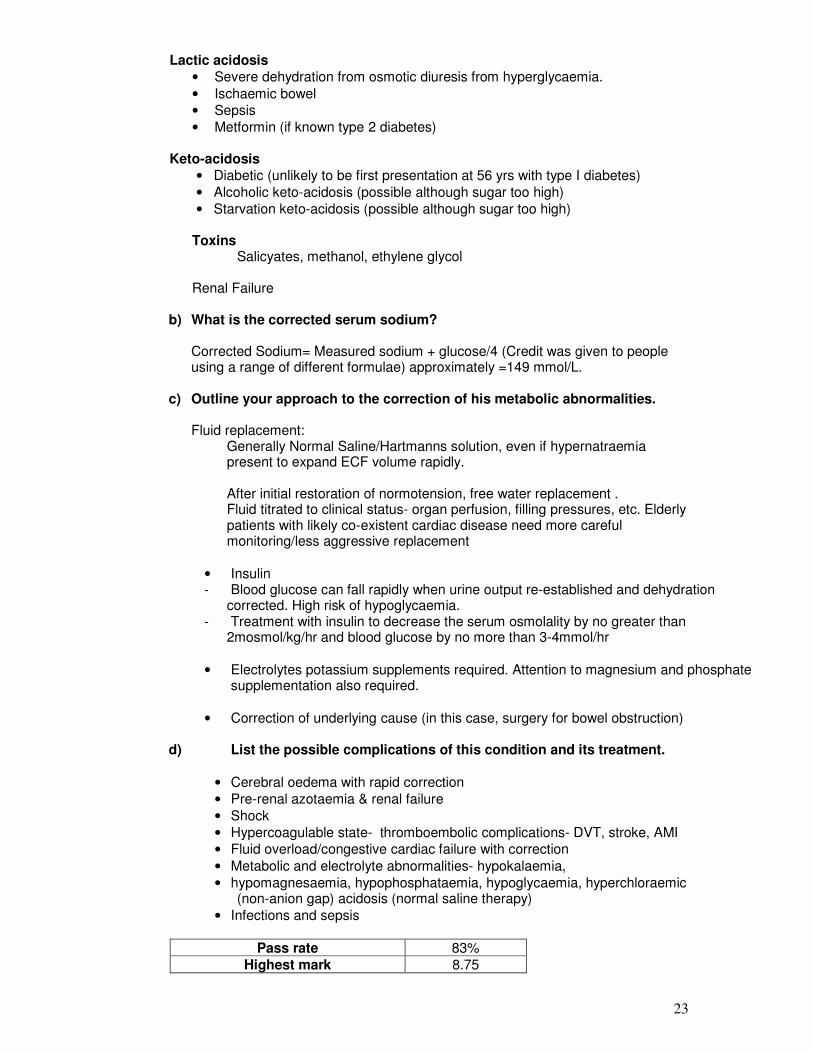
23
Lactic acidosis • Severe dehydration from osmotic diuresis from hyperglycaemia.
• Ischaemic bowel
• Sepsis
• Metformin (if known type 2 diabetes)
Keto-acidosis • Diabetic (unlikely to be first presentation at 56 yrs with type I diabetes)
• Alcoholic keto-acidosis (possible although sugar too high)
• Starvation keto-acidosis (possible although sugar too high)
Toxins Salicyates, methanol, ethylene glycol
Renal Failure
b) What is the corrected serum sodium? Corrected Sodium= Measured sodium + glucose/4 (Credit was given to people using a range of different formulae) approximately =149 mmol/L.
c) Outline your approach to the correction of his metabolic abnormalities.
Fluid replacement:
Generally Normal Saline/Hartmanns solution, even if hypernatraemia present to expand ECF volume rapidly. After initial restoration of normotension, free water replacement . Fluid titrated to clinical status- organ perfusion, filling pressures, etc. Elderly patients with likely co-existent cardiac disease need more careful monitoring/less aggressive replacement
• Insulin - Blood glucose can fall rapidly when urine output re-established and dehydration
corrected. High risk of hypoglycaemia. - Treatment with insulin to decrease the serum osmolality by no greater than
2mosmol/kg/hr and blood glucose by no more than 3-4mmol/hr
• Electrolytes potassium supplements required. Attention to magnesium and phosphate supplementation also required.
• Correction of underlying cause (in this case, surgery for bowel obstruction) d) List the possible complications of this condition and its treatment.
• Cerebral oedema with rapid correction
• Pre-renal azotaemia & renal failure
• Shock
• Hypercoagulable state- thromboembolic complications- DVT, stroke, AMI
• Fluid overload/congestive cardiac failure with correction
• Metabolic and electrolyte abnormalities- hypokalaemia,
• hypomagnesaemia, hypophosphataemia, hypoglycaemia, hyperchloraemic (non-anion gap) acidosis (normal saline therapy)
• Infections and sepsis
Pass rate 83%
Highest mark 8.75
24
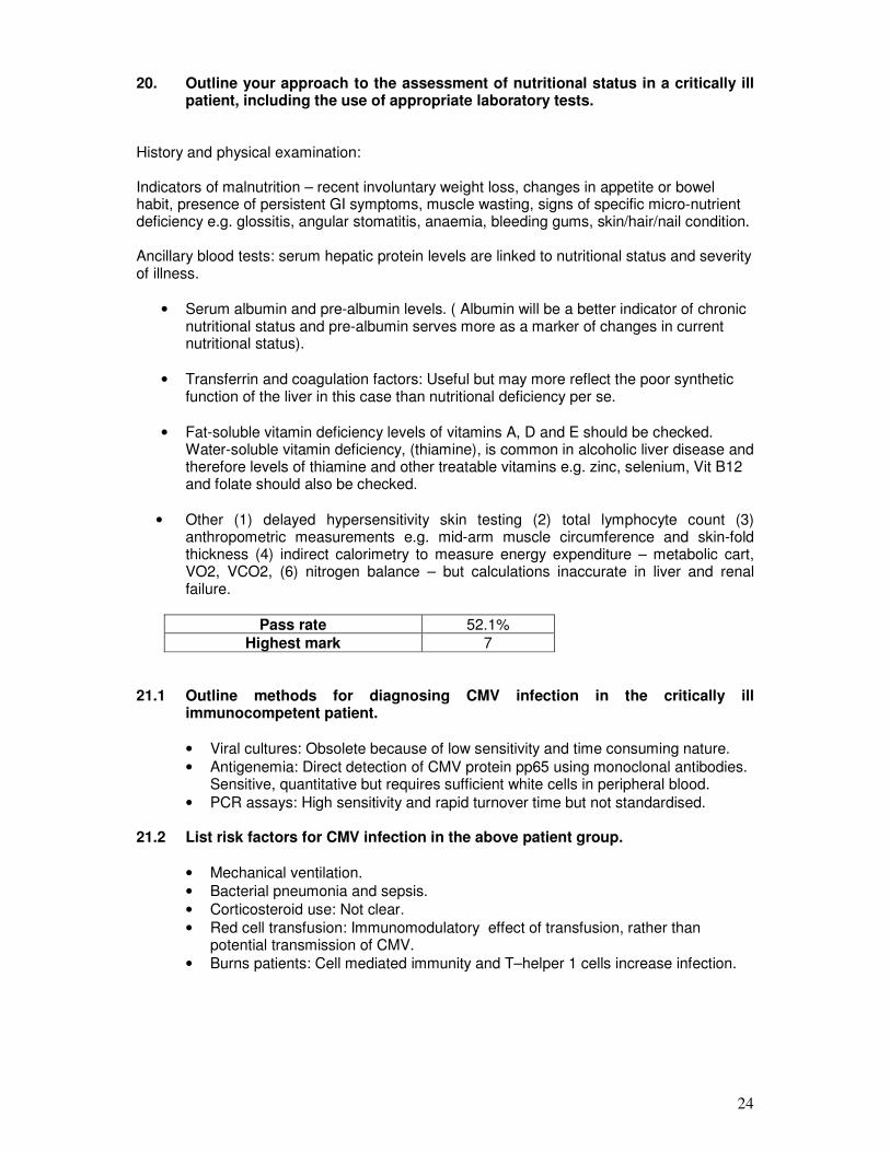
20. Outline your approach to the assessment of nutritional status in a critically ill
patient, including the use of appropriate laboratory tests. History and physical examination: Indicators of malnutrition – recent involuntary weight loss, changes in appetite or bowel habit, presence of persistent GI symptoms, muscle wasting, signs of specific micro-nutrient deficiency e.g. glossitis, angular stomatitis, anaemia, bleeding gums, skin/hair/nail condition. Ancillary blood tests: serum hepatic protein levels are linked to nutritional status and severity of illness.
• Serum albumin and pre-albumin levels. ( Albumin will be a better indicator of chronic nutritional status and pre-albumin serves more as a marker of changes in current nutritional status).
• Transferrin and coagulation factors: Useful but may more reflect the poor synthetic function of the liver in this case than nutritional deficiency per se.
• Fat-soluble vitamin deficiency levels of vitamins A, D and E should be checked. Water-soluble vitamin deficiency, (thiamine), is common in alcoholic liver disease and therefore levels of thiamine and other treatable vitamins e.g. zinc, selenium, Vit B12 and folate should also be checked.
• Other (1) delayed hypersensitivity skin testing (2) total lymphocyte count (3) anthropometric measurements e.g. mid-arm muscle circumference and skin-fold thickness (4) indirect calorimetry to measure energy expenditure – metabolic cart, VO2, VCO2, (6) nitrogen balance – but calculations inaccurate in liver and renal failure.
Pass rate 52.1%
Highest mark 7 21.1 Outline methods for diagnosing CMV infection in the critically ill
immunocompetent patient.
• Viral cultures: Obsolete because of low sensitivity and time consuming nature.
• Antigenemia: Direct detection of CMV protein pp65 using monoclonal antibodies. Sensitive, quantitative but requires sufficient white cells in peripheral blood.
• PCR assays: High sensitivity and rapid turnover time but not standardised. 21.2 List risk factors for CMV infection in the above patient group.
• Mechanical ventilation.
• Bacterial pneumonia and sepsis.
• Corticosteroid use: Not clear.
• Red cell transfusion: Immunomodulatory effect of transfusion, rather than potential transmission of CMV.
• Burns patients: Cell mediated immunity and T–helper 1 cells increase infection.
25
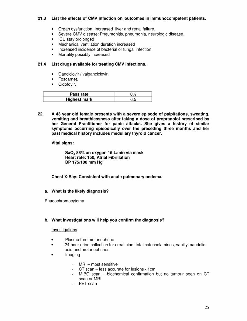
21.3 List the effects of CMV infection on outcomes in immunocompetent patients.
• Organ dysfunction: Increased liver and renal failure.
• Severe CMV disease: Pneumonitis, pneumonia, neurologic disease.
• ICU stay prolonged
• Mechanical ventilation duration increased
• Increased incidence of bacterial or fungal infection
• Mortality possibly increased 21.4 List drugs available for treating CMV infections.
• Ganciclovir / valganciclovir.
• Foscarnet.
• Cidofovir.
Pass rate 8%
Highest mark 6.5 22. A 43 year old female presents with a severe episode of palpitations, sweating,
vomiting and breathlessness after taking a dose of propranolol prescribed by her General Practitioner for panic attacks. She gives a history of similar symptoms occurring episodically over the preceding three months and her past medical history includes medullary thyroid cancer.
Vital signs:
SaO2 88% on oxygen 15 L/min via mask Heart rate: 150, Atrial Fibrillation BP 175/100 mm Hg
Chest X-Ray: Consistent with acute pulmonary oedema.
a. What is the likely diagnosis?
Phaeochromocytoma
b. What investigations will help you confirm the diagnosis?
Investigations
• Plasma free metanephrine
• 24 hour urine collection for creatinine, total catecholamines, vanillylmandelic acid and metanephrines
• Imaging
- MRI – most sensitive - CT scan – less accurate for lesions <1cm - MIBG scan – biochemical confirmation but no tumour seen on CT
scan or MRI - PET scan
26
c. Outline your immediate management of this patient.
• Admission to ICU or HDU for close monitoring
• Increase inspired oxygen concentration
• Start alpha blockade with IV phentolamine to control BP acutely and start phenoxybenzamine orally. Rate control of AF with calcium channel blocker
• Once alpha blockade established, beta blockade can be added
• IV fluid replacement as vasodilation occurs to normalise blood volume
• Some authorities recommend magnesium sulphate infusion
• Screen for myocardial damage with serial troponins, ECG and echo. Echo may show Takutsubo type abnormality
d. List four complications of this condition.
• Malignancy
• Death
• Myocardial infarction
• Arrhythmias
• Seizures
• Stroke
Pass rate 58%
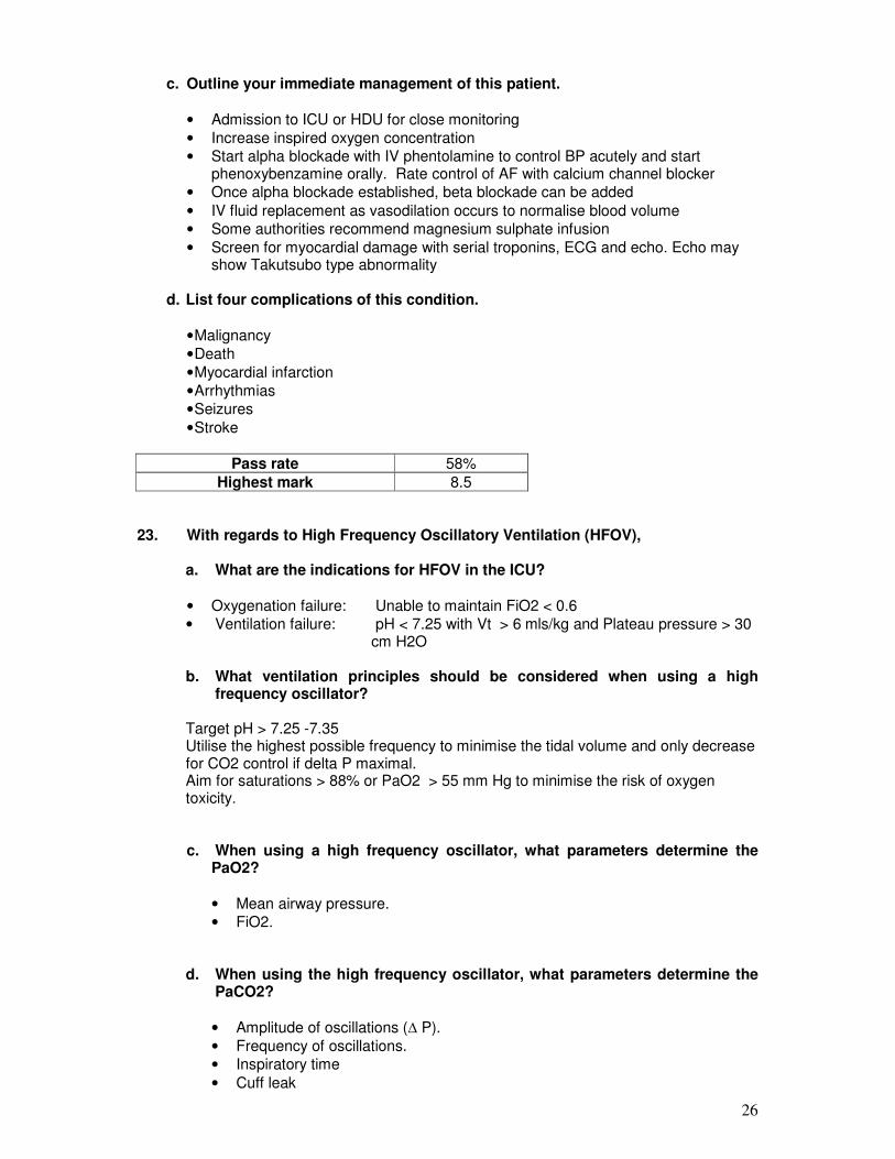
Highest mark 8.5 23. With regards to High Frequency Oscillatory Ventilation (HFOV),
a. What are the indications for HFOV in the ICU?
• Oxygenation failure: Unable to maintain FiO2 < 0.6
• Ventilation failure: pH < 7.25 with Vt > 6 mls/kg and Plateau pressure > 30 cm H2O
b. What ventilation principles should be considered when using a high
frequency oscillator?
Target pH > 7.25 -7.35 Utilise the highest possible frequency to minimise the tidal volume and only decrease for CO2 control if delta P maximal. Aim for saturations > 88% or PaO2 > 55 mm Hg to minimise the risk of oxygen toxicity.
c. When using a high frequency oscillator, what parameters determine the PaO2?
• Mean airway pressure.
• FiO2. d. When using the high frequency oscillator, what parameters determine the
PaCO2?
• Amplitude of oscillations (∆ P).
• Frequency of oscillations.
• Inspiratory time
• Cuff leak
27
e. Briefly outline the mechanisms of gas transport during HFOV.
Gas transport during HFOV is thought to occur via
• Bulk flow of gas in alveolar units close to proximal airways
• Asymmetrical velocity profiles and Taylor dispersion.
• In addition, asymmetrical filling of adjacent alveoli (termed pendelluft) due to differing emptying times, collateral ventilation through non-airway connections and cardiogenic mixing are other postulated mechanisms.
Pass rate 25%
Highest mark 8
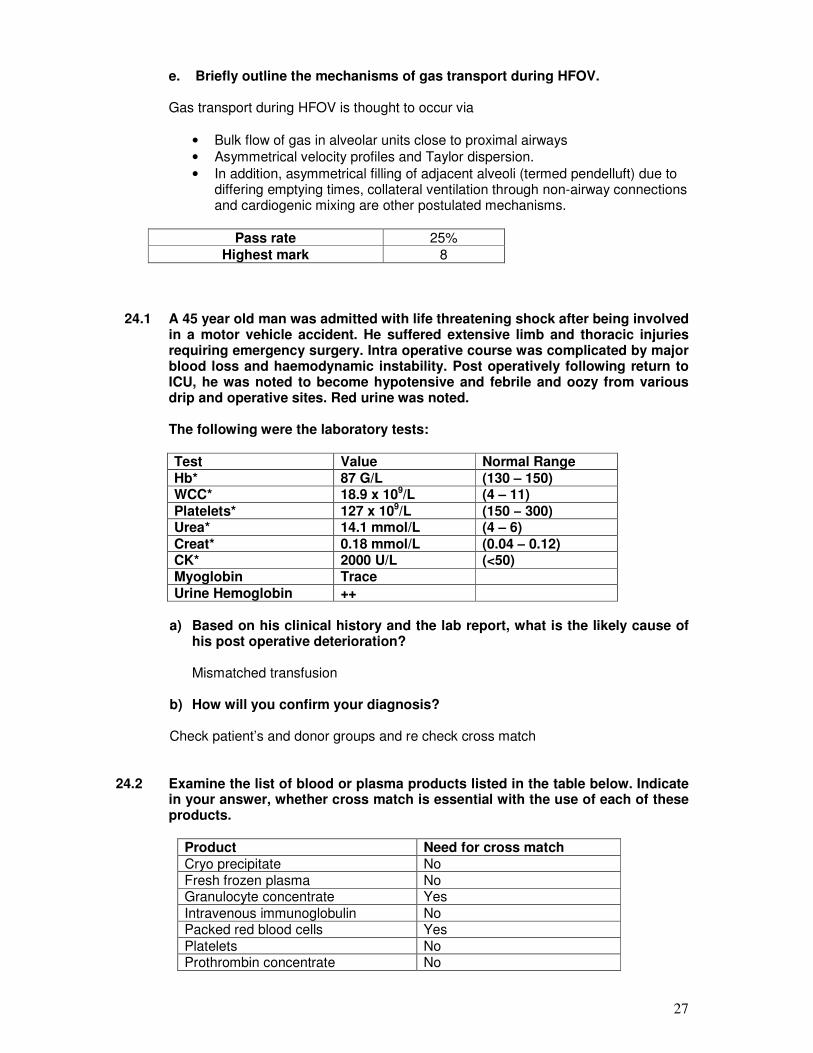
24.1 A 45 year old man was admitted with life threatening shock after being involved
in a motor vehicle accident. He suffered extensive limb and thoracic injuries requiring emergency surgery. Intra operative course was complicated by major blood loss and haemodynamic instability. Post operatively following return to ICU, he was noted to become hypotensive and febrile and oozy from various drip and operative sites. Red urine was noted.
The following were the laboratory tests:
Test Value Normal Range Hb* 87 G/L (130 – 150) WCC* 18.9 x 109/L (4 – 11) Platelets* 127 x 109/L (150 – 300) Urea* 14.1 mmol/L (4 – 6) Creat* 0.18 mmol/L (0.04 – 0.12) CK* 2000 U/L (<50) Myoglobin Trace Urine Hemoglobin ++
a) Based on his clinical history and the lab report, what is the likely cause of
his post operative deterioration? Mismatched transfusion
b) How will you confirm your diagnosis? Check patient’s and donor groups and re check cross match
24.2 Examine the list of blood or plasma products listed in the table below. Indicate in your answer, whether cross match is essential with the use of each of these products.
Product Need for cross match Cryo precipitate No Fresh frozen plasma No Granulocyte concentrate Yes
Intravenous immunoglobulin No Packed red blood cells Yes
Platelets No Prothrombin concentrate No
28
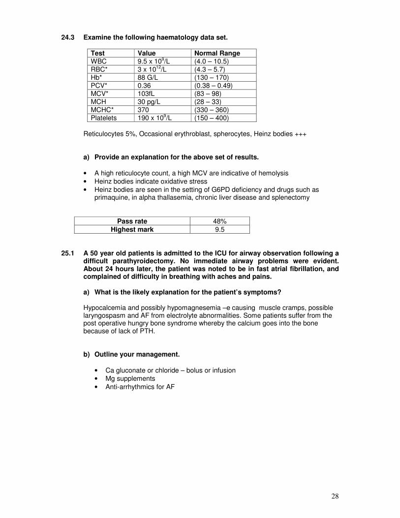
24.3 Examine the following haematology data set.
Test Value Normal Range WBC 9.5 x 10⁹/L (4.0 – 10.5) RBC* 3 x 1012/L (4.3 – 5.7) Hb* 88 G/L (130 – 170) PCV* 0.36 (0.38 – 0.49) MCV* 103fL (83 – 98) MCH 30 pg/L (28 – 33) MCHC* 370 (330 – 360) Platelets 190 x 109/L (150 – 400)
Reticulocytes 5%, Occasional erythroblast, spherocytes, Heinz bodies +++
a) Provide an explanation for the above set of results.
• A high reticulocyte count, a high MCV are indicative of hemolysis
• Heinz bodies indicate oxidative stress
• Heinz bodies are seen in the setting of G6PD deficiency and drugs such as primaquine, in alpha thallasemia, chronic liver disease and splenectomy
Pass rate 48%
Highest mark 9.5
25.1 A 50 year old patients is admitted to the ICU for airway observation following a
difficult parathyroidectomy. No immediate airway problems were evident. About 24 hours later, the patient was noted to be in fast atrial fibrillation, and complained of difficulty in breathing with aches and pains.
a) What is the likely explanation for the patient’s symptoms?
Hypocalcemia and possibly hypomagnesemia –e causing muscle cramps, possible laryngospasm and AF from electrolyte abnormalities. Some patients suffer from the post operative hungry bone syndrome whereby the calcium goes into the bone because of lack of PTH.
b) Outline your management.
• Ca gluconate or chloride – bolus or infusion
• Mg supplements
• Anti-arrhythmics for AF
29
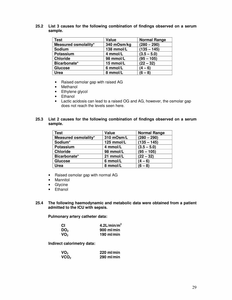
25.2 List 3 causes for the following combination of findings observed on a serum
sample.
Test Value Normal Range Measured osmolality* 340 mOsm/kg (280 – 290) Sodium 138 mmol/L (135 – 145) Potassium 4 mmol/L (3.5 – 5.0) Chloride 98 mmol/L (95 – 105) Bicarbonate* 15 mmol/L (22 – 32) Glucose 6 mmol/L (4 – 6) Urea 8 mmol/L (6 – 8)
• Raised osmolar gap with raised AG
• Methanol
• Ethylene glycol
• Ethanol
• Lactic acidosis can lead to a raised OG and AG, however, the osmolar gap does not reach the levels seen here.
25.3 List 2 causes for the following combination of findings observed on a serum
sample.
Test Value Normal Range Measured osmolality* 310 mOsm/L (280 – 290) Sodium* 125 mmol/L (135 – 145) Potassium 4 mmol/L (3.5 – 5.0) Chloride 98 mmol/L (95 – 105) Bicarbonate* 21 mmol/L (22 – 32) Glucose 6 mmol/L (4 – 6) Urea 8 mmol/L (6 – 8)
• Raised osmolar gap with normal AG
• Mannitol
• Glycine
• Ethanol 25.4 The following haemodynamic and metabolic data were obtained from a patient
admitted to the ICU with sepsis.
Pulmonary artery catheter data:
CI 4.2L/min/m2 DO2 900 ml/min VO2 190 ml/min
Indirect calorimetry data:
VO2 220 ml/min VCO2 290 ml/min
30
a) Why is the VO2 different between the two methods? (Assume no
measurement errors).
Indirect calorimetry also measures lung oxygen consumption.
b) What changes in patient management will you consider based on the indirect calorimetry data?
A high RQ suggests excess carbohydrate load. Reduce caloric intake and consider changing to a higher fat intake.
Pass rate 56%
Highest mark 7.25
26. You are called to assess a 38 year old female with respiratory failure in the
Emergency Department. This is her first pregnancy and she is 28 weeks pregnant after several attempts at IVF. She is positive for Swine-Origin Influenza Virus (H1N1). Arterial blood gas on a FiO2 of 0.8 shows:
pH 7.31 pCO2 48 mm Hg pO2 55 mm Hg Bicarbonate 18 mmol/L
Evaluation of the foetal heart reveals significant bradycardia.
a) Outline the specific challenges in this case that distinguish it from a similar illness in a previously healthy 38 year old male.
• Precious pregnancy in older, primiparous patient.
• Known high incidence of morbidity and mortality in mother and foetus with H1N1 Influenza infection with severe CAP.
• Requirement to work closely with specialist obstetric team and rationalising potentially conflicting priorities eg. timing of delivery of foetus.
• Anatomical and Physiological considerations during pregnancy- elevated diaphragm and decreased FRC, decreased chest wall compliance, increased risk of aspiration during intubation, pressure of gravid uterus on IVC (and aorta) decreasing venous return (and increasing afterload) in the supine position.
• Maintaining effective foeto-placental circulation while optimising maternal outcome.
• Safety of various drugs in pregnancy eg. anti-virals, sedatives.
• History of severe asthma complicating current episode of severe CAP likely to make ventilatory strategy more complex.
• Importance of keeping family members well informed of considerations and likelihood of poor foetal outcome as priority will be given to mother’s survival.
31
b) Outline your specific approach to the management of this case.
Immediate-
• Clinical scenario described requires rapid resuscitation.
• Airway- Secure early, rapid sequence induction. Anticipate difficult airway (ensure help and difficult airway equipment available.
• Breathing- Ventilate with protective lung strategy. Example of Settings – SIMV/PC, FiO2-1.0 PC to achieve Tidal Volumes of 6-8ml/kg, Low rate- 6-8/min I:E ratio 1:3-4 to allow adequate expiratory time, PEEP 10-15cm titrated to oxygenation. Close monitoring with regular blood gas evaluation. Tolerate hypercapnia (although not ideal for foetus) if poorly compliant lungs. Position at least 30 degrees head-up to optimise respiratory mechanics. Sedate heavily to minimise oxygen consumption. Neuromuscular blockade if required to facilitate ventilation.
• Circulation-. Fluid resuscitate (likely to be volume depleted) to clinical endpoints, vasoconstrictors to maintain perfusion pressure (eg MAP>60mmHg). Assessment of cardiac output if unstable haemodynamics with these measures (eg. echocardiogram, PiCCO, PA catheter, ScVO2)- High cardiac output expected due to pregnancy and infection. Inotropes if cardiac output low. Position slightly left lateral to relieve IVC compression.
• Early specialist obstetric evaluation to determine foetal condition, position of placenta and risk versus benefit of delivery of foetus may need to be considered carefully taking into consideration maternal and foetal factors.
Pass rate 54%
Highest mark 7.75
32
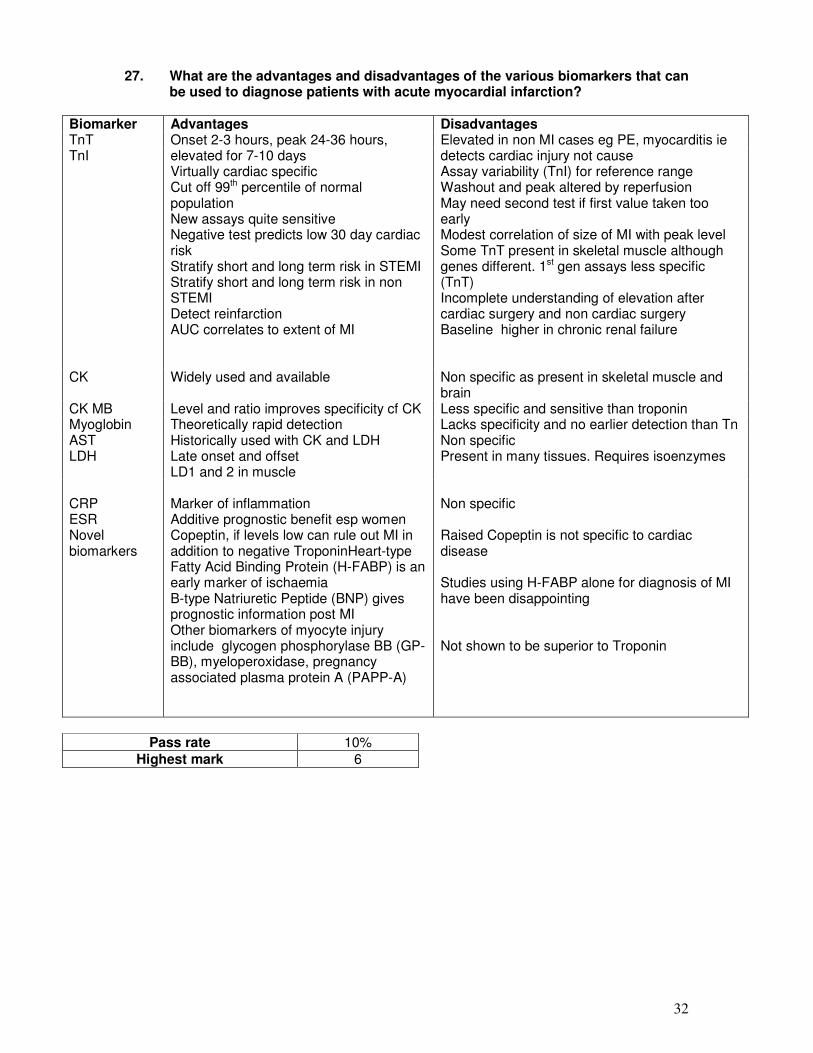
27. What are the advantages and disadvantages of the various biomarkers that can
be used to diagnose patients with acute myocardial infarction?
Biomarker Advantages Disadvantages TnT Onset 2-3 hours, peak 24-36 hours,
elevated for 7-10 days Virtually cardiac specific Cut off 99th percentile of normal population New assays quite sensitive Negative test predicts low 30 day cardiac risk Stratify short and long term risk in STEMI Stratify short and long term risk in non STEMI Detect reinfarction AUC correlates to extent of MI
Elevated in non MI cases eg PE, myocarditis ie detects cardiac injury not cause Assay variability (TnI) for reference range Washout and peak altered by reperfusion May need second test if first value taken too early Modest correlation of size of MI with peak level Some TnT present in skeletal muscle although genes different. 1st gen assays less specific (TnT) Incomplete understanding of elevation after cardiac surgery and non cardiac surgery Baseline higher in chronic renal failure
TnI
CK Widely used and available Non specific as present in skeletal muscle and brain
CK MB Level and ratio improves specificity cf CK Less specific and sensitive than troponin Myoglobin Theoretically rapid detection Lacks specificity and no earlier detection than Tn AST Historically used with CK and LDH Non specific LDH Late onset and offset
LD1 and 2 in muscle Present in many tissues. Requires isoenzymes
CRP ESR Novel biomarkers
Marker of inflammation Additive prognostic benefit esp women Copeptin, if levels low can rule out MI in addition to negative TroponinHeart-type Fatty Acid Binding Protein (H-FABP) is an early marker of ischaemia B-type Natriuretic Peptide (BNP) gives prognostic information post MI Other biomarkers of myocyte injury include glycogen phosphorylase BB (GP-BB), myeloperoxidase, pregnancy associated plasma protein A (PAPP-A)
Non specific Raised Copeptin is not specific to cardiac disease Studies using H-FABP alone for diagnosis of MI have been disappointing Not shown to be superior to Troponin
Pass rate 10%
Highest mark 6
33
28. You have been asked to review a three year old child who was trapped in a
house fire and is now in the Paediatric Emergency Department. There is no history available from the child’s carer and you observe that the child is drowsy and confused and has a persistent cough. His heart rate is 140 beats per minute, blood pressure 70/40 mmHg. Respiratory rate is 54 breaths per minute and oxygen saturations are 94 % on high flow oxygen via a non re-breather mask.
a. Briefly outline the initial priorities in management.
• Resuscitation including primary and secondary survey
• Assessment and management of potential airway burn injury – mention consideration of early intubation,
• Obtain large bore iv access and administration of fluid bolus (20mls/kg) for probable hypovolaemic shock- mention that groins are usually spared in burns and are a good site for clean skin vas cath access.
• Look for signs of traumatic injury and assess extent of body surface area and depth of burn
• Awareness of risk of hypothermia
• Seek collateral history for past medical history and medication history and history of acute events
b. List the features from the history and your examination of this child which would suggest a significant airway injury.
• Burns occurring in a closed space
• Cough, stridor, hoarseness of voice
• Burns to face, lips, mouth, pharynx or nasal mucosa
• Soot in sputum, nose or mouth
• Hypoxaemia or
• Dyspnoea
• Carboxyhaemoglobin levels > 2%
• Acute confusional state or depressed level of consciousness
c. List 4 likely causes for his altered conscious state.
• Traumatic brain injury
• Carbon monoxide / CN – poisoning
• Hypoxic insult
• Other pathology precipitating loss of consciousness eg seizure-related, hypoglycaemia, drug ingestion
Pass rate 85%
Highest mark 8.75
34
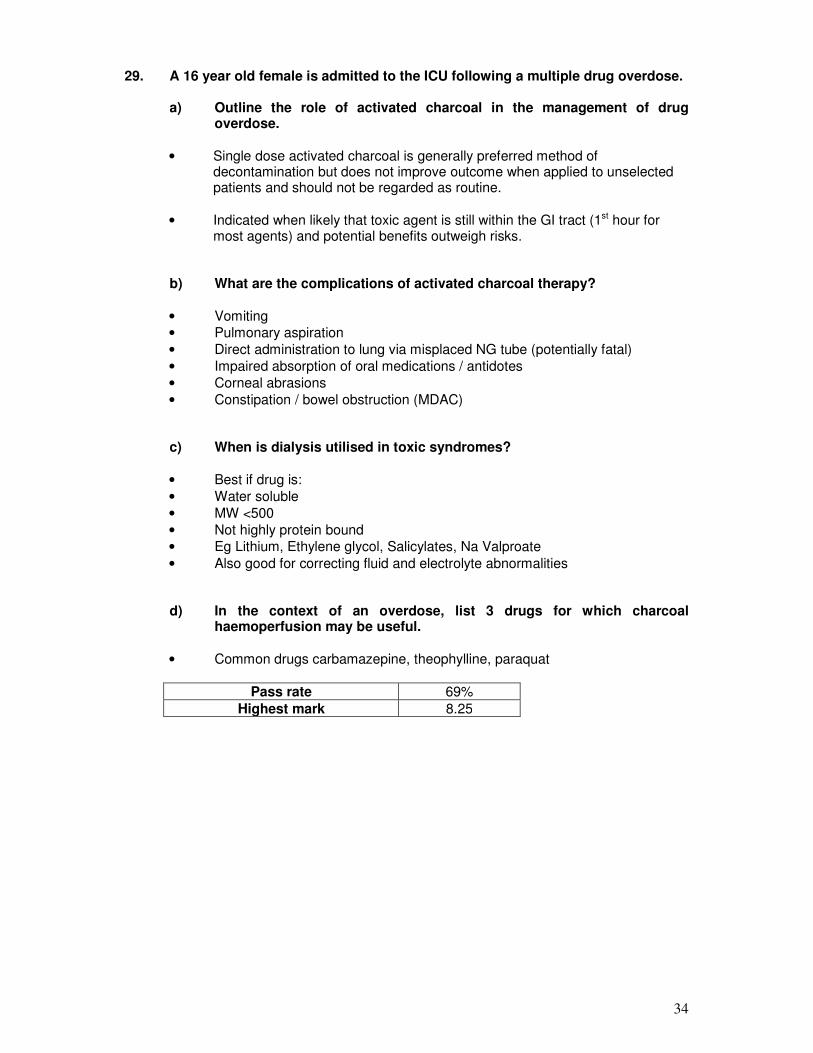
29. A 16 year old female is admitted to the ICU following a multiple drug overdose.
a) Outline the role of activated charcoal in the management of drug overdose.
• Single dose activated charcoal is generally preferred method of
decontamination but does not improve outcome when applied to unselected patients and should not be regarded as routine.
• Indicated when likely that toxic agent is still within the GI tract (1st hour for most agents) and potential benefits outweigh risks.
b) What are the complications of activated charcoal therapy?
• Vomiting
• Pulmonary aspiration
• Direct administration to lung via misplaced NG tube (potentially fatal)
• Impaired absorption of oral medications / antidotes
• Corneal abrasions
• Constipation / bowel obstruction (MDAC)
c) When is dialysis utilised in toxic syndromes?
• Best if drug is:
• Water soluble
• MW <500
• Not highly protein bound
• Eg Lithium, Ethylene glycol, Salicylates, Na Valproate
• Also good for correcting fluid and electrolyte abnormalities
d) In the context of an overdose, list 3 drugs for which charcoal haemoperfusion may be useful.
• Common drugs carbamazepine, theophylline, paraquat
Pass rate 69%
Highest mark 8.25
35
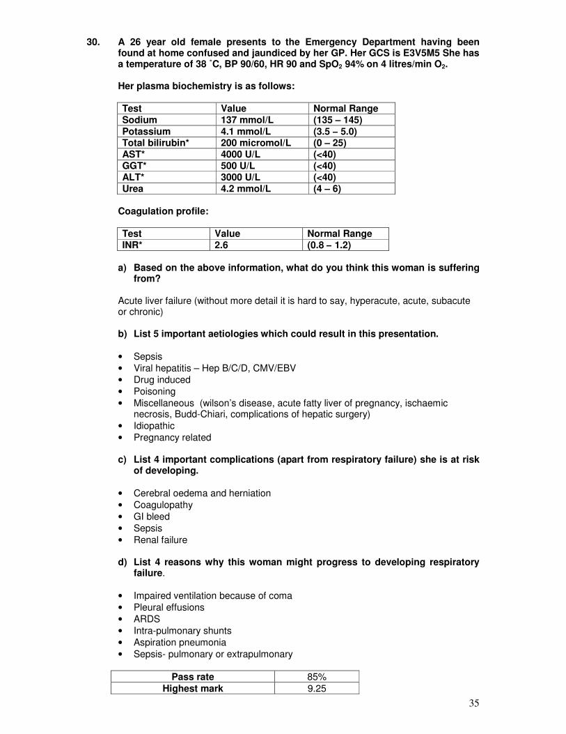
30. A 26 year old female presents to the Emergency Department having been found at home confused and jaundiced by her GP. Her GCS is E3V5M5 She has a temperature of 38 ˚C, BP 90/60, HR 90 and SpO2 94% on 4 litres/min O2.
Her plasma biochemistry is as follows: Test Value Normal Range Sodium 137 mmol/L (135 – 145) Potassium 4.1 mmol/L (3.5 – 5.0) Total bilirubin* 200 micromol/L (0 – 25) AST* 4000 U/L (<40) GGT* 500 U/L (<40) ALT* 3000 U/L (<40) Urea 4.2 mmol/L (4 – 6)
Coagulation profile:
Test Value Normal Range INR* 2.6 (0.8 – 1.2)
a) Based on the above information, what do you think this woman is suffering
from?
Acute liver failure (without more detail it is hard to say, hyperacute, acute, subacute or chronic)
b) List 5 important aetiologies which could result in this presentation.
• Sepsis
• Viral hepatitis – Hep B/C/D, CMV/EBV
• Drug induced
• Poisoning
• Miscellaneous (wilson’s disease, acute fatty liver of pregnancy, ischaemic necrosis, Budd-Chiari, complications of hepatic surgery)
• Idiopathic
• Pregnancy related
c) List 4 important complications (apart from respiratory failure) she is at risk of developing.
• Cerebral oedema and herniation
• Coagulopathy
• GI bleed
• Sepsis
• Renal failure
d) List 4 reasons why this woman might progress to developing respiratory failure.
• Impaired ventilation because of coma
• Pleural effusions
• ARDS
• Intra-pulmonary shunts
• Aspiration pneumonia
• Sepsis- pulmonary or extrapulmonary
Pass rate 85%
Highest mark 9.25
36
Hot Case Section - Royal Adelaide Hospital 1) This is an 82 year old man with diabetes and hyperlipidaemia who underwent an aortic
valve replacement and CABG 1 week ago. His pre-operative left ventricular ejection fraction was 20% and he required an intra aortic balloon pump post operatively. His post operative course was complicated by a brief asystolic arrest and he remains ventilated 1 week post operatively. Candidates were asked to assess the patient for causes of slow respiratory wean. Ongoing issues included – persistent inotrope requirement, encephalopathy, search for sepsis, consideration of diuresis +/- tracheostomy.
2) 79 year old male – D3 post Grade 5 SAH. Aneurysm coiled within 48 hrs of
presentation. Candidates asked to perform a neurological examination and asked to discuss investigations and management of SAH, spasm and hydrocephalus.
3) A 30 year old man admitted following an MVA. On presentation, awake but
hemodynamically unstable requiring an urgent trauma laparotomy. Post op complicated by new onset respiratory failure requiring reintubation and found to have H1N1. Candidates asked to discuss management of respiratory failure secondary to H1N1 and nutritional management.
4) A 40 year old lady with SLE and bilateral renal transplants admitted with high grade
fever, productive cough and respiratory failure. Issues for discussion included differential diagnosis of pneumonia in immunocompromised patients, H1N1,CMV and role of NIV in pneumonia in immunocompromised patients.
5) 63 year old patient admitted following an MVA with severe thoracic injuries. Topics for
discussion included EMST principles, flail chest, damage control surgery, DVT prophylaxis and ventilatory wean.
6) An 82 year old male admitted with NSTEMI and APO secondary to severe AS and CAD.
He required emergent cardiac surgery. Discussion centred around management of cardio-respiratory wean, post op AF and role of levosimendan.
7) A 73 year old man with DM and dialysis dependent CRF. In ICU following elective AVR
and CABG. Candidates were asked to discuss glycemic control, fluid management in dialysis dependant cardiac surgery and complications following cardiac surgery.
37
Flinders Medical Centre
1) A 65 year old male admitted with pneumonia and septic shock and MODS. Background h/o arthropathy requiring immunosuppression. Problems included: slow respiratory wean, MODS, CVVHDF, obvious features of steroid therapy and a deforming polyarthopathy
2. A 45 year old male admitted with chronic liver disease and haematemesis. The
discussion focusing on GI bleed management
3) A 40 year old man with past history of psychiatric illness admitted with seizures and hyponatremia. Failed one extubation and required nasal reintubation. Candidates asked to assess suitability for extubation. Discussion on suitability of extubation, airway assessment, assessment of power and demonstration of a systematic approach.
4) A 64 year old male admitted with altered sensorium and jaundice. Candidates asked to discuss an approach to altered sensorium in this context.
5) A 46 year old female with a past history of antiphospholipid syndrome and a mechanical MVR admitted with ascending cholangitis complicated by a cardiac arrest. Required emergent re do MVR as well as an urgent laparotomy.
Current problems included:
a) Slow respiratory wean b) Ongoing tracheostomy bleed c) CVVHDF for ARF d) MODS e) Evidence of intravascular hemolysis
6) A 76 year old male with a past history of IHD and CABG admitted with acute hyperkalemia and ARF in the contect of a recent hospitalization for pneumonia. Current issues included: VAP, RV dysfunction, and residual acute renal failure.
Queen Elizabeth Hospital
1) A 59 year old male with a previous splenectomy presented with a 2/52 h/o leg pain and
swelling following an insect bite. Current problems included aortic regurgitation , endocarditis and left above knee amputation and respiratory failure
2) A 71 year old male with previous h/o gastric cancer and liver metastases was admitted
with a parapneumonic effusion and empyema requiring a thoracotomy. Post operative course complicated by hemoptysis. Ongoing problems included cachexia, pleural effusions, recent thoracotomy, management of hemoptysis and intercostal drains
3) A 38 year old man with severe respiratory failure secondary to H1N1 and complicated
by ARDS.
38
Viva Section
VIVA 1 A 55 year old lady presents to the Emergency Department irritable, confused and complaining of a severe headache. Her husband reports that she had been very drowsy since earlier that day when she was heard to suddenly cry out. She was apparently well the day before. Examination findings include Temp 37.8 deg C, HR 95, BP 150/80mmHg, RR 24, Pulse oximetry 91%, GCS E2, V3, M6. List the most likely differential diagnoses. Pass rate: 79%, Highest mark: 75% VIVA 2 A 70 year old man is admitted to ICU from theatre following routine coronary artery bypass grafting. Surgery was uneventful. He is brought back intubated and ventilated. Following handover, you are going to continue ventilation on the ICU ventilator. What controls do you do you need to set and how would you determine the value of the settings? Pass rate: 72%, Highest mark: 98% VIVA 3 A 25 year old cyclist fell of his cycle at high speed and was found unconscious. In the Emergency Department initial observations are pulse 108/minute, blood pressure 100/60 mm Hg, saturation 89% on 15 litres/min oxygen via face mask. He is unconscious, tachypnoeic but has symmetrical chest movement, what would be the immediate management of this patient? What would be the immediate management of this patient? Pass rate: 100%, Highest mark: 95% VIVA 4 A 74-yr-old man with a past history of hypertension and NIDDM has just undergone a 3-vessel graft and aortic valve replacement for aortic stenosis. You are on duty when the patient is transferred to ICU postoperatively. On admission, you note the following vital signs and haemodynamic observations: Temperature 35.5 0 C Heart rate 66 bpm, sinus rhythm Serum HCO3 19 mEq/L Systemic blood pressure 94/60 mm Hg Serum lactate 2.4 mmol/L Pulmonary artery pressure 22/10 mm Hg Cardiac index 1.7 L/min/m2 Systemic vascular resistance index 1600 dynes. sec. cm-5
What would you like to know about his pre-operative health status and work-up that would influence your post-operative management? Pass rate: 86%, Highest mark: 83%
39
VIVA 5 You are asked to see a 72 year old man who is 5 days post elective left hemicolectomy for cancer. His co-morbidities include COPD, ischaemic heart disease and rheumatoid arthritis. On assessment he is drowsy but responsive to voice. He is poorly perfused, HR =120, BP = 100/60. His heart sounds and lung fields are normal; his abdomen is diffusely tender and distended. He has intravenous access, and has been commenced on fluids and O2 . Please outline your most likely diagnosis and a differential diagnosis for this patient’s deterioration. Pass rate: 79%, Highest mark: 87% COMMUNICATION STATION You have taken over the care of Matthew, a 28 year old male, admitted to the Intensive Care Unit overnight following a motor bike accident. His GCS was 3 at the scene and was intubated and ventilated by the paramedics. He was noted to have dilated non reactive pupils, and feeble respirations, prior to intubation. An urgent CT head showed severe diffuse axonal injury with a 1.5 cm midline shift, evidence of transtentorial herniation but no drainable intracranial collection. The consensus opinion of the neurosurgical and ICU team was that the outcome was dismal and would not be altered by ICP monitoring and neuro protective measures. The findings on your clinical assessment the following morning are as follows: GCS -3, Pupils 5mm not reactive to light, absent corneal cough and gag reflexes. Spontaneous respirations present (6-8 breaths per minute). He has been off sedation, analgesics and neuromuscular blocking agents since admission. The patient’s next of kin (Pat) is waiting to talk to you. The nursing staff informs you that while Pat was briefed by senior registrar (John) over night about the poor outcome, Pat was hoping for a miracle. Pass rate: 86%, Highest mark: 74% PROCEDURE STATION You are working as an ICU specialist in a small regional hospital. You are called to give urgent assistance with a 65 year old male who has presented to the Emergency Department with increasing shortness of breath one week after discharge from a Metropolitan hospital following an apparently uncomplicated cardiac surgery. He has rapidly deteriorated. A junior registrar and nurse are in attendance. How will you respond to this crisis? The rest of the question focused on the differential diagnosis of a cardiac arrest post cardiac surgery, approach to BLS and pericardiocentesis. Pass rate: 70%, Highest mark: 85% RADIOLOGY STATION There were 4 Chest X-Rays and 2 CT scans for interpretation. Problems included identification of artifacts on the X-Rays, lung collapse, and pneumothoraces. CT scans included a carotid territory infarct and a lung CT showing extensive bilateral interstitial pneumoninits. Pass rate: 72%, Highest mark: 75%