Embed Size (px)
Citation preview
i
EVALUATING THE OCCURRENCE OF SCHISTOSOMA HAEMATOBIUM AND
GEOHELMINTHES INFECTION IN RESIDENTS OF TWO VILLAGES IN
MSAMBWENI DISTRICT OF COAST PROVINCE, KENYA.
Matonge, Paul Mwambua B.Ed. Sc.
Registration Number I56/CE/10718/06
A thesis submitted in partial fulfillment of the requirements for the award of the degree of
Master of Science in Applied Medical Parasitology in the School of Pure and Applied
Sciences of Kenyatta University.
June 2013
ii
DECLARATION
I declare that this is my original work and has not been submitted for the award of degree
in any other University or for any other award.
Mr. Paul M. Matonge
Sign ………………………………………… Date…………………………………
We Kenyatta university supervisors declare that the candidate undertook the work under
our supervision and that the thesis is submitted for examination with our approval.
Dr. Lucy Kamau
Department of Zoological Sciences,
Kenyatta University,
P.O. Box 43844-00100
Nairobi.
Sign ………………………………………… Date…………………………………
Dr. Margaret Muturi
Department of Medical Laboratory Sciences,
Kenyatta University,
P. O. Box 43844-00100
Nairobi.
Sign ………………………………………… Date…………………………………
iii
DEDICATION
This thesis is dedicated to my family and friends;
The ones that have stood with me all the way and those I have lost along the way.
iv
ACKNOWLEDGEMENT
I am grateful to my supervisors; Dr. Lucy Kamau and Dr. Margaret Muturi, who have
been very helpful and encouraging throughout the study. Their patient guidance equipped
me with the necessary knowledge and skills required in the study. The assistance of
several friends with the collection of field data is acknowledged. Particular mention is
made of Ephraim Adel Odek of the department of vector borne diseases, and all medical
technicians at Msambweni district hospital.
I thank Dr. Bernard Kirui and Mr. Joseph Langat both of Kenya Marine and Fisheries
Research Institute, and Mr. Isaack Njagi of Zoological Sciences Department of Kenyatta
University for their valuable support. I would also like to thank my family and friends for
their support and understanding.
My sincere gratitude goes to Case Western Reserve University for funding part of this
research.
v
TABLE OF CONTENTS
DECLARATION ................................................................................................................ ii
DEDICATION ................................................................................................................... iii
ACKNOWLEDGEMENT ................................................................................................. iv
TABLE OF CONTENTS .................................................................................................... v
LIST OF TABLES ........................................................................................................... viii
LIST OF FIGURES ........................................................................................................... ix
ABBREVIATIONS AND ACRONYMS ........................................................................... x
ABSTRACT ....................................................................................................................... xi
CHAPTER ONE: INTRODUCTION ................................................................................. 1
1.1 Background ............................................................................................................... 1
1.2 Statement of the problem .......................................................................................... 2
1.3 Justification ............................................................................................................... 3
1.4 Research questions .................................................................................................... 4
1.5 Hypothesis................................................................................................................. 4
1.6 Objectives of the study.............................................................................................. 5
1.6.1General objective ................................................................................................ 5
1.6.2 Specific objectives ............................................................................................. 5
CHAPTER TWO: LITERATURE REVIEW ..................................................................... 6
2.1 Helminthes infections in humans .............................................................................. 6
2.2 Hookworm disease .................................................................................................... 7
2.3 Ascariasis .................................................................................................................. 9
2.4 Schistosomiasis ....................................................................................................... 11
2.5 Control of helminthes infections ............................................................................. 14
2.5.1 Anthelmintic drug treatment ............................................................................ 14
2.5.2 Improved sanitation ......................................................................................... 15
vi
2.5.3 Health education .............................................................................................. 15
2.5.4 Other control measures .................................................................................... 16
CHAPTER THREE: MATERIALS AND METHODS ................................................... 17
3.1 Study area................................................................................................................ 17
3.2 Study design ............................................................................................................ 18
3.3 Study population ..................................................................................................... 19
3.4 Data collection ........................................................................................................ 19
3.5 Collection of samples .............................................................................................. 20
3.5.1 Collection of fecal samples .............................................................................. 20
3.5.2 Collection of urine samples ............................................................................. 20
3.6 Determining of hematuria status ............................................................................. 21
3.7 Preparation and examination of urine filters ........................................................... 21
3.8 Examination of stool samples for geohelminthes ................................................... 22
3.8.1 Microscopic examination of feacal specimens ................................................ 22
3.8.2 Quantification of hookworm eggs ....................................................................... 22
3.9 Determination of hemoglobin levels .............................................................. 23
3.10 Data analysis ......................................................................................................... 24
3.11 Permission ............................................................................................................. 24
3.12 Ethical consideration ............................................................................................. 24
3.13 Exclusion criteria .................................................................................................. 25
3.14 Confidentiality ...................................................................................................... 25
CHAPTER FOUR: RESULTS ......................................................................................... 26
4.1 Prevalence of parasitic infections ........................................................................... 26
4.2 Co-infection with urinary schistosomiasis and soil transmitted helminthes ........... 27
4.3 Occurrence of parasitic infection in different age groups and sex .......................... 28
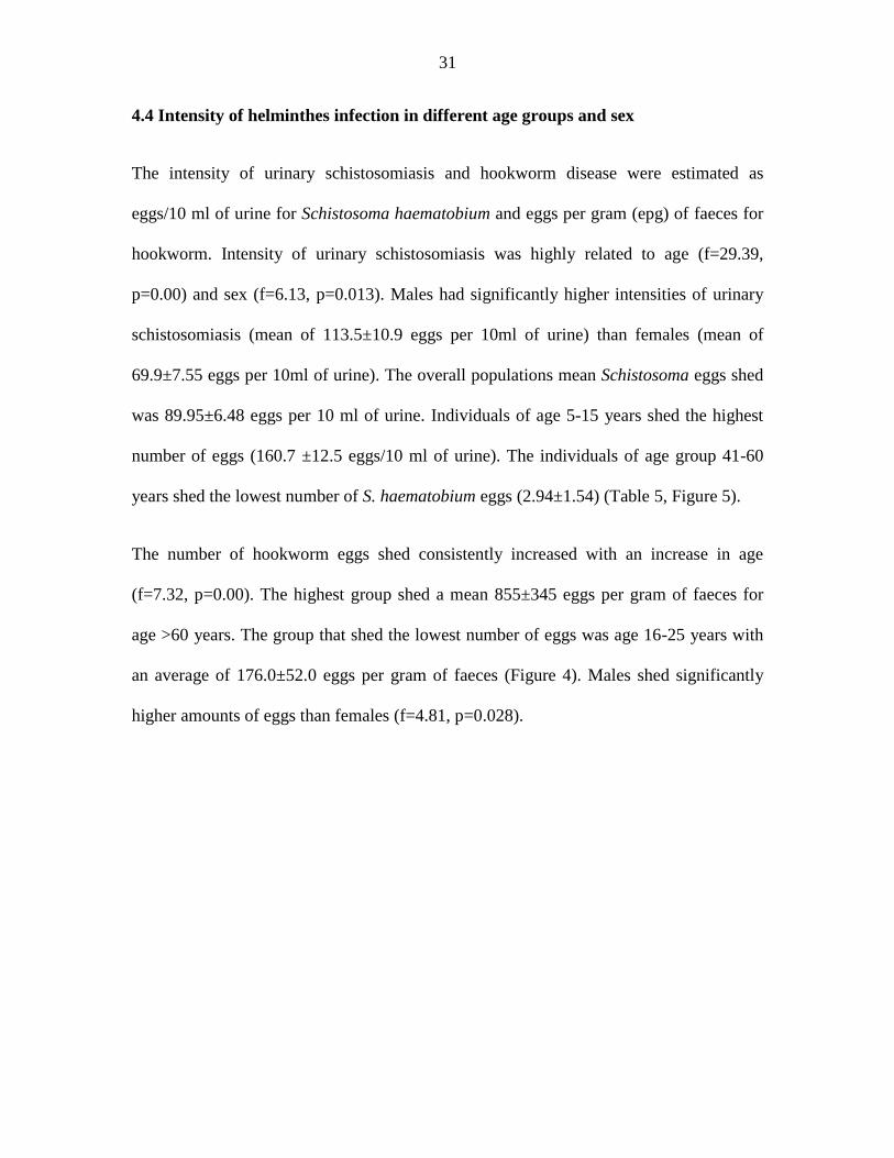
4.4 Intensity of helminthes infection in different age groups and sex .......................... 31
4.5 Schistosoma haematobium and hematuria .............................................................. 34
4.6 Hookworm infection and haemoglobin level .......................................................... 35
vii
CHAPTER FIVE: DISCUSION ....................................................................................... 37
5.1 Occurrence of parasitic infections .......................................................................... 37
5.2 Occurrence and intensity of parasitic infection in different age groups and sex .... 39
5.3 Occurrence of trichuriasis in the two villages......................................................... 42
5.4 Schistosoma haematobium and hematuria .............................................................. 43
5.5 Hookworm disease intensity and anaemia .............................................................. 44
CHAPTER SIX: CONCLUSION AND RECOMMENDATIONS .................................. 45
6.1 Conclusions ............................................................................................................. 45
6.2 Recommendations .......................................................................................... 46
REFERENCES ................................................................................................................. 49
APPENDICES .................................................................................................................. 57
viii
LIST OF TABLES
Table 1. Percentage occurrence of urinary schistosomiasis and soil transmitted
helminthes in Mwagundu and Bomani. ............................................................................ 27
Table 2. Percentage occurrence of soil transmitted infection in Schistosoma haematobium
infected individuals. .......................................................................................................... 28
Table 3. Percentage prevalence of parasitic diseases by age. ........................................... 29
Table 4. Percentage occurrence S. haematobium and geohelminthes infections by age in
males and females. ............................................................................................................ 30
Table 5. Mean eggs shed expressed as eggs per 10 ml of urine for Schistosoma
haematobium and eggs per gram of faeces for hookworm in different age groups. ......... 32
Table 6. Proportion of eggs shed by individuals with different intensities of Urinary
schistosomiasis and hookworm disease. ........................................................................... 34
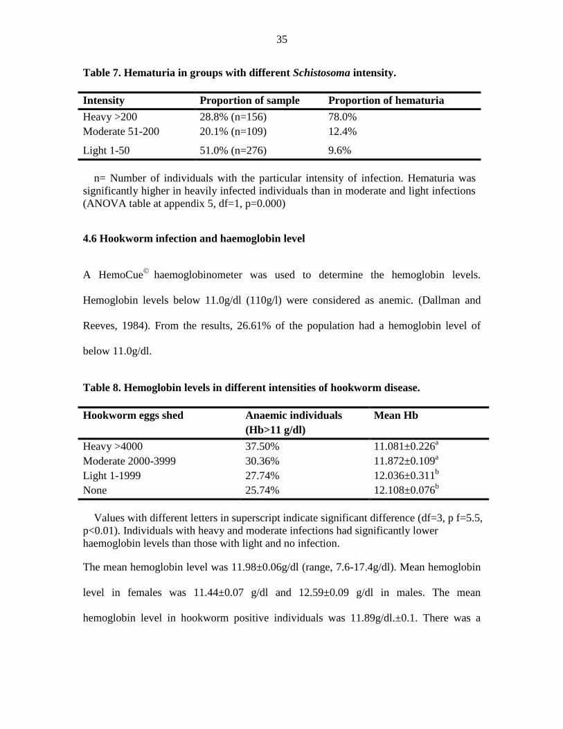
Table 7. Hematuria in groups with different Schistosoma intensity. ................................ 35
Table 8. Hemoglobin levels in different intensities of hookworm disease. ...................... 35
ix
LIST OF FIGURES
Figure 1. Life cycle of Ascaris lumbricoides. .................................................................. 10
Figure 2. Water masses in Msambweni division where Mwagundu and Bomani are
located. .............................................................................................................................. 18
Figure 3. Percentage prevalence of parasitic diseases in different age groups. ................ 29
Figure 4. Scatter plots showing average Schistosoma and hookworm eggs shed versus
age. .................................................................................................................................... 32
Figure 5. Plot showing eggs of Schistosoma haematobium shed by individuals of
different age groups. ......................................................................................................... 32
x
ABBREVIATIONS AND ACRONYMS
EPG : Eggs per Gram of Faeces
CDC : Centre for Disease Control
DALYs: Disability-Adjusted Life Years
LF : Lymphatic Filariasis
MDGs : Millennium Development Goals
NGOs : Non Governmental Organizations
NTDs : Neglected Tropical Diseases
RFC : Relative Centrifugal Force
STH : Soil Transmitted Helminthes
WHO : World Health Organization
xi
ABSTRACT
The presence of multiple concurrent infections, or polyparasitism, is quite common in
schistosomiasis endemic areas. Msambweni District in the Coast Province of Kenya is
highly endemic for Schistosoma haematobium. Previous studies showed an overall
prevalence of schistosomiasis in Msambweni District at 40% to 60%. This is a prevalence
way above the national prevalence of approximately 23%. There has been continued
support by various agencies, including the government and non-governmental
organizations (NGO’s) in providing treatment and control of the disease in the area.
However, the long term impact of these programs in the reduction of the overall
prevalence of the disease has not been fully evaluated. This survey of 1232 people aged
5-78 years in two villages in Vingujini Sub-Location Msambweni District of Coast
Province, Kenya was conducted to determine prevalence of Schistosoma haematobium
and soil transmitted helminthes infections. Urine and stool samples were collected and
examined for eggs of Schistosoma haematobium and intestinal helminthes. Haematuria
was determined using urine dip strips. Hemoglobin levels were determined to establish
the relationship between the intensity of hookworm disease and anemia. The overall
occurrence of parasitic infections were, 43.99% for Schistosoma haematobium, 29.63%
for hookworm disease, 0.49% for ascariasis and 24.59% for trichuriasis (N=1232). Only
32.71% were free from any of the parasitic infections screened. Age was related to
infection with schistosomiasis (f=95.17, p>0.01), hookworm disease (f=11.51, p= 0.010)
and trichuriasis (f=26.46, p>0.01). Infection with Schistosomiasis was highly correlated
with Trichuris infection (r=0.96, p=0.006). Intensities of Schistosomiasis and hookworm
were highly related to age (p<0.01). High intensities of Schistosoma haematobium were
associated with haematuria (f=639.99, p<0.01). Intensity with hookworm was correlated
to anemia (r=-0.091, p<0.01). Individuals with heavy and light infections were more
likely to suffer from anemia than individuals with low intensities or the non-infected
(f=5.5, p<0.01). The results of this study have shown that the problem of urinary
schistosomiasis and soil transmitted helminthes persists despite the control programs and
enhancement of these control programs is required.
1
CHAPTER ONE: INTRODUCTION
1.1 Background
Infection with soil transmitted helminthes, also known as geohelminthes and
schistosomiasis is common in developing countries. It is also common to find individuals
co-infected with schistosomiasis and soil transmitted helminthes. In areas where more
than one species of Schistosoma occurs, some individuals may be infected with more than
one species, for example Schistosoma mansoni and Schistosoma haematobium concurrent
infections (Charles and Madeline, 2008).
Studies have often been based on examining specific parasite’s role in causing
morbidities such as anemia or under-nutrition (Engels and Savioli, 2006). Other
researchers have focused on advanced pathogen specific disease outcomes such as site
specific damages to tissues and organs as the only signs of infection attributable to
morbidities, leaving the non-specific morbidities such as anemia and under-nutrition
unacknowledged. However, lack of statistical significance does not necessarily mean
absence of a clinically relevant effect.
Over the last decade, there has been convincing evidence to indicate the negative impact
of parasitism, especially chronic parasitism on human growth and development (Stoltzfus
et al., 1997), including cognitive development (Albonico et al., 1998), site specific
morbidities, granulomatous reactions and fibrosis in specific organs due to repeated tissue
inflammation (Van der Werf and de Vlas, 2001) and anemia (Stephenson et al., 1985b).
Other effects include periportal liver fibrosis, portal hypertension with haematemesis and
2
splenomegally caused by Schistosoma mansoni and S. japonicum (Vennervald et al.,
2004).
There is also growth stunting and caloric under-nutrition as a result of hookworm
infection, cystitis and urethritis with ulceration and haematuria caused by Schistosoma
haematobium which can progress to bladder cancer in chronic situations (Van der Werf
and de Vlas, 2001). Parasitic infections also lead to increased susceptibility to co-
infection, fatigue, poor exercise tolerance and decreased work output (Brooker et al.,
2004).
Some parasitic infections such as Ascariasis and Trichuriasis are known to cause the
highest intensity infections in school children (Brooker et al., 2004). Hookworm
infections however frequently occur in equally high intensities in both children and adult
populations of both genders; and are an important health threat to adolescent girls,
women of reproductive age and to outcomes in pregnancy who have a high blood demand
as a result of menstruation and child birth (Adenusi and Ogunyomi, 2003).
1.2 Statement of the problem
In Msambweni area, surveys have established that the prevalence of Schistosoma
haematobium in school age children ranges from 60% to 85% with an overall area
prevalence of 40% to 50% (Muchiri et al., 1996). Control programs of parasites which
are based on oral drug delivery have been developed and partially implemented as means
to control morbidity within these affected populations (WHO, 2002). However questions
remain about the long-term impact of the programs on schistosomiasis and geohelminthes
transmission. Treatment of the most heavily infected segment of the population, i.e.
3
school age children, has been suggested as the best practical means of reducing
contamination of local water by Schistosoma eggs (WHO, 2001). Although treatment has
been shown to significantly reduce Schistosoma haematobium egg output by more than
90% among treated subjects over the short term (King et al., 1988), the actual impact of
long-term, population-based treatment programs on year-to-year transmission of
schistosomiasis has not been fully explored in this area. This calls for an evaluation to
establish the impact of the current control measures on the prevalence of these diseases.
This study investigated the occurrence of schistosomiasis and geohelminthes and the
relationship between infection with hookworm disease and anemia in residents of 5 years
and above in Bomani and Mwagundu villages in Vingujini sub-location of Msambweni
district in the coast province of Kenya.
1.3 Justification
Evaluation of disease control methods is an important integral part for effective disease
control. This project aimed at developing data on both single and concurrent infections
with geohelminthes and schistosomiasis. It also captured the relationship between
infection with hookworm disease and hemoglobin levels. This data can be utilized to
determine the impact of control programs on the overall prevalence of schistosomiasis
and geohelminthes in the area. The data obtained may also be used to assist in evidence-
based decision-making, formulating strategies and providing technical decisions for
formulating integrated, multi-disease based strategies for prevention, control and/or
elimination; all based on evidence and with a clear vision towards fulfillment of the
Millennium Development Goals (MDGs) and the development of neglected diseases
agenda.
4
1.4 Research questions
i. What soil transmitted helminthes occur in people of ages five years up to 78 years
in Bomani and Mwagundu villages of Msambweni location in Msambweni
district at the Coast province of Kenya?
ii. What is the occurrence of urinary schistosomiasis in people of ages five years up
to 78 years living in Bomani and Mwagundu villages?
iii. What is the occurrence of co-infections of Schistosoma haematobium and
geohelminthes in people of ages five years up to 78 years in Bomani and
Mwagundu villages?
iv. What is the relationship between infection with hookworm disease and levels of
hemoglobin in people of ages five years up to 78 years in Bomani and Mwagundu
villages?
1.5 Hypothesis
Ho- People aged five years and above in Bomani and Mwagundu villages in Vingujini
sub-location of Msambweni location at the coast province of Kenya suffers neither
urinary schistosomiasis nor soil transmitted infections.
5
1.6 Objectives of the study
1.6.1General objective
To determine the occurrence of urinary schistosomiasis and geohelminthes in residents of
ages five years up to 78 years in Bomani and Mwagundu villages of Msambweni location
in Msambweni district of the Coast province in Kenya.
1.6.2 Specific objectives
i. To determine the occurrence of geohelminthes and urinary schistosomiasis
infections in people of ages five years up to 78 years in Bomani and Mwagundu
villages of Msambweni location in Msambweni district of the Coast province in
Kenya.
ii. To determine the occurrence of concurrent multiple helminthes infections in
people of ages five years up to 78 years.
iii. To determine the relationship between hookworm disease and hemoglobin levels
in people of ages five years up to 78 years.
iv. To determine the relationship between the intensity of urinary schistosomiasis and
haematuria.
6
CHAPTER TWO: LITERATURE REVIEW
2.1 Helminthes infections in humans
The world health organization (WHO) classifies soil-transmitted helminthes (STH),
schistosomiasis, lymphatic filariasis (LF), onchocerciasis and trachoma as neglected
tropical diseases (NTDs). In 2002, WHO estimated that NTDs contributes to
approximately 5% of the 457.7 million burden of disease according to the disability-
adjusted life years (DALYs) due to infectious diseases (WHO Expert Committee, 2002).
The impact of NTDs does not only stem from the approximately more than half a million
deaths it causes annually, but largely from disability and morbidity, sometimes affecting
a major proportion of the population in endemic areas (Hotez et al., 2006).
Subtle but often chronic NTDs manifestation may not be easily recognized because of
lack of appropriate diagnostic tools leading to underestimation of the disease burden.
Clinical manifestations are also easily overlooked and hence these diseases are often
under-reported. A study by Hotez and others led to an increase in DALYs from 4.7
million, as estimated by WHO, to 39 million, or about 8% of the disease burden due to
infectious diseases (Hotez et al., 2006)
The morbidities caused by soil transmitted helminthes (STHs) and schistosomiasis are
most commonly associated with heavy infections (Crompton, 1999; Montressor et al.,
2002). The four most common soil transmitted helminthes in the world are Ascaris
lumbricoides, Trichuris trichiura, and the hookworm diseases caused by Necator
americanus and Ancylostoma duodenale (de Silva et al., 2003). Global infection rates
with these diseases are estimated at 1.221 billion people for A. lumbricoides, 795 million
7
for T. trichiura and 740 million for hookworms (Hotez et al., 2005). Infections with
Strongyloides stercolaris are also common, but detailed information on the prevalence of
strongyloidiasis is lacking because of difficulties in diagnosing human infections.
2.2 Hookworm disease
Hookworm disease is caused by parasitic nematodes in the order Strongyloidae, family
Ancylostomatidae. The two species that are responsible for the disease in humans are
Necator americanus and Ancylostoma duodenale. The adult worm lives in the small
intestine of its host (Stoll, 1962). Necator americanus predominates in the Americas, sub-
Saharan Africa, Southeast Asia, China and Indonesia, while Ancylostoma duodenale is
predominantly in the Middle East, northern Africa, India and southern Europe (Hotez et
al., 2005).
Hookworm infection is generally considered to be asymptomatic; however, hookworm is
an extremely dangerous infection because its damage is ‘silent and insidious’ (Stoll,
1962). Hookworm transmission occurs by skin contact with infective third-stage larvae
(L3) that have the ability to penetrate through the skin, frequently entering the body
through the hands, feet, arms, or legs. A. duodenale L3 also can be ingested. Hookworm
Larvae invasion of the skin may give rise to intense, local itching, usually on the foot or
lower leg, which are followed by lesions that appear like insect bites, and cause blisters
that last for a week or more. A creeping eruption called cutaneous larva migrans occur
when animal hookworm larvae the most common of which is Ancylostoma braziliense,
penetrate human skin. People who have been exposed to very large numbers of larvae
sometimes experience coughing, chest pain, wheezing and fever as the larvae begin to
8
break into the alveoli and travel up the trachea. Epigastric pain, indigestion, nausea,
vomiting, constipation, and diarrhea that occur early or in later stages, are other signs of
hookworm infection, although gastrointestinal symptoms tend to improve with time (John
et al., 2006).
Severe hookworm infections are characterized by anemia and protein deficiency
(Bethony et al., 2006). The presence of between 40 and 160 adult hookworms in the
human intestine results in blood loss sufficient to cause anemia and malnutrition. These
result mainly from adult hookworms in the small intestine ingesting blood, rupturing
erythrocytes and degrading hemoglobin in the host (Hotez et al., 2005). Long term blood
loss can manifest itself through facial and peripheral edema; eosinophilia and pica which
is caused by iron deficiency anemia in some hookworm infected patients (John et al.,
2006). Children who suffer from chronic hookworm infection can also suffer from
growth retardation as well as intellectual and cognitive impairments which lead to
reduced school performance and attendance, and adversely affect future productivity and
wage-earning potential (Hotez et al., 2005).
Hookworm prevalence and intensity can be higher among adult males than other
members of the same population. This is because hookworm infection also tends to be
occupational, for example, plantation workers, coal miners and other work groups
maintain a high prevalence of infection among themselves by contaminating their work
environment (Bethony et al., 2006). However, in most endemic areas, women are the
most severely affected by anemia, mainly because they have much higher physiological
needs for iron (menstruation and repeated pregnancy). In pregnant women, anemia as a
result of hookworm disease results in outcomes that are adverse for both the mother and
9
her infant, including low birth weight, impaired milk production, and increased risk of
death for both the mother and the child. (Hotez et al., 2005).
2.3 Ascariasis
Ascariasis is a human disease caused by a parasitic roundworm Ascaris lumbricoides.
More than 1.221 billion people are infected with this worm, primarily in Africa and Asia
(Hotez et al., 2005). Adult worms live in the lumen of the small intestine. A female may
produce approximately 200,000 eggs per day, which are passed with the faeces.
Unfertilized eggs may be ingested but are not infective. Fertile eggs embryonate and
become infective after 18 days to several weeks, depending on the environmental
conditions the optimum being moist, warm and shaded soil. After infective eggs are
swallowed by humans, the larvae hatch and invade the intestinal mucosa, and are carried
via the portal, then systemic circulation and/or lymphatics to the lungs. The larvae mature
further in the lungs in 10 to 14 days, where they penetrate the alveolar walls, ascend the
bronchial tree to the throat, from where they are swallowed. Upon reaching the small
intestine, they develop into adult worms. Between 2 and 3 months are required from
ingestion of the infective eggs to oviposition by the adult female. Adult worms can live
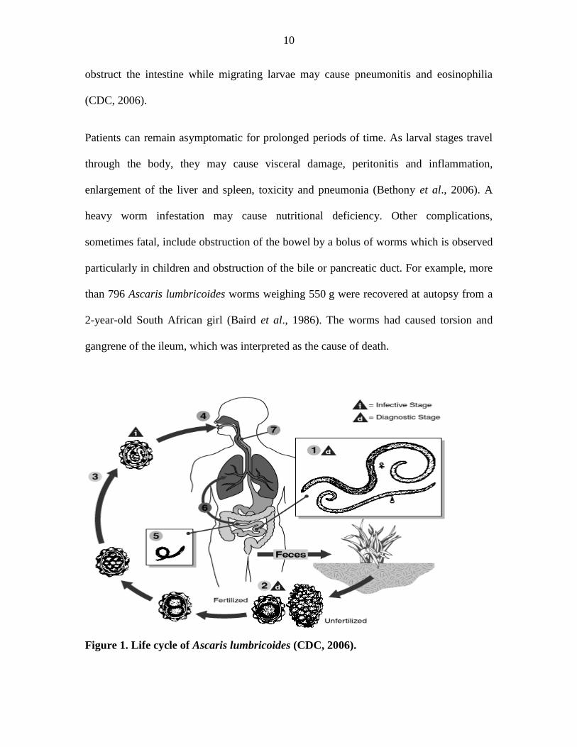
between 1 to 2 years (figure 1) (Wu and Jones, 2000).
First appearance of eggs in stools is 60-70 days after initial infection with the larvae. In
larval Ascariasis, symptoms occur 4-16 days after infection. The final symptoms are
gastrointestinal discomfort and vomiting, fever, and observation of live worms in stools.
Some patients may have pulmonary symptoms or neurological disorders during migration
of the larvae. However there are generally few or no symptoms. A bolus of worms may
10
obstruct the intestine while migrating larvae may cause pneumonitis and eosinophilia
(CDC, 2006).
Patients can remain asymptomatic for prolonged periods of time. As larval stages travel
through the body, they may cause visceral damage, peritonitis and inflammation,
enlargement of the liver and spleen, toxicity and pneumonia (Bethony et al., 2006). A
heavy worm infestation may cause nutritional deficiency. Other complications,
sometimes fatal, include obstruction of the bowel by a bolus of worms which is observed
particularly in children and obstruction of the bile or pancreatic duct. For example, more
than 796 Ascaris lumbricoides worms weighing 550 g were recovered at autopsy from a
2-year-old South African girl (Baird et al., 1986). The worms had caused torsion and
gangrene of the ileum, which was interpreted as the cause of death.
Figure 1. Life cycle of Ascaris lumbricoides (CDC, 2006).
11
Ascaris takes most of its nutrients from the partially digested host food in the intestine.
There is limited evidence that it can also pierce the intestinal mucous membrane and feed
on blood, but this is not its usual source of nutrition. As a result, Ascaris infection does
not produce anemia associated with some other roundworm infections (Wu and Jones,
2000).
2.4 Schistosomiasis
The species of parasitic trematodes of the family Schistosomatidae associated with
human infections are; Schistosoma haematobium, S. intercalatum, S. japonicum, S.
mansoni and S. mekongi (Chitsulo, 2000). Urinary schistosomiasis, in which the bladder
is affected, is caused by infection with S. haematobium which only occurs in Africa.
Intestinal schistosomiasis results from infections with S. mansoni, which occurs in the
Middle East, South America and Africa; and from infections with S. japonicum, which
occurs in parts of China and the Philippines (Ross et al., 2002). Two other schistosome
species are known to cause intestinal schistosomiasis in restricted geographical areas; for
example, S. intercalatum, found in central Africa, and S. mekongi found in Cambodia and
the Lao People’s Democratic Republic. Schistosomiasis is estimated to affect 200 million
people worldwide (de Silva et al., 2003), and is usually a chronic disease (Van Der Werf
and De Vlas, 2001).
Many infections are sub clinically symptomatic, with mild anemia and malnutrition being
common in endemic areas. Acute schistosomiasis also known as Katayama’s fever may
occur weeks after the initial infection, especially by S. mansoni and S. japonicum.
12
Manifestations include: abdominal pain, cough, diarrhea, eosinophilia which is
characterized by extremely high eosinophil granulocyte count, fever, fatigue,
hepatosplenomegaly –which is the enlargement of both the liver and the spleen (Van Der
Werf and De Vlas, 2001). Occasionally central nervous system lesions occur: ectopic S.
japonicum eggs in the brain may cause cerebral granulomatous disease. Granulomatous
lesions around ectopic eggs in the spinal cord from S. mansoni and S. haematobium
infections may result in a transverse myelitis with flaccid paraplegia (Hodder et al.,
2000).
Continuing infection may cause granulomatous reactions and fibrosis in the affected
organs. This may result in manifestations that include; colonic polyposis with bloody
diarrhea mostly caused by S. mansoni, portal hypertension with hematemesis and
splenomegaly in S. mansoni or S. japonicum infection. Infection with Schistosoma
haematobium results in cystitis and urethritis, with haematuria. In some cases it can
progress to bladder cancer. Other pathological effects include pulmonary hypertension in
S. mansoni and S. japonicum infections, and more rarely glomerulonephritis as a result of
S. haematobium infection (Ross et al., 2002). In sub-Saharan Africa where
schistosomiasis constitutes an important public health problem, a survey in 2000 of
disease-specific mortality reported that 70 million individuals out of 682 million had
experienced haematuria and 32 million dysuria associated with S. haematobium infection.
It was estimated that 18 million suffered bladder wall pathology and 10 million
hydronephrosis. Infection with S. mansoni was estimated to cause diarrhea in 0.78
million individuals, blood in stool in 4.4 million and hepatomegaly in 8.5 million. Using
the very limited data available, mortality rates due to non-functioning kidney (from S.
13
haematobium) and haematemesis (from S. mansoni) have been estimated at 150 000 and
130 000 per year, respectively (WHO, 2010).
Schistosoma haematobium and S. mansoni are endemic in Kenya. In 1938, Dowdswell
reported that the prevalence of S. haematobium among children was 78 % around
kavirondo gulf (Lake Victoria). In 1948 both S. haematobium and S. mansoni were
reported in the Taveta region of coast province (Heisch, 1948). Since then, the disease
prevalence has remained high. Treating children of school going age has been shown as
the most cost-effective way of reducing prevalence of scistosomiasis (Partnership for
Child Development, 1997; Bundy et al., 2006; Brooker et al., 2008). The World Health
Assembly, in May 2001, resolved to regularly treat at least 75% of school-aged children
and other high-risk groups by 2010 (WHO, 2002). In 1994, WHO suggested a control
strategy for schistosomiasis that is based on the prevalence among 7- to 14- year-old
school children. In areas where prevalence in this group is greater than 50%, treatment
should be administered to the entire population. Where prevalence is between 20 and
50% in this age group, all children aged 5-19 years should be treated for schistosomiasis.
Where prevalence is less than 20%, only children with a positive test should be treated
(WHO, 1994). Light microscopy of repeated stool and/or urine examinations to detect
and quantify distinctive schistosome eggs is the ‘gold’ standard of parasitological
diagnosis (Bergquist et al., 2009).
Praziquantel (PZQ) is the drug of choice in the treatment of all forms of schistosomiasis.
It is relatively cheap, non-toxic and easy to administer. It is also produced in a number of
countries (Utzinger et al., 2003). The drug can also be used on pregnant women. Studies
in Gezira, in South Sudan showed no significant difference in the rates of abortion and
14
preterm deliveries between pregnant women who had received PZQ compared with those
who had not received it (Adam et al., 2004). Although the drug is cheap and readily
available, it does not have residual effects and has to be taken repeatedly for every re-
infection (Mutapi et al., 1998; King et al., 1990)
2.5 Control of helminthes infections
Parasitic diseases, particularly helminthes infections can be effectively controlled. Failure
to sustain control of parasites may be due to development of drug resistance or the failure
to implement proven strategies as a result of decreased resources within the health
system. Other limitations include decentralization of health management through health-
sector reform and the lack of financial and human resources (Stephenson et al., 2000).
2.5.1 Anthelmintic drug treatment
Drug treatment in helminthes infections referred to as deworming, targets the reduction
of morbidities by decreasing the worm burden. Repeated chemotherapy at regular
intervals called periodic deworming in high risk groups can keep the levels of infection
below those associated with morbidity and often results in improvement in child health
and development (Albonico et al., 2004). Anthelminthic drug treatments often prevent
the development of irreversible consequences of schistosomiasis that occur in adulthood
as a result of childhood infections. Frequent and periodic deworming may reduce
transmission over time; for example intensity peaks are reduced among school age
children (Adams et al., 2004). The effectiveness of periodic deworming is however
diminished by low efficacy of single dose mebendazole and albendazole for treatment of
hookworm and Trichuriasis (Adams et al., 2004). High rates of post treatment re-
15
infection for STHs occur in areas of high endemicity and also diminished treatment
efficacy occurs with repeated use of either single doze mebendazole or single doze
albendazole for treatment of hookworm and Trichuriasis, possibly as a result of
anthelminthic resistance (Albonico et al., 2004).
2.5.2 Improved sanitation
Improved sanitation can result in reduced soil and water contamination with parasitic
organisms. Sanitation is the only intervention that has been known to eliminate STH
infections. The success of sanitation in control of STH infections relies on the percentage
of the population the measure will cover. This is limited by the high cost of implementing
this strategy (Asaolu and Ofoezie, 2003). Provision of reliable clean water and
connection to public sewer systems remains a pipe dream in most third world countries
due to the costs involved. WHO estimates that more than 300 million Africans still lack
access to safe drinking water. In addition, when used as the primary means of control, it
can take years or even decades for sanitation to be effective since it does not result in
treatment of active infections (Brooker et al., 2004).
2.5.3 Health education
Health education targets encouraging healthy behavior to reduce transmission and re-
infection; for example, communities may be educated on the importance of using latrines
and hygienic behavior like proper disposal of fecal material and household water
treatment, to reduce contamination of soil and water which prevents re-infection with soil
transmitted helminthes (Montressor et al., 2002). The aim of health education is to
develop a sense of responsibility for health conditions by individuals, as members of
16
families, and as communities. Assessment of habits and attitudes of the people that relate
to spread and frequency of the disease will provide information of specific means to
make the necessary changes in behavior required to control infectious diseases.
2.5.4 Other control measures
In specific epidemiological conditions, environmental or chemical control of snails can
be useful tools for reducing the transmission of schistosomiasis. Controlling populations
of snail hosts through the use of molluscicides at one time was considered the only
effective way to preventing large-scale infection in communities living near aquatic
habitats. With the advent of safe drugs, such as praziquantel, this strategy has declined in
popularity, though it still plays a crucial role in controlling the spread of the disease.
17
CHAPTER THREE: MATERIALS AND METHODS
3.1 Study area
Vingujini sub-location is situated in the Msambweni District of Coast Province,
approximately 50 km south of the city of Mombasa, along the Indian Ocean. The area has
a monsoon-type of climate, with the period from January to March being hot and dry.
There are two rainy season's with heavy rains in April to June and short rains in October
to November. The total annual rainfall ranges between 1000 to 1600 mm. The mean
monthly temperature varies from a high of 26.7°C (August) and minimum of 23.5°C
(July). Relative humidity is about 95 % due to close proximity to the sea (McClanahan,
1988).
The area is on the coastal plain; an area sometimes referred to as the ‘coral rag’. This is a
narrow strip of corals, sand and alluvial deposits extending 3-20 km from seashore. It lies
0-30 meters above sea level. Vingujini Sub-Location comprises of seven villages;
Vingujini, Bomani, Mwaembe, Mwagundu, Sawasawa, Tumbe and Kisimachande.
Mwaembe, Mwagundu, Sawasawa, Tumbe and Kisimachande villages border the ocean
line. Bomani and Vingujini are located further inland (Ministry of Agriculture, 2008).
Bomani village is traversed by the Mombasa Lungalunga road and in some areas it has
characteristics of a modern slum, with overcrowding and poor drainage facilities. The
economic activities in Bomani are mostly trading and farming. Mwagundu village has
typical rural characteristics where the households are spread apart. The predominant
economic activities in this village are farming and small scale fishing. In both villages,
rainfall causes seasonal flooding which provides necessary conditions for breeding of
18
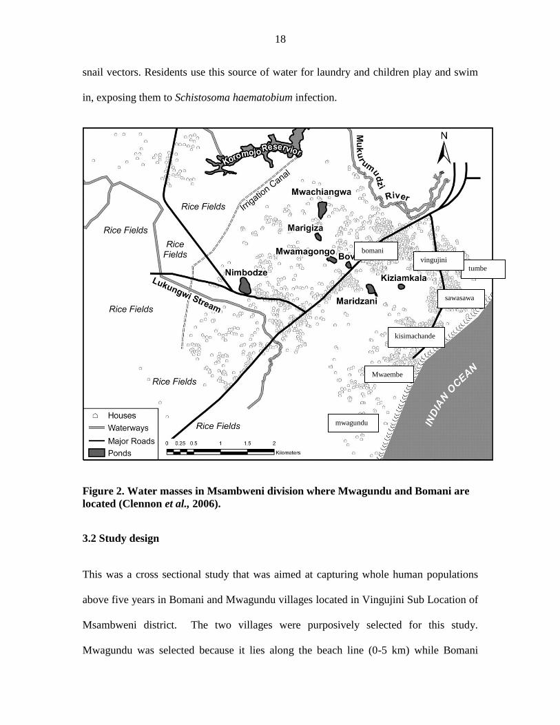
snail vectors. Residents use this source of water for laundry and children play and swim
in, exposing them to Schistosoma haematobium infection.
Figure 2. Water masses in Msambweni division where Mwagundu and Bomani are
located (Clennon et al., 2006).
3.2 Study design
This was a cross sectional study that was aimed at capturing whole human populations
above five years in Bomani and Mwagundu villages located in Vingujini Sub Location of
Msambweni district. The two villages were purposively selected for this study.
Mwagundu was selected because it lies along the beach line (0-5 km) while Bomani
bomani
Mwaembe
mwagundu
kisimachande
sawasawa
tumbe
vingujini
19
village lies furthest from the beach line (12-20 km). The estimated population of
Vingujini sublocation is 12,662 (Kenya National Bureau of Statistics, 2009).
Prior to the study, all houses in the study area were visited and given a code. All house
hold members were given a registration number related to their position in household for
example, 01 for household head, 02 for spouse of household head and 03 for other
members of the household. This coded registration was used for labeling all samples
collected during the study.
3.3 Study population
A prior demographic survey showed population above 5 years in Mwagundu village was
689. This consisted of 342 (49.64%) female and 347 (50.36%) male. The population in
Bomani above 5 years was 1,399; 727 (51.97%) were female while 672 (48.03%) were
males. The main economic activities in the area are agriculture and fishing.
3.4 Data collection
Information meetings were held with community members prior to the study. The people
were informed that collection of samples would be done in primary schools convenient to
the villages. The stool and urine samples collected were transported to Msambweni
district hospital clinical laboratory for examination. Urine samples were examined for
eggs of Schistosoma haematobium. Hematuria was determined using urine dip strips.
Stool samples were examined microscopically for intestinal helminthes. Blood samples
were collected to determine hemoglobin levels in order to establish the relationship
between hookworm disease and anemia.
20
3.5 Collection of samples
All the individuals who had been registered during the demographic study were invited
on specific days for sampling. It was expected that the whole population of residents
above 5 years in each of the two villages would be sampled according to WHO
recommendations for areas where prevalence with schistosomiasis is above 50%.
One hundred people were invited per day for a period of 21 days. Each individual
provided a fecal and urine sample. The sample containers were labeled with the
respondents’ number codes that had been assigned during the time of the demographic
survey. Blood samples for determining hemoglobin status were collected at primary
schools located in the two villages using capillary blood collection tubes.
3.5.1 Collection of fecal samples
Approximately ten grams of fresh fecal specimens were provided by the study subjects in
clean clearly labeled plastic sealable sample containers. The individuals were advised to
provide the first stool of the day. Collected fecal specimens were transported in cool
boxes to the Msambweni district hospital clinical laboratory. All the samples were
analyzed for parasites within two hours of collection.
3.5.2 Collection of urine samples
About 30 ml of urine was collected from each individual in clean receptacles. The urine
samples were collected between 10.00 am and 2.00 pm, to coincide with the period when
excretion of S. haematobium eggs is highest (Fleck and Moody, 1988).
21
3.6 Determining of hematuria status
The hematuria status of each sample was observed by gross examination and also using
reagent strips (medi-test combi®). The reagent strips were dipped into urine inside a test
tube. Microhematuria were evaluated according to the manufacturers’ instructions and
recorded.
3.7 Preparation and examination of urine filters
Polycarbonate membrane filters (Whatman/GE™) of 13 mm diameter and 12 µm pore
size were used for filtration of Schistosoma haematobium eggs from urine. The filters
were carefully placed on the filter support of the filter holder using blunt-ended forceps.
The filter holder was then re-assembled and attached to the end of a 10 ml luer syringe.
The plunger was then removed from the syringe after which the syringe was filled with
well-mixed urine sample to the 10 ml mark, and the plunger was then replaced. Holding
the syringe over a beaker, the urine was slowly passed through the filter. The filter holder
was unscrewed and placed on specially designed racks labeled with the study subjects
demographic codes and transported to Msambweni district hospital clinical laboratory to
be examined microscopically.
The filters were removed at the laboratory from filter holders using a blunt-ended forceps
and transferred to a microscope slide, face upwards. Using a teat dropper, a drop of lugols
iodine was added, and then the slide was covered with a cover glass and examined
microscopically using 10x magnification with the condenser iris closed sufficiently to
give good contrast. The entire filter was systematically examined for eggs of Schistosoma
haematobium. The number of eggs in the preparation was counted and reported per 10ml
22
of urine. 1-50 eggs per 10ml of urine were considered as light infection, 51-200 eggs per
10ml of urine as moderate infection and above 200 eggs per 10ml of urine as heavy
infection.
3.8 Examination of stool samples for geohelminthes
Stool samples collected were examined microscopically. Kato katz technique was used to
quantify hookworm eggs.
3.8.1 Microscopic examination of feacal specimens
Saline and iodine slide preparations were made for fresh faecal samples. A drop of saline
was placed on one half of a microscope slide and a drop of iodine on the other half. With
an applicator stick, a portion of faeces approximately the size of a match head was picked
and mixed with the saline drop on the slide. Similarly another portion of the same size
was mixed with the drop of iodine. A cover slip was placed separately on each
preparation. These were examined microscopically at a magnification of 10x and 100x
for motile geohelminthes such as the larvae of Stercolaris spiralis and helminthes eggs
and cysts.
3.8.2 Quantification of hookworm eggs
Kato katz technique was used to quantify the intensity of hookworm infection. A plastic
template with a hole (accommodating 41.7mg of formed faeces) was placed at the centre
of a microscope slide. Approximately 10g of freshly collected faeces was placed on the
surface of a glazed tile. An 80 mm plastic screen was pressed on top of the faecal
material using a gloved hand. The upper surface of the screen was then scraped with a
23
flat spatula to collect the filtered faeces. The collected filtered faeces in the screen were
added in the hole of the template so that it became completely filled. The template was
then removed carefully so that the cylinder of faeces was left on the slide. The faecal
material was then covered with a cellophane strip that had been pre-soaked in glycerol-
malachite green solution. The microscope slide was then inverted and pressed on a
smooth tile to spread the faecal material evenly. The slide was carefully removed by
gently sliding it sideways to avoid separating the cellophane strip. The slide was placed
with the cellophane upwards. The smear was examined in a systematic manner for
hookworm eggs and the number reported. This number was then multiplied by 24 as per
the manufacturer’s instructions to give number of eggs per gram of faeces (epg). Intensity
was estimated as proposed by WHO where 1-1999epg were considered as light
infections, 2000-3999epg as moderate and more than 4000epg were considered to be
heavy infections.
3.9 Determination of hemoglobin levels
Hemoglobin levels were determined for each participant using a HemoCue®
haemoglobinometer. Capillary blood samples were collected by finger prick in the middle
finger of left hand, after cleaning and massaging the finger to facilitate blood flow. A
standard cuvette (10mm) was filled with blood from the finger-prick. The test was
performed as stated by the manufacturer. After calibration of the HemoCue®
haemoglobinometer machine, hemoglobin values were read and recorded to one decimal
point. The HemoCue®
photometer was checked on a daily basis using the control cuvette
and a standard of known concentration.
24
3.10 Data analysis
Data obtained from this study was recorded and analyzed using computer statistical
software; spreadsheets Microsoft excel (2007) and minitab14. Data on the number of
people infected with schistosomiasis and geohelminthes was pooled in five age groups of
5-15, 16-25, 26-40, 41-60 and >60. Mean infections were determined for schistosomiasis,
geohelminthes infections and for co-infections with Schistosoma and geohelminthes.
Data transformations were done to normalize it. Analysis of Variance, ANOVA (general
linear model and one way) were done on weighted means to determine differences in the
mean infections between the villages, sex and age groups. ANOVA was also carried out
to determine differences in hemoglobin levels in individuals with different infection
intensities of hookworm disease and also for the relationship between hematuria and
intensity with Schistosoma haematobium. Group comparisons were done using the tukey
test at 95% confidence interval. Correlation was examined between infection with
Schistosoma haematobium and soil transmitted helminthes using Pearson coefficients.
3.11 Permission
Permission to carry out the study was obtained from the School of Pure and Applied
Sciences, Kenyatta University and the Ministry of Health, Msambweni district hospital.
3.12 Ethical consideration
All work was done according to the guidelines for human experimentation in clinical
research as stipulated by the Ministry of Health of Kenya. Ethical clearance was obtained
from KEMRI before the commencement of the study. All subjects included in the study
25
were required to give oral informed consent and a signed written consent was given by
household heads and all adult participants. For respondents below the age of 12 years,
consent was sought from their parents or legal guardians before inclusion in the study.
3.13 Exclusion criteria
Children below 5 years were excluded from this study. This is because the incidence of
schistosomiasis is rare in this group since they do not normally actively participate in
activities that expose them to infection (Chitsulo et al., 2000).
3.14 Confidentiality
The information obtained from subjects was treated confidentially and was used only for
the purposes of the current research study.
26
CHAPTER FOUR: RESULTS
4.1 Prevalence of parasitic infections
Fifty nine per cent (1,232) of the total 2,088 legible subjects turned out for screening. Out
of these, 495 subjects were from Mwagundu village. This accounts for 72.37% of the
target population (689 people aged 5 years or more) in this village. Of the 495 people
sampled, 258 (52.12%) were female and 237 (47.88%) male. A total of 737 subjects from
Bomani village were involved in the study. This accounts for 37.81% of the target
population (1,399 people aged 5 years or more). Out of this population, 409 (55.50%)
were female and 328 (44.50%) male.
The overall occurrence of parasitic infections were, 43.99% for Schistosoma
haematobium, 29.63% for hookworm disease, 0.49% for ascariasis and 24.59% for
trichuriasis (N=1,232). Only 32.71% were free from any of the parasitic infections
screened. The occurrence of urinary Schistosomiasis and soil transmitted helminthes in
the two villages are represented in table 2.
27
Table 1. Percentage occurrence of urinary schistosomiasis and soil transmitted
helminthes in Mwagundu and Bomani.
Village Disease Male Female Population
Mwagundu Schistosomiasis 43.04 ±6.45 46.51 ±6.43 44.85 ±6.15
Hookworm disease 29.96 ± 4.43 25.68 ±5.09 27.68 ±4.10
Ascariasis 1.22 ±0.13 - 0.68 ±0.06
Trichuriasis 24.89 ±3.45 18.60 ±3.59 21.62 ±2.98
*
Bomani Schistosomiasis 44.82 ±7.01 42.30 ±6.58 43.42 ±6.23
Hookworm disease 33.54 ±4.66 28.85 ±4.35 30.94 ±3.31
Ascariasis - 1.22 ±0.75 0.68 ±0.41
Trichuriasis 29.27 ±3.63 24.45 ±3.25 26.59 ±3.18
*
* Shows there is a significant difference in the occurrence of trichuriasis between
Mwagundu and Bomani (ANOVA table in appendix 3, f=4.9, df=1, p=0.045).
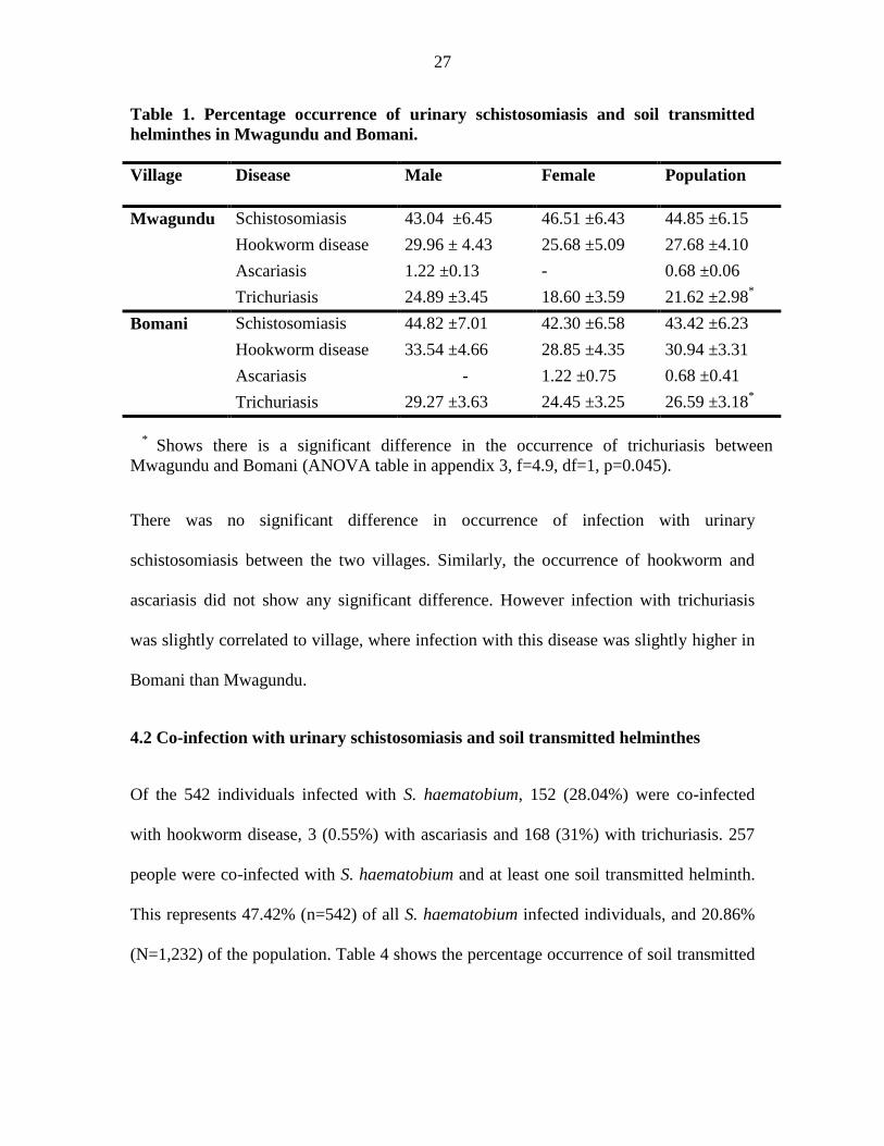
There was no significant difference in occurrence of infection with urinary
schistosomiasis between the two villages. Similarly, the occurrence of hookworm and
ascariasis did not show any significant difference. However infection with trichuriasis
was slightly correlated to village, where infection with this disease was slightly higher in
Bomani than Mwagundu.
4.2 Co-infection with urinary schistosomiasis and soil transmitted helminthes
Of the 542 individuals infected with S. haematobium, 152 (28.04%) were co-infected
with hookworm disease, 3 (0.55%) with ascariasis and 168 (31%) with trichuriasis. 257
people were co-infected with S. haematobium and at least one soil transmitted helminth.
This represents 47.42% (n=542) of all S. haematobium infected individuals, and 20.86%
(N=1,232) of the population. Table 4 shows the percentage occurrence of soil transmitted
28
infection in Schistosoma haematobium infected individuals. Infection with S.
haematobium was highly correlated with trichuriasis infection (r=0.96, p=0.006).
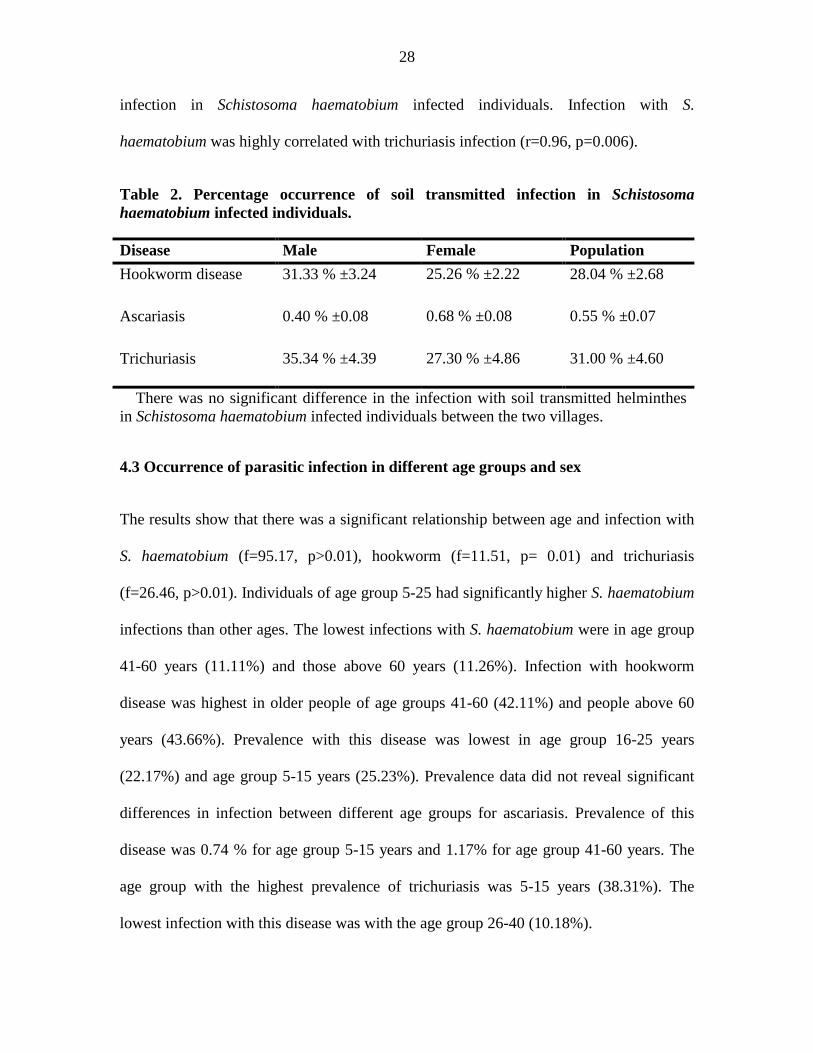
Table 2. Percentage occurrence of soil transmitted infection in Schistosoma
haematobium infected individuals.
Disease Male Female Population
Hookworm disease 31.33 % ±3.24 25.26 % ±2.22 28.04 % ±2.68
Ascariasis 0.40 % ±0.08
0.68 % ±0.08 0.55 % ±0.07
Trichuriasis 35.34 % ±4.39 27.30 % ±4.86 31.00 % ±4.60
There was no significant difference in the infection with soil transmitted helminthes
in Schistosoma haematobium infected individuals between the two villages.
4.3 Occurrence of parasitic infection in different age groups and sex
The results show that there was a significant relationship between age and infection with
S. haematobium (f=95.17, p>0.01), hookworm (f=11.51, p= 0.01) and trichuriasis
(f=26.46, p>0.01). Individuals of age group 5-25 had significantly higher S. haematobium
infections than other ages. The lowest infections with S. haematobium were in age group
41-60 years (11.11%) and those above 60 years (11.26%). Infection with hookworm
disease was highest in older people of age groups 41-60 (42.11%) and people above 60
years (43.66%). Prevalence with this disease was lowest in age group 16-25 years
(22.17%) and age group 5-15 years (25.23%). Prevalence data did not reveal significant
differences in infection between different age groups for ascariasis. Prevalence of this
disease was 0.74 % for age group 5-15 years and 1.17% for age group 41-60 years. The
age group with the highest prevalence of trichuriasis was 5-15 years (38.31%). The
lowest infection with this disease was with the age group 26-40 (10.18%).
29
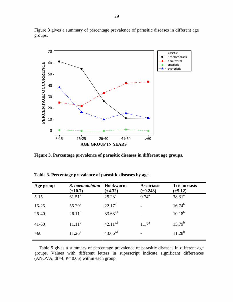
Figure 3 gives a summary of percentage prevalence of parasitic diseases in different age
groups.
Figure 3. Percentage prevalence of parasitic diseases in different age groups.
Table 3. Percentage prevalence of parasitic diseases by age.
Age group S. haematobium
(±10.7)
Hookworm
(±4.32)
Ascariasis
(±0.243)
Trichuriasis
(±5.12)
5-15 61.51a 25.23
a 0.74
a 38.31
a
16-25 55.20a 22.17
a - 16.74
b
26-40 26.11b
33.63a,b
- 10.18b
41-60 11.11b 42.11
c,b 1.17
a 15.79
b
>60 11.26b 43.66
c,b - 11.28
b
Table 5 gives a summary of percentage prevalence of parasitic diseases in different age
groups. Values with different letters in superscript indicate significant differences
(ANOVA, df=4, P< 0.05) within each group.
AGE GROUP IN YEARS
PE
RC
EN
TA
GE
OC
CU
RR
EN
CE
>6041-6026-4016-255-15
70
60
50
40
30
20
10
0
Variable
ascariasis
trichuriasis
Schistosomiasis
hookworm
30
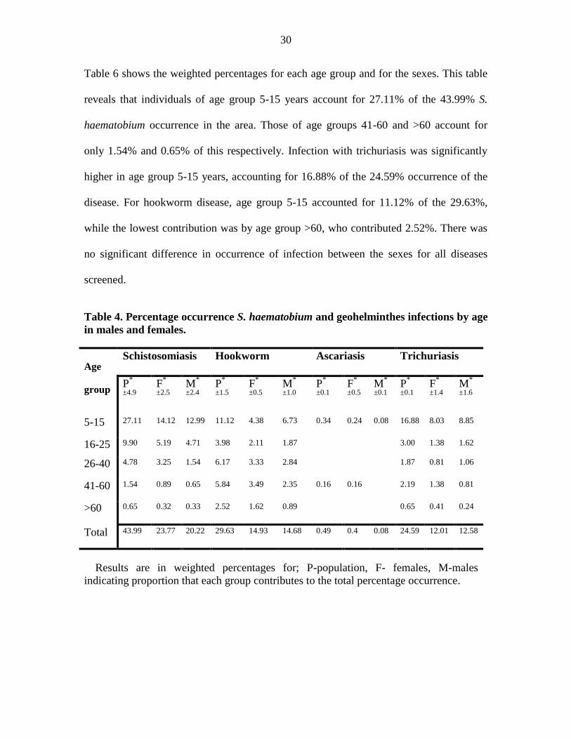
Table 6 shows the weighted percentages for each age group and for the sexes. This table
reveals that individuals of age group 5-15 years account for 27.11% of the 43.99% S.
haematobium occurrence in the area. Those of age groups 41-60 and >60 account for
only 1.54% and 0.65% of this respectively. Infection with trichuriasis was significantly
higher in age group 5-15 years, accounting for 16.88% of the 24.59% occurrence of the
disease. For hookworm disease, age group 5-15 accounted for 11.12% of the 29.63%,
while the lowest contribution was by age group >60, who contributed 2.52%. There was
no significant difference in occurrence of infection between the sexes for all diseases
screened.
Table 4. Percentage occurrence S. haematobium and geohelminthes infections by age
in males and females.
Age
group
Schistosomiasis
Hookworm
Ascariasis Trichuriasis
P*
±4.9 F
*
±2.5 M
*
±2.4 P
*
±1.5 F
*
±0.5 M
*
±1.0 P
*
±0.1 F
*
±0.5 M
*
±0.1 P
*
±0.1
F*
±1.4 M
*
±1.6
5-15 27.11 14.12 12.99 11.12 4.38 6.73 0.34 0.24 0.08 16.88 8.03 8.85
16-25 9.90 5.19 4.71 3.98 2.11 1.87 3.00 1.38 1.62
26-40 4.78 3.25 1.54 6.17 3.33 2.84 1.87 0.81 1.06
41-60 1.54 0.89 0.65 5.84 3.49 2.35 0.16 0.16 2.19 1.38 0.81
>60 0.65 0.32 0.33 2.52 1.62 0.89 0.65 0.41 0.24
Total 43.99 23.77 20.22 29.63 14.93 14.68 0.49 0.4 0.08 24.59 12.01 12.58
Results are in weighted percentages for; P-population, F- females, M-males
indicating proportion that each group contributes to the total percentage occurrence.
31
4.4 Intensity of helminthes infection in different age groups and sex
The intensity of urinary schistosomiasis and hookworm disease were estimated as
eggs/10 ml of urine for Schistosoma haematobium and eggs per gram (epg) of faeces for
hookworm. Intensity of urinary schistosomiasis was highly related to age (f=29.39,
p=0.00) and sex (f=6.13, p=0.013). Males had significantly higher intensities of urinary
schistosomiasis (mean of 113.5±10.9 eggs per 10ml of urine) than females (mean of
69.9±7.55 eggs per 10ml of urine). The overall populations mean Schistosoma eggs shed
was 89.95±6.48 eggs per 10 ml of urine. Individuals of age 5-15 years shed the highest
number of eggs (160.7 ±12.5 eggs/10 ml of urine). The individuals of age group 41-60
years shed the lowest number of S. haematobium eggs (2.94±1.54) (Table 5, Figure 5).
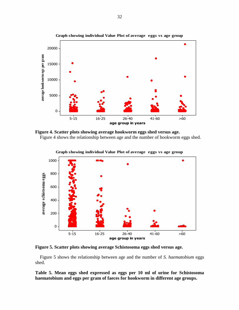
The number of hookworm eggs shed consistently increased with an increase in age
(f=7.32, p=0.00). The highest group shed a mean 855±345 eggs per gram of faeces for
age >60 years. The group that shed the lowest number of eggs was age 16-25 years with
an average of 176.0±52.0 eggs per gram of faeces (Figure 4). Males shed significantly
higher amounts of eggs than females (f=4.81, p=0.028).
32
age group in years
aver
age
hoo
kw
orm
eg
s pe
r g
ram
>6041-6026-4016-255-15
20000
15000
10000
5000
0
Graph showing individual Value Plot of average eggs vs age group
Figure 4. Scatter plots showing average hookworm eggs shed versus age.
Figure 4 shows the relationship between age and the number of hookworm eggs shed.
age group in years
ave
rag
e s
chis
tso
ma
eg
gs
>6041-6026-4016-255-15
1000
800
600
400
200
0
Graph showing individual Value Plot of average eggs vs age group
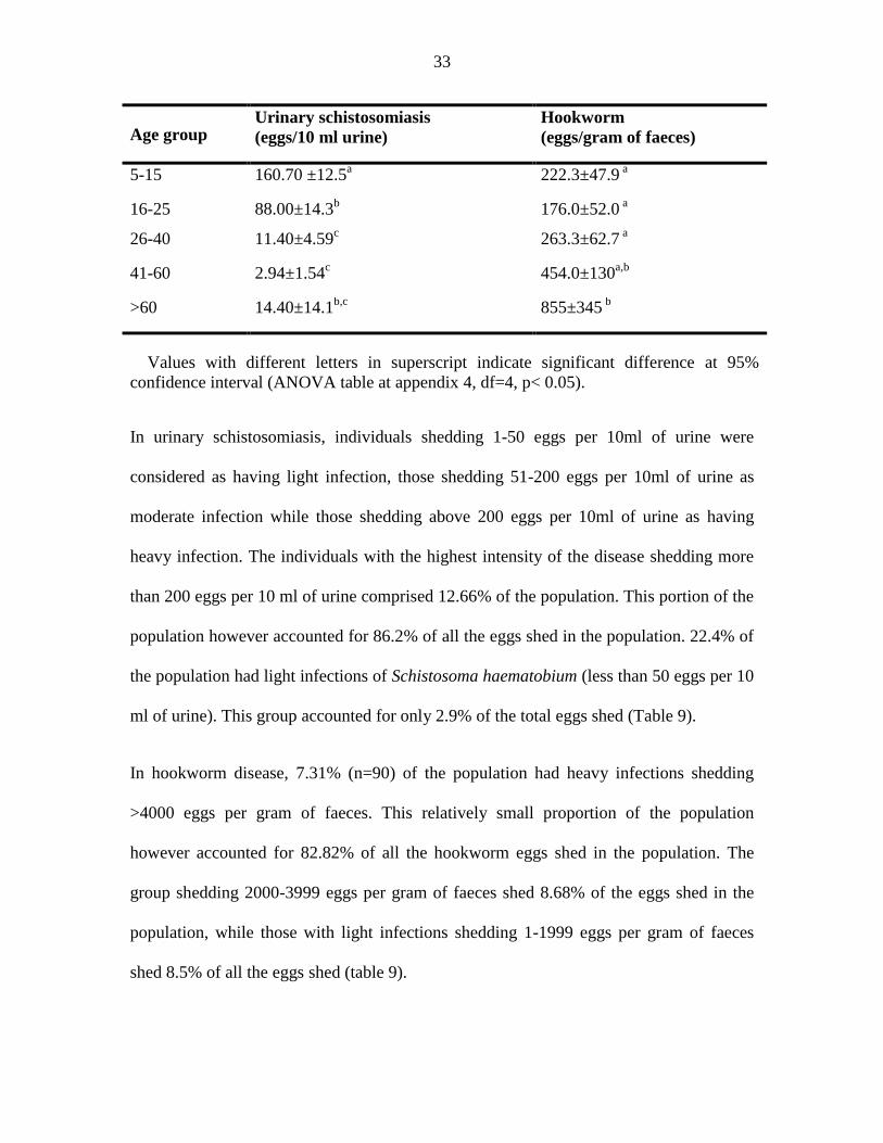
Figure 5. Scatter plots showing average Schistosoma eggs shed versus age.
Figure 5 shows the relationship between age and the number of S. haematobium eggs
shed.
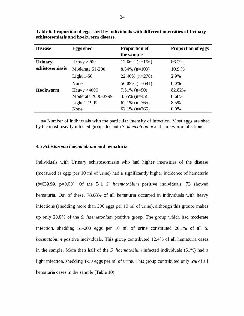
Table 5. Mean eggs shed expressed as eggs per 10 ml of urine for Schistosoma
haematobium and eggs per gram of faeces for hookworm in different age groups.
33
Age group Urinary schistosomiasis
(eggs/10 ml urine)
Hookworm
(eggs/gram of faeces)
5-15 160.70 ±12.5a
222.3±47.9 a
16-25 88.00±14.3b
176.0±52.0 a
26-40 11.40±4.59c
263.3±62.7 a
41-60 2.94±1.54c
454.0±130a,b
>60 14.40±14.1b,c
855±345 b
Values with different letters in superscript indicate significant difference at 95%
confidence interval (ANOVA table at appendix 4, df=4, p< 0.05).
In urinary schistosomiasis, individuals shedding 1-50 eggs per 10ml of urine were
considered as having light infection, those shedding 51-200 eggs per 10ml of urine as
moderate infection while those shedding above 200 eggs per 10ml of urine as having
heavy infection. The individuals with the highest intensity of the disease shedding more
than 200 eggs per 10 ml of urine comprised 12.66% of the population. This portion of the
population however accounted for 86.2% of all the eggs shed in the population. 22.4% of
the population had light infections of Schistosoma haematobium (less than 50 eggs per 10
ml of urine). This group accounted for only 2.9% of the total eggs shed (Table 9).
In hookworm disease, 7.31% (n=90) of the population had heavy infections shedding
>4000 eggs per gram of faeces. This relatively small proportion of the population
however accounted for 82.82% of all the hookworm eggs shed in the population. The
group shedding 2000-3999 eggs per gram of faeces shed 8.68% of the eggs shed in the
population, while those with light infections shedding 1-1999 eggs per gram of faeces
shed 8.5% of all the eggs shed (table 9).
34
Table 6. Proportion of eggs shed by individuals with different intensities of Urinary
schistosomiasis and hookworm disease.
Disease Eggs shed Proportion of
the sample
Proportion of eggs
Urinary
schistosomiasis
Heavy >200 12.66% (n=156) 86.2%
Moderate 51-200 8.84% (n=109) 10.9.%
Light 1-50 22.40% (n=276) 2.9%
None 56.09% (n=691) 0.0%
Hookworm Heavy >4000 7.31% (n=90) 82.82%
Moderate 2000-3999 3.65% (n=45) 8.68%
Light 1-1999 62.1% (n=765) 8.5%
None 62.1% (n=765) 0.0%
n= Number of individuals with the particular intensity of infection. Most eggs are shed
by the most heavily infected groups for both S. haematobium and hookworm infections.
4.5 Schistosoma haematobium and hematuria
Individuals with Urinary schistosomiasis who had higher intensities of the disease
(measured as eggs per 10 ml of urine) had a significantly higher incidence of hematuria
(f=639.99, p=0.00). Of the 541 S. haematobium positive individuals, 73 showed
hematuria. Out of these, 78.08% of all hematuria occurred in individuals with heavy
infections (shedding more than 200 eggs per 10 ml of urine), although this groups makes
up only 28.8% of the S. haematobium positive group. The group which had moderate
infection, shedding 51-200 eggs per 10 ml of urine constituted 20.1% of all S.
haematobium positive individuals. This group contributed 12.4% of all hematuria cases
in the sample. More than half of the S. haematobium infected individuals (51%) had a
light infection, shedding 1-50 eggs per ml of urine. This group contributed only 6% of all
hematuria cases in the sample (Table 10).
35
Table 7. Hematuria in groups with different Schistosoma intensity.
Intensity Proportion of sample Proportion of hematuria
Heavy >200 28.8% (n=156) 78.0%
Moderate 51-200 20.1% (n=109) 12.4%
Light 1-50 51.0% (n=276) 9.6%
n= Number of individuals with the particular intensity of infection. Hematuria was
significantly higher in heavily infected individuals than in moderate and light infections
(ANOVA table at appendix 5, df=1, p=0.000)
4.6 Hookworm infection and haemoglobin level
A HemoCue©
haemoglobinometer was used to determine the hemoglobin levels.
Hemoglobin levels below 11.0g/dl (110g/l) were considered as anemic. (Dallman and
Reeves, 1984). From the results, 26.61% of the population had a hemoglobin level of
below 11.0g/dl.
Table 8. Hemoglobin levels in different intensities of hookworm disease.
Hookworm eggs shed Anaemic individuals
(Hb>11 g/dl)
Mean Hb
Heavy >4000 37.50% 11.081±0.226a
Moderate 2000-3999 30.36% 11.872±0.109a
Light 1-1999 27.74% 12.036±0.311b
None 25.74% 12.108±0.076b
Values with different letters in superscript indicate significant difference (df=3, p f=5.5,
p<0.01). Individuals with heavy and moderate infections had significantly lower
haemoglobin levels than those with light and no infection.
The mean hemoglobin level was 11.98±0.06g/dl (range, 7.6-17.4g/dl). Mean hemoglobin
level in females was 11.44±0.07 g/dl and 12.59±0.09 g/dl in males. The mean
hemoglobin level in hookworm positive individuals was 11.89g/dl.±0.1. There was a
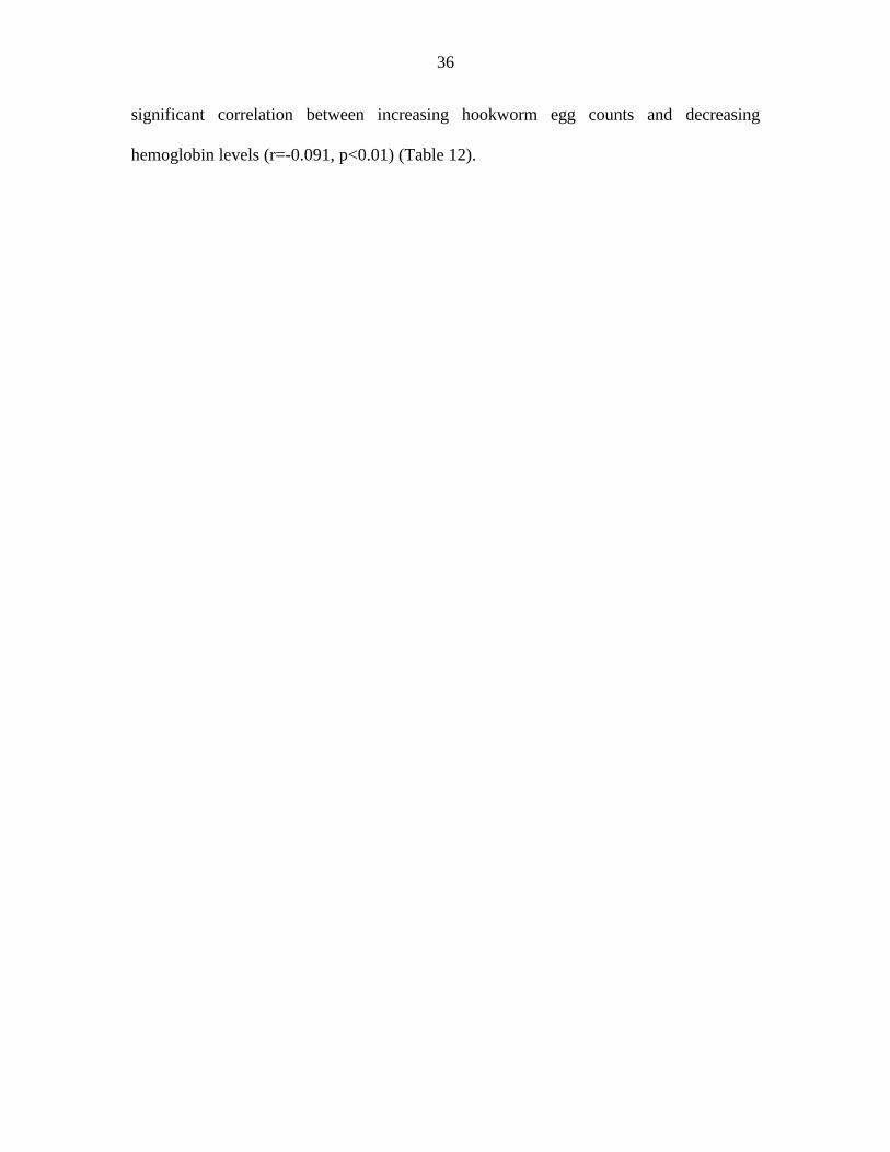
36
significant correlation between increasing hookworm egg counts and decreasing
hemoglobin levels (r=-0.091, p<0.01) (Table 12).
37
CHAPTER FIVE: DISCUSION
5.1 Occurrence of parasitic infections
Of the four diseases studied, urinary schistosomiasis had the highest prevalence for both
villages (44.85% ±6.15 in Mwagundu and 43.42% ±6.23 in Bomani; overall prevalence
of 43.99%). This is a prevalence way above the national prevalence of approximately
23%. It is however a significantly lower prevalence compared to the prevalence of 50-
70% reported in previous years in this area (King et al., 1988, 1990; Muchiri, 1996). This
decrease can be attributed to the regular annual oral therapy since 1984 which has had a
marked impact on the prevalence and intensity of Schistosoma haematobium (King et al.,
1988, 1990, 1991; Muchiri, 1996; Satayathum et al., 2006).
This study has revealed that many people infected with urinary schistosomiasis are also
infected with other helminthes (47.42%). Individuals who were infected with
Schistosoma haematobium were more likely to be infected with hookworm disease and
trichuriasis than those that were not infected with the disease (r=0.96, p=0.006). Previous
studies on the effect of multiple concurrent helminth infections have revealed that the
degree of mobidity is also related to the number of different species harbored (Booth et
al., 1998; Buck et al., 1978). Studies done in Tanzania also revealed that children with
two or more species of helminthes generally carry heavier infections of each species than
children carrying single species infections (Buck et al., 1978). Biological interactions
have been established among several helminthes species with respect to anemia in
children (Ezeamama et al., 2008) and infection with urinary schistosomiasis and soil
38
transmitted helminthes are both associated with increased risk of anemia (Olds et al.,
1999; Friedman et al., 2005).
Ascariasis is the most common soil transmitted helminth in the world (WHO, 1981). This
study observed a relatively low infection levels with ascariasis in Msambweni (0.68%
±0.06 in Mwagundu and 0.68% ±0.41 in Bomani compared to the global picture of this
disease (26% global prevalence). This may be attributed to the soil type in this area,
which is mainly sandy. Ascaris eggs develop best in less permeable clay soils, with
survival ability increasing with the soil depth (Crompton, 1989a). Clay soils are believed
to prevent egg dispersal by water while sandy soils have poor water retention properties
(Mizgajska, 1993).
Occurrence of hookworm disease and trichuriasis were 29.63% and 24.59% respectively.
Severe hookworm infections are characterized by anemia and protein deficiency
(Bethony et al., 2006). Children who suffer from chronic hookworm infection can also
suffer from growth retardation as well as intellectual and cognitive impairments which
lead to poor school performance and attendance, and adversely affect future productivity
and wage-earning potential (Hotez et al., 2005). Deworming pregnant women especially
is of particular importance in improving outcomes of pregnancies and in reducing child
mortality by improving child birth weight. Regular deworming helps reduce malnutrition
and improves motor and language development in young children. It also has a positive
effect on nutritional status, physical fitness, growth, and language development in
school–age children; and improves maternal hemoglobin levels as well as birth weight
and child survival (Albonico et al., 2004; Hotez et al., 2005).
39
Only one tablet of mebendazole or albendazole per individual is required for treatment of
soil transmitted helminthes (Adams et al., 2004). The drugs can be administered by
persons without medical training. Until new approaches become available, whether a
hookworm vaccine or improved sanitation infrastructures, antihelminthic therapy for
school–age children remains the most practical way to control helminth infections in the
developing world.
5.2 Occurrence and intensity of parasitic infection in different age groups and sex
The results obtained shows that age is a highly significant factor in infections with
Schistosoma haematobium, hookworm disease and trichuriasis. Infection with Urinary
schistosomiasis was highly related to age (f=95.17, p>0.01). Individuals of age group 5-
25 had significantly higher schistosome infections than other ages. A study done in
Blantyre, Malawi in 2006 revealed that people in this age group were more likely to come
into contact with an open water source through swimming, playing and other activities
than other age groups, thereby exposing themselves to schistosome infection (Atupele et
al., 2009). Infection with S. haematobium and trichuriasis diseases was significantly
lower in individuals of age group >60. However highest infection with hookworm disease
was seen among older age groups (43.66% in the age group >60 years and 42.11 in the
age group 41-60 years). While heavy hookworm burdens still occur among children in
some tropical areas (Stephenson et al., 1989; Labiano-Abello et al., 1999), in most of the
world studied to date the peak prevalence and infection intensities for hookworm occurs
in individuals in middle age, or even over the age of 50 years (Gandhi et al., 2001;
Bethony et al., 2002). In this study, infection with this disease was lowest among
individuals of age group 16-25 years (22.17%). Better hygiene practices and frequency in
40
use of shoes are attributed with low incidence of the disease. Studies conducted in 2005-
2007 in a rural community in central Thailand revealed that villagers exposed to soil by
walking barefoot were 4.2 times at a greater risk of hookworm infection (Vittaya et al.,
2011). This is attributed to the fact that acquiring a hookworm infection is directly related
to exposure to soil where filariform larvae, the infective stage, live in and penetrate the
skin. Trichuriasis infections were highest among individuals of age 5-15 years. This is
consistent with previous cross sectional surveys carried out in areas where ascariasis and
trichuriasis is endemic. Such studies revealed three patterns as follows; firstly, the
prevalence with these diseases rises rapidly once infancy has passed and tends to remain
high. Secondly, intensity rises rapidly and peaks during childhood (among 5–15 year-
olds) before declining steadily. Thirdly, the frequency distribution of numbers of worms
per host is overdispersed, where only a small fraction of the population harbors most
worms (Crompton, 2001).
A common measurement of helminthes infection is the estimate of intensity of infection.
In urinary schistosomiasis and soil transmitted infections, intensity is a continuous,
quantitative variable expressed as eggs/10 ml of urine in Schistosoma haematobium and
eggs per gram of faeces for soil transmitted helminthes and intestinal Schistosomiasis.
Quantitative measurements of intensity, though more difficult to obtain, are more
sensitive to change, especially repeated measurements on a cohort than prevalence
measures. Although fundamentally different measurements, prevalence and intensity are
related: prevalence rises with increasing intensity to an upper limit of 100% but intensity
has no theoretical upper limit. It is accepted (WHO, 1998) that rising intensity is
accompanied by an increased risk of developing morbidity and disease for example,
41
pathology of the urogenital tract; hepatomegaly and hepatosplenomegaly in
Schistosomiasis. Egg counts are conventionally transformed to log(x+1) to include zero
counts and normalize their distribution for statistical analysis. Egg counts give a
reflection of the number of worms present at a specific time. They also show significant
changes when, for example, children acquire increasing worm loads due to cumulative
exposure over time, or worm numbers drop after successful treatment.
In this study, the individual Schistosoma haematobium and hookworm egg counts are
distributed asymmetrically; 86.18% of all Schistosoma eggs are passed by 12.66% of the
subjects and the rest passing few or no eggs. Individuals of age group 5-15 and 16-25
passed significantly higher levels of schistosoma eggs in urine as compared to the other
age groups. In hookworm infection, older individuals shed significantly higher amounts
of eggs in faeces, the highest being those >60 years of age who shed an average 855±345
eggs per gram of faeces. The nutritional and health status of older individuals in
developing countries is often poor (Tucker, 2001), which makes them vulnerable to
morbidities as a result of chronic and high intensity of infection. The incidence of
increased intensity and prevalence has been observed in China and other places (Bethony,
2002). The group with the least intensity was age group 16-25 years who shed an average
of 176.0±52.0 eggs per gram of faeces. In this study, individuals of 5-15 years had the
highest prevalence of trichuriasis (38.31%) followed by those of 16-25 (16.74%).
Previous studies revealed that the prevalence and intensity of trichuriasis peak at 10-20
years and then starts to decline (Kabatereine et al., 2004). Studies on infection with
Ascaris after peak intensity has been reached at about 5–10 years of age, clearly
demonstrate the difference between the prevalence and age intensity profiles. Such
42
studies have revealed a marked decline in intensity after this period and it remains at low
levels throughout adulthood, whereas prevalence rates remain high (Kabatereine et al.,
2004).
Intensity of Schistosoma haematobium infection was sex related (f=6.13, p=0.013), while
infection with hookworm disease showed no significant difference in the sexes (f=3.29,
p=0.07) which is however closer to a significant level than for analysis done using
prevalence data alone (f=1.24, p=0.284). In the previous analyses of published data
intensity of infection with hookworm and Schistosoma haematobium was significantly
higher overall in males than females. Previous Meta-analysis studies to investigate
whether there is a consistent host sex bias in infection with helminth infections among
non-human hosts indicated a tendency for infection prevalence to be higher in males in
many types of host-parasite associations, particularly for nematode infections in birds and
mammals. By contrast, intensity of infection showed no clear sex bias except for
nematodes parasitizing mammals, differences in infection intensity being significantly
male-biased (Poulin, 1996). Explanation for this bias is largely lacking, with some
research pointing to the differences in the activities that the members of different gender
are involved in, while other studies suggesting that the difference is in the physiology
particularly the effect of the male hormones on suppression of immunity (Hotez et al.,
2003).
5.3 Occurrence of trichuriasis in the two villages
In the present study, prevalence of trichuriasis was significantly higher in Bomani village
compared to Mwagundu (p=0.045). This may be attributed to the fact that Bomani has
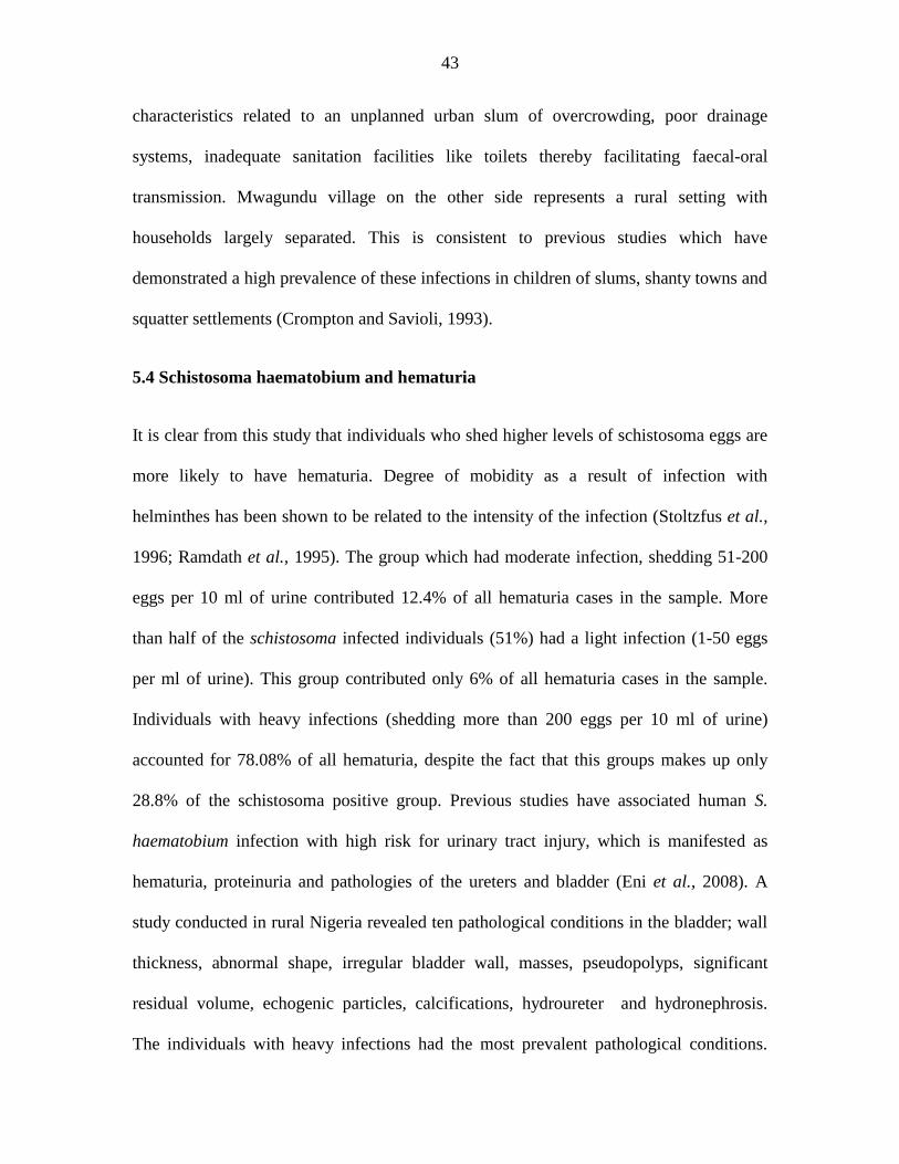
43
characteristics related to an unplanned urban slum of overcrowding, poor drainage
systems, inadequate sanitation facilities like toilets thereby facilitating faecal-oral
transmission. Mwagundu village on the other side represents a rural setting with
households largely separated. This is consistent to previous studies which have
demonstrated a high prevalence of these infections in children of slums, shanty towns and
squatter settlements (Crompton and Savioli, 1993).
5.4 Schistosoma haematobium and hematuria
It is clear from this study that individuals who shed higher levels of schistosoma eggs are
more likely to have hematuria. Degree of mobidity as a result of infection with
helminthes has been shown to be related to the intensity of the infection (Stoltzfus et al.,
1996; Ramdath et al., 1995). The group which had moderate infection, shedding 51-200
eggs per 10 ml of urine contributed 12.4% of all hematuria cases in the sample. More
than half of the schistosoma infected individuals (51%) had a light infection (1-50 eggs
per ml of urine). This group contributed only 6% of all hematuria cases in the sample.
Individuals with heavy infections (shedding more than 200 eggs per 10 ml of urine)
accounted for 78.08% of all hematuria, despite the fact that this groups makes up only
28.8% of the schistosoma positive group. Previous studies have associated human S.
haematobium infection with high risk for urinary tract injury, which is manifested as
hematuria, proteinuria and pathologies of the ureters and bladder (Eni et al., 2008). A
study conducted in rural Nigeria revealed ten pathological conditions in the bladder; wall
thickness, abnormal shape, irregular bladder wall, masses, pseudopolyps, significant
residual volume, echogenic particles, calcifications, hydroureter and hydronephrosis.
The individuals with heavy infections had the most prevalent pathological conditions.
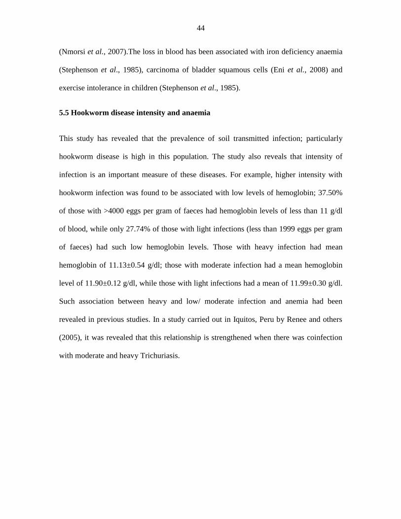
44
(Nmorsi et al., 2007).The loss in blood has been associated with iron deficiency anaemia
(Stephenson et al., 1985), carcinoma of bladder squamous cells (Eni et al., 2008) and
exercise intolerance in children (Stephenson et al., 1985).
5.5 Hookworm disease intensity and anaemia
This study has revealed that the prevalence of soil transmitted infection; particularly
hookworm disease is high in this population. The study also reveals that intensity of
infection is an important measure of these diseases. For example, higher intensity with
hookworm infection was found to be associated with low levels of hemoglobin; 37.50%
of those with >4000 eggs per gram of faeces had hemoglobin levels of less than 11 g/dl
of blood, while only 27.74% of those with light infections (less than 1999 eggs per gram
of faeces) had such low hemoglobin levels. Those with heavy infection had mean
hemoglobin of 11.13±0.54 g/dl; those with moderate infection had a mean hemoglobin
level of 11.90±0.12 g/dl, while those with light infections had a mean of 11.99±0.30 g/dl.
Such association between heavy and low/ moderate infection and anemia had been
revealed in previous studies. In a study carried out in Iquitos, Peru by Renee and others
(2005), it was revealed that this relationship is strengthened when there was coinfection
with moderate and heavy Trichuriasis.
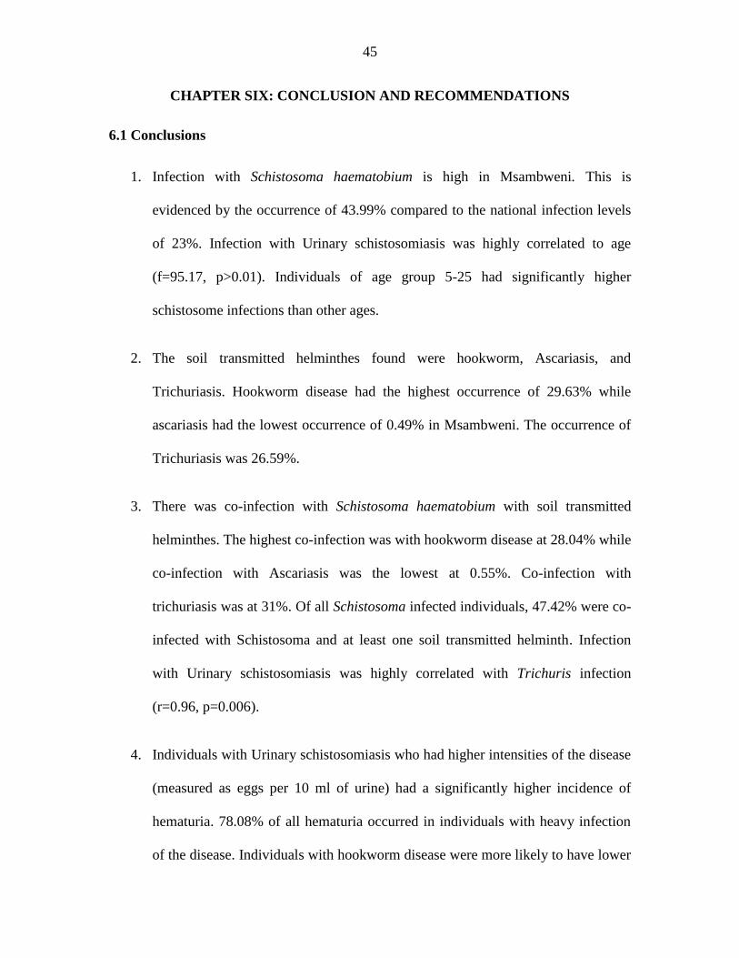
45
CHAPTER SIX: CONCLUSION AND RECOMMENDATIONS
6.1 Conclusions
1. Infection with Schistosoma haematobium is high in Msambweni. This is
evidenced by the occurrence of 43.99% compared to the national infection levels
of 23%. Infection with Urinary schistosomiasis was highly correlated to age
(f=95.17, p>0.01). Individuals of age group 5-25 had significantly higher
schistosome infections than other ages.
2. The soil transmitted helminthes found were hookworm, Ascariasis, and
Trichuriasis. Hookworm disease had the highest occurrence of 29.63% while
ascariasis had the lowest occurrence of 0.49% in Msambweni. The occurrence of
Trichuriasis was 26.59%.
3. There was co-infection with Schistosoma haematobium with soil transmitted
helminthes. The highest co-infection was with hookworm disease at 28.04% while
co-infection with Ascariasis was the lowest at 0.55%. Co-infection with
trichuriasis was at 31%. Of all Schistosoma infected individuals, 47.42% were co-
infected with Schistosoma and at least one soil transmitted helminth. Infection
with Urinary schistosomiasis was highly correlated with Trichuris infection
(r=0.96, p=0.006).
4. Individuals with Urinary schistosomiasis who had higher intensities of the disease
(measured as eggs per 10 ml of urine) had a significantly higher incidence of
hematuria. 78.08% of all hematuria occurred in individuals with heavy infection
of the disease. Individuals with hookworm disease were more likely to have lower
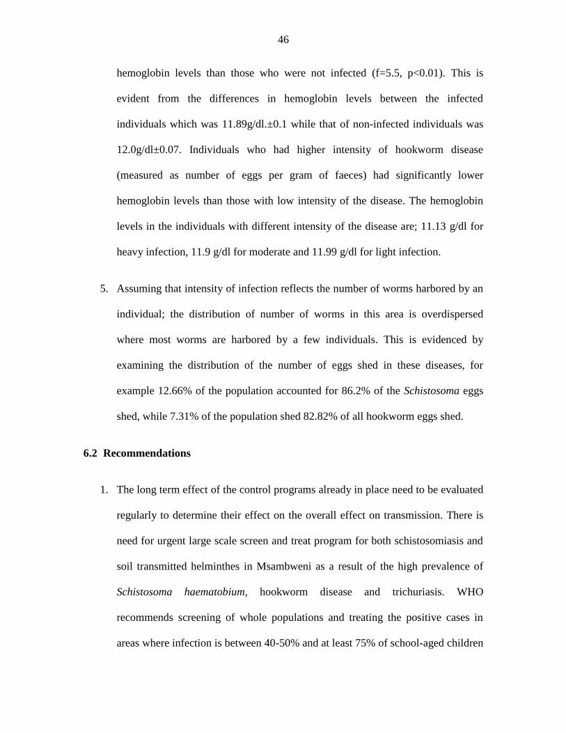
46
hemoglobin levels than those who were not infected (f=5.5, p<0.01). This is
evident from the differences in hemoglobin levels between the infected
individuals which was 11.89g/dl.±0.1 while that of non-infected individuals was
12.0g/dl±0.07. Individuals who had higher intensity of hookworm disease
(measured as number of eggs per gram of faeces) had significantly lower
hemoglobin levels than those with low intensity of the disease. The hemoglobin
levels in the individuals with different intensity of the disease are; 11.13 g/dl for
heavy infection, 11.9 g/dl for moderate and 11.99 g/dl for light infection.
5. Assuming that intensity of infection reflects the number of worms harbored by an
individual; the distribution of number of worms in this area is overdispersed
where most worms are harbored by a few individuals. This is evidenced by
examining the distribution of the number of eggs shed in these diseases, for
example 12.66% of the population accounted for 86.2% of the Schistosoma eggs
shed, while 7.31% of the population shed 82.82% of all hookworm eggs shed.
6.2 Recommendations
1. The long term effect of the control programs already in place need to be evaluated
regularly to determine their effect on the overall effect on transmission. There is
need for urgent large scale screen and treat program for both schistosomiasis and
soil transmitted helminthes in Msambweni as a result of the high prevalence of
Schistosoma haematobium, hookworm disease and trichuriasis. WHO
recommends screening of whole populations and treating the positive cases in
areas where infection is between 40-50% and at least 75% of school-aged children
47
in the high-burden regions should be regularly treated with praziquantel by the
year 2010 for schistosomiasis and where prevalence is above 20 for soil
transmitted helminthes (WHO, 2001).
2. Further research should examine sufficient replicate samples to avoid
misclassifying light infections as negative if egg counts are near the threshold of
egg detection, especially after treatment.
3. Disease surveys should include a measure of intensity. Also, new reliable
methods should be devised to assess intensity and severity of parasitic diseases
especially for diseases that have proved difficult to quantify through the currently
developed diagnostic methods. This is because prevalence data alone do not
provide information on the numbers of worms harbored whereas intensity has a
direct effect on morbidity.
4. This study has revealed that infection with shistosomiasis is correlated to
infection with soil transmitted helminthes. Research on interactions between
schistosomiasis and soil transmitted helminthes is scanty. Possible interactions
between these parasites need to be investigated further.
6.3 Limitations of the study
One limitation of this study is that some of the study subjects may have been treated for
urinary schistosomiasis and soil transmitted helminthes a short period before the study.
This may have had an impact on the overall occurrence of these diseases. Obtaining such
information from the subjects would have given largely unreliable information since most
48
rural populations do not seek information on the kinds of diseases they seek treatment
from. Treated subjects may test negative even when they still harbor the worms as result
of low number of eggs shed and low worm burdens resulting from chemotherapy.
49
REFERENCES
Adam, I.; Elwasila, E.T.; Homeida, M. (2004). Is praziquantel therapy safe during
pregnancy? Transactions of the Royal Society of Tropical Medicine and Hygiene, 98;
540–543.
Adenusi, A.A.; Ogunyomi, E.O.A. (2003). Relative prevalence of the hookworm species,
Necator americanus and Ancylostoma duodenale in an urban community in Ogun state,
Nigeria. African Journal of Biotechnology, 2(11); 470- 473.
Albonico, M.; Stoltzfus, R.J.; Savioli, L.; Tiesch, J.M.; Chjaway, H.M.; Ercole, E.;
Cancrini, G. (1998.) Epidemiological evidence for a differential effect of hookworm
species, Ancylostoma duadenale and Nacator americanus, on iron status of school
children. International Journal of Epidemiology, 27; 530-537.
Albonico, M.; Engels, D.; Savioli, L. (2004). Monitoring drug efficacy and early
detection of drug resistance in human soil-transmitted nematodes: a pressing public
health agenda for helminth control. International Journal of Parasitology, 34;1205–1210.
Allen, A.V.H.; Ridley, D.S. (1970). Further observations on formal-ether concentration
technique for faecal parasites. Journal of Clinical Pathology, 23; 545-546.
Asaolu, S.O.; Ofoezie, I.E. (2003). The role of health education and sanitation in the
control of helminth infections. Acta Tropica, 86 ; 283-294.
Atupele, P.; Kapito, T.; Victor, M.; Steven, R.M.; Young, S.; Dan, B.; Cameron, B.;
Sarah, R.(2009). Prevalence distribution and risk factors for Schistosoma haematobium
infection among school children in Blantre, Malawi. PLoS Neglected Tropical Diseases,
3(1); 361.
Baird, J.K.; Mistrey, M.; Pimsler, M.; Connor, D. H. (1986). Fatal human ascariasis
following secondary massive infection. American Journal of Tropical Medicine and
Hygiene, 35(2); 214-218.
Beaver, P.C.; Jung, R.C.; Cup, E.W. (1984). Clinical parasitology, Trichuris trichiura. 9th
ed. Lea & Febiger publishers, Philadelphia.
Bergquist, R.; Johansen, M.V.; Utzinger, J. (2009). Diagnostic dilemmas in
helminthology: what tools to use and when? Trends in Parasitology, 25; 151–156.
Bethony, J.; Brooker, S.; Albonico, M.; Geiger,; Loukas,; Diemert,; Hotez. P.J. (2006).
Soil transmitted helminth infections; ascariasis, trichuriasis and hookworm. Lancet, 367;
1521-1532.
Bethony, J.; Chen, J.Z.; Lin, S.X.; Xiao, S.H.; Zhan, B.; Li, S.W.; Xue, H.C.; Xing, F.Y.;
Humphries, D.; Wang, Y.; Chen, G.; Foster, V.; Hawdon, J.M.; Hotez, P.J. (2002).
Emerging patterns of hookworm infection: influence of aging on the intensity of Necator
50
infection in Hainan Province, People’s Republic of China. Clinical Infectious Diseases,
35(11); 1336-44.
Birgitte, J.; Vennerveld, D.; David W. (2005). Working paper for the scientific working
group meeting on schistosomiasis research, convened by the special programme for
research and training in tropical disease. Geneva: WHO/ TDR.
Booth, M.; Bundy, D.A.P.; Albonico, M,; Chwaya, H.M.; Alawi, K.S.; Savioli, L.
(1998a). Association among multiple geohelminth species infection in school children
from Pemba Island. Parasitology, 116; 85-93.
Brooker, S.; Peshu, N.; Warn, P.A.; Mosobo, M.; Guyatt, H.L.; Marsh, K.; Snow, R.W.
(1999). The epidemiology of hookworm infection and its contribution to anemia among
pre-school children on the Kenyan coast. Transactions of the Royal Society of Tropical
Medicine and Hygiene, 93; 240-246.
Brooker, S.; Kabatereine, N. B.; Fleming, F.; Devlin, N. (2008). Cost and cost
effectiveness of nationwide school-based helminth control in Uganda: intra-country
variation and effects of scaling-up. Health Policy and Planning. 23; 24–35.
Brooker, S.; Whawell, S.; Kabatereine, N.B.; Fenwick, A.; Anderson, R.M. (2004).
Evaluating the epidemiological impact of national control programmes for helminthes.
Trends in Parasitology. 11; 537-545.
Buck, A.A.; Anderson, R.I.; MacRae, A.A.; Fain, A. (1978). Epidemiology of poly-
parasitism I. Combined effects on the state of health. Tropenmedizin und Parasitologie,
29; 253-268.
Bundy, D.A.; Farthing, M.J.G.; Keusch, G.T.; Wakelin, D. (1995). Epidemiology and
transmission of intestinal helminthes in enteric infection, intestinal helminthes. 2nd
ed.
London: Chapman & Hall medicine publishers.
Bundy, D. A. P.; Shaeffer, S.; Jukes, M.; Beegle, K.; Gillespie, A.; Drake, L.; Seung-Hee
Frances Lee,; Hoffman, A. M.; Jones, J.; Mitchell, A.; Wright, C.; Barcelona, D.;
Camara, B.; Golmar, C.; Savioli, L.; Takeuchi, T.; Sembene, M. (2006). School based
health and nutrition programs. In Disease Control Priorities in Developing Countries. (2nd
ed.). NY, USA: The World Bank and Oxford University Press.
CDC. (2006). life cycle of Ascaris lumbricoides
http://www.dpd.cdc..gov/dpdx/HTML/ImageLibrary/Ascariasis_il.asp?body=AF/Ascaria
sis/body_Ascariasis_il11.htm .
Charles, H. King, Madeline Dangerfield-Cha. (2008). The unacknowledged impact of
chronic schistosomiasis.Center for Global Health and Diseases, Case Western Reserve
University School of Medicine 10900 Euclid Avenue, Cleveland, Ohio 44106-7286,
USA.
51
Chitsulo L. et al. (2000). The global status of schistosomiasis and its control. Acta
Tropica, 77(1); 44-51.
Clennon, J. A.; Mungai, P. L.; Muchiri, E. M.; King, C. H.; Kitron, U. (2006). Spatial
and temporal variations in local transmission of Schistosoma haematobium in
Msambweni, Kenya. American Journal of Tropical Medicine and Hygiene, 75(6); 1034–
1041.
Crompton, D. W. T. (2001). Advances in parasitology, vol. 48, p. 285-375. Academic
Press, Boston, Mass.USA.
Crompton, D.W. (1999). How much helminthiasis is there in the world? Journal of
Parasitology, 8; 397-403.
Crompton, D.W.T.; Savioli L. (1993) Intestinal parasitic infections and urbanization.
Bulletin of the World Health Organization, 71; 1 –7.
Crompton, D.W.T,; Nesheim, M.C.; Pawlowski, Z.S. (1989). Biology of Ascaris
lumbricoides. In: Ascariasis and its prevention and control. London: Taylor & Francis.
Dallman, P.R.; Reeves, J.D. (1984). Laboratory diagnosis of iron deficiency. In: Iron
nutrition in infancy and childhood. Nestle Nutrition Workshop Series, Vol 4. New York:
Raven Press.
de Silva, N.R.; Brooker, S.; Hotez, P.J.; Montresso, A.; Engeles, D.; Savioli, L. (2003).
Soil transmitted helminth infection: updating the global picture. Trends in Parasitology.
19; 547-51.
Dowdeswell, R.M. (1938). Schistosomiasis in the Kavirondo District of Kenya colony.
Transactions of the Royal Society of Tropical Medicine and Hygiene, 31(6); 78-94.
Engels, D.; Chitsulo, L.; Montresor, A.; Savioli, L. (2002). The global status of
schistosomiasis and its control. Acta Tropica, 82; 139–146.
Engels. D.; Savioli, L. (2006). Reconsidering the underestimated burden caused by
neglected tropical diseases. Trends in Parasitology, 22; 363–366.
Eni , U.; Na’aya, H.; Nggada, H.; Dogo, D. (2008). Carcinoma of the urinary bladder in
Maiduguri: The Schictosomiasis connection. The Internet Journal of Oncology, 2(5).
DOI:10.5580/459.
Ezeamama, A.; McGarvey, S.; Acosta, L.; Zierler, S.; Manalo, D.; Wu, H.; Kurtis, J.;
Mor, V.; Olveda, R.; Friedman, J. (2008). The synergistic effect of concomitant
schistosomiasis, hookworm and trichuris infections on children's anemia burden. PLoS
Neglected Tropical Diseases, 2(6); 245.
Fleck, S.L.; Moody, A.H. (1988). Diagnostic techniques in Medical Parasitology. John
Wright Publishers. London.
52
Friedman, J.; Kanzaria, H.;, McGarvey, S. (2005). Human schistosomiasis and anemia:
the relationship and potential mechanisms. Trends in Parasitology, 21; 386-392.
Gandhi, N.S.; Chen, J.Z.; Koshnood, K.; Xing, F,Y,; Li, S,W,; Liu, Y,R,; Bin, Z.;
Haechou, X.; Chong,-Jin T.; Wang, Yan; Wensen, W.; Dungxing, H.; Chong, C.; Shuhua,
X.; Hawdon, J.M.; Hotez, P.J. (2001). Epidemiology of Necator americanus hookworm
infections in Xiulongkan Village, Hainan Province, China: High prevalence and intensity
among middle-aged and elderly residents. Journal of Parasitology, 87; 739-43.
Heisch R.B. (1948). A parasitological survey of Taveta. East African Medical Journal,
25(2); 78-94.
Hodder, S.L.; Mahmoud, A.A.F.; Sorenson, K.; Weinert, D.M.; Stein, R.L.; Ouma, J.H.;
Koech, D.; King, C.H. (2000). Predisposition to urinary tract epithelial metaplasia in
Schistosoma haematobium infection. American Journal of Tropical Medicine and
Hygiene, 63; 133–138.
Hotez, P.; Bethony, J.; Bottazzi, M.E.; Brooker, S.; Buss, P. (2005). Hookworm; the great
infection of mankind. PLoS Neglected Tropical Diseases, 2(3); 67.
Hotez, J. P.; de Silva, N.; Brooker, S.; Bethony, J. (2003). Soil transmitted helminth
infections; the nature, causes and burden of the condition. DCPP working paper No. 3
(20); 18.
Hotez, P.; Ottesen, E.; Fenwick, A. et al. (2006).The neglected tropical diseases: the
ancient afflictions of stigma and poverty and the prospects for their control and
elimination. Advances in Experimental Medicine and Biology, 582; 23–33.
Hotez, P.J.; Molyneux, D.H.; Fenwick, A.; Molyneux, D. (2006). Incorporating a rapid-
impact package for neglected tropical diseases with programs for HIV/AIDS,
tuberculosis, and malaria. PLoS Neglected Tropical Diseases, 3; 102.
John, D.T.; William, A. Petri. (2006). Markell and Voge’s Medical Parasitology. Sanders
Publishers. Philadelphia, USA
Kabatereine, N.B.; Brooker, S.; Tukahebwa, E.M.; Kazibwe, F.; Onapa, A.W. (2004).
Epidemiology and geography of Schistosoma mansoni in Uganda: Implications for
planning control. Tropical Medicine and International Health. 9(3); 372–380.
Kaminsky, R.G. (1993). Evaluation of three methods for laboratory diagnosis of
Strongyloides stercolaris infection. Journal of Parasitology, 79; 277-280.
Kenya National Bureau of Statistics. (2009). The Kenya national persons and housing
population census. Nairobi.http/www.knbs.or.ke/finddata.php
King, C.H.; Lombardi, G.; Lombardi, C.; Greenblatt, R.; Hodder, S.; Kinyanjui, H,;
Ouma, J.; Odhiambo, O.; Bryan, P.J.; Muruka, J.; Magak, P.; Weinert, D.; Mackay, W.;
Ransohoff, D.; Houser, H.; Koech, D.; Siongok, T.K.; Mahmoud, A.A.F. (1988).
53
Chemotherapy-based control of schistosomiasis haematobia. I. Metrifonate versus
praziquantel in control of intensity and prevalence of infection. American Journal of
Tropical Medicine and Hygiene, 39; 295–305.
King, C.H,; Keating, C.E.; Muruka, J.F.; Ouma, J.H.; Houser, H.; Arap Siongok, T.K.;
Mahmoud, A.A.F. (1988). Urinary tract morbidity in Schistosoma haematobium:
associations with age and intensity of infection in an endemic area of coast province,
Kenya. American Journal of Tropical Medicine and Hygiene, 39; 361-368.
King, C.H.; Lombardi, G.; Lombardi, C.; Greenblatt, R.; Hodder, S.; Kinyanjui, H,;
Ouma, J.; Odiambo, O.; Bryan, P.J.; Muruka, J.; Magak, P.; Weinert, D.; Mackay, W.;
Ransohoff, D.; Houser, H.; Koech, D.; Siongok, T.K.; Mahmoud, A.A.F. (1990).
Chemotherapy-based control of schistosomiasis haematobia. II. Metrifonate versus
praziquantel in control of infection associated morbidity. American Journal of Tropical
Medicine and Hygiene, 42; 587-595.
King, C.H.; Muchiri, E.M,; Ouma, J.H.; Koech, D., (1991). Chemotherapy-based control
of Schistosoma haematobium. IV. Impact of annual chemotherapy on prevalence and
intensity of Schistosoma haematobium infection in an endemic area of Kenya. American
Journal of Tropical Medicine and Hygiene, 45; 498-508.
Labiano-Abello, N.; Canese, J.; Velazquez, J.M;, Hawdon, J.M.; Wilson, M.L.; Hotez,
P.J. (1999) Epidemiology of hookworm infection in Itagua, Paraguay: A cross sectional
study. Memorias do Instituto Oswaldo Cruz, 94; 583-586.
McClanahan, T.R. (1988). Seasonality in East Africa’s coastal waters. Marine Ecology
Progressive Series, 44(2); 191-199.
Montresor, A.; Crompton, D.W.T.; Gyorkos, T.W.; Savioli, l. (2002). Helminth control in
school age children: a guide for managers of control programmes. Geneva: World Health
Organization.
Ministry of Agriculture. (2008). Annual report on district performance. G.O.K.Nairobi.
Mizgajska H. (1993). The distribution and survival of eggs of Ascaris suum in six
different natural soil profiles. Acta Parasitologia, 38; 170 – 174.
Muchiri, E.M.; Ouma, J.H.; King, C.H. (1996). Dynamics and control of Schistosoma
haematobium transmission in Kenya: An overview of the Msambweni Project. Journal of
Tropical Medicine and Hygiene, 55; 127-134.
Mutapi, F.; Hagan, P.; Ndhlovu, P.D.; woolhouse, M.E.J. (1998). Changes in specific
anti-egg antibody levels of infection following infection in children. Parasite
Immunology, 20; 595-600.
Nmorsi, O.P.; Ukwandu, N.C.; Ogoinja, S.; Blackie, H.O.; Odoke, M.A. (2007). Urinary
tract pathology in Schistosoma haematobium infected rural Nigerians. Southeast Asian
Journal of Tropical Medicine and Public Health, 38(1); 32-37.
54
Olds, G.R.; King, C.; Hewlett, J. et al. (1999). Double-blind placebo-controlled study of
concurrent administration of albendazole and praziquantel in schoolchildren with
schistosomiasis and geohelminths. Journal of Infectious Diseases, 179; 996–1003.
Partnership for Child Development. (1997) This wormy world:Fifty years on.
Parasitology Today, 13(11).
Poulin, R. (1996). Sexual inequalities in helminth infections: a cost of being a male? The
American Naturalist, 147; 287–295.
Ramdath, D.D.; Simeon, D.T., Wong, M.S.; Grantham-McGregor, S.M. (1995). Iron
status with varying intensities of Trichuris trichiura infection. Parasitology, 110; 347-
351.
Renee, L.; Martin, C.;Eduardo, G.; Theresa, W.G. (2005). Relationship between intensity
of soil-transmitted helminth infections and anemia during pregnancy. American Journal
of Tropical Medicine and Hygiene, 73(4); 783–789.
Ross, A.G.; Bartley, P.B.; Sleigh, A.C.; Olds, G.R.; Li, Y.; Williams, G.M.; McManus
D.P. ( 2002). Schistosomiasis. New England Journal of Medicine, 346; 1212-1220.
Satayathum S. A.; Muchiri E. M.; Ouma J. H.; Whalen, C.C.; King, C. H. Factors
affecting infection or reinfection with schistosoma haematobium in coastal kenya:
survival analysis during a nine-year, school-based treatment program (2006). American
Journal of Tropical Medicine and Hygiene, 75(1); 83–92.
Savioli, S.; Hatz, C.; Dixon. H.; Kisumku. U.M.; Mott, K.E. (1990). Control of morbidity
due to Schistosoma haematobium on Pemba Island: egg excretion and haematuria as
indicators of infection. American Journal of Tropical Medicine and Hygiene, 43; 289-
295.
Stephenson, L.S.; Latham, M.C.; Ottesen, E.A. (2000). Malnutrition and parasitic
helminth infections. Parasitology, 121 (supplement); 523-28.
Stephenson, L.S.; Latham, M.C.; Kurz, K.M.; Kinoti ,S.N.; Brigham,H. (1989).
Treatment with a single dose of albendazole improves growth of Kenyan schoolchildren
with hookworm, Trichuris trichiura, and Ascaris lumbricoides infection. American
Journal of Tropical Medicine and Hygiene, 41; 78-87.
Stephenson, L.S.; Latham, M.C.; Kurz, K.M.; Miller, D.; Kinoti, S.N.; Oduor, M.L.
(1985a). Urinary iron loss and physical fitness of Kenyan children with urinary
schistosomiasis. American Journal of Tropical Medicine and Hygiene, 34; 322-330.
Stephenson, L.S.; Latham, M.C.; Kurz, K.M.; Kinoti, S.N.; Oduor, M.L.; Crompton,
D.W.T.(1985b). Relationships of schistosoma haematobium, hookworm and malarial
infections and metrifonate treatment to hemoglobin level in Kenyan school children.
American Journal of Tropical Medicine and Hygiene, 34; 519-528.
55
Stoltzfus, R.J.; Albonica, M.; Chwaya, H.M.; Savioli, l.; Tielsch, J.M.; Schulze, K.J.;
Yip, R. (1996). Hemoquent determination of hookworm-related blood loss and its role in
iron deficiency in African children. American Journal of Tropical Medicine and Hygiene,
110; 347-351.
Stoltzfus, R.J.; Dreyfuss, H.M.; Chwaya, H.M.; Albonico, M. (1997). Hookworm control
as a strategy to control iron deficiency anaemia. Nutrition Reviews, 55; 223-232.
Stoll, N.R. (1962). On endemic hookworm, where do we stand today? Experimental
Parasitology, 12; 241-252.
Tucker, K.L. (2001). Eat a variety of healthful foods: old advice with new support.
Nutrition Reviews, 59; 156-158.
Utzinger, J.; Keiser, J.; Shuhua, X., Tanner, M.; Singer, B.H. (2003). Combination
chemotherapy of schistosomiasis in laboratory studies and clinical trials. Antimicrobial
Agents and Chemotherapy, 47(5); 1487-1495.
Van Der Werf, M.J.; De Vlas, S.J. (2001). Morbidity and infection with schistosomes and
soil transmitted helminthes. Acta Tropica, 86; 1-3.
Vennervald, B.J.; Kenty, L.; Butterworth, A.E.; Kariuki, C.H.; Kadzo, H.; Ireri, E.;
Amaganga, C.; Kimani, G.; Mwatha J.; Otedo, A.; Booth, M.; Ouma, J.H.; Dune, W.
(2004). Detailed clinical and ultrasound examination of children and adolescents in a
schistosoma mansoni endemic arera in Kenya: hepatosplenic disease in the absence of
portal fibrosis. Tropical medicine and international health, 9; 461-470.
Vittaya, J.; Wongwarit, A.; Mathirut, M.; Rommanee, K.; Ram, R.; Rebecca, J.T;
Phunlerd, P.; Tawee, N.; Paanjit, T.; Saovanee, L. (2011). Incidence and risk factors of
hookworm infection in a rural community of central Thailand. American Journal of
Tropical Medicine and Hygiene, 84(4); 594–598.
WHO (2002). Prevention and control of schistosomiasis and soil-transmitted
helminthiasis: report of a WHO expert committee. WHO Technical Report Series No.
912; 1–57. Geneva: WHO.
World Health Organization (1994). The control of schistosomiasis. WHO Technical
Report Series No. 830. Geneva: WHO.
WHO (1998). Report of the WHO Informal Consultation on schistosomiasis control.
WHO/CDS/CPC/SIP/ 99(2). Geneva: WHO.
WHO (2001) Schistosomiasis and soil transmitted helminth infections. Fifty fourth
World Health Assembly, resolution WHA54.19. www.http://who.int/gb/
WHO. (1981) Intestinal protozoan and helminthic infection. Report of WHO Scientific
Group. WHO Technical Report Series no. 666. Geneva: WHO.
56
WHO. (2010) Partners for Parasite Control (PPC) www.who.int/wormcontrol.
WHO. (1993). The control of schistosomiasis; Second report of the WHO Expert
Committee. Technical report series no. 830. Geneva:
Wu, M.L.; Jones, V.A. (2000). Ascaris lumbricoides. Archives of Pathology &
Laboratory Medicine, 124(1); 174-5.
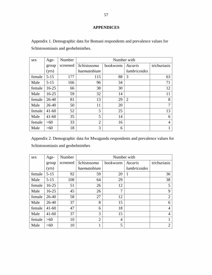
57
APPENDICES
Appendix 1. Demographic data for Bomani respondents and prevalence values for
Schistosomiasis and geohelminthes.
sex Age-
group
(yrs)
Number
screened
Number with
Schistosoma
haematobium
hookworm Ascaris
lumbricoides
trichuriasis
female 5-15 177 115 88 3 63
Male 5-15 166 96 34 71
female 16-25 66 38 30 12
Male 16-25 59 32 14 11
female 26-40 81 13 29 2 8
Male 26-40 50 11 20 7
female 41-60 52 5 25 13
Male 41-60 35 5 14 6
female >60 33 2 16 4
Male >60 18 3 6 1
Appendix 2. Demographic data for Mwagundu respondents and prevalence values for
Schistosomiasis and geohelminthes
sex Age-
group
(yrs)
Number
screened
Number with
Schistosoma
haematobium
hookworm Ascaris
lumbricoides
trichuriasis
female 5-15 92 59 20 1 36
Male 5-15 108 64 29 38
female 16-25 51 26 12 5
Male 16-25 45 26 7 9
female 26-40 58 27 12 2
Male 26-40 37 8 15 6
female 41-60 47 6 18 4
Male 41-60 37 3 15 4
female >60 10 2 4 1
Male >60 10 1 5 2
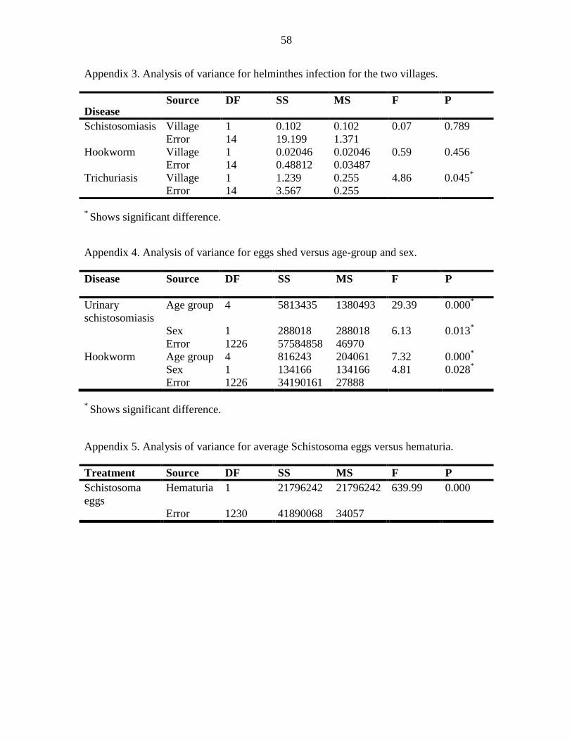
58
Appendix 3. Analysis of variance for helminthes infection for the two villages.
Disease Source DF SS MS F P
Schistosomiasis Village 1 0.102 0.102 0.07 0.789
Error 14 19.199 1.371
Hookworm Village 1 0.02046 0.02046 0.59 0.456
Error 14 0.48812 0.03487
Trichuriasis Village 1 1.239 0.255 4.86 0.045*
Error 14 3.567 0.255
* Shows significant difference.
Appendix 4. Analysis of variance for eggs shed versus age-group and sex.
Disease Source DF SS MS F P
Urinary
schistosomiasis
Age group 4 5813435 1380493 29.39 0.000*
Sex 1 288018 288018 6.13 0.013*
Error 1226 57584858 46970
Hookworm Age group 4 816243 204061 7.32 0.000*
Sex 1 134166 134166 4.81 0.028*
Error 1226 34190161 27888
* Shows significant difference.
Appendix 5. Analysis of variance for average Schistosoma eggs versus hematuria.
Treatment Source DF SS MS F P
Schistosoma
eggs
Hematuria 1 21796242 21796242 639.99 0.000
Error 1230 41890068 34057


![Deep, multi-stage transcriptome of the schistosomiasis vector … · 2017. 8. 28. · schistosomiasis - Schistosoma mansoni [7], Schistosoma japonicum [53] and Schistosoma haematobium](https://img.pdfslide.net/doc/110x75/60f8a53e7bdd0764ad39282d/deep-multi-stage-transcriptome-of-the-schistosomiasis-vector-2017-8-28-schistosomiasis.jpg)