Embed Size (px)
Citation preview
EXAMINING WAYS TO IMPROVE
ANKLE MOBILITY DURING THE OVERHEAD SQUAT LIFT
by
George Larson
A thesis submitted to the Faculty of the University of Delaware in partial
fulfillment of the requirements for the degree of Master of Science in Exercise Science
Summer 2014
© 2014 George Larson
All Rights Reserved
All rights reserved
INFORMATION TO ALL USERSThe quality of this reproduction is dependent upon the quality of the copy submitted.
In the unlikely event that the author did not send a complete manuscriptand there are missing pages, these will be noted. Also, if material had to be removed,
a note will indicate the deletion.
Microform Edition © ProQuest LLC.All rights reserved. This work is protected against
unauthorized copying under Title 17, United States Code
ProQuest LLC.789 East Eisenhower Parkway
P.O. Box 1346Ann Arbor, MI 48106 - 1346
UMI 1567813Published by ProQuest LLC (2014). Copyright in the Dissertation held by the Author.
UMI Number: 1567813
EXAMINING WAYS TO IMPROVE ANKLE MOBILITY
DURING THE OVERHEAD SQUAT LIFT
by
George Larson
Approved: __________________________________________________________
Thomas W. Kaminski, Ph.D., ATC, FNATA, FACSM
Professor in charge of thesis on behalf of the Advisory Committee
Approved: __________________________________________________________
William B. Farquhar, Ph.D.
Chair of the Department of Kinesiology and Applied Physiology
Approved: __________________________________________________________
Kathleen S. Matt, Ph.D.
Dean of the College of Health Sciences
Approved: _________________________________________________________
James G. Richards, Ph.D.
Vice Provost for Graduate and Professional Education
iii
ACKNOWLEDGMENTS
Several people aided in the accomplishment of this project. First, I would like
to thank my advisor, Dr. Thomas Kaminski, for his patience, time, and support
throughout the stages of this two-year process. Additionally, I would like to
acknowledge Dr. Todd Royer and Dr. Joseph Glutting for serving on my thesis
committee and aiding in the writing process and statistical analysis, respectively.
Without permission from Dr. Kelly Starrett, this project would not have been
possible. I am thankful that he took time out of his busy schedule to offer advice and
support over many phone calls from San Francisco, California. I would also like to
acknowledge Jaclyn Caccese, my doctoral student mentor, who assisted and was
always enthusiastic in helping me with project design, understanding instrumentation,
data collection, and running statistics. Johanna Lukk and Nathanial Rosal were also a
huge help during data collection and deserve recognition. I would like to thank my
fellow lab mates for their varying contributions to this project and my overall graduate
experience: Andrea DiTrani, Aaron Struminger, Yong Woo An, Sean Stryker, Shawn
Hanlon, Molly Johnson, Kelly McGuire, Kelsey Shonk, and Alex Salinas. Finally, I
would like to acknowledge the Strength and Conditioning Staff for their support
during this project: Brian Hess, Trevor Williams, Melissa Boldt, and Kyle Hobbs.
The past two years would not have been possible without the love and support
of my parents, George and Michele, and my sister, Jennifer. You all offered me peace
of mind during this stressful endeavor.
iv
TABLE OF CONTENTS
LIST OF TABLES ........................................................................................................ vi
LIST OF FIGURES ...................................................................................................... vii ABSTRACT .................................................................................................................. ix
Chapter
1 INTRODUCTION .............................................................................................. 1
2 SUBJECTS AND METHODS ........................................................................... 4
2.1 Experimental Approach to the Problem .................................................... 4
2.2 Subjects ...................................................................................................... 5 2.3 Procedures ................................................................................................. 5
2.3.1 Biomechanical Measurements ....................................................... 7
2.3.2 Interventions ................................................................................. 8
2.4 Post-Intervention Testing ........................................................................ 10
2.5 Statistical Analysis .................................................................................. 10
3 RESULTS ......................................................................................................... 11
3.1 Ankle Dorsiflexion Range-of-Motion ..................................................... 11
3.2 Torso Angle ............................................................................................. 12
3.3 Squat Depth ............................................................................................. 12
3.4 Shin Angle ............................................................................................... 13
3.5 Knee Flare ............................................................................................... 13
3.6 Foot Width ............................................................................................... 13
3.7 Ankle Eversion ........................................................................................ 13
4 DISCUSSION ................................................................................................... 14
5 TABLES AND FIGURES ................................................................................ 19
REFERENCES ............................................................................................................. 39
v
Appendix
A INFORMED CONSENT FORM ..................................................................... 42
B DATA COLLECTION SHEET ....................................................................... 51 C SPECIFIC AIMS .............................................................................................. 52 D BACKGROUND AND SIGNIFICANCE ....................................................... 56
D.1 Background ............................................................................................. 56
D.2 Significance -
The Squat and how it Applies to Modern Sport and Strength and
Conditioning Programs ........................................................................... 58
D.3 Significance - Execution of the Squat .................................................... 61
D.3.1 Stance Variation .......................................................................... 61
D.3.2 Sitting Back ................................................................................. 62
D.3.3 Muscle Activation ........................................................................ 66
D.4 Significance - Forces Placed on the Knee .............................................. 67
D.5 Significance - Assumptions How to Squat ............................................. 68
D.6 Significance - Contemporary Concepts on Squat Execution .................. 70
D.6.1 Hip Mobility and the Squat ......................................................... 71
D.6.2 Neutral Spine and the Squat ........................................................ 71
D.6.3 Ankle Mobility and the Squat ...................................................... 71
D.7 Significance - Flexibility vs. Mobility .................................................... 74
D.7.1 Soft Tissue ................................................................................... 75
D.7.2 Stretching ..................................................................................... 75
D.7.3 Joint Mobilization ........................................................................ 76
D.8 Summary ................................................................................................. 76
E SPSS ANCOVA DATA TABLES ................................................................... 77

vi
LIST OF TABLES
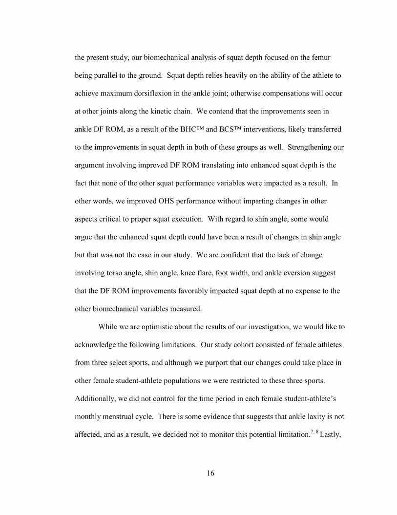
Table 1: Participant demographics (Mean ± SD) ..................................................... 19
vii
LIST OF FIGURES
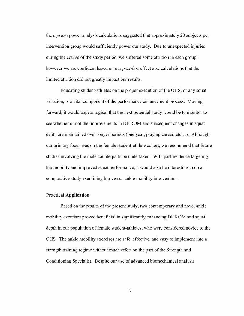
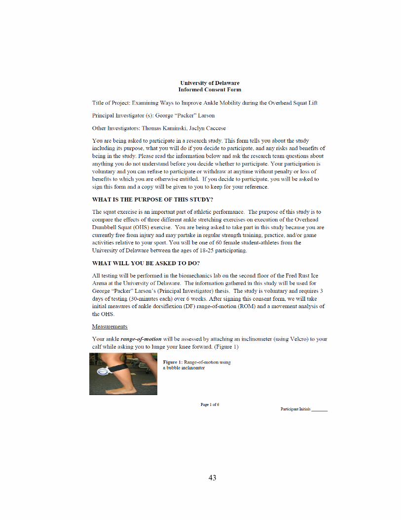
Figure 1: Range-of-motion using a bubble inclinometer ........................................ 20
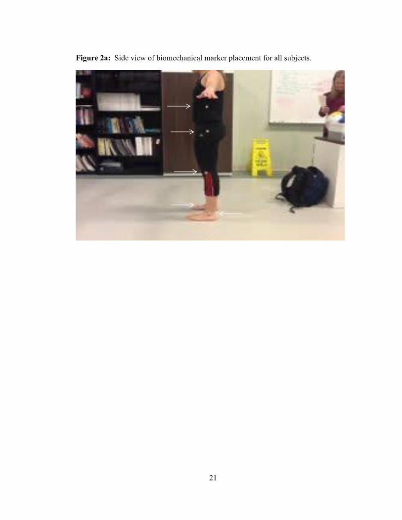
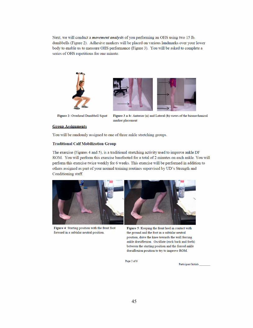
Figure 2a: Side view of biomechanical marker placement for all subjects. ............. 21
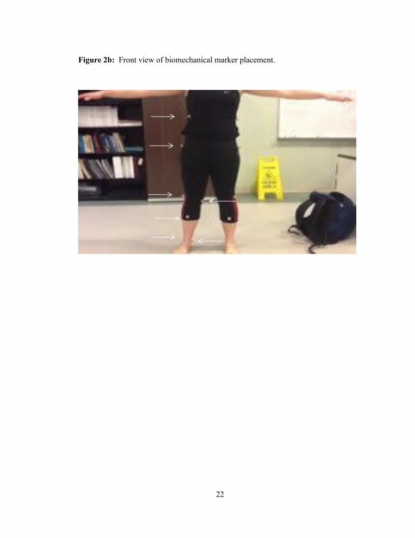
Figure 2b: Front view of biomechanical marker placement.. ................................... 22
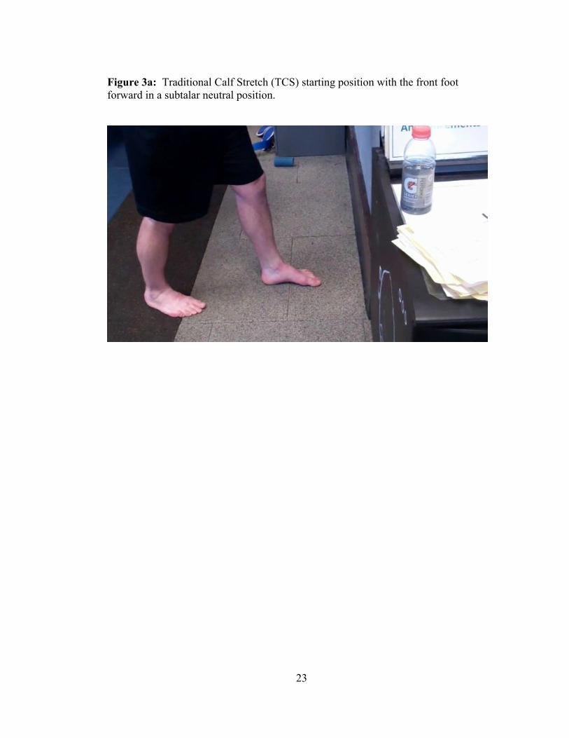
Figure 3a: Traditional Calf Stretch (TCS) starting position with the front foot
forward in a subtalar neutral position ...................................................... 23
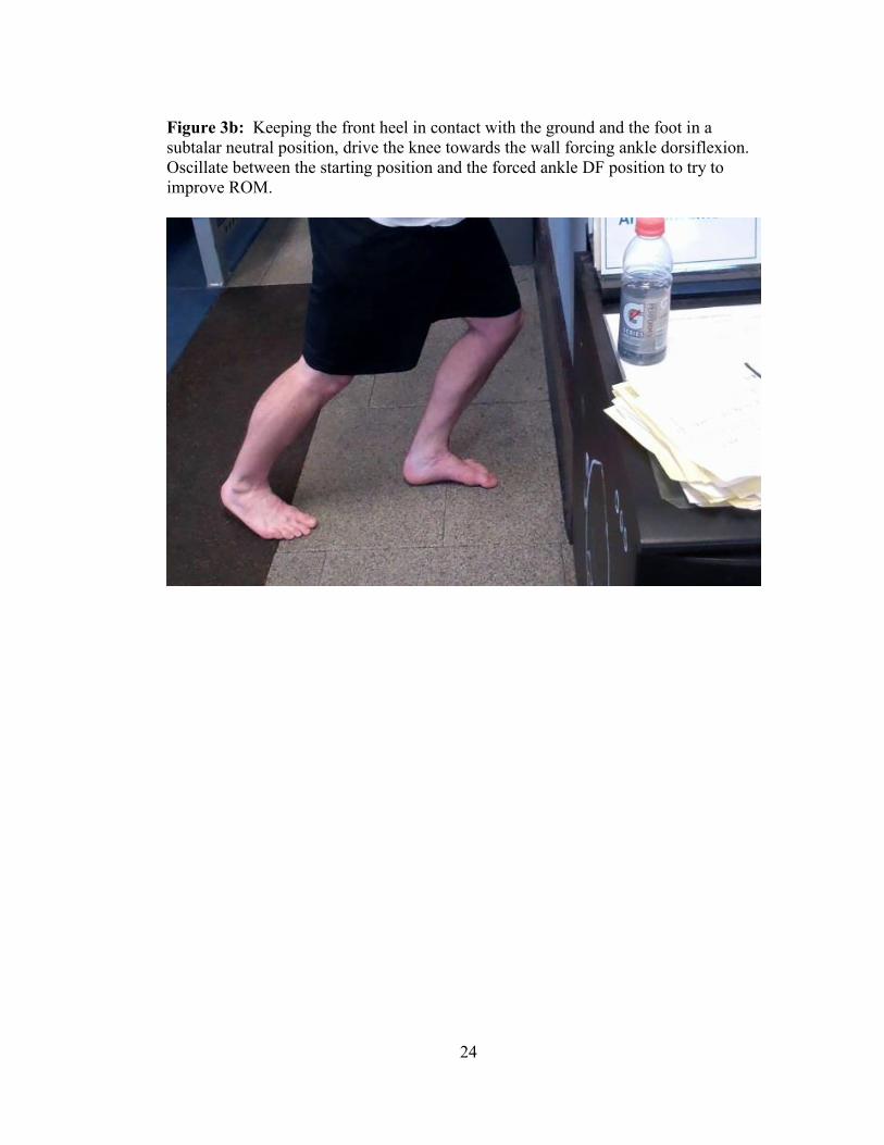
Figure 3b: Oscillate between the starting position and the forced ankle DF position
to try to improve ROM ............................................................................ 24
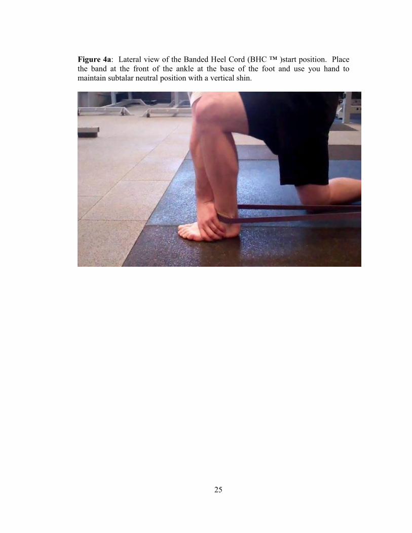
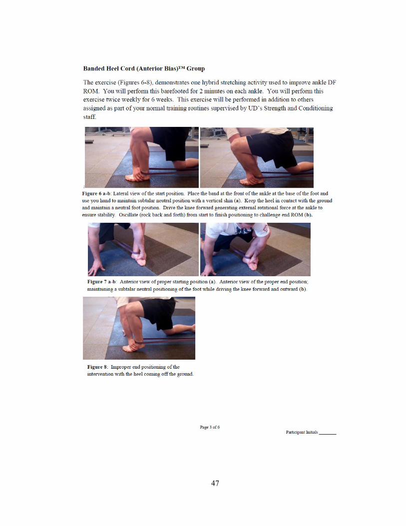
Figure 4a: Lateral view of the Banded Heel Cord (BHC ™ )start position. Place
the band at the front of the ankle at the base of the foot and use you
hand to maintain subtalar neutral position with a vertical shin. .............. 25
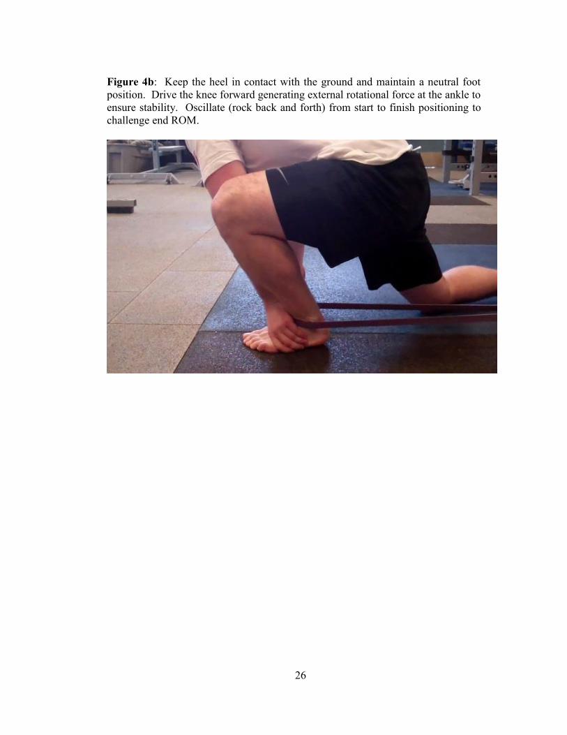
Figure 4b: Keep the heel in contact with the ground and maintain a neutral foot
position. Drive the knee forward generating external rotational force
at the ankle to ensure stability. Oscillate (rock back and forth) from
start to finish positioning to challenge end ROM. ................................... 26
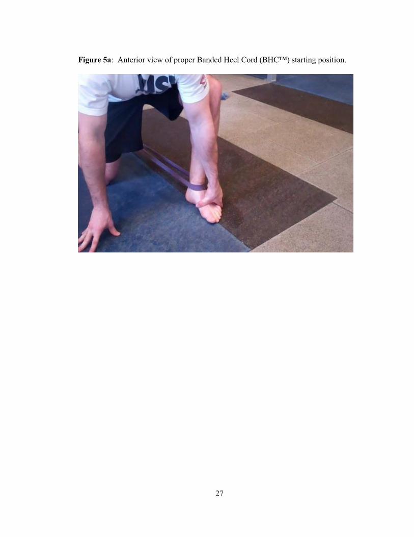
Figure 5a: Anterior view of proper Banded Heel Cord (BHC™) starting position
. ............................................................................................................. ...27
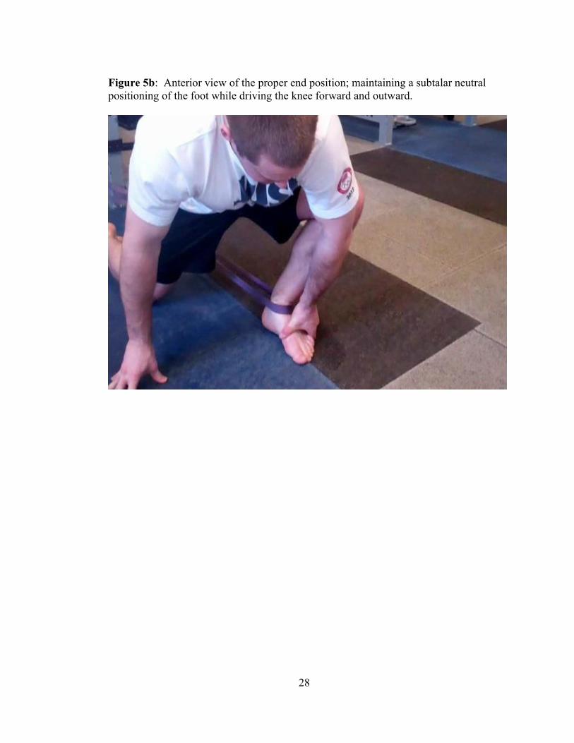
Figure 5b: Anterior view of the proper end position; maintaining a subtalar
neutral positioning of the foot while driving the knee forward and
outward. ................................................................................................ ...28
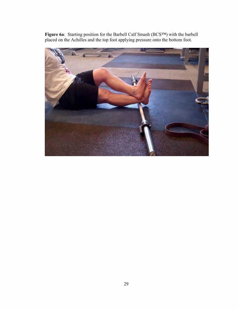
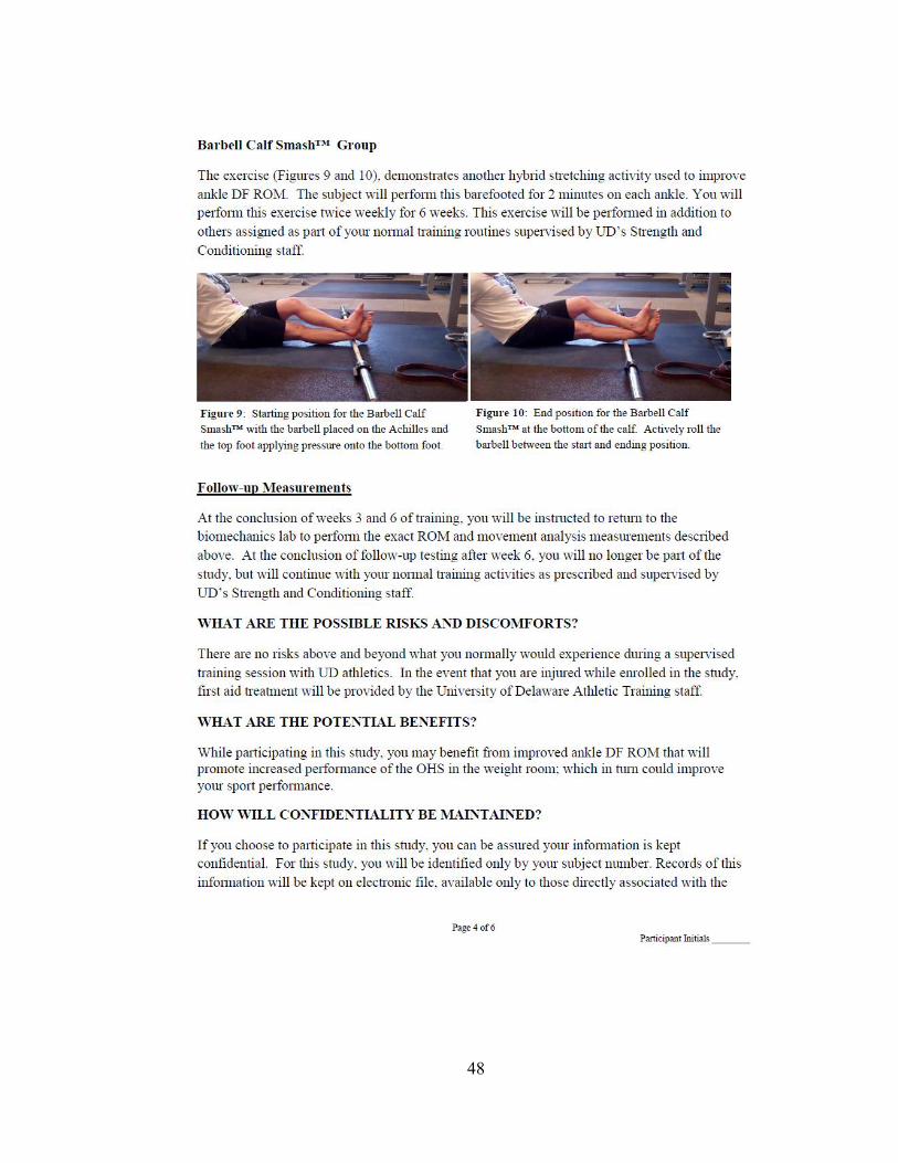
Figure 6a: Starting position for the Barbell Calf Smash (BCS™) with the barbell
placed on the Achilles and the top foot applying pressure onto the
bottom foot. ............................................................................................. 29
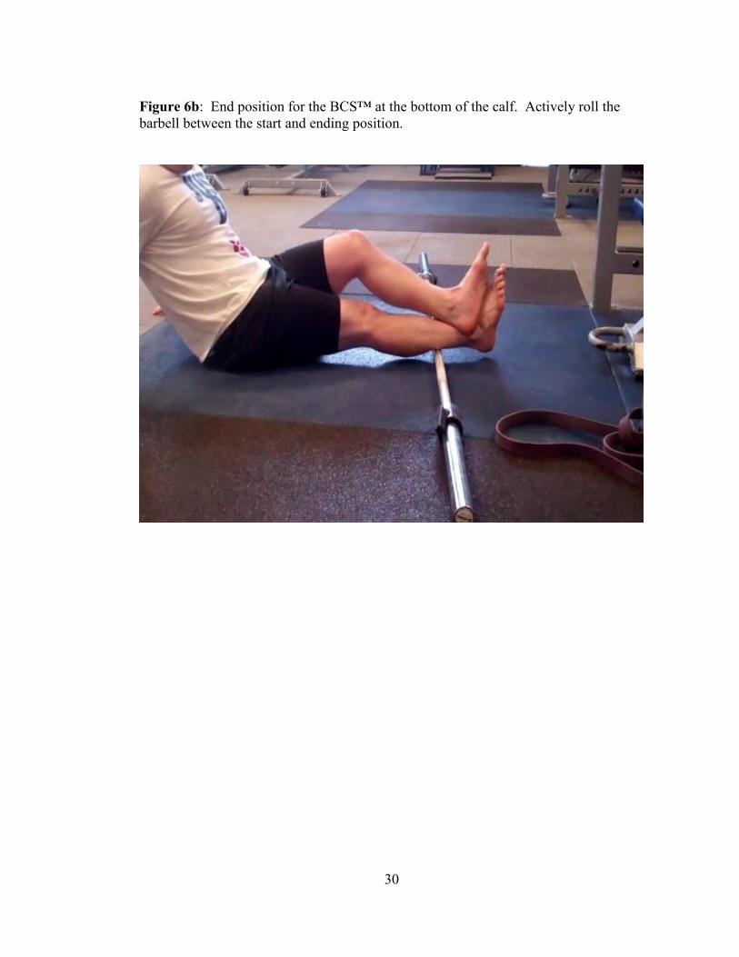
Figure 6b: End position for the BCS™ at the bottom of the calf. Actively roll the
barbell between the start and ending position. ........................................ 30
viii
Figure 7: Right DF ROM between the TCS, BHC™, and BCS™ intervention
groups. Note: * indicates significance between baseline and final
measurement. ........................................................................................... 31
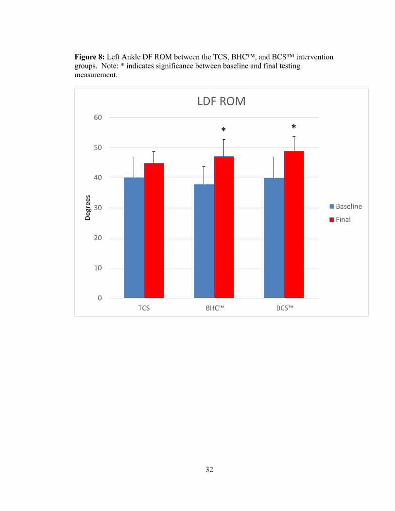
Figure 8: Left DF ROM between the TCS, BHC™, and BCS™ intervention
groups. Note: * indicates significance between baseline and final
measurement. ........................................................................................... 32
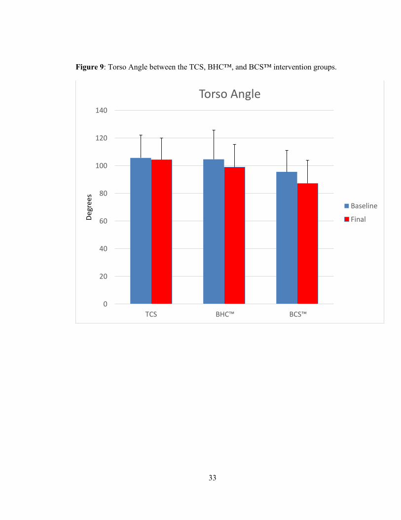
Figure 9: Torso Angle between the TCS, BHC™, and BCS™ intervention
groups. ..................................................................................................... 33
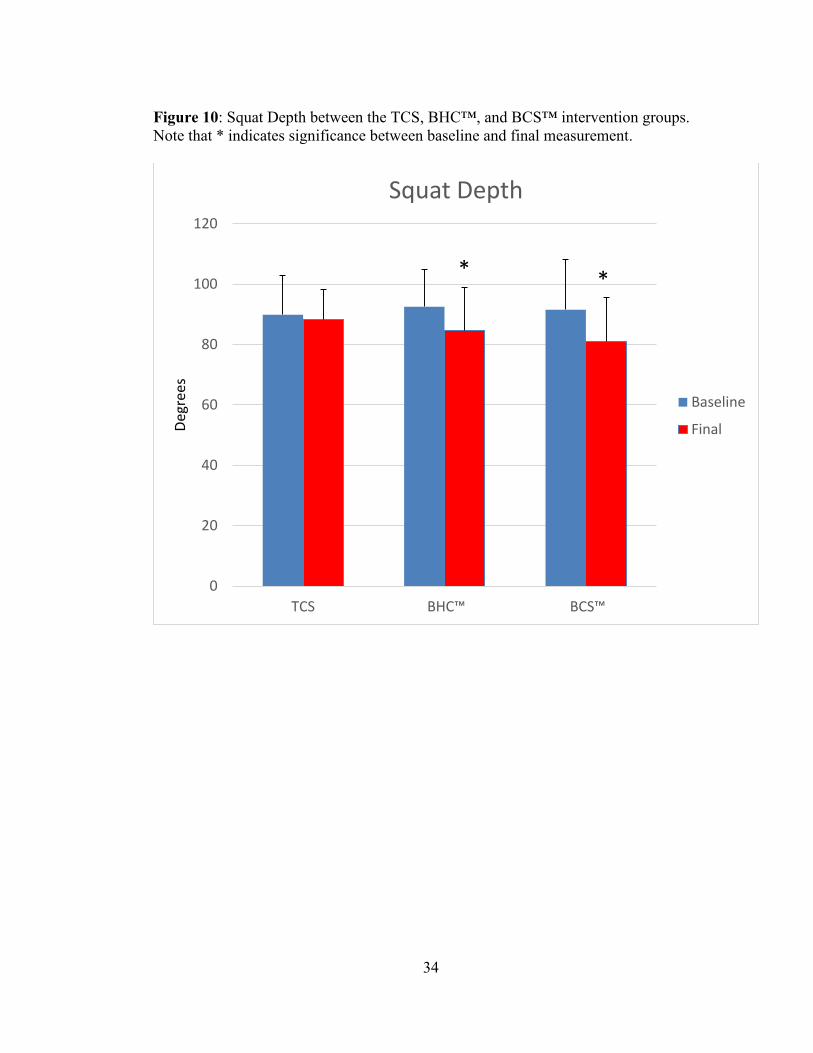
Figure 10: Squat Depth between the TCS, BHC™, and BCS™ intervention groups.
Note that * indicates significance between baseline and final
measurement. ........................................................................................... 34
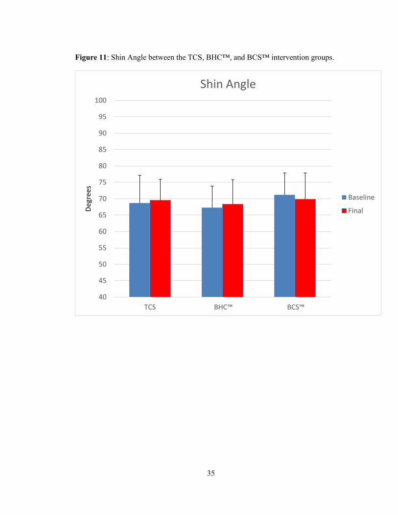
Figure 11: Shin Angle between the TCS, BHC™, and BCS™ intervention groups.
................................................................................................................. 35
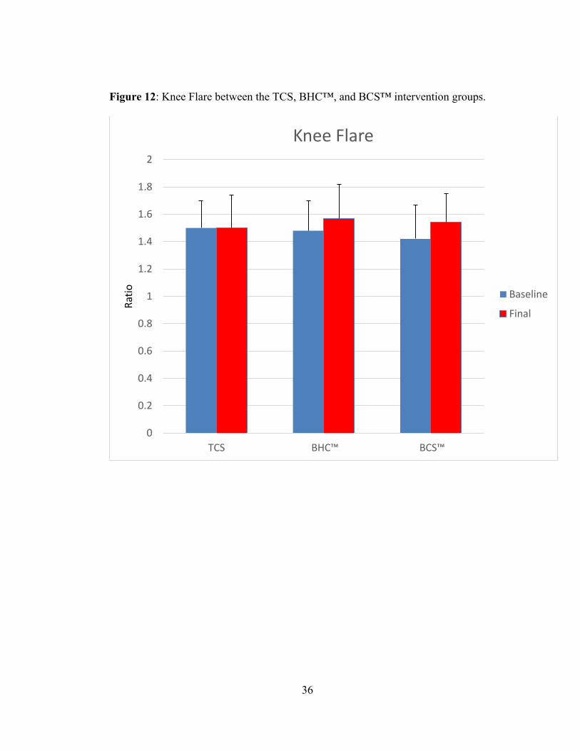
Figure 12: Knee Flare distance (cm) between the TCS, BHC™, and BCS™
intervention groups. ................................................................................. 36
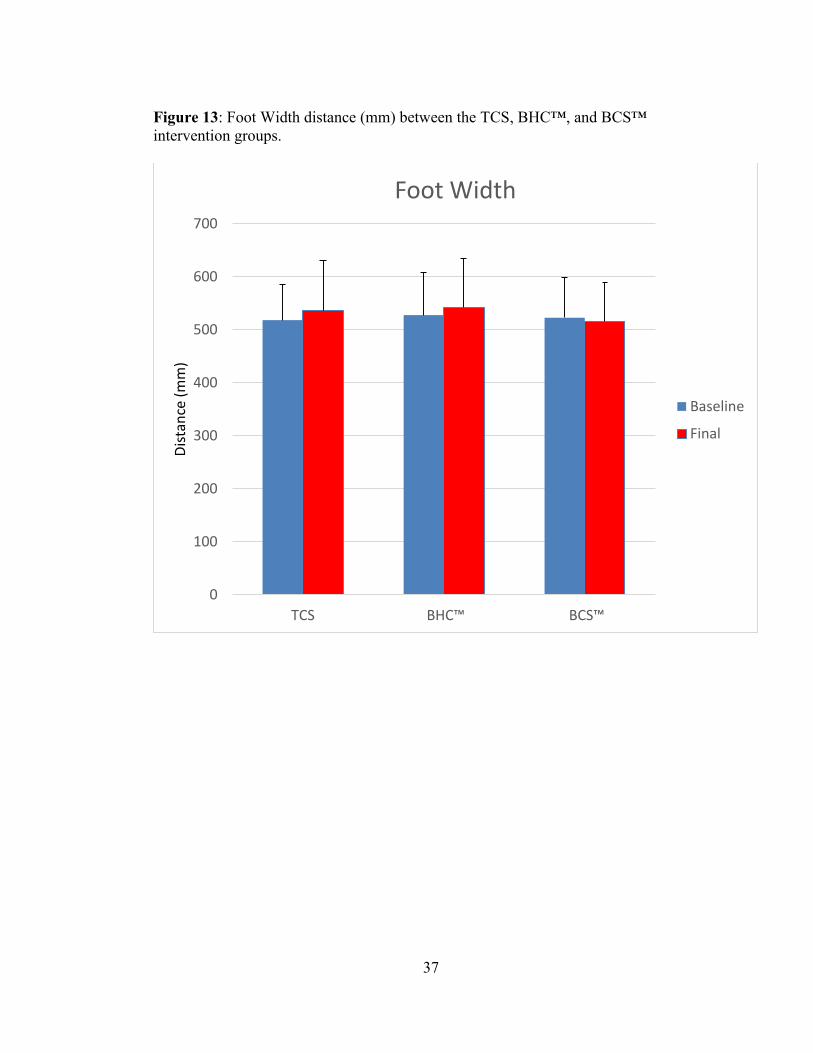
Figure 13: Foot Width distance (cm) between the TCS, BHC™, and BCS™
intervention groups .................................................................................. 37
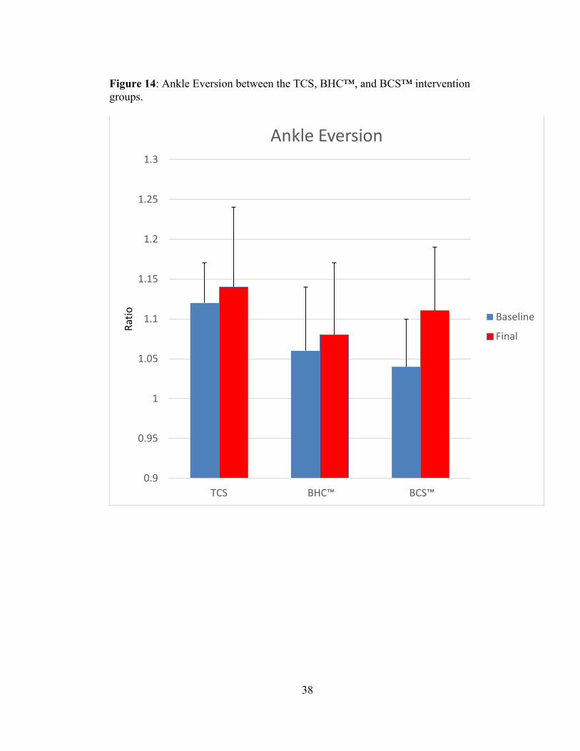
Figure 14: Ankle Eversion between the TCS, BHC™, and BCS™ intervention
groups ...................................................................................................... 38
ix
ABSTRACT
To perform a proper squat, athletes must be mobile in the ankle, knee, and hip while
maintaining a strong torso to protect the musculature surrounding the spine. Deficits
in mobility, especially at the foot and ankle can cause compensation resulting in the
widening of the feet and/or turning the toes outward to achieve proper squat depth.
The purpose of this study is to analyze the overhead squat (OHS) in a group of female
collegiate athletes and implement an appropriate ankle dorsiflexion mobility exercise
program to improve squat performance. A total of 44 female student-athletes (age 18-
25) were randomly divided into three treatment interventions: (1) Traditional Calf
Stretch technique; (2) a joint-capsule release intervention termed “Banded Heel Cord
(Anterior Bias)™”; and (3) a soft tissue intervention termed “Barbell Calf Smash™”.
A series of measurements were made to analyze the OHS including ankle dorsiflexion
motion and movement analysis (torso angle, squat depth, shin angle, knee flare, foot
width, and ankle eversion motion) at baseline and following 6 weeks of training.
Analysis of covariance (ANCOVA) tests were used for comparisons between and
within the three intervention groups. Improvements in DF ROM following the 6 week
intervention period averaged greater than 20% in both the BHC™ and BCS™ groups;
leading to significant improvements in squat depth in both groups as well. Two
contemporary and novel ankle mobility exercises proved beneficial in significantly
x
enhancing DF ROM and squat depth in our population of female student-athletes, who
were considered novice to the OHS.
Keywords: strength, power, MobilityWOD™, biomechanics, dorsiflexion
Formatted for submission to: Journal of Strength & Conditioning Research
1
Chapter 1
INTRODUCTION
The field of Strength and Conditioning has evolved tremendously over the
years to accommodate the needs of the modern athlete. Sport coaches are
acknowledging the importance of year-round strength and conditioning programs for
their teams. Athletes, ranging from novice to the professional level, seek sport
performance specialists during their off-seasons to train and gain a competitive edge.
Athletes need cardiovascular endurance, strength, power, coordination, agility, speed,
flexibility, and mobility training to be successful in today’s sporting endeavors. It is
recognized that training these variables in a weight room or sports performance setting
can lead to success in sport and injury prevention, especially in the female
population.11-14, 16-18
The squat, no matter the variation, is a fundamental strengthening exercise we
perform in everyday life that is essential to many, if not all, strength and conditioning
programs working with athletes or an athletic population. To perform a proper squat,
a person must be mobile in the ankle, knee, and hip while maintaining a strong torso to
protect the musculature surrounding the spine. If an athlete lacks mobility and
strength, they may compensate their technique and increase their risk of injury. A
common compensation is to widen the feet and/or turn the toes outward to achieve
proper squat depth.1, 3, 5, 10, 22
While stance width is highly debatable, this study will
2
look at ankle mobility and how it may be a limiting factor during the execution of the
squat maneuver. Increased ankle dorsiflexion (DF) range-of-motion (ROM) leads to
increased force production which can carry over to skills essential in sports requiring
sprinting, cutting, and jumping.22
Conversely, compensation of ankle DF ROM
during the squat may in turn be limiting the potential for force production during many
sport performance maneuvers.
Flexibility has always been an area of interest to Strength and Conditioning
Specialists, with a primary focus on lengthening muscles that are short and tight.
Many athletes have incorporated dynamic warm-ups or static stretching exercises to
correct issues with flexibility and prepare for on-the-field sport performance.
Mobility, or mobilization, is a movement-based integrated full-body approach that
addresses all the elements that limit movement and performance including short and
tight muscles, soft tissue restriction, joint capsule restriction, motor control problems,
joint range-of-motion dysfunction, and neural dynamic issues.22
Therefore, mobility is
a tool that can be used to address issues involving poor movement and performance
patterns in the athletic population. General joint mobility is important for execution in
sport and training maneuvers and can involve a variety of interventions.22
Specific to
the squat maneuver, ankle DF ROM is of importance.19
Traditional methods used to
improve DF ROM have included techniques (manual or device-aided) that stretch the
calf musculature. Recently, alternative techniques to improve ankle DF ROM have
been introduced, however there is no evidence to support their superiority versus
traditional methods. Whereas traditional techniques have focused on lengthening the
3
calf musculature, proponents of these alternative techniques suggest that DF ROM can
be enhanced by improvements in joint capsule integrity as well as improvements in
soft tissue pliability.4-6, 10, 15, 22
A dilemma facing Strength and Conditioning Specialists each year is the arrival
of an incoming recruitment class and their experience with the squat maneuver,
including the Overhead Dumbbell Squat (OHS). Proper execution is especially
important with the female athletic population who are at greater risk for lower
extremity injury.11, 12, 13
Therefore the purpose of this study was to analyze the OHS in
a group of female collegiate athletes and implement an appropriate ankle DF mobility
exercise program to improve squat performance. The study was guided by the
following aims: to evaluate a group of female athletes from select sports at a large
collegiate athletic program to identify which athletes can perform a proper OHS, and
then to determine the effect of three different ankle mobility interventions on measures
of change in squat performance and ankle DF ROM. We hypothesized that
improvements in ankle DF ROM through mobility interventions would improve
overall positioning of the body during the squat maneuver.
4
Chapter 2
SUBJECTS AND METHODS
2.1 Experimental Approach to the Problem
The study included one independent variable with 3 levels, and seven dependent
variables. The independent variable in this study was the intervention effect of the
Traditional Calf Stretch (TCS), Banded Heel Cord (Anterior Bias)™ (BHC™), and
Barbell Calf Smash™ (BCS™) exercises. The dependent variables included: ankle
DF ROM, torso angle, squat depth, shin angle, knee flare, foot width, and ankle
eversion motion. A pretest-posttest randomized design was employed with subjects
executing the OHS. To our knowledge, no other study has observed changes in squat
position during a continuous, or multiple repetition set, making this the first functional
experiment for this movement. We utilized the OHS because it forces the subjects to
squat in the most upright torso position and requires sufficient ankle DF ROM to reach
proper depth (femur parallel to the ground). Dumbbells were chosen to use during the
OHS so as to reduce the risk of allowing the subjects to compensate their torso
position by widening their hands. If the subjects attempted to widen their hands, they
would drop the dumbbells and be unable to complete a successful repetition, leading
us to believe that an ankle mobility issue may be the cause of the failed repetition.
5
2.2 Subjects
A total of 60 female student-athletes, ranging in age from 18-25 years, were
recruited and randomly divided into the three treatment interventions. The student-
athletes were recruited from the Tennis, Golf, and Rowing/Crew teams. All subjects
signed an informed consent agreement approved by the university’s IRB (451551-1)
and provided preliminary demographic information (Table 1). All subjects were free
from injury at the time of the study. Due to injuries sustained during their given
athletic season, 16 of the participants withdrew from the study leaving 44 healthy
participants for the final analysis.
2.3 Procedures
All testing occurred in a climate controlled Biomechanics Laboratory with
subjects in bare feet and clothing that did not restrict movement. Dorsiflexion ROM
was assessed by attaching an inclinometer to the calf while asking the subject to lunge
the knee forward, keeping the heel on the ground (Figure 1). Movement (degrees) was
read from the inclinometer with a single measurement derived from both ankles.
Biomechanical analysis was assessed during the execution of the OHS. Prior to
performing the OHS, a set of biomechanical (adhesive reflective) markers were
attached to each subject; including placement on the top of both feet in line with the
2nd
metatarsal, mid-tibia of each leg, on both lateral and medial malleoli of each ankle,
the lateral and medial epicondyles of each knee, the greater trochanter of each femur,
6
and on the fifth intercostal space on the right and left side of the body in-line with the
marker on the greater trochanter (Figure 2). The movement (kinematic) analysis
enabled for the measurements of: torso angle, squat depth, shin angle, knee flare, foot
width, and ankle eversion. To ensure accuracy between pre and post-test
measurements (6 weeks), each marker was measured from a specific point either on
the body or between markers during the initial baseline setup. For example, the toe
marker was measured as a distance (cm) from the position it was placed on the top of
the foot to the end of the 2nd
metatarsal. Distances were also recorded between: the
medial ankle and medial knee, the lateral ankle and lateral knee, the tibial tuberosity to
mid-tibia, the lateral knee and the greater trochanter, and lastly from the greater
trochanter and the ribcage.
Subjects were asked to perform 5 OHS repetitions continuously while holding
the dumbbells with their palms facing away (forward) from them. The subjects were
instructed to squat as low as possible keeping their elbows locked out and their heels
on the ground. Being novice to the OHS, no instruction was given to our subjects with
regard to foot width and position, knee flare, or action of the torso. Fifteen- pound
dumbbells were used by all subjects to execute the OHS. The thirty pound total
weight (one dumbell/hand) is nearly equivalent to the official women’s Olympic
barbell of 15 kg (33 lbs). The entire baseline measurement session lasted 30 minutes.
7
2.3.1 Biomechanical Measurements
Torso Angle
Torso angle (degrees) was measured from the angle created between the
biomechanical markers on the outside of the ribcage, the marker on the greater
trochanter, and the marker on the lateral knee.
Squat Depth
Squat depth (degrees) was determined from the angle formed between the
greater trochanter, the lateral femoral epicondyle, and the lateral ankle.
Shin Angle
Shin angle (degrees) was formed by the markers placed on the lateral femoral
epicondyle, the lateral malleolus, and the top of the foot in line with the 2nd
metatarsal.
Knee Flare
Knee flare was determined by the ratio of the distance (mm) between the
biomechanical markers placed on each greater trochanter, and the distance between
the markers placed on each lateral epicondyle of the knee at the bottom of each squat
repetition.
Foot Width
Foot width was determined from the distance (mm) between the biomechanical
markers on the lateral malleoli on each ankle at the bottom position of each squat
repetition.
8
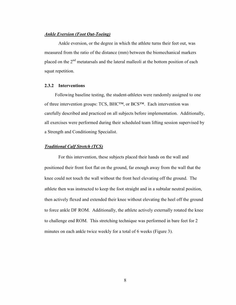
Ankle Eversion (Foot Out-Toeing)
Ankle eversion, or the degree in which the athlete turns their feet out, was
measured from the ratio of the distance (mm) between the biomechanical markers
placed on the 2nd
metatarsals and the lateral malleoli at the bottom position of each
squat repetition.
2.3.2 Interventions
Following baseline testing, the student-athletes were randomly assigned to one
of three intervention groups: TCS, BHC™, or BCS™. Each intervention was
carefully described and practiced on all subjects before implementation. Additionally,
all exercises were performed during their scheduled team lifting session supervised by
a Strength and Conditioning Specialist.
Traditional Calf Stretch (TCS)
For this intervention, these subjects placed their hands on the wall and
positioned their front foot flat on the ground, far enough away from the wall that the
knee could not touch the wall without the front heel elevating off the ground. The
athlete then was instructed to keep the foot straight and in a subtalar neutral position,
then actively flexed and extended their knee without elevating the heel off the ground
to force ankle DF ROM. Additionally, the athlete actively externally rotated the knee
to challenge end ROM. This stretching technique was performed in bare feet for 2
minutes on each ankle twice weekly for a total of 6 weeks (Figure 3).
9
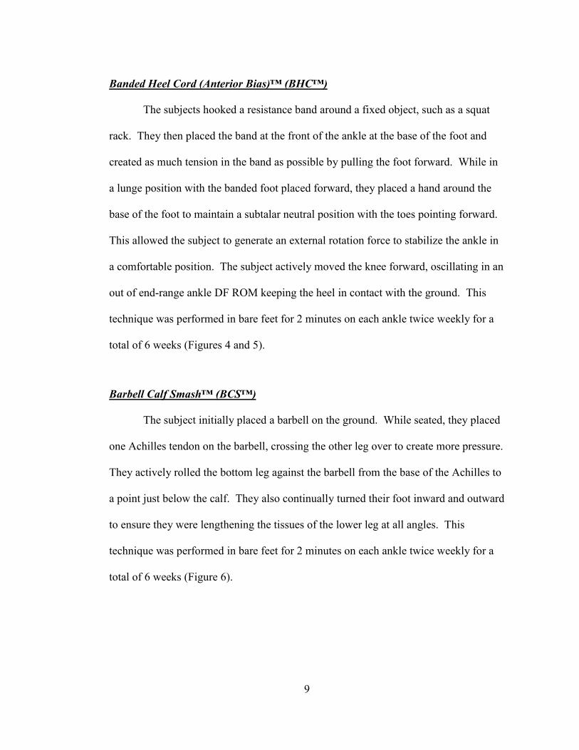
Banded Heel Cord (Anterior Bias)™ (BHC™)
The subjects hooked a resistance band around a fixed object, such as a squat
rack. They then placed the band at the front of the ankle at the base of the foot and
created as much tension in the band as possible by pulling the foot forward. While in
a lunge position with the banded foot placed forward, they placed a hand around the
base of the foot to maintain a subtalar neutral position with the toes pointing forward.
This allowed the subject to generate an external rotation force to stabilize the ankle in
a comfortable position. The subject actively moved the knee forward, oscillating in an
out of end-range ankle DF ROM keeping the heel in contact with the ground. This
technique was performed in bare feet for 2 minutes on each ankle twice weekly for a
total of 6 weeks (Figures 4 and 5).
Barbell Calf Smash™ (BCS™)
The subject initially placed a barbell on the ground. While seated, they placed
one Achilles tendon on the barbell, crossing the other leg over to create more pressure.
They actively rolled the bottom leg against the barbell from the base of the Achilles to
a point just below the calf. They also continually turned their foot inward and outward
to ensure they were lengthening the tissues of the lower leg at all angles. This
technique was performed in bare feet for 2 minutes on each ankle twice weekly for a
total of 6 weeks (Figure 6).
10
2.4 Post-Intervention Testing
At the conclusion of the 6 week training period, our subjects were instructed to
return to the Biomechanics Laboratory for follow-up OHS biomechanical analysis.
The exact procedures described above were again used at this post-intervention
session.
2.5 Statistical Analysis
Statistical analysis was performed using SPSS version 20.0 software (IBM, Armonk,
NY). Analysis of Covariance (ANCOVA) was utilized for comparisons between, and
within, the three intervention groups (TCS, BHC™, BCS™). Separate ANCOVA
tests were utilized for each of the 7 dependent variables between baseline and post-
intervention measurements. Post-hoc comparisons were apportioned using Bonferroni
adjustment. The significance level was set at p ≤ 0.05 for all analyses.
11
Chapter 3
RESULTS
3.1 Ankle Dorsiflexion Range-of-Motion
Dorsiflexion ROM values across the three intervention groups, both pre and post-test,
ranged from 25° to 57° . The results of the ANCOVA are presented separately for
both the right and left ankles. There was a significant difference in DF ROM for the
right ankle measurements [F (2, 40) = 7.82, p = .001]. The effect size for right ankle
DF ROM was small (d = .29). Post hoc comparisons showed that the TCS group had
significantly lower DF ROM values than either the BHC™ (p = .002) or the BCS™ (p
= .008) groups. The BHC™ versus BCS™ comparison was not statistically
significant. The right ankle showed greater improvements in DF ROM after 6 weeks
of intervention training using the modern BHC™ (37.3° to 48.3° = 11° [29%
improvement]) and BCS™ (39.8° to 49.4° = 9.6° [24% improvement]) interventions
as opposed to the TCS (41.4° to 44.8° = 3.4° [8% improvement]) technique as
compared to their baseline values (Figure 7).
There was a significant difference in DF ROM for the left ankle measurements
[F (2, 40) = 4.22, p = .022]. The effect size for left ankle DF ROM was small (d =
.27). Post hoc comparisons showed that the TCS group had significantly lower values
than BCS group (p = .041). None of the other post hoc comparisons were significant.
The left ankle showed greater improvements in DF ROM after 6 weeks of intervention
12
training using the modern BHC™ (37.8° to 47.0° = 9.2° [24% improvement]) and
BCS™ (39.9° to 48.9° = 8° [20% improvement]) interventions as opposed to the TCS
(40.1° to 44.8° = 4.7° [12% improvement]) technique as compared to their baseline
values (Figure 8).
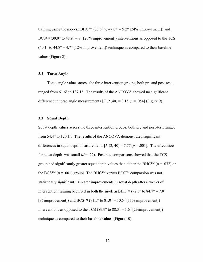
3.2 Torso Angle
Torso angle values across the three intervention groups, both pre and post-test,
ranged from 61.6° to 137.1°. The results of the ANCOVA showed no significant
difference in torso angle measurements [F (2 ,40) = 3.15, p = .054] (Figure 9).
3.3 Squat Depth
Squat depth values across the three intervention groups, both pre and post-test, ranged
from 54.4° to 120.1°. The results of the ANCOVA demonstrated significant
differences in squat depth measurements [F (2, 40) = 7.77, p = .001]. The effect size
for squat depth was small (d = .22). Post hoc comparisons showed that the TCS
group had significantly greater squat depth values than either the BHC™ (p = .032) or
the BCS™ (p = .001) groups. The BHC™ versus BCS™ comparsion was not
statistically significant. Greater improvements in squat depth after 6 weeks of
intervention training occurred in both the modern BHC™ (92.5° to 84.7° = 7.8°
[8%improvement]) and BCS™ (91.5° to 81.0° = 10.5° [11% improvement])
interventions as opposed to the TCS (89.9° to 88.3° = 1.6° [2%improvement])
technique as compared to their baseline values (Figure 10).
13
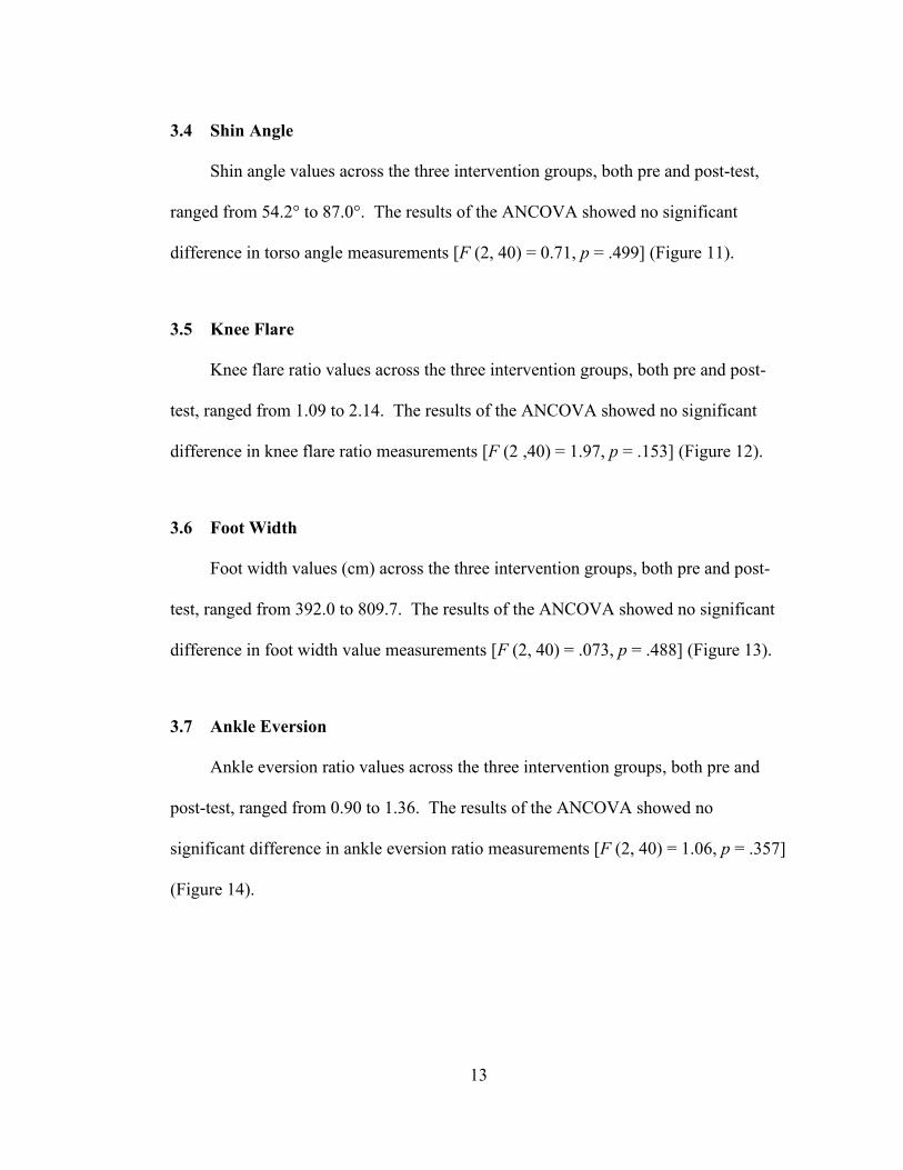
3.4 Shin Angle
Shin angle values across the three intervention groups, both pre and post-test,
ranged from 54.2° to 87.0°. The results of the ANCOVA showed no significant
difference in torso angle measurements [F (2, 40) = 0.71, p = .499] (Figure 11).
3.5 Knee Flare
Knee flare ratio values across the three intervention groups, both pre and post-
test, ranged from 1.09 to 2.14. The results of the ANCOVA showed no significant
difference in knee flare ratio measurements [F (2 ,40) = 1.97, p = .153] (Figure 12).
3.6 Foot Width
Foot width values (cm) across the three intervention groups, both pre and post-
test, ranged from 392.0 to 809.7. The results of the ANCOVA showed no significant
difference in foot width value measurements [F (2, 40) = .073, p = .488] (Figure 13).
3.7 Ankle Eversion
Ankle eversion ratio values across the three intervention groups, both pre and
post-test, ranged from 0.90 to 1.36. The results of the ANCOVA showed no
significant difference in ankle eversion ratio measurements [F (2, 40) = 1.06, p = .357]
(Figure 14).
14
Chapter 4
DISCUSSION
Strength and Conditioning Specialists have long relied on the execution of the
squat maneuver to aid in improving athletic performance. This study involved an
examination of increasing ankle DF ROM and observing if such an improvement leads
to better overall positioning during the OHS maneuver. The most significant finding
of this study was that increased ankle DF ROM did, in fact, lead to better positioning
during the OHS. The use of two unique and contemporary exercises for improving DF
ROM in our cohort of female athletes proved to be very beneficial in bringing about
this change. Additionally, our methodology for analyzing the OHS allowed us to
efficiently evaluate the squat maneuver and use it as a tool to track changes in OHS
performance after the intervention period had concluded.
Improvements in DF ROM following the 6 week intervention period averaged
greater than 20% in both the BHC™ and BCS™ groups. Interestingly, and although
not significant, there were improvements of up to 12% in the control group subjects
performing traditional DF flexibility exercises. Both the BHC™ and BCS™ training
groups performed a unique set of exercises aimed at improving DF ROM. The
BHC™ exercise involved a self-administered joint mobilization activity intended to
improve ankle (talocrural) joint function. Traditionally, such ankle joint mobilizations
have been restricted to relieving pain and improving ROM in injured joints.9 To our
15
knowledge, this is the first study that has involved such techniques in an uninjured
population. Deficits in DF ROM have been related to restricted posterior glide of the
talus on the tibia.7 Joint mobilizations, especially those involving anterior to posterior
gliding of the talus on the tibia, have been shown to be effective in improving ankle
DF ROM.23
The self-administered BHC™ intervention proved to be beneficial in
improving DF ROM in our cohort of subjects and involved an anterior to posterior
glide maneuver. Starrett has referred to this phenomenon as “clearing adhesions from
the joint” to improve DF ROM.22
Likewise, DF ROM improvements were evident in
the BCS™ intervention group. This exercise involved a hybrid myofascial release
technique using a self-administered “calf smash” against a barbell. Myofascial pain in
the calf muscles has been documented to cause biomechanical abnormalities,
especially with gait.24
Additionally, taut bands on skeletal muscle and fascia can
cause stiffness and restricted ROM even in the absence of pain.21
Simons et al. used
the terminology “trigger point pressure release” as a myofascial technique that
lengthens sarcomeres and was effective in increasing ROM and reducing muscle
tension.20,21
Subsequently, from a strength and conditioning perspective this is a soft
tissue modality that can be easily adopted. One of the benefits to a Strength and
Conditioning Specialist is that both of these effective DF ROM “self-administered”
interventions require very little time and effort on the part of the coach.
In executing the OHS, squat depth is a vital component. Depending on
philosophical viewpoint, proper squat depth has included the idea of femur parallel to
the ground, hamstring parallel to the ground, or quadriceps parallel to the ground. In
16
the present study, our biomechanical analysis of squat depth focused on the femur
being parallel to the ground. Squat depth relies heavily on the ability of the athlete to
achieve maximum dorsiflexion in the ankle joint; otherwise compensations will occur
at other joints along the kinetic chain. We contend that the improvements seen in
ankle DF ROM, as a result of the BHC™ and BCS™ interventions, likely transferred
to the improvements in squat depth in both of these groups as well. Strengthening our
argument involving improved DF ROM translating into enhanced squat depth is the
fact that none of the other squat performance variables were impacted as a result. In
other words, we improved OHS performance without imparting changes in other
aspects critical to proper squat execution. With regard to shin angle, some would
argue that the enhanced squat depth could have been a result of changes in shin angle
but that was not the case in our study. We are confident that the lack of change
involving torso angle, shin angle, knee flare, foot width, and ankle eversion suggest
that the DF ROM improvements favorably impacted squat depth at no expense to the
other biomechanical variables measured.
While we are optimistic about the results of our investigation, we would like to
acknowledge the following limitations. Our study cohort consisted of female athletes
from three select sports, and although we purport that our changes could take place in
other female student-athlete populations we were restricted to these three sports.
Additionally, we did not control for the time period in each female student-athlete’s
monthly menstrual cycle. There is some evidence that suggests that ankle laxity is not
affected, and as a result, we decided not to monitor this potential limitation.2, 8
Lastly,
17
the a priori power analysis calculations suggested that approximately 20 subjects per
intervention group would sufficiently power our study. Due to unexpected injuries
during the course of the study period, we suffered some attrition in each group;
however we are confident based on our post-hoc effect size calculations that the
limited attrition did not greatly impact our results.
Educating student-athletes on the proper execution of the OHS, or any squat
variation, is a vital component of the performance enhancement process. Moving
forward, it would appear logical that the next potential study would be to monitor to
see whether or not the improvements in DF ROM and subsequent changes in squat
depth are maintained over longer periods (one year, playing career, etc…). Although
our primary focus was on the female student-athlete cohort, we recommend that future
studies involving the male counterparts be undertaken. With past evidence targeting
hip mobility and improved squat performance, it would also be interesting to do a
comparative study examining hip versus ankle mobility interventions.
Practical Application
Based on the results of the present study, two contemporary and novel ankle
mobility exercises proved beneficial in significantly enhancing DF ROM and squat
depth in our population of female student-athletes, who were considered novice to the
OHS. The ankle mobility exercises are safe, effective, and easy to implement into a
strength training regime without much effort on the part of the Strength and
Conditioning Specialist. Despite our use of advanced biomechanical analysis

18
equipment, we contend that even a simple video tape analysis of the OHS, along with
the easy DF ROM measurement technique, that the majority of Strength and
Conditioning Specialists could employ such measurements.
19
Chapter 5
TABLES AND FIGURES
Table 1: Participant demographics (Mean ± SD).
Characteristic
Group
TCS
(N = 14)
BHC™
(N=16)
BCS™
(N=14)
Age (years) 19.4 ± 1.2 19.4 ± 0.8 19.4 ± 1.2
Mass (kg) 62.3 ± 10.6 64.2 ± 10.7 63.4 ± 9.6
Height (cm) 166.0 ± 8.50 168.0 ± 8.50 166.0 ± 6.10
20
Figure 1: Range-of-motion using a bubble inclinometer.
21
Figure 2a: Side view of biomechanical marker placement for all subjects.
22
Figure 2b: Front view of biomechanical marker placement.
23
Figure 3a: Traditional Calf Stretch (TCS) starting position with the front foot
forward in a subtalar neutral position.
24
Figure 3b: Keeping the front heel in contact with the ground and the foot in a
subtalar neutral position, drive the knee towards the wall forcing ankle dorsiflexion.
Oscillate between the starting position and the forced ankle DF position to try to
improve ROM.
25
Figure 4a: Lateral view of the Banded Heel Cord (BHC ™ )start position. Place
the band at the front of the ankle at the base of the foot and use you hand to
maintain subtalar neutral position with a vertical shin.
26
Figure 4b: Keep the heel in contact with the ground and maintain a neutral foot
position. Drive the knee forward generating external rotational force at the ankle to
ensure stability. Oscillate (rock back and forth) from start to finish positioning to
challenge end ROM.
27
Figure 5a: Anterior view of proper Banded Heel Cord (BHC™) starting position.
28
Figure 5b: Anterior view of the proper end position; maintaining a subtalar neutral
positioning of the foot while driving the knee forward and outward.
29
Figure 6a: Starting position for the Barbell Calf Smash (BCS™) with the barbell
placed on the Achilles and the top foot applying pressure onto the bottom foot.
30
Figure 6b: End position for the BCS™ at the bottom of the calf. Actively roll the
barbell between the start and ending position.

31
Figure 7: Right Ankle DF ROM between the TCS, BHC™, and BCS™ intervention
groups. Note: * indicates significance between baseline and final measurement.
0
10
20
30
40
50
60
TCS BHC™ BCS™
Deg
ree
s RDF ROM
Baseline
Final
* *
32
Figure 8: Left Ankle DF ROM between the TCS, BHC™, and BCS™ intervention
groups. Note: * indicates significance between baseline and final testing
measurement.
0
10
20
30
40
50
60
TCS BHC™ BCS™
Deg
ree
s
LDF ROM
Baseline
Final
* *
33
Figure 9: Torso Angle between the TCS, BHC™, and BCS™ intervention groups.
0
20
40
60
80
100
120
140
TCS BHC™ BCS™
Deg
ree
s Torso Angle
Baseline
Final
34
Figure 10: Squat Depth between the TCS, BHC™, and BCS™ intervention groups.
Note that * indicates significance between baseline and final measurement.
0
20
40
60
80
100
120
TCS BHC™ BCS™
Deg
ree
s Squat Depth
Baseline
Final
* *
35
Figure 11: Shin Angle between the TCS, BHC™, and BCS™ intervention groups.
40
45
50
55
60
65
70
75
80
85
90
95
100
TCS BHC™ BCS™
Deg
ree
s Shin Angle
Baseline
Final
36
Figure 12: Knee Flare between the TCS, BHC™, and BCS™ intervention groups.
0
0.2
0.4
0.6
0.8
1
1.2
1.4
1.6
1.8
2
TCS BHC™ BCS™
Rat
io
Knee Flare
Baseline
Final
37
Figure 13: Foot Width distance (mm) between the TCS, BHC™, and BCS™
intervention groups.
0
100
200
300
400
500
600
700
TCS BHC™ BCS™
Dis
tan
ce (
mm
) Foot Width
Baseline
Final
38
Figure 14: Ankle Eversion between the TCS, BHC™, and BCS™ intervention
groups.
0.9
0.95
1
1.05
1.1
1.15
1.2
1.25
1.3
TCS BHC™ BCS™
Rat
io
Ankle Eversion
Baseline
Final
39
REFERENCES
1. Andrews, R. All about the squat. PrecisionNutrition.com web site.
http://www.precisionnutrition.com/all-about-the-squat Updated December 12,
2012. Accessed February 7, 2013.
2. Beynnon, B. D.. "The effect of estradiol and progesterone on knee and ankle
joint laxity." Am J Sports Med 33 (9): 1298-1304, 2005.
3. Chang, DE, Buschbacher, LP and Edlich, RF. Limited joint mobility in power
lifters." Am J Sports Med 16(3):280-284, 1988.
4. Comfort, P. and Kasim, P. "Optimizing Squat Technique." Strength Cond J.
29(6): 10, 2007.
5. Cressey, E. The importance of ankle mobility. EricCressey.com web site. http://www.ericcressey.com/the-importance-of-ankle-mobility Updated
December 20, 2009. Accessed February 7, 2013.
6. Czaprowski, D, Biernat, R, and Kędra, A. Squat-rules of performing and
most common mistakes." Pol. J. Sport Tourism 19(1): 3-7, 2012.
7. Denegar CR, Miller S. Can chronic ankle instability be prevented? Rethinking
management of lateral ankle sprains. J Ath Train 37:430-435, 2002.
8. Ericksen, H, and Gribble, PA. Sex differences, hormone fluctuations, ankle
stability, and dynamic postural control. J Athl Train. 47(2): 143-148, 2012
9. Green T. Refshauge K, Crosbie J, Adams R. A randomized controlled trial of a
passive accessory joint mobilization on acute ankle inversion sprains, Phys
Ther. 81: 984-994, 2001.
10. Hansel, D. Mobility and stability in the deep squat. Deanna Hansel web site.
http://deannahansel.myefolio.com/samples Updated April 19, 2010. Accessed
February 7, 2013.
11. Hewett, TE, Stroupe AL, Nance, TA, and Noyes FR. Plyometric training in
female athletes decreased impact forces and increased hamstring torques. Am J
Sports Med. 24(6): 765-73, 1996.
40
12. Hewett, TE, Lindenfeld, TN, Riccobene JV, and Noyes, FR. The effect of
neuromuscular training on the incidence of knee injury in female athletes a
prospective study. Am J Sport Med. 27(6): 699-706, 1999.
13. Hutchinson, MR and Ireland, ML. Knee injuries in female athletes. Sports
Med. 19(4): 288-302, 1995.
14. Kraemer, WJ., Ratamess N, Fry, AC, Triplett-McBride T, Koziris LP, Bauer
JA, Lynch, JM, and Fleck, SJ. Influence of resistance training volume and
periodization on physiological and performance adaptations in collegiate
women tennis players. Am J Sports Med. 28(5): 626-633, 2000.
15. Kritz, M, Cronin, J, and Hume, P. The bodyweight squat: a movement screen
for the squat pattern. Strength Cond J. 31(1):76-85, 2009.
16. Lindbeck, L, and Kjellberg, K. Gender differences in lifting technique.
Ergonomics 44(2): 202-214.
17. Mandelbaum, BR, Silvers HJ, Watanabe, DS, Knarr JF, Thomas SD, Griffin,
LY, Kirkendall, DT, and Garrett W. Effectiveness of a neuromuscular and
proprioceptive training program in preventing anterior cruciate ligament
injuries in female athletes 2-year follow-up. Am J Sports Med. 33(7): 1003-
010, 2005.
18. Myer, GD, Ford, KR, Palumbo OP, and Hewett, TE. Neuromuscular training
improves performance and lower-extremity biomechanics in female athletes. J
Strength Cond Res. 19(1): 51-60, 2005.
19. Schoenfeld, BJ. Squatting kinematics and kinetics and their application to
exercise performance. J Strength Cond Res. 24(12): 3497-3506, 2010.
20. Simons, DG. Review of enigmatic MTrPs as a common cause of enigmatic
musculoskeletal pain and dysfunction. J Electromyogr Kines. 14(1): 95-107,
2004.
21. Simons, DG, Travell, JG, Simons, L. Myofascial Pain and Dysfunction: The
Trigger Point Manual, The Upper Extremities. Volume 1. Baltimore, MD:
Williams and Wilkins. 1983.
22. Starrett, K. Becoming a Supple Leopard; The Ultimate Guide to Resolving
Pain, Preventing Injury, and Optimizing Athletic Performance. Las Vegas, NV:
Victory Belt Publishing Inc., 2013.
41
23. Vicenzino B, Branjerdporn M, Teys P, Jordan K. Initial changes in posterior
talar glide and dorsiflexion of the ankle after mobilization with movement in
individuals with recurrent ankle sprain. J Orthop Sports Phys. 36:464-471,
2006.
24. Wu, S, Hong, C, You, J, Chen, C, Wang, L, Su, F. Therapeutic effect on the
change of gait performance in chronic calf myofascial pain syndrome: a time
series case study. J. Musculoskelet Pain. 13(3): 33-43, 2005.

42
Appendix A
INFORMED CONSENT FORM
43

44
45

46
47
48

49

50

51
Appendix B
DATA COLLECTION SHEET
52
Appendix C
SPECIFIC AIMS
The field of Strength and Conditioning has evolved tremendously over the years to
accommodate the needs of the modern athlete. Sport coaches are acknowledging the
importance of year-round strength and conditioning programs for their teams.
Athletes, ranging from novice to the professional level, seek sport performance
specialists during their off-seasons to train and gain a competitive edge. Athletes need
cardiovascular, strength, power, coordination, agility, speed, flexibility, and mobility
training to be successful in today’s sporting endeavors. It is recognized that training
these variables in a weight room or sports performance setting can lead to success in
sport and injury prevention, especially in the female population.35,35,38,41, 43, 44,52
The squat, no matter the variation, is a fundamental strengthening exercise we
perform in everyday life that is essential to many, if not all, Strength and Conditioning
programs working with athletes or an athletic population. To perform a proper squat,
a person must be mobile in the ankle, knee, and hip while maintaining a strong torso to
protect the musculature surrounding the spine. If an athlete lacks mobility and
strength, they may compensate their technique and increase their risk of injury. A
common compensation is to widen the feet and/or turn the toes outward to achieve
proper squat depth.4, 13, 19, 33, 58
While stance width is highly debatable, this study will
53
look at ankle mobility and how it may be a limiting factor during the execution of the
squat maneuver. Increased ankle dorsiflexion (DF) range-of-motion (ROM) leads to
increased force production which can carry over to skills essential in sports requiring
sprinting, cutting, and jumping.58
Compensation of ankle DF ROM during the squat
may in turn be limiting the potential for force production during many sport
performance maneuvers.
Flexibility has always been an area of interest to Strength and Conditioning and
Sports Performance Specialists. Flexibility focuses on lengthening muscles that are
short and tight. To correct this issue, many athletes have performed dynamic warm-
ups or static stretching exercises to correct issues with flexibility and prepare for on-
the-field sport performance. Mobility, or mobilization, is “a movement-based
integrated full-body approach that addresses all the elements that limit movement and
performance including short and tight muscles, soft tissue restriction, joint capsule
restriction, motor control problems, joint range-of-motion dysfunction, and neural
dynamic issues.”58
Therefore, mobility is a tool used to address issues with poor
movement and performance patterns in the athletic population. General joint mobility
is important for execution in sport and training maneuvers.58
There are a variety of
methods used to improve joint mobility. Specific to the squat maneuver, ankle DF
ROM is of extreme importance.55
Traditionally, Strength and Conditioning Specialists
would try to improve dorsiflexion ROM with ways, or devices, that stretch the calf
musculature i.e. slant boards, etc... Recently, some specialists have explored

54
alternative techniques to improve ankle DF ROM; however, no studies have attempted
to explore which method is superior.
Utilizing a large collegiate female student-athlete population, Overhead Dumbbell
Squat (OHS) performance will be carefully examined, and those unable to properly
execute the movement will be identified. Therefore the purpose of this study is to
analyze the OH Squat in a group of female collegiate athletes and implement an
appropriate ankle dorsiflexion mobility exercise program to increase squat
performance.
The following aims will guide this research effort:
Specific Aim 1: To evaluate a group of female athletes from select sports at the
University of Delaware to identify which athletes can perform a proper OHS.
Hypothesis 1: We anticipate an overwhelming majority (greater than 75%) of these
female athletes will be identified as having improper squatting technique, thus creating
the participants for this intervention study. Studies have shown that the ability to
correctly perform a proper squat for a trained athlete is low17, 50, 58
, and considering the
OHS is the most difficult squat variation55
, we anticipate the majority of our subjects
to have poor technique in the OHS.
Specific Aim 2: We intend to determine the effect of three different ankle mobility
interventions on measures of change in squat performance and ankle dorsiflexion
ROM.
Hypothesis 2.1: Athletes in each of the 3 mobility intervention groups will show
improvement in squat performance and ankle dorsiflexion ROM.4, 31, 33, 51, 55
Hypothesis 2.2: Although we anticipate all three interventions will positively impact
squat performance, subjects assigned to the modern MobilityWOD™ interventions
will demonstrate superior performance in the depth of the OHS as measured by an
increase in getting to or below thigh parallel to the ground.
55
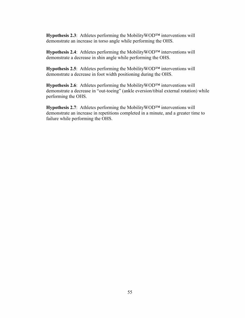
Hypothesis 2.3: Athletes performing the MobilityWOD™ interventions will
demonstrate an increase in torso angle while performing the OHS.
Hypothesis 2.4: Athletes performing the MobilityWOD™ interventions will
demonstrate a decrease in shin angle while performing the OHS.
Hypothesis 2.5: Athletes performing the MobilityWOD™ interventions will
demonstrate a decrease in foot width positioning during the OHS.
Hypothesis 2.6: Athletes performing the MobilityWOD™ interventions will
demonstrate a decrease in “out-toeing” (ankle eversion/tibial external rotation) while
performing the OHS.
Hypothesis 2.7: Athletes performing the MobilityWOD™ interventions will
demonstrate an increase in repetitions completed in a minute, and a greater time to
failure while performing the OHS.
56
Appendix D
BACKGROUND AND SIGNIFICANCE
D.1 Background
To understand the importance of the squat exercise, we must understand the
history of weight-lifting. The genealogy of lifting traces back to the beginning of
recorded history where man's fascination with physical prowess can be found among
numerous ancient writings.1
The earliest reference to formal strength training occurs in
Chinese texts dating back as far as 3600BC. when emperors made their subjects
exercise daily and potential soldiers had to pass weight-lifting tests before being
allowed to enter the armed forces.63
There has also been much evidence of weight-
training used in ancient Egypt and India, while the Greeks left numerous sculptures of
their athletes training with weights. 63
The 6th
century BC was known as the ‘Age of
Strength’ and weight-lifting competitions involved lifting heavy stones. 63
Weight
training was not confined to men: a wall mosaic from a Roman villa in Piazza Almeria
in Sicily depicted a girl exercising with weights. 63
While weight-lifting has its origins
dating so far back, the science and expansion of the sport was never deeply studied
until around the 16th
century. From that time on, the emergence of books about
weight-training began to explode from all around the world, most notably in England,
France, and Russia. 63
The first modern day Olympics were held in 1896 and
weightlifting was included as an official sport.1 As weight-lifting emerged as an
Olympic sport, the 1900’s saw an explosion in the popularity of weight-training and
57
competitions in basic feats of strength. With this comes the origin and popularity of
the squat exercise in sport.
While the basic movement of the squat maneuver can be traced back thousands
of years, the popularity of the squat can be pin-pointed to the date October 21, 1921.
A young German immigrant, Henry Steinborn, set a world record by squatting a
402lb. barbell.60
Steinborn did not do this conventionally. A barbell was loaded with
402lb. on the ground and Steinborn upended the barbell, leaning to one side and
placing his hands on it slightly beyond shoulder width. He then squatted down,
allowing the barbell to rock across his shoulders, and stood up with the weight then
reversed the process after squatting the weight for many repetitions.60
This single
event propelled the squatting exercise to become relevant in sport not only in America,
but worldwide. 60
In the 1930’s, 40’s, and 50’s, Olympic lifters, such as 1956
American Olympic weight-lifting gold medalist Paul Anderson, began utilizing the
squat in their training protocols.54
The squat as an exercise began to gain popularity as
a lift in strongman competition, and in 1972 an International Powerlifting Federation
(IPF) set the squat as one of the three movements an athlete will perform to compete
in what is now known as the sport of Powerlifting.62
Today, Powerlifting is a
recognized sport that tests athletes in the Bench Press, Deadlift, and Squat. While all
three of these lifts are utilized in modern Strength and Conditioning Programs, the
Squat is the exercise that defines lower body strength.59
58
D.2 Significance – The Squat and How it Applies to Modern Sport and Strength
and Conditioning Programs
The bilateral squat is one of the most prevalent exercises used in strength
training world-wide.18
It is a fundamental movement pattern that requires mobility at
the ankle, hip and thoracic spine and stability at the foot, knee, and lumbar spine.42
The popularity of the squat is certainly a reflection of its practicality. Humans
throughout time have used variations of the squat pattern to accomplish various tasks
associated with activities of daily living.2, 14
A significant amount of research has been
dedicated to establish the resisted squat as an effective exercise for enhancing strength
and power performances4, 11, 22, 23, 25, 26, 30
,which makes it one of the most widely used
exercises for increasing physical strength and power.4 The effects of the squat are
easily reflected in the most basic sport movement of sprinting. This interest is a result
of the defined relationship between the ability to apply force into the ground to
increase running velocity.64, 65, 66
Several investigators have found a strong correlation
between ground reaction force (GRF) or impulse magnitude and sprinting velocity.37,
65
Several investigations have shown the relationship between GRF capabilities and
sprinting performance.37, 64, 65
In addition Weyand et al., reported that maximal
sprinting velocity was a product of GRF and not the leg speed of runners; in fact
swing time (stride frequency) for the legs of slow and fast runners was identical at
approximately 0.360 seconds.65
Hunter et al. has reported a significant correlation
between GRF horizontal impulse and sprinting velocity.37
Therefore, it is evident that
59
strength or maximal force production is an essential component to maximal sprinting
velocity.
Given the known relationship between GRF and sprinting velocity and the
contribution from the major muscle groups of the lower body, it has been shown that
the squat strengthens the proper lower body musculature to apply a greater GRF. A
study by McBride et al.47
reported that both 10 and 40 yard sprint times increased
when an athlete could squat greater than 2.1 times their body weight when compared
to those who could only squat 1.9 times their body weight. This finding is consistent
with previous studies, Wisloff et al.66
, that showed improved 10 yard sprint times in
athletes who could squat more weight. In summary, it appears that horizontal ground
reaction force, net GRF, and net impulse all demonstrate strong correlations with
sprinting velocity whereas leg speed is not a factor in increased sprinting velocity.46, 47,
64, 65 Thus, one of the primary factors determining sprinting velocity is the ability to
generate large GRF with the lower-body musculature, making this area an obvious site
of interest for maximizing sprinting ability. It can be speculated then that a focus of
resistance training on increasing lower-body structural multiple-joint movements of
strength (i.e., free weight squat) compared to single leg movements is warranted.
Despite the stereotype, the squat exercise is not just reserved for male athletes.
Although American women first began strength training for sports in the 1950s to
improve their performance in track and field, they have traditionally participated in
strength training less than men.24
Such exercise has not been considered feminine,
and a lack of research and information regarding the effects of training on women has
60
made it a predominantly male activity. Women's participation was particularly limited
until 1972, when Title IX mandated equal access to educational programs--including
athletics--for men and women in schools that receive federal funding.24
Since then,
women's sports participation has exploded, traditional gender roles have loosened, and
strength training has grown in popularity among active women. Since the advent of
Title IX, according to the National Federation of State High School Associations, the
number of girls playing high school sports has grown more than tenfold, from 294,000
in 1971 to nearly 3.2 million in 2012. 24
There has been an equal growth in the female
sport participation in the collegiate level during this time frame. In 2002, the number
of female athletes competing in collegiate athletics was reported to be 158,469.8
The
number of female athletes reported in 2011 was reported to be 191,131.8
The biggest
difference a female athlete will encounter in the transition between high school and
college athletics is the incorporation of a strength and conditioning program as a year-
round training.9
Like their male counterparts, girls have started to specialize early in their
careers, working on just one sport year-round, often as a way to capture the attention
of college coaches. With more scholarship money available than ever, girls feel
pressured to specialize at a young age in the hopes of winning a spot on an elite team
or gaining an edge in the increasingly competitive college admissions game. Despite
persistent warnings from orthopedic surgeons and athletic trainers, young athletes bent
on specialization continue to suffer from preventable overuse injuries (tendonitis
61
bursitis, stress fractures, etc…).12
According to the American College of Sports
Medicine, 50 percent of these overuse injuries are preventable.12
Additionally, of
special concern for female athletes is damage to the anterior cruciate ligament (ACL),
as they are four to five times more likely than their male counterparts to suffer injury
to this structure. Compounding matters is that once girls begin to menstruate, they also
develop a tendency to utilize their quadriceps muscle more than their hamstrings,
making them more vulnerable to ACL injury. This is turn enables female athletes to
jump and land in a more erect posture, further stressing the ACL.29
Therefore, as
Strength and Conditioning Specialists, getting female athletes to utilize proper
running, jumping, and landing techniques is apparent in an attempt to prevent injury.
One such exercise at the core of each of these sport movements is the squat.
D.3 Significance - Execution of the Squat
There are many variations of the squat technique, including stance width, foot
positioning, and squat depth. However, previous research has indicated that the
optimal squat technique is a wide stance(≥ shoulder width) with natural foot
positioning, unrestricted movement of the knees, and full depth (femur parallel to the
floor) while the lordotic curve of the lumbar spine is maintained with a forward or
upward gaze.18
D.3.1 Stance Variation
In a narrow stance squat, the mechanical demand is distributed across the hip
and knee extensors and ankle plantar-flexors.25
As the stance width increases, the
62
demand placed on the ankle plantar-flexors decreases and the demand placed on the
hip and knee extensors increases.25
With extremely wide stance widths, it is possible
that the ankle dorsiflexors are required. Squats that do not allow the knees to move
forward (ankle dorsiflexion) result in greater forward trunk lean30
, which increases
loading of the lumbar spine.10
Therefore, the decision to use one technique versus
another cannot simply be made by considering which technique allows the most
weight to be lifted.
D.3.2 Sitting Back
Sitting back into the squat, also known as the hip hinge by McGill49
, should be
used to initiate the eccentric portion of the lift. Sitting back allows the gluteus
maximus, a powerful hip extensor, to immediately become a part of the lift,
particularly increasing activation in a deeper squat. Without this posterior shift, the
squat exercise will emphasize the quadriceps; deemphasizing the gluteus maximus
throughout the lift. Research shows that sitting back and preventing the knees from
moving too far beyond the toes does increase hip torque.30
Sitting back to minimize
anterior translation of the knees will also decrease torque at the knee joints.30, 45
The
quadriceps are still a major component of the lift, but now, the gluteal muscles can
share the load more evenly distributing forces throughout the lower extremities. For
those with knee pain, this can make an immediate difference in their ability to perform
the lift. For athletes without current knee issues, it can be a way to avoid future
problems because of overloading.15
To achieve a parallel squat or deeper, the knees
will travel past the toes to a degree, but it should be clear that ‘‘sitting back’’ is not a
63
way of preventing this but rather limiting excessive anterior shift of the tibia at the
knee.15
Engaging the gluteal muscles by sitting back also has the effect of preventing
excessive lumbar lordosis, a common cause of spondylolytic disorders.15
The gluteus
maximus has the ability to resist excessive anterior tilting of the pelvis because it
offsets the pull of the lumbar paraspinals, to keep the lumbar spine in a neutral
position.15
The ability to maintain a neutral lumbar spine throughout the lift has been
shown to increase stability through the spine, allowing it the ability to bear greater
compressive loads, and reduces shear forces.32
Another biomechanical advantage of
sitting back is to reduce the ankle dorsiflexion moment; in other words maintaining a
vertical shin angle. As the knees travel past the toes, dorsiflexion requirements become
greater. An athlete with ‘‘stiff ankles’’ will do one of three things: (a) the heels could
come off the floor and increase shear forces at the knee45
(b) the heels come off the
floor causing the athlete to lose balance, and (c) excessive subtalar pronation occurs
along with femoral internal rotation with the result being unacceptable valgus at the
knees.15
The combination of increased knee valgus and anterior tibial shear forces has
also been shown to increase stress on the anterior cruciate ligament.45
Teaching
athletes to sit back to initiate the squat can have several important benefits, including a
more even distribution of load between the hip and knee extensors, maintaining a
neutral spine, keeping the heels on the floor, and preventing valgus collapse of the
knees. Each of these can lead to a safer and more effective squat during training and
potentially lead to greater athletic performance.
64
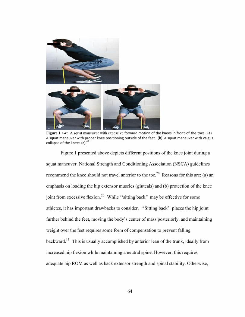
Figure 1 a-c: A squat maneuver with excessive forward motion of the knees in front of the toes. (a) A squat maneuver with proper knee positioning outside of the feet. (b) A squat maneuver with valgus collapse of the knees (c).
42
Figure 1 presented above depicts different positions of the knee joint during a
squat maneuver. National Strength and Conditioning Association (NSCA) guidelines
recommend the knee should not travel anterior to the toe.20
Reasons for this are: (a) an
emphasis on loading the hip extensor muscles (gluteals) and (b) protection of the knee
joint from excessive flexion.20
While ‘‘sitting back’’ may be effective for some
athletes, it has important drawbacks to consider. ‘‘Sitting back’’ places the hip joint
further behind the feet, moving the body’s center of mass posteriorly, and maintaining
weight over the feet requires some form of compensation to prevent falling
backward.15
This is usually accomplished by anterior lean of the trunk, ideally from
increased hip flexion while maintaining a neutral spine. However, this requires
adequate hip ROM as well as back extensor strength and spinal stability. Otherwise,
65
an athlete must flex the spine to maintain balance, presenting well-documented risks
that contradict accepted squatting guidelines.20, 49
Harvey et al.34
has shown that
restricted anterior movement of the knees during squatting increased loads at the hip
but also caused excessive forward lean of the trunk and was likely to inappropriately
transfer load to the lower back. Although reduced knee flexion is proposed to decrease
stress in the knee, it may also compound risk to the spine by limiting hip ROM
afforded by the 2-joint hamstring muscles.15
Thus, in an attempt to protect the knee,
‘‘sitting back’’ may pose additional risk to the spine.
Recent evidence also suggests that excess flexion can aggravate hip joint
pathology in some athletes.45
The major determinant of lower extremity joint loads
during the squat is the location of the GRF. Especially with higher barbell loads, GRF
location is driven primarily by the position of the upper body because it has the
greatest mass.15
Whether one ‘‘sits back’’ or not, forward lean of the trunk and/or
greater barbell mass places greater relative demand on the hip joint.32, 34
The
difference is that ‘‘sitting back’’ requires this forward lean to maintain stability. If
instead the knees are allowed to remain at or even beyond the toes, a greater range of
trunk position is possible. Forward lean can be allowed if hip ROM and back strength
permit. Alternatively, a more erect position can be maintained while still keeping
weight over the feet. Shifting weight forward has the added benefit of increasing
torque at the ankle joint, providing greater training stimulus to the plantar flexors.32
A
more anterior knee position usually implies greater ankle dorsiflexion and knee
flexion, which are often said to pose a risk to the knee. Evidence to date however
66
suggests that thigh-parallel squats are safe for healthy athletes, although deeper
squatting might pose additional risk to the knee menisci or ligaments (principally the
posterior cruciate ligament).30
In the presence of a knee injury, reduced barbell loads
and/or limited squat depth to control knee flexion are better options than a strategy that
could pose undue risk to the spine.15
Many athletes that have restrictions in DF ROM
are asked to ‘‘sit back,’’ causing similar concerns related to torso angle. Corrective
measures could include mobility/flexibility training, limitation of squat depth, changes
in barbell load, and/or elevating the heels.13
D.3.3 Muscle Activation
There is continued debate among strength and conditioning experts regarding the most
appropriate foot placement and squat depth, not only in terms of stresses on the knee
but also in terms of recruitment of muscles. Electromyographic studies have found
that increased squat depth (half squat 45°, parallel squat 90°, full-depth squat 125°)
resulted in a greater percentage contribution of the gluteus maximus during the full-
depth squat.11, 53
During the eccentric phase of the weighted-back squat, the relative
contributions of 4 muscle groups (vastus medialis, vastus lateralis, biceps femoris,
gluteus maximus) at the 3 depths tested were not statistically different.11
Therefore,
muscle activation did not differ based on squat depth. However, it is important to
mention loads used were submaximally (25% body weight and 100–125% body
weight) and recruitment patterns may change at near-maximal loads.11
Rotating the feet (neutral, 30–40°medial, 80° lateral rotation) while performing
the squat, regardless of depth and stance width (75–140% shoulder width), has been
67
shown to have no noticeable effect on muscle activity of the lower leg (rectus femoris,
vastus medialis, vastus lateralis, adductor longus, semimembranosus, semitendinosus,
and biceps femoris).27, 28, 48, 53, 56
Two studies indicate stance width variation does
alter muscle recruitment patterns by increasing activity of the adductor longus when a
wide stance is used (> shoulder width).48, 53
However, rotating the feet outward may
place greater demands on the ligaments of the knee .58
When there is a lack of DF
ROM, there is a tendency to lack the ability to drive the weight up through the heels.
An athlete will anteriorly displace the weight to the toes which can cause an anterior
shift during the movement and increase rotational torque at the knee.19
D.4 Significance - Forces Placed on the Knee
A study calculating forces during the squat movement reported patello-femoral
joint (PFJ) forces during the ascent phase of a deep squat regardless of the speed were
present (reference). Another study determined that PFJ forces increase with greater
amounts of knee flexion during the squat maneuver.18
Research has also demonstrated
increases in tibio-femoral joint (TFJ) forces during the squat and leg press when there
was a greater amount of knee flexion.27
It has also been shown that the squat exercise
produces significantly less anterior displacement when compared to the open kinetic
chain exercise of leg extension, producing less strain on the anterior cruciate ligament
(ACL).6, 28, 39
Similar results regarding peak posterior cruciate ligament (PCL) forces
have also been reported, where forces >4.5 times body mass during isokinetic and
isometric leg extension, compared with 3.5 times body mass during the squat.28, 61
This suggests that the squat may result in a lower risk of injury than a simple leg
68
extension exercise. However, it is worth mentioning that PCL forces increase with
increased knee flexion, or depth, during the squat.57, 68
Despite this increase, the PCL
can sustain much greater forces to failure than the ACL. Other research has found that
the ACL was subject to small forces during the squat when the knee was less than 50°
of flexion and when the angles increased, the PCL rather than the ACL receive greater
loads.27, 61
ACL forces were also much lower when the squat was performed with
heals on the ground compared with when the squat was performed with heels elevated
during both the descent and the ascent phases.61
Therefore, it is important to gain
ankle dorsiflexion ROM so we may perform a squat with our heels on the ground
rather than displacing the weight towards our toes to lessen the forces placed on the
knee.
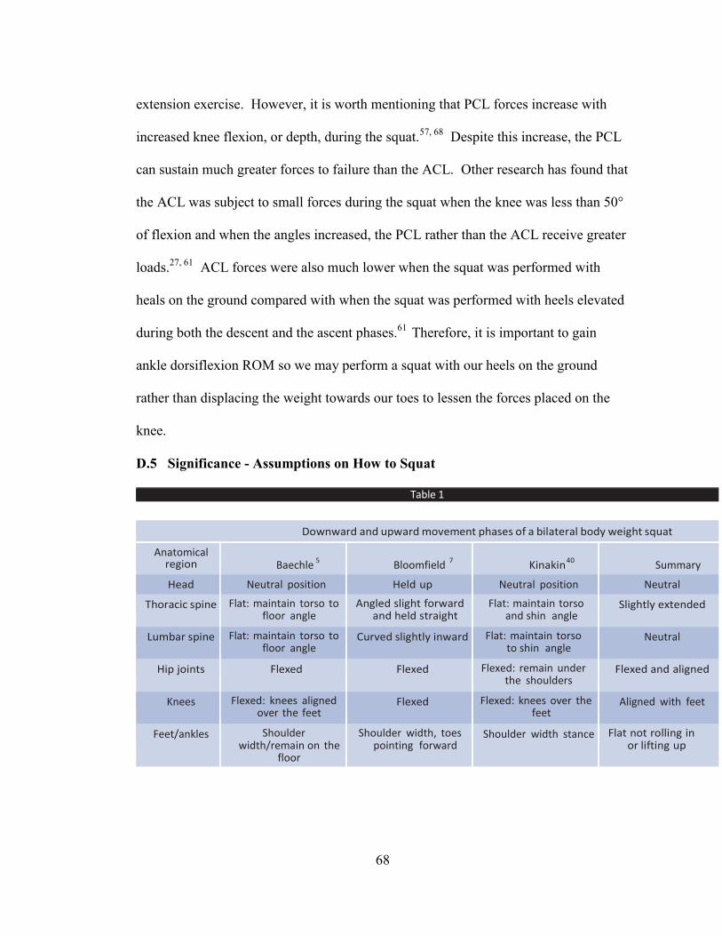
D.5 Significance - Assumptions on How to Squat
Table 1
Kinematic considerations of the bilateral bodyweight squat
Downward and upward movement phases of a bilateral body weight squat
Anatomical region
Baechle 5
Bloomfield 7
Kinakin 40
Summary
Head Neutral position Held up Neutral position Neutral
Thoracic spine Flat: maintain torso to floor angle
Angled slight forward and held straight
Flat: maintain torso and shin angle
Slightly extended
Lumbar spine Flat: maintain torso to floor angle
Curved slightly inward Flat: maintain torso to shin angle
Neutral
Hip joints Flexed Flexed Flexed: remain under the shoulders
Flexed and aligned
Knees Flexed: knees aligned over the feet
Flexed Flexed: knees over the feet
Aligned with feet
Feet/ankles Shoulder width/remain on the
floor
Shoulder width, toes pointing forward
Shoulder width stance Flat not rolling in or lifting up
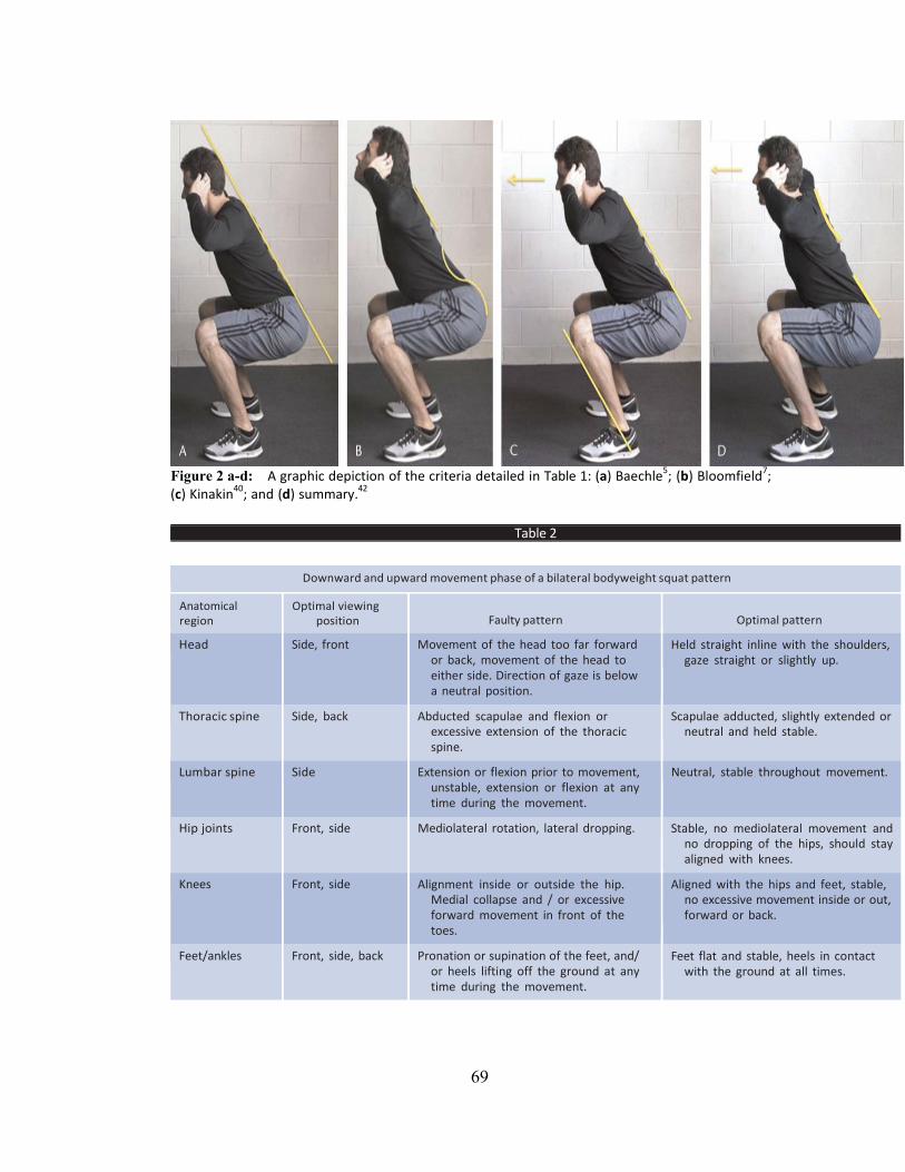
69
Figure 2 a-d: A graphic depiction of the criteria detailed in Table 1: (a) Baechle
5; (b) Bloomfield
7;
(c) Kinakin40
; and (d) summary.42
Table 2
Criteria and optimal viewing position for identifying faulty movement patterns related to a bilateral squat pattern
Downward and upward movement phase of a bilateral bodyweight squat pattern
Anatomical region
Optimal viewing position
Faulty pattern
Optimal pattern
Head Side, front Movement of the head too far forward or back, movement of the head to either side. Direction of gaze is below a neutral position.
Held straight inline with the shoulders, gaze straight or slightly up.
Thoracic spine Side, back Abducted scapulae and flexion or excessive extension of the thoracic spine.
Scapulae adducted, slightly extended or neutral and held stable.
Lumbar spine Side Extension or flexion prior to movement, unstable, extension or flexion at any time during the movement.
Neutral, stable throughout movement.
Hip joints Front, side Mediolateral rotation, lateral dropping. Stable, no mediolateral movement and no dropping of the hips, should stay aligned with knees.
Knees Front, side Alignment inside or outside the hip. Medial collapse and / or excessive forward movement in front of the toes.
Aligned with the hips and feet, stable, no excessive movement inside or out, forward or back.
Feet/ankles Front, side, back Pronation or supination of the feet, and/ or heels lifting off the ground at any time during the movement.
Feet flat and stable, heels in contact with the ground at all times.
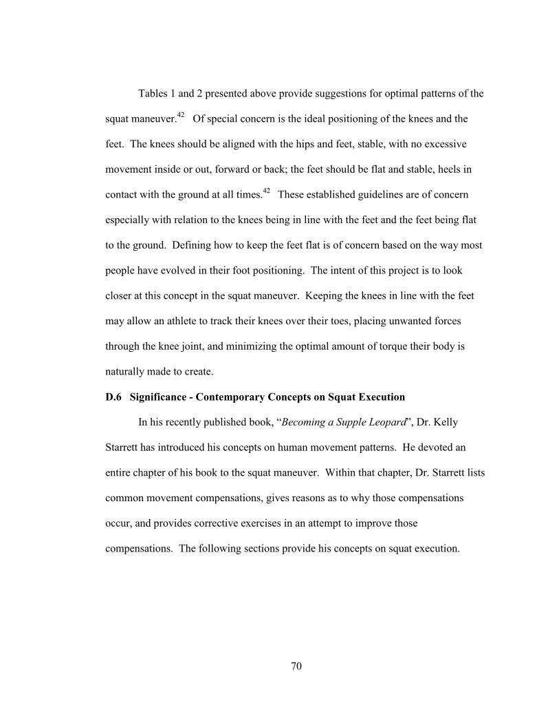
70
Tables 1 and 2 presented above provide suggestions for optimal patterns of the
squat maneuver.42
Of special concern is the ideal positioning of the knees and the
feet. The knees should be aligned with the hips and feet, stable, with no excessive
movement inside or out, forward or back; the feet should be flat and stable, heels in
contact with the ground at all times.42
These established guidelines are of concern
especially with relation to the knees being in line with the feet and the feet being flat
to the ground. Defining how to keep the feet flat is of concern based on the way most
people have evolved in their foot positioning. The intent of this project is to look
closer at this concept in the squat maneuver. Keeping the knees in line with the feet
may allow an athlete to track their knees over their toes, placing unwanted forces
through the knee joint, and minimizing the optimal amount of torque their body is
naturally made to create.
D.6 Significance - Contemporary Concepts on Squat Execution
In his recently published book, “Becoming a Supple Leopard”, Dr. Kelly
Starrett has introduced his concepts on human movement patterns. He devoted an
entire chapter of his book to the squat maneuver. Within that chapter, Dr. Starrett lists
common movement compensations, gives reasons as to why those compensations
occur, and provides corrective exercises in an attempt to improve those
compensations. The following sections provide his concepts on squat execution.
71
D.6.1 Hip Mobility and the Squat
When observing an athlete performing a squat, it’s important to look at torso
position, hip mobility, knee position, and ankle mobility.58
Deficits in the squat
maneuver are often associated with lack of mobility in the ankle and hip. 58
Deficits
in hip mobility are typically associated with deficits in internal hip rotation, thus
allowing the knees to collapse inward. 58
Additionally, we compensate by leaning the
trunk forward, rounding the truck, or overextending the trunk. 58
These
compensations may lead to an increase in a back injury by placing unnecessary forces
on the vertebrae. 58
Therefore, closely examining hip mobility deficiencies within the
squat maneuver is of concern.
D.6.2 Neutral Spine and the Squat
Neutral spine is a term synonymous with “setting the core”. To obtain a
neutral spine, athletes are told to squeeze the gluteal muscles together, bringing the
pelvis and hips closer to the femur. 58
Furthermore, athletes are instructed to squeeze
the ribcage downward in an attempt to anchor it toward the hips. 58
These two
techniques when combined allow an athlete to brace their core and keep it in a neutral
position. This position prevents the athlete from overextending or rounding their back,
protecting the spine.
D.6.3 Ankle Mobility and the Squat
Modern footwear has an elevated heel, resulting in a restriction of the heel cord
and reflecting in poor ankle dorsiflexion ROM. 58
This restriction has led to calf
stiffness and the turning out of the toes during the squat maneuver. 58
Many athletes
72
have to turn their feet out dramatically in order to achieve full depth during the squat.
58 While this is clearly functional, it’s not optimal. In fact, when the feet turn out past
15 degrees, it can result in poor motor control, deficits in internal hip rotation, a tight
anterior chain (quads, etc.), and stiff ankles. 58
The further out the feet go, the less
effective the hip rotators are at resisting the valgus forces at the knee created by the
body. 58
Somewhere between 5-12 degrees appears to be an area where an athlete can
achieve proper depth and create optimal amounts of torque throughout the squat. 58
Athletes will turn out their feet in order to “un-impinge” the hip. 58
While this
mechanically “un-impinges” the hip, the mechanical advantage of being in a stable
position is lost, making it harder to keep the knees out without allowing them to track
over the foot. 58
Tracking the knee outside the feet creates a stable knee position,
while positioning the knee over the feet creates an unstable knee position. 58
The modern elevated shoe heel has also led to navicular drop. 58
The medial
malleolus starts to drop inward from a flat foot position which causes tibial torsion. 58
When there is navicular drop, there is a collapse in the whole kinetic chain. Internal
tibial torsion puts the knee in a valgus position. 58
Valgus positioning puts heavy
strain on the ACL. When the feet turn out, in addition to navicular drop, there is
increased potential exposure of the knee to a valgus position because there is a
decreased ability to flare the knees outside of the toes when squatting, increasing the
chance to collapse the knee during the movement. 58
Therefore when squatting, a
subtalar neutral position with the feet straight should be considered optimal so there is
a reduced chance to collapse the knee when squatting; thus reducing the chances of
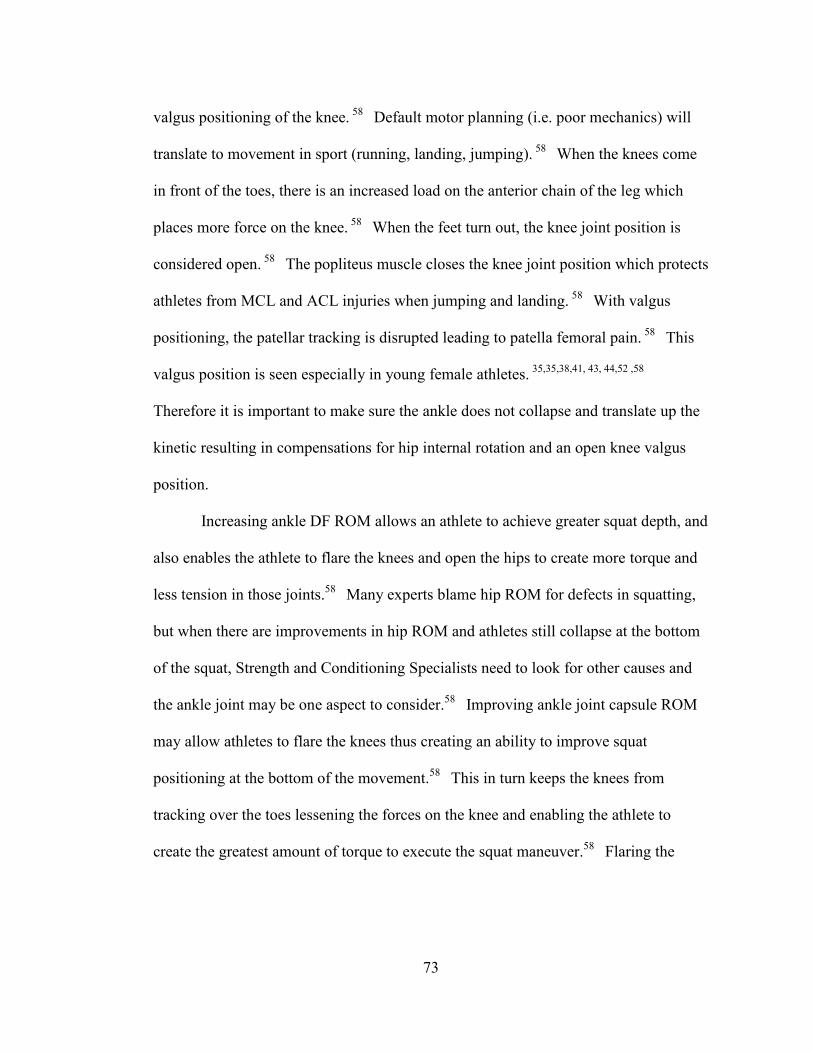
73
valgus positioning of the knee. 58
Default motor planning (i.e. poor mechanics) will
translate to movement in sport (running, landing, jumping). 58
When the knees come
in front of the toes, there is an increased load on the anterior chain of the leg which
places more force on the knee. 58
When the feet turn out, the knee joint position is
considered open. 58
The popliteus muscle closes the knee joint position which protects
athletes from MCL and ACL injuries when jumping and landing. 58
With valgus
positioning, the patellar tracking is disrupted leading to patella femoral pain. 58
This
valgus position is seen especially in young female athletes. 35,35,38,41, 43, 44,52 ,58
Therefore it is important to make sure the ankle does not collapse and translate up the
kinetic resulting in compensations for hip internal rotation and an open knee valgus
position.
Increasing ankle DF ROM allows an athlete to achieve greater squat depth, and
also enables the athlete to flare the knees and open the hips to create more torque and
less tension in those joints.58
Many experts blame hip ROM for defects in squatting,
but when there are improvements in hip ROM and athletes still collapse at the bottom
of the squat, Strength and Conditioning Specialists need to look for other causes and
the ankle joint may be one aspect to consider.58
Improving ankle joint capsule ROM
may allow athletes to flare the knees thus creating an ability to improve squat
positioning at the bottom of the movement.58
This in turn keeps the knees from
tracking over the toes lessening the forces on the knee and enabling the athlete to
create the greatest amount of torque to execute the squat maneuver.58
Flaring the
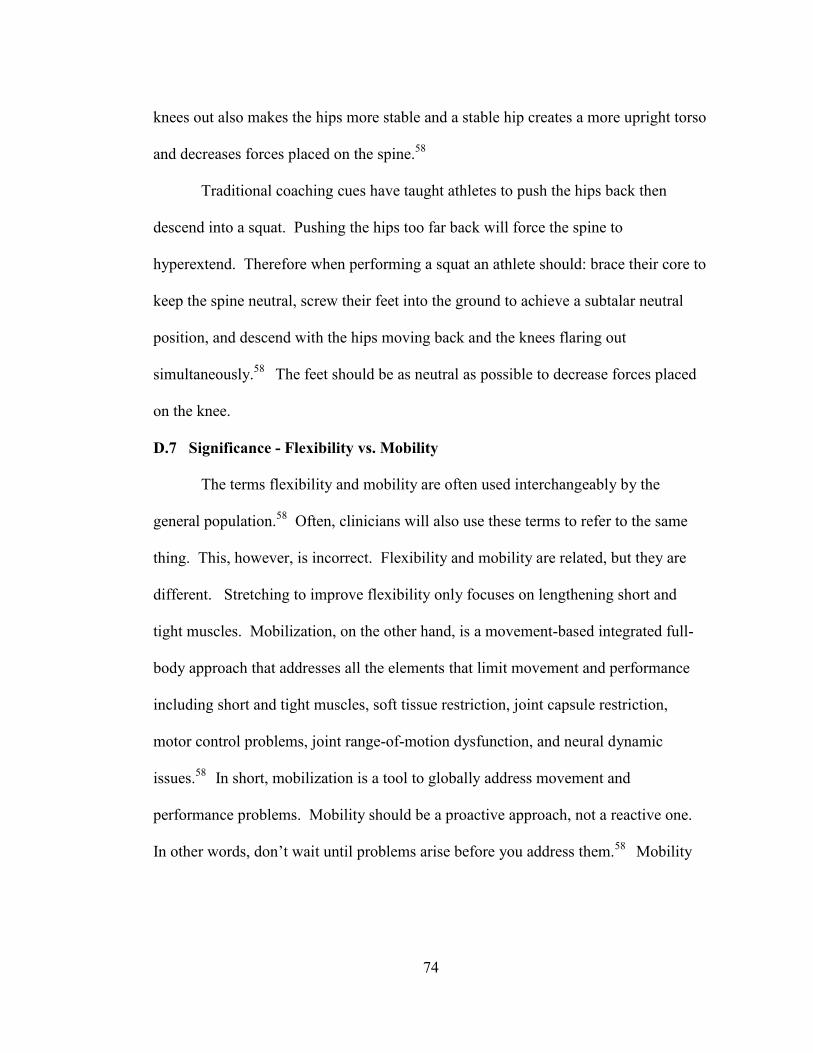
74
knees out also makes the hips more stable and a stable hip creates a more upright torso
and decreases forces placed on the spine.58
Traditional coaching cues have taught athletes to push the hips back then
descend into a squat. Pushing the hips too far back will force the spine to
hyperextend. Therefore when performing a squat an athlete should: brace their core to
keep the spine neutral, screw their feet into the ground to achieve a subtalar neutral
position, and descend with the hips moving back and the knees flaring out
simultaneously.58
The feet should be as neutral as possible to decrease forces placed
on the knee.
D.7 Significance - Flexibility vs. Mobility
The terms flexibility and mobility are often used interchangeably by the
general population.58
Often, clinicians will also use these terms to refer to the same
thing. This, however, is incorrect. Flexibility and mobility are related, but they are
different. Stretching to improve flexibility only focuses on lengthening short and
tight muscles. Mobilization, on the other hand, is a movement-based integrated full-
body approach that addresses all the elements that limit movement and performance
including short and tight muscles, soft tissue restriction, joint capsule restriction,
motor control problems, joint range-of-motion dysfunction, and neural dynamic
issues.58
In short, mobilization is a tool to globally address movement and
performance problems. Mobility should be a proactive approach, not a reactive one.
In other words, don’t wait until problems arise before you address them.58
Mobility
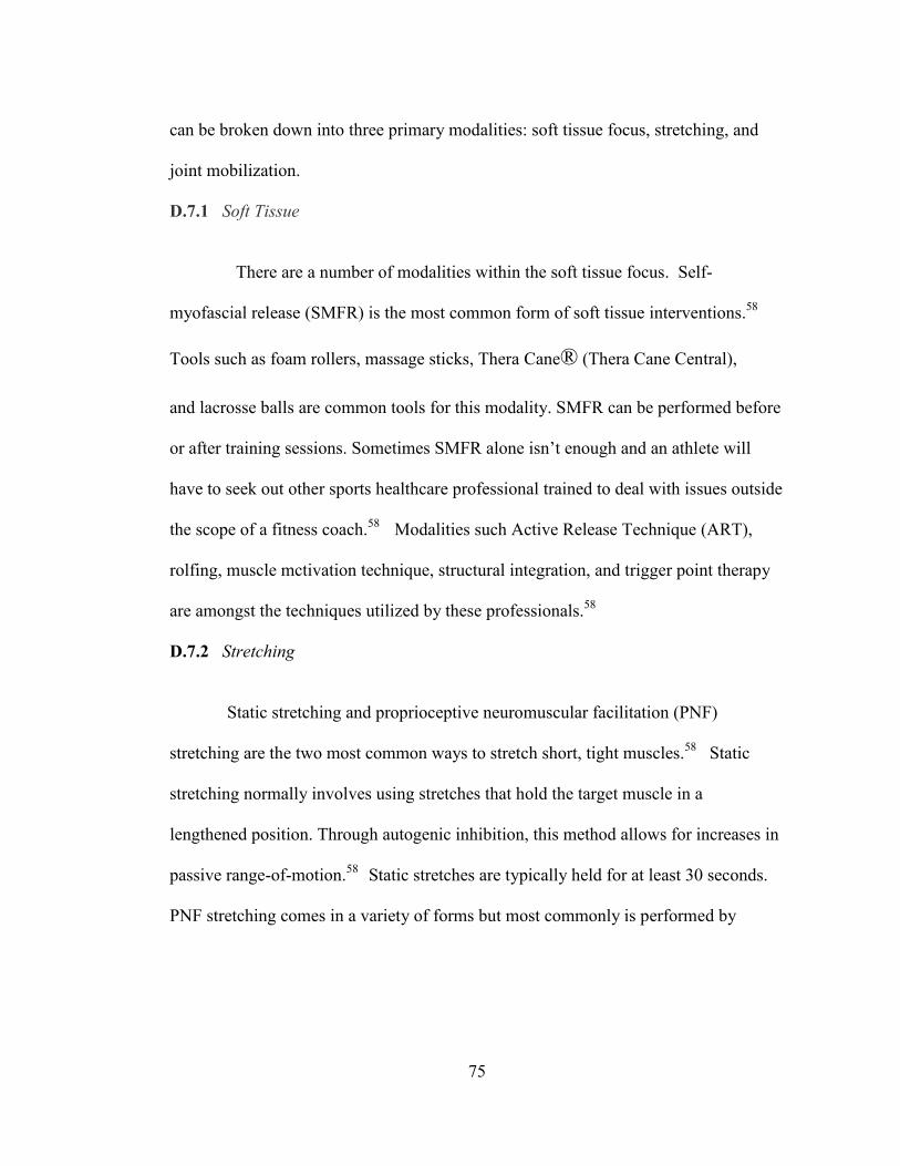
75
can be broken down into three primary modalities: soft tissue focus, stretching, and
joint mobilization.
D.7.1 Soft Tissue
There are a number of modalities within the soft tissue focus. Self-
myofascial release (SMFR) is the most common form of soft tissue interventions.58
Tools such as foam rollers, massage sticks, Thera Cane® (Thera Cane Central),
and lacrosse balls are common tools for this modality. SMFR can be performed before
or after training sessions. Sometimes SMFR alone isn’t enough and an athlete will
have to seek out other sports healthcare professional trained to deal with issues outside
the scope of a fitness coach.58
Modalities such Active Release Technique (ART),
rolfing, muscle mctivation technique, structural integration, and trigger point therapy
are amongst the techniques utilized by these professionals.58
D.7.2 Stretching
Static stretching and proprioceptive neuromuscular facilitation (PNF)
stretching are the two most common ways to stretch short, tight muscles.58
Static
stretching normally involves using stretches that hold the target muscle in a
lengthened position. Through autogenic inhibition, this method allows for increases in
passive range-of-motion.58
Static stretches are typically held for at least 30 seconds.
PNF stretching comes in a variety of forms but most commonly is performed by
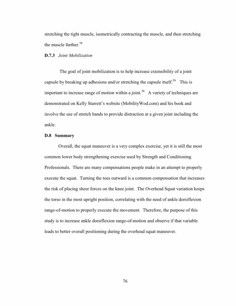
76
stretching the tight muscle, isometrically contracting the muscle, and then stretching
the muscle further.58
D.7.3 Joint Mobilization
The goal of joint mobilization is to help increase extensibility of a joint
capsule by breaking up adhesions and/or stretching the capsule itself.58
This is
important to increase range of motion within a joint.58
A variety of techniques are
demonstrated on Kelly Starrett’s website (MobilityWod.com) and his book and
involve the use of stretch bands to provide distraction at a given joint including the
ankle.
D.8 Summary
Overall, the squat maneuver is a very complex exercise, yet it is still the most
common lower body strengthening exercise used by Strength and Conditioning
Professionals. There are many compensations people make in an attempt to properly
execute the squat. Turning the toes outward is a common compensation that increases
the risk of placing sheer forces on the knee joint. The Overhead Squat variation keeps
the torso in the most upright position, correlating with the need of ankle dorsiflexion
range-of-motion to properly execute the movement. Therefore, the purpose of this
study is to increase ankle dorsiflexion range-of-motion and observe if that variable
leads to better overall positioning during the overhead squat maneuver.
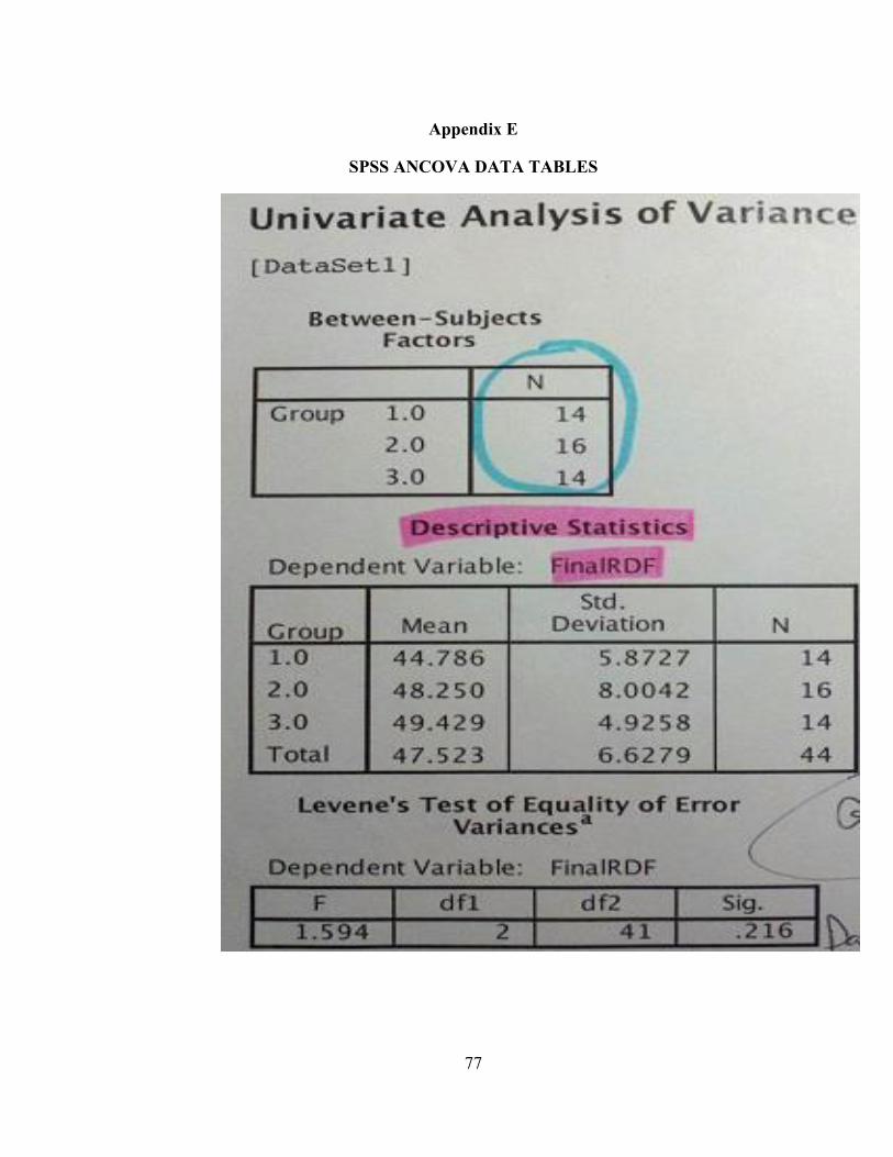
77
Appendix E
SPSS ANCOVA DATA TABLES

78
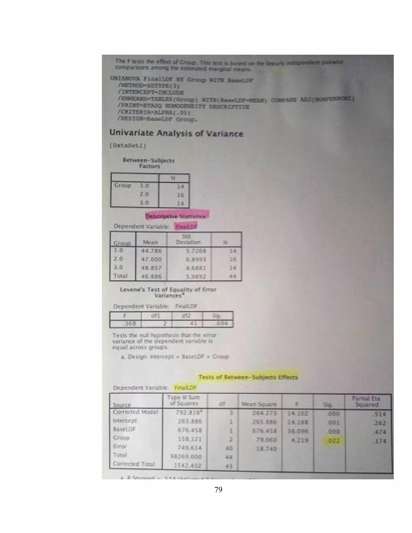
79
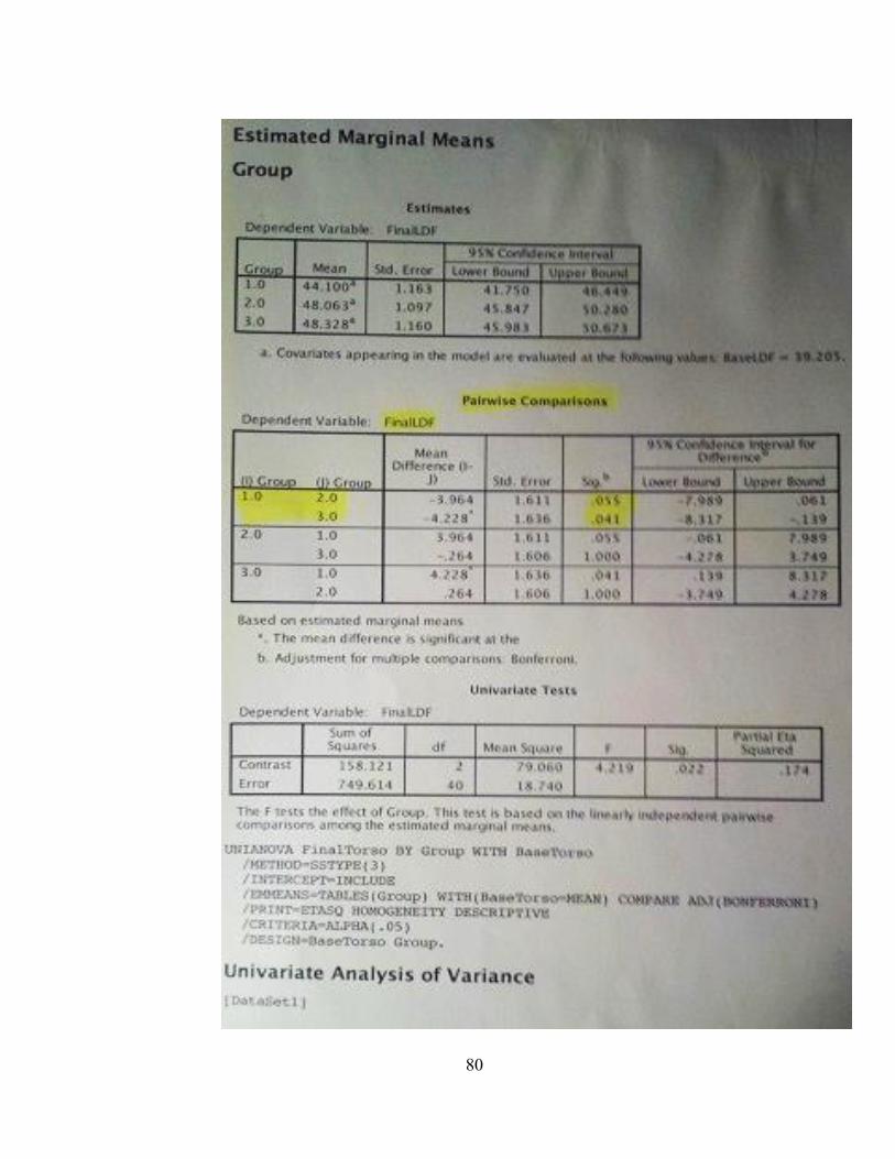
80
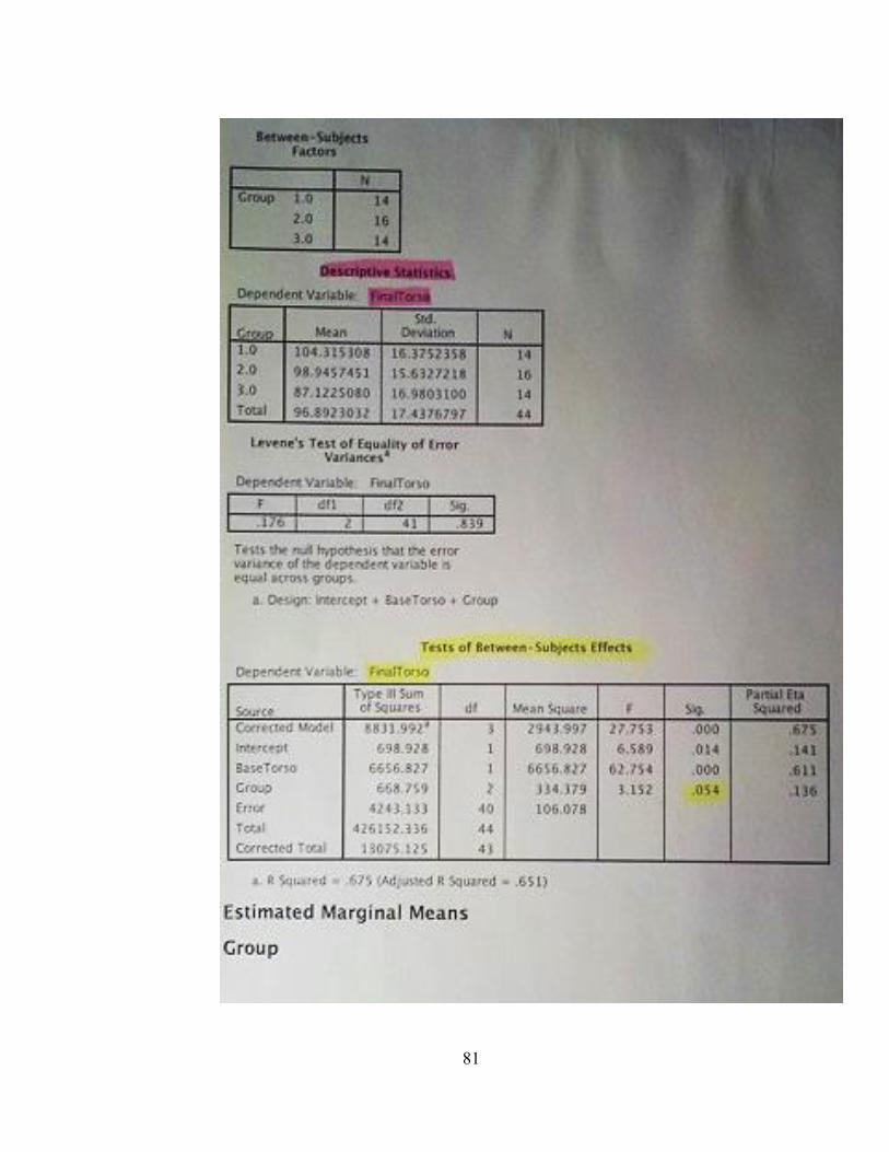
81
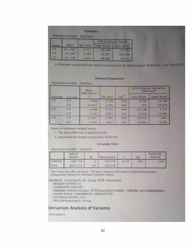
82
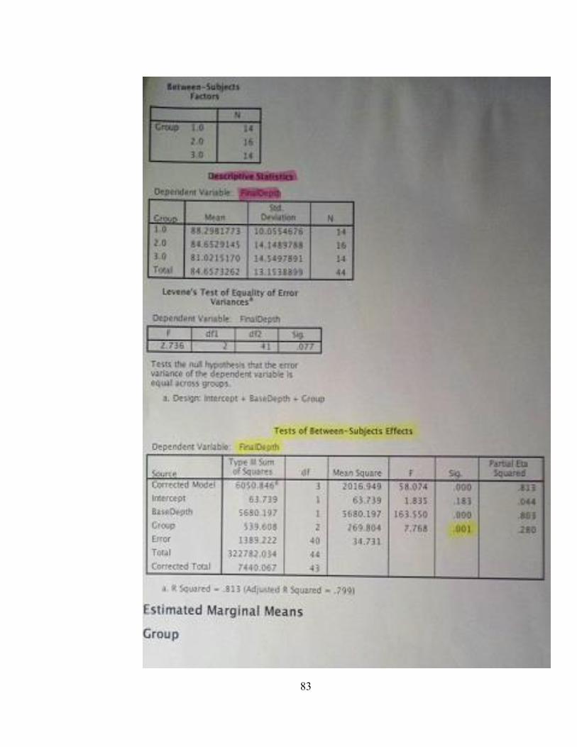
83
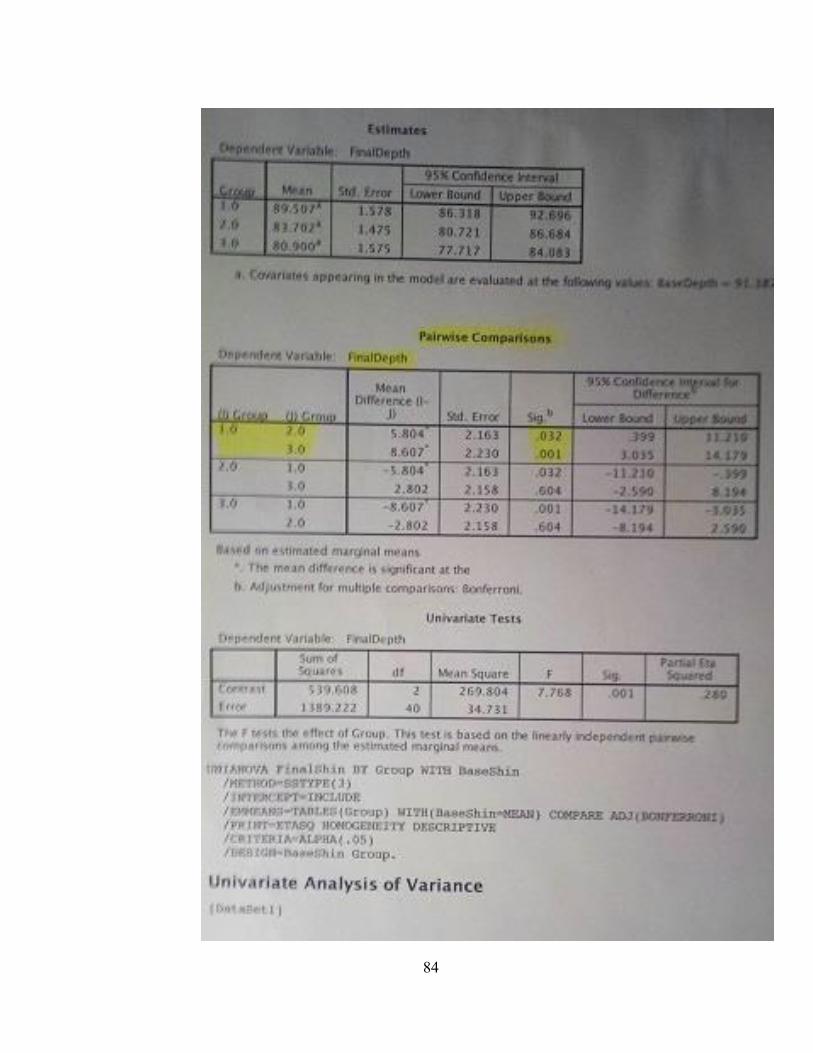
84

85
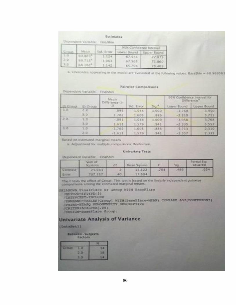
86

87
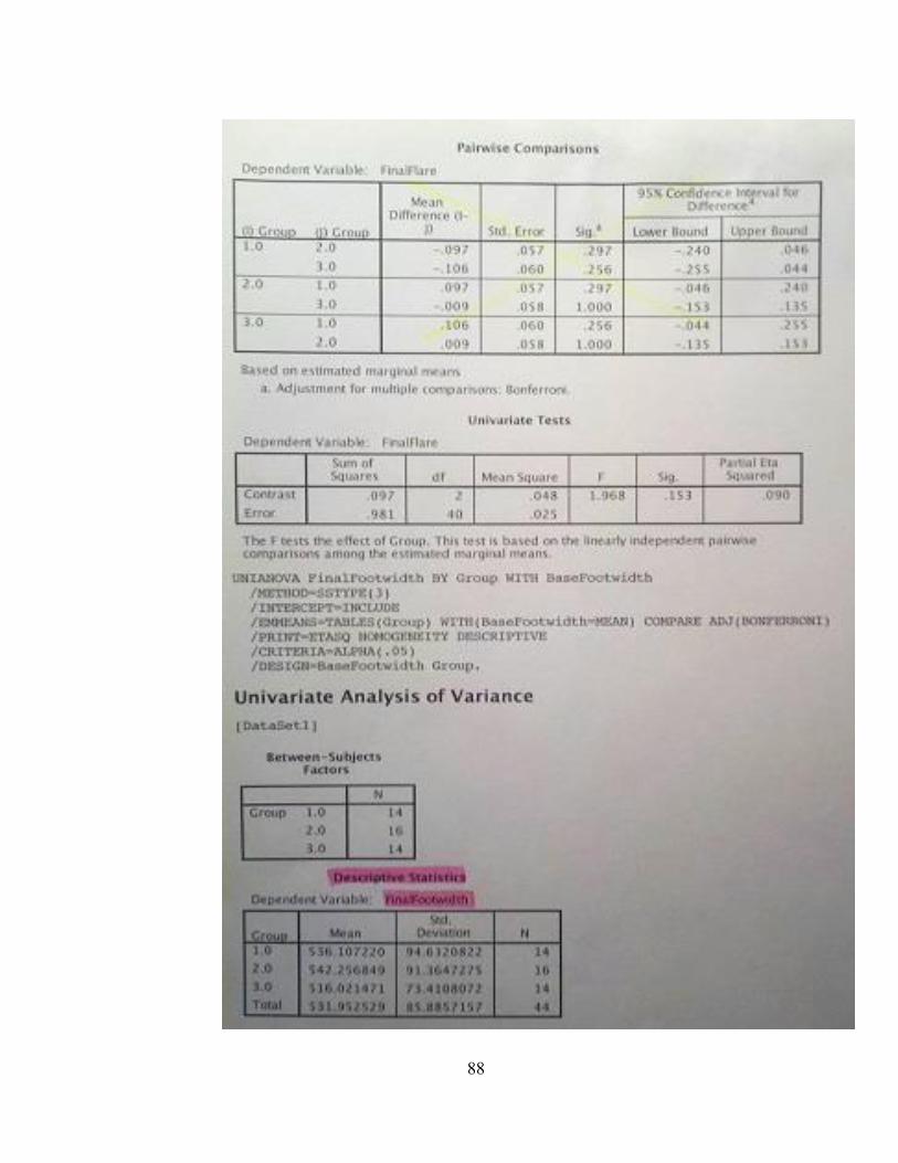
88

89

90

91
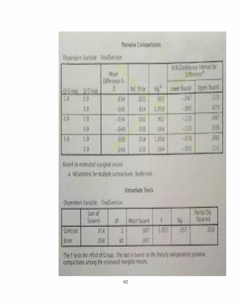
92