Embed Size (px)
Citation preview
Instructions for use
Title EXCESS DIETARY UREA INTAKE IN EWES AND ITS EFFECT ON OVULATION RATE AND EMBRYODEVELOPMENT
Author(s) BISHONGA, Christopher; ROBINSON, John J.; MCEVOY, Tim G.; FINDLAY, Patson; AITKEN, Raymond P.;ROBERTSON, Irene
Citation Japanese Journal of Veterinary Research, 44(3), 139-151
Issue Date 1996-12-18
DOI 10.14943/jjvr.44.3.139
Doc URL http://hdl.handle.net/2115/2566
Type bulletin (article)
File Information KJ00002398261.pdf
Hokkaido University Collection of Scholarly and Academic Papers : HUSCAP
lPn. l. Vet. Res. 44(3): 139-151 (1996) FULL PAPER
EXCESS DIETARY UREA INTAKE IN EWES AND ITS EFFECT ON OVULATION RATE
AND EMBRYO DEVELOPMENT.
Christopher BISHONGA\ John 1. ROBINSON2, Tim G. MCEVOy2, Patson FINDLAy2, Raymond P. AITKEN2 and Irene ROBERTSON2
(Accepted for publication: Oct. 18, 1996)
ABSTRACT
The effect of excess dietary urea on ovulation and early embryo develop
ment of sheep was studied. Thirty Border Leicester x Scottish Black face ewes
randomly assigned to three treatments were given a basal control diet (C) which
met energy requirements for body weight maintenance. Other treatments were
basal diet plus 24g of urea/day (low urea, L) or plus 48g (high urea, H)/day.
The reproductive cycles of the ewes were synchronized using a single
injection of prostaglandin (PGF 2«) and progesterone by an intravaginal controlled
internal drug release (CIDR) device for 12 days. Ovulation was induced by the
use of pregnant mare serum gonadotrophin (PMSG). Ewes were inseminated
approximately 52 hours after CIDR device removal using a laparoscopic techni
que. Embryos were recovered at Day 4 or Day 11 after insemination from half
of the ewes from each treatment group.
There were no significant differences in ovulation rates among the three
groups. The embryo recovery rates were not affected by day of recovery. At
embryo recovery on Day 4, 7/13 in C, 3/6 in Land 017 in H embryos were
morulae. After 72 hours of in vitro culture 6/10 in C, 2/3 in Land 0/4 in H
embryos developed to the blastocyst stage. Pregnancies sustained were C 6/8,
L 517 and H 113 of the autotransfers. Throughout the experiment plasma urea
levels were significantly affected by diet (p < O. 01). Plasma ammonia levels in
the H group were significantly higher than those in the C and L groups (p < O. 05)
for 4 hours after each feed. There was no treatment effect on plasma prog
esterone concentration. The luteinizing hormone (LH) surge onset time and
amplitude were not correlated to ovulation rate and were not affected by
treatment. It is concluded that high circulating concentrations of plasma urea
1. Laborotary of Theriogenology. Department of Veterinary Clinical Sciences, Graduate School of Veterinary Medicine. Hokkaido University. Kita 18, Nishi 9, Kitaku, Sapporo, 060.
Hokkaido, Japan. 2. Department of Physiology, Rowett Research Institute Bucksburn, Aberdeen, AB2 9SB
Scotland. U. K.
140 Christopher BISHONGA et al.
and ammonia have an adverse effect on early embryo development. This effect
was independent of any alterations in progesterone and LH concentrations.
Key Words: dietary urea, embryo development, ewes
INTRODUCTION
In sheep production systems, low producing animals that are solely dependent on low quality forages are often given dietary supplements of urea or other sources of non-protein nitrogen (NPN) to alleviate the protein shortage arising from a low yield of
microbial protein20). The diets of high producing animals, in contrast, are supplemented with high quality protein that is largely undegradable (UDP) in the rumen, or their diets include high quality forage with a level of rumen degradable protein (RDP) that is grossly in excess of that needed by rumen microbes for maximum microbial protein synthesis. Whether or not the level and quality of protein (unde
graded versus readily degradable) has any detrimental effect on reproduction is uncertain. Although there are reports of normal fertility of cows on protein diets, there are other studies in which excess dietary protein has been associated with a reduction in fertility in COWS3,6, 7,11,15). Where adverse effects have been noted, they
have on occasion been ascribed to RDp3,9). The reported variation in fertility in
various studies3,4,6,l1) has been attributed to suboptimal reproductive management
(such as treatment of uterine disorders and estrus detection programs) during the postpartum period and not to dietary protein4
). Elrod6) concluded that the adverse
effect on fertility manifests itself regardless of the source and degradability of the protein when crude protein is fed in excess of animal requirements. He asserted that the effect is brought about by changes in uterine pH. However, the mechanisms by which this pH change is modulated by excess dietary protein has not been identified. Despite the claim that excess dietary protein can reduce fertility, there is little or no evidence that fertility was reduced in dairy cows fed a diet with a protein level as low as 13.2% or as high as 20%4,11). The fertility criteria measured in these studies were
days to first ovulation, number of services per conception, conception rate to first service, pregnancy rate and calving interval. There is however, a lack of similar
studies in sheep. The objective of the present study was to investigate the effect of varying levels
of dietary urea (RDP) during and after the preovulatory priming period on ovulation and the early embryo development in sheep.
MATERIALS AND METHODS
Thirty 2-6 years old Greyface (Border Leceister x Scottish Blackface) ewes were brought indoors during late April with their lambs and maintained on an artificial
photoperiod of 12 hours of light, 12 hours of darkness each day. At four weeks the
Excess dietary urea and embryo development 141
lambs were weaned. After weaning the lambs, a week later the ewes were placed on
the experimental diets. The body weights were measured at the start and end of the
experimental period. The ewes were ranked in ascending order of their initial
weights, and each consecutive trio was randomly allocated to the three treatments.
The treatments were C: basal diet 1600g, L: basal diet 1600g plus 24g urea (low
urea), and H: basal diet 1600g plus 48g urea supplement. All diets were weighed
separately for each ewe and supplemented with minerals and vitamins. Vrea supple
ments were mixed into the basal diet at the time of feeding. Sodium sulphate was
added as source of sulphur in the ratio of 14: 1 by weight, urea: sulphur. The diet
was fed at the rate of 800 g/feed twice daily at 08:00 and 16:00 hours. The diet was
fed in the period before, during and after progesterone priming and up to 30 days after
intrauterine artificial insemination. Water was available at all times. The basal diet composition in g/kg was: hay 832, molasses 147, dicalciumphosphate 10, sodium
chloride 10 and minerals and vitamins 1. Hay had 89.62% dry matter and the
nitrogen on fresh basis was 0.834%. Ewes were housed and fed in individual
concrete-floor pens. After one week on these diets each ewe received a single
intramuscular injection of 150,ug of PGF2a (Cloprostenol; Estrumate, Cooper Animal Health, Crewe, UK). Estrus was synchronized by using a CIDR device containing
0.3g progesterone (InterAg Hamilton, New Zealand). This was inserted into the
vagina at 40 hours after the PGF 2a injection and remained in position for a period of
12 days. Ovulation was induced with an intramuscular injection of 800 IV PMSG (Intervet UK, Ltd .. Cambridge) given at the time of CIDR withdrawal. The ewes
were then exposed to a vasectomized ram at 2-hour intervals begining 24 hours after CIDR removal for the determination of the time of estrus onset. Estrus was defined
as the time at which ewes stood to be mounted by the vasectomized ram.
Throughout this experiment, fresh semen was used for insemination. It was
collected from the same Suffolk ram by an artificial vagina and immediately diluted 1: 1
with phosphate buffered saline (PBS pH 7.4) kept at 37°C. Intrauterine artificial
insemination (lUAI) at 52 hours after CIDR device removal using a laparoscopic
technique was carried out as described by Scudamore23). Approximately 100x 106 of
semen was deposited into each uterine horn.
Embryo recovery Anesthesia was induced and maintained after intubation using halothane, nitrous
oxide and oxygen. At laparotomy, the number of corpora lutea were counted and the embryos were recovered by retrograde flushing of each oviduct at Day 4 after IUAI 12
).
At Day 11 after IVAI at laparotomy, embryos were recovered by flushing both the
uterine horns and oviducts as described by Wallace26). Embryos were collected in
ovum culture media supplemented with 10% v/v heat-treated fetal calf serum (FCS).
In all recoveries at Day 4 and Day 11, one embryo was returned (autotransfer) when
one or more embryos were recovered. At Day 4 a fine glass pipette attached by
142 Christopher BISHONGA et al.
rubber tubing to a Iml syringe was used to suck up the ovum from collection dish. The pipette was inserted through the puncture wound created by an 18 gauge blunted
needle about 2 ems caudal to the utero-tubal junction. For Day 11 embryos, a thick glass pipette (0.5 cm diameter drawn to a blunted point, 0.3 cm diameter) was used. This was inserted into the opening in the uterine horn that had been used previously to recover the embryos. Embryos transferred back to the donor (autotransfer) were considered viable if the ewe was diagnosed as pregnant at Day 18 from the time of IUAI. Pregnancy was diagnosed by determining the plasma progesterone concentration in Day 18 blood samples.
Embryos recovered at Day 4 and not autotransfered were cultured for 72 hours in tissue culture media 199 (TCM-199) supplemented with 10% FCS at 3TC in an atmosphere of 5% C02 in air. Every 24 hours, individual ova were assessed at 200x for development and Quality based on the criteria described by Lindner and Wright16
).
B food sampling Blood samples were collected by jugular vein puncture using a vacutainer into
plastic tubes with heparin starting just before insertion of the CIDR device and then once daily until Day 30 after insemination. The two-hourly samples were collected over a twenty four hour period starting 24 hours after CIDR device removal. These samples were analysed for LH, plasma urea and plasma ammonia concentration. Plasma urea and plasma ammonia
Plasma urea and plasma ammonia concentrations were determined on an autoanalyzer by a method based on that described by Talke and Schubert24
).
Progesterone Progesterone content was determined by radioimmunoassay following extraction
as described by Djahanbahkch5). The lower limit of detection of the assay was 0.15 g/ml. Since the progesterone measurements were all done in a single assay, only the intra-assay coefficient calculated for 3 Quality control pools was measured in duplicate, and was 9%. The mean recovery of progesterone in the extraction was (± SEM)
61.5±1.0% or N=150. Plasma luteinizing hormone
Plasma LH concentration was measured in duplicate by a direct double antibody radioimmunoassay technique described by McNeilly19}. Only samples taken at two hourly intervals between 24 and 48 hours after the CIDR device removal were assayed for LH to determine the onset and magnitude of the LH surge. Three tubes in duplicate were set up for Quality control. The intra-assay coefficient of variation was
10.5%. Statistical Analysis
The body weights of the ewes, the plasma urea and plasma ammonia concentrations, progesterone levels, LH surge peak and time intervals were compared using the Student's t-test. Chi-square analysis was applied to investigate treatment differences
...
Excess dietary urea and embryo development 143
in the proportion of the ewes with and without embryos recovered from them at Day 4 and Day 11 after insemination. Where the numbers were too small, the data are presented without statistical analysis.
RESULTS
Body weights There were no treatment effects in the final body weights of the ewes. During
the period of study, all ewes maintained their body weights (kg ± SEM: initial weight 65.5±0.9, final weight 65.6±1.1). Progesterone
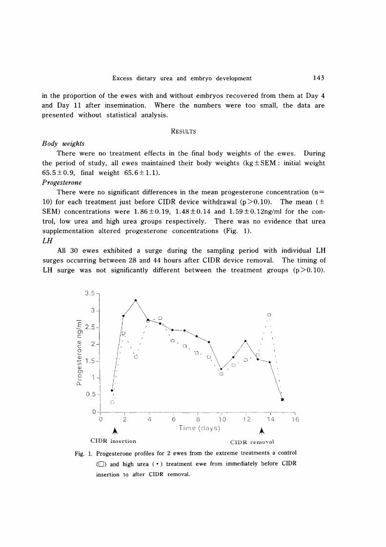
There were no significant differences in the mean progesterone concentration (n =
10) for each treatment just before CIDR device withdrawal (p >0.10). The mean (± SEM) concentrations were 1. 86 ± 0.19, 1. 48 ± 0.14 and 1. 59 ± 0.12ng/ml for the control, low urea and high urea groups respectively. There was no evidence that urea supplementation altered progesterone concentrations (Fig. 1).
LH All 30 ewes exhibited a surge during the sampling period with individual LH
surges occurring between 28 and 44 hours after CIDR device removal. The timing of LH surge was not significantly different between the treatment groups (p >0.10).
3.5
3
~ 2.5·· Q)
c Q.l C o '-
2-
(j)
t; 1.5 Q.l Q)
o '-(L
1
0.5 -
0-- -------,-----,---i-----r----------f------------,-------I -----1
o 2 if 6 8 10 12 14 16
Tirne (days)
crDR insertion CIDR removal
Fig. 1. Progesterone profiles for 2 ewes from the extreme treatments a control
(0) and high urea (.) treatment ewe from immediately before ClOR
insertion to after CIDR removal.
144 Christopher BISHONGA et al.
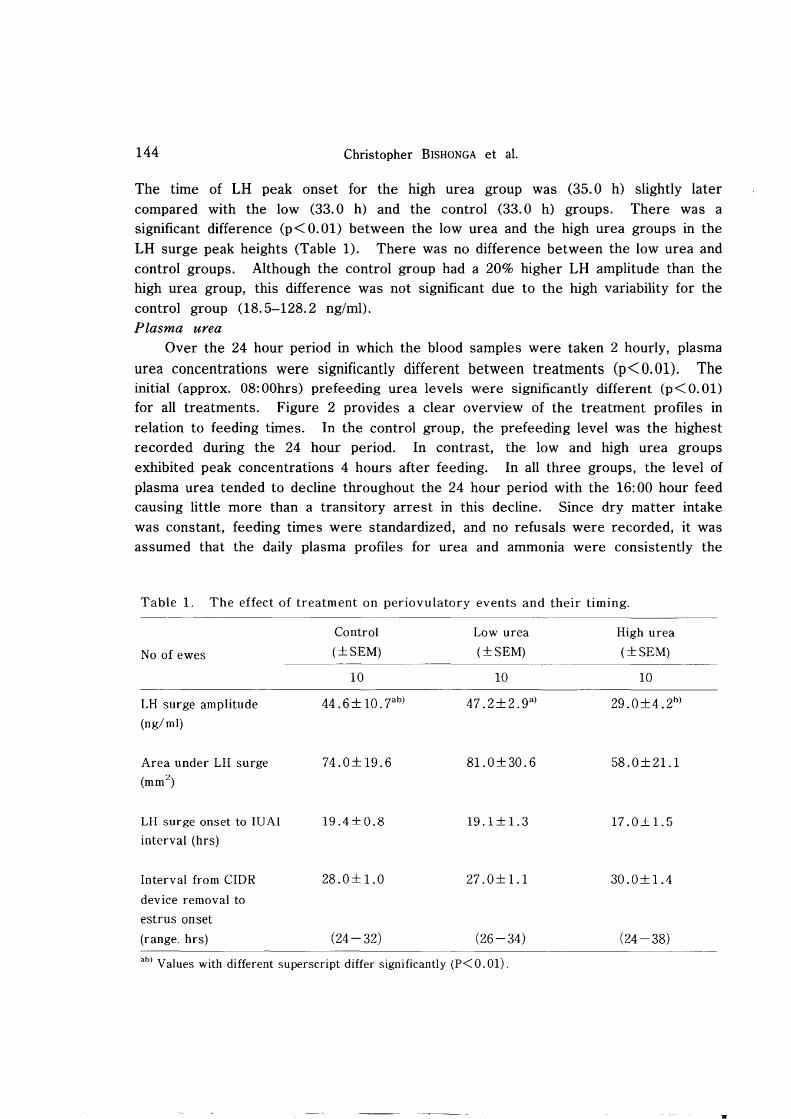
The time of LH peak onset for the high urea group was (35.0 h) slightly later compared with the low (33.0 h) and the control (33.0 h) groups. There was a significant difference (p<O.Ol) between the low urea and the high urea groups in the LH surge peak heights (Table 1). There was no difference between the low urea and control groups. Although the control group had a 20% higher LH amplitude than the high urea group, this difference was not significant due to the high variability for the control group (1S. 5-12S. 2 ng/ml). Plasma urea
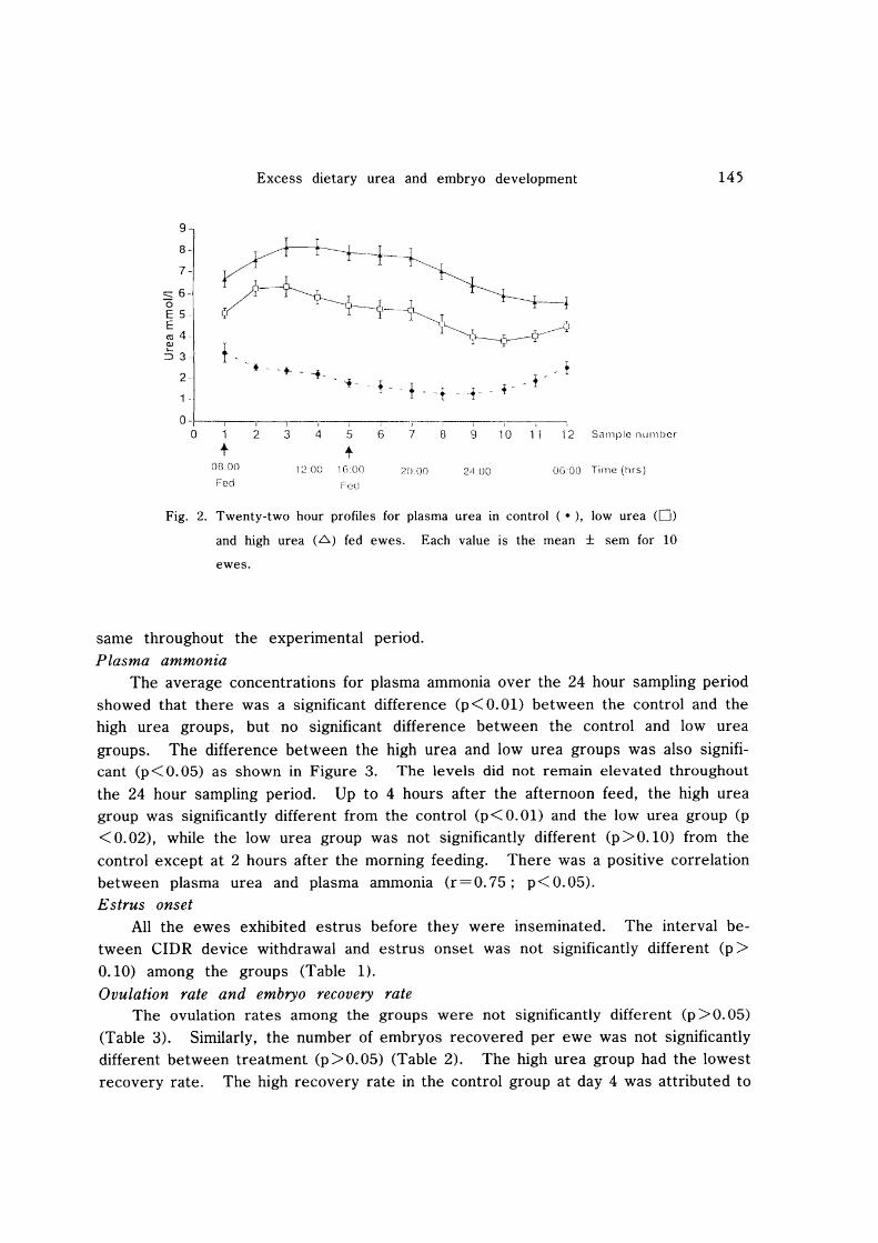
Over the 24 hour period in which the blood samples were taken 2 hourly, plasma
urea concentrations were significantly different between treatments (p< O. 01). The initial (approx. OS:OOhrs) prefeeding urea levels were significantly different (p<O.Ol) for all treatments. Figure 2 provides a clear overview of the treatment profiles in relation to feeding times. In the control group, the prefeeding level was the highest recorded during the 24 hour period. In contrast, the low and high urea groups exhibited peak concentrations 4 hours after feeding. In all three groups, the level of plasma urea tended to decline throughout the 24 hour period with the 16: 00 hour feed causing little more than a transitory arrest in this decline. Since dry matter intake was constant, feeding times were standardized, and no refusals were recorded, it was assumed that the daily plasma profiles for urea and ammonia were consistently the
Table 1. The effect of treatment on periovulatory events and their timing.
Control Low urea High urea
No of ewes (±SEM) (±SEM) (±SEM)
10 10 10
LH surge amplitude 44.6± 10. 7ab) 47.2±2.9a
) 29.0±4.2b)
(ng/ml)
Area under LH surge 74.0± 19.6 81.0±30.6 58.0±21.1 (mm 2
)
LH surge onset to IUAI 19.4±O.8 19.1 ± 1. 3 17.0±1.5 interval (hrs)
Interval from CIDR 28.0± 1.0 27 .O± 1.1 30.0± 1.4
device removal to
estrus onset
(range. hrs) (24- 32) (26-34) (24-38)
ab) Values with different superscript differ significantly (P< 0.01) .
Excess dietary urea and embryo development
9
8-
7-
::::::6 (5 E 5-E co 4-III
~ 3-
2-
1 -
o ~, --~--~~--__ -I
o 1 2 3 4 5
t t 0800 1200 1(i.On
Fed Fell
-r---, I
6 7 0 9 10 11 12
20.00 211 DO 0600
Sample nllmber
Time (hrs)
Fig. 2. Twenty-two hour profiles for plasma urea in control ( .), low urea (0)
and high urea (~) fed ewes. Each value is the mean ± sem for 10
ewes.
same throughout the experimental period.
Plasma ammonia
145
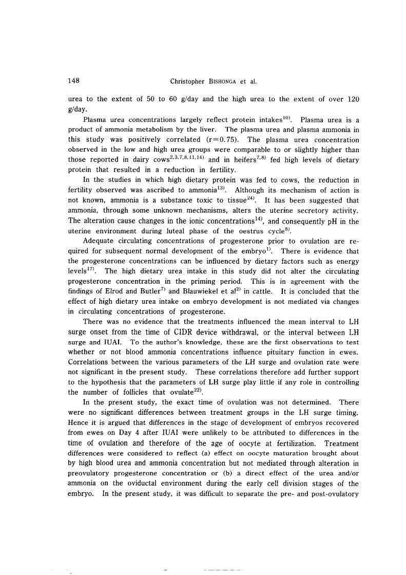
The average concentrations for plasma ammonia over the 24 hour sampling period showed that there was a significant difference (p<O.Ol) between the control and the high urea groups, but no significant difference between the control and low urea
groups. The difference between the high urea and low urea groups was also significant (p<O.05) as shown in Figure 3. The levels did not remain elevated throughout
the 24 hour sampling period. Up to 4 hours after the afternoon feed, the high urea group was significantly different from the control (p < 0.01) and the low urea group (p
< 0.02), while the low urea group was not significantly different (p >0.10) from the
control except at 2 hours after the morning feeding. There was a positive correlation between plasma urea and plasma ammonia (r=0.75; p< 0.05). Estrus onset
All the ewes exhibited estrus before they were inseminated. The interval between CIDR device withdrawal and estrus onset was not significantly different (p>
0.10) among the groups (Table 1).
Ovulation rate and embryo recovery rate The ovulation rates among the groups were not significantly different (p >0.05)
(Table 3). Similarly, the number of embryos recovered per ewe was not significantly different between treatment (p>0.05) (Table 2). The high urea group had the lowest
recovery rate. The high recovery rate in the control group at day 4 was attributed to

146 Christopher BISHONGA et al.
160
140 -
S 120-0 E 100 E ro 80-'c 0 E 60-E <! 40-
20-
0-I---r--,-, ----~r_--___r--i·---J--------·r--~T~---I---\--- I
0 1 2 3 11 5 6 7 8 9 10 11 -12 S<Jmple numbel
t t 08:00
Fed \200 1G 00
Foci
20.00 O(JOO Time (hrs)
Fig. 3. Twenty-two hour profiles for plasma ammonia in Controls ( .). low urea
(0) and high urea (.6) fed ewes. Each value is a mean ± sem for 10
ewes.
Table 2. Treatment mean values for embryo recoveries on Day 4 and Day 11 after insemination (in percentage).
Treatment
Day of No of ewes Control Low urea High urea
recovery per group (±SEM) (±SEM) (±SEM)
4 5 56 .0 ± 14 . oa} 29.0± 14.4 39.0±16.7
11 5 44.0±17.8 51.0±20.8 1O.O± 1O.oa}
a) Values with same superscript are significantly different (P<O.05).
one ewe which showed a typical superovulatory response. The recovery rate In the
control group at Day 4 was significantly different from the recovery at Day 11 in the
high urea group (p<0.05).
Day of recovery (hence method of recovery) had no effect on the number of
embryos recovered (x2 = L 04). One ewe from the control exhibited a superovulatory
response. There is no reason to believe that this was anything other than a chance
result. If the data of that ewe are removed from the mean, the ovulation rates
become 3.4 ± O. 7 for the control, 2.9 ± O. 5 for the low and 2.4 ± 4. 2 for the high urea
groups. Two ova from a ewe on the high urea treatment were retarded, and were
without spermatozoa on the zona pellucida.
Excess dietary urea and embryo development 147
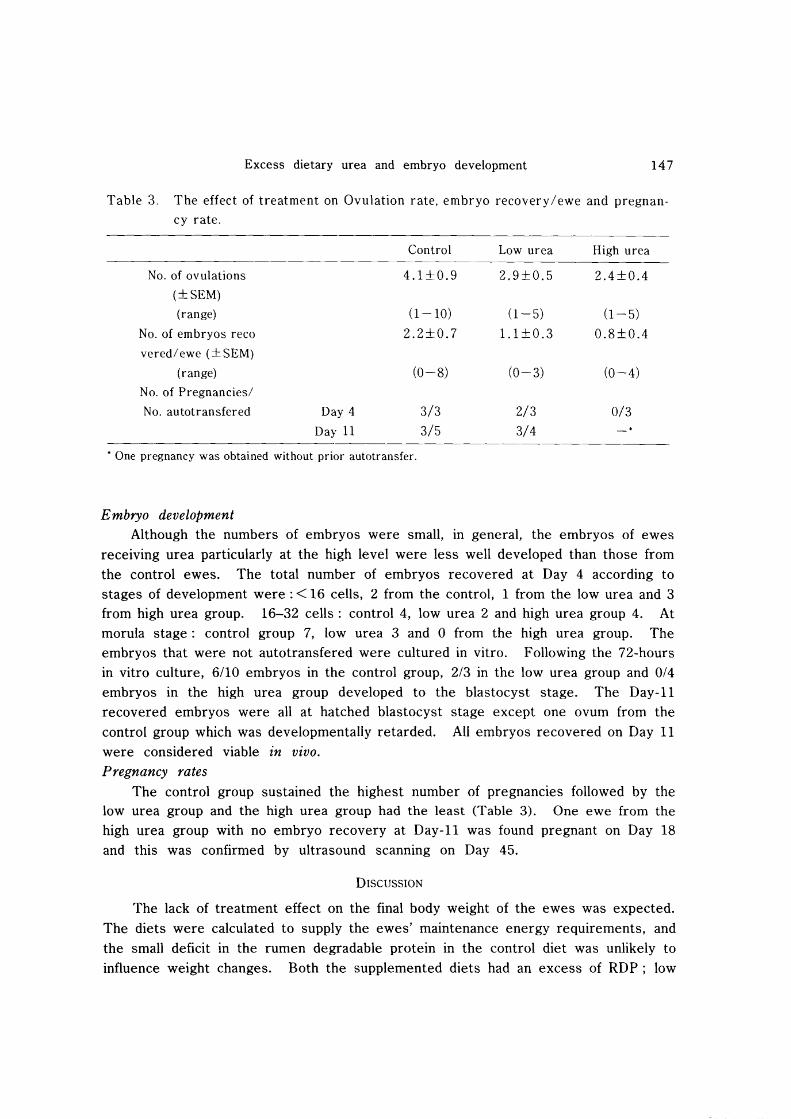
Table 3. The effect of treatment on Ovulation rate, embryo recovery/ewe and pregnan
cy rate.
Control Low urea High urea
No. of ovulations 4.1±0.9 2.9±0.5 2.4±0.4
(±SEM)
(range) (1-10) (1-5) (1-5)
No. of embryos reco 2.2±0.7 1.1 ±0.3 0.8±0.4
vered/ ewe (± SEM)
(range) (0-8) (0-3) (0-4)
No. of Pregnancies/
No. autotransfered Day 4 3/3 2/3 0/3
Day 11 3/5 3/4
• One pregnancy was obtained without prior autotransfer.
Embryo development Although the numbers of embryos were small, in general, the embryos of ewes
receiving urea particularly at the high level were less well developed than those from
the control ewes. The total number of embryos recovered at Day 4 according to stages of development were: < 16 cells, 2 from the control, 1 from the low urea and 3 from high urea group. 16-32 cells: control 4, low urea 2 and high urea group 4. At morula stage: control group 7, low urea 3 and 0 from the high urea group. The embryos that were not autotransfered were cultured in vitro. Following the 72-hours in vitro culture, 6/10 embryos in the control group, 2/3 in the low urea group and 0/4
embryos in the high urea group developed to the blastocyst stage. The Day-II recovered embryos were all at hatched blastocyst stage except one ovum from the
control group which was developmentally retarded. All embryos recovered on Day 11 were considered viable in vivo. Pregnancy rates
The control group sustained the highest number of pregnancies followed by the low urea group and the high urea group had the least (Table 3). One ewe from the high urea group with no embryo recovery at Day-II was found pregnant on Day 18 and this was confirmed by ultrasound scanning on Day 45.
DISCUSSION
The lack of treatment effect on the final body weight of the ewes was expected.
The diets were calculated to supply the ewes' maintenance energy requirements, and the small deficit in the rumen degradable protein in the control diet was unlikely to influence weight changes. Both the supplemented diets had an excess of RDP; low
148 Christopher BISHONGA et aI.
urea to the extent of 50 to 60 g/day and the high urea to the extent of over 120
g/day. Plasma urea concentrations largely reflect protein intakeslO). Plasma urea is a
product of ammonia metabolism by the liver. The plasma urea and plasma ammonia in this study was positively correlated (r = O. 75). The plasma urea concentration observed in the low and high urea groups were comparable to or slightly higher than those reported in dairy COWS2,3,7,8,1l,14) and in heifers7,8) fed high levels of dietary
protein that resulted in a reduction in fertility. In the studies in which high dietary protein was fed to cows, the reduction in
fertility observed was ascribed to ammonia 13). Although its mechanism of action is not known, ammonia is a substance toxic to tissue24
). It has been suggested that ammonia, through some unknown mechanisms, alters the uterine secretory activity.
The alteration cause changes in the ionic concentrations14), and consequently pH in the
uterine environment during luteal phase of the oestrus cycleS). Adequate circulating concentrations of progesterone prior to ovulation are re
quired for subsequent normal development of the embryo!). There is evidence that the progesterone concentrations can be influenced by dietary factors such as energy levels17
). The high dietary urea intake in this study did not alter the circulating progesterone concentration in the priming period. This is in agreement with the findings of Elrod and Butler7
) and Blauwiekel et a12) in cattle. It is concluded that the
effect of high dietary urea intake on embryo development is not mediated via changes in circulating concentrations of progesterone.
There was no evidence that the treatments influenced the mean interval to LH
surge onset from the time of CIDR device withdrawal, or the interval between LH surge and IVAI. To the author's knowledge, these are the first observations to test whether or not blood ammonia concentrations influence pituitary function in ewes. Correlations between the various parameters of the LH surge and ovulation rate were not significant in the present study. These correlations therefore add further support to the hypothesis that the parameters of LH surge play little if any role in controlling the number of follicles that ovulate22
).
In the present study, the exact time of ovulation was not determined. There were no significant differences between treatment groups in the LH surge timing. Hence it is argued that differences in the stage of development of embryos recovered from ewes on Day 4 after IUAI were unlikely to be attributed to differences in the
time of ovulation and therefore of the age of oocyte at fertilization. Treatment differences were considered to reflect (a) effect on oocyte maturation brought about by high blood urea and ammonia concentration but not mediated through alteration in preovulatory progesterone concentration or (b) a direct effect of the urea and/or ammonia on the oviductal environment during the early cell division stages of the embryo. In the present study, it was difficult to separate the pre- and post-ovulatory
Excess dietary urea and embryo development 149
influences on embryo development. Abscence of spermatozoa on zona pellucida on some ova implies that the high urea
treatment may have led to a hostile uterine and/or oviductal environment that compromised sperm viability.
In view of treatment effects on progesterone levels and periovulatory endocrine events, the observation that there were less pregnancies established and even fewer pregnancies sustained in ewes on the high urea supplemented diet suggests that urea supplementation caused early embryo mortality. The results indicate that the effect was manifest by Day 4 post-insemination. Thus, the general picture from this study is that an excess of RDP in the form of dietary urea is detrimental to the establishment of pregnancy in sheep. The detrimental effects manifest right through the pre-implantation stages. These findings, albeit generated from a fairly extreme experimental model, may well have implications for current grassland husbandry practices in which dairy cows in particular graze spring pastures that are very high in rumen degradable proteins.
ACKNOWLEDGEMENTS
We thank Prof. Hiroshi Kanagawa and Dr Jose Arceo N. Bautista for their advice on the manuscript.
REFERENCES
1) ASHWORTH, C. J., SALES, D. J. & WILMUT, I. : Evidence of an association between
the survival of embryos and the periovulatory plasma progesterone concentration in the ewe. The riogeno logy. 54: 749-759, 1989.
2) BLAUWIEKEL, R., KINCAID, R. L. & REVEES, J. J. : Effect of high crude protein on
pituitary and ovarian function in Holstein cows. J. Dairy Sci. 69: 439-446, 1986.
3) CANIFIELD, R. W., SNIFFEN, C. j. & BUTLER, W. R. : Effects of excess degradable
protein on postpartum reproduction and energy balance in dairy cattle. ]. Dairy Sci. 73: 2342-2349, 1990.
4) CARROLL, D. J., BARON, R. A., ANDERSON, G. W. & SMITH, R. D.: Influence of
protein intake and feeding strategy on reproductive performance of dairy cows. J. Dairy Sci. 71: 3470-3481, 1988.
5) DJAHANBAHKCH, 0., SWANSTON, I. I., CORRIE, j. E. T. & MCNEILLY, A. S. : Predic
tion of ovulation by progesterone. Lancet. 2(8256): 1164-1165, 1981.
6) ELROD, C. C.: High dietary protein and high fertility can we have both? In:
Proceedings of Cornell Nutrition Conference for Feed Manufacturers, 54th Meeting,
October 13-15. Ithaca, New York, 1992.
7) ELROD, C. C. & BUTLER, W. R. : Reduction of fertility due to reduction of uterine
pH in heifers fed excess ruminally degradable protein. J. Anim. Sci. 71 : 694-701,
1993. 8) ELROD, C. c., VAN AMBURGH. M. & BUTLER, W. R. : Alterations of pH in response
to increased dietary protein in cattle is unique to the uterus. ]. Anim. Sci. 71 :
150 Christopher BISHONGA et al.
702-706, 1993.
9) FOLMAN, Y., NEUMARK, H., KAIM, M. & KAUFMANN, W.: Performance, rumen and
blood metabolites in high yielding cows fed varying protein percents and protected
soyabean. J. Dairy Sci. 64: 759-768, 1981.
10) HARMEYER, I. & MARTENS, H.: Aspects of urea metabolism in ruminants with
reference to the goat. J. Dairy Sci. 63: 1707-1728, 1980.
11) HOWARD, H. P., AASLETH, E. P., ADAMS, G. D. & BUSH, L. j. : Influence of dietary
protein on reproductive performance of dairy cows. j. Dairy Sci. 70: 1563-1571,
1987.
12) HUNTER, G. L., ADAMS, C. E. & ROWSON, L. E. A.: Interbreed ovum transfer in
sheep. J. Agric. Sci. 46: 143-149, 1955. 13) JORDAN, E. R. & SWANSON, L. V. : Serum progesterone and luteinizing hormone in
dairy cattle fed varying levels of crude protein. J. Anim. Sci. 48: 1154-1158, 1979.
14) JORDAN, E. R., CHAPMAN, T. E., HOLTAN, D. W. & SWANSON, L. V. : Relationship of
dietary crude protein to compostion of uterine secretions and blood in high produc
ing postpartum dairy cows. j. Dairy Sci. 66: 1854-1862, 1983.
15) KAIM, M., FOLMAN, Y. & NEUMARK, H.: The effect of protein intake and lactation
number on postpartum body weight loss and reproductive performance of dairy
cows. j. Anim. Prod. 37: 227-235, 1983. 16) LINDNER, G. M. & WRIGHT, R. W. JR. : Bovine embryo morphology and evaluation.
Theriogenology. 20: 407-416, 1983.
17) MCEVOY, T. G., ROBINSON, J. J., AITKEN, R. P., KYLE, C. E. & ROBERTSON, I. S. : The effect of feeding level during a 12-day progesterone priming period on the viability of embryos collected from superovulated ewes. B SAP Winter Meeting.
Scarbrough. Anim. Prod. 56: 432, 1993.
18) MCKELVEY, W. A. C., ROBINSON, j. J. & AITKEN, R. P.: The evaluation of a
laparoscopic insemination technique in ewes. Theriogenology 24: 519-533, 1985.
19) MCNEILLY, A. S., JONASSEN, 1. A. & FRASER, H. M.: Suppression of follicular
development after chro,nic LHRH immuno-neutralization in ewes. J. Reprod. Fertil. 76: 481-490, 1986.
20) o RSKOV, E. R. & ROBINSON, J. J. : The application of modern concepts of ruminant protein nutrition to sheep production systems. Livestock Sci. 8: 330-350, 1981.
21) RENARD, 1. -P., Du MESNIL Du DUISSON, F., WINTENBERGERTORRES, S. & MENEZO,
Y. : Invitro culture of cow embryos from day 6 and day 7. In: ROWSON, L. E. A. ed. Egg Transfer in Cattle. pp 159-164. Commission of the European Communities: Luxembourg. 1976.
22) RHIND, S. M. : Nutrition: its effects on reproductive performance and its hormonal control in female sheep and goats. In: Speedy, A. W. ed. Progress in Sheep and
Goat Research. pp 25-51, C. A. B International Wallingford, Oxon. 1992.
23) SCUDAMORE, C. L. : Studies in superovulating ewes of factors influencing the yield of
fertilized ova, and their capacity for development. Ph. D. thesis. Rowett Research
Institute. University of Aberdeen. 1991. 24) TALKE, H. & SCHUBERT, G. E.: Enzyme determination of urea in blood serum by

Excess dietary urea and embryo development
the Warburg optical test. Klin. Wochschr. 43: 174-175, 1965.
25) VISEK, J. W. : Ammonia: its effects on biological systems, metabolic hormones and reproduction. J. Dairy Sci. 67: 481-498, 1982.
26) WALLACE, J. M. AITKEN, R. P. & CHEYNE, M. A. : Conceptus development in vivo
endometrial and conceptus protein release in vitro following blastocyst transfer to ewes induced to ovulate at 28 days postpartum. Reprod. Fertil. Dev. 5 : 191-200,
1993.
151