Embed Size (px)
Citation preview
Excitons and Excess Electrons in Nanometer Size MolecularPolyoxotitanate Clusters: Electronic Spectra, Exciton Dynamics, andSurface StatesJianhua Bao,† Zhihao Yu,† Lars Gundlach,† Jason B. Benedict,‡ Philip Coppens,‡ Hung Cheng Chen,§
John R. Miller,§ and Piotr Piotrowiak*,†
†Department of Chemistry, Rutgers University, Newark, New Jersey 07102, United States‡Department of Chemistry, University at Buffalo, SUNY, Buffalo, New York 14260, United States§Chemistry Department, Brookhaven National Laboratory, Upton, New York 11973, United States
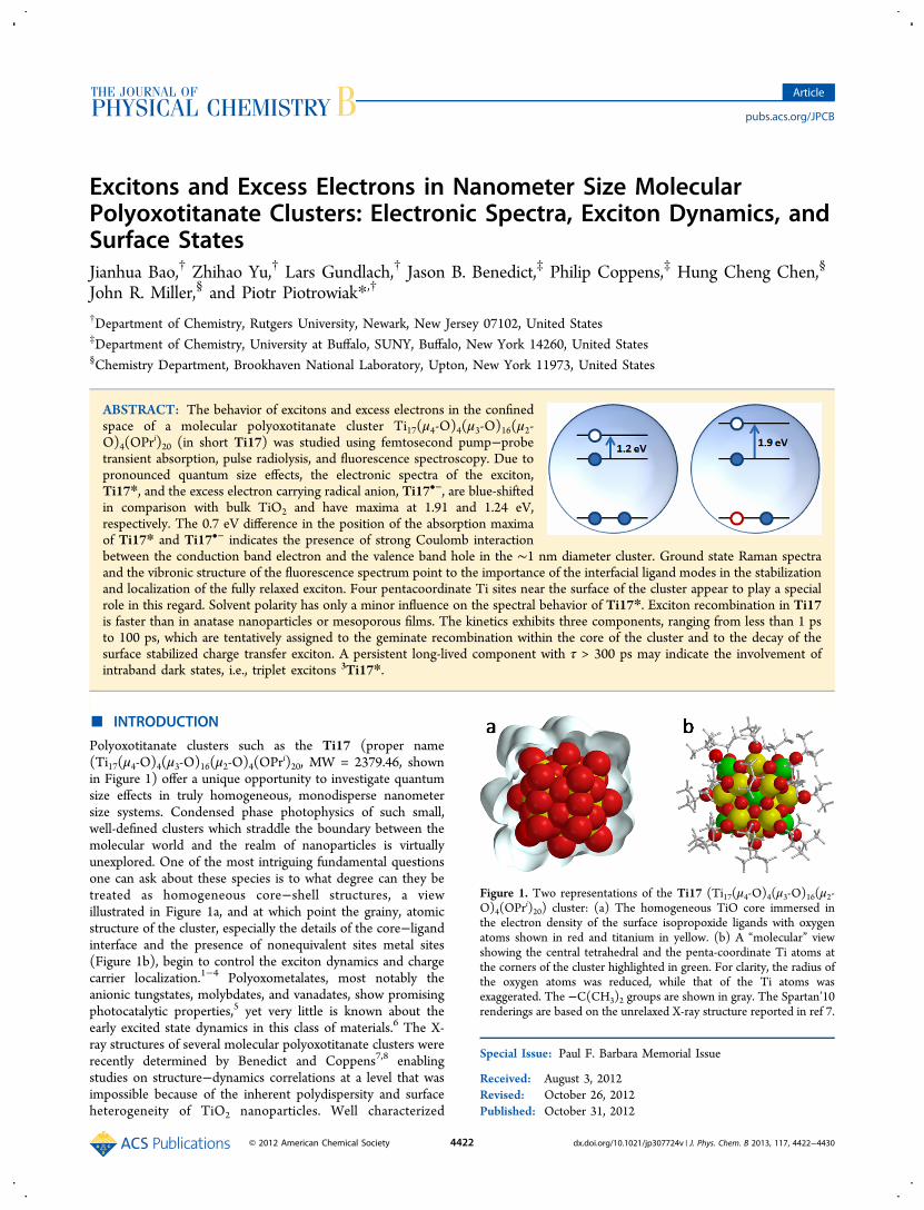
ABSTRACT: The behavior of excitons and excess electrons in the confinedspace of a molecular polyoxotitanate cluster Ti17(μ4-O)4(μ3-O)16(μ2-O)4(OPr
i)20 (in short Ti17) was studied using femtosecond pump−probetransient absorption, pulse radiolysis, and fluorescence spectroscopy. Due topronounced quantum size effects, the electronic spectra of the exciton,Ti17*, and the excess electron carrying radical anion, Ti17•−, are blue-shiftedin comparison with bulk TiO2 and have maxima at 1.91 and 1.24 eV,respectively. The 0.7 eV difference in the position of the absorption maximaof Ti17* and Ti17•− indicates the presence of strong Coulomb interactionbetween the conduction band electron and the valence band hole in the ∼1 nm diameter cluster. Ground state Raman spectraand the vibronic structure of the fluorescence spectrum point to the importance of the interfacial ligand modes in the stabilizationand localization of the fully relaxed exciton. Four pentacoordinate Ti sites near the surface of the cluster appear to play a specialrole in this regard. Solvent polarity has only a minor influence on the spectral behavior of Ti17*. Exciton recombination in Ti17is faster than in anatase nanoparticles or mesoporous films. The kinetics exhibits three components, ranging from less than 1 psto 100 ps, which are tentatively assigned to the geminate recombination within the core of the cluster and to the decay of thesurface stabilized charge transfer exciton. A persistent long-lived component with τ > 300 ps may indicate the involvement ofintraband dark states, i.e., triplet excitons 3Ti17*.
■ INTRODUCTION
Polyoxotitanate clusters such as the Ti17 (proper name(Ti17(μ4-O)4(μ3-O)16(μ2-O)4(OPr
i)20, MW = 2379.46, shownin Figure 1) offer a unique opportunity to investigate quantumsize effects in truly homogeneous, monodisperse nanometersize systems. Condensed phase photophysics of such small,well-defined clusters which straddle the boundary between themolecular world and the realm of nanoparticles is virtuallyunexplored. One of the most intriguing fundamental questionsone can ask about these species is to what degree can they betreated as homogeneous core−shell structures, a viewillustrated in Figure 1a, and at which point the grainy, atomicstructure of the cluster, especially the details of the core−ligandinterface and the presence of nonequivalent sites metal sites(Figure 1b), begin to control the exciton dynamics and chargecarrier localization.1−4 Polyoxometalates, most notably theanionic tungstates, molybdates, and vanadates, show promisingphotocatalytic properties,5 yet very little is known about theearly excited state dynamics in this class of materials.6 The X-ray structures of several molecular polyoxotitanate clusters wererecently determined by Benedict and Coppens7,8 enablingstudies on structure−dynamics correlations at a level that wasimpossible because of the inherent polydispersity and surfaceheterogeneity of TiO2 nanoparticles. Well characterized
Special Issue: Paul F. Barbara Memorial Issue
Received: August 3, 2012Revised: October 26, 2012Published: October 31, 2012
Figure 1. Two representations of the Ti17 (Ti17(μ4-O)4(μ3-O)16(μ2-O)4(OPr
i)20) cluster: (a) The homogeneous TiO core immersed inthe electron density of the surface isopropoxide ligands with oxygenatoms shown in red and titanium in yellow. (b) A “molecular” viewshowing the central tetrahedral and the penta-coordinate Ti atoms atthe corners of the cluster highlighted in green. For clarity, the radius ofthe oxygen atoms was reduced, while that of the Ti atoms wasexaggerated. The −C(CH3)2 groups are shown in gray. The Spartan’10renderings are based on the unrelaxed X-ray structure reported in ref 7.
Article
pubs.acs.org/JPCB
© 2012 American Chemical Society 4422 dx.doi.org/10.1021/jp307724v | J. Phys. Chem. B 2013, 117, 4422−4430
polyoxotitanate clusters such as that described here Ti17 canserve as very attractive, experimentally and theoreticallytractable models for colloidal TiO2, the most widely studiedwide band gap semiconductor substrate for hybrid photovoltaicmaterials9 and a potent photocatalyst for hydrogen produc-tion10 and pollutant remediation.11
In this paper, we report the results of combined laser pump−probe and pulse radiolysis experiments aimed at the under-standing of the exciton dynamics, charge localization, andelectron−hole coupling in the Ti17 cluster. Optical excitationof Ti17 generates Ti17* excitons, i.e., electron−hole pairs inwhich the charges interact with one another, while pulseradiolysis places a single excess electron in the lowest emptyorbital (LUMO) or the conduction band (CB) of the cluster,leading to the formation of the corresponding radical anion,Ti17•−. In the case of a single excess charge, the confinementaffects only the quantized kinetic energy of the electron, whilefor the exciton the kinetic, Coulomb, and exchange energy areall affected by the size of the nanoparticle.1−3 The combinationof pulse radiolysis and optical spectroscopy gives a morecomplete view of the electronic states of Ti17 and allows abetter grasp of the electron−hole interaction in the confinedvolume of the cluster.The crystallographic structure of the polyoxotitanate core of
Ti17 resembles most closely brookite, the least commonmetastable phase of crystalline TiO2.
7 The core of the cluster isterminated by 20 isopropoxide groups (Figure 1b). As a result,the outer shell of the cluster is for the most part hydrophobic,resulting in good solubility in even mildly polar organicsolvents. There are four five-coordinate titanium ions whichform a square centered around the unique tetrahedral Ti ionpositioned in the middle of the cluster (shown in green inFigure 1b). The remaining 12 Ti ions have the most typical, 6-fold coordination. The system has an approximate S4 ratherthan spherical symmetry, and there is considerable disorder inthe orientation of the isopropyl groups. As it will be seen, theexistence of nonequivalent and coordinatively unsaturated Tisites has a significant influence on the electronic properties ofthe cluster.Because titanium dioxide is a large band gap II−VI
semiconductor with a strong ionic character,18,19 the onset ofquantum size effects is expected to occur for much smallerparticles than in the case of chalcogenides and other small bandgap covalent solids. Serpone et al. concluded that quantum sizeeffects were not significant in anatase particles down to ∼2.1nm diameter, which corresponds to approximately 200 Tiatoms;20 however, the oxotitanate core of Ti17 contains only17 Ti atoms and is approximately 10 times smaller by volume.Its furthest Ti centers span 8.3 Å, and the maximum distancebetween the surface oxygen atoms is 11.4 Å. At thesedimensions, quantum confinement effects are clearly man-ifested in the ground state electronic absorption spectrum ofTi17. The line shape analysis performed by Benedict andCoppens placed the band edge of Ti17 at 4.3 eV (∼290 nm)and the lowest indirect transition at 3.4 eV (∼365 nm).7 Forcomparison, the band gaps of bulk anatase, rutile, and brookiteare 3.2, 3.0, and 3.3 eV, respectively (Table 1).We have found that the small size of the cluster has an even
more dramatic influence on the spectral properties of Ti17*and Ti17•− than on the ground state absorption. In bulksemiconductors and large nanoparticles, excitons can dissociateand form free carriers. In rutile, the exciton binding energy is assmall as 4 meV, and in anatase, it is believed to be larger, on the
order of 20 meV, i.e., still approximately only 1 kBT.18,19 In the
case of Ti17, because of the confinement, the charges remainstrongly coupled to one another and cannot fully dissociate.Classical Coulomb interaction calculated at the maximumelectron−hole separation permitted by the 1 nm diameter ofthe TiO core of the cluster is approximately 250 meV, i.e.,equivalent to 10 kBT at room temperature. Better estimates canbe obtained using Brus type expressions2−4 which account forthe electron−phonon and hole−phonon coupling (nuclearreorganization in the molecular terminilogy) by introducing anadjustable effective mass for both charges; however, since theseparameters are not known for the cluster, we will use the roughvalue of 0.25 eV as a convenient zeroth-order reference point.The results reported here show that the simple core−shell
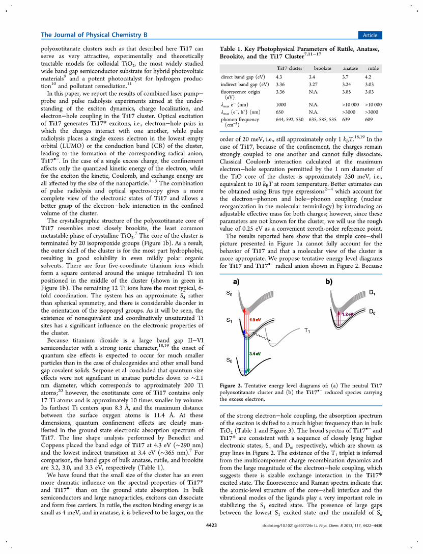
picture presented in Figure 1a cannot fully account for thebehavior of Ti17 and that a molecular view of the cluster ismore appropriate. We propose tentative energy level diagramsfor Ti17 and Ti17•− radical anion shown in Figure 2. Because
of the strong electron−hole coupling, the absorption spectrumof the exciton is shifted to a much higher frequency than in bulkTiO2 (Table 1 and Figure 3). The broad spectra of Ti17•− andTi17* are consistent with a sequence of closely lying higherelectronic states, Sn and Dn, respectively, which are shown asgray lines in Figure 2. The existence of the T1 triplet is inferredfrom the multicomponent charge recombination dynamics andfrom the large magnitude of the electron−hole coupling, whichsuggests there is sizable exchange interaction in the Ti17*excited state. The fluorescence and Raman spectra indicate thatthe atomic-level structure of the core−shell interface and thevibrational modes of the ligands play a very important role instabilizing the S1 excited state. The presence of large gapsbetween the lowest S1 excited state and the manifold of Sn
Table 1. Key Photophysical Parameters of Rutile, Anatase,Brookite, and the Ti17 Cluster7,11−17
Ti17 cluster brookite anatase rutile
direct band gap (eV) 4.3 3.4 3.7 4.2indirect band gap (eV) 3.36 3.27 3.24 3.03fluorescence origin(eV)
3.36 N.A. 3.85 3.03
λmax e− (nm) 1000 N.A. >10 000 >10 000
λmax {e−, h+} (nm) 650 N.A. >3000 >3000
phonon frequency(cm−1)
644, 592, 550 635, 585, 535 639 609
Figure 2. Tentative energy level diagrams of: (a) The neutral Ti17polyoxotitanate cluster and (b) the Ti17•− reduced species carryingthe excess electron.
The Journal of Physical Chemistry B Article
dx.doi.org/10.1021/jp307724v | J. Phys. Chem. B 2013, 117, 4422−44304423
states as well as between the D0 ground state and the Dn
manifold of doublets is not consistent with the bulk-like pictureof Ti17. We postulate that the S1 and D0 states are rathertightly localized and stabilized by nuclear reorganization at thecore−ligand interface, while the higher, densely spaced Sn andDn states involve primarily the orbitals of the polyoxotitanatecore and hence are of a more bulk-like character.In the body of the paper, we will continue to apply Ti17* as
a general symbol for the Ti17 excited state or exciton, while theS1, Sn, and T1 terms will be used when more precise labels arenecessary for clarity.
■ EXPERIMENTAL METHODS
Synthesis. The synthesis, purification, and structuralcharacterization of the Ti17 polyoxotitanate clusters weredescribed in detail in the literature.7,21,22
Sample Preparation. Benzonitrile (Acros) and dibutylether (Sigma-Aldrich) were dried on 4 Å molecule sieves andused without further purification. Samples for the transientabsorption measurements were prepared by dissolvingapproximately 3 mg of solid Ti17 in 2 mL of dry solvent.After 30 min, the clear solution was transferred into a standardquartz cuvette with an airtight valve. The entire process wascarried out in a glovebox.Steady-State Spectroscopy. Absorption spectra were
measured with a Varian Cary 500 spectrometer. Fluorescencespectra were recorded on a Varian Cary Eclipse fluorimeter(resolution 2.5 nm).Raman Microspectroscopy. Approximately 1 mg of the
Ti17 or Ti17cat4 cluster was spread in an indentation in aclean brass cell and covered by a glass slide with vacuum oilalong the edge to prevent air from entering the sample. Samplepreparation was carried out in a glovebox. Raman spectra wereacquired using a Kaiser Optical Systems Confocal RamanMicroprobe equipped with a 785 nm diode laser. A 100× oilimmersion objective was used to focus approximately 6−10mW of the single mode power within the sample volume of ∼2μm3. The spectral coverage ranged from 100 to 3450 cm−1, andthe resolution was 4 cm−1. The spectra were acquired using 4accumulations of 60 s exposure time each.
Pump−Probe Transient Absorption Spectroscopy inthe Visible and IR. 70 fs pulses at 795 nm were provided by ahome-built 1.25 kHz multipass Ti:sapphire amplifier seeded bya Spectra-Physics Tsunami oscillator. The output of theamplifier was split to generate independently tunable pumpand probe pulses. The pump beam (∼25 fs, 3.5 mW, 300 nm)was produced by frequency doubling the output of anoncollinear optical parametric amplifier (Topas White, LightConversion). The pump was modulated at 17 Hz with the helpof a mechanical chopper. The probe beam was delayed withrespect to the pump using a computer-controlled delay stageand focused into a 2 mm sapphire plate to generate white lightcontinuum. The white light was overlapped with the pumpbeam in a 10 mm fused silica cuvette containing the sample.The probe light was dispersed with a monochromator (OrielMS257 with a 1200 lines/mm grating) and detected with a Siphotodiode (Thorlabs, DET110). The absorption spectra werecollected in the lock-in mode (Stanford Research, SR 810) withthe monochromator scanning the wavelength range and thepump−probe delay fixed at the desired value. The relativepolarization of the pump and probe beams was set at the“magic angle”.The IR transients were obtained using as a probe sub-100 fs
pulses produced by a home-built IR OPA based on parametricdown-conversion in KNbO3 and pumped by the samemultipass Ti:sapphire amplifier as above. In order to optimizethe relative duty cycles, the output of the IR detector (InAsphotodiode) was sampled with a boxcar integrator (StanfordResearch 280) the analog output of which served as the inputfor the lock-in amplifier. Instead of using IR interference filters,we took advantage of the narrow transparency window ofacetonitrile at 2.65 μm to select the desired wavelength. TheTi17 sample was prepared in the same fashion as describedabove. The anatase film on a thin silica substrate was preparedas described in the literature.23 It was placed in a cuvettecontaining acetonitrile in order to ensure comparableexperimental conditions.
Pulse Radiolysis. The spectra of Ti17•− were obtained atthe Laser-Electron Accelerator Facility (LEAF) at theBrookhaven National Laboratory. LEAF and the methodsused are described elsewhere.24 Briefly, an electron pulse (≤120ps duration) was focused into a quartz cell with an optical path
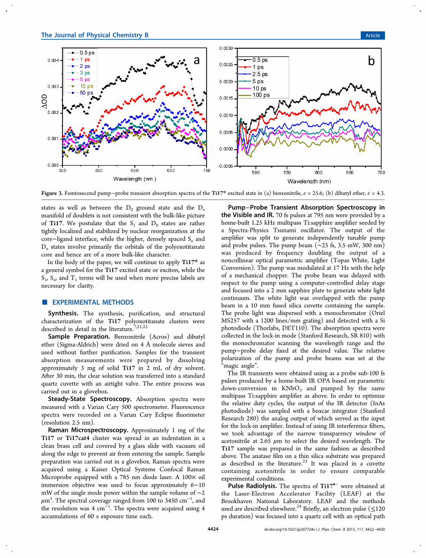
Figure 3. Femtosecond pump−probe transient absorption spectra of the Ti17* excited state in (a) benzonitrile, ε = 25.6; (b) dibutyl ether, ε = 4.3.
The Journal of Physical Chemistry B Article
dx.doi.org/10.1021/jp307724v | J. Phys. Chem. B 2013, 117, 4422−44304424
length of 20 mm containing the solution of interest. Themonitoring light source was a 65 W xenon arc lamp pulsed to afew hundred times its normal intensity. Wavelengths wereselected using 10 nm bandpass interference filters. Transientabsorption signals were detected with either an FND-100Qsilicon diode (λ ≤ 1000 nm, 2 ns, EG&G) or a GAP-500LInGaAs diode (λ ≥ 1100 nm, 8 ns, Germanium Power Devices)and digitized with a Tektronix TDS-680B or a LeCroy 8420Aoscilloscope. The electron pulses ionized the solvent,benzonitrile, to produce its anions and cations. Electronsfrom the solvent anions (“excess electrons”) were transferred toTi17, reducing it and producing Ti17•−, i.e., the radical anionof the cluster. Benzonitrile cations cannot oxidize Ti17, and as aresult, the corresponding Ti17•+ species was not observed. Thiswas confirmed by experiments on Ti17 solutions saturated withSF6, which selectively scavenges the excess electrons. Underthese conditions, neither Ti17•+ nor Ti17•− were observed.Computation. The calculation of the Raman and IR
absorption spectra of the isopropoxide anion and the oxoisopropoxyl radical was carried out at the DFT B3LYP levelwith the 6-31G* basis set using the Spartan’10 softwarepackage by Wavefunction, Inc. The resulting vibrationalfrequencies were not scaled.
■ RESULTS AND DISCUSSIONExcitation of Ti17 with 300 nm ∼25 fs pulses results in theformation of the S1 excited state which is characterized by abroad and featureless spectrum which spans nearly the entirevisible range from 500 to 750 nm (Figure 3). The large spectralwidth, lack of structure, and gradual falloff on the high energyside are consistent with a closely spaced sequence of higherelectronic excited states of the cluster (Figure 2). Thetransitions to the closely lying S2, ..., Sn electronic states havediminishing oscillator strength and suggest the onset ofcontinuum (ionization of the Ti17* excited state) at around2.5 eV. The spectra in two solvents, dibutyl ether andbenzonitrile, which differ considerably in polarity (ε = 4.3 forthe former and ε = 25.6 for the latter), are nearlyindistinguishable from one another. The spectrum in dibutylether does appear to be somewhat broader and possibly slightlyred-shifted; however, even in the presence of these minordifferences, one must conclude that the influence of solventpolarity on the spectrum of Ti17* is small. This suggests thatthe isopropoxide ligands, which form a dense cover on thesurface of the cluster, reduce the solvation effects by shieldingthe core of the cluster from the surrounding medium. Inessence, the ligands themselves act as the first solvation shell forthe exciton and diminish the polarization of the bulk solvent.Alternatively, such weak solvent dependence could indicate thatthe relaxed S1 state has little CT character and that the electronand the hole are not spatially well separated. This explanation isunlikely given the presence of nonequivalent Ti centers and theionic nature of the TiO core.The 650 nm (1.9 eV) maximum of the Ti17* spectrum lies
at a much higher energy than that for the bulk exciton orconduction band electron in TiO2, which also has a very broadand featureless spectrum with λmax > 10 000 nm, i.e., <0.1eV.17,25 This dramatic blue shift of more than 1.8 eV points tothe presence of strong quantum size effects and increasedelectron−hole coupling in the 1 nm cluster, as discussed in theIntroduction. In order to confirm that Ti17* does not havelower-lying electronic transitions, we also probed the responseof the directly excited cluster and anatase in the near IR (1200−
Figure 4. (a) Overlayed normalized transients collected at severalwavelengths from 550 to 680 nm showing homogeneous decaykinetics. (b) Decay of the excited state of Ti17 in dibutyl ethermonitored at 680 nm (excitation at 300 nm) fitted with abiexponential function with τ1 = 0.75 ps and τ2 = 5.3 ps. A longertime scale decay fitted with τ3 = 102 ps is shown in the inset. (c) IRtransient responses of the Ti17 cluster (red) and anatase film (black)probed at 2.65 μm.
The Journal of Physical Chemistry B Article
dx.doi.org/10.1021/jp307724v | J. Phys. Chem. B 2013, 117, 4422−44304425
1400 nm) and IR (2650 nm) spectral regions. The resultingtransients obtained for Ti17 and a mesoporous TiO2 film at2.65 μm are shown in Figure 4c. The directly excited TiO2 filmshows at this wavelength strong absorption in agreement withthe findings of Katoh et al.;25 however, the only signal that wewere able to detect in the case of Ti17 is the small nonlinearartifact at t = 0. This verifies that Ti17* has no low-lyingallowed electronic transitions below the 700 nm. It should bementioned that photoexcited Ti17, unlike rutile or anatase,does not give a response in the terahertz frequency range.26
The lack of IR and terahertz absorption corroborates thepresence of strong electron−hole coupling in Ti17* andsuggests that the cluster does not have a true conduction band.It is more likely that the appearance of the spectrum of Ti17* isdue to a sequence of closely spaced localized and delocalizedd−d transitions in the core of the cluster.Exciton Relaxation and Recombination Dynamics.
The broad transient absorption spectra of the Ti17* excitedstate decay homogeneously, as shown in Figures 3 and 4a.While there appears to be a barely perceptible shift to the blueduring the initia1 1 ps following the excitation, the low signal-to-noise ratio of the available data, combined with the largewidth of the spectrum, do not justify a more detailed analysis ofthe spectral dynamics. Similarly to the Ti17* absorptionspectrum, its decay dynamics is weakly dependent on solventpolarity and the systematic differences between the benzonitrileand dibutyl ether results were smaller than the fittinguncertainties. This further supports the picture of an excitonshielded from the solvent by the ligand shell.The kinetics in the 0−30 ps time window, which was
examined most extensively in this work, is dominated by twocomponents, τ1 = 0.75 ps and τ2 = 5.3 ps, which togetherconstitute more than 60% of the overall decay (Figure 3b andTable 2). Since the pump−probe data were collected in the“magic angle” configuration and since the spectral evolution isminor, these rates reflect purely the population decay. Thefastest subpicosecond rate most likely corresponds to thegeminate recombination of the initial electron−hole pairs whichdid not fully equilibrate with the 550−640 cm−1 phonon modesof the TiO core of the cluster (Table 1). The surviving excitonsfollow two paths: they either decay with the rate τ2, which isconsistent with vibrational cooling, or acquire charge transfercharacter with one of the charges stabilized at the core−ligandinterface, consistent with the view that in small nanocrystallitesthe interaction with the surface dominates the electron and holedynamics.1 Skinner et al. found that in colloidal TiO2 theelectron localizes within a picosecond on coordinativelyunsaturated Ti centers,27 which in the case of Ti17* suggeststhe central tetrahedral Ti atom (Figure 1b) as the location ofthe negative charge in the relaxed excited state. The crucialinvolvement of surface states in stabilizing the hole iselaborated in more detail in the following sections of thepaper. Longer scans up to 300 ps (Figure 4b, Table 2) allowedus to determine the third decay time, τ3 = 102 ps, which weascribe to the recombination of the fully relaxed, surface-stabilized exciton. Only less than 30% of the initial excited statepopulation reaches this stage.
Lastly, fitting of the long scan transients always returned a 5−10% A∞ component which does not decay at the 300 ps timescale. While we cannot rule out the presence of small amountsof impurities or photodamage of the cluster itself, even thoughprecautions were taken to minimize the latter, it is plausiblethat the longest lived absorption component originates fromthe “dark” intraband states, i.e., triplet excitons 3Ti17*, or inthe molecular terminology, simply the T1 triplet state of thecluster (Figure 2). As it will be shown below, the electron−holecoupling in the confined space of Ti17 appears to be large, onthe order of 6000 cm−1 or 30 kBT. This coupling consists of theCoulomb and exchange interaction, the latter of which leads tothe singlet−triplet splitting of the excited state. One cannoteasily determine the partitioning of the overall interaction intothe Coulomb and exchange components. However, if the S−Tsplitting is larger than kBT, which it is very likely to be given thelarge magnitude of the overall electron−hole interaction, adistinct, slower recombination dynamics of the triplet excitonshould be expected. A definitive proof of the existence of suchtriplet states would be to detect low temperature phosphor-escence of Ti17. These efforts are under way.
Spectrum of the Excess Electron and the Electron−Hole Coupling in the Excited State of Ti17. In bulksemiconductors, the direct electron−hole coupling is small. Asmentioned earlier, the exciton binding is only 4 meV in rutileand 20 meV in anatase.18,19 These values contain both thedirect electrostatic interaction and the lattice relaxation(phonon trapping) effects. Because the perturbation is small,the absorption spectra of the exciton and the free conductionband electron are usually very similar to one another.Furthermore, at room temperature, the exciton rapidlydissociates into free carriers. In the case of a ∼1 nm diameterparticle such as the Ti17 cluster, even if the phonon (nuclearreorganization) effects were negligible, the two charges cannotmigrate far from one another. Since the exciton in Ti17 cannotfully dissociate, the electron and the hole remain coupled by thelarge Coulomb interaction and the absorption spectra of Ti17*and Ti17•− should differ markedly. For this reason,independent spectral characterization of the Ti17•− radicalanion is very important if one wishes to understand thebehavior of the quantum confined excess electron in theabsence and presence of the electrostatic perturbation exertedby the hole. The comparison of the absorption spectra ofTi17•− and Ti17* offers insights into the interplay between thepure “particle-in-the-box” confinement which in the zeroth-order approximation affects only the kinetic energy of theexcess electron in Ti17•− and the Coulomb and exchangeinteractions, which are also modified by the restricted spaceavailable to the electron−hole pair in Ti17*.2−4
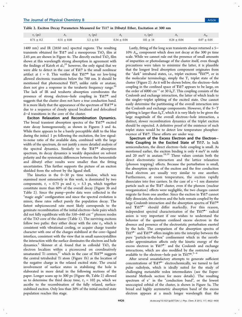
After several unsatisfactory attempts to generate sufficientconcentrations of Ti17•− electrochemically, we turned to fastpulse radiolysis, which is ideally suited for the study ofchallenging metastable redox intermediates (see the Exper-imental Methods section for more details). The resultingspectrum of e− in the “conduction band”, or the lowestunoccupied orbital of the cluster, is shown in Figure 5a. Thebroad and highly asymmetric absorption band of the excesselectron appears at a much longer wavelength than the
Table 2. Exciton Decay Parameters Measured for Ti17 in Dibutyl Ether, Excitation at 300 nm
τ1 (ps) A1 τ2 (ps) A2 τ3 (ps) A3 A∞
0.75 ± 0.2 0.31 ± 0.06 5.3 ± 0.8 0.34 ± 0.04 102 ± 18 0.28 ± 0.04 0.07 ± 0.05
The Journal of Physical Chemistry B Article
dx.doi.org/10.1021/jp307724v | J. Phys. Chem. B 2013, 117, 4422−44304426
spectrum of Ti17*. The absorption maximum lies at 1000 nm(1.24 eV), i.e.,1.1 eV higher than that of the conduction bandelectron in bulk TiO2 and 0.67 eV lower than that of the Ti17*exciton, simultaneously confirming the presence of pronouncedquantum size effects in Ti17•− and strong electrostaticinteraction between the electron and the hole in Ti17*. Therise on the low energy side is very sharp, while the high energyfalloff is much more gradual. The spectrum cannot besatisfactorily fitted with a single Gaussian or Lorentzian lineshape; however, a sequence of three 1900 cm−1 wide Gaussianpeaks centered at 9920, 11 710, and 13 550 cm−1 reproduces itperfectly (Figure 5b, green lines). Given the low resolution ofthe experimental spectrum, one should be prudent not tooverinterpret the origin of the modulation seen in Figure 5a.Indeed, a single Gaussian convoluted with an exponentialfunction gives a very satisfactory fit, too (Figure 5b, blue line).Nonetheless, the asymmetric shape of the band alone indicatesthat it consists of a sequence of transitions to a series of closelyspaced higher states with diminishing oscillator strength,similarly as the spectrum of Ti17*, albeit shifted to lowerenergy. It is more likely that these are closely lying higherelectronic doublet states D2, ..., Dn shown in Figure 2 ratherthan a vibronic progression of the same electronic transition.Indeed, the apparent ∼1800 cm−1 separation between thefeatures does not correspond to any vibrational mode of theTiO core or the ligands. In the future, we will attempt to obtainbetter resolved spectra of Ti17•− and address this point inmore detail.The 0.67 eV shift between the spectra of Ti17•− and Ti17*
can be used to set a rough limit for the electron−hole distancein the relaxed excited state of Ti17. In a vacuum, 0.67 eVcorresponds to the potential energy of two point chargesseparated by ∼2.1 nm, i.e., further apart than both the diameterof the TiO core and the size of the entire Ti17 cluster. Scaledby the high frequency dielectric constant of anatase, εopt ≈ 6,the estimated electron−hole separation is reduced to r+− = 0.35nm. This value is nearly identical with the 3.50 Å distancebetween the central tetrahedral and the outer, six-coordinate Tiatoms and it is only slightly shorter than the 4.2 Å distancebetween the center of the cluster and the five-coordinate Tiatoms based on the X-ray diffraction structure determination.7
Such crude classical electrostatic estimates ignore the atomicstructure of the medium, charge delocalization, as well as
exchange interactions and should be taken with caution. Eventhe use of the bulk dielectric constant of anatase isquestionable, since it is has been convincingly argued that innanoparticles the effective ε is lower.28 Nevertheless, theysuggest that the relaxed exciton in Ti17 has substantial charge-separated character, most likely with the electron occupying theterahedral center of the cluster and the hole localized at theelectron-rich Ti−isopropoxide interface between the core andthe ligand shell. The emerging picture of a charge transferexcited state and the involvement of the ligand−core interfaceare corroborated by the fluorescence and Raman spectroscopyresults discussed in the next section. Excited states of otherpolyoxometalates (POMs), e.g., the [W10O32]
4−, are alsobelieved to be of the LMCT (ligand-to-metal charge-transfer)character, with the electron residing at one of the metalcenters.6 Furthermore, in colloidal TiO2, the conduction bandelectron localizes on the coordinatively unsaturated Ti ions.27
The large Coulomb interaction in the excited state of Ti17suggests that the exchange coupling K between the unpairedHOMO−LUMO (or valence band−conduction band) elec-trons is also substantial in this system. As mentioned earlier, the0.67 eV spectral shift observed for the Ti17* exciton incomparison with the Ti17•− radical anion is a composite resultof both interactions. Exchange interaction K raises the energy ofthe singlet exciton by K and lowers the energy of the otherwisedegenerate triplet state by the same amount. Coulomb andexchange integrals have different distance dependence, with theshort-range part of the latter scaling as the inverse of thecommon volume sampled by the unpaired spins.29−31 If weaccept the 2.35 nm Bohr radius of the free exciton in anatasededuced recently by Hormann et al.32 and combine it with theparticle size dependence of the electron−hole exchange energyderived by Takagahara,29 an over 100-fold increase in theexchange interaction is predicted for the Ti17 cluster incomparison with bulk anatase TiO2. For a reference, in the caseof the fully delocalized π-electron system of C60, which issimilar in size to Ti17, the singlet−triplet splitting is on theorder of 0.3−0.4 eV.33,34 Naturally, any localization of the holeand the electron away from one another through chargeseparation or phonon coupling, both of which certainly takeplace in Ti17*, will affect these values. Nevertheless, as long asthe singlet−triplet splitting remains greater than the thermalenergy, ES−T = 2|K| > kBT, it is likely to manifest itself in the
Figure 5. (a) Absorption spectra of the Ti17•− species at different delay times obtained by picosecond pulse radiolysis in benzonitrile. (b) Gaussian(green) and stretched Gaussian (blue) line shape fitting of the Ti17•− absorption band obtained at 50 ns delay. The solid red line is the sum of thethree Gaussian functions calculated only at the frequencies of the experimental data.
The Journal of Physical Chemistry B Article
dx.doi.org/10.1021/jp307724v | J. Phys. Chem. B 2013, 117, 4422−44304427
exciton recombination dynamics as a slow decay channel whichshould appear in the time-resolved transient absorptionexperiments.The issue of the electron−hole exchange interaction and the
resulting singlet−triplet splitting is of great importance forquantum confinement effects at the smallest particle size limit.One should note that the size dependent position of the tripletstate in the band gap or HOMO−LUMO gap of the species hasimplications for the emission efficiency of quantum dots, as wellas the multiple exciton generation (MEG)35 and singlet fission(SF)36 processes which have been attracting a lot of attentionrecently because of their potential importance for solar energyconversion. Both phenomena critically depend on the spacingbetween the energy levels which participate in the partitioningof the initial excitation. The possibility of using the size andshape of the particle to control the electron−hole exchangeinteraction29,37 and hence being able to place the triplet excitonat an arbitrary position within the band gap opens attractivepossibilities for the design of light conversion materials. Weplan to address the possible involvement of triplet excitons inthe photophysics of Ti17 by low temperature luminescence andtransient absorption studies.Fluorescence and Raman Spectra of Ti17: The
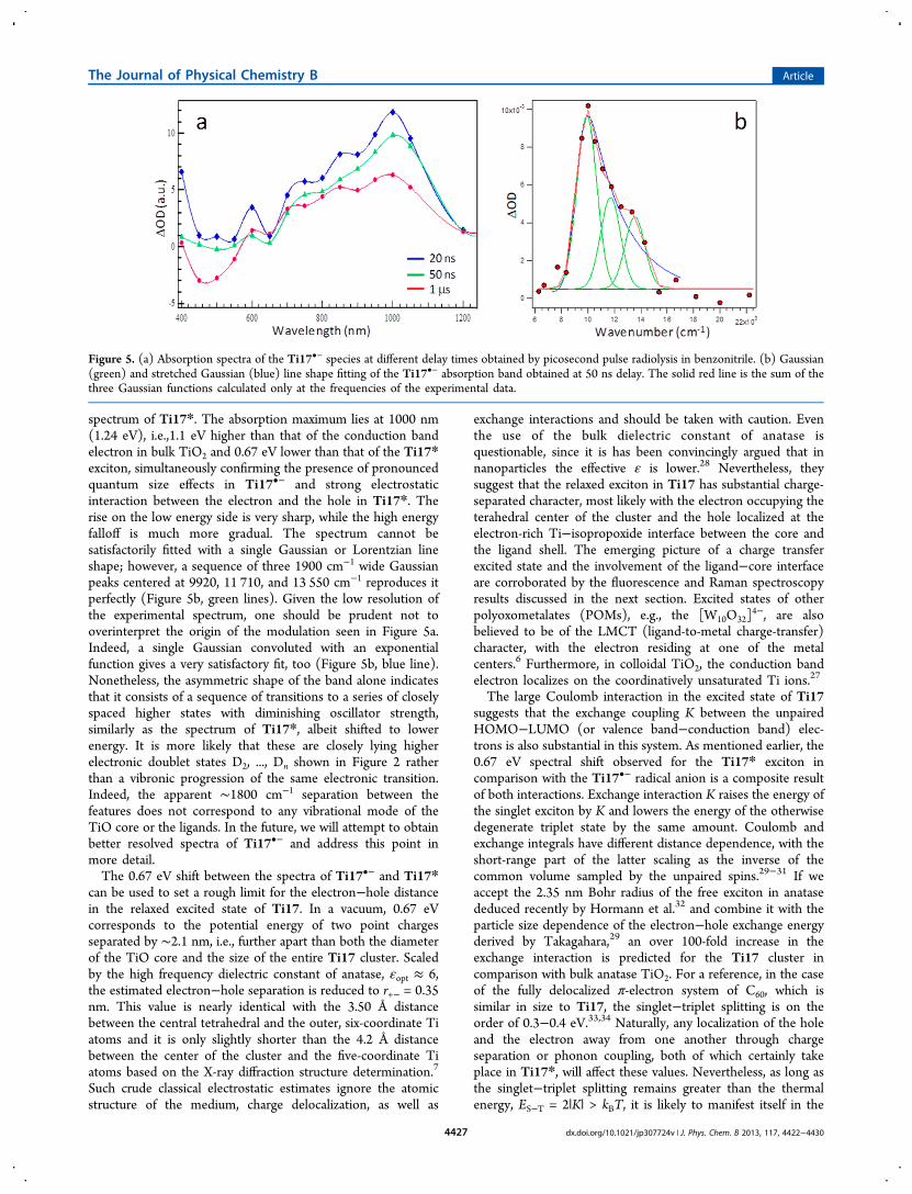
Involvement of High-Frequency Ligand Modes. Ti17exhibits a UV fluorescence band peaking at 365 nm. Theemission is broad and extends into the violet portion of thevisible spectral range. Although the emission is weak, Φ ≤ 1%,it can be readily measured even at room temperature (Figure6a). This finding is significant because it provides additionalvaluable information about the electronic structure of thecluster. The position of the maximum at 365 nm is in perfectagreement with the indirect band gap of 3.36 eV deduced byBenedict and Coppens on the basis of their analysis of theabsorption onset of Ti17.7 Furthermore, the fluorescence bandshows two discernible vibronic peaks separated by approx-imately 1290 ± 150 cm−1. The coupling between the emissiveexciton and the high frequency promoting mode offers hintsabout the charge localization in the fully relaxed excited state.
The brookite-like vibrational spectrum of the TiO core of thecluster is similar to those of the bulk phases of TiO2 (Table 1and Figure 6b) with a peak at a frequency of 644 cm−1, i.e.,much lower than the separation of the vibronic shoulders in thefluorescence spectrum. Therefore, it is doubtful that thevibronic structure of the fluorescence spectrum results fromthe coupling between the exciton and the core modes of thecluster. The involvement of the ligand modes at the organic−inorganic interface seems much more likely. Indeed, the groundstate Raman spectrum of solid Ti17 shows a very prominentcomposite peak at 1030 cm−1 and another one at 1190 cm−1
(Figure 6b) which correspond to the combination COstretching and CH wagging modes of the isopropoxideligands38,39 forming the organic shell of the cluster. We believethat the CO modes are responsible for the vibrational structureof the fluorescence band. If this hypothesis is correct, it wouldsuggest at least one of the charges, most likely the hole of therelaxed exciton, is localized at the core−ligand interface, whereit couples to the vibrations of the isopropoxide group.Very interestingly, the CO Raman bands of Ti17 originate
almost exclusively from four ligands bound to the penta-coordinate Ti sites at the corners of the cluster. As it is depictedin Figure 1b and discussed in the Introduction, the clustercontains four five-coordinate and as a result coordinativelyunsaturated Ti centers (shown in green) which are terminatedwith a single, strongly polarized isopropoxide ligand. Benedictand Coppens found that, upon selective substitution of the five-coordinate Ti centers with bidentate catechol, all surfacetitanium sites of the resulting Ti17cat4 cluster (proper nameTi17(μ4-O)4(μ3-O)16(μ2-O)4(cat)4(OPr
i)16) become hexa-coor-dinate.7 Our results show that upon this substitution the strongCO band nearly completely disappears from the Ramanspectrum (Figure 6b), despite the fact that there are still 16isopropoxide ligands which remain bound to the surface of thecore. The explanation for this puzzling observation lies in thedegree of the purely ionic vs charge-transfer bondinginteraction between the ligand and the surface titanium centers.The CO modes of the closed-shell free isopropoxide ion arestrong IR absorbers; however, because of the localized “hard”
Figure 6. (a) Room temperature fluorescence spectrum of Ti17 in dibutyl ether showing vibronic bands at 365 and 383 nm (excitation at 340 nm).The spectrum corrected for the solvent background is shown in red. (b) Raman spectrum of solid Ti17 showing at 1029 cm−1 a prominent C−Ostretch/CH wagging combination mode of the four isopropoxide ligands bound to the pentacoordinate Ti sites (black) and the correspondingRaman spectrum of solid Ti17cat (red), in which the peak corresponding to the C−O stretch of the isopropoxides bound to the five coordinate Ticenters is absent.
The Journal of Physical Chemistry B Article
dx.doi.org/10.1021/jp307724v | J. Phys. Chem. B 2013, 117, 4422−44304428
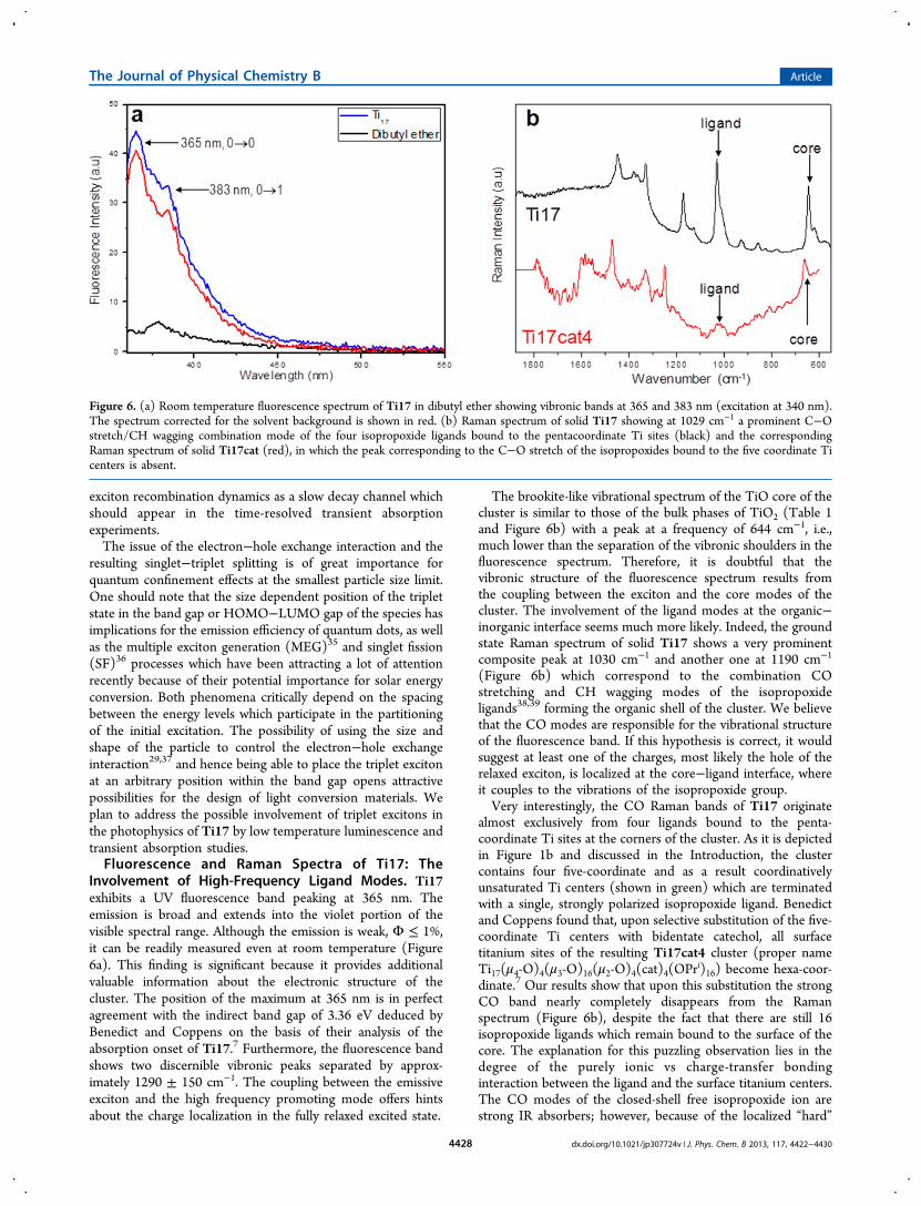
charge distribution, they are weak Raman scatterers. This isillustrated by the calculated Raman and IR spectra shown inFigure 7. On the contrary, the same vibrational modes of the
corresponding open-shell electroneutral oxo-radical are weak IRabsorbers in the 750−1500 cm−1 region; however, they havemuch larger Raman cross sections (Figure 7, shown in red).This implies that any shift of electron density from theisopropoxide anion onto the metal center will give rise tostronger Raman scattering in the CO region. It appears thatisopropoxides bound to the hexa-coordinate Ti centers retaintheir primarily ionic character and that there is little charge shiftbetween their oxygen atoms and the d-orbitals of the metal. Asa result, they make only a minor contribution to the CO regionof the Raman spectrum of Ti17. On the other hand,isopropoxides bound to the penta-coordinate Ti centers donatea large fraction of their charge to the d-orbitals of the titaniumions. Such electron density shift renders them considerably lessionic and increases the overall polarizability of the local −C−O−Ti− interface, leading to a strong Raman signature.40 Ininorganic chemistry literature, this type of charge-transferinteraction is often referred to as “π-bonding”.41
On the basis of the presented fluorescence and Raman dataalone, it is not possible to unequivocally decide whether thehole in the excited state of Ti17 becomes localized on one ofthe coordinatively unsaturated five-coordinate titanium centersor on one of the more numerous six-coordinate sites. High levelcalculations on the complete cluster will be necessary in orderto settle this question. Nevertheless, it is safe to conclude thatligand modes at the interface play a more significant role instabilizing the lowest electronic excited state of the Ti17 clusterthan either the solvent reorganization or the core frequencies.This further bolsters the picture of a CT excited state with thehole localized at the core−ligand interface and the electron onthe central tetrahedral titanium ion. The implications of thelarge ground state ligand-to-metal charge shift at the five-coordinate centers, which is so strikingly evident in the Raman
spectra, extend beyond the excited state dynamics of Ti17.Because of the charge shift, these sites behave as reactive “hot-spots” on the surface of the cluster and for this reason theligand substitution reactions occur with high selectivity only atthese Ti centers. The charge shift must also have an influenceon the redox and potential catalytic activity of the cluster bothin the ground and in the excited state.
■ CONCLUSIONSThe 1 nm diameter Ti17(μ4-O)4(μ3-O)16(μ2-O)4(OPri)20polyoxotitanate cluster with a brookite-like core displayscomplex photophysics which combines primarily molecularcharacteristics with some traits typical of larger nanoparticlesand bulk TiO2. The high frequency modes at the ligand−coreinterface were found to play a key role in the stabilization andlocalization of the excited state. On the basis of our results, weproposed a tentative energy level diagram of the cluster (Figure2). Due to spatial confinement, the absorption spectrum of theD0 ground state of the Ti17•− radical anion is shifted from theIR range typical of the excess electron in bulk II−VIoxides17,25,42 to 1000 nm. The spectrum of the S1 excitedstate of the cluster is blue-shifted even further, indicating thepresence of large Coulomb and exchange interactions betweenthe spatially confined electron and hole. The appearance of thebroad and featureless spectra of the S1 and D0 states of Ti17 isconsistent with band-like progressions of closely lying upperelectronic states, Sn and Dn, respectively. It is likely that thehigher states involve primarily the TiO core of Ti17 andtherefore are of more bulk-like character than the lowest states.DFT calculations show a large Ti d-orbital contribution to low-lying empty orbitals of the cluster, suggesting that the S1 and D1spectra of Ti17 may be due to d−d transitions.The electron−hole recombination in Ti17 is faster than in
colloidal anatase or mesoporous films of TiO2; however, it doesnot follow single exponential decay. The fast, 0.7−5 pscomponents, which account for ∼60% of the decay, mostlikely originate from the recombination of the initial excitonsbefore they relax and become stabilized at the core−ligandinterface. The slower, 100 ps component is assigned to thedecay of the fully relaxed exciton. Lastly, we consistentlydetected a long-lived component accounting for 5−10% of theinitial population which we tentatively ascribe to the formationof a lower-lying triplet state with a distinct, slowerrecombination dynamics.Our findings suggest that the relaxed excited state of Ti17
has charge transfer character, with one of the charges (hole)localized at the core−ligand interface and the other on theunique tetrahedral Ti at the center of the cluster. The vibronicfeatures of the emission spectrum as well as the ground stateRaman spectra show that the ligand modes are strongly coupledto the overall electron density of the cluster. Polyoxometalatesand colloidal TiO2 have been successfully employed in anumber of photocatalytic reactions,5,6 and there are indicationsthat the brookite phase possesses more reactive surface sitesthan either anatase or rutile.43,44 A better understanding of thelocalization and dynamics of electrons and holes in clusterssimilar to Ti17 should facilitate further development of catalyticapplications of this class of materials.The authors hope that the presented data will stimulate high-
level quantum mechanical calculations, which are necessary fora conclusive interpretation of the experimental results,especially when it comes to the orientation of the transitiondipole, the electron and hole localization sites, as well as the
Figure 7. Calculated Raman (a) and IR (b) spectra of theisopropoxide anion (blue) and the oxo isopropoxyl radical (red).Calculations were performed at the B3LYP DFT level (Spartan’10)without frequency scaling.
The Journal of Physical Chemistry B Article
dx.doi.org/10.1021/jp307724v | J. Phys. Chem. B 2013, 117, 4422−44304429
magnitude of the Coulomb and exchange interaction in theexcited state of Ti17. Transient absorption depolarizationmeasurements in the <500 fs window will be used to probe theearliest steps of Ti17* relaxation dynamics. Low temperaturephotoluminescence experiments will be performed to betterresolve the vibronic structure of the fluorescence spectrum ofTi17 and to search for the phosphorescence spectrum of the3Ti17* triplet state.
■ AUTHOR INFORMATION
Corresponding Author*E-mail: [email protected].
NotesThe authors declare no competing financial interest.
■ ACKNOWLEDGMENTS
The work at Rutgers University was supported by Division ofChemical Sciences, Geosciences, and Biosciences, Office ofBasic Energy Sciences of the U.S. Department of Energythrough Grant No. DE-FG02-06ER15828 to P.P. The work atUniversity at Buffalo was funded by the Division of ChemicalSciences, Geosciences, and Biosciences, Office of Basic EnergySciences of the U.S. Department of Energy through Grant DE-FG02-02ER15372 to P.C. The femtosecond laser instrumenta-tion used to carry out this research was funded by NationalScience Foundation CRIF Grant No. 0342432 to P.P. Theauthors gratefully acknowledge support of the Division ofChemical Sciences, Geosciences, and Biosciences, Office ofBasic Energy Sciences of the U.S. Department of Energythrough Grant No. DE-AC02-98-CH10886 to all authors, andfor use of the LEAF Facility of the BNL Accelerator Center forEnergy Research. We are grateful to Prof. Richard Mendelsohnfor the help with the Raman measurements, Prof. Frieder Jaeklefor the access to one of his glove boxes, and Prof. Galoppini forthe loan of the spectro-electrochemistry setup. We thank Prof.Victor Batista and his group for sharing with us theircomputational results on the related Ti17cat4 polyoxotitanatecluster.
■ REFERENCES(1) Alivisatos, A. P.; Harris, A. L.; Levinos, N. J.; Steigerwald, M. L.;Brus, L. E. J. Chem. Phys. 1988, 89, 4001−4011.(2) Brus, L. J. Phys. Chem. 1986, 90, 2555−2560.(3) Brus, L. E. J. Chem. Phys. 1983, 79, 5566−5571.(4) Brus, L. E. J. Chem. Phys. 1984, 80, 4403−4409.(5) Pope, M. T.; Achim, M. Polyoxometalates: from platonic solids toanti-retroviral activity; Kluwer Academic Publishers: Dordrecht, TheNetherlands, 1994.(6) Duncan, D. C.; Netzel, T. L.; Hill, C. L. Inorg. Chem. 1995, 34,4640−4646.(7) Benedict, J. B.; Coppens, P. J. Am. Chem. Soc. 2010, 132, 2938−2944.(8) Benedict, J. B.; Freindorf, R.; Trzop, E.; Cogswell, J.; Coppens, P.J. Am. Chem. Soc. 2010, 132, 13669−13671.(9) Ardo, S.; Meyer, G. J. Chem. Soc. Rev. 2009, 38, 115−164.(10) Fujishima, A.; Honda, K. Nature 1972, 238, 37−38.(11) Linsebigler, A. L.; Lu, G. Q.; Yates, J. T. Chem. Rev. 1995, 95,735−758.(12) Amtout, A.; Leonelli, R. Phys. Rev. B 1995, 51, 6842−6851.(13) Gonzalez, R. J.; Zallen, R.; Berger, H. Phys. Rev. B 1997, 55,7014−7017.(14) Lottici, P. P.; Bersani, D.; Braghini, M.; Montenero, A. J. Mater.Sci. 1993, 28, 177−183.
(15) Martin, S. T.; Herrmann, H.; Hoffmann, M. R. J. Chem. Soc.,Faraday Trans. 1994, 90, 3323−3330.(16) Mattsson, A.; Osterlund, L. J. Phys. Chem. C 2010, 114, 14121−14132.(17) Yamakata, A.; Ishibashi, T.; Onishi, H. Chem. Phys. Lett. 2001,333, 271−277.(18) Chiodo, L.; Garcia-Lastra, J. M.; Iacomino, A.; Ossicini, S.;Zhao, J.; Petek, H.; Rubio, A. Phys. Rev. B 2010, 82.(19) Hendry, E.; Wang, F.; Shan, J.; Heinz, T. F.; Bonn, M. Phys. Rev.B: Condens. Matter Mater. Phys. 2004, 69.(20) Serpone, N.; Khairutdinovt, D. L. a. R. J. Phys. Chem. 1995, 99,16646−16654.(21) Steunou, N.; Kickelbick, G.; Boubekeur, K.; Sanchez, C. M. J.Chem. Soc., Dalton Trans. 1999, 3653−3655.(22) Steunou, N.; Portal, R.; Sanchez, C. High Pressure Res. 2001, 20,63−70.(23) Ernstorfer, R.; Gundlach, L.; Felber, S.; Storck, W.; Eichberger,R.; Willig, F. J. Phys. Chem. B 2006, 110, 25383−25391.(24) Wishart, J. F.; Cook, A. R.; Miller, J. R. Rev. Sci. Instrum. 2004,75, 4359−4366.(25) Yoshihara, T.; Katoh, R.; Furube, A.; Tamaki, Y.; Murai, M.;Hara, K.; Murata, S.; Arakawa, H.; Tachiya, M. J. Phys. Chem. B 2004,108, 3817−3823.(26) Schmuttenmaer, C. Personal communication.(27) Skinner, D. E.; Colombo, D. P.; Cavaleri, J. J.; Bowman, R. M. J.Phys. Chem. 1995, 99, 7853−7856.(28) Wang, L. W.; Zunger, A. Phys. Rev. Lett. 1994, 73, 1039−1042.(29) Takagahara, T. Phys. Rev. B 1993, 47, 4569.(30) Scholes, G. D. ACS Nano 2008, 2, 523−537.(31) Scholes, G. D.; Rumbles, G. Nat. Mater. 2006, 5, 683−696.(32) Hormann, U.; Kaiser, U.; Albrecht, M.; Geserick, J.; Husing, N.J. Phys.: Conf. Ser. 2010, 209, 012039.(33) Arbogast, J. W.; Darmanyan, A. P.; Foote, C. S.; Rubin, Y.;Diederich, F. N.; Alvarez, M. M.; Anz, S. J.; Whetten, R. L. J. Phys.Chem. 1991, 95, 11−12.(34) Foote, C. Photophysical and photochemical properties offullerenes. In Top. Curr. Chem.; Mattay, J., Ed.; Springer-Verlag:Berlin/Heidelberg, 1994; Vol. 169, pp 347−363.(35) Stewart, J. T.; Padilha, L. A.; Qazilbash, M. M.; Pietryga, J. M.;Midgett, A. G.; Luther, J. M.; Beard, M. C.; Nozik, A. J.; Klimov, V. I.Nano Lett. 2012, 12, 622−628.(36) Schwerin, A. F.; Johnson, J. C.; Smith, M. B.; Sreearunothai, P.;Popovic, D.; Cerny, J.; Havlas, Z.; Paci, I.; Akdag, A.; MacLeod, M. K.;et al. J. Phys. Chem. A 2010, 114, 1457−1473.(37) Brovelli, S.; Schaller, R. D.; Crooker, S. A.; García-Santamaría,F.; Chen, Y.; Viswanatha, R.; Hollingsworth, J. A.; Htoon, H.; Klimov,V. I. Nat. Commun. 2011, 2, 1−8.(38) Bell, J. V.; Heisler, J.; Tannenbaum, H.; Goldenson, J. Anal.Chem. 1953, 25, 1720−1724.(39) Lynch, C. T.; Mazdiyasni, K. S.; Smith, J. S.; Crawford, W. J.Anal. Chem. 1964, 36, 2332−2337.(40) Moran, P. D.; Bowmaker, G. A.; Cooney, R. P.; Finnie, K. S.;Bartlett, J. R.; Woolfrey, J. L. Inorg. Chem. 1998, 37, 2741−2748.(41) Chisholm, M. H.; Davidson, E. R.; Huffman, J. C.; Quinlan, K.B. J. Am. Chem. Soc. 2001, 123, 9652−9664.(42) Yoshihara, T.; Katoh, R.; Furube, A.; Murai, M.; Tamaki, Y.;Hara, K.; Murata, S.; Arakawa, H.; Tachiya, M. J. Phys. Chem. B 2004,108, 2643−2647.(43) Koelsch, M.; Cassaignon, S.; Guillemoles, J. F.; Jolivet, J. R. ThinSolid Films 2002, 403, 312−319.(44) Lin, H. F.; Li, L. P.; Zhao, M. L.; Huang, X. S.; Chen, X. M.; Li,G. S.; Yu, R. C. J. Am. Chem. Soc. 2012, 134, 8328−8331.
The Journal of Physical Chemistry B Article
dx.doi.org/10.1021/jp307724v | J. Phys. Chem. B 2013, 117, 4422−44304430


![Relaxing Electrons in Solids · been able to capture the dynamics of core-excitons in solids in real-time. [25] The world of nanosensors may be physically small, but the demand is](https://img.pdfslide.net/doc/110x75/5f5c59fe3248872e5926c714/relaxing-electrons-in-solids-been-able-to-capture-the-dynamics-of-core-excitons.jpg)