Embed Size (px)
DESCRIPTION
Exercise 44. Survey of Embryonic Development. Objectives:. Fertilization, zygote, morula, blastocyst, gastrula, fetus, chorion, chorionic villi, placenta, amnion, yolk sac, umbilical cord Embryonic structures and functions. Fertilization. - PowerPoint PPT Presentation
Citation preview
Exercise 44Exercise 44
Survey of Embryonic Survey of Embryonic DevelopmentDevelopment
Objectives:Objectives:
Fertilization, zygote, morula, Fertilization, zygote, morula, blastocyst, gastrula, fetus, chorion, blastocyst, gastrula, fetus, chorion, chorionic villi, placenta, amnion, chorionic villi, placenta, amnion, yolk sac, umbilical cordyolk sac, umbilical cord
Embryonic structures and Embryonic structures and functionsfunctions
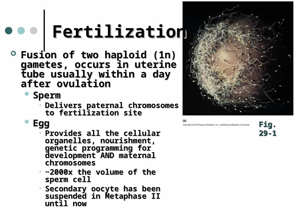
FertilizationFertilization Fusion of two haploid (1n) Fusion of two haploid (1n)
gametes, occurs in uterine gametes, occurs in uterine tube usually within a day after tube usually within a day after ovulationovulation SpermSperm
• Delivers paternal chromosomes Delivers paternal chromosomes to fertilization siteto fertilization site
Egg Egg • Provides all the cellular Provides all the cellular
organelles, nourishment, genetic organelles, nourishment, genetic programming for development programming for development AND maternal chromosomesAND maternal chromosomes
• ~2000x the volume of the sperm ~2000x the volume of the sperm cellcell
• Secondary oocyte has been Secondary oocyte has been suspended in Metaphase II until suspended in Metaphase II until nownow
Fig. 29-1Fig. 29-1
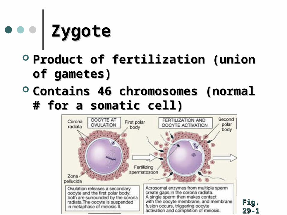
ZygoteZygote
Product of fertilization (union of Product of fertilization (union of gametes)gametes)
Contains 46 chromosomes (normal # for Contains 46 chromosomes (normal # for a somatic cell)a somatic cell)
Fig. 29-1Fig. 29-1
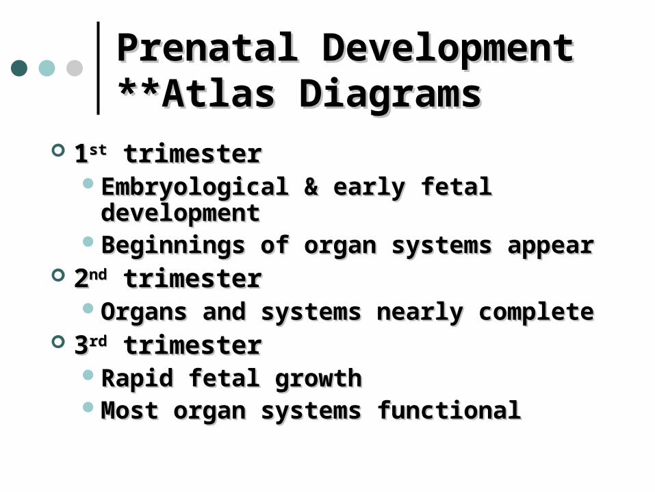
Prenatal Development Prenatal Development **Atlas Diagrams**Atlas Diagrams
11stst trimester trimesterEmbryological & early fetal developmentEmbryological & early fetal developmentBeginnings of organ systems appearBeginnings of organ systems appear
22ndnd trimester trimesterOrgans and systems nearly completeOrgans and systems nearly complete
33rdrd trimester trimesterRapid fetal growthRapid fetal growthMost organ systems functionalMost organ systems functional
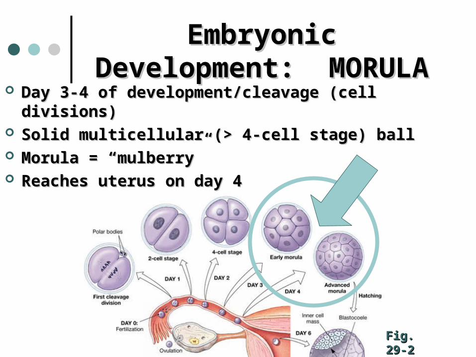
Embryonic Development: Embryonic Development: MORULAMORULA
Day 3-4 of development/cleavage (cell divisions)Day 3-4 of development/cleavage (cell divisions) Solid multicellular (> 4-cell stage) ballSolid multicellular (> 4-cell stage) ball Morula = “mulberry”Morula = “mulberry” Reaches uterus on day 4 Reaches uterus on day 4
Fig. 29-2Fig. 29-2
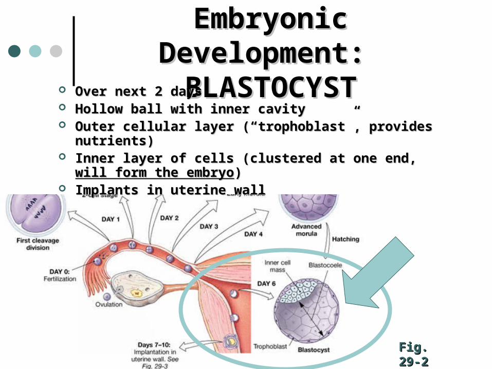
Embryonic Development: Embryonic Development: BLASTOCYSTBLASTOCYST
Fig. 29-2Fig. 29-2
Over next 2 daysOver next 2 days Hollow ball with inner cavityHollow ball with inner cavity Outer cellular layer (“trophoblast”, provides nutrients)Outer cellular layer (“trophoblast”, provides nutrients) Inner layer of cells (clustered at one end, Inner layer of cells (clustered at one end, will form the will form the
embryoembryo)) Implants in uterine wallImplants in uterine wall
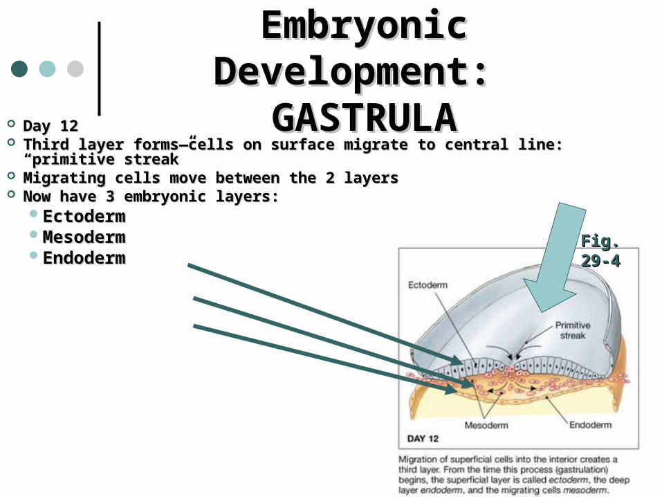
Embryonic Development: Embryonic Development: GASTRULAGASTRULA
Day 12Day 12 Third layer forms—cells on surface migrate to central line: “primitive Third layer forms—cells on surface migrate to central line: “primitive
streak”streak” Migrating cells move between the 2 layers Migrating cells move between the 2 layers Now have 3 embryonic layers:Now have 3 embryonic layers:
EctodermEctodermMesoderm Mesoderm Endoderm Endoderm
Fig. 29-4Fig. 29-4
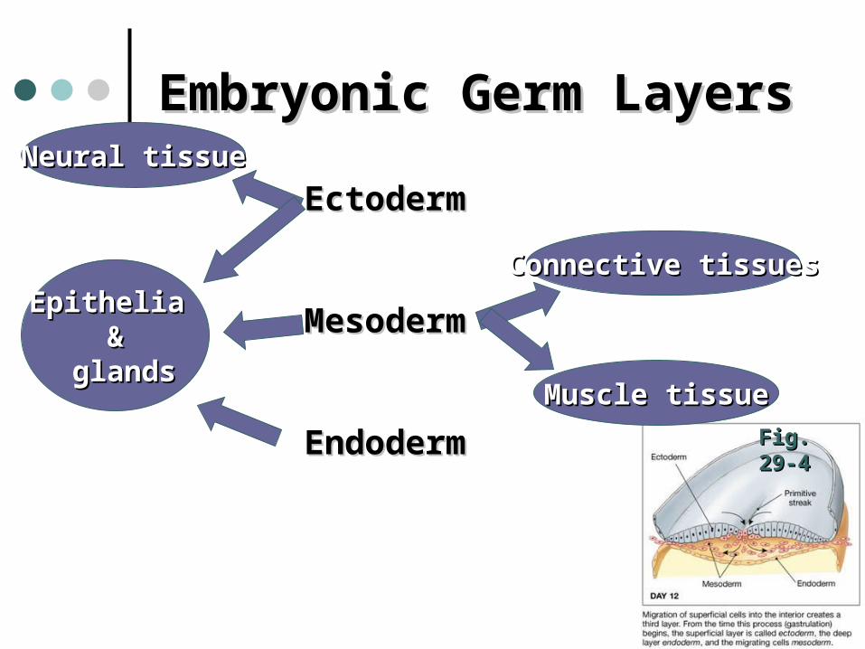
Embryonic Germ LayersEmbryonic Germ Layers
EctodermEctoderm
MesodermMesoderm
Endoderm Endoderm Fig. 29-4Fig. 29-4
Neural tissueNeural tissue
Epithelia Epithelia &&
glandsglands
Connective tissuesConnective tissues
Muscle tissueMuscle tissue
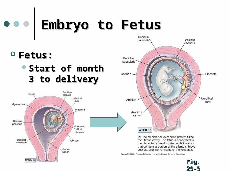
Embryo to FetusEmbryo to Fetus
Fetus: Fetus: Start of month 3 to Start of month 3 to
deliverydelivery
Fig. 29-5Fig. 29-5
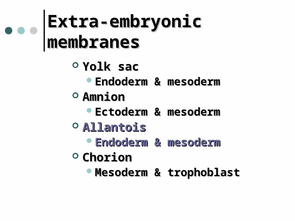
Extra-embryonic membranesExtra-embryonic membranes
Yolk sacYolk sacEndoderm & mesodermEndoderm & mesoderm
AmnionAmnionEctoderm & mesodermEctoderm & mesoderm
AllantoisAllantois Endoderm & mesodermEndoderm & mesoderm
ChorionChorionMesoderm & trophoblastMesoderm & trophoblast
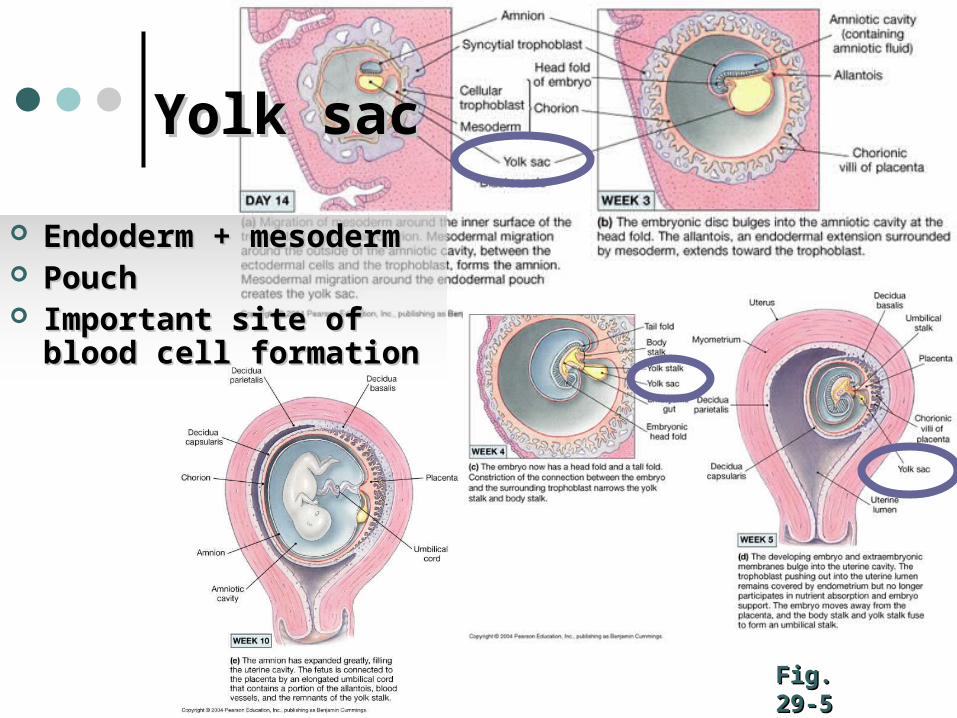
Yolk sacYolk sac
Fig. 29-5Fig. 29-5
Endoderm + mesodermEndoderm + mesoderm PouchPouch Important site of blood Important site of blood
cell formationcell formation
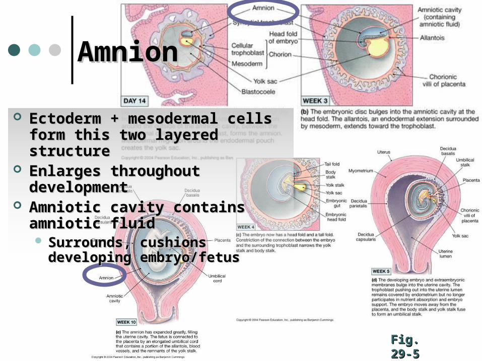
Fig. 29-5Fig. 29-5
Ectoderm + mesodermal cells Ectoderm + mesodermal cells form this two layered structureform this two layered structure
Enlarges throughout Enlarges throughout developmentdevelopment
Amniotic cavity contains Amniotic cavity contains amniotic fluidamniotic fluid Surrounds, cushions Surrounds, cushions
developing embryo/fetusdeveloping embryo/fetus
AmnionAmnion
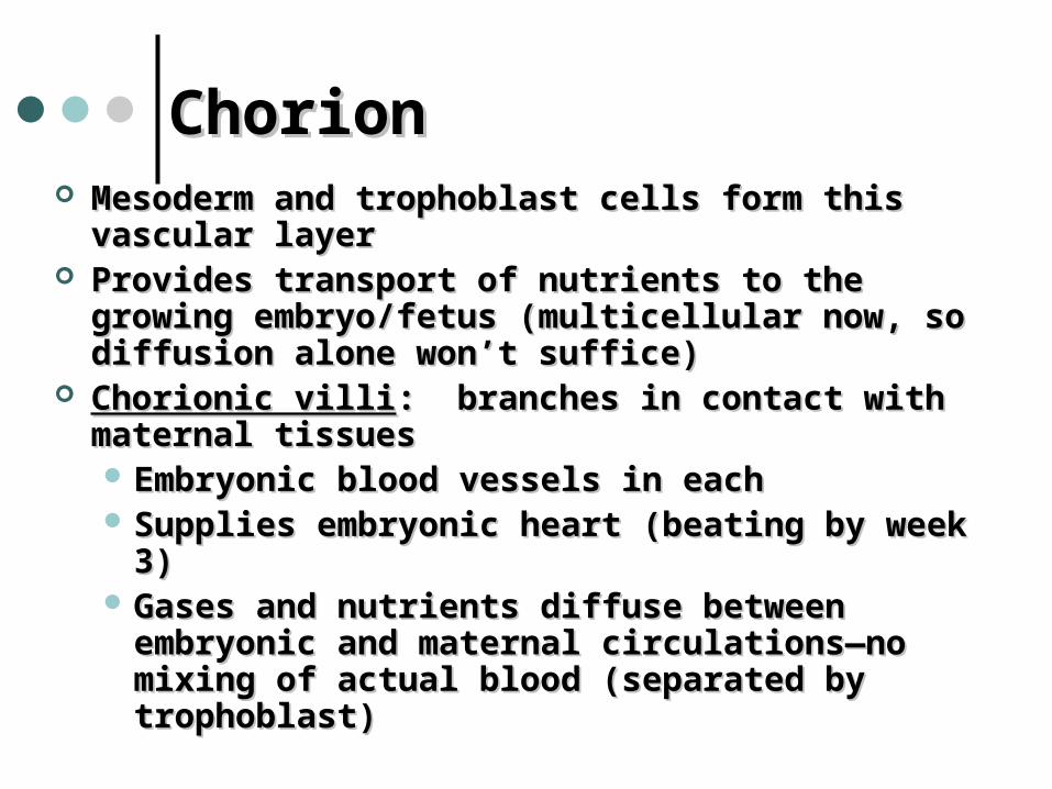
ChorionChorion Mesoderm and trophoblast cells form this vascular Mesoderm and trophoblast cells form this vascular
layerlayer Provides transport of nutrients to the growing Provides transport of nutrients to the growing
embryo/fetus (multicellular now, so diffusion alone embryo/fetus (multicellular now, so diffusion alone won’t suffice)won’t suffice)
Chorionic villiChorionic villi: branches in contact with maternal : branches in contact with maternal tissuestissues Embryonic blood vessels in each Embryonic blood vessels in each Supplies embryonic heart (beating by week 3)Supplies embryonic heart (beating by week 3) Gases and nutrients diffuse between embryonic Gases and nutrients diffuse between embryonic
and maternal circulations—no mixing of actual and maternal circulations—no mixing of actual blood (separated by trophoblast)blood (separated by trophoblast)
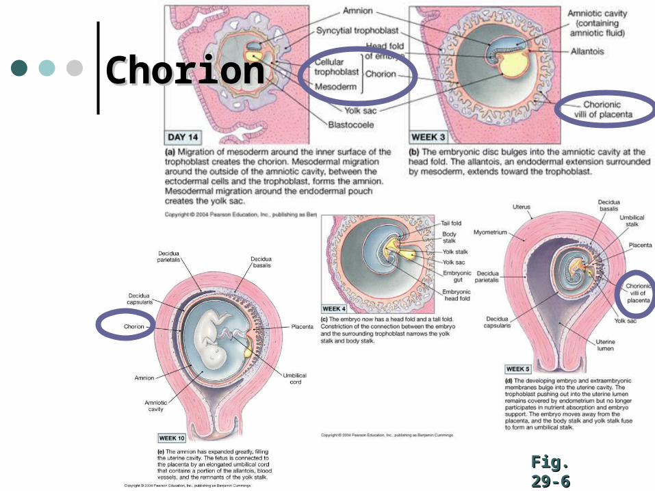
Fig. 29-6Fig. 29-6
ChorionChorion
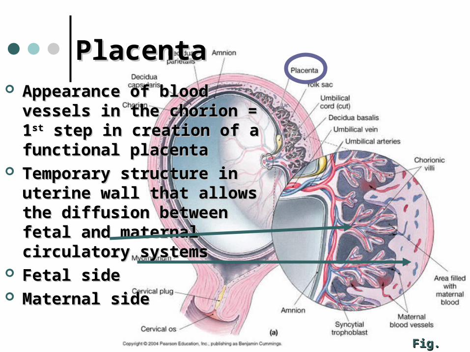
PlacentaPlacenta Appearance of blood vessels in Appearance of blood vessels in
the chorion = 1the chorion = 1stst step in creation step in creation of a functional placentaof a functional placenta
Temporary structure in uterine Temporary structure in uterine wall that allows the diffusion wall that allows the diffusion between fetal and maternal between fetal and maternal circulatory systemscirculatory systems
Fetal sideFetal side Maternal sideMaternal side
Fig. 29-6Fig. 29-6
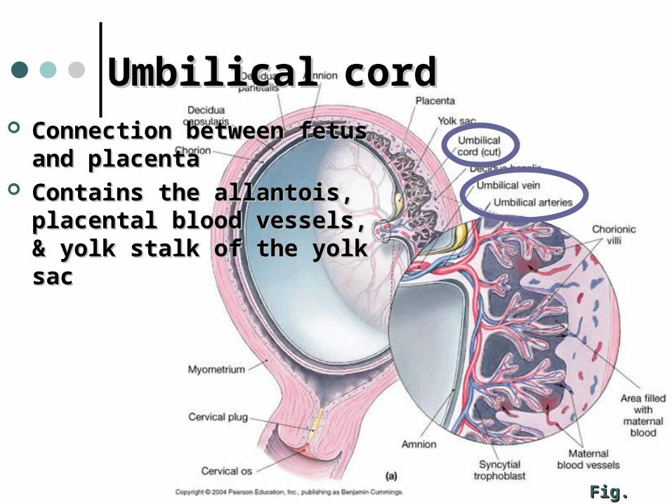
Umbilical cordUmbilical cord Connection between fetus and Connection between fetus and
placentaplacenta Contains the allantois, placental Contains the allantois, placental
blood vessels, & yolk stalk of blood vessels, & yolk stalk of the yolk sacthe yolk sac
Fig. 29-6Fig. 29-6
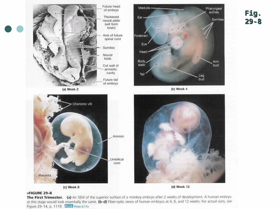
Fig. 29-8Fig. 29-8
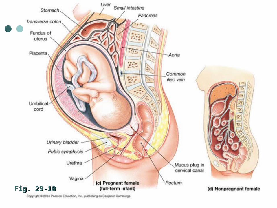
Fig. 29-10Fig. 29-10

Video:Video:
The Miracle of LifeThe Miracle of Life


![Vitti] : (Hiroshi Yamaoka) : "J 7 (Sam Kawa) : ANTENNNA VWD-300 . exercise 35 exercise 36 exercise 37 exercise 38 exercise 39 exercise 40 : exercise 41](https://img.pdfslide.net/doc/110x75/5b479fdd7f8b9a824f8c0adb/anthony-vitti-hiroshi-yamaoka-j-7-sam-kawa-antennna-vwd-300-exercise.jpg)


![Exercise 3(A) Page: 44...Concise Selina Solutions for Class 9 Maths Chapter 3- Compound Interest [Using Formula] Exercise 3(A) Page: 44 1. Find the amount and the compound interest](https://img.pdfslide.net/doc/110x75/5e9d53c86523b60bc73abae9/exercise-3a-page-44-concise-selina-solutions-for-class-9-maths-chapter-3-.jpg)


![STATEWIDE MEDICAL AND HEALTH EXERCISE SWMHE EXERCISE DEBRIEF [Exercise Name/Exercise Date] SWMHE EXERCISE DEBRIEF](https://img.pdfslide.net/doc/110x75/56649d755503460f94a56498/statewide-medical-and-health-exercise-swmhe-exercise-debrief-exercise-nameexercise.jpg)
![STATEWIDE MEDICAL AND HEALTH EXERCISE PHASE III: TABLETOP EXERCISE [Exercise Name/Exercise Date]](https://img.pdfslide.net/doc/110x75/56649e535503460f94b48b86/statewide-medical-and-health-exercise-phase-iii-tabletop-exercise-exercise.jpg)