Embed Size (px)
Citation preview
Chapter 4
A Geological Chapter
Mr. Spruce, Mrs. Palm, and Mrs. Magnolia complain of pain in the right upper quadrant.
4.1. Mr. Spruce is not only feeling bad; he also has a moderate fever. A right parasagittal scan is performed. The liver is normal in the area of the section, but obviously ...
Fig.4.1 .. ...:. ....
. . . there is an infundibular cholelithiasis (L below) with a handsome acoustic shadow posterior to the stone (open arrow).
Fig. 4.1
Is the fever related to an inflammation of the gall bladder?
35
F. S. Weill et al., Exercises in Diagnostic Ultrasonography of the Abdomen© Editions Vigot Freres, Paris 1982
In principle, no: there is neither dilatation, nor stasis, nor ... ?
... nor thickening of the gall bladder wall.
Now you should ask the sonologist a few questions: Did the transducer's application trigger pain? Was palpation of the gall bladder painful under realtime control? For Mr. Spruce, the answer in each case is no.
Why the fever?
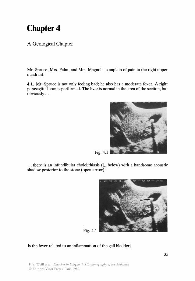
You have no doubt noticed ...
Fig. 4.1
. .. a pleural effusion (t above), marked by the presence of a supradiaphragmatic sonolucency, posteriorly limited by the thoracic wall, which normally is not visible. Absence of dilatation of the hepatic veins, of which certain segments are necessarily cut in this paramedial section, as well as evaluation of the vena cava, show it is not an effusion of cardiac origin. We can hold the lithiasis responsible for the pain, but this patient also exhibits a pleuropathy.
1 a) Morison's pouch. b) Perihepatic recesses. c) Juxtavesicular region. d) Perisplenic region . e) Lesser sac. f) Paracolic gutters . g) Douglas' pouch
36

4.2. Mrs. Palm is also experiencing pain on the right side.
Fig. 4.2 Fig.4.2_~_
This sagittal section eloquently shows a lithiasis, this time with multiple stones (1 above) casting a broad acoustic shadow (white arrow). Thus in this geological chapter you have unearthed some new stones. The thickness of the gall bladder wall is normal. The gall bladder is not particularly sensitive on palpation. Thus there is neither objective nor subjective sign to indicate cholecystitis.
Let's go on the next case.
Not so fast you say, and you're right: there is still something important to be seen ...
Fig. 4.2
We find a small fluid effusion in the most anterior part of the sUbdiaphragmatic area (l above). We will confirm the presence of this ascites by examining other sensitive areas which you should enumerate before checking below l .
Consequently, we are confronted with a diagnostic problem much greater than the simple lithiasis discovered earlier. Actually, it was carcinoma of the ascending colon, unrecognized at that point, with peritoneal spread.
37
a
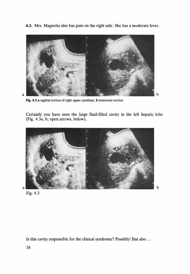
4.3. Mrs. Magnolia also has pain on the right side. She has a moderate fever.
b Fig. 4.3. a sagittal section of right upper quadrant, b transverse section
Certainly you have seen the large fluid-filled cavity in the left hepatic lobe (Fig. 4.3a, b; open arrows, below).
b Fig. 4.3
Is this cavity responsible for the clinical syndrome? Possibly! But also ...
38
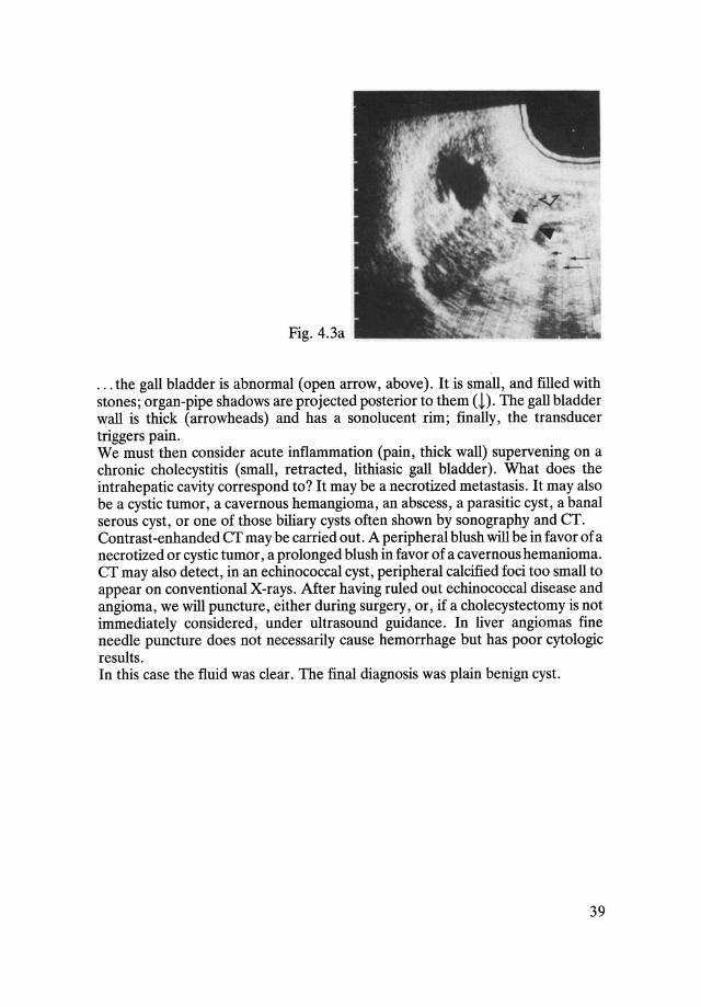
Fig.4.3a
... the gall bladder is abnormal (open arrow, above). It is small, and filled with stones; organ-pipe shadows are projected posterior to them a). The gall bladder wall is thick (arrowheads) and has a sonolucent rim; finally, the transducer triggers pain. We must then consider acute inflammation (pain, thick wall) supervening on a chronic cholecystitis (small, retracted, lithiasic gall bladder). What does the intrahepatic cavity correspond to? It may be a necrotized metastasis. It may also be a cystic tumor, a cavernous hemangioma, an abscess, a parasitic cyst, a banal serous cyst, or one of those biliary cysts often shown by sonography and CT. Contrast -enhanded CT may be carried out. A peripheral blush will be in favor of a necrotized or cystic tumor, a prolonged blush in favor of a cavernous hemanioma. CT may also detect, in an echinococcal cyst, peripheral calcified foci too small to appear on conventional X-rays. After having ruled out echinococcal disease and angioma, we will puncture, either during surgery, or, if a cholecystectomy is not immediately considered, under ultrasound guidance. In liver angiomas fine needle puncture does not necessarily cause hemorrhage but has poor cytologic results. In this case the fluid was clear. The final diagnosis was plain benign cyst.
39
a
c
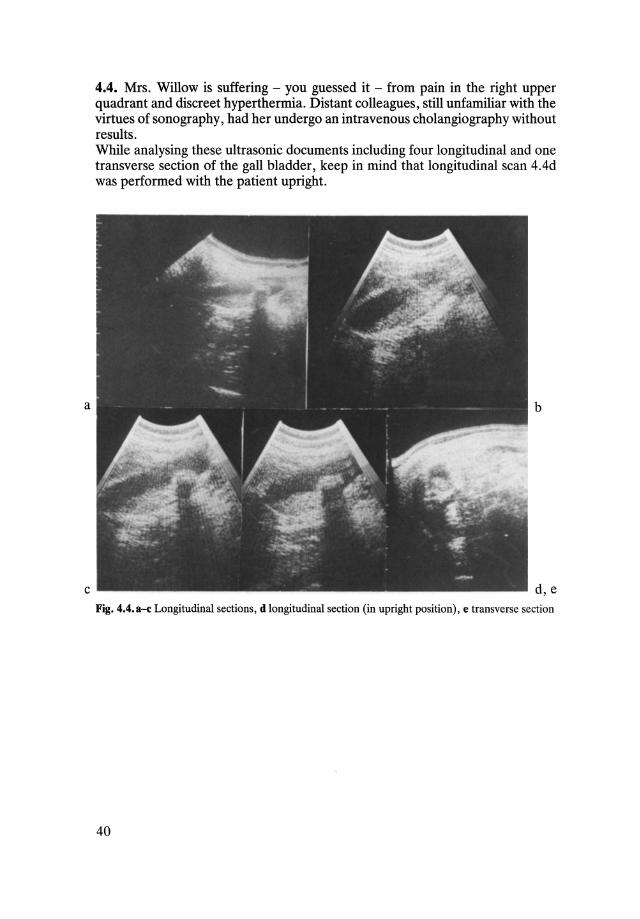
4.4. Mrs. Willow is suffering - you guessed it - from pain in the right upper quadrant and discreet hyperthermia. Distant colleagues, still unfamiliar with the virtues of sonography, had her undergo an intravenous cholangiography without results. While analysing these ultrasonic documents including four longitudinal and one transverse section of the gall bladder, keep in mind that longitudinal scan 4.4d was performed with the patient upright.
b
d,e Fig. 4.4.a-c Longitudinal sections, d longitudinal section (in upright position), e transverse section
40
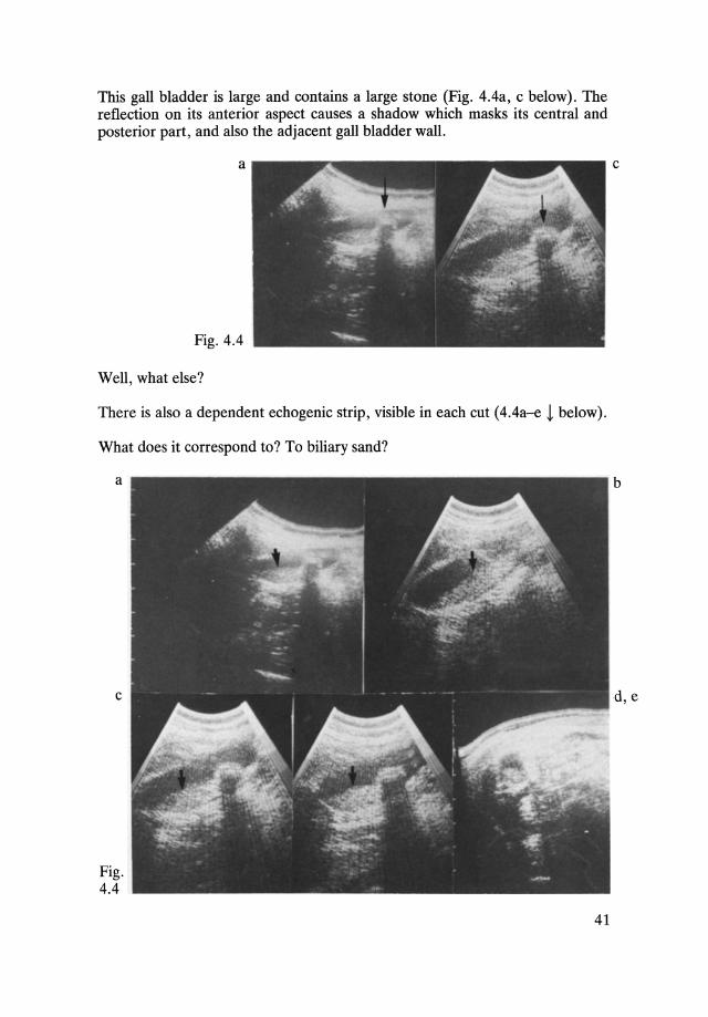
This gall bladder is large and contains a large stone (Fig. 4.4a, c below). The reflection on its anterior aspect causes a shadow which masks its central and posterior part, and also the adjacent gall bladder wall.
a
Fig. 4.4
Well, what else?
There is also a dependent echogenic strip, visible in each cut (4.4a-e ~ below).
What does it correspond to? To biliary sand?
a
c
Fig. 4.4
41
c
b
d,e
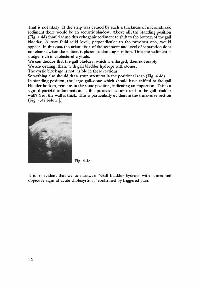
That is not likely. If the strip was caused by such a thickness of microlithiasic sediment there would be an acoustic shadow. Above all, the standing position (Fig. 4.4d) should cause this echogenic sediment to shift to the bottom of the gall bladder. A new fluid-solid level, perpendicular to the previous one, would appear. In this case the orientation of the sediment and level of separation does not change when the patient is placed in standing position. Thus the sediment is sludge, rich in cholesterol crystals. We can deduce that the gall bladder, which is enlarged, does not empty. We are dealing, then, with gall bladder hydrops with stones. The cystic blockage is not visible in these sections. Something else should draw your attention in the positional scan (Fig. 4.4d). In standing position, the large gall-stone which should have shifted to the gall bladder bottom, remains in the same position, indicating an impaction. This is a sign of parietal inflammation. Is this process also apparent in the gall bladder wall? Yes, the wall is thick. This is particularly evident in the transverse section (Fig. 4.4e below t).
Fig.4.4e
It is so evident that we can answer: "Gall bladder hydrops with stones and objective signs of acute cholecystitis," confirmed by triggered pain.
42
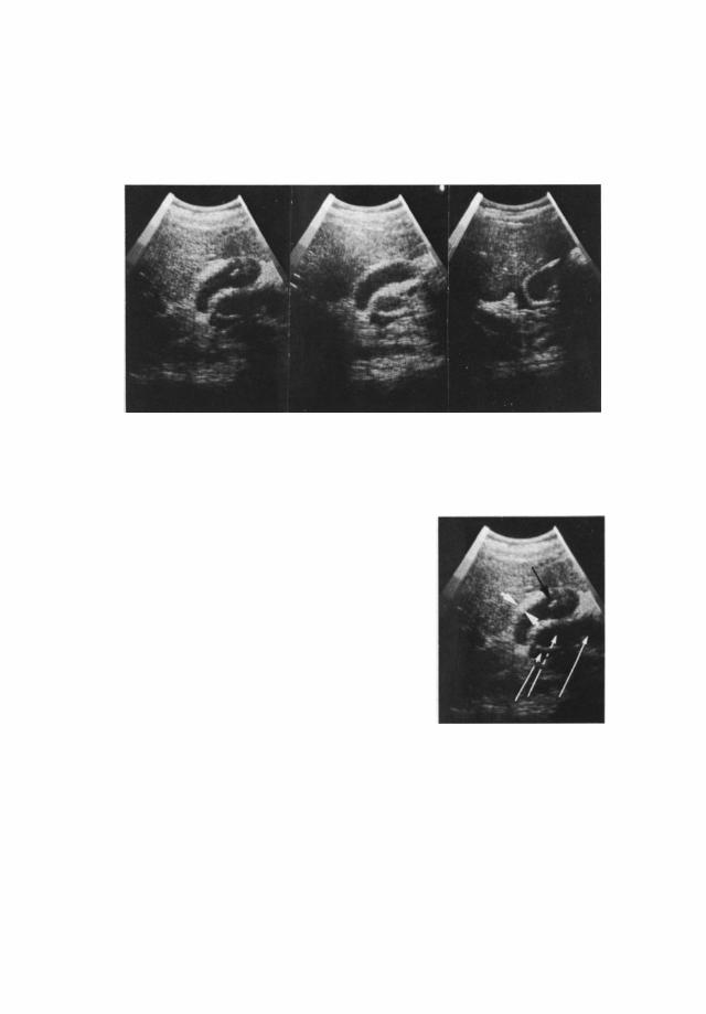

Fig.4.5b
Fluid is also demonstrated around these collections (white arrows). What we are seeing then, are dilated fluid-filled intestinal loops, caused by a paralytic ileus and floating in an intraperitoneal effusion. The presence of a paralytic ileus is not surprising in this picture of acute cholecystitis; but proof of distinct peritoneal involvement indicates it is time to sharpen up those Swiss knives.
44
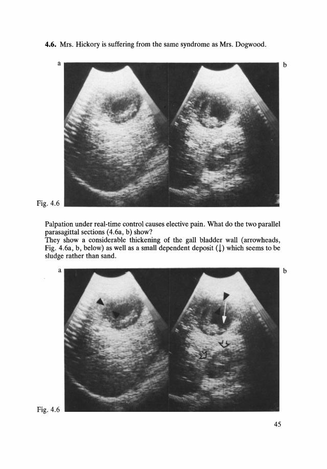
4.6. Mrs. Hickory is suffering from the same syndrome as Mrs. Dogwood.
a
Fig. 4.6
Palpation under real-time control causes elective pain. What do the two parallel parasagittal sections (4.6a, b) show? They show a considerable thickening of the gall bladder wall (arrowheads, Fig. 4.6a, b, below) as well as a small dependent deposit (!) which seems to be sludge rather than sand.
a
Fig. 4.6
45
b
b
a
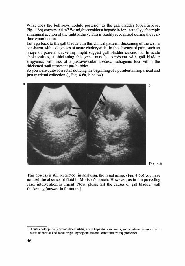
What does the bull's-eye nodule posterior to the gall bladder (open arrows, Fig. 4.6b) correspond to? We might consider a hepatic lesion; actually, it's simply a marginal section of the right kidney. This is readily recognized during the realtime examination. Let's go back to the gall bladder. In this clinical pattern, thickening of the wall is consistent with a diagnosis of acute cholecystitis. In the absence of pain, such an image of parietal thickening might suggest gall bladder carcinoma. In acute cholecystities, a thickening this great may be consistent with gall bladder empyema, with risk of a juxtavesicular abscess. Echogenic foci within the thickened wall represent gas bubbles. So you were quite correct in noticing the beginning of a purulent intraparietal and juxtaparietal collection (~ Fig. 4.6a, b below).
b
Fig. 4.6
This abscess is still restricted: in analysing the renal image (Fig. 4.6b) you have noticed the absence of fluid in Morison's pouch. However, as in the preceding case, intervention is urgent. Now, please list the causes of gall bladder wall thickening (answer in footnote l ).
1 Acute cholecystitis, chronic cholecystitis, acute hepatitis, carcinoma, ascitic edema, edema due to stasis of cardiac and renal origin, hypoglobulineroia, other infiltrating processes
46
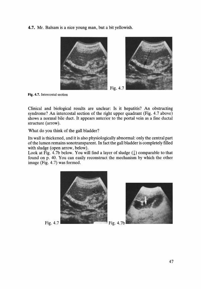
4.7. Mr. Balsam is a nice young man, but a bit yellowish.
Fig. 4.7
Fig. 4.7. Intercostal section
Clinical and biological results are unclear: Is it hepatitis? An obstructing syndrome? An intercostal section of the right upper quadrant (Fig. 4.7 above) shows a normal bile duct . It appears anterior to the portal vein as a fine ductal structure (arrow) .
What do you think of the gall bladder?
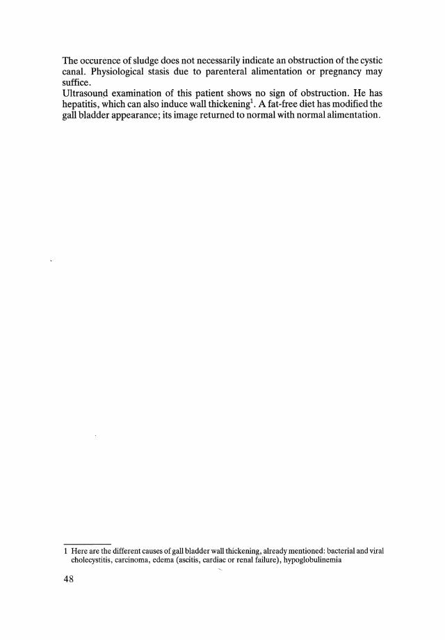
Its wall is thickened, and it is also physiologically abnormal: only the central part of the lumen remains sonotransparent. In fact the gall bladder is completely filled with sludge (open arrow, below). Look at Fig. 4.7b below. You will find a layer of sludge (1) comparable to that found on p. 40. You can easily reconstruct the mechanism by which the other image (Fig. 4.7) was formed.
Fig. 4.7 Fig. 4.7b
47
The occurence of sludge does not necessarily indicate an obstruction of the cystic canal. Physiological stasis due to parenteral alimentation or pregnancy may suffice. Ultrasoun~ examination of this patient shows no sign of obstruction. He has hepatitis, which can also induce wall thickeningl. A fat-free diet has modified the gall bladder appearance; its image returned to normal with normal alimentation.
1 Here are the different causes of gall bladder wall thickening, already mentioned: bacterial and viral cholecystitis, carcinoma, edema (ascitis, cardiac or renal failure), hypoglobulinemia
48
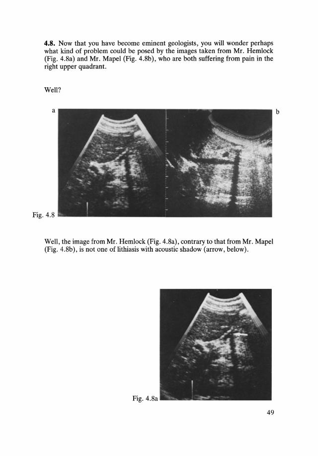
4.8. Now that you have become eminent geologists, you will wonder perhaps what kind of problem could be posed by the images taken from Mr. Hemlock (Fig. 4.8a) and Mr. Mapel (Fig. 4.8b), who are both suffering from pain in the right upper quadrant.
Well?
a
Fig. 4.8
Well, the image from Mr. Hemlock (Fig. 4.8a), contrary to that from Mr. Mapel (Fig. 4.8b), is not one of lithiasis with acoustic shadow (arrow, below).
Fig. 4.8a
49
b
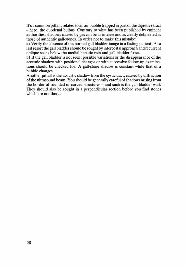
It's a common pitfall, related to an air bubble trapped in part of the digestive tract - here, the duodenal bulbus. Contrary to what has been published by eminent authorities, shadows caused by gas can be as intense and as clearly delineated as those of authentic gall-stones. In order not to make this mistake: a) Verify the absence of the normal gall bladder image in a fasting patient. As a last resort the gall bladder should be sought by intercostal approach and recurrent oblique scans below the medial hepatic vein and gall bladder fossa. b) If the gall bladder is not seen, possible variations or the disappearance of the acoustic shadow with positional changes or with successive follow-up examinations should be checked for. A gall-stone shadow is constant while that of a bubble changes. Another pitfall is the acoustic shadow from the cystic duct, caused by diffraction of the ultrasound beam. You should be generally careful of shadows arising from the border of rounded or curved structures - and such is the gall bladder wall. They should also be sought in a perpendicular section before you find stones which are not there.
50