Embed Size (px)
Citation preview
Respiratory Medicine (2011) 105, 659e666
ava i lab le at www.sc iencedi rec t .com
journa l homepage: www.e lsev ier .com/ locate / rmed
REVIEW
Exogenous lipoid pneumonia. Clinical andradiological manifestations
Edson Marchiori a,b,*, Glaucia Zanetti b,c, Claudia Mauro Mano a,d,Bruno Hochhegger b,e
a Fluminense Federal University, Rio de Janeiro, Brazilb Federal University of Rio de Janeiro, Rio de Janeiro, Brazil
Received 19 August 2010; accepted 1 December 2010Available online 23 December 2010
KEYWORDSExogenous lipoidpneumonia;Computed tomography;Aspiration pneumonia;Bronchoalveolar lavage;Lung diseases
* Corresponding author. Rua Thomazfax: þ55(21)26299017.
E-mail addresses: edmarchiori@[email protected] (B. Hochc Rua Coronel Veiga, 733/504. Centrd Rua Dr. Paulo Alves, 110/802 E. Ine Rua Joao Alfredo, 558/301, CEP 90
0954-6111/$ - see front matter ª 201doi:10.1016/j.rmed.2010.12.001
Summary
Lipoid pneumonia results from the pulmonary accumulation of endogenous or exogenous lipids.Host tissue reactions to the inhaled substances differ according to their chemical characteris-tics. Symptoms can vary significantly among individuals, ranging from asymptomatic to severe,life-threatening disease. Acute, sometimes fatal, cases can occur, but the disease is usuallyindolent. Possible complications include superinfection by nontuberculous mycobacteria,pulmonary fibrosis, respiratory insufficiency, cor pulmonale, and hypercalcemia. The radiolog-ical findings are nonspecific, and the disease presents with variable patterns and distribution.For this reason, lipoid pneumonia may mimic many other diseases. The diagnosis of exogenouslipoid pneumonia is based on a history of exposure to oil, characteristic radiological findings,and the presence of lipid-laden macrophages on sputum or BAL analysis. High-resolutioncomputed tomography (HRCT) is the best imaging modality for the diagnosis of lipoid pneu-monia. The most characteristic CT finding in LP is the presence of negative attenuation valueswithin areas of consolidation. There are currently no studies in the literature that define thebest therapeutic option. However, there is a consensus that the key measure is identifying and
Cameron, 438, Valparaiso, CEP 25685.120, Petropolis, Rio de Janeiro, Brazil. Tel.:þ55 (24) 22492777;
ail.com (E. Marchiori), [email protected] (G. Zanetti), [email protected] (C.M. Mano),hegger).o, CEP 25655-504, Petropolis, Rio de Janeiro, Brazil. Tel.: þ55(24)22429156.ga, CEP 24210-445, Niteroi, Rio de Janeiro. Brazil.050-230, Porto Alegre, Brazil.
0 Elsevier Ltd. All rights reserved.
660 E. Marchiori et al.
discontinuing exposure to the offending agent. Treatment in patients without clinical symp-toms remains controversial, but in patients with diffuse pulmonary damage, aggressive thera-pies have been reported. They include whole lung lavage, systemic corticosteroids, andthoracoscopy with surgical debridement.ª 2010 Elsevier Ltd. All rights reserved.
Contents
Definition and classification . . . . . . . . . . . . . . . . . . . . . . . . . . . . . . . . . . . . . . . . . . . . . . . . . . . . . . . . . . . . . . 660Etiopathogenesis and pathophysiology . . . . . . . . . . . . . . . . . . . . . . . . . . . . . . . . . . . . . . . . . . . . . . . . . . . . . . . 660Clinical findings . . . . . . . . . . . . . . . . . . . . . . . . . . . . . . . . . . . . . . . . . . . . . . . . . . . . . . . . . . . . . . . . . . . . . . . 661Diagnosis . . . . . . . . . . . . . . . . . . . . . . . . . . . . . . . . . . . . . . . . . . . . . . . . . . . . . . . . . . . . . . . . . . . . . . . . . . . . 661
Clinical history . . . . . . . . . . . . . . . . . . . . . . . . . . . . . . . . . . . . . . . . . . . . . . . . . . . . . . . . . . . . . . . . . . . 661Bronchoalveolar lavage . . . . . . . . . . . . . . . . . . . . . . . . . . . . . . . . . . . . . . . . . . . . . . . . . . . . . . . . . . . . . 661Chest radiograph . . . . . . . . . . . . . . . . . . . . . . . . . . . . . . . . . . . . . . . . . . . . . . . . . . . . . . . . . . . . . . . . . 662Computed tomography . . . . . . . . . . . . . . . . . . . . . . . . . . . . . . . . . . . . . . . . . . . . . . . . . . . . . . . . . . . . . 662Histopathology . . . . . . . . . . . . . . . . . . . . . . . . . . . . . . . . . . . . . . . . . . . . . . . . . . . . . . . . . . . . . . . . . . . 663
Complications . . . . . . . . . . . . . . . . . . . . . . . . . . . . . . . . . . . . . . . . . . . . . . . . . . . . . . . . . . . . . . . . . . . . . . . . 664Treatment . . . . . . . . . . . . . . . . . . . . . . . . . . . . . . . . . . . . . . . . . . . . . . . . . . . . . . . . . . . . . . . . . . . . . . . . . . . 664Natural history and outcome . . . . . . . . . . . . . . . . . . . . . . . . . . . . . . . . . . . . . . . . . . . . . . . . . . . . . . . . . . . . . 664Conflict of interest statement . . . . . . . . . . . . . . . . . . . . . . . . . . . . . . . . . . . . . . . . . . . . . . . . . . . . . . . . . . . . 665References . . . . . . . . . . . . . . . . . . . . . . . . . . . . . . . . . . . . . . . . . . . . . . . . . . . . . . . . . . . . . . . . . . . . . . . . . . 665
Definition and classification
Several pulmonary complications may be caused by theaspiration of different substances into the airways andairspaces. Lipoid pneumonia (LP) is an uncommon conditionthat results from the pulmonary accumulation of fatlikecompounds of animal, vegetable or mineral origin.1,2 It canbe classified into endogenous and exogenous forms.
In endogenous form, also called “cholesterol” or“golden” pneumonia, the fatlike materials are derived fromthe lung itself.3,4 It usually develops when lipids that nor-mally reside in the lung tissue emost commonly cholesteroland its esters e are released from destroyed alveolar cellwalls distal to an obstructive airway lesion or from lungtissue damaged by a suppurative process, or due to lipidstorage diseases.3,5,6
The exogenous form can be classified into acute andchronic. Chronic exogenous LP results from long-term,recurrent inhalation exposure to oil, while the acute form issecondary to accidental aspiration of a large quantity oflipid material over a short period of time.7,8
Etiopathogenesis and pathophysiology
Exogenous LP is caused by the inhalation of oils present infood, radiographic contrast media, or oil-based medica-tions such as laxatives.1,9 Traditional oil-based popularmedicines are used to treat various diseases in children,and are often associated with respiratory diseases.10 Inadults, most cases result from the use of oil-based laxatives(olive oil, cod liver oil and paraffin oil) for the treatment ofconstipation, followed by the nasal instillation of oilyproducts for chronic rhinopharyngeal diseases.3,11
Chronic constipation is a frequent symptom in the pedi-atric population.Mineral oil is also used in children for partialsmall bowel obstruction by Ascaris lumbricoides.12,13
However, it must be remembered that infants and smallchildren often object vigorously to ingest the oil, resulting ingagging that precipitates aspiration.14
Different substances called pyrofluids are used by “fire-eaters” (performers that “swallow” or “spit” fire). The mostcommon is the petroleum-derivative kerdan, characterizedbyits reduced viscosity and rapid diffusion throughout the bron-chial tree. After flame blowing, the fire-eater takes a deepbreath, and can aspirate the kerdan remaining in the mouth.8
It is noteworthy that fire-eater pneumonitis has morpho-logical, radiographic and clinical profiles entirely differentfrom chronic lipoid pneumonia. The history of accidentalaspiration during “fire-eating” demonstrations is character-istic. Symptoms occur in the first 12 h after aspiration, andinclude chest pain, dyspnea, cough, fever, and hemoptysis.The disease has, in most of cases, a favorable evolution. TheHRCT usually shows bilateral lung consolidations, oftenassociated with cavitary lesions (pneumatoceles).8
Other less commonly reported causes of LP include:aspiration of milk,15,16 poppy seed oil,3 and egg yolk16;occupational exposure to paraffin (paraffin dropletsreleased by machines) in cardboard crockery factories17; oilblasting in industries; use of spray paint18; contact withplastic paint19; aspiration of oils used in industry as lubri-cants and cutting fluids, in either fluid or spray form, forturning, milling and grinding operations3; cleaning of newcars protected by paraffin, using hot water generated bycompressed air jets20; cleaning oil-containing vats;siphoning diesel fuel19,21; smoking blackfat tobacco, a Ken-tucky product coated with oil and petroleum jelly to flavorand moisturize the leaf6; accidental aspiration of vaseline
Exogenous lipoid pneumonia 661
used in the placement of nasogastric tubes22; longstandinguse of petroleum jelly (Vaseline, Vicks�) at bedtime23;aspiration of spray lubricant WD-40 (very popular oil spray,for home use)24; excessive use of lip balm (Chap Stick,a lipstick that contains petrolatums and lipids) and offlavored lip gloss6; and facial application of petrolatum forerythrodermic psoriasis.25 Some authors also described thedevelopment of LP secondary to suicide attempt by mineraloil immersion.21,26
Factors that increase the risk of exogenous LP includeextremes of age; anatomical or structural abnormalities of thepharynx and esophagus, such as Zenker diverticulum, gastro-esophageal fistula, hiatal hernia, gastroesophageal reflux,achalasia; psychiatric disorders; episodes of loss ofconsciousness; neuromuscular disorders that result in swal-lowingdysfunctionor affect thecough reflex; and,particularlyin children, forcedoil intake.1,7,27e29Because theyfloat on thetopof stomachfluids,oilsmayenter theairwaywhenaspiratedby patients with gastroesophageal reflux disease.3,6e8,27,30
Critically ill patients receiving nasoenteric feeding mayeasily develop aspiration pneumonia, as a result ofa retrograde regurgitation of feeding solutions.31
In some countries the digestive form of Chagas disease,where patients with megacolon have severe and prolongedperiods of constipation (over 60 days), should also beremembered.32,33 The combination of two factors (con-stipation caused by functional disorder of colon, andtendency to aspiration due to megaesophagus) enhances therisk of lipoid pneumonia in patients with Chagas disease.34
In many cases there is no predisposing condition and theexcessive use of oily substances is the presumed cause.1,2,29
In adults, 25% of cases of lipoid pneumonia occur in indi-viduals without any predisposing factors.3,14
It is noteworthy that the aspiration of mineral oil usuallyoccurs unnoticed because it does not induce reactiveresponses in the airways, such as glottic closure or cough.These substances introduced into the nose can easily andsilently reach the bronchial tree of sleeping patientswithout eliciting a normal protective cough reflex11 andmay impair mucociliary transport, subsequently reducingtheir clearance from the respiratory tract.3,14
Clinical findings
The clinical symptoms are nonspecific, and vary accordingto the patient’s age, duration of oil intake, and the amountand quality of oil aspirated.14,35
The time of exposure to oil varies widely in the casesdescribed in the literature, reaching up to severalyears.3,7,8,30,36,37 In almost all studies, the amount of oilconsumed was not quantified.3,7,8,30,36,37
In the elderly, these pneumonias are usually chronic,progressive, and asymptomatic. For this reason, it may bediscovered only as an incidental autopsy finding. Respira-tory failure requiring ventilatory support is an unusualpresentation of the disease.21 In children, massive aspira-tion may present with acute clinical manifestations andearly radiological findings.36,38
Symptoms can vary significantly among individuals, rangingfrom asymptomatic to severe, life-threatening disease.39
When present, symptoms are usually mild.26 Lipoid
pneumonia usually presents with chronic cough, sometimesproductive, and dyspnea.40 Less common problems includechest pain, hemoptysis, weight loss, and intermittent fever.Physical examination of the chest may be normal or maypresentdullness topercussion, crackles,wheezes,or rhonchi.6
Most cases of lipoid pneumonia shows a discrepancy inseverity between the radiological and clinical findings.Patients are often asymptomatic with extensive imagingfindings, which are discovered incidentally on routine chestradiographs.2,6,28
Extrathoracic symptoms, such as vomiting, stomachpain, dysphagia, vertigo and fainting, are occasionallydescribed in the literature.8 The presence of hypertrophicosteoarthropathy has been reported in a five-year-old childwith a history of forced feeding with animal fat (ghee).41
Since its clinical and radiological presentations arenonspecific, lipoid pneumonia may mimic many otherdiseases,14 especially bacterial pneumonia, presenting withfever and cough.19 Occasionally, it can also simulate lungtumours,42 pulmonary tuberculosis or cystic fibrosis43.
Diagnosis
The diagnosis of exogenous lipoid pneumonia is based ona history of exposure to oil with radiological findings inkeeping with the disease and the presence of lipid-ladenmacrophages on sputum or bronchoalveolar lavage (BAL)analysis.3,28 It should be considered, however, that none ofthese findings alone is diagnostic of lipoid pneumonia.
Clinical history
Exogenous lipoid pneumonias may be difficult to diagnosebecause a history of oil ingestion is often missed. Theexposure is often identified only retrospectively after thediagnosis is made, when a directed history is taken fromthe patient or their parents.7,29,35,42
Bronchoalveolar lavage
Bronchoscopy with BAL has been reported to be successfulin establishing the diagnosis of lipoid pneumonia. It allowsthe assessment of airway anatomy to rule out other causesof chronic nonresolving pneumonia.1,14 Plugging with oil hasbeen noted bronchoscopically and on post-mortem exami-nation.43 The macroscopic appearance of BAL may suggestthe diagnosis, revealing a whitish or turbid fluid, with fatglobules on the fluid surface.6,14,29 In a series of 10 childrenwith LP, Sias et al.44 have found that all of them showeda milky BAL fluid on gross examination.
Besides the diagnostic value, BAL has therapeuticimplications in some cases.44 The biochemical analysis ofthe collected fluid can help to confirm the diagnosis, whenusing fat stains.45 The specific type of oil aspirated can bedetermined by chemical analysis, infrared spectroscopy orchromatography.6,44,46
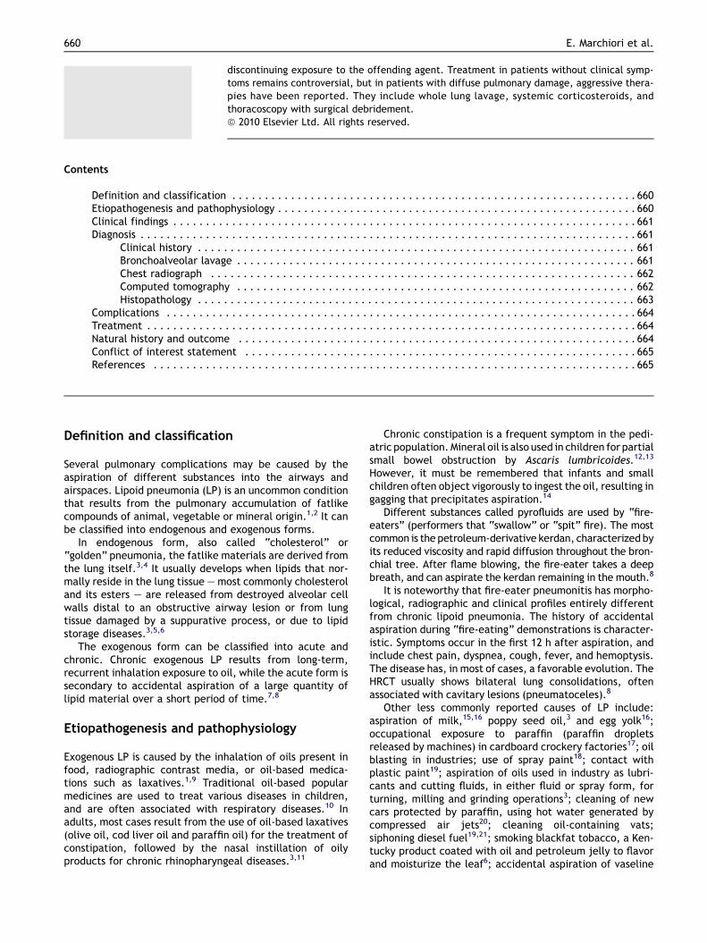
The cytological demonstration of lipid-laden macrophagesis consistent with the diagnosis of exogenous LP,14 althoughfalse-negative results may also occur.14 Both the presence oflipid-ladenmacrophages per se (Fig. 1), and the finding of highlipid-laden macrophage index (LLMI)47 are nonspecific
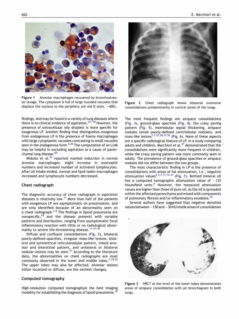
Figure 1 Alveolar macrophages recovered by bronchoalveo-lar lavage. The cytoplasm is full of large rounded vacuoles thatdisplace the nucleus to the periphery (oil red O stain, �400).
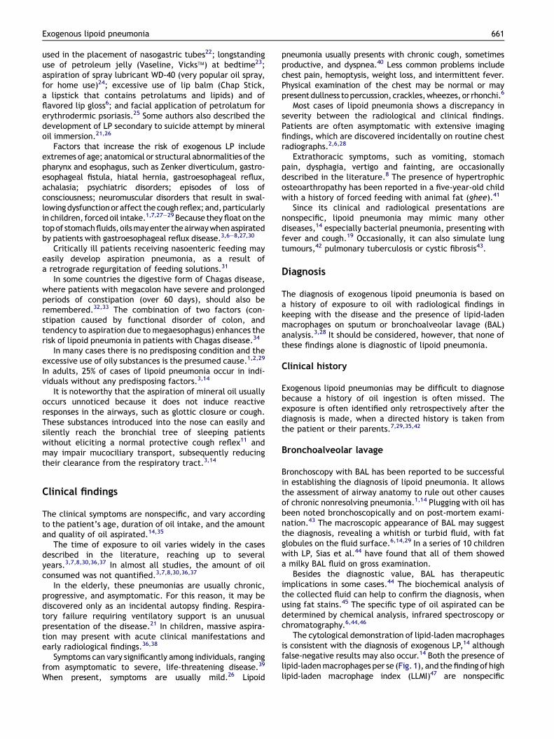
Figure 2 Chest radiograph shows bilateral extensiveconsolidations predominantly in central zones of the lungs.
Figure 3 HRCT at the level of the lower lobes demonstratesareas of airspace consolidation with air bronchogram in bothlungs.
662 E. Marchiori et al.
findings, andmay be found in a variety of lung diseases wherethere is no clinical evidence of aspiration.47e49 However, thepresence of extracellular oily droplets is more specific forexogenous LP. Another finding that distinguishes exogenousfrom endogenous LP is the presence of foamy macrophageswith large cytoplasmic vacuoles contrasting to small vacuolesseen in the endogenous form.6,44 The computation of an LLMImay be helpful in excluding aspiration as a cause of paren-chymal lung disease.50
Midulla et al.35 reported marked reduction in normalalveolar macrophages, slight increase in eosinophilnumbers and increased number of activated lymphocytes.After oil intake ended, normal and lipid-laden macrophagesincreased and lymphocyte numbers decreased.
Chest radiograph
The diagnostic accuracy of chest radiograph in aspirationdiseases is relatively low.11 More than half of the patientswith exogenous LP are asymptomatic on presentation, andare only identified because of an abnormality seen ona chest radiograph.2,6 The findings in lipoid pneumonia arenonspecific,29 and the disease presents with variablepatterns and distribution, ranging from asymptomatic focalinflammatory reaction with little or no radiological abnor-mality to severe life-threatening disease.11,27,46
Diffuse and confluent consolidations (Fig. 2), bilateralpoorly-defined opacities, irregular mass-like lesions, bilat-eral and symmetrical reticulonodular pattern, mixed alve-olar and interstitial pattern, and unilateral or bilateralnodular lesions may be seen.51 According to the literaturedata, the abnormalities on chest radiographs are mostcommonly observed in the lower and middle lobes.1,29,42
The upper lobes may also be affected. Alveolar lesionseither localized or diffuse, are the earliest changes.
Computed tomography
High-resolution computed tomographyis the best imagingmodality for establishing the diagnosis of lipoid pneumonia.42
The most frequent findings are airspace consolidations(Fig. 3), ground-glass opacities (Fig. 4), the crazy pavingpattern (Fig. 5), interlobular septal thickening, airspacenodules (small poorly-defined centrilobular nodules), andmass-like lesions1,7,27,28,37,52 (Fig. 6). None of these aspectsare a specific radiological feature of LP. In a study comparingadults and children, Marchiori et al.37 demonstrated that theconsolidations were significantly more frequent in children,while the crazy paving pattern was more commonly seen inadults. The prevalence of ground-glass opacities or airspacenodules did not differ between the two groups.
The most characteristic finding in LP is the presence ofconsolidations with areas of fat attenuation, i.e., negativeattenuation values2,3,7,11,14,37 (Fig. 7). Bottled mineral oilhas a computed tomographic attenuation value of �132Hounsfield units.6 However, the measured attenuationvalues are higher than those of pure oil, as the oil is spreadedwithin the affected parenchyma andmixedwith componentsof pulmonary fibrosis and/or inflammatory exudates.52
Several authors have suggested that negative densitiesvaluesbetween�150and�30HUinsideareasofconsolidation
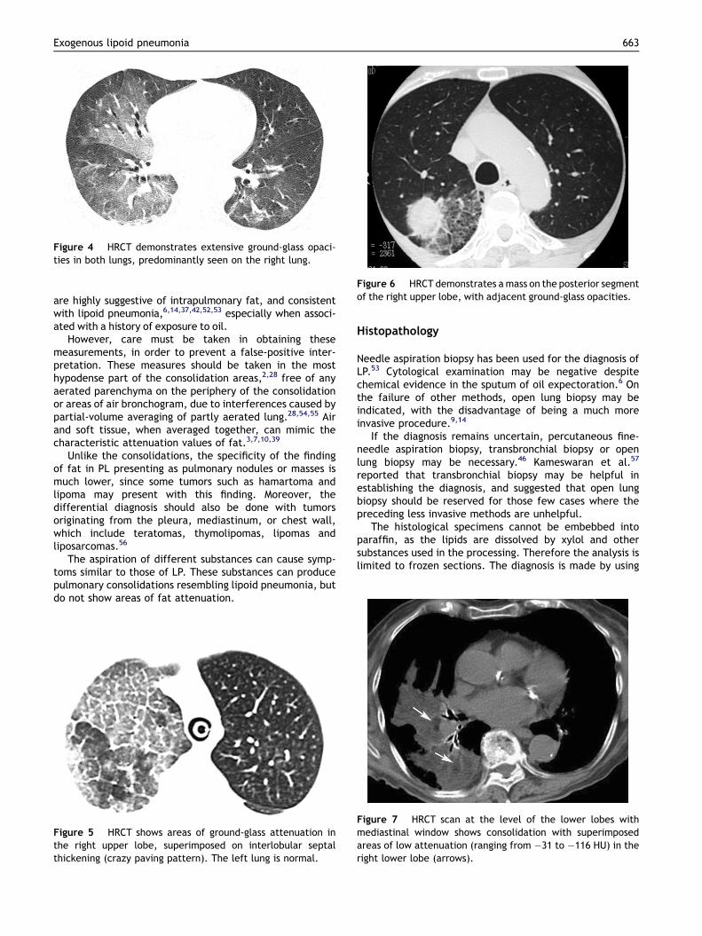
Figure 4 HRCT demonstrates extensive ground-glass opaci-ties in both lungs, predominantly seen on the right lung.
Figure 6 HRCT demonstrates amass on the posterior segmentof the right upper lobe, with adjacent ground-glass opacities.
Exogenous lipoid pneumonia 663
are highly suggestive of intrapulmonary fat, and consistentwith lipoid pneumonia,6,14,37,42,52,53 especially when associ-ated with a history of exposure to oil.
However, care must be taken in obtaining thesemeasurements, in order to prevent a false-positive inter-pretation. These measures should be taken in the mosthypodense part of the consolidation areas,2,28 free of anyaerated parenchyma on the periphery of the consolidationor areas of air bronchogram, due to interferences caused bypartial-volume averaging of partly aerated lung.28,54,55 Airand soft tissue, when averaged together, can mimic thecharacteristic attenuation values of fat.3,7,10,39
Unlike the consolidations, the specificity of the findingof fat in PL presenting as pulmonary nodules or masses ismuch lower, since some tumors such as hamartoma andlipoma may present with this finding. Moreover, thedifferential diagnosis should also be done with tumorsoriginating from the pleura, mediastinum, or chest wall,which include teratomas, thymolipomas, lipomas andliposarcomas.56
The aspiration of different substances can cause symp-toms similar to those of LP. These substances can producepulmonary consolidations resembling lipoid pneumonia, butdo not show areas of fat attenuation.
Figure 5 HRCT shows areas of ground-glass attenuation inthe right upper lobe, superimposed on interlobular septalthickening (crazy paving pattern). The left lung is normal.
Histopathology
Needle aspiration biopsy has been used for the diagnosis ofLP.53 Cytological examination may be negative despitechemical evidence in the sputum of oil expectoration.6 Onthe failure of other methods, open lung biopsy may beindicated, with the disadvantage of being a much moreinvasive procedure.9,14
If the diagnosis remains uncertain, percutaneous fine-needle aspiration biopsy, transbronchial biopsy or openlung biopsy may be necessary.46 Kameswaran et al.57
reported that transbronchial biopsy may be helpful inestablishing the diagnosis, and suggested that open lungbiopsy should be reserved for those few cases where thepreceding less invasive methods are unhelpful.
The histological specimens cannot be embebbed intoparaffin, as the lipids are dissolved by xylol and othersubstances used in the processing. Therefore the analysis islimited to frozen sections. The diagnosis is made by using
Figure 7 HRCT scan at the level of the lower lobes withmediastinal window shows consolidation with superimposedareas of low attenuation (ranging from �31 to �116 HU) in theright lower lobe (arrows).
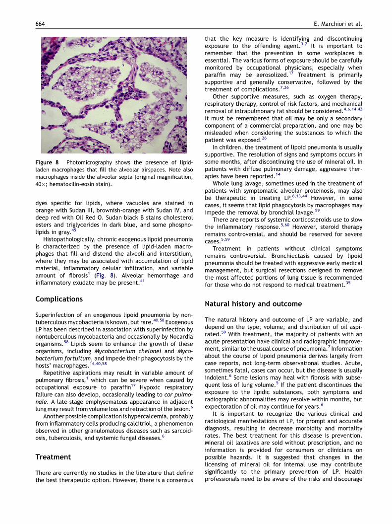
Figure 8 Photomicrography shows the presence of lipid-laden macrophages that fill the alveolar airspaces. Note alsomacrophages inside the alveolar septa (original magnification,40�; hematoxilin-eosin stain).
664 E. Marchiori et al.
dyes specific for lipids, where vacuoles are stained inorange with Sudan III, brownish-orange with Sudan IV, anddeep red with Oil Red O. Sudan black B stains cholesterolesters and triglycerides in dark blue, and some phospho-lipids in gray.45
Histopathologically, chronic exogenous lipoid pneumoniais characterized by the presence of lipid-laden macro-phages that fill and distend the alveoli and interstitium,where they may be associated with accumulation of lipidmaterial, inflammatory celular infiltration, and variableamount of fibrosis1 (Fig. 8). Alveolar hemorrhage andinflammatory exudate may be present.41
Complications
Superinfection of an exogenous lipoid pneumonia by non-tuberculousmycobacteria is known, but rare.40,58 ExogenousLP has been described in association with superinfection bynontuberculous mycobacteria and occasionally by Nocardiaorganisms.58 Lipids seem to enhance the growth of theseorganisms, including Mycobacterium chelonei and Myco-bacterium fortuitum, and impede their phagocytosis by thehosts’ macrophages.14,40,58
Repetitive aspirations may result in variable amount ofpulmonary fibrosis,1 which can be severe when caused byoccupational exposure to paraffin17 Hypoxic respiratoryfailure can also develop, occasionally leading to cor pulmo-nale. A late-stage emphysematous appearance in adjacentlungmay result from volume loss and retraction of the lesion.6
Another possible complication is hypercalcemia, probablyfrom inflammatory cells producing calcitriol, a phenomenonobserved in other granulomatous diseases such as sarcoid-osis, tuberculosis, and systemic fungal diseases.6
Treatment
There are currently no studies in the literature that definethe best therapeutic option. However, there is a consensus
that the key measure is identifying and discontinuingexposure to the offending agent.3,7 It is important toremember that the prevention in some workplaces isessential. The various forms of exposure should be carefullymonitored by occupational physicians, especially whenparaffin may be aerosolized.17 Treatment is primarilysupportive and generally conservative, followed by thetreatment of complications.7,26
Other supportive measures, such as oxygen therapy,respiratory therapy, control of risk factors, and mechanicalremoval of intrapulmonary fat should be considered.4,6,14,42
It must be remembered that oil may be only a secondarycomponent of a commercial preparation, and one may bemisleaded when considering the substances to which thepatient was exposed.26
In children, the treatment of lipoid pneumonia is usuallysupportive. The resolution of signs and symptoms occurs insome months, after discontinuing the use of mineral oil. Inpatients with diffuse pulmonary damage, aggressive ther-apies have been reported.14
Whole lung lavage, sometimes used in the treatment ofpatients with symptomatic alveolar proteinosis, may alsobe therapeutic in treating LP.6,13,44 However, in somecases, it seems that lipid phagocytosis by macrophages mayimpede the removal by bronchial lavage.59
There are reports of systemic corticosteroids use to slowthe inflammatory response.5,60 However, steroid therapyremains controversial, and should be reserved for severecases.5,59
Treatment in patients without clinical symptomsremains controversial. Bronchiectasis caused by lipoidpneumonia should be treated with aggressive early medicalmanagement, but surgical resections designed to removethe most affected portions of lung tissue is recommendedfor those who do not respond to medical treatment.35
Natural history and outcome
The natural history and outcome of LP are variable, anddepend on the type, volume, and distribution of oil aspi-rated.59 With treatment, the majority of patients with anacute presentation have clinical and radiographic improve-ment, similar to the usual course of pneumonia.7 Informationabout the course of lipoid pneumonia derives largely fromcase reports, not long-term observational studies. Acute,sometimes fatal, cases can occur, but the disease is usuallyindolent.6 Some lesions may heal with fibrosis with subse-quent loss of lung volume.5 If the patient discontinues theexposure to the lipidic substances, both symptoms andradiographic abnormalities may resolve within months, butexpectoration of oil may continue for years.6
It is important to recognize the various clinical andradiological manifestations of LP, for prompt and accuratediagnosis, resulting in decrease morbidity and mortalityrates. The best treatment for this disease is prevention.Mineral oil laxatives are sold without prescription, and noinformation is provided for consumers or clinicians onpossible hazards. It is suggested that changes in thelicensing of mineral oil for internal use may contributesignificantly to the primary prevention of LP. Healthprofessionals need to be aware of the risks and discourage
Exogenous lipoid pneumonia 665
the uncontrolled use of mineral oil, especially for the veryyoung and the elderly.
Conflict of interest statement
All authors inform that there are none conflicts of interest.
References
1. Franquet T, Gimenez A, Bordes R, Rodriguez-Arias JM,Castella J. The crazy-paving pattern in exogenous lipoidpneumonia: CT-pathologic correlation. AJR 1998;170:315e7.
2. Laurent F, Philippe JC, Vergier B, et al. Exogenous lipoidpneumonia: HRCT, MR, and pathologic findings. Eur Radiol1999;9:1190e6.
3. Gondouin A, Manzoni PH, Ranfaing E, et al. Exogenous lipidpneumonia: a retrospective multicentre study of 44 cases inFrance. Eur Respir J 1996;9:1463e9.
4. Sharma A, Ohri S, Bambery P, Singh S. Idiopathic endogenouslipoid pneumonia. Indian J Chest Dis Allied Sci 2006;48:143e5.
5. Chin NK, Hui KP, Sinnah R, Chan TB. Idiopathic lipoid pneu-monia in an adult treated with prednisolone. Chest 1994;105:956e7.
6. Spickard A, Hirchmann JV. Exogenous lipoid pneumonia. ArchIntern Med 1994;154:686e92.
7. Baron SR, Haramati LD, Riviera VT. Radiological and clinicalfindings in acute and chronic exogenous lipoid pneumonia. JThorac Imaging 2003;18:217e24.
8. Gentina T, Tillie-Leblond I, Birolleau S, et al. Fire-eater’s lung:seventeen cases and a review of the literature. Medicine(Baltimore) 2001;80:291e7.
9. Asnis DS, Saltzman HP, Melchert A. Shark oil pneumonia. Anoverlooked entity. Chest 1993;103:976e7.
10. Hoffman LR, Yen EH, Kanne JP, Effmann EL, Gibson RL, VanNiel CW. Lipoid pneumonia due to Mexican folk remedies:cultural barriers to diagnosis. Arch Pediatr Adolesc Med 2005;159:1043e8.
11. Franquet T, Gimenez A, Roson N, Torrubia S, Sabate JM,Perez C. Aspiration diseases: findings, pitfalls, and differentialdiagnosis. RadioGraphics 2000;20:673e85.
12. de Oliveira GA, Del Caro SR, Bender Lamego CM, Mercon deVargas PR, Vervloet VE. Radiographic plain film and CT findingsin lipoid pneumonia in infants following aspiration of mineraloil used in the treatment of partial small bowel obstruction byAscaris lumbricoides. Pediatr Radiol 1985;15:157e60.
13. Azevedo Sias S, Oliveira Caetano R, Dutra Comarella J, deOliveira E, Santos Ferreira A, Quirico-Santos T. Successfultreatment of lipoid pneumonia associated with bowelobstruction by Ascaris lumbricoides. J Trop Pediatrics 2009.doi:10.1093/tropej/fmp119.
14. Bandla HP, Davis SH, Hopkins NE. Lipoid pneumonia: a silentcomplication ofmineral oil aspiration. Pediatrics 1999;103:E19.
15. Bromer RS, Wolman IJ. Lipoid pneumonia in infants and chil-dren. Radiology 1939;1:1e7.
16. Pinkerton H. Oils and fats e Their entrance into and fats in thelungs of infants and children: a clinical and pathologic report.AJDC 1928;33:259e85.
17. Descatha A, Mompoint D, Ameille J. Occupacional paraffin-induced pulmonary fibrosis: a 25-year follow-up. Occup Med2006;56:504e6.
18. Libuse HM, Rohatgi PK. Diffuse cholesterol granulomatouspneumonitis in a patient exposed to spray paints. Chest 2000;118:303S.
19. Fernandez AA, Dıez JM, Vime RL, Santos DG, Botello LN,Chinarro BJ. Neumonıa lipoidea em relacion com exposicionlaboral a pinturas. Arch Bronconeumol 2003;39:133e5.
20. Pujol JL, Barneon G, Bousquet J, Michel FB, Godard P. Inter-stitial disease induced by occupational exposure to paraffin.Chest 1990;97:234e6.
21. Hussain IR, Edenborough FP, Wilson RS, Stableforth DE. Severelipoid pneumonia following attempted suicide by mineral oilimmersion. Thorax 1996;51:652e3.
22. Mora RB, Martinez PM, Martinez MC, Garcia LA, Cruz ML,Gascon FS. Neumonıa lipoidea aguda debida a la aspiracionaccidental de vaselina utilizada em um sondaje nasogastrico.Arch Bronconeumol 2000;36:485e7.
23. Brown AC, Slocum PC, Putthoff SL, Wallace WE, Foresman BH.Exogenous lipoid pneumonia due to nasal application ofpetroleum jelly. Chest 1994;106:1311e2.
24. Glynn KP, Gale N. Exogenous lipoid pneumonia due to inhala-tion of spray lubricant (DW-40 lung). Chest 1990;97:1265e6.
25. Cohen MA, Galbut B, Kerdel FA. Exogenous lipoid pneumoniacaused by facial application of petrolatum. J Am Acad Der-matol 2003;49:1128e30.
26. Nogue S, Sanz P, Borondo JC, Picon M, Red G, Mestre G. Fatallipoid pneumonia due to bronco-aspiration of isoparaffin afteringestion of an organophosphate insecticide. Acta AnaesthesiolScand 2003;47:777e9.
27. Lee JY, Lee KS, Kim TS, et al. Squalene-induced extrinsic lipoidpneumonia: serial radiologic findings in nine patients. J Com-put Assist Tomogr 1999;23:730e5.
28. Lee KH, Kim WS, Cheon JE, Seo JB, Kim IO, Yeon KM. Squaleneaspiration pneumonia in children: radiographic and CT findingsas the first clue to diagnosis. Pediatr Radiol 2005;35:619e23.
29. Lee KS, Muller NL, Hale V, Newell Jr JD, Lynch DA, Im JG.Lipoid pneumonia: CT findings. J Comput Assist Tomogr 1995;19:48e51.
30. Lee JS, Im JC, Song KS, Seo JB, Lim TH. Exogenous lipoidpneumonia: high-resolution CT findings. Eur Radiol 1999;9:287e91.
31. Umuro�glu T, Takil A, Irmak P, et al. Effects of multiplepulmonary aspirations of enteral solutions on lung tissuedamage. Clin Nutr 2006;25:45e50.
32. Garcia SB, Aranha AL, Garcia FR, et al. A retrospective study ofhistopathological findings in 894 cases of megacolon: what isthe relationship between megacolon and colonic cancer? RevInst Med Trop Sao Paulo 2003;45:91e3.
33. Rochitte CE, Nacif MS, Oliveira Jr AC, Siqueira-Batista R,Marchiori E, Uellendahl M, Higuchi ML. Cardiac magneticresonance in Chaga’s disease. Artif Organs 2007;31(4):259e67.
34. Marchiori E, Zanetti G, Nobre LF, Takayassu TC, Irion KL. Lipoidpneumonia complicating chagasic megaesophagus. High-reso-lution CT findings. J Thorac Imaging 2010;25:179e82.
35. Midula F, Strappini PM, Ascoli V, et al. Bronchoalveolar lavagecell analysis in a child with chronic lipid pneumonia. Eur RespirJ 1998;11:239e42.
36. Annobil SH, Morad NA, Khurana P, Kameswaran M, Ogunbiyi O,al-Malki T. Reaction of human lungs to aspirated animal fat(ghee): a clinicopathological study. Virchows Arch 1995;426(3):301e5.
37. Marchiori E, Zanetti G, Mano CM, Irion KL, Daltro PA,Hochhegger B. Lipoid Pneumonia in 53 patients after aspirationof mineral oil: comparison of high-resolution computedtomography findings in adults and children. J Comput AssistTomogr 2010;34:9e12.
38. Requena-Kassarjlan Y. An infant with respiratory distress. ClinPediatr 2001;40:507e9.
39. Zanetti G, Marchiori E, Gasparetto TD, Escuissato DL. SoaresSouza A Jr. Lipoid pneumonia in children following aspiration ofmineral oil used in the treatment of constipation: high-resolu-tion CT findings in 17 patients. Pediatr Radiol 2007;37:1135e9.
40. Talwar A, Mayerhoff R, London D, Shah R, Stanek A, Epstein M,False-positive PET. Scan in a pacient with lipoid pneumoniasimulating lung cancer. Clin Nucl Med 2004;29:426e8.
666 E. Marchiori et al.
41. Hugosson C, Bahabri S, Rifai A, Al-Dalaan A. Hipertrophicoteoarthropathy caused by lipoid pneumonia. Pediatr Radiol1995;25:482e3.
42. Brechot JM, Buy JN, Laaban JP, Rochemaure J. Computedtomography and magnetic resonance findings in lipoid pneu-monia. Thorax 1991;46:738e9.
43. Balakrishnan S. Lipoid pneumonia in infants and children inSouth India. Brit Med J 1973;4:329e31.
44. Sias SM, Daltro PA, Marchiori E, Ferreira AS, Caetano RL,Silva CS, et al. Clinic and radiological improvement of lipoidpneumonia with multiple bronchoalveolar lavages. PediatrPulmonol 2009;44:309e15.
45. Lauque D, Dongay G, Levade T, Caratero C, Carles P. Bron-choalveolar lavage in liquid paraffin pneumonitis. Chest 1990;98:1149e55.
46. Ciravegna B, Sacco O, Moroni C, et al. Mineral oil child withanoxic encephalopathy: treatment by whole lung lavage.Pediatr Pulmonol 1997;23:233e7.
47. Ding Y, Simpson PM, Schellhase DE, Tryka AF, Ding L,Parham DM. Limited reliability of lipid-laden macrophage indexrestricts its use as a test for pulmonary aspiration: comparisonwith a simple semiquantitative assay. Pediatr Dev Pathol 2002;5:551e8.
48. Kazachkov MY, Muhlebach MS, Livasy CA, Noah TL. Lipid-ladenmacrophage index and inflammation in bronchoalveolar lavagefluids in children. Eur Respir J 2001;18:790e5.
49. Knauer-Fisher S, Ratgen F. Lipid-laden macrophages in bron-choalveolar lavage fluid as a marker for pulmonary aspiration.Pediatr Pulmonol 1999;27:419e22.
50. Corwin RW, Irwin RS. The lipid-laden alveolar macrophage asa marker of aspiration in parenchymal lung disease. Am RevRespir Dis 1985;132:576e81.
51. Rossi SE, Erasmus JJ, Volpacchio M, Franquet T, Castiglioni T,MacAdams HP. “Crazy-paving” pattern at thin-section CT of thelungs: radiologic- pathologic overview. Radiographics 2003;23:1509e19.
52. Betancourt SL, Martinez-Jimenez S, Rossi SE, Truong MT,Carrillo J, Erasmus JJ. Lipoid pneumonia: spectrum of clinicaland radiologic manifestations. AJR Am J Roentgenol 2010;194:103e9.
53. Wheeler PS, Stitik FP,HutchinsGM, KlinefelterHF, Siegelman SS.Diagnosis of lipoid pneumonia by computed tomography. JAMA1981;245:65e6.
54. Hugosson CO, Riff EJ, Moore CC, Akhtar M, Tufenkeji HT. Lipoidpneumonia in infants: a radiological-pathological study.Pediatr Radiol 1991;21:193e7.
55. Tahon F, Berthezene Y, Hominal S, Blineau N, Guerin JC,Cinotti L, et al. Exogenous lipoid pneumonia with unusual CTpattern and FDG positron emission tomography scan findings.Eur Radiol 2002;12:171e3.
56. Gaerte SC, Meyer CA,Winer-MuramHT, Tarver RD, Conces Jr DJ.Fat-containing lesions of the chest. Radiographics; 2002:S61e78. 22 Spec No.
57. Kameswaran M, Annobil SH, Benjamin B, Salim M. Bronchos-copy in lipoid pneumonia. Arch Dis Child 1992;67:1376e7.
58. Ridaura-Sanz C, Lopez-Corella E, Salazar-Flores M. Exogenouslipoid pneumonia superinfected with acid-fast bacilli in infants:a report of nine cases. Fetal Pediatr Pathol 2006;25:107e17.
59. Meltzer E, Guranda L, Vassilenko L, Krupsky M, Steinlauf S,Sidi Y. Lipoid pneumonia: a preventable complication. Isr MedAssoc J 2006;8:33e5.
60. Ayvasian F, Steward DS, Merkel CG, Frederick WW. Diffuselipoid pneumonitis successfully treated with prednisone. Am JMed 1966;43:930e4.
本文献由“学霸图书馆-文献云下载”收集自网络,仅供学习交流使用。
学霸图书馆(www.xuebalib.com)是一个“整合众多图书馆数据库资源,
提供一站式文献检索和下载服务”的24 小时在线不限IP
图书馆。
图书馆致力于便利、促进学习与科研,提供最强文献下载服务。
图书馆导航:
图书馆首页 文献云下载 图书馆入口 外文数据库大全 疑难文献辅助工具