Embed Size (px)
Citation preview

2940
Abstract – Early diagnosis of diabetes melli-tus can significantly improve therapeutic strate-gies and overall health span. Identifying bio-markers as a tool for determining the risk of de-veloping diabetes as well as a monitoring strat-egy for progression of the disease state would be useful in predicting potential complications while simultaneously improving our ability to prevent and treat diabetes. Extracellular vesi-cles (EV) have recently emerged as prominent mediators of intercellular communication and as a potential source for the discovery of novel bio-markers. A deeper understanding of the cargo molecules present in EVs obtained from type 1 diabetes mellitus (T1D) patients may aid in the identification of novel diagnostic and prognos-tic biomarkers, and can potentially lead to the discovery of new therapeutic targets.
Key WordsExosomes, Type 1 diabetes, Biomarkers, Therapy,
Islets.
Introduction
Diabetes mellitus refers to a group of meta-bolic disorders characterized by elevated blood glucose levels attributed to an ineffective, insuf-ficient or absent production of insulin. Long-term complications of the disease have been associated with macro and microvascular problems, leading to heart diseases, stroke, blindness and kidney diseases. Accurate monitoring of the progression towards these complications and responses to ther-apy at early stages, will allow for improvement in preventive and therapeutic strategies. Biomarkers can be objective indicators of the medical condi-tions of a patient, which can be measured accu-rately and reproducibly. The ideal biomarker for diabetes should detect disease trait (risk factor or
risk marker), pathogenesis, disease state (preclini-cal or clinical), or disease rate of progression and prevent disease onset or help assess the possible therapies1. Extracellular vesicles (EVs) are small vesicles that have emerged as important mediators in cellular communication2, which contain pro-teins, DNA and RNA species, (miRNA, mRNA, tRNAs, etc.)3,4 as well as lipids and metabolites. Specific sources of EVs carrying distinct cargos have been associated with both normal physiologic and disease states. Their content can reflect biolog-ical events and disease progression5. This review focuses on the potential use of EVs as a novel class of biomarkers relevant to type 1 diabetes mellitus.
Classification of extracellular vesiclesEVs are a heterogeneous population of mem-
brane bound structures released by cells into the extracellular space. EVs are released from most cells types 6 and can be isolated from most body fluids such as serum, plasma, urine, saliva and cerebrospinal fluid. Their potential role for di-agnostic and therapeutic clinical use is currently under investigation. A consensus nomenclature for these heterogeneous vesicles is not fully estab-lished. However, EVs recently have been classi-fied based on their tissue/cell-specific origin and size (Table I) or their biogenesis (Table II).
Microvesicles Microvesicles arise through direct outward
membrane protrusion, budding, and fission of the plasma membrane and subsequent release of the vesicles into the extracellular space 7. Microves-icles can range from 50-1000 nm in size and are heterogeneous in shape. Several molecular local-ized changes undergo in the plasma membrane that result in the formation of the microvesicles, in-cluding changes in lipid, protein composition and
European Review for Medical and Pharmacological Sciences 2017; 21: 2940-2956
M. GARCIA-CONTRERAS1,2,3, R.W. BROOKS4, L. BOCCUZZI1, P.D. ROBBINS4, C. RICORDI1,2
1Diabetes Research Institute, University of Miami, Miami, FL, USA2Ri.Med Foundation, Palermo, Italy3School of Dentistry and Medicine, Catholic University of Valencia, Valencia, Spain4Department of Molecular Medicine and The Center on Aging, The Scripps Research Institute, Jupiter, FL, USA
Corresponding Author: Camillo Ricordi, MD; e-mail: [email protected]
Exosomes as biomarkers and therapeutictools for type 1 diabetes mellitus

Exosomes in type 1 diabetes mellitus
2941
calcium levels8. These are unique mechanisms of EV formation in comparison to exosomes, which are formed intracellularly within multivesicular bodies. This novel mechanism of microvesicle for-mation results in the regulated release of EVs con-taining specifically enriched molecular cargoes.
Retrovirus-like particles (RLPs)Retrovirus-like particles (RLPs) are those EVs
that resemble retroviral vesicles, but are non-infec-tious because they do not contain the full comple-ment of genes required for cellular entry or viral propagation9. RLPs contain a subset of retroviral proteins, they are released from cells after a viral infection. Certain viruses can use RLPs to facilitate their propagation and entry into neighboring cells10.
Apoptotic bodies or blebsApoptotic bodies or blebs are the largest EVs
with a size range of 50-5000 nm. Apoptotic bod-
ies are vesicles produced from cells undergoing programmed cell death or apoptosis. Apoptotic bodies bud directly from the plasma membrane like microvesicles and contain fragmented nuclei as well as fragmented cytoplasmic organelles11.
Exosomes Exosomes (EXOs) are small (about 30-200 nm
in diameter) lipid vesicles derived from multive-sicular bodies that are released by most cell types. EXOs are present in different body fluids including serum, urine, cerebral spinal fluid, and saliva and bronchiolar lavage fluid. EXOs have emerged as important mediators in cell communication, trans-ferring proteins, lipids, DNA and RNA species (miRNA, mRNA, tRNAs, etc.) between cells2,12. Given that the cargo of EVs reflects the status of the cell type of origin there is a growing interest in the potential of EVs, in particular, exosomes, for diagnosis and therapy in multiples diseases4,13.
Table I. Classification of extracellular vesicles based on tissue/cell-specific origin.
Type of EVs Cellular origin Size (nm) Markers References
Cardiosomes Cardiomyocytes 40-300 CAV-3, flotillin-1 86Ectosomes Neutrophils or Monocytes 100-1000 CD14, TyA and C1a, 87, 88Microparticles Platelets in blood (PMPs) 100-1000 Both CD31 PMPs: CD42 89 or endothelial cells (EMPs) Oncosomes Malignant cells 1000-10000 CAV-1 90Prostatosomes or Seminal fluid 30-200 CD48, CD244 91 prostasomes Tolerosomes Serum antigen-fed mice, 40 MHC II, CD68 92 intestinal epithelial cells and LAMP1Vexosomes Adeno-associated virus vectors (AVV) 50-200 AAV capsids 93
Table II. Classification of extracellular vesicles based on biogenesis.
Type of EVs Origin Size (nm) Markers Content
Exosomes Endolysosomal 30-200 Tetraspanins: CD63, mRNA, microRNA, non-coding pathway CD9, CD81. RNAs, cytoplasmic and ESCRT components: membrane proteins, including TSG101, flotilin, Alix receptors and major histocompatibility moleculesMicrovesicles Cell surface 50-1000 ARF6, VCAMP3, mRNA, microRNA, non-coding integrins, selectins, RNAs, cytoplasmic and CD40 ligand membrane proteins, including receptorsRetrovirus like Plasma membrane 75-100 gag Retroviral particles env, rec, particles and polApoptotic bodies Cell surface 50-5000 Thrombospondin, C3b, Nuclear fractions, or blebs Annexin V, phosphati- cell organelles dylserine
M. Garcia-Contreras, R.W. Brooks, L. Boccuzzi, P.D. Robbins, C. Ricordi
2942
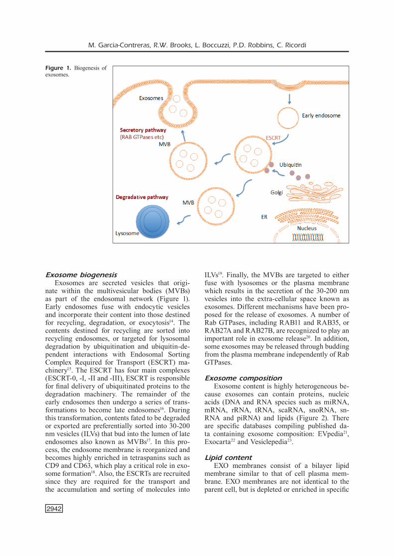
Exosome biogenesisExosomes are secreted vesicles that origi-
nate within the multivesicular bodies (MVBs) as part of the endosomal network (Figure 1). Early endosomes fuse with endocytic vesicles and incorporate their content into those destined for recycling, degradation, or exocytosis14. The contents destined for recycling are sorted into recycling endosomes, or targeted for lysosomal degradation by ubiquitination and ubiquitin-de-pendent interactions with Endosomal Sorting Complex Required for Transport (ESCRT) ma-chinery15. The ESCRT has four main complexes (ESCRT-0, -I, -II and -III), ESCRT is responsible for final delivery of ubiquitinated proteins to the degradation machinery. The remainder of the early endosomes then undergo a series of trans-formations to become late endosomes16. During this transformation, contents fated to be degraded or exported are preferentially sorted into 30-200 nm vesicles (ILVs) that bud into the lumen of late endosomes also known as MVBs17. In this pro-cess, the endosome membrane is reorganized and becomes highly enriched in tetraspanins such as CD9 and CD63, which play a critical role in exo-some formation18. Also, the ESCRTs are recruited since they are required for the transport and the accumulation and sorting of molecules into
ILVs19. Finally, the MVBs are targeted to either fuse with lysosomes or the plasma membrane which results in the secretion of the 30-200 nm vesicles into the extra-cellular space known as exosomes. Different mechanisms have been pro-posed for the release of exosomes. A number of Rab GTPases, including RAB11 and RAB35, or RAB27A and RAB27B, are recognized to play an important role in exosome release20. In addition, some exosomes may be released through budding from the plasma membrane independently of Rab GTPases.
Exosome composition Exosome content is highly heterogeneous be-
cause exosomes can contain proteins, nucleic acids (DNA and RNA species such as miRNA, mRNA, rRNA, tRNA, scaRNA, snoRNA, sn-RNA and piRNA) and lipids (Figure 2). There are specific databases compiling published da-ta containing exosome composition: EVpedia21, Exocarta22 and Vesiclepedia23.
Lipid contentEXO membranes consist of a bilayer lipid
membrane similar to that of cell plasma mem-brane. EXO membranes are not identical to the parent cell, but is depleted or enriched in specific
Figure 1. Biogenesis of exosomes.

Exosomes in type 1 diabetes mellitus
2943
lipids. EXOs contain sphingomyelin, ganglio-sides, cholesterol and desaturated lipids24. EXOs also contain phosphatidylserine in the outer leaflet, which facilitates their internalization by recipient cells25. In addition, EXOs contain en-zymes involved in lipid metabolism and receptors and adhesion molecule and the lipid composition of EXOs derived from different cell sources can vary widely.
RNA contentEXOs contain diverse RNA species such as
miRNA, mRNA, rRNA, tRNA, scaRNA, snoR-NA, snRNA and piRNA26, but are enriched in smaller, non-coding RNAs compared to the pa-rental cell. Defined sequence motifs27 and post-transcriptional modifications28 in the 3’ end in miRNAs may promote specific miRNA and mR-NA packaging into EXOs. The EXO RNA con-tent can be transferred to recipient cells where it plays a functional role2,29.
DNA contentEXOs can transport DNA (exoDNA) that re-
flects the parental cell genomic DNA (gDNA)30. Recent studies have shown that both mitochon-drial DNA (mtDNA) and fragmented chromo-somal DNA are found in EXOs31,32. Furthermore,
EXOs from plasma of cancer patients and cancer cell lines contain different double-stranded ge-nomic DNA fragments containing the tumor-as-sociated mutations3,33. Like RNA, this exosomal DNA also can be transferred between cells and can influence the function of the recipient cells, possibly playing important roles in pathological conditions12,34.
Protein contentProteins are found in the membrane or in
the hydrophilic core of the EVs. These proteins include tetraspanins (CD9, CD63, and CD81 anti-gens), epithelial cell adhesion molecule (EpCam), and other adhesion proteins. The EXOs protein signature is unique and contained cell line-spe-cific proteins, but in some instances, is related to the cell type and EXOs biogenesis24. For example, EXOs originated from the endosome compart-ment are more enriched in tetraspanins and major histocompatibility complex class II.
Exosome isolation methodsEXOs have been isolated from biological flu-
ids including blood, urine, cerebrospinal fluid, tears, saliva and nasal secretions, ascites, and se-men. All currently used protocols for purification co-purify different subtypes of EXOs (Table III).
Figure 2. Schematic im-age of the molecular com-position of an exosome.

M. Garcia-Contreras, R.W. Brooks, L. Boccuzzi, P.D. Robbins, C. Ricordi
2944
There is no universal consensus as to the best method for isolation.
Ultracentrifugation Ultracentrifugation is the most commonly
used method to isolate EVs. This method consists of several sequential centrifugation steps, usually with the first steps being a 300 x g spin for 10 min followed by a 10.000 x g spin 30 min to elimi-nate intact cells, dead cells and cell debris. After
depletion of cells and large apoptotic bodies by low-speed centrifugation, the EVs are pelleted in the final step at 100.000 x g for 70 min35.
Sucrose-gradient centrifugation Sucrose-gradient centrifugation is use in com-
bination with ultracentrifugation to remove con-taminating non-vesicular particles35. This procedure allows separation of vesicles according to their den-sity, classically reported between 1.1 and 1.19 g/ml.
Table III. Classification of extracellular vesicles based on biogenesis.
Method Type Principle Advantages Disadvantages References
Ultracentri- Differential Sedimentation Gold standard Time consuming 35 fugation based on size Inability to separate exosomes and density from microvesicles Expensive equipment Sucrose- Flotation based Removes protein Time consuming 35 gradient on density contamination Inability to separate exosomes Collection from microvesicles morphological Expensive equipment intact microparticlesAffinity- Immunobeads Separation Fast Not suited for large sample 35 based based on Semi-quantitative volumes captured affinity characterization Captured extracellular interactions of the surface vesicles may not retain phenotype can be biological functionality tissue-specific Co-purification of protein aggregates. Low yield.Filtration Microfiltration Separation Easy and fast Protein contamination based on size Small sample volume 35 limitations Inability to separate exosomes from microvesicles Exosomes can adhere to the filtration membranes and become lost. Also, since the additional force is applied to pass the analyzed liquid through the membranes, the exosomes can potentially be deformed or damaged.Chroma- Size-exclusion Separation Collection morpho- Small sample volume limitations 36 tography based on size logical intact Time consuming microparticles Requires specialized equipment Polymer Polymer based Separation Fast Low purity and specificity 38, 94 precipitation based on High yield Protein contamination Polyethylene Requires small glycol sample amount precipitation Microfluidic Devices Mechanics of Increases Large sample volume 95 fluid flow throughput and limitations based allows multiplexing Reduced cost, sample size and processing time

Exosomes in type 1 diabetes mellitus
2945
Affinity-based captured of extracellular vesicles
Affinity-based isolation enables the selective capture of specific subpopulations using antibod-ies to CD63, CD81, CD82, CD9, Alix, annexin, EpCAM and Rab535. These could be used either alone or in combination. For this application, the antibodies can be immobilized on magnetic beads, chromatography matrices, plates, and microfluidic devices.
Size exclusion chromatography/filtration
Separation of EVs based on the size is achieved through the use of chromatography36 or microfil-tration37. Column chromatography allows for se-quential elution of EV size fractions from a single column. Microfiltration is used in combination with ultracentrifugation to eliminate dead cells, apoptotic bodies and large debris.
Polymer precipitation Volume-excluding polymers such as polyeth-
ylene glycol (PEG) are used for the precipitation of extracellular vesicles, although the purity is lower than with some other isolation methods38. Contamination of EVs pellets with non-exosomal materials remains a problem for polymer-precip-
itation methods. In addition, the polymer sub-stance present in the isolate may interfere with down-stream analysis.
Microfluidics The application of these methods to biological
fluids has not yet been described extensively39. However, recent advances in microfluidic-based technologies have made it possible to extract EVs from the blood in an easily reproducible, conve-nient manner. It is likely that this approach will become more prevalent.
Exosomes characterization methods
EXO quantification can be carried out directly or indirectly. There are a variety of methods (Ta-ble IV) that are currently used for analysis, but there is a lack of consensus in the field.
Protein quantification The total protein content of the purified EVs
can be determined by the Bradford assay, which also provides indirect quantification of EV35. However, the presence of proteins which are not associated to the EVs content can impact the total protein content and therefore bias EV concentra-tion determination.
Table IV. Summary of exosome characterization methods.
Method Characteristics Advantages Disadvantages References
Protein Analysis of protein Easy and low cost No specific information 35 quantification concentration (non-specific) Transmission Assessment of Direct evidence for No quantification 35 electron morphology, the presence of EV Need an expert in TEM microscopy size and markersScanning electron Assessment of For the presence of EV No quantification 96 microscopy morphology, Need an expert in SEM size and markersAtomic force Assessment of Direct evidence Need an expert 41 microscopy morphology and size and quantificationELISA Specific EV proteins Specific for exosome Unreliable 42 proteins Technical troublesNanoparticle Analysis of absolute Quantification No distinguishment 43, 96 tracking analysis concentration of EV from aggregated of particles and proteins particle size Flow cytometry Detection of Detection specific Low detection threshold 44, 97 EV markers EV markersWestern blot Detection of specific Detection specific Cannot determine 35 EV proteins EV subset the presence of EVs
M. Garcia-Contreras, R.W. Brooks, L. Boccuzzi, P.D. Robbins, C. Ricordi
2946
Transmission electron microscopy Morphological examination is typically carried
out using transmission electron microscopy (TEM) is an established technique that provides the direct evidence for the presence of EVs. EV suspensions are applied to grids, fixed, stained with osmium tetroxide or uranyl acetate, and contrasted by embedding in methylcellulose. EVs preparations examined by TEM show EVs with a cup shaped appearance, which is an artifact of the preparation procedure40. TEM can be combined with immuno-gold staining using gold conjugated antibodies to detect the presence of specific markers.
Scanning electron microscopy Although TEM is considered a standard tool
for characterizing the morphology of exosomes, scanning electron microscopy (SEM) is a newer, alternative approach to analyze EVs morphology and structure.
Atomic force microscopyAtomic Force Microscopy (AFM) is used for
topographic imaging of EVs. AFM also can be used to analyze the mechanical properties of nanoparticles41.
ELISAAlternatively, exosome ELISA kits (System
Biosciences) allow investigators to quantify the number of exosomes based on the level of the exosome-associated proteins including CD9, CD63, and CD 8142.
Nanoparticle tracking analysis and resistive pulse sensing
Nanosight™ nanoparticle tracking analysis uses light diffraction patterns to measure the size and the concentration of exosomes43. Sim-ilarly, direct quantification of exosomes can be performed using the qNano Gold (Izon Science) which measures nanoparticles using the tunable resistive pulse sensing (TRPS) principle, report-ing concentration as a function of a defined size range. Both are label-free methods.
Flow cytometry Traditional flow cytometry has been wide-
ly used for membrane marker identification44. Here it is recommended to assess not only the presence of selected membrane surface markers, but also the absence of contaminants, and to include appropriate isotype controls. There are inherent problems with this approach, as these
instruments have traditionally been developed to measure whole cells, which are orders of magni-tude larger than exosomes and contain more of each membrane protein. The use of antibodies coupled to beads can allow for detecting EXO surface proteins although not necessarily in a quantitative manner. Imaging flow cytometry has emerged as new alternative with increased fluo-rescence sensitivity, low background, and image confirmation ability45.
Western blotting Western blotting (WB) is a convenient meth-
od to detect the presence of surface markers include tetraspanins (CD9, CD63, CD81, and CD82), MHC molecules, and cytosolic proteins or cytoskeletal proteins. Isolated EVs are lysed, and the proteins are separated and analyzed 35. However, WB alone cannot determine the pres-ence of EVs.
Function of exosomes Physiological function of exosomes
In the 1960s and 70s, studies suggested that membrane fragments and vesicles in the extracel-lular compartment or blood may have originated from specific or regular cellular activity46. In 1983, it was reported that vesicles harboring transferrin receptors were jettisoned by reticulocytes as part of their differentiation into red blood cells47,48. It was not until 1996, that exosomes were first shown to be involved in cell-to-cell communication and possibly play a role in antigen presentation6. More recent studies have focused on the potential func-tions of exosomes in different cell types. The function of exosomes depends on the origin and molecules packaged within. It has been observed that multiple cell types, including pancreatic islets of Langerhans, release EXOs that can transfer their cargos to target cells49. EXOs can transfer not only proteins, but also have been shown to transfer mRNA and miRNAs molecules which are taken up functionally by target cells2. This horizontal transfer of information via exosomes as a new mechanism of cell-to-cell communication has been reported in multiple cell models.
Exosomes in pathological processes: role in type 1 diabetes (T1D)
In addition to their biological function, exo-somes are hypothesized to be involved in patho-logical processes and disease pathogenesis in-cluding cancer5,34, virus50,51 infection and autoim-mune diseases such as type 1 diabetes mellitus52.

Exosomes in type 1 diabetes mellitus
2947
Type 1 diabetes mellitus (T1DM)Type 1 diabetes mellitus, also known as in-
sulin dependent diabetes mellitus, is primarily a childhood associated autoimmune disease char-acterized by the destruction of insulin-producing ß cells in the pancreatic islets of Langerhans. Because of the autoimmune destruction of the insulin-producing ß-cells, there is an insulin de-ficiency and the body is unable to control blood sugar. Since insulin is the primary anabolic hor-mone that regulates blood glucose level, patients with T1D require the administration of exoge-nous insulin through multiple daily injections, guided by daily blood glucose measurements, for survival. Additionally, long-term type 1 dia-betes due to the chronic hyperglycemia can lead to multiple complications including retinopathy, nephropathy, neuropathy and cardiomyopathy.
Pancreas and pancreatic islets of Langerhans
The pancreas is the organ in the body which functions as both endocrine and exocrine gland. Almost all the pancreas (95%) consist of exo-crine tissue which is composed of acinar cells that produce digestive enzymes for digestion. The remaining is endocrine tissue comprised of large clusters of cells called islets of Langerhans, each containing thousands of cells. Islets are composed of different types of secretory cells: α-cells that secrete glucagon when glucose is low, β-cells that secrete insulin when glucose is elevated, δ-cells that secrete somatostatin to regulate α and β cells, PP cells that secrete pan-creatic polypeptide, e-cells that secrete ghrelin53, neurons that produce AcCholine and Norepin, serotonin-producing enterochromaffin cells and gastrin-producing G1 cells54. Non-secretory cell types also are found within islets such as endo-thelial cells and immune cells. Due to the impor-tance of the pancreas in regulating and balancing hormone levels in the body, damage or disease to the organ leads to severe metabolic imbalances, as in the case of diabetes.
Paracrine interaction within islets of Langerhans: exosomes
Paracrine interactions within the islet of Lang-erhans serves to orchestrate hormonal secretion and promote islet health and survival55. These paracrine interactions between cells within islets are mediated by peptides and neurotransmit-ters56,57 secreted by pancreatic islet cells and, as very recently reported, by EXOs49 (Table V).
Thus EXOs are the emerging players that medi-ate paracrine communication between different cell types in islets2,58. Insulin-producing ß-cells lines59,60 and islets49 have been shown to re-lease exosomes in the typical size range (30-200 nm size) and morphology. Interestingly, analysis of the content of islet-derived EXOs revealed the presence of insulin transcripts and insu-lin, C-peptide proteins, GAD65, low levels of glucagon and endothelial nitric oxide synthase, suggesting that these EXOs were mostly of insu-lin-producing ß-cell origin49. Furthermore, many miRNAs have been found to be enriched in EXOs derived from Islets of Langerhans49 and insulin-producing ß-cells59. Islet-derived EXOs have the capacity to interact and transfer their content to surrounding cells such as endothelial cells49. Pro-inflammatory cytokines secreted by immune cells contribute to the immune attack of islets. Exposure of insulin-producing ß-cells and islets to pro-inflammatory cytokines, revealed that EXO release was induced containing protein involved in the TNF signaling pathway, auto-antigens and immunostimulatory chaperones61,62. Moreover, insulin-producing ß-cells exposed to pro-inflammatory cytokines revealed an altered miRNA signature compare to non-cytokine-stim-ulated cells59,61. In addition, EXOs released by insulin-producing ß-cells exposed to pro-inflam-matory cytokines have been shown to confirm apoptotic effects on recipient cells59,63.
Exosomes and pathogenesis of type 1 diabetes mellitus
Type 1 diabetes mellitus is a tissue specif-ic autoimmune disease, characterized by T-cell mediated destruction of the insulin-producing ß-cells. Although the initial triggering events of T1D are unknown, multiple factors likely are involved in the induction and pathogenesis of disease including genetic predisposing factors, exogenous infectious pathogens, non-infectious environment agents, endogenous antigens and physiological stress events64. HLA genes are the major risk genes for T1D, HLA class II gene variants play a major role controlling disease susceptibility via the presentation of autoanti-gen peptides to autoreactive T cells65. Antigen presentation is mediated by antigen presenting cells (APC). Recently, has been reported that Inflammation in the T1D pancreas play a role in modifying the presentation of autoantigen pep-tides from insulin-producing ß-cells66. Emerging evidence indicates that EXOs play a role in the

M. Garcia-Contreras, R.W. Brooks, L. Boccuzzi, P.D. Robbins, C. Ricordi
2948
initiation of autoimmune responses in the islets. For example, rat and human pancreatic islets release intracellular ß-cell autoantigens (GAD65, IA-2, and proinsulin) in EXOs, which are then taken up by activated dendritic cells, a major an-tigen presenting cell (APC) type, to activate auto-reactive T and B cells (Figure 3)62,67-69. Providing evidence that insulin-producing ß-cells released EXOs can cause and accelerate diabetes onset in vivo by stimulate the autoimmune responses67,68.
Exosomes as Biomarkers in type 1 diabetes mellitus
Exosomes have emerged as a potential source of biomarkers for disease diagnosis and moni-toring of therapeutic responses. The underlying autoimmune process of T1D occurs before the
onset of clinical diabetes and this asymptomatic period provides an excellent opportunity for the prediction and prevention of the disease (Figure 4)1. However, there is a lack of suitable biomark-ers for the identification and stratification of the high-risk population for specific intervention and lack of surrogate biomarkers to evaluate the efficacy of intervention in T1D. Currently the most useful biomarkers for T1D risk prediction are susceptibility genes and islet autoantibodies. Autoantibodies also are used as a predictive marker to identify subjects at risk, for diagnosis and during follow-up of patients. However, au-toantibodies appear relatively late in the disease process, thus limiting their value in early disease prediction. The major autoantibodies in T1D are GAD65, IA-2 also known as ICA512, and in-
Table V. Summary of findings on exosomes released by insulin-producing ß-cells.
Year Cell type Species Culture Primary Findings References conditions isolation method
2016 Pancreatic Human Cytokines Ultracentri- ß-Cells Release Autoantigens GAD65, 62 Islets and rat fugation IA-2, and Proinsulin in Exosomes Together With Cytokine-Induced Enhancers of Immunity 2015 MIN6B1 Mouse Cytokines Ultracentri- Transfer of exosomal microRNAs 59 fugation transduce apoptotic signals between ß-cells 2014 Pancreatic Human Control Ultracentri- ß cell-endothelium cross-talk of 49 Islets fugation extracellular vesicles released from human pancreatic islets 2014 Islet Stem Mouse Control Ultracentri- Exosomes released by islet-derived 52 Cells fugation and mesenchymal stem cells trigger Exoquick-TC kit autoimmune responses2014 NIT-1 Mouse Cytokines Ultracentri- EXOs isolated from the culture medium of 63 fugation INS-1 cells treated with cytokines at a low concentration inhibit apoptosis induced by a high concentration of cytokines2014 MIN6 Mouse Cytokines Ultracentri- ß-cell exosomes loaded with miR-29 98 fugation stimulate TNF-alpha, IL-6, and IL-10 cytokine secretion from splenocytes isolated from diabetes-prone NOD mice in vitro 2012 NHI 6F Rat Cytokines Ultracentri- Specific proteins, specific sites of protein 61 Tu28 Control fugation phosphorylation and N-linked sialylation in proteins associated with microvesicles from β-cells. 2011 MIN6 Mouse Control Ultracentri- Insulinoma-released EVs are immuno- 67 fugation stimulatory and can activate autoreactive T cells spontaneously 2009 NIT-1 Mouse Control Ultracentri- Insulinoma cells share some common 60 fugation properties with exosomes from lymphocyte and cancer cells and also differ from them in some properties

Exosomes in type 1 diabetes mellitus
2949
sulin. GAD65 autoantibody positivity tends to be stable, IA-2 autoantibodies tend to decrease with disease duration and insulin autoantibodies cannot be used after initiation of insulin therapy. The risk associated with subjects carrying auto-antibodies to two or three of these autoantigens. Genetic factors have been implicated as triggers in the pathogenesis of T1D, especially HLA genes that are involved in antigen presentation. Interest-ingly, the combination of high-risk HLA genes with autoantibodies further increases positive prediction. However, these markers do not fully meet the needs. Exosomes are released in phys-iological and pathological conditions. The EXO cargo is indicative of the state of the cells that can be potentially used for diagnosis. In numer-ous disease has been shown that healthy subjects and patients release exosomes with different con-tent into the circulation, which can be measured as biomarkers. For instance, insulin-producing ß-cells release EXOs containing-specific miR-NAs or proteins including autoantibodies that can be potentially used for T1D diagnosis (Table VI)49,59,61,62. Over the past few years, numerous studies have demonstrated that EXOs cargo is im-plicated in numerous diseases included metabolic diseases. In T1D context, EXOs have been used as biomarkers of numerous complications such
as nephropathy70-73 or retinopathy71,72,74. More-over, recent studies have been shown that EXOs could be used for monitoring noninvasively islet transplantation outcome (Figure 4)75. Further-more, EXOs provide advantages versus classi-cal methods for the identification of biomarkers including: 1) EXOs can be easily isolated from biological fluids such as blood or urine; 2) EXOs are relatively stable and can be long-term stored at -80°C; 3) provide protease/nuclease controlled environment increasing molecule stability at the time; 4) allow for concentration of specific mole-cules of interest in easy to isolate particles; and 5) a subset can be isolated using a specific anti-cell surface marker antibody followed by analysis of specific cargo proteins/RNAs4. These character-istics make them very attractive for diagnostic applications in a clinical setting.
Exosomes as therapeutic tools in type 1 diabetes mellitus
EXOs also are potential novel targets for ther-apeutic intervention. EXOs are natural carriers of functional DNA, RNA and proteins2,76 and can be used as a therapeutic delivery of these mole-cules and/or synthetic drugs77. However, systemic deliver of exosomes accumulates in the liver and spleen78. Here approaches to target EXOs to spe-
Figure 3. Summary Role of possible role of exosomes in type 1 dia-betes pathogenesis.
M. Garcia-Contreras, R.W. Brooks, L. Boccuzzi, P.D. Robbins, C. Ricordi
2950
cific cell types would enhance their therapeutic potential. Obtaining EXOs from genetically mod-ified cells designed to load molecular contents and/or directional targeting molecules would allowing targeting of disease sites in vivo. EXOs have shown potential as a therapeutic agent in treating T1D (Table VII). In particular, EXOs from human urine derived stem cells can prevent kidney inju-ry in T1D rats by the transfer of growth factors, transforming growth factor-ß1, angiogenin and bone morphogenetic protein-779. Recent studies on EXOs from mesenchymal stem cells (MSCs), have been shown to have an immunomodulatory effect
in culture through a T1D pro-inflammatory re-sponse80,81. Furthermore, delivery of specific miR-NAs-using EXOs also can be a therapeutic tool. For example, EXOs from bone marrow stromal cell from diabetic rats conferred neuro-restorative effects in a rat stroke model by the transfer of miR-14582. In other studies, MSC derived exosomes have been used as small RNA carriers in T1D83. In addition to nucleic acids, exosomes have been successfully used to deliver drugs such as cur-cumin that had an effect on T1D mice after stroke ameliorating neurovascular dysfunction84. Current therapeutic alternatives to daily insulin injections
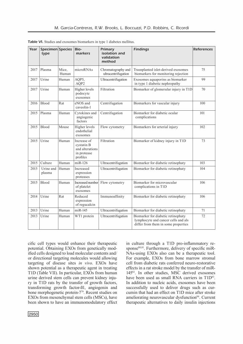
Table VI. Studies and exosomes biomarkers in type 1 diabetes mellitus.
Year Specimen Species Bio- Primary Findings References type markers isolation and validation method
2017 Plasma Mice, microRNAs Chromatography and Trasnplanted islet-derived exosomes 75 Human ultracentrifugation biomarkers for monitoring rejection2017 Urine Human AQP5, Ultracentrifugation Exosomes aquaporins as biomarker 99 AQP2 in type 1 diabetic nephropathy2017 Urine Human Higher levels Filtration Biomarker of glomerular injury in T1D 70 podocyte exosomes 2016 Blood Rat eNOS and Centrifugation Biomarkers for vascular injury 100 caveolin-12015 Plasma Human Cytokines and Centrifugation Biomarker for diabetic ocular 101 angiogenic complications factors 2015 Blood Mouse Higher levels Flow cytometry Biomarkers for arterial injury 102 endothelial exosomes 2015 Urine Human Increase of Filtration Biomarker of kidney injury in T1D 73 cystatin B and alterations in protease profiles2015 Culture Human miR-126 Ultracentrifugation Biomarker for diabetic retinophaty 1032015 Urine and Human Increased Ultracentrifugation Biomarker for diabetic retinophaty 104 plasma expression proteases 2015 Blood Human Increased number Flow cytometry Biomarker for microvascular 106 of platelet complications in T1D exosomes2014 Urine Rat Reduced Immunoaffinity Biomarker for diabetic retinophaty 106 expression of regucalcin2013 Urine Human miR-145 Ultracentrifugation Biomarker for diabetic retinophaty 712013 Urine Human WT1 protein Ultracentrifugation Biomarker for diabetic retinophaty 72 lymphocyte and cancer cells and als differ from them in some properties

Exosomes in type 1 diabetes mellitus
2951
Figure 4. Biomarkers and opportunities for T1D prevention and intervention.
Table VII. Studies showing therapeutic applications of exosomes in type 1 diabetes mellitus.
Year Mechanism Findings References
2017 Bone marrow exosomes transfer Improve hyperglycemia 107 miR-106b and miR-222 2016 Urine-derived stem cells transfer VEGF, Polyuria improved in vivo and prevent 79 TGF-β1, angiogenin and BMP-7 apoptosis in kidney cells 2016 Bone-marrow stromal cells derived Neurorestorative effects in T1D 82 exosomes secretion of miR-145 stroke rats2016 Human endothelial progenitor exosomes Acceleration cutaneous wound healing in diabetes 108, 1092016 Mesenchymal stem cell derived Improve islet transplantation 83 exosomes as anti-miR-375 carriers 2016 Bone marrow-derived exosomes Repairing diabetes-induced damaged 110 neurons and astrocytes 2016 Hsp20-engineered exosomes Therapeutic agent for diabetic cardiomyopathy 1112016 miR-let7-engineered MSC exosomes Attenuate renal fibrosis in T1D 1122016 Urine-derived stem cells Prevention of kidney complications in T1D 792015 Murine pancreatic ß-cell line exosomes Differentiation of bone marrow cells 85 into insulin-producing cells in the subcutaneous Matrigel platforms 2015 Fibrocyte derived EXOs release hsp90α, Treatment of diabetic ulcers 113 total and activated signal transducer and activator of transcription 3, proangiogenic (miR-126, miR-130a, miR-132) and anti- inflammatory (miR124a, miR-125b) microRNAs, and a microRNA regulating collagen deposition (miR-21).

M. Garcia-Contreras, R.W. Brooks, L. Boccuzzi, P.D. Robbins, C. Ricordi
2952
for T1D are invasive organ transplant procedures, only available for the most severe cases of T1D due to significant complications associated with the procedure. Nevertheless, clinical islet cell trans-plantation has emerged as a less invasive thera-peutic alternative to whole pancreas transplant for the treatment of the most severe cases of T1D. In combination with the islet transplantation setting, exosomes may represent an exciting new therapy not only for improvement of islet function83, but also for the induction of transplant tolerance81 and as alternative source to obtain insulin-producing ß-cells as previously shown85.
Conclusions
EXOs provide a potential source of cell or tissue type specific biomarkers for the diagnosis, treatment and evaluation of disease progression of T1D. Further characterization of EXOs, their mechanisms of action and their relationship with different stages of disease will provide the basis for a better understanding of the role of EXOs in the pathogenesis of T1D and their possible use as therapeutic tools.
Acknowledgements: This work was supported by the Diabetes Research Institute Foundation, Miami, FL, USA.
Declaration of interest:The authors declare no competing financial interests.
References
1) Sattar N. Biomarkers for diabetes prediction, pathogenesis or pharmacotherapy guidance? Past, present and future possibilities. Diabet Med 2012; 29: 5-13.
2) Valadi H, Ekstrom K, Bossios A, Sjostrand M, Lee JJ, Lotvall JO. Exosome-mediated transfer of mRNAs and microRNAs is a novel mechanism of genetic exchange between cells. Nat Cell Biol 2007; 9: 654-659.
3) thakur Bk, ZhaNg h, Becker a, Matei i, huaNg Y, coSta-Silva B, ZheNg Y, hoShiNo a, BraZier h, XiaNg J, WilliaMS c, rodrigueZ-Barrueco r, Silva JM, ZhaNg W, hearN S, eleMeNto o, PakNeJad N, MaNova-todorova k, Welte k, BroMBerg J, PeiNado h, lYdeN d. Double-stranded DNA in exosomes: a novel biomarker in cancer detection. Cell Res 2014; 24: 766-769.
4) cheNg l, SharPleS ra, ScicluNa BJ, hill aF. Exosomes provide a protective and enriched source of miRNA for biomarker profiling com-pared to intracellular and cell-free blood. J Extracell Vesicles 2014; 3. doi: 10.3402/jev.v3.23743. eCollection 2014.
5) Skog J, WurdiNger t, vaN riJN S, MeiJer dh, gaiNche l, SeNa-eSteveS M, currY Wt, Jr., carter BS, krichevSkY aM, BreakeField Xo. Glioblastoma mi-crovesicles transport RNA and proteins that pro-mote tumour growth and provide diagnostic bio-markers. Nat Cell Biol 2008; 10: 1470-1476.
6) raPoSo g, NiJMaN hW, Stoorvogel W, leiJeNdekker r, hardiNg cv, MelieF cJM, geuZe hJ. B lympho-cytes secrete antigen-presenting vesicles. J Exp Med 1996; 183: 1161-1172.
7) MuralidharaN-chari v, claNcY JW, SedgWick a, d'SouZa-SchoreY c. Microvesicles: mediators of extracellular communication during cancer pro-gression. J Cell Sci 2010; 123: 1603-1611.
8) rataJcZak J, WYSocZYNSki M, haYek F, JaNoWSka-WiecZorek a, rataJcZak MZ. Membrane-derived microvesicles: important and underappreciated mediators of cell-to-cell communication. Leukemia 2006; 20: 1487-1495.
9) iZquierdo-uSeroS N, PuertaS Mc, BorraS Fe, BlaNco J, MartiNeZ-Picado J. Exosomes and retroviruses: the chicken or the egg? Cell Microbiol 2011; 13: 10-17.
10) SchWaB a, MeYeriNg SS, lePeNe B, iordaNSkiY S, vaN hoek Ml, hakaMi rM, kaShaNchi F. Extracellular vesicles from infected cells: potential for direct pathogenesis. Front Microbiol 2015; 6: 1132.
11) elMore S. Apoptosis: a review of programmed cell death. Toxicol Pathol 2007; 35: 495-516.
12) cai J, Wu g, JoSe Pa, ZeNg c. Functional trans-ferred DNA within extracellular vesicles. Exp Cell Res 2016; 349: 179-183.
13) lakhter aJ, SiMS ek. Minireview: emerging roles for extracellular vesicles in diabetes and related met-abolic disorders. Mol Endocrinol 2015; 29: 1535-1548.
14) graNt Bd, doNaldSoN Jg. Pathways and mecha-nisms of endocytic recycling. Nat Rev Mol Cell Biol 2009; 10: 597-608.
15) raiBorg c, SteNMark h. The ESCRT machinery in endosomal sorting of ubiquitylated membrane proteins. Nature 2009; 458: 445-452.
16) Stoorvogel W, StrouS gJ, geuZe hJ, oorSchot v, SchWartZ al. Late endosomes derive from early endosomes by maturation. Cell 1991; 65: 417-427.
17) adell Ma, vogel gF, Pakdel M, Muller M, liNdNer h, heSS MW, teiS d. Coordinated binding of Vps4 to ESCRT-III drives membrane neck constriction during MVB vesicle formation. J Cell Biol 2014; 205: 33-49.
18) PolS MS, kluMPerMaN J. Trafficking and function of the tetraspanin CD63. Exp Cell Res 2009; 15: 1584-1592.
19) Wollert t, hurleY Jh. Molecular mechanism of multivesicular body biogenesis by ESCRT com-plexes. Nature 2010; 464: 864-U873.
20) BoBrie a, coloMBo M, raPoSo g, thérY c. Exosome secretion: molecular mechanisms and roles in immune responses. Traffic 2011; 12: 1659-1668.
Exosomes in type 1 diabetes mellitus
2953
21) kiM dk, lee J, SiMPSoN rJ, lotvall J, gho YS. EVpedia: a community web resource for prokary-otic and eukaryotic extracellular vesicles re-search. Semin Cell Dev Biol 2015; 40: 4-7.
22) keerthikuMar S, chiSaNga d, ariYaratNe d, al SaFFar h, aNaNd S, Zhao k, SaMuel M, PathaN M, JoiS M, chilaMkurti N, gaNgoda l, MathivaNaN S. ExoCarta: a web-based compendium of exosomal cargo. J Mol Biol 2016; 428: 688-692.
23) kalra h, SiMPSoN rJ, Ji h, aikaWa e, altevogt P, aSkeNaSe P, BoNd vc, BorràS Fe, BreakeField X, BudNik v, BuZaS e, caMuSSi g, claYtoN a, cocucci e, FalcoN-PereZ JM, gaBrielSSoN S, gho YS, guPta d, harSha hc, heNdriX a, hill aF, iNal JM, JeNSter g, kräMer-alBerS eM, liM Sk, lloreNte a, lötvall J, Marcilla a, MiNcheva-NilSSoN l, NaZareNko i, NieuWlaNd r, Nolte-'t hoeN eN, PaNdeY a, Patel t, PiPer Mg, PluchiNo S, PraSad tS, raJeNdraN l, raPoSo g, record M, reid ge, SáNcheZ-Madrid F, SchiFFelerS rM, SilJaNder P, SteNSBalle a, Stoorvogel W, taYlor d, therY c, valadi h, vaN BalkoM BW, váZqueZ J, vidal M, WauBeN Mh, YáñeZ-Mó M, Zoeller M, MathivaNaN S. Vesiclepedia: a compen-dium for extracellular vesicles with continuous community annotation. PLoS Biol 2012; 10: e1001450.
24) PocSFalvi g, StaNlY c, vilaSi a, FiuMe i, caPaSSo g, turiak l, BuZaS ei, vekeY k. Mass spectrometry of extracellular vesicles. Mass Spectrom Rev 2016; 35: 3-21.
25) FitZNer d, SchNaarS M, vaN roSSuM d, kriShNaMoorthY g, diBaJ P, Bakhti M, regeN t, haNiSch uk, SiMoNS M. Selective transfer of exosomes from oligoden-drocytes to microglia by macropinocytosis. J Cell Sci 2011; 124: 447-458.
26) huaNg X, YuaN t, tSchaNNeN M, SuN Z, JacoB h, du M, liaNg M, dittMar rl, liu Y, liaNg M, kohli M, thiBodeau SN, BoardMaN l, WaNg l. Characterization of human plasma-derived exosomal RNAs by deep sequencing. BMC Genomics 2013; 14: 319.
27) Batagov ao, kuZNetSov va, kurochkiN iv. Identification of nucleotide patterns enriched in secreted RNAs as putative cis-acting elements targeting them to exosome nano-vesicles. BMC Genomics 2011; 12 Suppl 3: S18.
28) villarroYa-Beltri c, gutierreZ-vaZqueZ c, SaNcheZ-caBo F, PereZ-herNaNdeZ d, vaZqueZ J, MartiN-coFreceS N, MartiNeZ-herrera dJ, PaScual-MoNtaNo a, MittelBruNN M, SaNcheZ-Madrid F. Sumoylated hnRNPA2B1 controls the sorting of miRNAs into exosomes through binding to specific motifs. Nat Commun 2013; 4: 2980.
29) raMachaNdraN S, PalaNiSaMY v. Horizontal transfer of RNAs: exosomes as mediators of intercellular communication. Wiley Interdiscip Rev RNA 2012; 3: 286-293.
30) laZaro-iBaNeZ e, SaNZ-garcia a, viSakorPi t, eScoBedo-lucea c, SilJaNder P, aYuSo-Sacido a, YliPerttula M. Different gDNA content in the subpopulations of prostate cancer extracellular vesicles: apoptotic bodies, microvesicles, and exosomes. Prostate 2014; 74: 1379-1390.
31) roNquiSt kg, roNquiSt g, carlSSoN l, larSSoN a. Human prostasomes contain chromosomal DNA. Prostate 2009; 69: 737-743.
32) gueSciNi M, guidoliN d, valloraNi l, caSadei l, gioacchiNi aM, tiBollo P, BattiStelli M, Falcieri e, BattiStiN l, agNati lF, Stocchi v. C2C12 myoblasts release micro-vesicles containing mtDNA and proteins involved in signal transduction. Exp Cell Res 2010; 316: 1977-1984.
33) kahlert c, Melo Sa, ProtoPoPov a, taNg J, Seth S, koch M, ZhaNg J, WeitZ J, chiN l, Futreal a, kalluri r. Identification of double-stranded genomic DNA span-ning all chromosomes with mutated KRAS and p53 DNA in the serum exosomes of patients with pancre-atic cancer. J Biol Chem 2014; 289: 3869-3875.
34) coSta-Silva B, aiello NM, oceaN aJ, SiNgh S, ZhaNg h, thakur Bk, Becker a, hoShiNo a, Mark Mt, MoliNa h, XiaNg J, ZhaNg t, theileN tM, garcía-SaNtoS g, WilliaMS c, ararSo Y, huaNg Y, rodrigueS g, SheN tl, laBori kJ, lothe iM, kure eh, herNaNdeZ J, douSSot a, eBBeSeN Sh, graNdgeNett PM, holliNgSWorth Ma, JaiN M, MallYa k, Batra Sk, JarNagiN Wr, SchWartZ re, Matei i, PeiNado h, StaNger BZ, BroMBerg J, lYdeN d. Pancreatic cancer exosomes initiate pre-metastatic niche formation in the liver. Nat Cell Biol 2015; 17: 816-826.
35) therY c, aMigoreNa S, raPoSo g, claYtoN a. Isolation and characterization of exosomes from cell cul-ture supernatants and biological fluids. Curr Protoc Cell Biol 2006; Chapter 3: Unit 3.22.
36) gaMeZ-valero a, MoNguio-tortaJada M, carreraS-PlaNella l, FraNqueSa M, BeYer k, BorraS Fe. Size-exclusion chromatography-based isolation minimally alters extracellular vesicles' characteristics com-pared to precipitating agents. Sci Rep 2016; 6: 33641.
37) MerchaNt Ml, PoWell dW, WilkeY dW, cuMMiNS td, deegeNS Jk, rood iM, McaFee kJ, FleiScher c, kleiN e, kleiN JB. Microfiltration isolation of human uri-nary exosomes for characterization by MS. Proteomics Clin Appl 4: 84-96.
38) rider Ma, hurWitZ SN, MeckeS dg, Jr. ExtraPEG: a polyethylene glycol-based method for enrichment of extracellular vesicles. Sci Rep 2016; 6: 23978.
39) kaNWar SS, duNlaY cJ, SiMeoNe dM, Nagrath S. Microfluidic device (ExoChip) for on-chip isola-tion, quantification and characterization of circu-lating exosomes. Lab Chip 2014; 14: 1891-1900.
40) gYorgY B, SZaBó tg, PáSZtói M, Pál Z, MiSJák P, aradi B, láSZló v, PálliNger e, PaP e, kittel a, NagY g, FaluS a, BuZáS ei. Membrane vesicles, current state-of-the-art: emerging role of extracellular vesicles. Cell Mol Life Sci 2011; 68: 2667-2688.
41) SharMa S, raSool hi, PalaNiSaMY v, MathiSeN c, SchMidt M, WoNg dt, giMZeWSki Jk. Structural-mechanical characterization of nanoparticle exo-somes in human saliva, using correlative AFM, FESEM, and force spectroscopy. ACS Nano 2010; 4: 1921-1926.
42) ueda k, iShikaWa N, tatSuguchi a, Saichi N, FuJii r, NakagaWa h. Antibody-coupled monolithic silica microtips for highthroughput molecular profiling of circulating exosomes. Sci Rep 2014; 4: 6232.
43) dragovic ra, gardiNer c, BrookS aS, taNNetta dS, FerguSoN dJ, hole P, carr B, redMaN cW, harriS al, doBSoN PJ, harriSoN P, SargeNt il. Sizing and phenotyping of cellular vesicles using nanoparti-cle tracking analysis. Nanomedicine 2011; 7: 780-788.
M. Garcia-Contreras, R.W. Brooks, L. Boccuzzi, P.D. Robbins, C. Ricordi
2954
44) MarcouX g, ducheZ ac, cloutier N, ProvoSt P, Nigrovic Pa, Boilard e. Revealing the diversity of extracellular vesicles using high-dimensional flow cytometry analyses. Sci Rep 2016; 6: 35928.
45) laNNigaN J, erdBruegger u. Imaging flow cytometry for the characterization of extracellular vesicles. Methods 2017; 112: 55-67.
46) hardiNg cv, heuSer Je, Stahl Pd. Exosomes: look-ing back three decades and into the future. J Cell Biol 2013; 200: 367-371.
47) hardiNg c, heuSer J, Stahl P. Receptor-mediated endocytosis of transferrin and recycling of the transferrin receptor in rat reticulocytes. J Cell Biol 1983; 97: 329-339.
48) PaN Bt, JohNStoNe rM. Fate of the transferrin re-ceptor during maturation of sheep reticulocytes in vitro: selective externalization of the receptor. Cell 1983; 33: 967-978.
49) FiglioliNi F, caNtaluPPi v, de leNa M, BeltraMo S, roMagNoli r, SaliZZoNi M, MelZi r, NaNo r, PieMoNti l, tetta c, BiaNcoNe l, caMuSSi g. Isolation, char-acterization and potential role in beta cell-endo-thelium cross-talk of extracellular vesicles re-leased from human pancreatic islets. PLoS One 2014; 9: e102521.
50) kliBi J, Niki t, riedel a, Pioche-durieu c, Souquere S, ruBiNSteiN e, le Moulec S, guigaY J, hiraShiMa M, gueMira F, adhikarY d, MautNer J, BuSSoN P. Blood diffusion and Th1-suppressive effects of galec-tin-9-containing exosomes released by Epstein-Barr virus-infected nasopharyngeal carcinoma cells. Blood 2009; 113: 1957-1966.
51) kalaMvoki M, du t, roiZMaN B. Cells infected with herpes simplex virus 1 export to uninfected cells exosomes containing STING, viral mRNAs, and microRNAs. Proc Natl Acad Sci U S A 2014; 111: E4991-4996.
52) rahMaN MJ, regN d, BaShratYaN r, dai Yd. Exosomes released by islet-derived mesenchy-mal stem cells trigger autoimmune responses in NOD mice. Diabetes 2014; 63: 1008-1020.
53) caBrera o, BerMaN dM, keNYoN NS, ricordi c, BerggreN Po, caicedo a. The unique cytoarchitec-ture of human pancreatic islets has implications for islet cell function. Proc Natl Acad Sci U S A 2006; 103: 2334-2339.
54) WieruP N, SuNdler F, heller rS. The islet ghrelin cell. J Mol Endocrinol 2014; 52: R35-49.
55) koh dS, cho Jh, cheN l. Paracrine interactions within islets of Langerhans. J Mol Neurosci 2012; 48: 429-440.
56) BeNNet WM, WaNg Zl, JoNeS PM, WaNg rM, JaMeS rF, loNdoN NJ, ghatei Ma, BlooM Sr. Presence of neuropeptide Y and its messenger ribonucleic acid in human islets: evidence for a possible paracrine role. J Clin Endocrinol Metab 1996; 81: 2117-2120.
57) rodrigueZ-diaZ r, MeNegaZ d, caicedo a. Neurotransmitters act as paracrine signals to regulate insulin secretion from the human pancre-atic islet. J Physiol 2014; 592: 3413-3417.
58) villarroYa-Beltri c, gutierreZ-vaZqueZ c, SaNcheZ-Madrid F, MittelBruNN M. Analysis of microRNA and protein transfer by exosomes during an immune synapse. Methods Mol Biol 2013; 1024: 41-51.
59) guaY c, MeNoud v, roMe S, regaZZi r. Horizontal transfer of exosomal microRNAs transduce apop-totic signals between pancreatic beta-cells. Cell Commun Signal 2015; 13: 17.
60) lee hS, JeoNg J, lee kJ. Characterization of vesi-cles secreted from insulinoma NIT-1 cells. J Proteome Res 2009; 8: 2851-2862.
61) PalMiSaNo g, JeNSeN SS, le BihaN Mc, laiNe J, Mcguire JN, Pociot F, larSeN Mr. Characterization of membrane-shed microvesicles from cyto-kine-stimulated beta-cells using proteomics strat-egies. Mol Cell Proteomics 2012; 11: 230-243.
62) ciaNciaruSo c, PhelPS ea, PaSquier M, haMeliN r, deMurtaS d, aliBaShe ahMed M, PieMoNti l, hiroSue S, SWartZ Ma, de PalMa M, huBBell Ja, BaekkeSkov S. Primary human and rat beta-cells release the intracellular autoantigens GAD65, IA-2, and pro-insulin in exosomes together with cytokine-in-duced enhancers of immunity. Diabetes 2017; 66: 460-473.
63) Zhu q, kaNg J, Miao h, FeNg Y, Xiao l, hu Z, liao dF, huaNg Y, JiN J, he S. Low-dose cytokine-in-duced neutral ceramidase secretion from INS-1 cells via exosomes and its anti-apoptotic effect. FEBS J 2014; 281: 2861-2870.
64) WaNg Z, Xie Z, lu q, chaNg c, Zhou Z. Beyond genetics: what causes type 1 diabetes. Clin Rev Allergy Immunol 2017; 52: 273-286.
65) McgiNtY JW, Marre Ml, BaJZik v, PigaNelli Jd, JaMeS ea. T cell epitopes and post-translationally mod-ified epitopes in type 1 diabetes. Curr Diab Rep 2015; 15-90.
66) deloNg t, WileS ta, Baker rl, BradleY B, BarBour g, reiSdorPh r, arMStroNg M, PoWell rl, reiSdorPh N, kuMar N, elSo cM, deNicola M, BottiNo r, PoWerS ac, harlaN dM, keNt Sc, MaNNeriNg Si, haSkiNS k. Pathogenic CD4 T cells in type 1 dia-betes recognize epitopes formed by peptide fu-sion. Science 2016; 351: 711-714.
67) SheNg h, haSSaNali S, NugeNt c, WeN l, haMiltoN-WilliaMS e, diaS P, dai Yd. Insulinoma-released exosomes or microparticles are immunostimula-tory and can activate autoreactive T cells spon-taneously developed in nonobese diabetic mice. J Immunol 2011; 87: 1591-1600.
68) BaShratYaN r, SheNg h, regN d, rahMaN MJ, dai Yd. Insulinoma-released exosomes activate autore-active marginal zone-like B cells that expand endogenously in prediabetic NOD mice. Eur J Immunol 2013; 43: 2588-2597.
69) voMuNd aN, ZiNSelMeYer Bh, hugheS J, calderoN B, valderraMa c, FerriS St, WaN X, kaNekura k, carrero Ja, uraNo F, uNaNue er. Beta cells transfer vesi-cles containing insulin to phagocytes for presen-tation to T cells. Proc Natl Acad Sci U S A 2015; 112: E5496-5502.
70) lYtvYN Y, Xiao F, keNNedY cr, PerkiNS Ba, reich hN, ScholeY JW, cherNeY dZ, Burger d. Assessment of urinary microparticles in normotensive patients with type 1 diabetes. Diabetologia 2017; 60: 581-584.
71) Barutta F, tricarico M, corBelli a, aNNaratoNe l, PiNach S, griMaldi S, BruNo g, ciMiNo d, taverNa d, deregiBuS Mc, raStaldi MP, PeriN Pc, grudeN g. Urinary exosomal microRNAs in incipient diabetic nephropathy. PLoS One 2013; 8: e73798.

Exosomes in type 1 diabetes mellitus
2955
72) kalaNi a, MohaN a, godBole MM, Bhatia e, guPta a, SharMa rk, tiWari S. Wilm's tumor-1 protein levels in urinary exosomes from diabetic patients with or without proteinuria. PLoS One 2013; 8: e60177.
73) MuSaNte l, tataruch d, gu d, liu X, ForSBloM c, grooP Ph, holthoFer h. Proteases and protease inhibitors of urinary extracellular vesicles in dia-betic nephropathy. J Diabetes Res 2015; 2015: 289734.
74) katoMe t, NaMekata k, MitaMura Y, SeMBa k, egaWa M, Naito t, harada c, harada t. Expression of intraocular peroxisome proliferator-activated re-ceptor gamma in patients with proliferative dia-betic retinopathy. J Diabetes Complications 2015; 29: 275-281.
75) vallaBhaJoSYula P, korutla l, haBertheuer a, Yu M, roStaMi S, YuaN cX, reddY S, liu c, korutla v, koeBerleiN B, troFe-clark J, rickelS Mr, NaJi a. Tissue-specific exosome biomarkers for noninva-sively monitoring immunologic rejection of trans-planted tissue. J Clin Invest 2017; 127: 1375-1391.
76) cai J, haN Y, reN h, cheN c, he d, Zhou l, eiSNer gM, aSico ld, JoSe Pa, ZeNg c. Extracellular ves-icle-mediated transfer of donor genomic DNA to recipient cells is a novel mechanism for genetic influence between cells. J Mol Cell Biol 2013; 5: 227-238.
77) Saari h, laZaro-iBaNeZ e, viitala t, vuoriMaa-laukkaNeN e, SilJaNder P, YliPerttula M. Microvesicle- and exosome-mediated drug delivery enhances the cytotoxicity of Paclitaxel in autologous pros-tate cancer cells. J Control Release 2015; 220: 727-737.
78) ohNo S, takaNaShi M, Sudo k, ueda S, iShikaWa a, MatSuYaMa N, FuJita k, MiZutaNi t, ohgi t, ochiYa t, gotoh N, kuroda M. Systemically injected exo-somes targeted to EGFR deliver antitumor mi-croRNA to breast cancer cells. Mol Ther 2013; 21: 185-191.
79) JiaNg ZZ, liu YM, Niu X, YiN JY, hu B, guo Sc, FaN Y, WaNg Y, WaNg NS. Exosomes secreted by hu-man urine-derived stem cells could prevent kid-ney complications from type I diabetes in rats. Stem Cell Res Ther 2016; 7: 24.
80) Favaro e, carPaNetto a, laMorte S, FuSco a, caorSi c, deregiBuS Mc, BruNo S, aMoroSo a, giovarelli M, Porta M, PeriN Pc, tetta c, caMuSSi g, ZaNoNe MM. Human mesenchymal stem cell-derived mi-crovesicles modulate T cell response to islet an-tigen glutamic acid decarboxylase in patients with type 1 diabetes. Diabetologia 2014; 57: 1664-1673.
81) Favaro e, carPaNetto a, caorSi c, giovarelli M, aNgeliNi c, cavallo-PeriN P, tetta c, caMuSSi g, ZaNoNe MM. Human mesenchymal stem cells and derived extracellular vesicles induce regulatory dendritic cells in type 1 diabetic patients. Diabetologia 2016; 59: 325-333.
82) cui c, Ye X, choPP M, veNkat P, Zacharek a, YaN t, NiNg r, Yu P, cui g, cheN J. miR-145 regulates diabetes-bone marrow stromal cell-induced neu-rorestorative effects in diabetes stroke rats. Stem Cells Transl Med 2016; 5: 1656-1667.
83) WeN d, PeNg Y, liu d, WeiZMaNN Y, Mahato ri. Mesenchymal stem cell and derived exosome as small RNA carrier and Immunomodulator to im-prove islet transplantation. J Control Release 2016; 238: 166-175.
84) aNuradha kalaNi PkaNt. Curcumin-encapsulated stem cell exosomes mitigates neuro-vascular mi-tochondrial dysfunction after stroke in T1DM Mice. FASEB J 2015; .
85) oh k, kiM Sr, kiM dk, Seo MW, lee c, lee hM, oh Je, choi eY, lee dS, gho YS, Park kS. In vivo differ-entiation of therapeutic insulin-producing cells from bone marrow cells via extracellular vesicle-mimetic nanovesicles. ACS Nano 2015; 9: 11718-11727.
86) WaldeNStroM a, geNNeBack N, hellMaN u, roNquiSt g. Cardiomyocyte microvesicles contain DNA/RNA and convey biological messages to target cells. PLoS One 2012; 7: e34653.
87) cocucci e, MeldoleSi J. Ectosomes and exosomes: shedding the confusion between extracellular vesicles. Trends Cell Biol 2015; 25: 364-372.
88) heSS c, Sadallah S, heFti a, laNdMaNN r, SchiFFerli Ja. Ectosomes released by human neutrophils are specialized functional units. J Immunol 1999; 163: 4564-4573.
89) NielSeN Mh, Beck-NielSeN h, aNderSeN MN, haNdBerg a. A flow cytometric method for characterization of circulating cell-derived microparticles in plas-ma. J Extracell Vesicles 2014; 3. doi: 10.3402/jev.v3.20795. eCollection 2014.
90) MiNciacchi vr, FreeMaN Mr, di viZio d. Extracellular vesicles in cancer: exosomes, microvesicles and the emerging role of large oncosomes. Semin Cell Dev Biol 2015; 40: 41-51.
91) taraZoNa r, delgado e, guarNiZo Mc, roNcero rg, Morgado S, SáNcheZ-correa B, gordillo JJ, deJuliáN J, caSado Jg. Human prostasomes express CD48 and interfere with NK cell function. Immunobiology 2011; 216: 41-46.
92) karlSSoN M, luNdiN S, dahlgreN u, kahu h, PetterSSoN i, teleMo e. "Tolerosomes" are pro-duced by intestinal epithelial cells. Eur J Immunol 2001;31: 2892-900.
93) Maguire ca, BalaJ l, SivaraMaN S, croMMeNtuiJN Mh, ericSSoN M, MiNcheva-NilSSoN l, BaraNov v, giaNNi d, taNNouS Ba, SeNa-eSteveS M, BreakeField Xo, Skog J. Microvesicle-associated AAV vector as a novel gene delivery system. Mol Ther 2012; 20: 960-971.
94) alvareZ Ml, khoSroheidari M, kaNchi ravi r, diSteFaNo Jk. Comparison of protein, microRNA, and mRNA yields using different methods of urinary exosome isolation for the discovery of kidney disease bio-markers. Kidney Int 2012; 82: 1024-1032.
95) gholiZadeh S, Shehata draZ M, ZarghooNi M, SaNati-NeZhad a, ghavaMi S, ShaFiee h, akBari M. Microfluidic approaches for isolation, detection, and characterization of extracellular vesicles: Current status and future directions. Biosens Bioelectron 2017; 9: 588-605.
96) Sokolova v, ludWig ak, horNuNg S, rotaN o, horN Pa, ePPle M, gieBel B. Characterisation of exo-somes derived from human cells by nanoparticle tracking analysis and scanning electron micros-copy. Colloids Surf B Biointerfaces 2011; 87: 146-150.

M. Garcia-Contreras, R.W. Brooks, L. Boccuzzi, P.D. Robbins, C. Ricordi
2956
97) laNNigaN J, erdBruegger u. Imaging flow cytometry for the characterization of extracellular vesicles. Methods 2017; 112: 55-67.
98) SalaMa a, Fichou N, allard M, duBreil l, de BeaurePaire l, viel a, Jégou d, BöSch S, Bach JM. MicroRNA-29b modulates innate and anti-gen-specific immune responses in mouse mod-els of autoimmunity. PLoS One 2014; 9: e106153.
99) roSSi l, Nicoletti Mc, carMoSiNo M, MaStroFraNceSco l, di FraNco a, iNdrio F, lella r, laviola l, giorgiNo F, Svelto M, geSualdo l, ProciNo g. Urinary excre-tion of kidney aquaporins as possible diagnostic biomarker of diabetic nephropathy. J Diabetes Res 2017; 2017: 4360357.
100) iShida k, taguchi k, hida M, WataNaBe S, kaWaNo k, MatSuMoto t, hattori Y, koBaYaShi t. Circulating microparticles from diabetic rats impair endothe-lial function and regulate endothelial protein ex-pression. Acta Physiol 2016; 216: 211-220.
101) tokarZ a, SZuścik i, kuśNierZ-caBala B, kaPuSta M, koNkoleWSka M, ŻurakoWSki a, georgeScu a, StęPień e. Extracellular vesicles participate in the trans-port of cytokines and angiogenic factors in dia-betic patients with ocular complications. Folia Med Cracov 2015; 55: 35-48.
102) WaNg Z, eMoNd ZM, FlYNN Me, SWaMiNathaN S, kiBBe Mr. Microparticle levels after arterial injury and NO therapy in diabetes. J Surg Res 2016; 200: 722-731.
103) MaZZeo a, BeltraMo e, iavello a, carPaNetto a, Porta M. Molecular mechanisms of extracellular vesicle-induced vessel destabilization in diabetic retinopathy. Acta Diabetol 2015; 52: 1113-1119.
104) aNderSeN h, FriiS ug, haNSeN PB, SveNNiNgSeN P, heNrikSeN Je, JeNSeN Bl. Diabetic nephropathy is associated with increased urine excretion of pro-teases plasmin, prostasin and urokinase and activation of amiloride-sensitive current in col-lecting duct cells. Nephrol Dial Transplant 2015; 30: 781-789.
105) SaleM Ma, adlY aa, iSMail ea, darWiSh YW, kaMel ha. Platelets microparticles as a link between micro- and macro-angiopathy in young patients with type 1 diabetes. Platelets 2015; 26: 682-688.
106) ZuBiri i, PoSada-aYala M, BeNito-MartiN a, Maroto aS, MartiN-loreNZo M, caNNata-ortiZ P, de la cueSta F, goNZaleZ-calero l, BarderaS Mg,FerNaNdeZ-FerNaNdeZ B, ortiZ a, vivaNco F, alvareZ-llaMaS g. Kidney tissue proteomics reveals regucalcin downregulation in response to diabetic nephrop-athy with reflection in urinary exosomes. Transl Res 2015; 166: 474-484.e4.
107) tSukita S, YaMada t, takahaShi k, MuNakata Y, hoSaka S, takahaShi h, gao J, Shirai Y, kodaMa S, aSai Y, SugiSaWa t, chiBa Y, kaNeko k, uNo k, SaWada S, iMai J, katagiri h. MicroRNAs 106b and 222 improve hyperglycemia in a mouse model of insulin-defi-cient diabetes via pancreatic beta-cell prolifera-tion. EBioMedicine 2017; 15: 163-172.
108) ZhaNg J, cheN c, hu B, Niu X, liu X, ZhaNg g, ZhaNg c, li q, WaNg Y. Exosomes derived from human endothelial progenitor cells accelerate cutaneous wound healing by promoting angio-genesis through Erk1/2 signaling. Int J Biol Sci 2016; 12: 1472-1487.
109) li X, JiaNg c, Zhao J. Human endothelial progen-itor cells-derived exosomes accelerate cutane-ous wound healing in diabetic rats by promoting endothelial function. J Diabetes Complications 2016; 30: 986-992.
110) NakaNo M, NagaiShi k, koNari N, Saito Y, chikeNJi t, MiZue Y, FuJiMiYa M. Bone marrow-derived mesenchy-mal stem cells improve diabetes-induced cognitive impairment by exosome transfer into damaged neu-rons and astrocytes. Sci Rep 2016; 6: 24805.
111) WaNg X, gu h, huaNg W, PeNg J, li Y, YaNg l, qiN d, eSSaNdoh k, WaNg Y, PeNg t, FaN gc. Hsp20-mediated activation of exosome biogenesis in cardiomyocytes improves cardiac function and angiogenesis in dia-betic mice. Diabetes 2016 ;65: 3111-3128.
112) WaNg B, Yao k, huuSkeS BM, SheN hh, ZhuaNg J, godSoN c, BreNNaN eP, WilkiNSoN-Berka Jl, WiSe aF, ricardo Sd. Mesenchymal stem cells deliver ex-ogenous microrna-let7c via exosomes to attenu-ate renal fibrosis. Mol Ther 2016; 24: 1290-301.
113) geiger a, Walker a, NiSSeN e. Human fibrocyte-de-rived exosomes accelerate wound healing in genetically diabetic mice. Biochem Biophys Res Commun 2015; 467: 303-309.