Embed Size (px)
Citation preview

EXPERIMENT #8
CELL DIVISION: MITOSIS & MEIOSIS
Introduction
Cells, the basic unit of life, undergo reproductive acts to maintain the flow of genetic informationfrom parent to offspring. The processes of mitosis and meiosis are cellular events in which aparental cell will ultimately pass on its genetic information (encoded within genes onh ) d h ll h i i h i i f h i f i I d ’chromosomes) to daughter cells, thus insuring the continuation of that information. In today’s
lab, you will be observing mitosis, in which the daughter cells will contain the same number ofchromosomes as the parent cell. You will also be introduced to meiosis, in which the daughter
ll ill t i tl h th b f h th t llcells will contain exactly have the number of chromosomes as the parent cell.
Goals
At the end of this laboratory, you will:
1. Be familiar with the stages of both mitosis and meiosis
2. Be able to identify the stages of mitosis in both animal and plant cells via the microscope

EXPERIMENT #8
CELL DIVISION: MITOSIS & MEIOSIS
Experimental Procedure
A. Mitosis
Mitosis, the process of cell division, is actually a part of a much larger process called the CELLCYCLE. The cell cycle is composed of 4 stages: G1 (growth or gap), S (synthesis of DNA), G2CYCLE. The cell cycle is composed of 4 stages: G1 (growth or gap), S (synthesis of DNA), G2(growth or gap), and M (mitosis). In order for a parental cell to divide into two daughter cells, itmust be “prepared” for this division. The growth phases occur both before and after the DNAsynthesis stage and are involved in producing the necessary cellular components for the act ofy g p g y pdivision. The S phase is involved in duplicating the DNA so that each daughter cell will receiveone set of chromosomes. The stages of G1, S, and G2 are collectively referred to asINTERPHASE. As soon as the cell exits G2, mitosis begins and the following events occur:Prophase, Metaphase, Anaphase, and Telophase. These are the four stages of mitosis, and veryspecific cellular events occur during each stage to insure that the cell divides properly. Thefollowing slides will illustrate and characterize each of these four stages of mitosis, as well as thestarting interphase stage.

EXPERIMENT #8
CELL DIVISION: MITOSIS & MEIOSIS
Experimental Procedure
A. Mitosis (continued)
1. Interphase
G1 S d G2 i h Thi i i d f h d DNA d li iG1, S, and G2 stages compose interphase. This stage is a period of growth and DNA duplication.Visually, the cell looks like a typical cell with a defined cell and nuclear membrane. The contentsof the nucleus are diffuse and appear to contain millions of stained dots (nuclear material). Thef ll i lid d i t i t h i b th i l ll ( hit fi h) d l t ll ( lli tfollowing slides depict interphase in both an animal cell (whitefish) and plant cell (allium roottip)

EXPERIMENT #8
CELL DIVISION: MITOSIS & MEIOSIS
Whitefish
Nuclear membraneCell membrane

EXPERIMENT #8
CELL DIVISION: MITOSIS & MEIOSIS
Alium root tipAlium root tip
Nuclear membrane
Cell wall

EXPERIMENT #8
CELL DIVISION: MITOSIS & MEIOSIS
Experimental Procedure
A. Mitosis (continued)
1. Prophase
P h i h i d b h d i f h diff h i i i ibl “ d lik ”Prophase is characterized by the condensation of the diffuse chromatin into visible “strand-like”chromosomes. Even though you cannot visualize it, the condensed chromosomes are arranged assister chromatids attached at their centromeres. The cell pictured on the following slides do noth i t t l b A th h t i ti hi h i t i ibl th f ll ihave an intact nuclear membrane. Another characteristic which is not visible on the followingslides are the centrioles (only in animal cells), which migrate to opposite poles and will becomethe core of the the microtubule organizing center (MTOC)

EXPERIMENT #8
CELL DIVISION: MITOSIS & MEIOSIS
Condensed chromosomesCell membrane

EXPERIMENT #8
CELL DIVISION: MITOSIS & MEIOSIS
Condensed chromosomesCondensed chromosomes

EXPERIMENT #8
CELL DIVISION: MITOSIS & MEIOSIS
Experimental Procedure
A. Mitosis (continued)
1. Metaphase
Th i l h f i i id if i h i d b h “li i ” f h hThe simplest phase of mitosis to identify is characterized by the “lining up” of the chromosomes along the metaphase or equatorial plate. Spindle fibers may or may not be visible connecting the centromeres of the sister chromatids to opposite ends of the cell.

EXPERIMENT #8
CELL DIVISION: MITOSIS & MEIOSIS
Chromosomes
MTOC
Spindle fibersSpindle fibers

EXPERIMENT #8
CELL DIVISION: MITOSIS & MEIOSIS
Chromosomes

EXPERIMENT #8
CELL DIVISION: MITOSIS & MEIOSIS
Experimental Procedure
A. Mitosis (continued)
1. Anaphase
A h i h i d b h i f h i h id i i di id lAnaphase is characterized by the separation of the sister chromatids into individualchromosomes. Kinetochore fibers attached to the kinetochore region of the centromere facilitatesthe movement of the chromosomes toward opposite ends of the cell. The kinetochores ride theki t h fib lik t i il d t k b t th l th ki t h fib thkinetochore fibers like a train on railroad track, but as they move along the kinetochore fibers thekinetochores disassemble “destroy” the tracks that they have just passed over. This division ofthe nuclear material is known as karyokinesis.

EXPERIMENT #8
CELL DIVISION: MITOSIS & MEIOSIS
MTOC
Spindle fibers
Chromosomes

EXPERIMENT #8
CELL DIVISION: MITOSIS & MEIOSIS
Chromosomes

EXPERIMENT #8
CELL DIVISION: MITOSIS & MEIOSIS
Experimental Procedure
A. Mitosis (continued)
1. Telophase
T l h i h i d b h l i f h i i d h f i fTelophase is characterized by the completion of chromosome migration and the reformation ofthe nuclear membrane. Cytokinesis, the process of dividing the cytoplasm, began in anaphaseand is now leaving some definitive characteristics during telophase. In animal cells, the
f th l f i d i di ti th t th ll h t d t l h Thappearance of the cleavage furrow is a good indication that the cell has entered telophase. Thecleavage furrow is an invagination of the cell membrane at the point at which the cell will be splitat. In plant cell, the appearance of the cell plate (a precursor to the cell wall) is a characteristic oftelophasetelophase.

EXPERIMENT #8
CELL DIVISION: MITOSIS & MEIOSIS
Cleavage furrow
Chromosomes

EXPERIMENT #8
CELL DIVISION: MITOSIS & MEIOSIS
Cell plateChromosomes
EXPERIMENT #8
CELL DIVISION: MITOSIS & MEIOSIS
Experimental Procedure
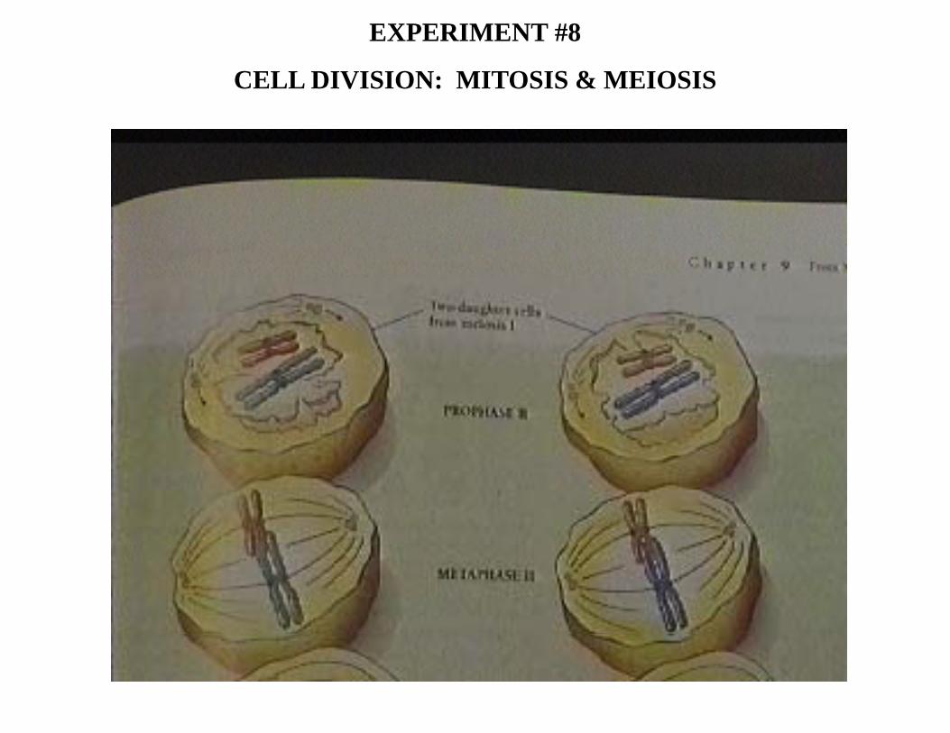
B. Meiosis
Meiosis is also a process by which there is a division of nuclear material, but instead ofproducing two daughter cells which contain the same number of chromosomes as the parent, fourproducing two daughter cells which contain the same number of chromosomes as the parent, fourhaploid daughter cells are produced. Each containing only half the number of chromosomes asthe parent. The reduction of the chromosome number in these sex cells or gametes is critical forthe process of sexual reproduction. When male and female gametes combine with each otherp p gduring fertilization, a zygote containing the proper number of chromosomes for that organismisproduced. This single fertilized egg will then undergo mitotic divisions to become a complexmulticellular organism with each cell containing the same genetic information encoded for by aset of chromosomes
Meiosis is divided into two steps: Meiosis I and Meiosis II. Each step of meiosis is divided intothe appropriate Prophase Metaphase Anaphase & Telophase stages with the number I or IIthe appropriate Prophase, Metaphase, Anaphase, & Telophase stages with the number I or IIfollowing it to identify meiosis I or meiosis II. Let us look at the stages of meiosis in orderbeginning with prophase I.

EXPERIMENT #8
CELL DIVISION: MITOSIS & MEIOSIS
Experimental Procedure
B. Meiosis - Prophase I
Prior to prophase I the cell completed a typical interphase stage where the cell grew and DNAwas synthesized. During prophase I, the same events that occurred in mitosis also occur here.was synthesized. During prophase I, the same events that occurred in mitosis also occur here.The chromatin condenses into visible chromosomes, the nuclear membrane disintegrates, andcentrioles migrate toward opposite poles. The unique event that occurs during prophase I issynapsis where homologous chromosomes line up and fuse together. This fusion allows they p g p gexchange of genetic material between two chromosomes in a process called crossing over. Thefollowing pictures are examples of prophase I and synapsis.

EXPERIMENT #8
CELL DIVISION: MITOSIS & MEIOSIS

EXPERIMENT #8
CELL DIVISION: MITOSIS & MEIOSIS

EXPERIMENT #8
CELL DIVISION: MITOSIS & MEIOSIS
Experimental Procedure
B. Meiosis - Metaphase I
During metaphase I, the pairs of homologous chromosomes line up on the metaphase plate. This is distinct from mitosis, where each replicated chromosome lines up by itself on the metaphaseis distinct from mitosis, where each replicated chromosome lines up by itself on the metaphase plate. This is due to the synapsis event during prophase I.

EXPERIMENT #8
CELL DIVISION: MITOSIS & MEIOSIS

EXPERIMENT #8
CELL DIVISION: MITOSIS & MEIOSIS
Experimental Procedure
B. Meiosis - Anaphase I
A pair of sister chromatids are separated from there homologous pair during anaphase I. Again this is different from mitosis in which individual sister chromatids are moved to opposite ends ofthis is different from mitosis in which individual sister chromatids are moved to opposite ends of the cell.

EXPERIMENT #8
CELL DIVISION: MITOSIS & MEIOSIS

EXPERIMENT #8
CELL DIVISION: MITOSIS & MEIOSIS
Experimental Procedure
B. Meiosis - Telophase I
Telophase I involves the division and separation of two daughter cells. The genetic make-up of these daughter cells is haploid. Even though they contain the same number of sister chromatidsthese daughter cells is haploid. Even though they contain the same number of sister chromatids that you would find in the original parent cell, chromosomes are counted based on the number of centromeres present in the cell. Since the sister chromatids are attached at the centromere, they contain only half the number of centromeres present in the parent cell and thus half the y p pchromosomes = haploid.

EXPERIMENT #8
CELL DIVISION: MITOSIS & MEIOSIS

EXPERIMENT #8
CELL DIVISION: MITOSIS & MEIOSIS

EXPERIMENT #8
CELL DIVISION: MITOSIS & MEIOSIS
Experimental Procedure
B. Meiosis - Prophase II and Metaphase II
Essentially another prophase stage, without the synapsis event. The same cellular events that take place during prophase of mitosis occur. During metaphase, replicated chromosomes line uptake place during prophase of mitosis occur. During metaphase, replicated chromosomes line up on the metaphase plate just like in mitosis.
EXPERIMENT #8
CELL DIVISION: MITOSIS & MEIOSIS

EXPERIMENT #8
CELL DIVISION: MITOSIS & MEIOSIS
Experimental Procedure
B. Meiosis - Anaphase II and Telophase II
Anaphase II involves the separation of sister chromatids and Telophase II deals with packaging them into distinct gametes (sex cells) which contain half the number of chromosomes as thethem into distinct gametes (sex cells) which contain half the number of chromosomes as the parent cell.

EXPERIMENT #8
CELL DIVISION: MITOSIS & MEIOSIS

EXPERIMENT #8
CELL DIVISION: MITOSIS & MEIOSIS

EXPERIMENT #8
CELL DIVISION: MITOSIS & MEIOSIS
Experimental Procedure
C. Aberrations in Meiosis - Non-disjunction
The ability of organisms to produce viable offspring lies in the proper re-establishment of thediploid number of chromosomes. Human cells possess 46 chromosomes arranged in 23 pairs.diploid number of chromosomes. Human cells possess 46 chromosomes arranged in 23 pairs.Each parent contributes 23 chromosomes via their sperm or egg to re-establish this diploidnumber. If there is a defect in the meiotic processes in the male or female, aberrant gametes canbe produced which contain abnormal numbers of chromosomes.p
Non-disjunction is the failure of homologous chromosomes to separate during anaphase I oranaphase II. This result in the production of gametes containing 1 extra or 1 less chromosome.When combined with a normal gamete the resulting fertilized egg and hence the offspring wouldWhen combined with a normal gamete, the resulting fertilized egg and hence the offspring wouldcontain 1 extra or 1 less chromosome (i.e. 45 or 47). Possessing one extra or one lesschromosome can lead to serious birth defects or miscarriages. The information carried in exactly46 chromosomes is so precise that possessing one extra or one less can inhibit the developmental46 chromosomes is so precise that possessing one extra or one less can inhibit the developmentalprocesses of the child to be.

EXPERIMENT #8
CELL DIVISION: MITOSIS & MEIOSIS
Conclusions
Hopefully, this tutorial has been insightful for the upcoming laboratory. You should be prepared for the following tasks:
1. Identification of the mitotic stages in both animal and plant cells1. Identification of the mitotic stages in both animal and plant cells
2. Understand the overall cell cycle and how mitosis is a phase of it.
3. Understand the similarities and differences between mitosis and meiosis.
4. Understand the consequences of errors in meiosis, specifically non-disjunction.