Embed Size (px)
Citation preview
Research ArticleExperimental and Clinical Applications of Chamaecyparis obtusaExtracts in Dry Eye Disease
Lian Cui,1,2 Hyo Seok Lee,3 Ying Li,2 Joo-Hee Choi,4 Je-Jung Yun,5 Ji Eun Jung,6 Won Choi,2
and Kyung Chul Yoon2
1The Affiliated Eye Hospital, Wenzhou Medical University, Wenzhou, China2Department of Ophthalmology and Center for Creative Biomedical Scientists, Chonnam National University,Gwangju, Republic of Korea3Balgeun Eye Clinic 21, Gwangju, Republic of Korea4College of Veterinary Medicine and BK 21 PLUS Project Team, Chonnam National University, Gwangju, Republic of Korea5Business Supporting Team, Center for Nano Bio Research, Jangseong, Jeonnam, Republic of Korea6Jeonnam Forest Resource Research Institute, Naju, Jeonnam, Republic of Korea
Correspondence should be addressed to Kyung Chul Yoon; [email protected]
Received 28 February 2017; Revised 22 September 2017; Accepted 27 September 2017; Published 26 December 2017
Academic Editor: Steven McAnulty
Copyright © 2017 Lian Cui et al. This is an open access article distributed under the Creative Commons Attribution License, whichpermits unrestricted use, distribution, and reproduction in any medium, provided the original work is properly cited.
Purpose. To investigate the effects of Chamaecyparis obtusa (CO) on human corneal epithelial (HCE) cells, a murine experimentaldry eye (EDE) model, and the efficacy of antioxidant eye mask in dry eye disease (DED) patients.Methods. 0.001%, 0.01%, and 0.1%CO extracts were used to treat HCE cells, cell viability, and production of antioxidative enzymes, and reactive oxygen species (ROS)were assessed. Afterwards, CO extracts or balanced salt solution (BSS) was applied in EDE. Clinical and experimental parameterswere measured at 7 days after treatment. In addition, DED patients were randomly assigned to wear either an eye mask containingCO extracts or a placebo. Clinical parameters were evaluated. Results. The viability of HCE cells and antioxidative enzymeexpression significantly improved after treatment with 0.1% CO extracts. Mice treated with 0.1% CO extracts showed significantimprovement in clinical parameters. During the trial, the clinical parameters significantly improved in the treatment group at 4weeks after application. Conclusions. 0.1% CO extracts could promote the expression of antioxidative proteins and ROSproduction. In addition, an eye mask containing CO extracts could improve DED clinical parameters. These suggest that COextracts may be useful as an adjunctive option for the DED treatment.
1. Introduction
Dry eye disease is one of the most common ophthalmicpathologies that results in symptoms of discomfort, visualdisturbance, and tear instability with potential damage tothe ocular surface [1]. Moreover, dry eye disease is a chronicocular disorder affecting about 14.5% of the world’s popula-tion including 17.9% of women and 10.5% of men, and theprevalence continues to rise [2]. The pathology of this condi-tion involves inflammation of the ocular surface, in which Tcells are highly involved [3, 4]. However, the pathogenesis ofthe disease has not yet been fully elucidated.
Recently, it was recognized that oxidative stress plays anotable role in dry eye disease [5–8]. Oxidative stress iscaused by an imbalance between the production of reactiveoxygen species (ROS) and the ability of biological systems’defense mechanisms necessary to eliminate the stress [9].Excessive oxidative stress is associated with ocular surfaceepithelial damage, as well as with a decrease in the secretoryfunction of the lacrimal gland [10]. The damaged epithelialcells then release cytokines and cause ocular surface inflam-mation, resulting in dry eye disease [11].
A variety of antioxidative treatments, such as omega-3essential fatty acids, blueberry component, xanthan gum,
HindawiOxidative Medicine and Cellular LongevityVolume 2017, Article ID 4523673, 12 pageshttps://doi.org/10.1155/2017/4523673
oral sea buckthorn oil, and green tea polyphenols, has beenshown to prevent or treat dry eye disease [5, 8, 12–14]. In aprevious study, we demonstrated the efficacy of mixedmedicinal plants with antioxidant and anti-inflammatoryproperties against oxidative stress induced by irradiationfrom a short wavelength light-emitting diode or desiccatingstress in human corneal epithelial (HCE) cells and in amouse model of experimental dry eye (EDE) [6, 15]. Inaddition, we also showed that glasses containing extractsof medicinal plants with antioxidative property couldimprove subjective and objective parameters in patientswith dry eye disease [16].
Chamaecyparis obtusa (CO) is a tropical tree speciesfound in Japan and in the southern regions of South Korea;the species has various biological activities, including cyto-toxic, antibacterial, antifungal, antioxidative, antiapoptotic,and anti-inflammatory effects [17–24]. Furthermore,although the biological activity of essential oil from CO isnot fully understood, the oil dose contains several types ofterpenes (sabinene, limonene, bornyl acetate, borneol, a-ter-pineol, and elemol), all of which have been shown to exertantioxidative and anti-inflammatory effects [23, 25].
Based on these effects, we hypothesized that COextracts have potential benefits in the treatment of dryeye disease that are similar to their protective effectsagainst oxidative stress and inflammation in other organsand tissues. In the present study, we investigated the roleof these CO extracts on oxidative stress and inflammatorymarkers in HCE cells and on clinical parameters in amouse model of EDE and in patients with dry eye disease.
2. Materials and Methods
2.1. Preparation of the Leaf Extract. Leaves of CO werecollected at Jangseong Province in South Korea. After har-vesting, the fresh leaves were washed with water and driedin a drying chamber using forced air under the temperatureof 40°C for 10 days after which dry matter with a watercontent of less than 5% remained. The dry leaves were thenmilled to a size of 0.5mm using a pin-type mill.
The leaf extract was processed with supercritical CO2extraction system (ISA-SCFE system, Ilshin Company,Daejeon, South Korea) in the Nano Bio Research Center atJangseong Province. Pure CO2 was applied using a syringepump. Each ground leaf, weighing between 100 g and 125 g,was placed in a separate chamber and extracted supercriti-cally; CO2 was used as the main extraction gas, C2H3OHwas used as the cosolvent, and the procedure was performedunder a pressure of 200 bars [26, 27]. During the extractionprocess, the pressure, temperature, and CO2 flow rate werecontrolled through adjustment of the regulating valves. Eachseparation vessel was set at a temperature of 40°C, a pressureof 200 bar, a CO2 flow rate of 60mL/min, and a cosolventflow rate of 3mL/min. The parameters were optimized usinga pretest and were determined based on extraction efficacyand operational performance. The pressure and temperaturewere optimized using an experimental design because theyare critical for extraction. After 2 hours of extraction time,the extraction vessel was depressurized and the extracts were
collected. The characteristics of CO extracts were clear yellowliquid with a density of 0.954 g/mL. It was stored in a cleanvial at −20°C until use.
Diluted solutions were prepared using either phos-phate buffer saline (PBS, for the in vitro experiments)or a balanced salt solution (BSS, for in vivo experimentsand the clinical trial) at concentrations of 0.001%, 0.01%,and 0.1%.
2.2. Cell Culture and Viability. A human SV-40 immortalizedcorneal epithelial cell line (CRL-11135, HCE-2; ATCC,Manassas, VA, USA) at passage 28 was cultured at 37°C ina humidified incubator containing 5% CO2. They were main-tained in Dulbecco’s Modified Eagle’s medium (CascadeBiologics, Portland, OR, USA). The medium was replacedevery other day and supplemented with human cornealgrowth supplement (Cascade Biologics) with 100U/mL ofpenicillin and 100μg/mL of streptomycin [28].
Cell viability was measured using the EZ-Cytox assaykit (Daeillab Service, Seoul, South Korea), which is basedon the cleavage of tetrazolium salt into water-soluble for-mazan by succinate-tetrazolium reductase [29]. Approxi-mately, the HCE cells (2× 10⁵ cells/well) were seeded ina 96-well plate 24 hours prior to treatment and exposedto different concentrations of CO extracts (0.001%,0.01%, and 0.1%) for another 1 hour. In addition, the cells(in 96-well plates) were added to 10μL of EZ-Cytoxreagent in each well, and the plate was reincubated for 3hours in a CO2 incubator. Absorbance was detected at a570 nm wavelength, and an EL× 808 microplate spectro-photometer reader (BioTek, Winooski, VT, USA) wasused. Hydrogen peroxide (H2O2; Fisher Scientific, Leices-tershire, UK) was used as a positive control to assess theeffect of the CO extracts on cell viability after oxidativestress. The cells were pretreated using CO extracts at theindicated concentrations for 1 hour before exposure toH2O2 (200μM). Exposure to H2O2 lasted for 30min.Thereafter, cell viability was measured using the EZ-Cytox assay. The CO extract-pretreated and H2O2-treatedcells were used in other in vitro analyses, and each assaywas performed in triplicate.
2.3. Measurement of Intracellular ROS Production. Intracel-lular ROS production was monitored using a dihydroethi-dium (DHE) assay kit (D-23107; Life Technologies™,Darmstadt, Germany). After 12 hours of treatment in a 96-well plate, the cells were washed three times using PBS andincubated with 5μM DHE at 37°C for 30minutes in thedark; they were then washed again three times using PBS.Cellular fluorescence was quantified using fluorescencemicroscopy (Eclipse TE-300; Nikon, Melville, NY, USA) atan excitation setting of 518nm and an emission setting of605 nm [30]. The DHE fluorescence intensities werequantified using Image J software (version 1.45; NIH,Bethesda, MD, USA.) and expressed as percentages normal-ized to a control.
2.4. Protein Extraction and Western Blot Analysis. Cellswere lysed in RIPA buffer (number 9806; Cell Signaling
2 Oxidative Medicine and Cellular Longevity
Technology, Danvers, MA, USA) containing a proteaseinhibitor cocktail (Sigma-Aldrich Corp., St. Louis, MO,USA) and phosphatase inhibitor cocktail I + II (Sigma-Aldrich Corp.). An NE-PER nuclear and cytoplasmic extrac-tion kit (number 78835; Thermo Fisher Scientific, San Jose,CA, USA) was used to extract nuclear proteins.
The expression levels of cleaved peroxiredoxin- (Prx-) 1,heme oxygenase- (HO-) 1, catalase (CAT), and cyclooxygen-ase- (COX-) 2 were examined by Western blot analysis.Protein concentrations were determined using the Bradfordprocedure. Proteins from each variable were separated bysodium dodecyl sulfate polyacrylamide gel electrophoresisand transferred to nitrocellulose membranes. The mem-branes were incubated at room temperature in a blockingsolution composed of 5% skimmed milk for 1 hour. Theywere then incubated with the primary antibodies overnightat 4°C. The samples were washed three times usingTris-buffered saline-Tween-20 (10mM Tris-HCl, pH7.6,150mMNaCl, 0.1% Tween-20); immunoreactive bands werethen developed by enhanced chemiluminescence usingEZ-Western Lumi Pico Western Blotting Detection Reagent(Daeil Lab Service) on a luminescent image analyzer (ImageQuant LAS4000 mini, GE Healthcare Life Sciences, LittleChalfont, Buckinghamshire, United Kingdom).
2.5. Mouse Model of Dry Eye and Experimental Procedure.This research protocol was approved by the ChonnamNational University Medical School Research InstitutionalAnimal Care and Use Committee (CNU IACUC-H-2015-11). All in vivo studies adhered to the Association forResearch in Vision and Ophthalmology Statement for theUse of Animals in Ophthalmic and Vision Research.
Female C57BL/6 mice aged 6 to 8 weeks were used inthese experiments. The mouse model of EDE was inducedby subcutaneous injection of 0.5mg/0.2mL scopolaminehydrobromide (Sigma-Aldrich Corp) four times a day(8AM, 11AM, 14PM, and 17PM) with exposure to an airdraft and 30% ambient humidity [3, 31]. The mice were ran-domly assigned to five to six groups based on the topicaltreatment administered as follows: (1) untreated control(UT), not exposed to desiccating stress or treated topically;(2) EDE control mice that received no eye drops; (3) EDEmice treated with BSS (Alcon, Fort Worth, TX, USA); (4)EDE mice treated with 0.001% CO extracts; (5) EDE micetreated with 0.01% CO extract; and (6) EDE mice treatedwith 0.1% CO extract. All treatment groups received 3μL ofeye drops 3 times a day (8AM, 12PM, and 5PM). The fol-lowing clinical parameters including tear volume, tear filmbreak-up time (TBUT), and corneal fluorescein stainingscores were measured at 7 days after treatment. Mice wereeuthanized 7 days after the induction of desiccating stress,and multiplex immunobead assay and conjunctival ROS levelanalysis were performed.
2.6. Measurement of Tear Volume. Tear volume wasmeasured using phenol red-impregnated cotton threads(Zone-Quick; Showa Yakuhin Kako Co., Tokyo, Japan) 3hours after the last scopolamine injection, as previouslydescribed [6, 32]. The threads were placed in the lateral
canthus for 20 seconds. When the thread was wet due totears, it turned red. The tear volume, expressed in microme-ters of red thread, was measured using a microscope (SMZ1500; Nikon, Melville, NY, USA). A standard curve wasderived to convert distance into a volume with a knownuptake volume of basic stock solution (1500mL of 0.9%saline combined with 5mL of 5M NaOH) over 20 s.
2.7. Evaluation of Tear Film Break-Up Time and CornealFluorescein Staining. Fluorescein (1%; 1μL) was instilledinto the inferior conjunctival sac; after three blinks, theTBUT was recorded in seconds using slit lamp biomicro-scopy (BQ-900; Haag-Streit, Bern, Switzerland) under acobalt blue light. Ninety seconds later, punctate stainingon the corneal surface was measured in a masked fashion.Each cornea was divided into four quadrants, each of whichwas scored individually. The intensity of corneal fluoresceinstaining score was calculated using a 4-point scale: 0,absent; 1, superficial stippling micropunctate staining with<30 spots; 2, punctate staining with >30 spots, but no dif-fuse staining; 3, severe diffuse staining, but no positive pla-que/patch; and 4, positive fluorescein plaque/patch [3]. Thescores of the four areas were summed to generate a finalgrade, ranging from 0 to 16.
2.8. Multiplex Immunobead Assay. Amultiplex immunobeadassay (Luminex 200; Luminex Corp., Austin, TX, USA) wasused to measure the concentrations of interleukin- (IL-) 17,interferon- (IFN-) γ, tumor necrosis factor- (TNF-) α, andIFN-γ-induced protein- (IP-) 10 in the conjunctiva, as previ-ously described [28, 31]. The tissues were collected andpooled in lysis buffer containing protease inhibitors for30minutes. The cell extracts were then centrifuged at14,000g at 4°C for 15 minutes, and the supernatants werestored at −70°C until required. The total protein concentra-tion of the supernatants was determined, and 25μL of eachsample was pipetted into assay plate wells. The wellscontained the appropriate cytokine and chemokine beadmixture (Millipore, Billerica, MA, USA), as well as mousemonoclonal antibodies specific for the mixture. Next, thesupernatant was washed three times and a biotinylated sec-ondary antibody mixture was applied for 30minutes in thedark at room temperature. The reactions were detected usingan analysis system (xPONENT, Austin, TX, USA) afterstreptavidin-phycoerythrin had been added. The concentra-tions of tissue cytokines and chemokines in the samples werecalculated through comparison with standard curves ofknown concentrations.
2.9. Measurement of Conjunctival ROS Production. The levelof intracellular ROS was measured using a DCF-DA assayaccording to the manufacturer’s protocol, as previouslydescribed [6]. The conjunctiva was surgically excised andwashed using PBS and 10μM DCF-DA (Molecular Probes,Eugene, OR, USA); it was then incubated for 30minutes at37°C. The cell pellet was analyzed using a FACScalibur flowcytometer (BD Biosciences, San Jose, CA, USA) at excitationand emission wavelengths of 488 and 530nm, respectively.The relative changes in DCF-DA fluorescence were
3Oxidative Medicine and Cellular Longevity

expressed as fold increase compared to that in the controltissue (conjunctivas harvested from mice that had not beenexposed to desiccant stress or topical treatment).
2.10. Patients and Study Design. Informed consent wasobtained from all patients in accordance with the Declarationof Helsinki, and the study protocol was approved by theInstitutional Review Board of Chonnam National UniversityHospital (CNUH-2015-232). The International StandardRandomized Controlled Trial Number (ISRCTN) isISRCTN10704205.
Patients aged between 20 and 60 years with dry eyedisease were recruited. The eligibility criteria for patientsentering the study were as follows: (1) one or more dryeye-related ocular symptoms (>3 months) such as dryness,irritation, and burning sensations; (2) ocular surface diseaseindex (OSDI) score> 15; (3) TBUT< 10 s; and (4) Schirmer’stest (with application of local anesthetic) value< 10mm for 5minutes [16]. Pregnant women were excluded from thestudy, as were patients with (1) active eye and periocular skininflammation, (2) vitamin A deficiency, (3) a history ofocular surgery< 3 months before the study, (4) a history ofwearing contact lenses, (5) a history of active treatment fordry eye (punctual occlusion or use of anti-inflammatory eyedrops< 1 month before the study), and (6) systemic condi-tions or medication that may have caused dry eye.
This prospective study included 50 patients who wererecruited at Chonnam National University Hospital inJanuary 2016. Patients were randomly assigned to either thetreatment or the placebo group. The treatment groupreceived a sleeping eye mask composed of an externalsupporting frame with masks that contain CO extracts atthe inside of the frontal part of the frame. Patients in the pla-cebo group were given an identical frame with masks that didnot contain the CO extracts. During the study, the partici-pants used the same mask for 7 hours when they slept atnight. The examinations were performed at baseline and 2and 4 weeks after eye mask application. At each visit, eachsubject underwent detailed ocular examinations includingbest-corrected visual acuity, slit lamp biomicroscopy, OSDI,TBUT, and Schirmer’s test. Participants were tested between10AM and 11AM at each visit. For safety reasons, allpatients were also questioned regarding any ocular symp-toms related to the eye mask at all visits.
Subjective dry eye symptoms were assessed using theOSDI score (range: 0–100) [33]. The following objective clin-ical parameters were evaluated: TBUT (seconds) and Schir-mer’s test (with anesthesia) [34]. These tests wereperformed by a single investigator (K.C.Y.), as described pre-viously. TBUT was assessed using slit lamp biomicroscopy(BQ-900; Haag-Streit, Bern, Switzerland), and a cobalt bluefilter 2 minutes after 2μL of 0.5% fluorescein had beeninstilled. This test was repeated three times, and the averagevalue was calculated. Schirmer’s test was performed 5minutes after instillation of a 2μL drop of 0.5% fluorescein,and 0.4% oxybuprocaine hydrochloride had been instilledin the conjunctival sac. A standard Schirmer’s test strip wasthen placed in the lateral canthus for another 5 minutes,and the patient closed their eyes. The length of wetting on
the strip was measured using a millimeter scale. All partici-pants included in the analysis completed the clinical trialwithout dropout participants. Compliance was monitoredby the examiner at the time of each visit through the wearingtime diary.
2.11. Statistical Analysis. The data are presented as mean± standard deviations. The statistical package for the socialsciences (SPSS) software (version 18.0; SPSS, Chicago, IL,USA) was used for statistical analyses. A Kruskal–Wallistest with Bonferroni post hoc analysis was used to comparecell viability, cytokine and chemokine levels, and DCF-DAvalue differences among the groups. The normal distribu-tion of the data was verified using the Kolmogorov–Smirnovtest. Statistical differences in tear volume, TBUT, and cor-neal fluorescein staining among the groups were deter-mined using one-way analysis of variance tests followedby Dunnett’s post hoc tests (sphericity assumptions wereevaluated with Mauchly’s test, and in the case of violation,the data were adjusted with an Epsilon Greenhouse–Geisser statistic). In the clinical study, to ensure similaritybetween the treatment and placebo groups at baseline, anindependent t-test was performed. Changes in the out-come measures over the follow-up period were assessedusing a repeated measure analysis of variance, followed byBonferroni’s post hoc test. A P value < 0.05 was consideredstatistically significant.
3. Results
3.1. CO Extracts Attenuated H2O2-Induced Cell Loss. As seenin Figure 1(a), CO extracts at 0.001%, 0.01%, and 0.1% con-centrations did not have any significant effect on the viabilityof HCE cells. The cell viability significantly decreased in thepositive control group after exposure to 200μM H2O2(75.06%± 2.98%, P < 0 01 versus control). The viabilities ofHCE cells in different CO pretreatment groups (0.001%,0.01%, and 0.1%) were 97.72%± 2.32%, 97.85%± 1.25%,and 94.70%± 1.03%, respectively (all P > 0 05 versus controland all P < 0 01 versus H2O2 control).
3.2. CO Extracts Prevented H2O2-Induced Oxidative Stress inHCE Cells. Figure 2(a) illustrates magnified representativeimages of DHE staining, as well as an analysis of relativefluorescence intensity. A significant increase in DHE fluo-rescence was seen after exposure to 200μM H2O2. How-ever, pretreatment with CO extracts significantly reducedthe DHE intensity in a concentration-dependent manner(0.001% CO: P = 0 96 versus positive control; 0.01% CO:P = 0 04 versus positive control; 0.1% CO: P < 0 01 versuspositive control; and P = 0 01 versus 0.001% CO).
3.3. Effect of CO Extracts on Antioxidative and Anti-Inflammatory Markers. As shown in Figure 3, the levelsof Prx-1, HO-1, and CAT were significantly decreasedafter exposure to H2O2. However, they increased in adose-dependent manner after pretreatment using COextracts. The level of COX-2 increased after exposure toH2O2 and was significantly lower in the 0.1% CO extractgroup than in the H2O2 control, the 0.001%, and the
4 Oxidative Medicine and Cellular Longevity

0.01% CO extract groups. In addition, the 0.001% and0.01% CO groups differed significantly from the positivecontrol group in this regard.
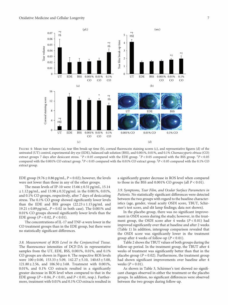
3.4. Tear Volume. There were no statistically significantdifferences in tear volumes among the groups at baseline(data not shown). Seven days after the desiccating stresswas applied; the mean tear volumes of the five groups were0.046± 0.014μL, 0.021± 0.006μL, 0.022± 0.004μL, 0.023± 0.004μL, and 0.037± 0.006μL, respectively, in the UT,BSS, 0.001%, 0.01%, and 0.1% CO groups. Tear volume in
the 0.1% CO treatment group was significantly greater thanthose in the other groups (all P < 0 01; Figure 4(a)).
3.5. Tear Film BUT. There were no statistically significantdifferences in TBUT among the groups at baseline (datanot shown). Seven days after desiccating stress had beenapplied, the TBUT was found to be 1.52± 0.16 s and 1.62± 0.12 s in the EDE and BSS groups, respectively; it wassignificantly shorter than that in the UT group (4.12± 0.33 s, P < 0 01; Figure 4(b)). The 0.01% and 0.1% COextract groups (1.84± 0.13 s and 1.97± 0.36 s, resp.) had
0
50
100
150
Cel
l via
bilit
y (%
)
Control 0.001%CO
0.01%CO
0.1%CO
(a)
0
50
100
150
Cel
l via
bilit
y (%
)
0.001%CO
0.01%CO
0.1%CO
H2O2 (200 �휇M)
Negativecontrol
Positivecontrol
⁎
† † †
(b)
Figure 1: Effect of pretreatment with Chamaecyparis obtusa (CO) extracts on the viability of human corneal epithelial (HCE) cells. (a) Theeffect of CO extracts (0.001%, 0.01%, and 0.1% concentrations) on the viability of HCE cells is shown. (b) The effect of CO extracts (0.001%,0.01%, and 0.1% concentrations) on HCE cells exposed to 200 μM hydrogen peroxide (H2O2). The results are shown as a percentage relativeto the control samples. ∗P < 0 01 compared with the control cells. †P < 0 01 compared with the cells exposed to H2O2.
Negativecontrol
Positivecontrol 0.1% CO0.01% CO0.001% CO
H2O2 (200 �휇M)
(a)
0
300
600
900
Negativecontrol
Positivecontrol
0.001%CO
0.1%CO
0.01%CO
H2O2 (200 �휇M)
DH
E in
tens
ity(%
relat
ive t
o co
ntro
l)
⁎ ⁎
†⁎
†‡⁎
(b)
Figure 2: Dihydroethidium (DHE) staining and the subsequent confocal fluorescence microscopy observations in hydrogen peroxide-(200 μM) treated human corneal epithelial cells with or without pretreatment with Chamaecyparis obtusa (CO) extracts (0.001%, 0.01%,and 0.1% concentrations). (a) One representative image selected from three individual experiments is shown. (b) Relative fluorescenceintensity (expressed as percent normalized to negative control) analysis is shown. ∗P < 0 05 compared with the control cells. †P < 0 05compared with the H2O2-exposed cells. ‡P < 0 05 compared with the 0.001% CO extract-pretreated cells.
5Oxidative Medicine and Cellular Longevity

significantly greater TBUT values than the EDE (P < 0 01and P = 0 03, resp.) and BSS (P < 0 01 and P = 0 01, resp.)groups. However, the 0.001% CO extract group did not differsignificantly from the EDE and BSS groups in terms ofTBUT. (1.68± 0.14 s; P = 0 27 and P = 0 98, resp.).
3.6. Corneal Fluorescein Staining. At baseline, the mean cor-neal fluorescein staining scores showed no significant differ-ences among the groups (data not shown). On day 7, thecorneal fluorescein staining score in the 0.001% CO groupwas 12.40± 0.70; it showed no significant difference com-pared with the EDE (13.80± 1.32, P = 0 09) and BSS groups(12.50± 1.18, P = 0 99; Figure 4(c)). The 0.01% CO extractgroup showed a significantly lower corneal fluorescein score
than the EDE group (12.20± 0.79, P = 0 04), and the 0.1%CO treatment group showed a significantly lower cornealfluorescein staining score than the EDE, BSS, 0.001%, and0.01% CO groups (10.20± 0.92, all P < 0 01).
3.7. Inflammatory Cytokine and Chemokine Levels in theConjunctival Tissue. The inflammatory cytokine and chemo-kine levels in conjunctival tissues are shown in Figure 5. Themean concentration of IFN-γ was 7.73± 0.75 pg/mL in the0.1% CO group. This was significantly lower than the con-centrations in the EDE, BSS, and 0.001% CO groups (14.54± 1.00 pg/mL, 12.84± 0.72 pg/mL, and 11.24± 0.74 pg/mLand P < 0 01, P < 0 01, and P = 0 03, resp.). The 0.01% COgroup showed significantly lower levels of IFN-γ than the
Negativecontrol
0.1%CO
0.01%CO
0.001%CO
Positivecontrol
H2O2 (200 �휇M)
HO-1
�훽-Actin
CAT
COX-2
Prx-1
(a)
0
0.4
0.8
1.2
1.6HO-1
0
0.4
0.8
1.2
1.6 CAT
0
0.4
0.8
1.2 COX-2
0
0.4
0.8
1.2
1.6 Prx-1
Negativecontrol
Positivecontrol
0.001%CO
0.01%CO
0.1%CO
Negativecontrol
Positivecontrol
0.001%CO
0.01%CO
0.1%CO
Negativecontrol
Positivecontrol
0.001%CO
0.01%CO
0.1%CO
Negativecontrol
Positivecontrol
0.001%CO
0.01%CO
0.1%CO
Rela
tive r
atio
of
signa
l/�훽-a
ctin
Relat
ive r
atio
of
signa
l/�훽-a
ctin
†‡§
†‡§
†† †
†‡§
†
⁎
⁎
⁎
⁎
⁎†⁎ ⁎ †
‡§
⁎
(b)
Figure 3: Western blot analyses to evaluate the effect of Chamaecyparis obtusa (CO) extracts on the expression of peroxiredoxin- (Prx-) 1,heme oxgenase- (HO-) 1, catalase (CAT), and cyclooxygenase-2 (COX-2) in the presence or absence of hydrogen peroxide (negativecontrol), H2O2 control (positive control), and 0.001%, 0.01%, and 0.1% CO extract groups. β-Actin was used as an internal control. (a)One representative data set obtained from among three individual experiments is shown. (b) Western blot band intensity analyses(relative ratio of signal/β-actin) are shown. ∗P < 0 05 compared with the control group. †P < 0 05 compared with the H2O2 control group.‡P < 0 05 compared with the 0.001% CO extracts group. §P < 0 05 compared with the 0.01% CO extract group.
6 Oxidative Medicine and Cellular Longevity
EDE group (9.76± 0.86 pg/mL, P = 0 02); however, the levelswere not lower than those in any of the other groups.
The mean levels of IP-10 were 15.66± 0.51 pg/mL, 15.14± 1.12 pg/mL, and 13.98± 0.32 pg/mL in the 0.001%, 0.01%,and 0.1% CO groups, respectively, after 7 days of desiccatingstress. The 0.1% CO group showed significantly lower levelsthan the EDE and BSS groups (22.23± 1.15 pg/mL and19.21± 0.89 pg/mL, P = 0 02 in both case). The 0.001% and0.01% CO groups showed significantly lower levels than theEDE group (P = 0 02, P < 0 01).
The concentrations of IL-17 and TNF-αwere lower in theCO treatment groups than in the EDE group, but there wereno statistically significant differences.
3.8. Measurement of ROS Level in the Conjunctival Tissue.The fluorescence intensities of DCF-DA in representativesamples from the UT, EDE, BSS, 0.001%, 0.01%, and 0.1%CO groups are shown in Figure 6. The respective ROS levelswere 100± 0.00, 153.33± 5.09, 142.27± 5.10, 140.63± 5.00,121.80± 2.56, and 106.50± 3.68. Treatment with 0.001%,0.01%, and 0.1% CO extracts resulted in a significantlygreater decrease in ROS level when compared to that in theEDE group (P = 0 04, P < 0 01, and P < 0 01, resp.). Further-more, treatment with 0.01% and 0.1% CO extracts resulted in
a significantly greater decrease in ROS level when comparedto those in the BSS and 0.001% CO groups (all P < 0 01).
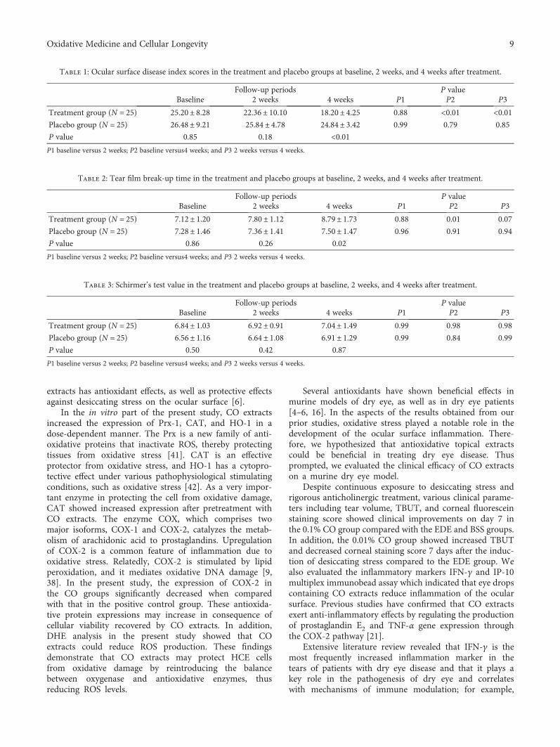
3.9. Symptoms, Tear Film, and Ocular Surface Parameters inPatients. No statistically significant differences were detectedbetween the two groups with regard to the baseline character-istics (age, gender, visual acuity OSDI score, TBUT, Schir-mer’s test score, and slit lamp findings; data not shown).
In the placebo group, there was no significant improve-ment in OSDI scores during the study; however, in the treat-ment group, the OSDI score after 4 weeks (P < 0 01) hadimproved significantly over that at baseline and after 2 weeks(Table 1) In addition, intergroup comparison revealed thatthe OSDI score was significantly lower in the treatmentgroup after 4 weeks of follow-up (P < 0 01).
Table 2 shows the TBUT values of both groups during thefollow-up period. In the treatment group, the TBUT after 4weeks of treatment was significantly better than that in theplacebo group (P = 0 02). Furthermore, the treatment grouphad shown significant improvements over baseline after 4weeks (P = 0 01).
As shown in Table 3, Schirmer’s test showed no signifi-cant changes observed in either the treatment or the placebogroups. In addition, no significant differences were observedbetween the two groups during follow-up.
0
0.01
0.02
0.03
0.04
0.05
0.06
0.07‡§
UT EDE BSS 0.001%CO
0.01%CO
0.1%CO
Tear
vol
ume
(�휇L)
‡§
⁎†
†⁎
(a)
UT EDE BSS 0.001%CO
0.01%CO
0.1%CO
0
1
2
3
4
5 ‡§‖
Tear
film
bre
ak-u
p tim
e
†
(sec)†⁎
⁎†⁎
(b)
0
4
8
12
16
UT EDE BSS 0.001%CO
0.01%CO
0.1%CO
Cor
neal
stai
ning
scor
e
‡§‖†⁎
‡§†⁎
⁎
(c)
EDE BSS
0.001% CO 0.01% CO 0.1% CO
UT
(d)
Figure 4: Mean tear volumes (a), tear film break-up time (b), corneal fluorescein staining scores (c), and representative figures (d) of theuntreated (UT) control, experimental dry eye (EDE), balanced salt solution (BSS), and 0.001%, 0.01%, and 0.1% Chamaecyparis obtusa (CO)extract groups 7 days after desiccant stress. ∗P < 0 05 compared with the EDE group. †P < 0 05 compared with the BSS group. ‡P < 0 05compared with the 0.001% CO extract group. §P < 0 05 compared with the 0.01% CO extract group. ∥P < 0 05 compared with the 0.1% COextract group.
7Oxidative Medicine and Cellular Longevity

4. Discussion
The protective properties of CO have been recognized toexert antigastropathic, anti-inflammatory, and antioxidativeactivity [35]. However, little is known about the bioactivityand potential clinical implications of CO on the health ofthe human eye. The present study indicated that CO might
have beneficial effects on eye and ocular surface disease. Inthe present study, the cellular viability has decreased subtlyin the 0.1% group. We thought that it may be due to mildtoxic reaction at a high concentration similar to generalsubstances. However, it did not show a statistically signif-icant change of the cellular viability in the present study.We also showed that CO reduced ROS generation andrebalanced homeostasis between oxygenases and antioxida-tive enzymes in HCE cells exposed to oxidative stress andthat it decreased inflammation and ROS levels in a murinedry eye model. The mechanisms of ROS reduction by COare as follows: phenolic compounds have been reported tobe one of the major constituents of CO [36]. The antiox-idant properties of phenolic compounds are related totheir structural characteristics, which are known to bedue to their role as direct ROS scavenging, reducing, andchain breaking antioxidant mechanisms [37].
Extensive researches during the past decade havedemonstrated that oxidative stress played an importantrole in ocular surface disease, including dry eye [9, 38].For example, in vitro studies have revealed that lipid andmitochondrial oxidative damage might cause inflammationin HCE cells [15, 38]. In addition, in vivo lipid oxidativeinjury has been observed in patients with dry eye, as wellas in those with Sjögren syndrome [39]. Higher oxidativestress may also explain the higher incidence and moresevere dry eye in elderly patients [40]. Our previous studyestablished that a natural mixture of plant ethyl alcohol
0
1
2
3
4
5
6
UT EDE BSS 0.001%CO
0.01%CO
0.1%CO
IL-17
(pg/
mL)
(a)
(pg/
mL)
0
4
8
12
16
20
UT EDE BSS 0.001%CO
0.01%CO
0.1%CO
IFN-�훾
‡§‡§‖†⁎
†⁎⁎
(b)
0
1
2
3
4
5
UT EDE BSS 0.001%CO
0.01%CO
0.1%CO
TNF-�훼
(pg/
mL)
(c)
0
5
10
15
20
25
UT EDE BSS 0.001%CO
0.01%CO
0.1%CO
IP-10
(pg/
mL)
‡§†⁎
†⁎⁎⁎
(d)
Figure 5: Concentrations of interleukin- (IL-) 17 (a), interferon- (IFN-) γ (b), tumor necrosis factor- (TNF-) α (c), and IFN-γ-inducibleprotein- (IP-) 10 (d), in the conjunctiva of the untreated (UT) control, experimental dry eye (EDE), balanced salt solution (BSS), and0.001%, 0.01%, and 0.1% Chamaecyparis obtusa (CO) extract groups. ∗P < 0 05 compared with the EDE group. †P < 0 05 compared withthe BSS group. ‡P < 0 05 compared with the 0.001% CO extract group. §P < 0 05 compared with the 0.01% CO extract group. ∥P < 0 05compared with the 0.1% CO extract group.
40
0
80
120
160
200
UT EDE BSS 0.001%CO
0.1%CO
0.01%CO
‡§
Mea
n flu
ores
cenc
ein
tens
ity
†⁎ ‡§†⁎†‡⁎
⁎
Figure 6: Mean conjunctival fluorescein intensity showing thereactive oxygen species (ROS) levels in the untreated (UT) control,experimental dry eye (EDE), balanced salt solution (BSS), and0.001%, 0.01%, and 0.1% Chamaecyparis obtusa (CO) extractsgroups. ∗P < 0 05 compared with the EDE group. †P < 0 05compared with the BSS group. ‡P < 0 05 compared with the0.001% CO extract group. §P < 0 05 compared with the 0.01% COextract group.
8 Oxidative Medicine and Cellular Longevity
extracts has antioxidant effects, as well as protective effectsagainst desiccating stress on the ocular surface [6].
In the in vitro part of the present study, CO extractsincreased the expression of Prx-1, CAT, and HO-1 in adose-dependent manner. The Prx is a new family of anti-oxidative proteins that inactivate ROS, thereby protectingtissues from oxidative stress [41]. CAT is an effectiveprotector from oxidative stress, and HO-1 has a cytopro-tective effect under various pathophysiological stimulatingconditions, such as oxidative stress [42]. As a very impor-tant enzyme in protecting the cell from oxidative damage,CAT showed increased expression after pretreatment withCO extracts. The enzyme COX, which comprises twomajor isoforms, COX-1 and COX-2, catalyzes the metab-olism of arachidonic acid to prostaglandins. Upregulationof COX-2 is a common feature of inflammation due tooxidative stress. Relatedly, COX-2 is stimulated by lipidperoxidation, and it mediates oxidative DNA damage [9,38]. In the present study, the expression of COX-2 inthe CO groups significantly decreased when comparedwith that in the positive control group. These antioxida-tive protein expressions may increase in consequence ofcellular viability recovered by CO extracts. In addition,DHE analysis in the present study showed that COextracts could reduce ROS production. These findingsdemonstrate that CO extracts may protect HCE cellsfrom oxidative damage by reintroducing the balancebetween oxygenase and antioxidative enzymes, thusreducing ROS levels.
Several antioxidants have shown beneficial effects inmurine models of dry eye, as well as in dry eye patients[4–6, 16]. In the aspects of the results obtained from ourprior studies, oxidative stress played a notable role in thedevelopment of the ocular surface inflammation. There-fore, we hypothesized that antioxidative topical extractscould be beneficial in treating dry eye disease. Thusprompted, we evaluated the clinical efficacy of CO extractson a murine dry eye model.
Despite continuous exposure to desiccating stress andrigorous anticholinergic treatment, various clinical parame-ters including tear volume, TBUT, and corneal fluoresceinstaining score showed clinical improvements on day 7 inthe 0.1% CO group compared with the EDE and BSS groups.In addition, the 0.01% CO group showed increased TBUTand decreased corneal staining score 7 days after the induc-tion of desiccating stress compared to the EDE group. Wealso evaluated the inflammatory markers IFN-γ and IP-10multiplex immunobead assay which indicated that eye dropscontaining CO extracts reduce inflammation of the ocularsurface. Previous studies have confirmed that CO extractsexert anti-inflammatory effects by regulating the productionof prostaglandin E2 and TNF-α gene expression throughthe COX-2 pathway [21].
Extensive literature review revealed that IFN-γ is themost frequently increased inflammation marker in thetears of patients with dry eye disease and that it plays akey role in the pathogenesis of dry eye and correlateswith mechanisms of immune modulation; for example,
Table 1: Ocular surface disease index scores in the treatment and placebo groups at baseline, 2 weeks, and 4 weeks after treatment.
Follow-up periods P valueBaseline 2 weeks 4 weeks P1 P2 P3
Treatment group (N = 25) 25.20± 8.28 22.36± 10.10 18.20± 4.25 0.88 <0.01 <0.01Placebo group (N = 25) 26.48± 9.21 25.84± 4.78 24.84± 3.42 0.99 0.79 0.85
P value 0.85 0.18 <0.01P1 baseline versus 2 weeks; P2 baseline versus4 weeks; and P3 2 weeks versus 4 weeks.
Table 2: Tear film break-up time in the treatment and placebo groups at baseline, 2 weeks, and 4 weeks after treatment.
Follow-up periods P valueBaseline 2 weeks 4 weeks P1 P2 P3
Treatment group (N = 25) 7.12± 1.20 7.80± 1.12 8.79± 1.73 0.88 0.01 0.07
Placebo group (N = 25) 7.28± 1.46 7.36± 1.41 7.50± 1.47 0.96 0.91 0.94
P value 0.86 0.26 0.02
P1 baseline versus 2 weeks; P2 baseline versus4 weeks; and P3 2 weeks versus 4 weeks.
Table 3: Schirmer’s test value in the treatment and placebo groups at baseline, 2 weeks, and 4 weeks after treatment.
Follow-up periods P valueBaseline 2 weeks 4 weeks P1 P2 P3
Treatment group (N = 25) 6.84± 1.03 6.92± 0.91 7.04± 1.49 0.99 0.98 0.98
Placebo group (N = 25) 6.56± 1.16 6.64± 1.08 6.91± 1.29 0.99 0.84 0.99
P value 0.50 0.42 0.87
P1 baseline versus 2 weeks; P2 baseline versus4 weeks; and P3 2 weeks versus 4 weeks.
9Oxidative Medicine and Cellular Longevity

it increases T-helper type 1 cells [43]. IP-10 is an IFN-γ/α- and TNF-α-inducible chemokine that is highlyexpressed by a variety of cells, including activated T lym-phocytes, NK cells, and monocytes [44]. Our presentfindings support the results of recent studies showing thattopical antioxidant also has anti-inflammatory activity ina murine dry eye model [6]. Furthermore, ROS levelsdetected using DCF-DA were notably lower in the0.01% and 0.1% CO extract groups than in the EDEand BSS groups, suggesting that CO extracts protect theocular surface from desiccating stress via antioxidativedefense mechanisms in the murine dry eye model.
Eventually, we used an eye mask containing CO extractsto demonstrate the antioxidative effects in patients with dryeye disease. Both OSDI score and TBUT had significantlyimproved in the treatment group after 4 weeks of wearingthe mask. In addition, significant differences between thetwo groups were observed in terms of OSDI score and TBUT.Taken together, these findings suggest that the maskscontaining CO extracts used in this study provide subjectiveand objective improvement in dry eye parameters, possiblyvia the antioxidative defense system.
Our findings are comparable to the results of previousstudies that have demonstrated the efficacy of antioxidativeand anti-inflammatory agents in improving dry eyeparameters [3, 5, 16]. For instance, a mixture of fournatural plant ethyl alcohol extracts decreased the levelsof inflammatory cytokines and oxidative stress markerson the ocular surface in the murine dry eye model andantioxidant glasses containing extracts of medical plantswhich improved the OSDI score and TBUT in patientswith dry eye disease [6, 16]. In addition, omega-3 polyun-saturated fatty acids, such as antioxidants, have beenfound to be effective in improving dry eye symptoms[45]. Furthermore, Kim et al. [46] reported that topicalvitamin A eye drops contribute to improvements inblurred vision, TBUT, Schirmer’s test, and findings fromimpression cytology in patients with dry eye disease.
In conclusion, CO extracts, especially at concentrationsof 0.01% and 0.1%, may protect the ocular surface fromdesiccating stress and resultant oxidative stress. In addition,wearing an antioxidative mask containing CO extractsimproved dry eye symptoms, as well as the clinical parame-ters of dry eye. CO extracts may be used as an adjunctivetherapeutic option for the treatment of dry eye disease.
Disclosure
This paper was presented at ARVO 2016 Annual Meetingand the 5th Asia Cornea Society Biennial ScientificMeeting 2016. The funders had no role in the studydesign, data collection and analysis, the decision topublish, or manuscript preparation.
Conflicts of Interest
None of the authors has any conflicting interests to disclose.
Acknowledgments
The study was supported by Basic Science ResearchProgram through the National Research Foundation ofKorea (NRF) funded by the Ministry of Science, ICT andFuture Planning (2017R1A2B4003367), the Forest Scienceand Technology Projects (Project no. S121313L50100) ofthe Korean Forest Service, and the CNUH BiomedicalResearch Institute (CRI 13906-22).
References
[1] “The definition and classification of dry eye disease: report ofthe Definition and Classification Subcommittee of the Interna-tional Dry Eye WorkShop (2007),” The Ocular Surface, vol. 5,no. 2, pp. 75–92, 2007.
[2] A. J. Paulsen, K. J. Cruickshanks, M. E. Fischer et al., “Dry eyein the beaver dam offspring study: prevalence, risk factors, andhealth-related quality of life,” American Journal of Ophthal-mology, vol. 157, no. 4, pp. 799–806, 2014.
[3] M. S. Sung, Z. Li, L. Cui et al., “Effect of topical 5-aminoimida-zole-4-carboxamide-1-β-d-ribofuranoside in a mouse modelof experimental dry eye,” Investigative Ophthalmology &Visual Science, vol. 56, no. 5, pp. 3149–3158, 2015.
[4] V. L. Perez, S. C. Pflugfelder, S. Zhang, A. Shojaei, andR. Haque, “Lifitegrast, a novel integrin antagonist for treat-ment of dry eye disease,” The Ocular Surface, vol. 14, no. 2,pp. 207–215, 2016.
[5] Z. Li, J. H. Choi, H. J. Oh, S. H. Park, J. B. Lee, and K. C. Yoon,“Effects of eye drops containing a mixture of omega-3 essentialfatty acids and hyaluronic acid on the ocular surface indesiccating stress-induced murine dry eye,” Current EyeResearch, vol. 39, no. 9, pp. 871–878, 2014.
[6] W. Choi, J. B. Lee, L. Cui et al., “Therapeutic efficacy of topi-cally applied antioxidant medicinal plant extracts in a mousemodel of experimental dry eye,”Oxidative Medicine and Cellu-lar Longevity, vol. 2016, Article ID 4727415, 10 pages, 2016.
[7] A. Higuchi, K. Takahashi, M. Hirashima, T. Kawakita, andK. Tsubota, “Selenoprotein P controls oxidative stress incornea,” PLoS One, vol. 5, no. 3, article e9911, 2010.
[8] J. Li, D. Ruzhi, X. Hua et al., “Blueberry component pterostil-bene protects corneal epithelial cells from inflammation viaanti-oxidative pathway,” Scientific Reports, vol. 6, no. 1, article19408, 2016.
[9] T. H. Wakamatsu, M. Dogru, and K. Tsubota, “Tearful rela-tions: oxidative stress, inflammation and eye diseases,” Arqui-vos Brasileiros de Oftalmologia, vol. 71, no. 6, Supplementary,pp. 72–79, 2008.
[10] Y. Uchino, T. Kawakita, M. Miyazawa et al., “Oxidative stressinduced inflammation initiates functional decline of tear pro-duction,” PLoS One, vol. 7, no. 10, article e45805, 2012.
[11] Y. Uchino, T. Kawakita, T. Ishii, N. Ishii, and K. Tsubota, “Anew mouse model of dry eye disease: oxidative stress affectsfunctional decline in the lacrimal gland,” Cornea, vol. 31,Supplement 1, pp. S63–S67, 2012.
[12] C. Amico, T. Tornetta, C. Scifo, and A. R. Blanco, “Antioxidanteffect of 0.2% xanthan gum in ocular surface corneal epithelialcells,” Current Eye Research, vol. 40, no. 1, pp. 72–76, 2015.
[13] P. S. Larmo, R. L. Jarvinen, N. L. Setala et al., “Oral sea buck-thorn oil attenuates tear film osmolarity and symptoms in
10 Oxidative Medicine and Cellular Longevity

individuals with dry eye,” The Journal of Nutrition, vol. 140,no. 8, pp. 1462–1468, 2010.
[14] S. D. Hsu, D. P. Dickinson, H. Qin et al., “Green tea polyphe-nols reduce autoimmune symptoms in a murine model forhuman Sjogren’s syndrome and protect human salivary acinarcells from TNF-α-induced cytotoxicity,” Autoimmunity,vol. 40, no. 2, pp. 138–147, 2007.
[15] J. B. Lee, S. H. Kim, S. C. Lee et al., “Blue light-induced oxida-tive stress in human corneal epithelial cells: protective effectsof ethanol extracts of various medicinal plant mixtures,” Inves-tigative Ophthalmology & Visual Science, vol. 55, no. 7,pp. 4119–4127, 2014.
[16] W. Choi, J. C. Kim, W. S. Kim et al., “Clinical effect of antiox-idant glasses containing extracts of medicinal plants in patientswith dry eye disease: a multi-center, prospective, randomized,double-blind, placebo-controlled trial,” PLoS One, vol. 10,no. 10, article e0139761, 2015.
[17] S.-C. Chien, J.-Y. Chang, C.-C. Kuo, C.-C. Hsieh, N.-S. Yang,and Y.-H. Kuo, “Cytotoxic and novel skeleton compoundsfrom the heartwood of Chamaecyparis obtusa var. formosana,”Tetrahedron Letters, vol. 48, no. 9, pp. 1567–1569, 2007.
[18] S. S. Cheng, H. T. Chang, C. L. Wu, and S. T. Chang, “Anti-ter-mitic activities of essential oils from coniferous trees againstCoptotermes formosanus,” Bioresource Technology, vol. 98,no. 2, pp. 456–459, 2007.
[19] P. Marimuthu, C.-L. Wu, H.-T. Chang, and S.-T. Chang,“Antioxidant activity of the ethanolic extract from the barkof Chamaecyparis obtusa var. formosana,” Journal of theScience of Food and Agriculture, vol. 88, no. 8, pp. 1400–1405, 2008.
[20] S. C. Cheng, W. H. Li, Y. C. Shi et al., “Antioxidant activity anddelayed aging effects of hot water extract from Chamaecyparisobtusa var. formosana leaves,” Journal of Agricultural andFood Chemistry, vol. 62, no. 18, pp. 4159–4165, 2014.
[21] B. S. An, J. H. Kang, H. Yang et al., “Anti-inflammatory effectsof essential oils from Chamaecyparis obtusa via thecyclooxygenase-2 pathway in rats,” Molecular MedicineReports, vol. 8, no. 1, pp. 255–259, 2013.
[22] E. J. Jeong, L. Hwang, M. Lee, K. Y. Lee, M. J. Ahn, and S. H.Sung, “Neuroprotective biflavonoids of Chamaecyparis obtusaleaves against glutamate-induced oxidative stress in HT22 hip-pocampal cells,” Food and Chemical Toxicology, vol. 64,pp. 397–402, 2014.
[23] E.-J. Hong, K.-J. Na, I.-G. Choi, K.-C. Choi, and E.-B. Jeung,“Antibacterial and antifungal effects of essential oils fromconiferous trees,” Biological and Pharmaceutical Bulletin,vol. 27, no. 6, pp. 863–866, 2004.
[24] D. Bae, H. Seol, H. G. Yoon et al., “Inhaled essential oil fromChamaecyparis obtuse ameliorates the impairments of cogni-tive function induced by injection of β-amyloid in rats,” Phar-maceutical Biology, vol. 50, no. 7, pp. 900–910, 2012.
[25] S. S. Joo, Y. M. Yoo, S. H. Ko et al., “Effects of essential oil fromChamaecypris obtusa on the development of atopic dermatitis-like skin lesions and the suppression of Th cytokines,” Journalof Dermatological Science, vol. 60, no. 2, pp. 122–125, 2010.
[26] H. Park, H. Lee, M. Shin et al., “Effects of cosolvents on thedecaffeination of green tea by supercritical carbon dioxide,”Food Chemistry, vol. 105, no. 3, pp. 1011–1017, 2007.
[27] P. Sookwong, P. Suttiarporn, P. Boontakham, P. Seekhow,S. Wangtueai, and S. Mahatheeranont, “Simultaneous quanti-fication of vitamin E, γ-oryzanols and xanthophylls from rice
bran essences extracted by supercritical CO2,” Food Chemistry,vol. 211, pp. 140–147, 2016.
[28] Z. Li, L. Cui, J. M. Yang et al., “The wound healing effects ofadiponectin eye drops after corneal alkali burn,” Current EyeResearch, vol. 41, no. 11, pp. 1424–1432, 2016.
[29] N. Chatterjee, H. J. Eom, and J. Choi, “A systems toxicologyapproach to the surface functionality control of graphene-cellinteractions,” Biomaterials, vol. 35, no. 4, pp. 1109–1127, 2014.
[30] Y. Xu, S. Wang, Q. Miao et al., “Protective role of hinokitiolagainst H2O2-induced injury in human corneal epithelium,”Current Eye Research, vol. 42, no. 1, pp. 47–53, 2016.
[31] Z. Li, J. M. Woo, S. W. Chung et al., “Therapeutic effect oftopical adiponectin in a mouse model of desiccating stress-induced dry eye,” Investigative Ophthalmology & Visual Sci-ence, vol. 54, no. 1, pp. 155–162, 2013.
[32] K. C. Yoon, K. Y. Ahn, W. Choi et al., “Tear production andocular surface changes in experimental dry eye after elimina-tion of desiccating stress,” Investigative Ophthalmology &Visual Science, vol. 52, no. 10, pp. 7267–7273, 2011.
[33] R. M. Schiffman, M. D. Christianson, G. Jacobsen, J. D. Hirsch,and B. L. Reis, “Reliability and validity of the ocular surfacedisease index,” Archives of Ophthalmology, vol. 118, no. 5,pp. 615–621, 2000.
[34] K. C. Yoon, H. Heo, S. K. Im, I. C. You, Y. H. Kim, and Y. G.Park, “Comparison of autologous serum and umbilical cordserum eye drops for dry eye syndrome,” American Journal ofOphthalmology, vol. 144, no. 1, pp. 86–92.e2, 2007.
[35] H. J. Park, S. K. Kim, W. S. Kang, J. M. Woo, and J. W. Kim,“Effects of essential oil from Chamaecyparis obtusa on cyto-kine genes in the hippocampus of maternal separation rats,”Canadian Journal of Physiology and Pharmacology, vol. 92,no. 2, pp. 95–101, 2014.
[36] T. P. Devasaqayam, J. C. Tilak, K. K. Boloor, K. S. Sane, S. S.Ghaskadbi, and R. D. Lele, “Free radicals and antioxidants inhuman health: currents status and future prospects,” The Jour-nal of the Association of Physicians of India, vol. 52, pp. 794–804, 2004.
[37] T. C. Chien, S. F. Lo, and C. L. Ho, “Chemical composition andanti-inflammatory activity of Chamaecyparis obtusa f.formo-sana wood essential oil from Taiwan. Nature product,” Com-munications, vol. 9, no. 5, p. 723, 2014.
[38] R. Deng, X. Hua, J. Li et al., “Oxidative stress markers inducedby hyperosmolarity in primary human corneal epithelial cells,”PLoS One, vol. 10, no. 5, article e0126561, 2015.
[39] T. H. Wakamatsu, M. Dogru, Y. Matsumoto et al., “Evaluationof lipid oxidative stress status in Sjögren syndrome patients,”Investigative Ophthalmology & Visual Science, vol. 54, no. 1,pp. 201–210, 2013.
[40] K. Tsubota, M. Kawashima, T. Inaba et al., “The antiagingapproach for the treatment of dry eye,” Cornea, vol. 31, Sup-plement 1, pp. S3–S8, 2012.
[41] Q. Le, K. Tabuchi, E. Warabi, and A. Hara, “The role of perox-iredoxin I in cisplatin-induced ototoxicity,” Auris NasusLarynx, vol. 44, no. 2, pp. 205–212, 2017.
[42] T. Ma, T. Chen, P. Li et al., “Heme oxygenase-1 (HO-1) pro-tects human lens epithelial cells (SRA01/04) against hydrogenperoxide (H2O2)-induced oxidative stress and apoptosis,”Experimental Eye Research, vol. 146, pp. 318–329, 2016.
[43] Y. Wei, N. Gadaria-Rathod, S. Epstein, and P. Asbell, “Tearcytokine profile as a noninvasive biomarker of inflammationfor ocular surface diseases: standard operating procedures,”
11Oxidative Medicine and Cellular Longevity
Investigative Ophthalmology & Visual Science, vol. 54, no. 13,pp. 8327–8336, 2013.
[44] Y. Wang, W. Yu, C. Shen et al., “Predictive value of serumIFN-γ inducible protein-10 and IFN-γ/IL-4 ratio for liverfibrosis progression in CHB patients,” Scientific Reports,vol. 7, article 40404, 2017.
[45] A. Olenik, “Effectiveness and tolerability of dietary supple-mentation with a combination of omega-3 polyunsaturatedfatty acids and antioxidants in the treatment of dry eyesymptoms: results of a prospective study,” Clinical Ophthal-mology, vol. 8, pp. 169–176, 2014.
[46] E. C. Kim, J. S. Choi, and C. K. Joo, “A comparison of vitaminA and cyclosporine A 0.05% eye drops for treatment of dry eyesyndrome,” American Journal of Ophthalmology, vol. 147,no. 2, pp. 206–213.e3, 2009.
12 Oxidative Medicine and Cellular Longevity

Submit your manuscripts athttps://www.hindawi.com
Stem CellsInternational
Hindawi Publishing Corporationhttp://www.hindawi.com Volume 2014
Hindawi Publishing Corporationhttp://www.hindawi.com Volume 2014
MEDIATORSINFLAMMATION
of
Hindawi Publishing Corporationhttp://www.hindawi.com Volume 2014
Behavioural Neurology
EndocrinologyInternational Journal of
Hindawi Publishing Corporationhttp://www.hindawi.com Volume 2014
Hindawi Publishing Corporationhttp://www.hindawi.com Volume 2014
Disease Markers
Hindawi Publishing Corporationhttp://www.hindawi.com Volume 2014
BioMed Research International
OncologyJournal of
Hindawi Publishing Corporationhttp://www.hindawi.com Volume 2014
Hindawi Publishing Corporationhttp://www.hindawi.com Volume 2014
Oxidative Medicine and Cellular Longevity
Hindawi Publishing Corporationhttp://www.hindawi.com Volume 2014
PPAR Research
The Scientific World JournalHindawi Publishing Corporation http://www.hindawi.com Volume 2014
Immunology ResearchHindawi Publishing Corporationhttp://www.hindawi.com Volume 2014
Journal of
ObesityJournal of
Hindawi Publishing Corporationhttp://www.hindawi.com Volume 2014
Hindawi Publishing Corporationhttp://www.hindawi.com Volume 2014
Computational and Mathematical Methods in Medicine
OphthalmologyJournal of
Hindawi Publishing Corporationhttp://www.hindawi.com Volume 2014
Diabetes ResearchJournal of
Hindawi Publishing Corporationhttp://www.hindawi.com Volume 2014
Hindawi Publishing Corporationhttp://www.hindawi.com Volume 2014
Research and TreatmentAIDS
Hindawi Publishing Corporationhttp://www.hindawi.com Volume 2014
Gastroenterology Research and Practice
Hindawi Publishing Corporationhttp://www.hindawi.com Volume 2014
Parkinson’s Disease
Evidence-Based Complementary and Alternative Medicine
Volume 2014Hindawi Publishing Corporationhttp://www.hindawi.com