Embed Size (px)
Citation preview
lable at ScienceDirect
Experimental Eye Research 91 (2010) 369e377
Contents lists avai
Experimental Eye Research
journal homepage: www.elsevier .com/locate/yexer
Evolution of damage in the lens after in vivo close to threshold exposureto UV-B radiation: Cytomorphological study of apoptosis
Konstantin Galichanin a,b,*, Stefan Löfgren a, Jan Bergmanson c, Per Söderberg b
a St. Erik’s Eye Hospital, Karolinska Institutet, Stockholm, SwedenbGullstrand Lab, Ophthalmology, Department of Neuroscience, Uppsala University Hospital, Uppsala, Swedenc Texas Eye Research and Technology Center, University of Houston College of Optometry, Houston, Texas, USA
a r t i c l e i n f o
Article history:Received 14 May 2010Accepted in revised form 10 June 2010Available online 17 June 2010
Keywords:lensultraviolet radiationcataractapoptosisrepairlight scatteringmicroscopy
* Address correspondence to: Konstantin Galichanmology, Dept. of Neuroscience, Uppsala University HSweden. Tel.: þ46 18 611 3716; fax: þ46 18 50 48 57
E-mail address: [email protected]
0014-4835/$ e see front matter � 2010 Elsevier Ltd.doi:10.1016/j.exer.2010.06.009
a b s t r a c t
The purpose of the present study was to investigate cataractogenesis and recovery of lens damage after invivo close to threshold ultraviolet (UV)-B radiation around 300 nm. Eighty six-week-old albino Spra-gueeDawley rats were familiarized to a rat restrainer five days prior to exposure. Groups of non-anesthetized rats were exposed unilaterally to 8 kJ/m2 UVR-300 nm. The animals were sacrificed at 1, 7,48 and 336 h following exposure. The lenses were extracted for imaging of dark-field lens macroanatomy and measurement of intensity of forward lens light scattering to quantify lens opacities. Threeexposed lenses and one non-exposed lens from each time interval were examined with light andtransmission electron microscopy (TEM). Macro anatomy and lens light scattering revealed that allcontralateral non-exposed lenses were clear. The degree of lens opacity (difference in lens light scat-tering between exposed and non-exposed lenses) increased during the 336 h, reaching a plateau towardsthe end of the observation period. Light microscopy and TEM demonstrated that apoptotic featuresappeared in the epithelium already 1 h after UVR exposure, and small vacuoles were seen in the outercortex. Epithelial damage occurs during the first 48 h after exposure and is followed by regenerativerepair at 336 h post-exposure. Apoptotic epithelial cells were phagocytized by adjacent epithelial cells.Cortical fiber cells exhibited increasing damage throughout the observation period without any clearrepair after 336 h. In conclusion, acute UVR-induced cataract is partly a reversible. Lens epithelium isa primary target for UVR exposure. Damage to cortical fiber cells remained irreversible.
� 2010 Elsevier Ltd. All rights reserved.
1. Introduction
Cataract is the number one cause of blindness in the world(Brian and Taylor, 2001; Evans et al., 2004; West, 2000). Phacoe-mulsification is the most prevalent cure of cataract in developedcountries (Asbell et al., 2005). In spite of the progress made incataract surgical techniques and materials during the last ten years,cataract continues to be a substantial public-health issue globally(Resnikoff et al., 2004). The Eye Disease Prevalence Research Grouphas projected that the number of US patients with cataract willincrease by 50% by 2020 (Congdon et al., 2004). To join the effortsfor blindness prevention the World Health Organizationwith otherinstitutions has launched the Global Initiative for the Elimination ofAvoidable Blindness “VISION 2020: the Right to Sight” (Thylefors,
in, Gullstrand Lab, Ophthal-ospital, SE-751 85 Uppsala,
.(K. Galichanin).
All rights reserved.
1998). The magnitude of the cataract problem emphasizes theimportance of development of cataract prevention and treatmentstrategies.
Solar radiation is the major source of UVR (Pitts, 1990). Exposureto sunlight has been correlated with the development of humansenile cataract (Hiller et al., 1977; ItalianeAmerican Cataract Study,1991; West and Valmadrid, 1995). Population-based studies inUnited States (Cruickshanks et al., 1992; Taylor et al., 1988),Australia (McCarty et al., 2000) and Japan (Sasaki et al., 2003)reported association between exposure to UVR-B and corticalcataract formation. Moreover, animal models have proven thatexposure to UVR-B induces cataract (Jose and Pitts, 1985; Löfgrenet al., 2003; Meyer et al., 2008; Michael et al., 1996; Pitts et al.,1977; Söderberg, 1988; Wegener, 1995).
The vertebrate ocular lens is a highly organized, compact andtransparent structure that has evolved to refract light entering theeye. The lens comprises densely packed fibers and a single layer ofepithelial cells on its anterior surface, enclosed by a thick elasticlens capsule. The entire homeostasis, in which the lens develops,differentiates and grows throughout life, is maintained by all the

K. Galichanin et al. / Experimental Eye Research 91 (2010) 369e377370
parts. (Kuszak and Costello, 2004) The essential structure-functionregimen of the lens can be altered by environmental factors such asUVR radiation (Hightower, 1995; Zigman, 1985). UVR-B photonsdamage both the lens epithelium and the lens fiber cells bydifferent mechanisms. In the lens epithelial cells, UVR-B irradiationleads to unscheduled DNA synthesis (Söderberg et al., 1986),formation of pyrimidine dimmers, DNA-DNA and DNA-proteincross linking, DNA single and double strand breaks (Kleiman et al.,1990), perturbation of calcium cell signaling and decline in reducedglutathione (Hightower et al., 1999), Na/K-ATPase inhibition(Hightower and McCready, 1992), increased membrane perme-ability (Hightower et al., 1994) and alterations in protein synthesis(Andley et al., 1990). In the lens fiber cells, UVR-B causes aggrega-tion of lens crystallins (Zigman et al., 1973), photolysis of humanlens a-crystallin and generation of reactive oxygen species (Andleyand Clark, 1989), mitochondrial rounding and movement cessation(Bantseev and Youn, 2006).
Besides all the above-listedmolecular processes, apoptosis playsa critical role in initiation of cataract in humans and animals (Li et al.,1995). The term apoptosis was first used by Kerr et al. in 1972 todescribe the morphological alterations of certain forms of pro-grammed or physiological cell death (Kerr et al.,1972). Transmissionelectron microscopy is the gold standard to confirm apoptosis anddistinguish it from necrosis. Morphologically, apoptosis is charac-terised by a condensation of both nuclear chromatin (karyopyk-nosis) and cytoplasm, which is followed by fragmentation of thenucleus (karyorhexis) and formation of apoptotic bodies containingnuclear material and closely packed cell organelles. Formedapoptotic bodies can be phagocytosed by macrophages or by adja-cent cells and subsequently shed from epithelial surfaces. There isno inflammatory reaction in apoptosis (Elmore, 2007).
Apoptosis is a morphologically and biochemically distinct modeof programmed cell deathwhich is genetically regulated and energydependent. The two best understood signaling apoptotic pathwaysin mammalian cells are the extrinsic or death receptor pathway andthe intrinsic or mitochondrial pathway. Cells exposed to UVRundergo apoptosis through the intrinsic pathway, causing DNAfragmentation and condensation of nuclear chromatin (Elmore,2007). Exposure to UVR causes apoptosis in the lens epithelialcells in vitro (Li and Spector,1996; Long et al., 2004). In our researchgroup, apoptosis has been investigated after in vivo UVR 300 nmexposure to the lens (Michael et al., 1998) and was found to bemediated by increased expression of p53 (Ayala et al., 2007).
Previously Söderberg revealed the sequence of microscopicalevents underlying cataractogenesis after high dose exposure toUVR 300 nm (Söderberg, 1988). Those events could be dueto apoptosis. The threshold dose for UVR cataract was later found tobe ten-fold lower than the dose used in the 1988 investigation(Söderberg et al., 2002). It is consequently important to investigatewhether the morphology of cataract after close to threshold UVR issimilar to that at ten times threshold, and to demonstrate chro-nology of apoptotic features in the lens.
2. Materials and methods
Non-anesthetized animals were exposed unilaterally in vivo toUVR-B. The intensity of forward lens light scattering was measuredin vitro at incrementing post exposure intervals. The lensmorphology was studied by light and transmission electronmicroscopy.
2.1. Animals
Eighty six-week-old albino SpragueeDawley (SD) female rats(Scanbur BK AB, Sollentuna, Sweden) were treated in accordance to
the ARVO Statement for the Use of Animals in Ophthalmic andVisual Research. Ethical approval was obtained from the NorthernStockholm Animal Experiments Ethics Committee, protocolnumber N184/04.
2.2. Exposure to ultraviolet radiation
2.2.1. UVR sourceUVR-B in the 300 nm wavelength region was generated with
a high-pressure mercury lamp (model 6828; Oriel, Stratford CT).The emerging radiation was collimated, passed through a waterfilter and focused into a double monochromator, set to delivera spectrum centered at 300 nm with dual peaks at 207.5 nm and302.6 nm (due to strong mercury lines near 300 nm) and 10.2 nmfull width at half maximum (Galichanin et al., 2010).
2.2.2. UVR exposureFive days prior to exposure all ratswere conditioned and fixed to
a newly developed rat restrainer (Galichanin et al., 2010). Each non-anesthetized animal was exposed unilaterally to double thresholddose 8 kJ/m2 UVR-300 nm for 15 min (Söderberg et al., 2002), whilethe contralateral eye was shielded during exposure and used asinternal control.
After a pre-determined post-exposure period, the rat wassacrificed by carbon dioxide asphyxiation, followed by cervicaldislocation. The eyes were enucleated and the lenses were extrac-ted. Remnants of the ciliary body were removed from the lensequator under a microscope, keeping the lens in balanced saltsolution (BSS; Alcon, USA).
2.3. Quantification of lens opacification
The degree of lens opacification was quantified in vitro bymeasurement of intensity of forward lens light scattering witha light dissemination meter (Söderberg et al., 1990).
2.4. Macroscopic imaging
Thereafter, the macroscopic appearance of the lens was docu-mented with digital photography in incident illumination againsta grid and in dark field illumination.
2.5. Light and transmission electron microscopy
After macro photography samples of 4 lenses from each post-exposure time interval were fixed in a 0.08 M cacodylate-bufferedsolution of 1.25% glutaraldehyde and 1% paraformaldehyde (pH 7.3)for at least 7 days at 4 �C. After that, the lenses were dissected intoequatorial rim, anterior and posterior surfaces, and furthermoredivided into 2 similar halfs (6 specimens per lens). Each piece of thelens was post-fixed in a 0.1 M cacodylate buffered solution of 1%osmium tetroxide supplemented with 1.5% potassium ferricyanidefor 1 h at 4 �C and thereafter dehydrated in a graded series ofethanol up to 100%, and embedded in epoxy resin.
Semithin sections of the embedded lens tissue were stainedwith 1 % toluidine blue for light microscopy. Ultrathin sections wereobtained and mounted on parallel bar copper grids. The sectionswere then double stained in 3.5 % uranyl acetate for 20 min at roomtemperature, followed by Reynold’s lead citrate for 10 min at roomtemperature. The grids with sections were examined in a Tecnai G2Bio Twin Spirit (FEI Company, USA) transmission electronmicroscope.

K. Galichanin et al. / Experimental Eye Research 91 (2010) 369e377 371
2.6. Experimental design
Altogether, 80 SD rats were randomly distributed in four latencygroups of 20 animals each: 1, 7, 48 and 336 h. These time intervalswere selected in a geometric scale because development of lenslight scattering has an exponential regression trend (Söderberg,1990). One eye in each animal was exposed to UVR-300 nm andthe intensity of light scattering was measured 3 times for each lensafter the post-exposure interval indicated by the group assignment.
2.7. Statistics
The significance level and the confidence coefficients were,considering the sample size and the expected contrasts, set to 0.05and 0.95, respectively. The paired difference in light scatteringbetween exposed and contralateral lens was used as primary datain the analyses. It was a priori decided to analyze contrast betweenpost-exposure intervals with orthogonal comparisons according tothe scheme: 336 h versus 48 h, 336 h and 48 h versus 7 h, and336 h, 48 h and 7 h versus 1 h.
3. Results
3.1. Evolution of intensity of forward light scattering
The difference of light scattering increased exponentiallydeclining with increasing post-exposure time (Fig. 1).
The variances for the various post-exposure intervals werecomparedwith Bartlett’s test and found to vary (test statistic¼ 114.8,c23;0.05¼7.81). For this reason, orthogonal t-tests were used for theorthogonal testing of contrasts among post-exposure intervals.Orthogonal comparison between the different post-exposure inter-vals revealed that there is no difference of light scattering inducedbetween 336 and 48 h (test statistic¼ 0.92, t0.95;38¼ 2.02), there isa difference of light scattering induced between 336 h and 48 hversus 7 h (test statistic¼ 3.43, t0.95;58¼ 2.00), and there is a differ-ence of light scattering between 336 h, 48 h and 7 h versus 1 h(test statistic¼ 2.75, t0.95;78¼ 1.99).
Considering the outcome of the orthogonal t-tests, the differ-ences of intensity of light scattering, Id, as a function of post-exposure interval, t, were fitted to a first order exponentialregression model, assuming an increase of light scattering towardsan asymptote, Ii, and an increase rate, k (Equation (1)).
Id ¼ Ii�1� e�kt
�(1)
The lens light scattering increased, with an exponential decline,with an increase rate of 0.02 h�1 corresponding to a time constant
-0.1
0.1
0.3
0 100 200 300 400
Post exposure time (hours)
g
n
i
r
e
t
t
a
c
S
t
h
g
i
L
s
n
e
L
)
C
D
E
t
(
Fig. 1. Evolution of difference of forward lens light scattering after in vivo exposure to8 kJ/m2 UVR at 300 nm. Error bars are 95% confidence intervals for the mean. The lineshows best fit to an exponential regression model.
(1/k) of 47 h and asymptote maximum light scattering of 0.16 tEDC(Fig. 1). The squared regression coefficient was 0.91.
3.2. Macroscopic appearance
Non-exposed lenses from all groups were clear and transparentwith smooth surface (Fig. 2).
The first macroscopic changes in the UVR-exposed lensesappeared at 7 h, as a slight haze in the epithelium with accentua-tion of the sutures (Fig. 3).
After 48 h prominent dot-like opacities were seen in theepitheliumwith a concentric demarcation line near the equator. At336 h the lens was again clear with the exception of small vacuoles(50e100 mm) in the equatorial cortex.
Three exposed lenses in the 336 h group developed severecataract, two of them with cortical cataract and one with bothcortical and nuclear cataract (Fig. 4).
3.3. Light and transmission electron microscopy
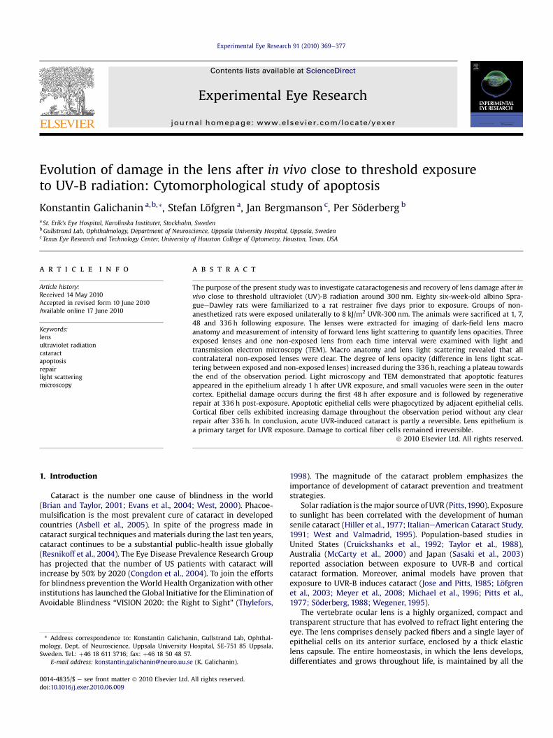
Sections of control lenses demonstrated a normal single layer ofepithelial cells, regular architecture of the nuclear bow, and the lensfibers were packed and oriented in order (Figs. 5 and 6).
One hour after UVR exposure, irregular epithelial cells occasion-ally appeared in the central zone (Fig. 3, 1 h: LM-A), but no distur-banceswere found in other areas of the epithelium. In ultrastructure,irregular epithelial cells in the central zone showed apoptoticfeatures (Fig. 3, 1 h: TEM-A). The nuclear bow (Fig. 3, 1 h: LM-B) andequatorial epithelial cells (Fig. 3, 1 h: TEM-B) appeared normal. Theouter posterior cortex showed small vacuoles (Fig. 3, 1 h: LM-C).Ultrastructurally, superficial lens fiber cells in the anterior, equatorialand posterior cortex had regular structure but there were extracel-lular vacuoles anteriorly (Fig. 3, 1 h: TEM-A) and posteriorly. Deeperfiber cells had normalmorphology throughout thewhole cell length.
At 7 h post-exposure, irregularly shaped epithelial cellsoccurred all over the central epithelial zone (Fig. 3, 7 h: LM-A). Thenuclear bow and the posterior region of the lens appeared normal(Fig. 3, 7 h: LM-B and C). Transmission electron microscopyrevealed that apoptotic cells exhibited chromatin condensation,intact cell membrane and cell convolution (Fig. 3, 7 h: TEM-A).Towards the equator, the epithelium became normal in appearance(Fig. 3, 7 h: TEM-B). The fiber order was disturbed in the outeranterior cortex while equatorial fibers appeared normal (Fig. 3, 7 h:LM-B) but therewere abundant vacuoles in the superficial posteriorcortex (Fig. 3, 7 h: LM-C). Deeper fiber cells were hexagonal intransverse section and arranged in typical order.
At 48 h after exposure, there were areas missing epithelial cellsand remaining epithelial cells contained nuclear and cytoplasmiccondensations, exhibiting apoptotic appearance, and were aggre-gated in multiple layers (Fig. 3, 48 h: LM-A). TEM verified that theepithelium was full of cells with apoptotic bodies, debris andextracellular spaces (Fig. 3, 48 h: TEM-A). The nuclei of these cellswere fragmented andwith dense chromatin. Themembranes of theepithelial cells were intact. Closer to the nuclear bow region,epithelial cells aggregated in multiple layers (Fig. 3, 48 h: TEM-B).Epithelial cells surrounding a target cell through the extensions ofpseudopodia and fusing its pseudopodia to engulf apoptotic bodies,resulting in formation of a phagosome or phago-lysosome,phagocytosis, was observed (Fig. 7).
The nuclear bow was deteriorated and cortical fiber cellsappeared swollen, partly fused and contained vacuoles (Fig. 3, 48 h:LM-B). In the posterior subcapsular area vacuoles were abundant(Fig. 3, 48 h: LM-C). Looking at TEM, superficial fiber cells wereheavily swollen and fusedwith disturbed orientation and numerousvacuoles, while deeper fiber cells remained accurately organized.

Fig. 2. Contralateral non-exposed lenses in bright-field (left) and dark-field (right) illumination. Grid square diameter is 0.79 mm.
Fig. 3. Macrographs (M) of lenses in dark-field illumination (column 1); light micrographs (LM) of central epithelial zone (column 2), nuclear bow region (column 3) and posteriorregion (column 4). Scale bar is 10 mm. Transmission electron micrographs (TEM) of lens epithelium (column 5) and nuclear bow region (column 6) after in vivo exposure to UVR. At1 h after exposure to UVR: LM-A, arrowheads indicate apoptotic epithelial cells with karyopyknosis, TEM-A, one apoptotic cell with condensed cytoplasm (black arrow) among twonormal epithelial cells (white arrow). TEM-B, elongating normal epithelial cells at transitional zone. At 7 h after exposure to UVR: TEM-A, rounding apoptotic cell with kar-yopyknosis and intact cell membrane. TEM-B, normal epithelial cells at transitional zone. At 48 h after exposure to UVR: LM-A, multilayering of the central epithelium and apoptoticcells detached from capsule (black arrowhead), loss of the epithelial cells (white arrowheads). LM-B, apoptotic bodies and debris disintegrating nuclear bow (black arrows). TEM-A,apoptotic bodies (black arrowheads), vacuoles (white arrows); c e capsule, f e fiber cells. TEM-B, debris of several apoptotic bodies (arrow). At 336 h after exposure to UVR: TEM-A,normal epithelial cells with subcapsular vacuoles (black arrows). TEM-B, swollen fiber cells with large extracellular granules; c e capsule, f e fiber cells.
K. Galichanin et al. / Experimental Eye Research 91 (2010) 369e377372

Fig. 4. UVR-exposed lens with nuclear and cortical cataract (A) and cortical cataract (B). Dark-field illumination photography.
K. Galichanin et al. / Experimental Eye Research 91 (2010) 369e377 373
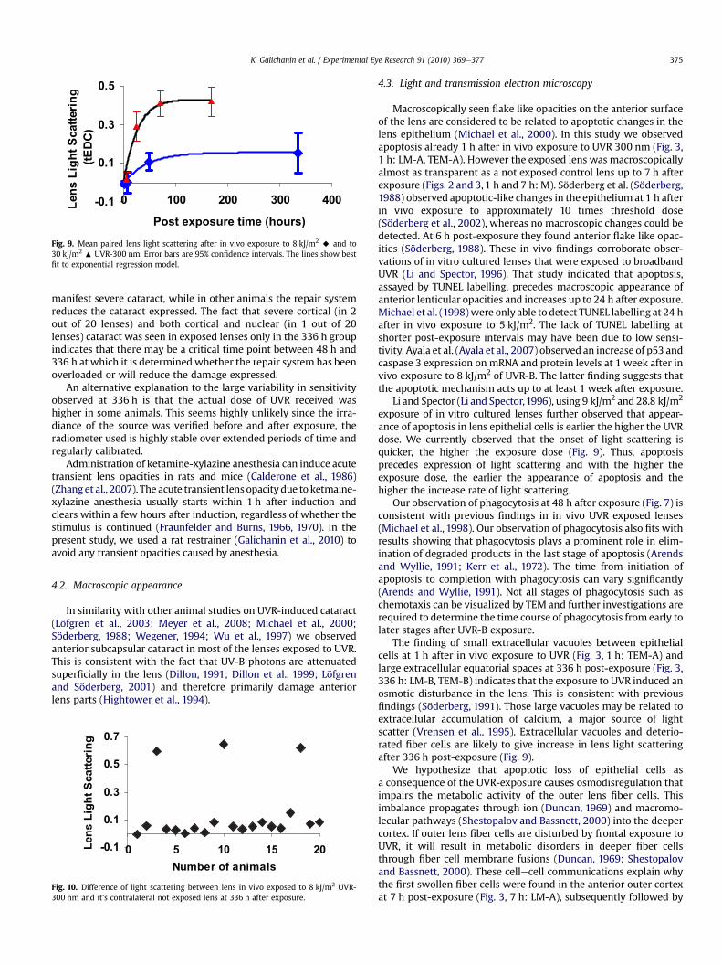
At 336 h after UVR exposure, the epithelium returned toa monolayer of cells (Fig. 3, 336 h: LM-A) but there was frequentlyaccumulations of vacuoles between the epithelial cells and thecapsule (Fig. 3, 336 h: TEM-A). The cortical fiber cells were stilldamaged, without any clear sign of repair (Fig. 3, 336 h: LM-B,TEM-B). The superficial anterior cortex had swollen and heavilyfused fiber cells. The superficial equatorial cortex was composed ofdisoriented, heavily swollen fiber cells with large intercellularvacuoles. The posterior outer cortex exhibited swollen and disor-ganized fiber cells (Fig. 8).
No alterations of the lens capsule were detected after theexposure to UVR. The lens nucleus could not be evaluatedbecause it became opaque after fixation and crumbled duringsectioning.
4. Discussion
This study investigated the evolution of cataract and chronologyof apoptotic events in the lens after in vivo exposure to close tothreshold dose of UVR.
Fig. 5. Sagittal sections of non-exposed rat lens. Central epithelial zone (A
4.1. Lens opacification
Light scattering increases exponentially after exposure to 8 kJ/m2
(Fig. 1), similar to the response to 30 kJ/m2 (Fig. 9) (Söderberg, 1990).However, at the lower dose the onset was slower and the level of
light scattering was lower. In other words, lens light scatteringevolves quicker with higher UVR dose and reaches a higher level.Moreover, the method of animal immobilisation, either restrainingas in the current experiment or anesthesia as in the foregoing study(Söderberg, 1990) does not alter the exponential trend of lensforward light scattering (Fig. 9). The higher level of long term lightscattering induced with a higher dose is consistent with previousfindings demonstrating a continuous dose response relationshipbetween dose of UVR and induced long term light scattering(Söderberg et al., 2003).
An analysis of the high variability of light scattering differencefor the 336 h post exposure interval (Fig. 1) demonstrated thatthree animals expressed a large difference of light scattering,0.6e0.7 tEDC, between exposed and contralateral not exposed lens,while the other animals expressed a lower difference (Fig. 10).
), nuclear bow region (B) and posterior region (C). Scale bar is 10 mm.
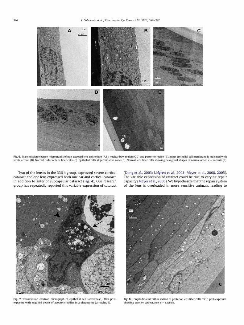
Fig. 6. Transmission electron micrographs of non-exposed lens epithelium (A,B), nuclear bow region (C,D) and posterior region (E). Intact epithelial cell membrane is indicated withwhite arrows (B). Normal order of lens fiber cells (C). Epithelial cells at germinative zone (D). Normal lens fiber cells showing hexagonal shapes in normal order, c e capsule (E).
K. Galichanin et al. / Experimental Eye Research 91 (2010) 369e377374
Two of the lenses in the 336 h group, expressed severe corticalcataract and one lens expressed both nuclear and cortical cataract,in addition to anterior subcapsular cataract (Fig. 4). Our researchgroup has repeatedly reported this variable expression of cataract
Fig. 7. Transmission electron micrograph of epithelial cell (arrowhead) 48 h post-exposure with engulfed debris of apoptotic bodies in a phagosome (arrowhead).
(Dong et al., 2003; Löfgren et al., 2003; Meyer et al., 2008, 2005).The variable expression of cataract could be due to varying repaircapacity (Meyer et al., 2005).We hypothesize that the repair systemof the lens is overloaded in more sensitive animals, leading to
Fig. 8. Longitudinal ultrathin section of posterior lens fiber cells 336 h post-exposure,showing swollen appearance. c e capsule.
-0.1
0.1
0.3
0.5
0 100 200 300 400
Post exposure time (hours)
g
n
i
r
e
t
t
a
c
S
t
h
g
i
L
s
n
e
L
)
C
D
E
t
(
Fig. 9. Mean paired lens light scattering after in vivo exposure to 8 kJ/m2 A and to30 kJ/m2 : UVR-300 nm. Error bars are 95% confidence intervals. The lines show bestfit to exponential regression model.
K. Galichanin et al. / Experimental Eye Research 91 (2010) 369e377 375
manifest severe cataract, while in other animals the repair systemreduces the cataract expressed. The fact that severe cortical (in 2out of 20 lenses) and both cortical and nuclear (in 1 out of 20lenses) cataract was seen in exposed lenses only in the 336 h groupindicates that there may be a critical time point between 48 h and336 h at which it is determined whether the repair system has beenoverloaded or will reduce the damage expressed.
An alternative explanation to the large variability in sensitivityobserved at 336 h is that the actual dose of UVR received washigher in some animals. This seems highly unlikely since the irra-diance of the source was verified before and after exposure, theradiometer used is highly stable over extended periods of time andregularly calibrated.
Administration of ketamine-xylazine anesthesia can induce acutetransient lens opacities in rats and mice (Calderone et al., 1986)(Zhanget al., 2007). The acute transient lens opacitydue to ketmaine-xylazine anesthesia usually starts within 1 h after induction andclears within a few hours after induction, regardless of whether thestimulus is continued (Fraunfelder and Burns, 1966, 1970). In thepresent study, we used a rat restrainer (Galichanin et al., 2010) toavoid any transient opacities caused by anesthesia.
4.2. Macroscopic appearance
In similarity with other animal studies on UVR-induced cataract(Löfgren et al., 2003; Meyer et al., 2008; Michael et al., 2000;Söderberg, 1988; Wegener, 1994; Wu et al., 1997) we observedanterior subcapsular cataract in most of the lenses exposed to UVR.This is consistent with the fact that UV-B photons are attenuatedsuperficially in the lens (Dillon, 1991; Dillon et al., 1999; Löfgrenand Söderberg, 2001) and therefore primarily damage anteriorlens parts (Hightower et al., 1994).
-0.1
0.1
0.3
0.5
0.7
0 5 10 15 20
Number of animals
gn
ir
et
ta
cS
th
gi
Ls
ne
L
Fig. 10. Difference of light scattering between lens in vivo exposed to 8 kJ/m2 UVR-300 nm and it’s contralateral not exposed lens at 336 h after exposure.
4.3. Light and transmission electron microscopy
Macroscopically seen flake like opacities on the anterior surfaceof the lens are considered to be related to apoptotic changes in thelens epithelium (Michael et al., 2000). In this study we observedapoptosis already 1 h after in vivo exposure to UVR 300 nm (Fig. 3,1 h: LM-A, TEM-A). However the exposed lens was macroscopicallyalmost as transparent as a not exposed control lens up to 7 h afterexposure (Figs. 2 and 3,1 h and 7 h: M). Söderberg et al. (Söderberg,1988) observed apoptotic-like changes in the epithelium at 1 h afterin vivo exposure to approximately 10 times threshold dose(Söderberg et al., 2002), whereas no macroscopic changes could bedetected. At 6 h post-exposure they found anterior flake like opac-ities (Söderberg, 1988). These in vivo findings corroborate obser-vations of in vitro cultured lenses that were exposed to broadbandUVR (Li and Spector, 1996). That study indicated that apoptosis,assayed by TUNEL labelling, precedes macroscopic appearance ofanterior lenticular opacities and increases up to 24 h after exposure.Michael et al. (1998)were only able to detect TUNEL labelling at 24 hafter in vivo exposure to 5 kJ/m2. The lack of TUNEL labelling atshorter post-exposure intervals may have been due to low sensi-tivity. Ayala et al. (Ayala et al., 2007) observed an increase of p53 andcaspase 3 expression on mRNA and protein levels at 1 week after invivo exposure to 8 kJ/m2 of UVR-B. The latter finding suggests thatthe apoptotic mechanism acts up to at least 1 week after exposure.
Li and Spector (Li and Spector,1996), using 9 kJ/m2 and 28.8 kJ/m2
exposure of in vitro cultured lenses further observed that appear-ance of apoptosis in lens epithelial cells is earlier the higher the UVRdose. We currently observed that the onset of light scattering isquicker, the higher the exposure dose (Fig. 9). Thus, apoptosisprecedes expression of light scattering and with the higher theexposure dose, the earlier the appearance of apoptosis and thehigher the increase rate of light scattering.
Our observation of phagocytosis at 48 h after exposure (Fig. 7) isconsistent with previous findings in in vivo UVR exposed lenses(Michael et al., 1998). Our observation of phagocytosis also fits withresults showing that phagocytosis plays a prominent role in elim-ination of degraded products in the last stage of apoptosis (Arendsand Wyllie, 1991; Kerr et al., 1972). The time from initiation ofapoptosis to completion with phagocytosis can vary significantly(Arends and Wyllie, 1991). Not all stages of phagocytosis such aschemotaxis can be visualized by TEM and further investigations arerequired to determine the time course of phagocytosis from early tolater stages after UVR-B exposure.
The finding of small extracellular vacuoles between epithelialcells at 1 h after in vivo exposure to UVR (Fig. 3, 1 h: TEM-A) andlarge extracellular equatorial spaces at 336 h post-exposure (Fig. 3,336 h: LM-B, TEM-B) indicates that the exposure to UVR induced anosmotic disturbance in the lens. This is consistent with previousfindings (Söderberg, 1991). Those large vacuoles may be related toextracellular accumulation of calcium, a major source of lightscatter (Vrensen et al., 1995). Extracellular vacuoles and deterio-rated fiber cells are likely to give increase in lens light scatteringafter 336 h post-exposure (Fig. 9).
We hypothesize that apoptotic loss of epithelial cells asa consequence of the UVR-exposure causes osmodisregulation thatimpairs the metabolic activity of the outer lens fiber cells. Thisimbalance propagates through ion (Duncan, 1969) and macromo-lecular pathways (Shestopalov and Bassnett, 2000) into the deepercortex. If outer lens fiber cells are disturbed by frontal exposure toUVR, it will result in metabolic disorders in deeper fiber cellsthrough fiber cell membrane fusions (Duncan, 1969; Shestopalovand Bassnett, 2000). These cellecell communications explain whythe first swollen fiber cells were found in the anterior outer cortexat 7 h post-exposure (Fig. 3, 7 h: LM-A), subsequently followed by
K. Galichanin et al. / Experimental Eye Research 91 (2010) 369e377376
disintegration of fiber cells all over the outer cortex at 48 h post-exposure (Fig. 3, 48 h: LM) and continued damage into the deepercortex at 336 h post-exposure (Fig. 3, 336 h: LM).
During normal lens growth, epithelial cells in the germinativezone proliferate and differentiate into fiber cells, while daughtercells migrate and contribute to the epithelial monolayer (Raffertyand Rafferty, 1981). The return of the epithelium to a monolayerof cells 336 h following UVR exposure (Fig. 3, 336 h: LM-A) may bedue to UVR triggered mitotic activity of stem cells within thegerminative zone. Such daughter cells may have repopulatedcentral zone area where epithelial cells were lost, thus recoveringlens capsule left naked or with remnants of cells that had under-gone apoptosis (Fig. 3, 336 h: TEM-A).
5. Conclusion
The higher the in vivo UVR dose, the faster the development oflens opacification and the higher the end level of lens opacification.Lens epithelial cells appear to be the primary target for UVRexposure. Apoptotic events in the epithelium precede, bothtemporally and spatially, macroscopic cataractogenesis. Epithelialdamage occurs during the first 48 h after exposure and is followedby regenerative repair at 336 h post-exposure. Damage to corticalfiber cells remained irreversible.
Acknowledgements
The authors thankMonica Aronsson for professional animal care,Dr. LindaMeyer for helpful discussions, Berit Spångberg for technicalassistance with light microscopy and Margaret Gondo for help withelectron microsopy imaging. The paper was presented in part as anabstract at the Association for Research in Vision and OphthalmologyCongress 2008. This workwas supported by the Karolinska InstitutetKID-funding, Swedish Radiation Protection Authority, KarolinskaInstitutet Eye Research Foundation, Karolinska Institutet ResearchFoundation, Gun och Bertil Stohnes Stiftelse, Swedish ResearchCouncil; project K2006-74X-15035-03-2, K2008-63X-15035-05-2,Konung Gustav V:s och Drottning Victorias Frimurarstiftelse.
References
Andley, U.P., Clark, B.A., 1989. Generation of oxidants in the near-UV photooxidationof human lens a-crystallin. Investigative Ophthalmology and Visual Science 30,706e713.
Andley, U.P., Walsh, A., Kochevar, I.E., Reddan, J.R., 1990. Effect of ultraviolet-Bradiation on protein synthesis in cultured lens epithelial cells. Current EyeResearch 9, 1099e1106.
Arends, M.J., Wyllie, A.H., 1991. Apoptosis: mechanisms and roles in pathology.International Review of Experimental Pathology 32, 223e254.
Asbell, P.A., Dualan, I., Mindel, J., Brocks, S., Ahmad, M., Epstein, S., 2005. Age relatedcataract. Lancet 365, 599e609.
Ayala, M.N., Strid, H., Jacobsson, U., Söderberg, P.G., 2007. p53 expression andapoptosis in the lens after ultraviolet radiation exposure. InvestigativeOphthalmology of Visual Science 48, 4187e4191.
Bantseev, V., Youn, H.Y., 2006. Mitochondrial “movement” and lens optics followingoxidative stress from. Annals of the New York Academy of Sciences 1091, 17e33.
Brian, G., Taylor, H.R., 2001. Cataract blindness: challenge for the 21 st century.Bulletin of the World Health Organization 79, 249e256.
Calderone, L., Grimes, P., Shalev, M., 1986. Acute reversible cataract induced byxylazine and by ketamine-xylazine anesthesia in rats and mice. ExperimentalEye Research 42, 331e337.
Cruickshanks, K.J., Klein, B.E., Klein, R., 1992. Ultraviolet light exposure and lensopacities: the Beaver Dam Eye Study. American Journal of Public Health 82,1658e1662.
Dillon, J., 1991. The photophysics and photobiology of the eye. Journal of Photo-chemistry and Photobiology B 10, 23e40.
Dillon, J., Zheng, L., Merriam, J.C., Gaillard, E.R., 1999. The optical properties of theanterior segment of the eye: implications for cortical cataract. Experimental EyeResearch 68, 785e795.
Dong, X., Ayala, M., Löfgren, S., Söderberg, P.G., 2003. Ultraviolet radiation-inducedcataract: age and maximum acceptable dose. Investigative Ophthalmology andVisual Science 44, 1150e1154.
Duncan, G., 1969. The site of the ion restricting membranes in the toad lens.Experimental Eye Research 8, 406e412.
Elmore, S., 2007. Apoptosis: a review of programmed cell death. ToxicologicPathology 35.
Evans, J.R., Fletcher, A.E., Wormand, R.P., 2004. Causes of visual impairment inpeople aged 75 years and older in Britain: an add-on study to the MRC Trial ofAssessment and Management of Older People in the Community. British Journalof Ophthalmology 88, 365e370.
Fraunfelder, F.T., Burns, R.P., 1966. Effect of lid-closure in drug-induced experi-mental cataracts. Archives of Ophthalmology 76, 599e601.
Fraunfelder, F.T., Burns, R.P., 1970. Acute reversible lens opacity: caused by drugs,cold, anoxia, asphyxia, stress, death and dehydration. Experimental EyeResearch 10, 19e30.
Galichanin, K., Wang, J., Lofgren, S., Soderberg, P., 2010. A new universal ratrestrainer for ophthalmic research. Acta Ophthalmologica.
Hightower, K., Duncan, G., Dawson, A., Wormstone, I., Reddan, J., Dziedzic, D., 1999.Ultraviolet irradiation (UVB) interrupts calcium cell signaling in lens epithelialcells. Photochemistry and Photobiology 69, 595e598.
Hightower, K., McCready, J., 1992. Mechanisms involved in cataract developmentfollowing near-ultraviolet radiation of cultured lenses. Current Eye Research 11,679e689.
Hightower, K.R., 1995. The role of the lens epithelium in development of UV cataract(Review). Current Eye Research 14, 71e78.
Hightower, K.R., Reddan, J.R., McCready, J.P., Dziedzic, D.C., 1994. Lens epithelium:a primary target of UVB irradiation. Experimental Eye Research 59, 557e564.
Hiller, R., Giacometti, L., Yuen, K., 1977. Sunlight and cataract: an epidemiologicinvestigation. American Journal of Epidemiology 105, 450e459.
ItalianeAmerican Cataract Study, G, 1991. Risk factors for age-related cortical,nuclear, and posterior subcapsular cataracts. American Journal of Epidemiology133, 541e553.
Jose, J.G., Pitts, D.G., 1985. Wavelength dependency of cataracts in albino micefollowing chronic exposure. Experimental Eye Research 41, 545e563.
Kerr, J.F., Wyllie, A.H., Currie, A.R., 1972. Apoptosis: a basic biological phenomenonwith wide-ranging implications in tissue kinetics. British Journal of Cancer 26,239e257.
Kleiman, N.J., Wang, R., Spector, A., 1990. Ultraviolet light induced DNA damage andrepair in bovine lens epithelial cells. Current Eye Research 9, 1185e1193.
Kuszak, J., Costello, M., 2004. The structure of the vertebrate lens. In: Lovicu, F.,Robinson, M. (Eds.), Development of the Ocular Lens. Cambridge UniversityPress, Cambridge, pp. 71e118.
Li, W.C., Kuszak, J.R., Dunn, K., Wang, R.R., Ma, W., Wang, G.M., Spector, A., Leib, M.,Cotiar, A.M., 1995. Lens epithelial cell apoptosis appears to be a commoncellular basis for non-congenital cataract development in humans and animals.Journal of Cell Biology 130, 169e181.
Li, W.C., Spector, A., 1996. Lens epithelial cell apoptosis is an early event in thedevelopment of UVB-induced cataract. Free Radical Biology and Medicine 20,301e311.
Long, A.C., Colitz, C.M., Bomser, J.A., 2004. Apoptotic and necrotic mechanisms ofstress-induced human lens epithelial cell death. Experimental Biology andMedicine (Maywood) 229, 1072e1080.
Löfgren, S., Michael, R., Soderberg, P.G., 2003. Impact of age and sex in ultravioletradiation cataract in the rat. Investigative Ophthalmology and Visual Science44, 1629e1633.
Löfgren, S., Söderberg, P.G., 2001. Lens lactate dehydrogenase inactivation after UV-B irradiation: an in vivo measure of UVR-B penetration. InvestigativeOphthalmology and Visual Science 42, 1833e1836.
McCarty, C.A., Nanjan, M.B., Taylor, H.R., 2000. Attributable risk estimates forcataract to prioritize medical and public health action. Investigative Ophthal-mology of Visual Science 41, 3720e3725.
Meyer, L., Dong, X., Wegener, A., Söderberg, P.G., 2008. Dose dependent cataracto-genesis and Maximum Tolerable Dose (MTD 2.3:16) for UVR - B induced cata-ract in C57BL/6J mice. Experimental Eye Research 86, 282e289.
Meyer, L.M., Soderberg, P., Dong, X., Wegener, A., 2005. UVR-B induced cataractdevelopment in C57 mice. Experimental Eye Research 81, 389e394.
Michael, R., Söderberg, P.G., Chen,E.,1996. Long-termdevelopmentof lensopacitiesafterexposure to ultraviolet radiation at 300 nm. Ophthalmic Research 28, 209e218.
Michael, R., Vrensen, G., van Marle, J., Gan, L., Söderberg, P.G., 1998. Apoptosis in therat lens after in vivo threshold dose ultraviolet irradiation. InvestigativeOphthalmology and Visual Science 13, 2681e2687.
Michael, R., Vrensen, G.F.J.M., Marle, J.V., Löfgren, S., Söderberg, P.G., 2000. Repair inthe rat lens after threshold ultraviolet radiation injury. Investigative Ophthal-mology and Visual Science 41, 204e212.
Pitts, D.G., 1990. Sunlight as an ultraviolet source. Optometry and Vision Science 67,401e406.
Pitts, D.G., Cullen, A.P., Hacker, P.D., 1977. Ocular effects of ultraviolet radiation from295 to 365 nm. Investigative Ophthalmology and Visual Science 16, 932e939.
Rafferty, N.S., Rafferty Jr., K.I., 1981. Cell population kinetics of the mouse lensepithelium. Journal of Cellular Physiology 107, 309e315.
Resnikoff, S., Pascolini, D., Etya’ale, D., Kocur, I., Pararajasegaram, R., Pokharel, G.P.,Mariotti, S.P., 2004. Global data on visual impairment in the year 2002. Bulletinof the World Health Organization 82, 844e851.
Sasaki, H., Kawakami, Y., Ono, M., Jonasson, F., Shui, Y.B., Cheng, H.M., Robman, L.,McCarty, C., Chew, S.J., Sasaki, K., 2003. Localization of cortical cataract insubjects of diverse races and latitude. Investigative Ophthalmology and VisualScience 44, 4210e4214.
K. Galichanin et al. / Experimental Eye Research 91 (2010) 369e377 377
Shestopalov, V.I., Bassnett, S., 2000. Expression of autofluorescent proteins revealsa novel protein permeable pathway between cells in the lens core. Journal ofCell Science 113, 1913e1921.
Söderberg, P.G., 1988. Acute cataract in the rat after exposure to radiation in the 300nm wavelength region. A study of the macro-, micro- and ultrastructure. ActaOphthalmologica (Copenh) 66, 141e152.
Söderberg, P.G., 1990. Development of light dissemination in the rat lens afterexposure to radiation in the 300 nm wavelength region. Ophthalmic Research22, 271e279.
Söderberg, P.G., 1991. Na and K in the lens after exposure to radiation in the 300nm wave length region. Journal of Photochemistry and Photobiology B 8,279e294.
Söderberg, P.G., Chen, E., Lindström, B., 1990. An objective and rapid method for thedetermination of light dissemination in the lens. Acta Ophthalmologica(Copenh) 68, 44e52.
Söderberg, P.G., Löfgren, S., Ayala, M., Dong, X., Kakar, M., Mody, V., 2002. Toxicity ofultraviolet radiation exposure to the lens expressed by maximum tolerable dose(MTD). Developments in Ophthalmology 35, 70e75.
Söderberg, P.G., Michael, R., Merriam, J.C., 2003. Maximum acceptable dose ofultraviolet radiation: a safety limit for cataract. Acta Ophthalmologica Scandi-navica 81, 165e169.
Söderberg, P.G., Philipson, B.T., Lindström, B., 1986. Unscheduled DNA synthesis inlens epithelium after in vivo exposure to UV radiation in the 300 nm wave-length region. Acta Ophthalmologica (Copenh) 64, 162e168.
Taylor, H.R., West, S.K., Rosenthal, F.S., Munoz, B., Newland, H.S., Abbey, H.,Emmett, E.A., 1988. Effect of ultraviolet radiation on cataract formation. NewEngland Journal of Medicine 319, 1429e1433.
Congdon, N., Vingerling, J.R., Klein, B.E., West, S., Friedman, D.S., Kempen, J.,O'Colmain, B., Wu, S.Y., Taylor, H.R., 2004. Prevalence of cataract and pseudo-phakia/aphakia among adults in the United States. Archives of Ophthalmology122, 487e494.
Thylefors, B., 1998. A global initiative for the elimination of avoidable blindness.American Journal of Ophthalmology 125, 90e93.
Vrensen, G.F.J.M., Sanderson, J., Willekens, B., Duncan, G., 1995. Calcium localizationand ultrastructure of clear and pCMPS-treated rat lenses. InvestigativeOphthalmology and Visual Science 36, 2287e2295.
Wegener, A.R., 1994. In vivo studies on the effect of UV-radiation on the eye lens inanimals. Documenta Ophthalmologica 3e4, 221e232.
Wegener, A.R., 1995. In vivo studies on the effect of UV-radiation on the eye lens inanimals. Documenta Ophthalmologica 88, 221e232.
West, S.K., 2000. Looking forward to 20/20: a focus on the epidemiology of eyediseases. Epidemiologic Reviews 22, 64e70.
West, S.K., Valmadrid, C.T., 1995. Epidemiology of risk factors for age-related cata-ract. Survey of Ophthalmology 39, 323e334.
Wu, K., Shui, Y., Kojima, M., Murano, H., Sasaki, K., Hockwin, O., 1997. Location andseverity of UV-B irradiation damage in rat lens. Japanese Journal of Ophthal-mology 41, 381e387.
Zhang, F., Löfgren, S., Söderberg, P.G., 2007. Interaction of anesthetic drugs and UVR-B irradiation in the anterior segment of the rat eye. Acta OphthalmologicaScandinavica 85, 745e752.
Zigman, S., 1985. Photobiology of the lens. In: The Ocular Lens. Structure, Functionand Pathology. Marcel Dekker, New York, pp. 301e347.
Zigman, S., Griess, G., Yulo, T., Schultz, J., 1973. Ocular protein alterations by near UVlight. Experimental Eye Research 15, 255e264.