Embed Size (px)
Citation preview
Experimental model of intervertebral discdegeneration by needle puncture in
Wistar rats
A.C. Issy1, V. Castania1, M. Castania1, C.E.G. Salmon2, M.H. Nogueira-Barbosa3,
E. Del Bel1 and H.L.A. Defino4
1Departamento de Morfologia, Fisiologia e Patologia Basica, Faculdade de Odontologia de Ribeirao Preto,
Universidade de Sao Paulo, Ribeirao Preto, SP, Brasil2Departamento de Fısica, Faculdade de Filosofia, Ciencias e Letras de Ribeirao Preto, Universidade de Sao Paulo,
Ribeirao Preto, SP, Brasil3Divisao de Radiologia, Departamento de Clınica Medica, Faculdade de Medicina de Ribeirao Preto,
Universidade de Sao Paulo, Ribeirao Preto, SP, Brasil4Departamento de Biomecanica, Medicina e Reabilitacao do Sistema Locomotor, Faculdade de Medicina de Ribeirao Preto,
Universidade de Sao Paulo, Ribeirao Preto, SP, Brasil
Abstract
Animal models of intervertebral disc degeneration play an important role in clarifying the physiopathological mechanisms and
testing novel therapeutic strategies. The objective of the present study is to describe a simple animal model of disc
degeneration involving Wistar rats to be used for research studies. Disc degeneration was confirmed and classified by
radiography, magnetic resonance and histological evaluation. Adult male Wistar rats were anesthetized and submitted to
percutaneous disc puncture with a 20-gauge needle on levels 6-7 and 8-9 of the coccygeal vertebrae. The needle was inserted
into the discs guided by fluoroscopy and its tip was positioned crossing the nucleus pulposus up to the contralateral annulus
fibrosus, rotated 3606 twice, and held for 30 s. To grade the severity of intervertebral disc degeneration, we measured the
intervertebral disc height from radiographic images 7 and 30 days after the injury, and the signal intensity T2-weighted
magnetic resonance imaging. Histological analysis was performed with hematoxylin-eosin and collagen fiber orientation using
picrosirius red staining and polarized light microscopy. Imaging and histological score analyses revealed significant disc
degeneration both 7 and 30 days after the lesion, without deaths or systemic complications. Interobserver histological
evaluation showed significant agreement. There was a significant positive correlation between histological score and
intervertebral disc height 7 and 30 days after the lesion. We conclude that the tail disc puncture method using Wistar rats is a
simple, cost-effective and reproducible model for inducing disc degeneration.
Key words: Intervertebral disc degeneration; Animal models; Histology; Wistar rats
Introduction
Degenerative disc disease (DDD) is one of the most
important public health problems and one of the most
common causes of low back pain (1,2). DDD is
characterized by complex serial progressive morphologi-
cal, biochemical and biomechanical changes of the
intervertebral disc that contribute to the impairment of
mechanical function and, in some cases, to the onset of
discogenic low back pain (3). The causes of DDD are
multifactorial and a clear understanding of the pathophy-
siology and pathogenesis of the condition is still lacking
(4,5).
The initiation and progression of the degenerative
cascade probably depend on multiple interdependent
factors. It has been suggested that these factors may
include reduced nutrient supply (6), hereditary factors
(7), altered mechanical loading (8), age (9), up-
regulated levels of proinflammatory cytokines and
associated catabolic enzymes (10), and decreased
diffusion of nutrients through the endplates (6).
Evidently, the occurrence of one or more of these
factors can promote several disc tissue changes (for a
review, see Ref. 11).
Correspondence: E. Del Bel, Departamento de Morfologia, Estomatologia e Fisiologia, Faculdade de Odontologia de Ribeirao Preto,
Universidade de Sao Paulo, 14049-904 Ribeirao Preto, SP, Brasil. E-mail: [email protected]
Received June 18, 2012. Accepted October 16, 2012. First published online March 15, 2013.
Brazilian Journal of Medical and Biological Research (2013) 46: 235-244, http://dx.doi.org/10.1590/1414-431X20122429
ISSN 1414-431X
www.bjournal.com.br Braz J Med Biol Res 46(3) 2013
Animal models of intervertebral disc degeneration play
an important role in clarifying pathomechanisms and
testing novel therapeutic strategies for this condition (12).
No ideal model for DDD currently exists, although several
categories of animal models have been developed. These
models can be classified as either experimentally induced
(mechanical or structural models) or spontaneous, which
include animals genetically altered or specially bred to
develop degenerative disc disease (13,14). Many rodent
disc degeneration models have been described and are
advantageous for DDD because of their low cost and
ease of care (15).
A recently developed needle puncture model has
shown altered morphological and biochemical features
similar to many of those found in human degenerative
discs (4,16). In the injury model, the use of needle
puncture has gained popularity mainly because of its
reproducibility and the short time required to produce the
desired degenerative effect (17). The rat tail disc has
been proposed as a platform for the puncture model
inducing disc degeneration (17-22). Rodent tail discs can
be easily manipulated to induce degeneration and
represent a desirable model for disc degeneration/
regeneration research (20). The tail disc is accessible to
intervention and does not require open exposure or tissue
retraction. As a result, tail disc annular puncture involves
a minimal risk of damage to surrounding structures. In
addition, while human disc degeneration is a complex and
multifactorial process that occurs over decades, it is clear
that focal annular injury is a potential initiator of disc
degeneration (23).
The needle puncture model was first described for use
in the lumbar spine of rabbits (16) and, because of its
advantages, was also used in rodents in a few publica-
tions, all of them using Sprague-Dawley rats (18,20-22). It
is important to point out that there are differences in
intervertebral discs regarding species, spinal level and
age (for a review, see Ref. 12). The properties of the discs
vary significantly at different locations within the spine
level of the same animal, e.g., caudal and lumbar discs of
the rat (24). The shape, profiles and relative sizes of the
intervertebral disc and adjacent spinal tissues vary
between species, with variations also occurring in the
ossification centers. The variation of properties (biome-
chanical, morphological and cellular) between regions
within the disc itself is also an important factor to consider
in choosing and designing a model (12). Also, there may
be variation between different colonies of the same animal
strain (12). A rodent DDD model using Wistar rats, which
are available in Brazil, has not been reported previously.
Thus, the objective of the present study is to describe
an experimental model of DDD using Wistar rats, induced
by needle puncture as a simple, less invasive and reliable
method to study intervertebral disc degeneration since
this method allows a complete experimental condition in
the tail of the same animal. The model was validated by
imaging (radiography and magnetic resonance) and
histological analysis.
Material and Methods
Experimental animals
Sixteen Wistar rats (300-350 g) obtained from the
Animal House of the Ribeirao Preto Campus, University of
Sao Paulo, Brazil, were used. Before the experiments, the
animals were housed in groups of 4 and kept at a
temperature of 23 ± 16C on a 12-h light-dark cycle. Food
and drinking water were available ad libitum. All experi-
ments were carried out in accordance with the Brazilian
Society of Neuroscience and Behavior guidelines for
animal care and all efforts were made to minimize animal
suffering.
Needle puncture technique
Animals were anesthetized with a combination of
ketamine and xylazine (10:7, 100 mg/kg injected intraper-
itoneally). The coccygeal intervertebral spaces Co6-7,
Co7-8, and Co8-9 were selected for the study. The needle
puncture was performed at coccygeal intervertebral levels
Co6-7 and Co8-9. The intervertebral level Co7-8
remained undisturbed as the control level. The selected
coccygeal intervertebral levels (Co6-7 and Co8-9) were
identified by digital palpation and confirmed by fluoro-
scopy. After tail skin antisepsis with alcohol iodate and
using fluoroscopy, a 20-gauge needle was inserted at the
level of the annulus fibrosus of Co6-7 (proximal) and Co8-
9 (distal), crossing the nucleus pulposus up to the
contralateral annulus fibrosus. After full penetration, the
needle was rotated 3606 twice and held for 30 s. The
depth of needle penetration was controlled by the
resistance of the contralateral annulus fibrosus.
Magnetic resonance imaging (MRI) acquisition
MRI was performed on 9 animals 30 days after
intervertebral disc puncture. Animals were anesthetized
as described before so they would remain immobile
throughout the entire MRI examination. Images were
acquired with a 3.0 T MRI machine (Philips, Achieva, The
Netherlands) using a dedicated coil for small animals. The
tail was inserted into a tube containing a 0.1 M CuSO4
solution to reduce the effects of susceptibility and to
increase the contrast in the image. We used a 2-D spin
echo-dual echo sequence with the following parameters:
repetition time = 9000 ms, echo times = 16 and 80 ms,
flip angle = 90, number of averages = 2, slice thickness
= 0.6 mm, field of view = 40 6 40 mm, in plane
resolution = 0.1 mm, 30 sagittal slices. The disc signal
intensity was calculated using the T2-weighted image
(echo time = 80 ms) for better visualization as an indirect
measure of disc hydration since it is well known that
reduction of water content is a common finding in
intervertebral disc degeneration. The mean signal inten-
236 A.C. Issy et al.
Braz J Med Biol Res 46(3) 2013 www.bjournal.com.br
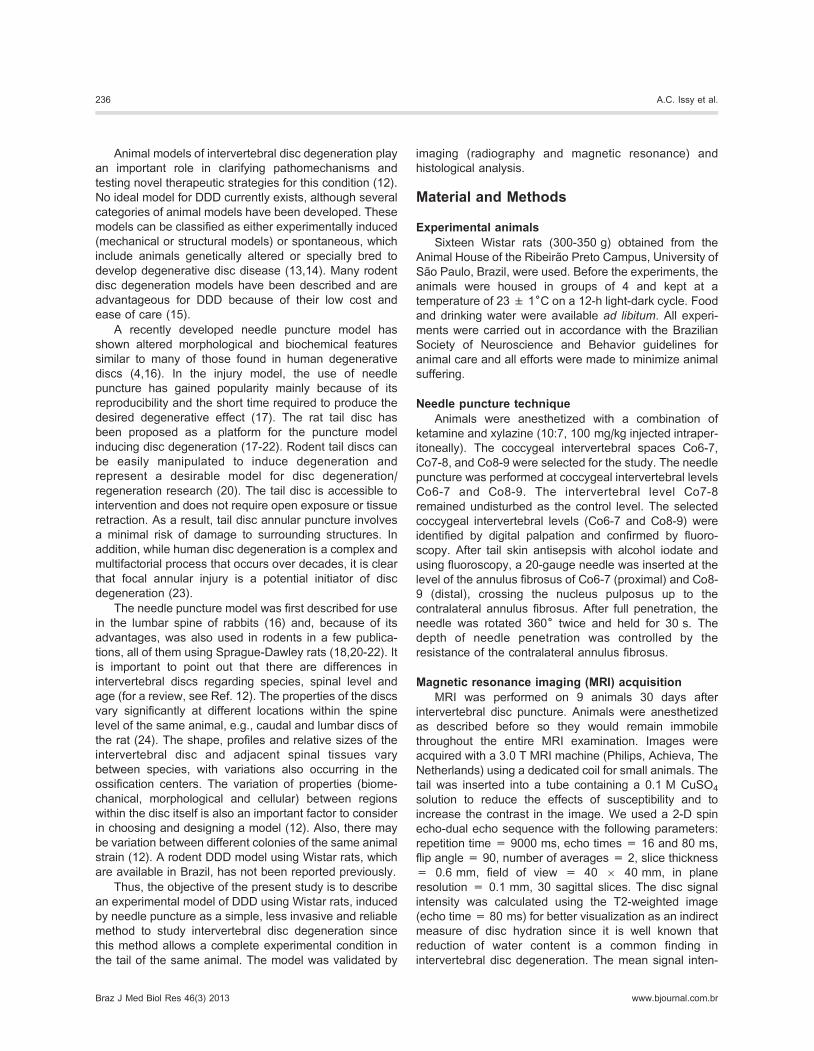
sity (brightness) in the control disc was set as reference
for the signal intensity of the injured discs in each animal
(Figure 1). Thus, the normalized intensity of the injured
discs had a value between 0 and 1.
Radiographic analysis
Radiographs of all rats were taken under anesthesia
just before and after intervertebral disc puncture at 7 (n =
7) or 30 (n = 9) days. The animals were placed on a
millimeter radiopaque scale to improve the identification of
the intervertebral disc level (Figure 2). Radiographic
images were taken with a Siemens MultixH instrument
(Germany; 35 kV, 3 mA, 2.5 ms, 1 m) and were scanned
and digitally stored using an image capture software
program. Extreme care was taken to maintain a consis-
tent level of anesthesia during radiography of each animal
and at each time point (before and after lesion) in order to
obtain a similar degree of muscle relaxation, which may
affect the disc height. Therefore, based on the method
proposed by Masuda et al. (4), the preoperative radio-
graph was always used as a baseline measurement.
Using digitized radiographs, measurements including the
proximal (PV) and distal (DV) vertebral body height and
intervertebral disc height (DH) were analyzed using the
public domain image analysis program developed by the
U.S. National Institutes of Health - ImageJ (http://imagej.
nih.gov/ij/) (Figure 2). Data were transported to the Excel
software and the intervertebral disc height was reported
as the DH index (DHI) (4). DHI was calculated by
averaging the measurements obtained from the anterior
(1), middle (2), and posterior (3) portions of the DH and
dividing them by the average height of the adjacent
vertebral body (Formula 1). Changes in the DHI of
punctured discs were expressed as %DHI and normalized
to the measured preoperative intervertebral disc height
(Formula 2).
DHI~2 � DH1zDH2zDH3ð Þ=½ PV1zPV2zPV3ð Þz(DV1zDV2zDV3)�
(Formula 1)
%DHI~(DHI post-lesion=DHI pre-lesion)�100 (Formula 2)
Figure 1. T2-weighted magnetic resonance imaging (MRI) of
intervertebral discs. A, Normal discs before lesion. B, Punctureddiscs 30 days after lesion. The first arrow indicates the distal
segment (Co8-9) and the second one the proximal segment
(Co6-7). C, ImageJ analysis. D, The x-axis indicates the distance
measured in millimeters and the y-axis indicates mean MRI
intensity profile along the yellow line. The peak value (± 60)
indicates the control segment and the depressions of the curve
indicate the punctured segments marked by dashed lines on the
graph. The results were normalized according to the respective
control.
Figure 2. Radiographic assessment of intervertebral disc
degeneration. A, Radiographic images were taken using a
radiopaque scale with millimeter markings to recognize the
intervertebral disc level of interest. Arrows indicate punctured
intervertebral discs. B, Using digitized radiographs, measure-
ments including the proximal (PV) and distal (DV) vertebral body
height and intervertebral disc height (DH) were analyzed using
the public domain image analysis program developed by the U.S.
National Institutes of Health - ImageJ (http://imagej.nih.gov/ij/).
1 = anterior; 2 = middle; 3 = posterior.
Injury model by needle puncture of the Wistar rat tail 237
www.bjournal.com.br Braz J Med Biol Res 46(3) 2013
Histological analysis
Animals were sacrificed at 7 (n = 7) or 30 (n = 9) days
after the tail needle puncture by excess anesthesia with
ketamine/xylazine. The whole discs with the vertebrae
adjacent to the punctured segments (Co6-7 and Co8-9) and
non-punctured segment (Co7-8) were removed and dis-
sected. Tissue was fixed in 4% paraformaldehyde, pH 7.4,
for 24 h and decalcified in 10% ethylenediaminetetraacetic
acid (EDTA) for 30 days, paraffin-embedded, and sectioned
to 5-mm thickness with a microtome. The sections were
stained with hematoxylin and eosin for histological score
and graded in a blind fashion using the definition
established by Norcross et al. (18), with some modifications
(Table 1), under a light microscope (LeicaH, Germany) at
106 magnification. This scale scores the disruption of the
collagenous architecture and cellularity of the nucleus but
not the vascular changes. The slides were graded based on
the histological appearance of the characteristics of the
nucleus pulposus and annulus fibrosus. The qualitative
analysis of collagen fiber organization was performed using
picrosirius red staining and polarized light.
Picrosirius red
Sagittal sections of intervertebral discs and adjacent
vertebral body segments were stained with picrosirius red
to visualize changes in collagen organization/orientation.
The slides were initially deparaffinized in xylene (3
changes - 10 min), followed by two ethanol washes - first
absolute ethanol (3 changes - 3 min) and next 95%
ethanol (v/v; 3 min), and washed rapidly in water. Tissue
sections were stained with picrosirius (0.5 g Sirius red and
500 mL saturated picric acid) for 1 h and were again
washed rapidly in water and 95% ethanol since Sirius red
is soluble in water. Tissue sections were then washed in
several changes of absolute ethanol (3 changes - 3 min)
cleared in xylene (3 changes - 3 min) and mounted.
Reliability of images and histological analyses
To assess the interobserver reliability of the analyses,
2 authors independently performed histological rating.
The degree of interobserver agreement regarding the
histological analyses was determined using the Cohen
kappa coefficient.
Statistical analyses
The normalized intensity values from MRI were
analyzed by the paired t-test. Intervertebral disc height
and histological score were analyzed by univariate
analysis of variance with condition (control or lesion
segments) as between-subject comparison. All tests were
followed by the Duncan test for multiple comparisons (P ,
0.05). Histological score and intervertebral disc height
data were tested by Pearson’s correlation analysis.
Results
MRI
The T2-weighted images retrieved from serial imaging
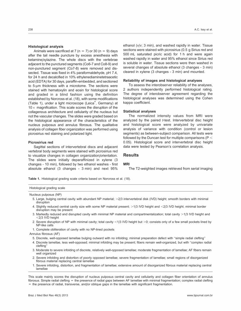
Table 1. Histological grading scale criteria based on Norcross et al. (18).
Histological grading scale
Nucleus pulposus (NP)
5. Large, bulging central cavity with abundant NP material; .2/3 intervertebral disk (IVD) height; smooth borders with minimaldisruption
4. Slightly reduced central cavity size with some NP material present; .1/3 IVD height and ,2/3 IVD height; minimal borderdisruption may be present
3. Markedly reduced and disrupted cavity with minimal NP material and compartmentalization; total cavity .1/3 IVD height and,2/3 IVD height
2. Severe disruption of NP with minimal cavity; total cavity ,1/3 IVD height but .0; consists only of a few small pockets lined byNP-like cells
1. Complete obliteration of cavity with no NP-lined pockets
Annulus fibrosus (AF)
5. Discrete, well-opposed lamellae bulging outward with no infolding; minimal preparation defect with ‘‘simple radial clefting’’
4. Discrete lamellae, less well-opposed; minimal infolding may be present; fibers remain well-organized, but with ‘‘complex radialclefting’’
3. Moderate to severe infolding of discrete, relatively well-opposed lamellae; moderate fragmentation of lamellae; AF fibers remainwell organized
2. Severe infolding and distortion of poorly opposed lamellae; severe fragmentation of lamellae; small regions of disorganizedfibrous material replacing central lamellae
1. Severe infolding, distortion, and fragmentation of lamellae; extensive amount of disorganized fibrous material replacing centrallamellae
This scale mainly scores the disruption of nucleus pulposus central cavity and cellularity and collagen fiber orientation of annulus
fibrosus. Simple radial clefting = the presence of radial gaps between AF lamellae with minimal fragmentation; complex radial clefting
= the presence of radial, transverse, and/or oblique gaps in the lamellae with significant fragmentation.
238 A.C. Issy et al.
Braz J Med Biol Res 46(3) 2013 www.bjournal.com.br
studies of the coccygeal disc showed degeneration of the
disc 30 days after needle puncture. A decrease of signal
intensity was observed in punctured discs and the control
discs did not reveal loss of the signal. The control disc
remained consistent over the 30-day period of evaluation.
Punctured discs showed a significant decrease of the MRI
signal compared to control discs (Co8-9: t8 = -4.75; P ,
0.001 and Co6-7: t8 = 8.49; P , 0.001) (paired t-test, P ,
0.05) (Figure 3) 30 days after the lesion. We did not detect
any difference in MRI signal between punctured discs
(Co8-9 and Co6-7).
Radiographic assessment
Radiographic assessment of the disc height was
performed considering the DHI, that was calculated by
averaging measures before and after disc needle punc-
ture. The control discs showed no significant differences
among time points. Punctured discs showed a significant
decrease in intervertebral DHI compared to control discs
[F(5,48) = 6.83; P = 0.001] 7 and 30 days after tail
needle puncture (univariate analysis of variance followed
by the post hoc Duncan test, P , 0.05; Table 2). The
effect of time after needle puncture on DHI was not
statistically significant. No significant difference in inter-
vertebral disc height was observed between punctured
discs (Co8-9 and Co6-7), or between 7 and 30 days after
lesion.
Histological score
Histological sections of the nucleus pulposus and
annulus fibrosus showed a range of morphological
changes after needle puncture. The nucleus pulposus
and annulus fibrosus of punctured discs showed sig-
nificant alterations compared to control discs 7 and 30
days after lesion [F(11,180) = 19.50; P = 0.001] by
univariate analysis of variance followed by the post hoc
Duncan test, P , 0.05 (Figures 4 and 5). No significant
difference in histological score was observed between
punctured discs (Co8-9 and Co6-7), or between 7 and 30
days after lesion. The only difference found was in the
annulus fibrosus score between 7 and 30 days after
lesion. Since we detected a significant degree of
histological interobserver agreement determined by the
Cohen kappa coefficient, the data are reported as the
average of observers 1 and 2 (Figure 4). Collagen fiber
orientation was assessed using picrosirius red staining
and polarized microscopy and is shown in Figure 6.
Correlations between techniques
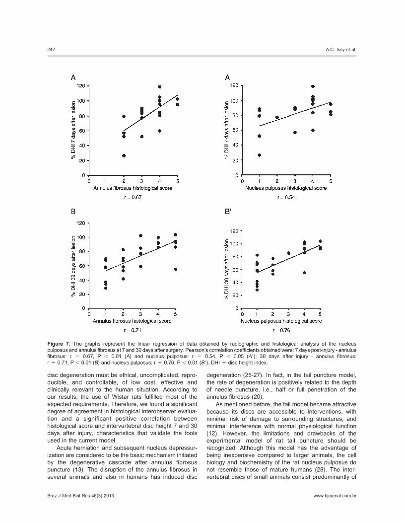
Pearson’s correlation (P , 0.05) analyses revealed a
significant positive correlation between histological score
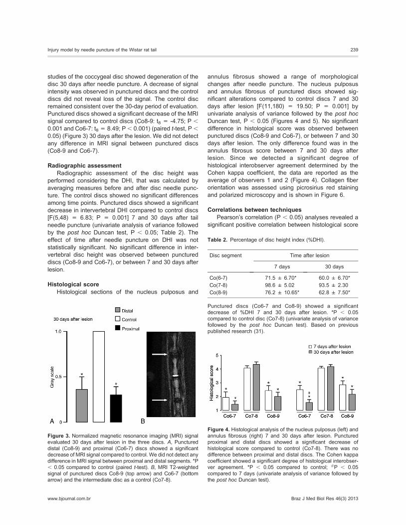
Figure 3. Normalized magnetic resonance imaging (MRI) signal
evaluated 30 days after lesion in the three discs. A, Punctureddistal (Co8-9) and proximal (Co6-7) discs showed a significant
decrease of MRI signal compared to control. We did not detect any
difference in MRI signal between proximal and distal segments. *P
, 0.05 compared to control (paired t-test). B, MRI T2-weighted
signal of punctured discs Co8-9 (top arrow) and Co6-7 (bottom
arrow) and the intermediate disc as a control (Co7-8).
Table 2. Percentage of disc height index (%DHI).
Disc segment Time after lesion
7 days 30 days
Co(6-7) 71.5 ± 6.70* 60.0 ± 6.70*
Co(7-8) 98.6 ± 5.02 93.5 ± 2.30
Co(8-9) 76.2 ± 10.65* 62.8 ± 7.50*
Punctured discs (Co6-7 and Co8-9) showed a significant
decrease of %DHI 7 and 30 days after lesion. *P , 0.05
compared to control disc (Co7-8) (univariate analysis of variance
followed by the post hoc Duncan test). Based on previous
published research (31).
Figure 4. Histological analysis of the nucleus pulposus (left) and
annulus fibrosus (right) 7 and 30 days after lesion. Punctured
proximal and distal discs showed a significant decrease of
histological score compared to control (Co7-8). There was no
difference between proximal and distal discs. The Cohen kappa
coefficient showed a significant degree of histological interobser-
ver agreement. *P , 0.05 compared to control; #P , 0.05
compared to 7 days (univariate analysis of variance followed by
the post hoc Duncan test).
Injury model by needle puncture of the Wistar rat tail 239
www.bjournal.com.br Braz J Med Biol Res 46(3) 2013
and intervertebral disc height 7 and 30 days after lesion in
both regions (nucleus pulposus and annulus fibrosus).
Linear regression curves are presented in Figure 7. We
did not calculate the correlation between histological
score and MRI because we tested only the latest time (30
days after lesion) by MRI.
Discussion
Percutaneous needle puncture induces degenerative
signs similar to those of human discs (16,18,20-22).
Needle puncture of the caudal discs of Wistar rats
induced early and clear degeneration very similar to that
previously reported for rabbits or Sprague-Dawley rats
(16,20-22). The described model is simple and economic
and seems to be appropriate for investigating the
pathogenesis of intervertebral disc degeneration. Image
and histological outcome measures validated the model
presented here and may strongly predict the severity of
disc degeneration.
The main features observed included disc space
narrowing, decreased disc height, water content reduc-
tion, and histological disorganization. Among classical
histological alterations, we detected changes ranging
from discrete disruption of the nucleus pulposus and a
small decrease of its cavity to its complete obliteration and
absence of nucleus pulposus cells. Similarly, we were
able to measure different degrees of lamellar disorganiza-
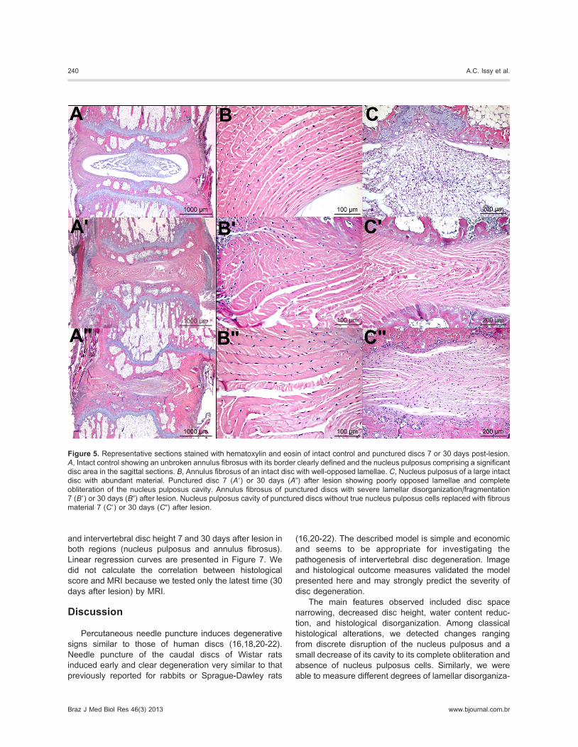
Figure 5. Representative sections stained with hematoxylin and eosin of intact control and punctured discs 7 or 30 days post-lesion.
A, Intact control showing an unbroken annulus fibrosus with its border clearly defined and the nucleus pulposus comprising a significant
disc area in the sagittal sections. B, Annulus fibrosus of an intact disc with well-opposed lamellae. C, Nucleus pulposus of a large intact
disc with abundant material. Punctured disc 7 (A9) or 30 days (A0) after lesion showing poorly opposed lamellae and complete
obliteration of the nucleus pulposus cavity. Annulus fibrosus of punctured discs with severe lamellar disorganization/fragmentation
7 (B9) or 30 days (B0) after lesion. Nucleus pulposus cavity of punctured discs without true nucleus pulposus cells replaced with fibrous
material 7 (C9) or 30 days (C0) after lesion.
240 A.C. Issy et al.
Braz J Med Biol Res 46(3) 2013 www.bjournal.com.br
tion of the annulus fibrosus. These results are similar to
those first obtained with Sprague Dawley rats (20-22).
We already know that in experimental models the
severity of intervertebral disc degeneration is directly
correlated to the needle gauge used. A larger diameter
needle consistently resulted in degeneration (16,20,21).
The severity of disc degeneration could be reasonably
calculated by the selection of needle gauge. Small
diameter needles, for example more than 30 gauge,
could be used to inject gene therapy agents or several
other biological agents for disc manipulation. According to
our results, the 20-gauge needle was able to induce
important disc degeneration 7 days after lesion, and these
results were not different 30 days post-lesion. Therefore,
our model did not reveal any spontaneous regeneration
and suggests a stable injury. In a recent publication,
Zhang et al. (22) reported that 21-gauge needle puncture
into the rat tail disc induces a rapid and progressive disc
degeneration process without spontaneous recovery.
Comparing our results for 7 and 30 days after injury, a
tendency to progressive disc degeneration was detected
in DHI (Table 2) and in the histological score (Figure 4).
This progressive injury is a relevant representation of disc
degeneration in humans and is suitable for evaluating the
effectiveness of new treatments (4). Also, the significant
correlations between %DHI values and histological results
(Figure 7) confirm that DHI can be used as a good
indicator of the injury (4).
Additionally, we did not find any difference in MRI
signal between proximal and distal segments (Figure 3).
The same finding was repeated in radiographic (Table 2)
and histological data (Figure 4). This independence
between the injury and different segments of the caudal
level suggest that in this model several conditions could
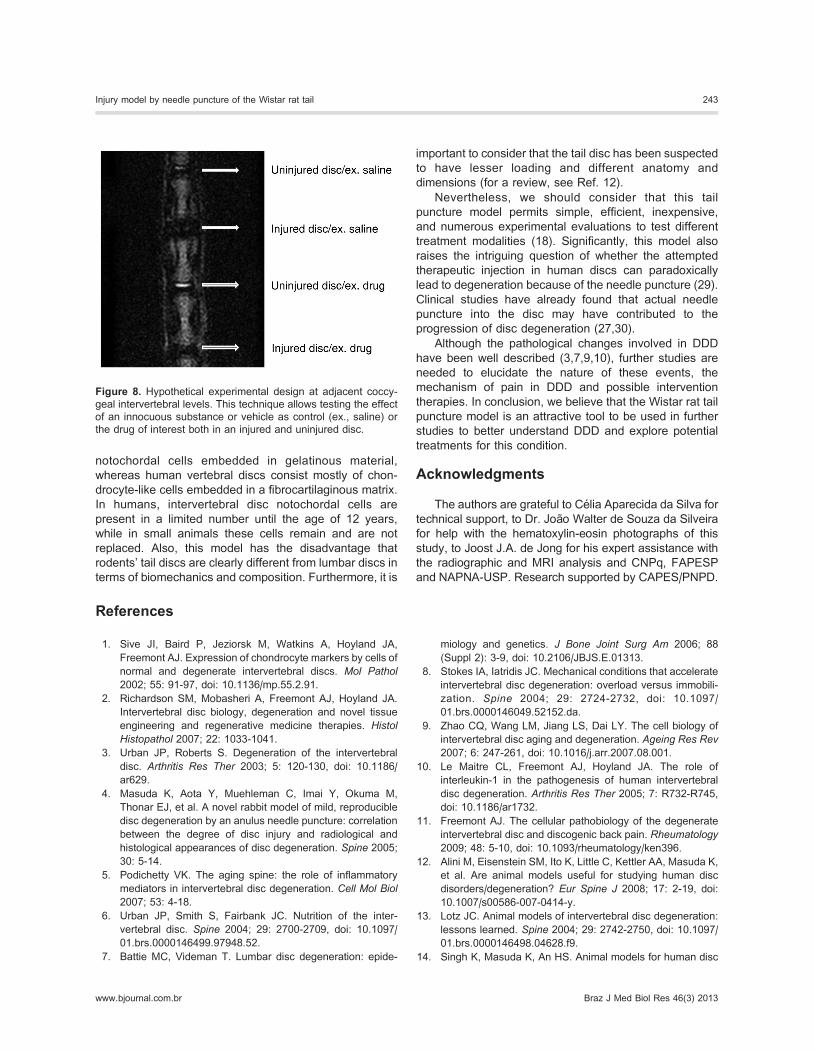
be tested in the same tail. Indeed, we conducted another
cohort of experiments (data not shown here) using
adjacent coccygeal intervertebral levels to promote a
complete experimental condition in order to test the effect
of an innocuous substance (as control) or the drug of
interest either in a lesioned or intact disc with success
(Figure 8).
The annulus puncture model for inducing disc degen-
eration was initially used in the lumbar spine of rabbits
(16). Rodent tail disc degeneration was later used
considering its easy manipulation since it does not require
a surgical procedure (20). Additionally, the tail disc
degeneration model induced by needle puncture is a
simplified low-cost procedure. To our knowledge, the use
of Wistar rats as an animal model for disc degeneration
has not been reported before. Basically, animal models of
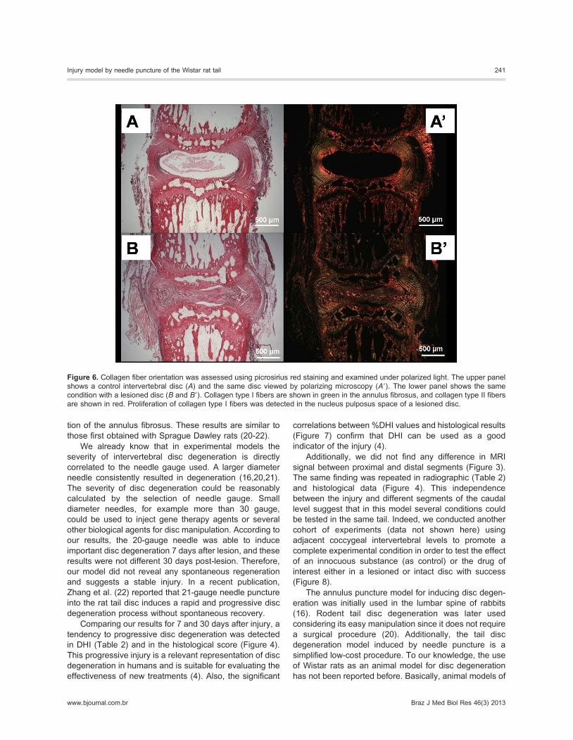
Figure 6. Collagen fiber orientation was assessed using picrosirius red staining and examined under polarized light. The upper panel
shows a control intervertebral disc (A) and the same disc viewed by polarizing microscopy (A9). The lower panel shows the same
condition with a lesioned disc (B and B9). Collagen type I fibers are shown in green in the annulus fibrosus, and collagen type II fibers
are shown in red. Proliferation of collagen type I fibers was detected in the nucleus pulposus space of a lesioned disc.
Injury model by needle puncture of the Wistar rat tail 241
www.bjournal.com.br Braz J Med Biol Res 46(3) 2013
disc degeneration must be ethical, uncomplicated, repro-
ducible, and controllable, of low cost, effective and
clinically relevant to the human situation. According to
our results, the use of Wistar rats fulfilled most of the
expected requirements. Therefore, we found a significant
degree of agreement in histological interobserver evalua-
tion and a significant positive correlation between
histological score and intervertebral disc height 7 and 30
days after injury, characteristics that validate the tools
used in the current model.
Acute herniation and subsequent nucleus depressur-
ization are considered to be the basic mechanism initiated
by the degenerative cascade after annulus fibrosus
puncture (13). The disruption of the annulus fibrosus in
several animals and also in humans has induced disc
degeneration (25-27). In fact, in the tail puncture model,
the rate of degeneration is positively related to the depth
of needle puncture, i.e., half or full penetration of the
annulus fibrosus (20).
As mentioned before, the tail model became attractive
because its discs are accessible to interventions, with
minimal risk of damage to surrounding structures, and
minimal interference with normal physiological function
(12). However, the limitations and drawbacks of the
experimental model of rat tail puncture should be
recognized. Although this model has the advantage of
being inexpensive compared to larger animals, the cell
biology and biochemistry of the rat nucleus pulposus do
not resemble those of mature humans (28). The inter-
vertebral discs of small animals consist predominantly of
Figure 7. The graphs represent the linear regression of data obtained by radiographic and histological analysis of the nucleus
pulposus and annulus fibrosus at 7 and 30 days after surgery. Pearson’s correlation coefficients obtained were: 7 days post-injury - annulus
fibrosus: r = 0.67, P , 0.01 (A) and nucleus pulposus: r = 0.54, P , 0.05 (A9); 30 days after injury - annulus fibrosus:
r = 0.71, P , 0.01 (B) and nucleus pulposus: r = 0.76, P , 0.01 (B9). DHI = disc height index.
242 A.C. Issy et al.
Braz J Med Biol Res 46(3) 2013 www.bjournal.com.br
notochordal cells embedded in gelatinous material,
whereas human vertebral discs consist mostly of chon-
drocyte-like cells embedded in a fibrocartilaginous matrix.
In humans, intervertebral disc notochordal cells are
present in a limited number until the age of 12 years,
while in small animals these cells remain and are not
replaced. Also, this model has the disadvantage that
rodents’ tail discs are clearly different from lumbar discs in
terms of biomechanics and composition. Furthermore, it is
important to consider that the tail disc has been suspected
to have lesser loading and different anatomy and
dimensions (for a review, see Ref. 12).
Nevertheless, we should consider that this tail
puncture model permits simple, efficient, inexpensive,
and numerous experimental evaluations to test different
treatment modalities (18). Significantly, this model also
raises the intriguing question of whether the attempted
therapeutic injection in human discs can paradoxically
lead to degeneration because of the needle puncture (29).
Clinical studies have already found that actual needle
puncture into the disc may have contributed to the
progression of disc degeneration (27,30).
Although the pathological changes involved in DDD
have been well described (3,7,9,10), further studies are
needed to elucidate the nature of these events, the
mechanism of pain in DDD and possible intervention
therapies. In conclusion, we believe that the Wistar rat tail
puncture model is an attractive tool to be used in further
studies to better understand DDD and explore potential
treatments for this condition.
Acknowledgments
The authors are grateful to Celia Aparecida da Silva for
technical support, to Dr. Joao Walter de Souza da Silveira
for help with the hematoxylin-eosin photographs of this
study, to Joost J.A. de Jong for his expert assistance with
the radiographic and MRI analysis and CNPq, FAPESP
and NAPNA-USP. Research supported by CAPES/PNPD.
References
1. Sive JI, Baird P, Jeziorsk M, Watkins A, Hoyland JA,
Freemont AJ. Expression of chondrocyte markers by cells of
normal and degenerate intervertebral discs. Mol Pathol
2002; 55: 91-97, doi: 10.1136/mp.55.2.91.
2. Richardson SM, Mobasheri A, Freemont AJ, Hoyland JA.
Intervertebral disc biology, degeneration and novel tissue
engineering and regenerative medicine therapies. Histol
Histopathol 2007; 22: 1033-1041.
3. Urban JP, Roberts S. Degeneration of the intervertebral
disc. Arthritis Res Ther 2003; 5: 120-130, doi: 10.1186/
ar629.
4. Masuda K, Aota Y, Muehleman C, Imai Y, Okuma M,
Thonar EJ, et al. A novel rabbit model of mild, reproducible
disc degeneration by an anulus needle puncture: correlation
between the degree of disc injury and radiological and
histological appearances of disc degeneration. Spine 2005;
30: 5-14.
5. Podichetty VK. The aging spine: the role of inflammatory
mediators in intervertebral disc degeneration. Cell Mol Biol
2007; 53: 4-18.
6. Urban JP, Smith S, Fairbank JC. Nutrition of the inter-
vertebral disc. Spine 2004; 29: 2700-2709, doi: 10.1097/
01.brs.0000146499.97948.52.
7. Battie MC, Videman T. Lumbar disc degeneration: epide-
miology and genetics. J Bone Joint Surg Am 2006; 88
(Suppl 2): 3-9, doi: 10.2106/JBJS.E.01313.
8. Stokes IA, Iatridis JC. Mechanical conditions that accelerate
intervertebral disc degeneration: overload versus immobili-
zation. Spine 2004; 29: 2724-2732, doi: 10.1097/
01.brs.0000146049.52152.da.
9. Zhao CQ, Wang LM, Jiang LS, Dai LY. The cell biology of
intervertebral disc aging and degeneration. Ageing Res Rev
2007; 6: 247-261, doi: 10.1016/j.arr.2007.08.001.
10. Le Maitre CL, Freemont AJ, Hoyland JA. The role of
interleukin-1 in the pathogenesis of human intervertebral
disc degeneration. Arthritis Res Ther 2005; 7: R732-R745,
doi: 10.1186/ar1732.
11. Freemont AJ. The cellular pathobiology of the degenerate
intervertebral disc and discogenic back pain. Rheumatology
2009; 48: 5-10, doi: 10.1093/rheumatology/ken396.
12. Alini M, Eisenstein SM, Ito K, Little C, Kettler AA, Masuda K,
et al. Are animal models useful for studying human disc
disorders/degeneration? Eur Spine J 2008; 17: 2-19, doi:
10.1007/s00586-007-0414-y.
13. Lotz JC. Animal models of intervertebral disc degeneration:
lessons learned. Spine 2004; 29: 2742-2750, doi: 10.1097/
01.brs.0000146498.04628.f9.
14. Singh K, Masuda K, An HS. Animal models for human disc
Figure 8. Hypothetical experimental design at adjacent coccy-
geal intervertebral levels. This technique allows testing the effect
of an innocuous substance or vehicle as control (ex., saline) or
the drug of interest both in an injured and uninjured disc.
Injury model by needle puncture of the Wistar rat tail 243
www.bjournal.com.br Braz J Med Biol Res 46(3) 2013
degeneration. Spine J 2005; 5: 267S-279S, doi: 10.1016/
j.spinee.2005.02.016.
15. Rousseau MA, Ulrich JA, Bass EC, Rodriguez AG, Liu JJ,
Lotz JC. Stab incision for inducing intervertebral disc
degeneration in the rat. Spine 2007; 32: 17-24, doi:
10.1097/01.brs.0000251013.07656.45.
16. Sobajima S, Kompel JF, Kim JS, Wallach CJ, Robertson
DD, Vogt MT, et al. A slowly progressive and reproducible
animal model of intervertebral disc degeneration character-
ized by MRI, X-ray, and histology. Spine 2005; 30: 15-24.
17. KeorochanaG, Johnson JS, Taghavi CE, Liao JC, Lee KB, Yoo
JH, et al. The effect of needle size inducing degeneration in the
rat caudal disc: evaluation using radiograph, magnetic reso-
nance imaging, histology, and immunohistochemistry. Spine J
2010; 10: 1014-1023, doi: 10.1016/j.spinee.2010.08.013.
18. Norcross JP, Lester GE, Weinhold P, Dahners LE. An in
vivo model of degenerative disc disease. J Orthop Res
2003; 21: 183-188, doi: 10.1016/S0736-0266(02)00098-0.
19. Ulrich JA, Liebenberg EC, Thuillier DU, Lotz JC. ISSLS
prize winner: repeated disc injury causes persistent
inflammation. Spine 2007; 32: 2812-2819, doi: 10.1097/
BRS.0b013e31815b9850.
20. Han B, Zhu K, Li FC, Xiao YX, Feng J, Shi ZL, et al. A
simple disc degeneration model induced by percutaneous
needle puncture in the rat tail. Spine 2008; 33: 1925-1934,
doi: 10.1097/BRS.0b013e31817c64a9.
21. Zhang H, La Marca F, Hollister SJ, Goldstein SA, Lin CY.
Developing consistently reproducible intervertebral disc
degeneration at rat caudal spine by using needle puncture.
J Neurosurg Spine 2009; 10: 522-530, doi: 10.3171/
2009.2.SPINE08925.
22. Zhang H, Yang S, Wang L, Park P, La Marca F, Hollister SJ,
et al. Time course investigation of intervertebral disc
degeneration produced by needle-stab injury of the rat
caudal spine: laboratory investigation. J Neurosurg Spine
2011; 15: 404-413, doi: 10.3171/2011.5.SPINE10811.
23. Iatridis JC, Michalek AJ, Purmessur D, Korecki CL.
Localized intervertebral disc injury leads to organ level
changes in structure, cellularity, and biosynthesis. Cell Mol
Bioeng 2009; 2: 437-447, doi: 10.1007/s12195-009-0072-8.
24. Elliott DM, Sarver JJ. Young investigator award winner:
validation of the mouse and rat disc as mechanical models
of the human lumbar disc. Spine 2004; 29: 713-722, doi:
10.1097/01.BRS.0000116982.19331.EA.
25. Moore RJ, Latham JM, Vernon-Roberts B, Fraser RD. Does
plate fixation prevent disc degeneration after a lateral
anulus tear? Spine 1994; 19: 2787-2790, doi: 10.1097/
00007632-199412150-00010.
26. Olsewski JM, Schendel MJ, Wallace LJ, Ogilvie JW, Gundry
CR. Magnetic resonance imaging and biological changes in
injured intervertebral discs under normal and increased
mechanical demands. Spine 1996; 21: 1945-1951, doi:
10.1097/00007632-199609010-00001.
27. Carragee EJ, Don AS, Hurwitz EL, Cuellar JM, Carrino JA,
Herzog R. 2009 ISSLS Prize Winner: Does discography
cause accelerated progression of degeneration changes in
the lumbar disc: a ten-year matched cohort study. Spine
2009; 34: 2338-2345, doi: 10.1097/BRS.0b013e3181ab5432.
28. Butler WF. Comparative anatomy and development of the
mammalian disc. In: Ghosh P (Editor), The biology of the
intervertebral disc. Boca Raton: CRC Press; 1989. p 108.
29. Kang JD. Does a needle puncture into the annulus fibrosus
cause disc degeneration? Spine J 2010; 10: 1106-1107, doi:
10.1016/j.spinee.2010.10.014.
30. Nassr A, Lee JY, Bashir RS, Rihn JA, Eck JC, Kang JD,
et al. Does incorrect level needle localization during anterior
cervical discectomy and fusion lead to accelerated disc
degeneration? Spine 2009; 34: 189-192, doi: 10.1097/
BRS.0b013e3181913872.
31. Issy-Pereira AC, Castania V, de Jong JJA, Defino HLA, Pitol
DL, Iyomasa MM, et al. Modelo de degeneracao do disco
intervertebral por puncao da cauda de ratos Wistar:
avaliacao histologica e radiografica. Coluna/Columna 2010;
9: 455-461, doi: 10.1590/S1808-18512010000400020.
244 A.C. Issy et al.
Braz J Med Biol Res 46(3) 2013 www.bjournal.com.br


![Inflammation in intervertebral disc degeneration and ... › pdf › inflammation-intervertebral-disc.pdf · which further compromises cell viability [8]. Various causes have been](https://img.pdfslide.net/doc/110x75/5f03406e7e708231d4084a08/inflammation-in-intervertebral-disc-degeneration-and-a-pdf-a-inflammation-intervertebral-discpdf.jpg)