Embed Size (px)
Citation preview
Experimental Models for the Study of Drug Resistance in Osteosarcoma: P-Glycoprotein-Positive, Murine
Osteosarcoma Cell Lines*
by HIDEYUKI TAKESHITA, MARK C. GEBHARDT, DEMPSEY S. SPRINGFIELD, KATSUYUKI KUSUZAKI, and HENRY J. MANKIN
J Bone Joint Surg AmVolume 78(3):366-75
March 1, 1996
©1996 by The Journal of Bone and Joint Surgery, Inc.
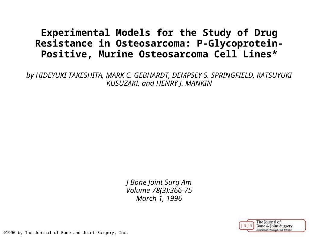
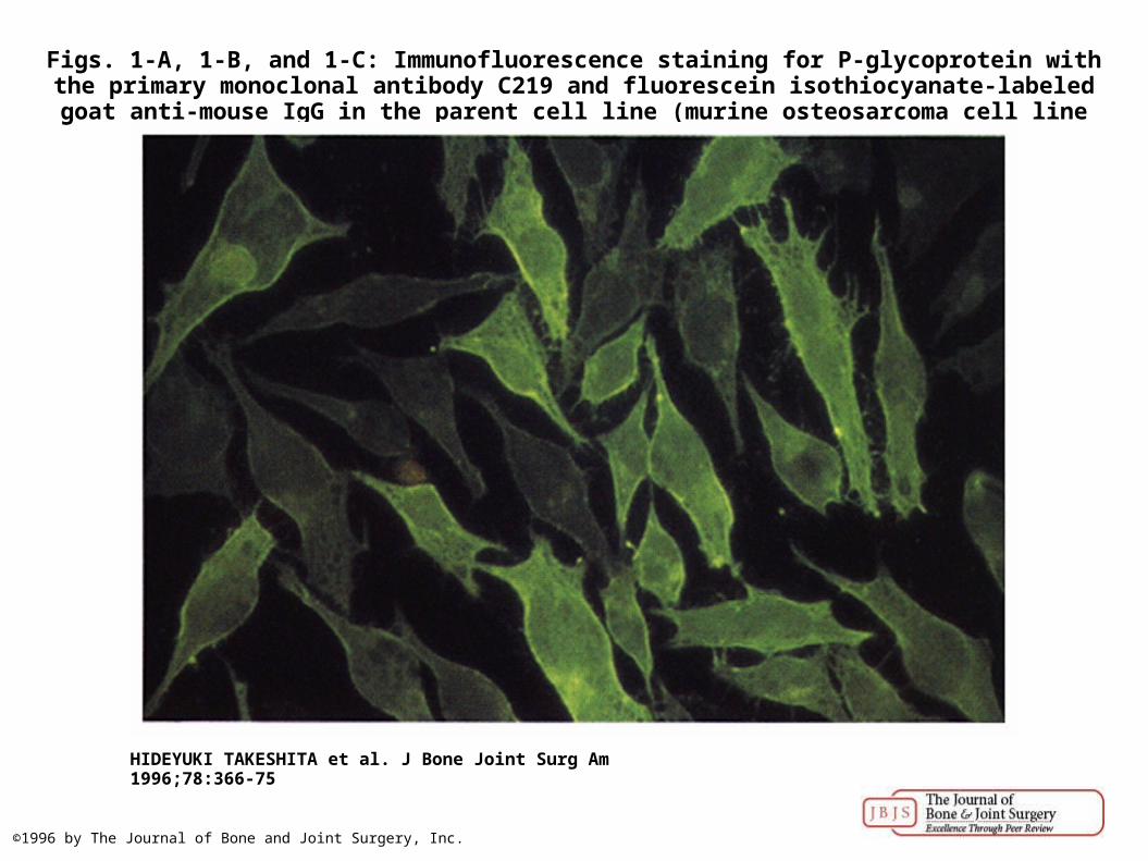
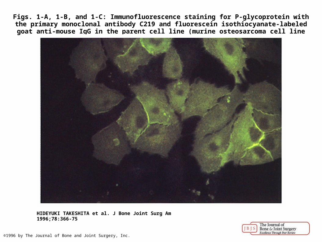
Figs. 1-A, 1-B, and 1-C: Immunofluorescence staining for P-glycoprotein with the primary monoclonal antibody C219 and fluorescein isothiocyanate-labeled goat anti-mouse IgG in the
parent cell line (murine osteosarcoma cell line developed at Massachusetts Ge...
HIDEYUKI TAKESHITA et al. J Bone Joint Surg Am 1996;78:366-75
©1996 by The Journal of Bone and Joint Surgery, Inc.
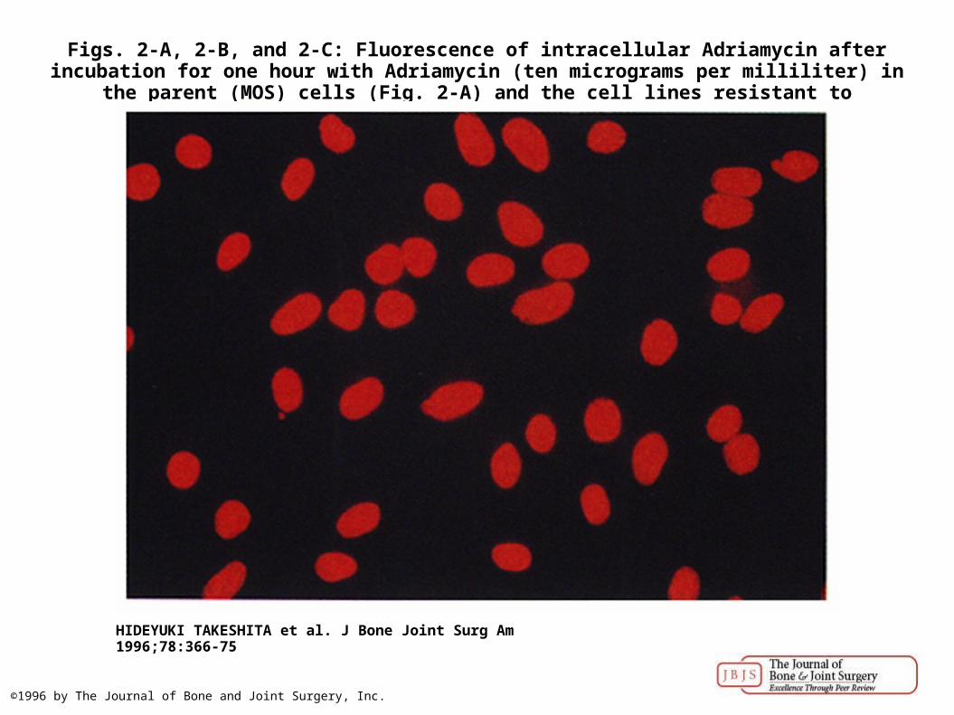
Figs. 2-A, 2-B, and 2-C: Fluorescence of intracellular Adriamycin after incubation for one hour with Adriamycin (ten micrograms per milliliter) in the parent (MOS) cells (Fig. 2-A) and the cell
lines resistant to Adriamycin, MOS/ADR1 (Fig. 2-B) and MOS/ADR2...
HIDEYUKI TAKESHITA et al. J Bone Joint Surg Am 1996;78:366-75
©1996 by The Journal of Bone and Joint Surgery, Inc.
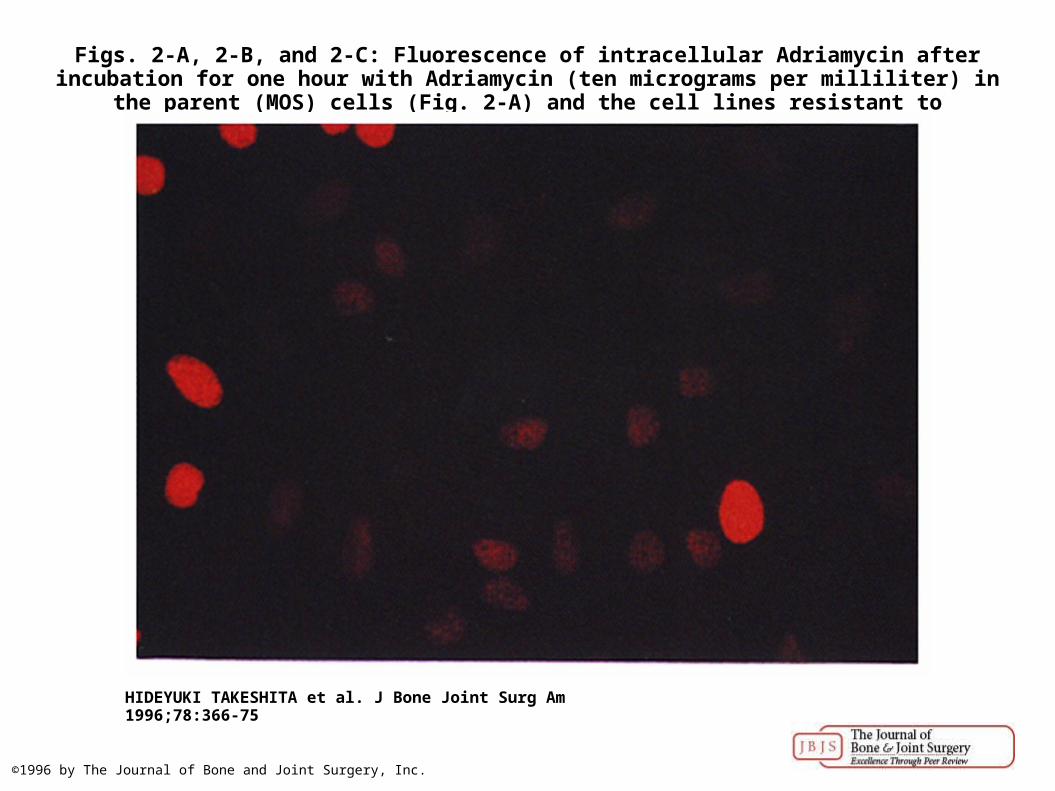
Figs. 2-A, 2-B, and 2-C: Fluorescence of intracellular Adriamycin after incubation for one hour with Adriamycin (ten micrograms per milliliter) in the parent (MOS) cells (Fig. 2-A) and the cell
lines resistant to Adriamycin, MOS/ADR1 (Fig. 2-B) and MOS/ADR2...
HIDEYUKI TAKESHITA et al. J Bone Joint Surg Am 1996;78:366-75
©1996 by The Journal of Bone and Joint Surgery, Inc.
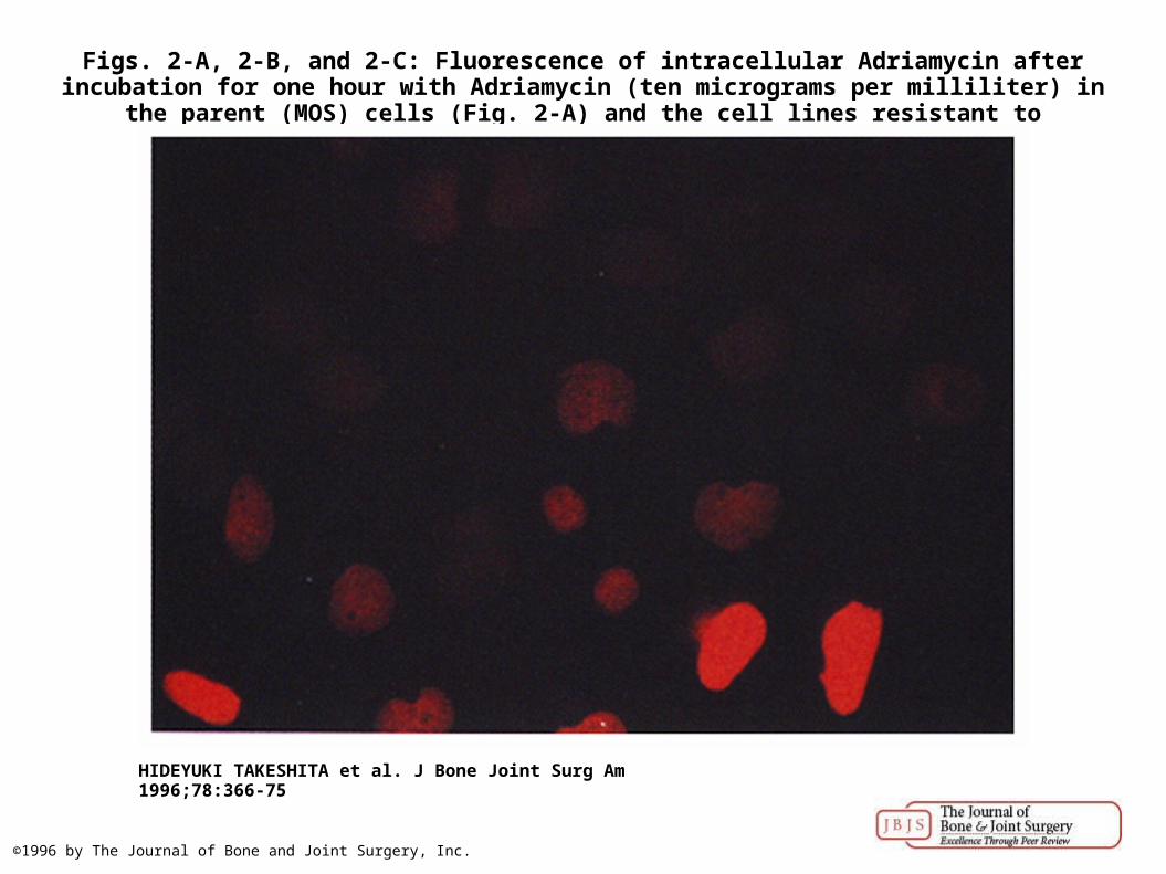
Figs. 2-A, 2-B, and 2-C: Fluorescence of intracellular Adriamycin after incubation for one hour with Adriamycin (ten micrograms per milliliter) in the parent (MOS) cells (Fig. 2-A) and the cell
lines resistant to Adriamycin, MOS/ADR1 (Fig. 2-B) and MOS/ADR2...
HIDEYUKI TAKESHITA et al. J Bone Joint Surg Am 1996;78:366-75
©1996 by The Journal of Bone and Joint Surgery, Inc.
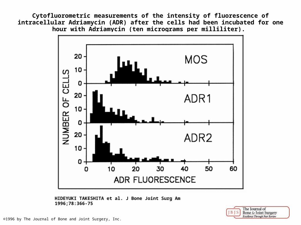
Cytofluorometric measurements of the intensity of fluorescence of intracellular Adriamycin (ADR) after the cells had been incubated for one hour with Adriamycin (ten micrograms per
milliliter).
HIDEYUKI TAKESHITA et al. J Bone Joint Surg Am 1996;78:366-75
©1996 by The Journal of Bone and Joint Surgery, Inc.
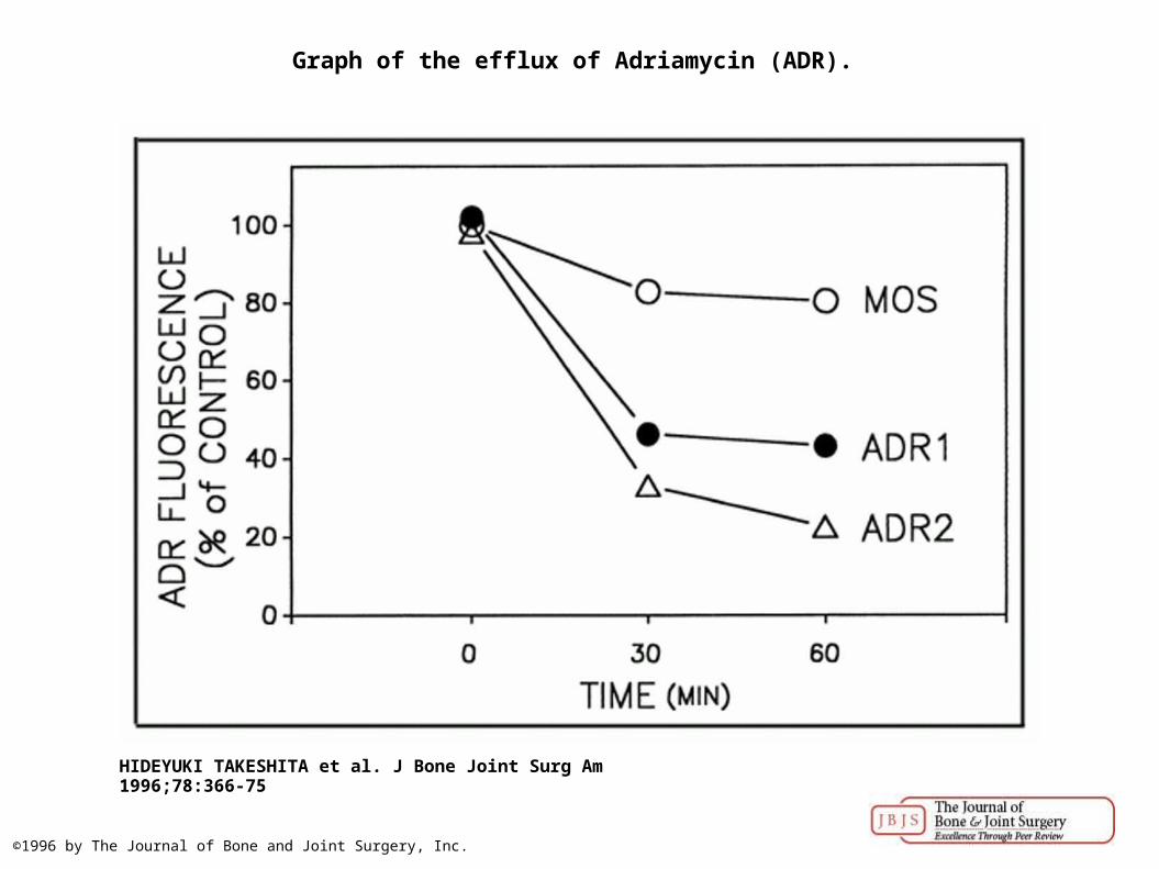
Graph of the efflux of Adriamycin (ADR).
HIDEYUKI TAKESHITA et al. J Bone Joint Surg Am 1996;78:366-75
©1996 by The Journal of Bone and Joint Surgery, Inc.
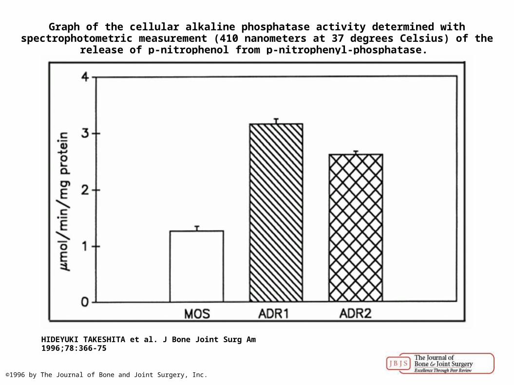
Graph of the cellular alkaline phosphatase activity determined with spectrophotometric measurement (410 nanometers at 37 degrees Celsius) of the release of p-nitrophenol from p-
nitrophenyl-phosphatase.
HIDEYUKI TAKESHITA et al. J Bone Joint Surg Am 1996;78:366-75
©1996 by The Journal of Bone and Joint Surgery, Inc.
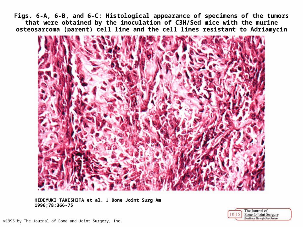
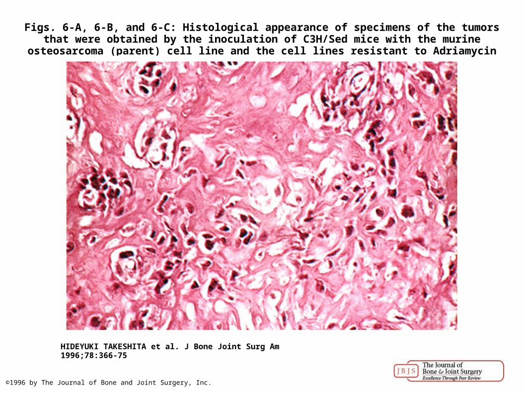
Figs. 6-A, 6-B, and 6-C: Histological appearance of specimens of the tumors that were obtained by the inoculation of C3H/Sed mice with the murine osteosarcoma (parent) cell line and the cell
lines resistant to Adriamycin (MOS/ADR1 and MOS/ADR2).
HIDEYUKI TAKESHITA et al. J Bone Joint Surg Am 1996;78:366-75
©1996 by The Journal of Bone and Joint Surgery, Inc.
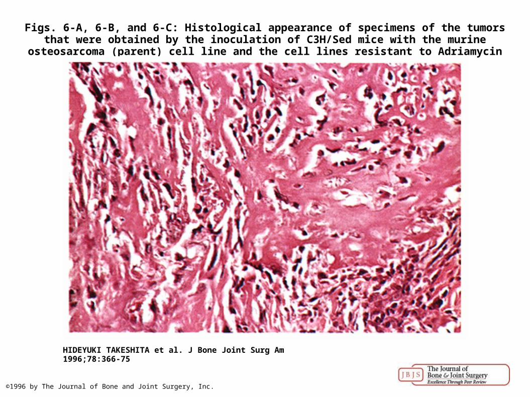
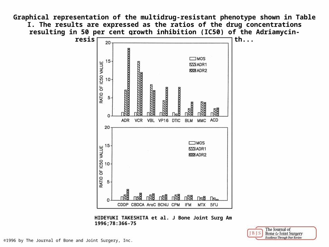
Graphical representation of the multidrug-resistant phenotype shown in Table I. The results are expressed as the ratios of the drug concentrations resulting in 50 per cent growth inhibition
(IC50) of the Adriamycin-resistant cell lines (ADR1 and ADR2) to th...
HIDEYUKI TAKESHITA et al. J Bone Joint Surg Am 1996;78:366-75
©1996 by The Journal of Bone and Joint Surgery, Inc.
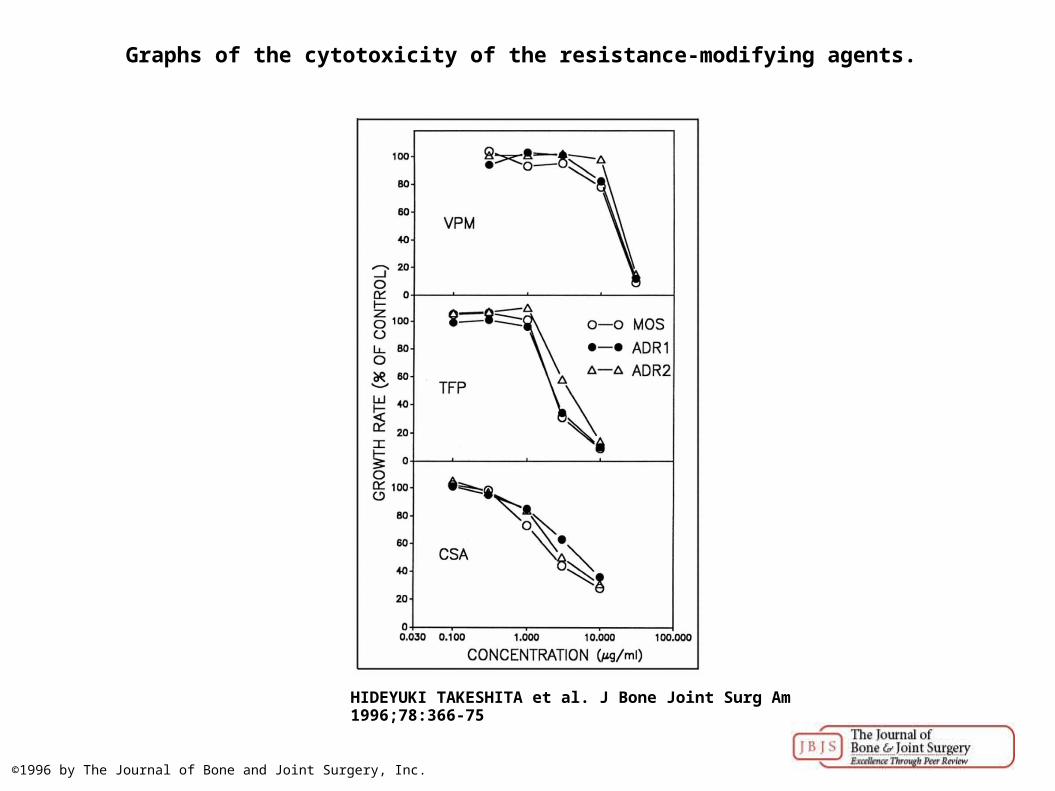
Graphs of the cytotoxicity of the resistance-modifying agents.
HIDEYUKI TAKESHITA et al. J Bone Joint Surg Am 1996;78:366-75
©1996 by The Journal of Bone and Joint Surgery, Inc.
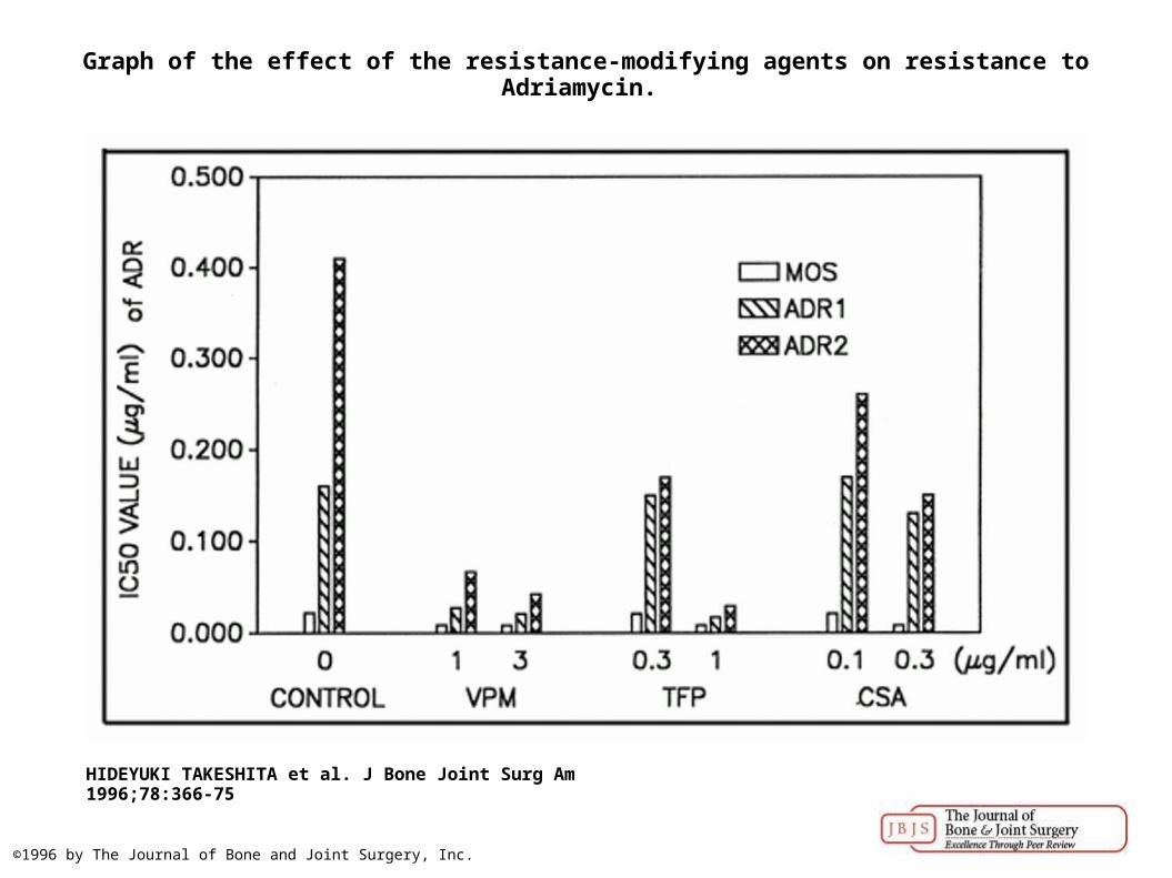
Graph of the effect of the resistance-modifying agents on resistance to Adriamycin.
HIDEYUKI TAKESHITA et al. J Bone Joint Surg Am 1996;78:366-75
©1996 by The Journal of Bone and Joint Surgery, Inc.