Embed Size (px)
Citation preview
DISEASES OF AQUATIC ORGANISMSDis Aquat Org
Vol. 87: 225–234, 2009doi: 10.3354/dao02123
Published December 3
INTRODUCTION
Cardiomyopathy syndrome (CMS) is a severe car-diac disease of unknown aetiology in farmed Atlanticsalmon Salmo salar. It was first diagnosed in 1985 inNorway, and later in Scotland and the Faeroe Islands(Amin & Trasti 1988, Bruno & Poppe 1996, Rodger &Turnbull 2000). Suspicious cases have also beenreported from Canada (Brocklebank & Raverty 2002).Outbreaks of CMS in farmed fish occur along most ofthe Norwegian coast, with the highest number ofaffected sites in mid-Norway (Kongtorp et al. 2006a).Most CMS cases are diagnosed during the late sea-water phase; typically from 14 to 18 mo after seatransfer. Mortality may be moderately elevated over along period, or suddenly high without prior symptoms(Ferguson et al. 1990, Brun et al. 2003). As CMS gener-ally affects large fish, the economic losses may be sub-stantial, even though the cumulative mortality may be
low or moderate (Brun et al. 2003, Østvik & Kjerstad2003).
Diagnosis of CMS is based on clinical findings,autopsy and histopathology. At autopsy, fish with CMStypically show skin haemorrhaging, raised scales andexophthalmia. Ascites and fibrinous casts on the liversurface are also common. The atrium and sinus veno-sus are usually enlarged, sometimes ruptured, andblood or blood clots often fill the pericardial cavity(Bruno & Poppe 1996). Histopathologically, CMS ischaracterised by inflammation and necrosis of endo-cardium and spongy myocardium in the atrium andventricle. Cellular infiltrates consist mainly of mono-nuclear cells, most probably lymphocytes and macro-phages. The compact myocardium is usually notaffected, but epicardial cell infiltrates may extend intothe compact layer along branches of the coronary ves-sel (Ferguson et al. 1990). Lesions may progress to sucha state that the wall of the atrium or sinus venosus
© Inter-Research 2009 · www.int-res.com*Email: [email protected]
Experimental transmission of cardiomyopathy syndrome (CMS) in Atlantic salmon Salmo salar
C. Fritsvold1,*, R. T. Kongtorp1, T. Taksdal1, I. Ørpetveit1, M. Heum1, T. T. Poppe2
1National Veterinary Institute, PO Box 750 Sentrum, 0106 Oslo, Norway2Norwegian School of Veterinary Science, PO Box 8146, 0033 Oslo, Norway
ABSTRACT: Cardiomyopathy syndrome (CMS) is a disease of unknown aetiology, having significanteconomic impact as it primarily affects large, farmed Atlantic salmon Salmo salar L. in seawater,close to harvest. In the present study, we have demonstrated that CMS is a transmissible diseaseunder experimental conditions. Histopathological lesions consistent with CMS were induced inAtlantic salmon post-smolts after injection of tissue homogenate from farmed fish diagnosed withCMS. Six weeks post-injection (p.i.), experimental fish started developing focal to multi-focal lesionsin the atrial endo- and myocardium, with subsequent progression to the ventricle. This proceededinto severe endocarditis and subsequent myocarditis with mononuclear cell infiltration of the atriumand, to a lesser degree, the spongy layer of the ventricle. These lesions were consistent withhistopathological findings in field outbreaks of CMS. From Week 33 p.i., lesions also appeared in thecompact myocardium, with focal epicarditis adjacent to focal myocardial lesions. In conclusion, theseresults indicate that CMS has an infectious aetiology and should be treated as a potentially conta-gious disease.
KEY WORDS: Atlantic salmon · Salmo salar · Cardiomyopathy syndrome · CMS · Experimental transmission · Myocarditis · Pathology · Cardiomyopathy · Transmission
Resale or republication not permitted without written consent of the publisher
OPENPEN ACCESSCCESS

Dis Aquat Org 87: 225–234, 2009
weakens or ruptures, with resultant haemopericar-dium and death. Previous studies indicate that CMS isa chronic disease developing over a period of severalmonths prior to the terminal clinical phase (Fergusonet al. 1990, Kongtorp et al. 2006a).
Several hypotheses on the cause of CMS have beenput forward, including environmental, immunologicaland microbiological factors (Kongtorp et al. 2005).Most of these hypotheses have not been studied fur-ther. Cardiac lesions in CMS may resemble pancreasdisease (PD) and heart and skeletal muscle inflamma-tion (HSMI), although the diseases are histopathologi-cally distinguishable in typical cases (Kongtorp et al.2004b, 2006b, McLoughlin & Graham 2007). PD iscaused by salmonid alphavirus (McLoughlin et al.1996, McLoughlin & Graham 2007). HSMI is experi-mentally transmissible and thought to be of viral aeti-ology (Kongtorp et al. 2004a). Both diseases causesevere myocardial inflammation and necrosis. Due tosimilarities with PD and HSMI, and the widespreadoccurrence of CMS, a viral aetiology has been sug-gested, although attempts to isolate virus from tissuesampled from fish with CMS have not yet been suc-cessful (Kongtorp et al. 2005). The aim of the presentstudy was therefore to investigate the transmissiblenature of CMS under experimental conditions.
MATERIALS AND METHODS
Experimental fish. A total of 496 healthy Atlanticsalmon smolts Salmo salar of a wild strain were used asexperimental fish. The fish had been hatched andgrown in a fresh water cultivation facility (Hellefoss) ineastern Norway, geographically isolated from the Nor-wegian population of farmed fish. The fish were notvaccinated, and the cultivation facility had no historyof disease. The experiment was performed at the Nor-wegian Institute of Water Research (NIVA), Solberg-strand, Akershus. Both the cultivation and researchfacilities are situated in an area with no commercialfish farms, physically and geographically well sepa-rated from the endemic area of CMS in Norway. Theresearch facility was approved for challenge experi-ments with unknown, suspected infectious agents bythe Norwegian Food Safety Authority. All effluentwater was filtrated (filter pores ≥300 µm) before beingtreated with hypochlorite, resulting in a total concen-tration of chlorine in effluent water of at least 35 mg l–1
30 min after treatment.The fish were transported to the research facility
(approximately 3 h transport time) in a water tank withoxygenation 10 wk before commencement of the ex-periment. There was no transport-related mortality.The fish had not been exposed to seawater, but most
fish had lost their parr markings and had a silveryappearance. The average length and weight were17 cm and 35 g, respectively, a fish size which fit wellwith the capacity at the facility.
Husbandry. At the research station, the fish werehoused indoors in a fibreglass tank. Osmoregulatorycapacity was tested after 2 wk of acclimatisation byexposing 7 fish to full salinity seawater for 24 h. Aftersedation in chlorobutanol (300 mg l–1), blood wassampled from the caudal vein of these fish, the fishwere euthanized by decapitation and the chloride con-centration was measured (Central Laboratory, Norwe-gian School of Veterinary Science) (Eisenman 1967,Tietz 1995). As the results indicated smoltification, theexperimental fish were transferred to seawater 7 dlater. After another 5 d, the fish were transferred to theexperimental tanks.
During the experiment, the fish were kept in cylin-drical fibreglass tanks with a conical bottom and cen-tral drainage, containing approximately 1.35 m3 ofwater. Water flow was 300 l h–1, providing a completewater exchange every 4.5 h. Seawater was pumpedfrom a depth of 60 m. Mean temperature was 8.5°C(range: 7.1 to 10.2°C) and mean salinity 33.8‰ (range:32.7 to 34.4‰). The fish were fed a commercial pel-leted feed with automatic feeders, at a feeding ratio ofapproximately 1% body weight d–1. A photoperiodregime of 10:14 h light:dark was used. Mortalities wereregistered, collected and stored at –18°C, and fishshowing aberrant behaviour were killed by a blow tothe head and decapitation before collection and similarstorage.
Preparation of inoculates. Tissue homogenate:Samples of cardiac and kidney tissue homogenatesfrom 6 Atlantic salmon collected during 2 field out-breaks of CMS were pooled and used for inoculation.Four of these fish were found dead in the cagesand displayed severe inflammation and necrosis ofendo- and myocardium in spongy tissue of the atriumand ventricle on histopathological examination. Theother 2 fish showed normal swimming behaviourand were caught live from the same cage as 2 of thedead fish. These fish had only mild inflammation incardiac tissue. Tissue samples were diluted 1:10 inLeibowitz L-15 cell culture media, homogenised andcentrifuged at 2500× g for 7 min. The supernatantwas further diluted 1:2 in L-15 supplemented withgentamycin (final concentration: 50 µg ml–1) beforeinoculation.
Negative control inoculate: Leibowitz L-15 cell cul-ture media, supplemented with gentamycin (50 µgml–1) was used as the negative control inoculate.
Challenge. The study was initiated after a 2 wkacclimatisation period in the experimental tanks. Ex-perimental fish were randomly allocated to 4 groups of
226
Fritsvold et al.: Experimental transmission of CMS
approximately 100 fish (range: 92 to 102), each groupin a separate tank. Injection of inoculates was per-formed after sedation in chlorobutanol (300 mg l–1).Duplicate groups of challenged fish (denoted Chal-lenges 1 and 2) were injected intraperitoneally (i.p.)with 0.2 ml supernatant from tissue homogenate. Sim-ilarly, duplicate groups of negative control fish (Con-trols 1 and 2) were injected i.p. with 0.2 ml of negativecontrol inoculate.
Sampling. Samples for histology, real-time reversetranscription-polymerase chain reaction (RRT-PCR)and microbiology were collected from 5 experimentalfish before commencement of the study. Post-chal-lenge, sampling was performed every 3 wk for a periodof 42 wk, resulting in a total of 14 samplings. At eachsampling, 5 experimental fish were collected fromeach group. Sampled fish were anesthetised in chlo-rine butanol (300 mg l–1) and killed by decapitation.Samples for RRT-PCR and cell culture were stored at–80°C until results from the histopathological exami-nation had been finalised.
As the number of fish in Control Group 1 was greatlyreduced by an outbreak of infectious pancreatic necro-sis (IPN), sampling from this group was not performedfrom Week 15 to Week 33. The last fish in this groupwere sampled 36 wk post injection (p.i.).
Histopathology. Tissue samples from gill, pseudo-branch, heart, liver, pyloric caeca with pancreas, mid-kidney, spleen and skeletal muscle were fixed in 10%neutral phosphate-buffered formalin and prepared byparaffin wax embedding and standard histologicaltechniques (Bancroft & Stevens 1990). Sections werestained with haematoxylin and eosin (H&E).
Sections of cardiac tissue were classified histologi-cally based on the presence of mononuclear endo- andmyocarditis, degeneration and necrosis in the spongylayer of the ventricle and atrium (Ferguson et al. 1990).The atrium, epicardium, compact and spongy layers ofthe ventricle and the endocardium of both cardiaccompartments were examined and evaluated. Thefindings were graded from 0 to 4 according to the cri-teria in Table 1 (see also illustrations in Fig. 1).
Microbiology. Bacteriology: Swabs from mid-kid-ney of 2 fish in each sampled group were cultivated onblood agar plates with and without 2% NaCl at 15 and22°C, respectively, for at least 6 d. Similar cultivationwas also performed on samples from both mid-kidneyand skin lesions from fish with skin ulcerations. Stan-dard procedures for identification of isolated bacteriawere performed.
RRT-PCR: After completion of the experiment, 76fish were examined for piscine nodavirus, salmonidalphavirus 3 (SAV3) and infectious pancreatic necrosisvirus (IPNV) by RRT-PCR. The fish examined werethose used for inoculate preparation, all fish from the
pre-challenge sampling, a selection of mortalities, allfish from 3 scheduled samplings and some fish withsevere heart lesions from scheduled samplings (seeTable 2). Total nucleic acids were extracted from heartand kidney tissue using the NucliSens® easyMAG™on-board protocol (bioMerieux) according to themanufacturer’s instructions. The nucleic acid con-centrations were determined using a Nanodrop ND-1000 (NanoDrop Technologies). Detection of nodavirusby RRT-PCR was performed according to Grove et al.(2006), but without quantification. RRT-PCR for detec-tion of SAV (Jansen et al. 2007) and for detection ofIPNV (Ørpetveit et al. 2007) was also performed. AllRRT-PCR reactions were performed on a StratageneMx3005P.
Cell culture: Post-experiment, heart and kidney tis-sue from 6 challenged fish sampled at 12, 18, 24 and27 wk p.i., all with moderate to severe (Grade 3 or 4)CMS-like histopathological atrial lesions and at leastGrade 2 lesions of the spongy myocardium of the ven-tricle, were examined in cell cultures according to rou-tine procedures at the National Veterinary Institute,Oslo, Norway. The tissue samples were homogenisedin cell culture medium (w/v 10%), and the homo-genates were cleared by low-speed centrifugation. AsIPNV is ubiquitous in Norwegian salmon farming(Melby et al. 1991, Jarp et al. 1995, 1996), the homo-genates were treated with a mix of polyclonal neutral-ising antibodies against IPNV serotype Sp and sero-type Ab. The homogenates were then inoculated ontocell cultures from bluegill fry fibroblast (BF)-2 cells(Wolf & Quimby 1966), epithelioma papulosum cyprini(EPC) (Fijan et al. 1983), rainbow trout gonad (RTG)-2(Wolf & Quimby 1962), chinook salmon embryo(CHSE)-214 (Lannan et al. 1984) and Atlantic salmonhead kidney (ASK) (Devold et al. 2000). Inoculatedcells were incubated for 1 wk at both 15 and 20°C inparallel and were regularly investigated with aninverted microscope for the occurrence of a cytopathiceffect (CPE). After 1 wk, the supernatants were pas-
227
Table 1. Salmo salar. Histological classification of lesions inendo-, epi- and/or myocardium
Score Description
0 No pathological findings, or slightly increasednumber of leukocytes
1 One or a few focal lesions, increased numberof leukocytes
2 Several distinct lesions and small to moderateincrease in number of leukocytes
3 Multifocal to confluent lesions and moderate tosevere increase in number of leukocytes
4 Severe confluent lesions comprising >75% ofthe tissue and massive leukocyte infiltration

Dis Aquat Org 87: 225–234, 2009228
Fig. 1. Salmo salar. Histological classification of lesions in cardiac atrium and spongy ventricle, in accordance with the gradingdescribed in Table 1. Grade 1: arrows indicate minor inflammatory lesions consisting of sparse, focal subendocardial infiltrationby mononuclear leukocytes and some degree of subendocardial vacuolisation, in both atrium and ventricle. Grade 2: several dis-tinct lesions with small to moderately increased number of leukocytes. Myocyte degeneration and necrosis are encircled in theatrium. Grade 3: Multifocal to confluent lesions with moderate to severe leukocyte infiltration. Grade 4: Arrows indicate hyper-trophic endocardial cells in the atrium forming empty tubes where almost all muscle fibres have been replaced by inflammatorycells, dominated by small mononuclear lymphocyte-like cells. Myocyte degeneration and necrosis are encircled in the atrium.Ventricular example is from a focal Grade 4 lesion. Haematoxylin and eosin staining. Scale bar (applies to all panels) = 50 µm
Fritsvold et al.: Experimental transmission of CMS
saged onto corresponding cells and incubated for a fur-ther week and investigated as described above.
RESULTS
Clinical signs
Only 2 fish showed clinical signs. One challengedfish was observed lying on the bottom of the tank at thefirst scheduled sampling and was included in the sam-pling. Another lethargic challenged fish was sampled12 wk p.i.
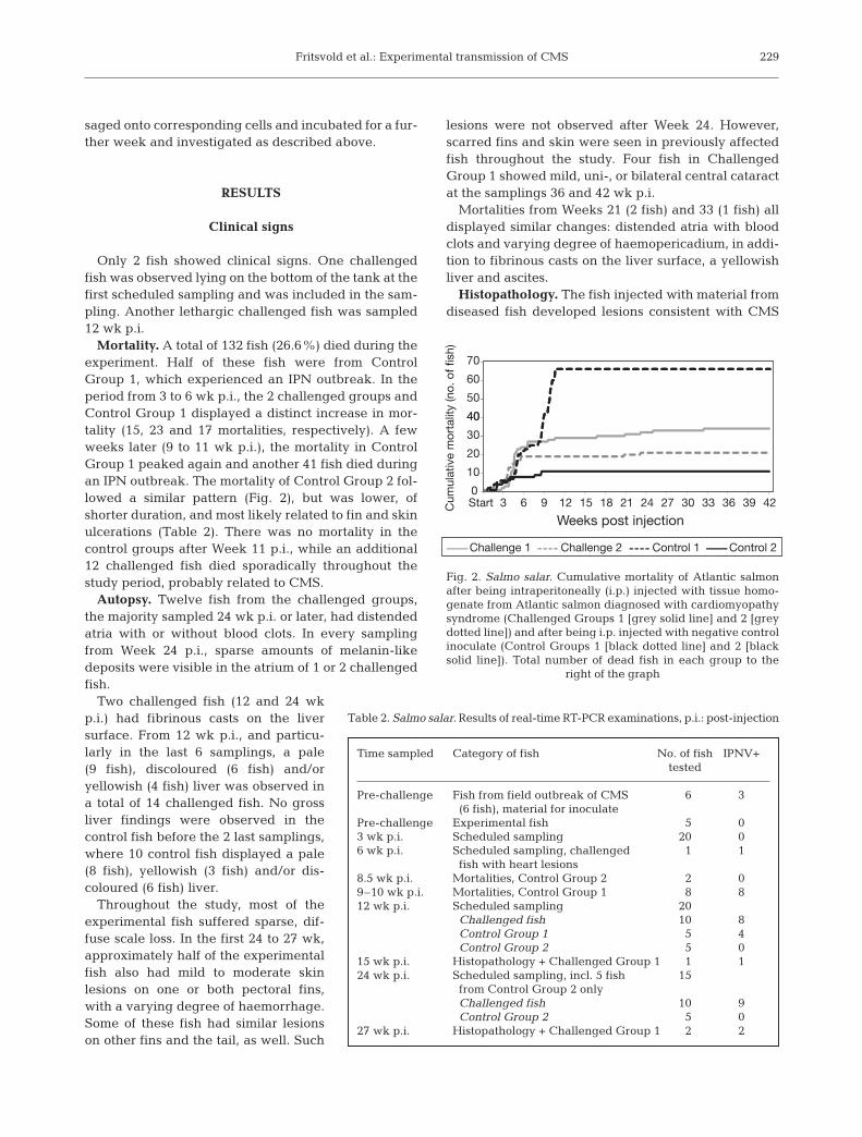
Mortality. A total of 132 fish (26.6%) died during theexperiment. Half of these fish were from ControlGroup 1, which experienced an IPN outbreak. In theperiod from 3 to 6 wk p.i., the 2 challenged groups andControl Group 1 displayed a distinct increase in mor-tality (15, 23 and 17 mortalities, respectively). A fewweeks later (9 to 11 wk p.i.), the mortality in ControlGroup 1 peaked again and another 41 fish died duringan IPN outbreak. The mortality of Control Group 2 fol-lowed a similar pattern (Fig. 2), but was lower, ofshorter duration, and most likely related to fin and skinulcerations (Table 2). There was no mortality in thecontrol groups after Week 11 p.i., while an additional12 challenged fish died sporadically throughout thestudy period, probably related to CMS.
Autopsy. Twelve fish from the challenged groups,the majority sampled 24 wk p.i. or later, had distendedatria with or without blood clots. In every samplingfrom Week 24 p.i., sparse amounts of melanin-likedeposits were visible in the atrium of 1 or 2 challengedfish.
Two challenged fish (12 and 24 wkp.i.) had fibrinous casts on the liversurface. From 12 wk p.i., and particu-larly in the last 6 samplings, a pale(9 fish), discoloured (6 fish) and/oryellowish (4 fish) liver was observed ina total of 14 challenged fish. No grossliver findings were observed in thecontrol fish before the 2 last samplings,where 10 control fish displayed a pale(8 fish), yellowish (3 fish) and/or dis-coloured (6 fish) liver.
Throughout the study, most of theexperimental fish suffered sparse, dif-fuse scale loss. In the first 24 to 27 wk,approximately half of the experimentalfish also had mild to moderate skinlesions on one or both pectoral fins,with a varying degree of haemorrhage.Some of these fish had similar lesionson other fins and the tail, as well. Such
lesions were not observed after Week 24. However,scarred fins and skin were seen in previously affectedfish throughout the study. Four fish in ChallengedGroup 1 showed mild, uni-, or bilateral central cataractat the samplings 36 and 42 wk p.i.
Mortalities from Weeks 21 (2 fish) and 33 (1 fish) alldisplayed similar changes: distended atria with bloodclots and varying degree of haemopericadium, in addi-tion to fibrinous casts on the liver surface, a yellowishliver and ascites.
Histopathology. The fish injected with material fromdiseased fish developed lesions consistent with CMS
229
40
50
60
70
Cum
ulat
ive
mor
talit
y (n
o. o
f fis
h)
0
10
20
30
40
Start 3 6 9 12 15 18 21 24 27 30 33 36 39 42
Weeks post injection
Challenge 1 Challenge 2 Control 1 Control 2
Table 2. Salmo salar. Results of real-time RT-PCR examinations, p.i.: post-injection
Time sampled Category of fish No. of fish IPNV+tested
Pre-challenge Fish from field outbreak of CMS 6 3(6 fish), material for inoculate
Pre-challenge Experimental fish 5 03 wk p.i. Scheduled sampling 20 06 wk p.i. Scheduled sampling, challenged 1 1
fish with heart lesions8.5 wk p.i. Mortalities, Control Group 2 2 09–10 wk p.i. Mortalities, Control Group 1 8 812 wk p.i. Scheduled sampling 20
Challenged fish 10 8Control Group 1 5 4Control Group 2 5 0
15 wk p.i. Histopathology + Challenged Group 1 1 124 wk p.i. Scheduled sampling, incl. 5 fish 15
from Control Group 2 onlyChallenged fish 10 9Control Group 2 5 0
27 wk p.i. Histopathology + Challenged Group 1 2 2
Fig. 2. Salmo salar. Cumulative mortality of Atlantic salmonafter being intraperitoneally (i.p.) injected with tissue homo-genate from Atlantic salmon diagnosed with cardiomyopathysyndrome (Challenged Groups 1 [grey solid line] and 2 [greydotted line]) and after being i.p. injected with negative controlinoculate (Control Groups 1 [black dotted line] and 2 [blacksolid line]). Total number of dead fish in each group to the
right of the graph

Dis Aquat Org 87: 225–234, 2009
(Ferguson et al. 1990). Detailed results are presentedin Figs. 3 & 4, and findings according to time are shownin Fig. 5. Focal to multi-focal inflammation became evi-dent in the atrium 6 wk p.i., with subsequent progres-sion to the spongy myocardium of the ventricle 9 wkp.i. The inflammation in both the atrium and ventriclewas dominated by mononuclear leukocytes, in addi-tion to necrosis of spongy myocardium and endo-cardium.
Early atrial lesions observed at 6 wk p.i. were mildto moderate. The number of affected fish increasedfrom 9 wk p.i. Severity of atrial lesions peaked at12 wk p.i., and remained at this level for the rest ofthe study. The most extensive atrial lesion (Grade 4)was recorded in a single fish sampled 24 wk p.i.Grade 1 to 4 atrial lesions were found in at least 60%of the challenged fish at all samplings from 9 wk p.i.,except at 33 wk p.i. Melanin-like deposits as observedat autopsy were located subendocardially in atriallesions and in degenerated atrial myocardium. In
the most severe foci, the deposits were fairly large(Fig. 6).
Mild lesions in the spongy layer of the ventricle(Grade 1) were initially detected in 3 fish with atrialchanges at 9 wk p.i. The first fish with more moderatespongy layer lesions (Grade 2) was registered at 12 wkp.i., and, in the following samplings, the number of fishwith Grade 2 lesions remained fairly constant. Ventric-ular spongy layer lesions of Grades 1 to 3 were found inat least 50% of the challenged fish at all samplings from12 wk p.i., except at 27 and 39 wk p.i. The most severeinflammatory changes observed in this tissue wereGrade 3, occurring in a few fish at 30, 33 and 42 wk p.i.
In the epicardium, a slightly increased number ofleukocytes were observed in 34 of the 130 challengedfish, but in accordance with Table 1, these weregraded 0. A total of 23 challenged fish displayed Grade1 lesions in the epicardium, and 2 fish displayed Grade2 lesions. The number of fish with epicardial changesdecreased towards the end of the study.
230
6789
10
4
3
0123456
3 6 9 12 15 18 21 24 27 30 33 36 39 42
No.
of f
ish
Weeks p.i.
2
1
0
Fig. 3. Salmo salar. Histological findings in the atrium of the challenged fish. The lesions were classified from 0 (normal) to 4 (se-vere changes) according to Table 1. No or very little atrial tissue was present in the histological samples of 1 or 2 fish at 6, 9, 21and 33 wk post-injection (p.i.) and could, therefore, not be evaluated in these fish. Due to high mortality in Challenge Group 1,
the last fish in this group were sampled 36 wk p.i.
5
6
7
8
9
10
No.
of f
ish 4
3
0
1
2
3
4
5
3 6 9 12 15 18 21 24 27 30 33 36 39 42
Weeks p.i.
2
1
0
Fig. 4. Salmo salar. Histological findings in the spongy layer of the ventricle of the challenged fish. The lesions were classifiedfrom 0 (normal) to 4 (severe changes) according to Table 1. Due to high mortality in Challenge Group 1, the last fish in this
group were sampled 36 wk post-injection (p.i.)

Fritsvold et al.: Experimental transmission of CMS
Mild (Grade 1) compact layer lesions were first reg-istered in a single fish at 18 wk p.i. and in 3 fish at21 wk p.i. Thereafter, 1 or a few well defined foci ofinflammatory cells in the compact layer, ranging inseverity from Grade 1 to 2, were observed in 1 to 3 ofthe fish sampled at 30, 33, 36 and 42 wk p.i. All 12 fishwith lesions in the compact layer also had inflamma-tory lesions in the spongy layer of the ventricle of iden-tical or greater severity, while 7 of them also displayedadditional focal Grade 1 or 2 lesions in the epicardium.
No CMS-like lesions were identified in the 5 fishsampled prior to challenge. None of the control fishhad pathological changes in the compact layer of theventricle, and only non-specific and sparse findingsgraded 0 or 1 according to Table 1 and Fig. 1 weredetected in the hearts of a few fish. During the firstdistinct mortality period, IPN was diagnosed byhistopathology and IPNV immunohistochemistry inboth challenged groups and Control Group 1, but notin Control Group 2.
Microbiology
Bacteriology. Bacteria were cultivated from 8 sam-pled fish (4 challenged) and 1 dead fish (control). Thebacterial growth was dominated by a mixed floraconsidered to be of little or no significance. In only 2fish (3 wk p.i.) were bacteria, subsequently identifiedas Vibrio sp. (dead, control fish) and sparse mixed flora(sampled, control fish), isolated from both kidney andskin or fin lesions.
RRT-PCR. All examined samples were negative fornodavirus and SAV by RRT-PCR. Three of the 6 fishused for inoculation of challenged fish had low to mod-erate levels of IPNV (Table 2). This was also the case inall examined fish from both challenged groups andControl Group 1. During the period of high mortality,moderate to low amounts of IPNV were found in allmortalities examined from the 2 challenge groupsand Control Group 1. Fish tested for IPNV in ControlGroup 2 were negative throughout the study.
Cell culture. No cytopathic effect was detected inany of the cell cultures at the chosen incubationtemperatures.
DISCUSSION
In the present study cardiac lesions consistent withCMS were successfully transmitted to naïve Atlanticsalmon Salmo salar post-smolts following i.p. injec-tion of tissue homogenate from diseased fish. Bothcharacteristics and severity of histopathologicalchanges were reproduced in experimental fish. Inaddition, typical clinical signs and gross lesions wereobserved in some fish (Ferguson et al. 1990, Poppe &Ferguson 2006). These results indicate that CMSmay be caused by an agent present in cardiac orrenal tissue.
The first histopathological lesions appeared in theatrium from 6 wk p.i., while lesions in the spongy layerof the ventricle did not appear until 9 wk p.i. Generally,ventricular lesions were not seen in experimental fishwithout associated, and mostly more severe, atriallesions. The explanation for this is uncertain, but thesame pattern is typically seen in fish sampled fromfield outbreaks (T. T. Poppe unpubl. data). In such fieldmaterial, atrial lesions are generally more severe thanventricular lesions, indicating a more advanced stageof the disease process in the atrium. The sequentialoccurrence of cardiac changes observed in the presentstudy emphasizes the importance of sampling bothatrial and ventricular tissues for histopathology. Evalu-ation of both tissues is a clear advantage in histopatho-logical diagnostics, in order to differentiate betweenCMS and similar heart diseases.
231
Atrium
Ventricle, spongy layer
Ventricle, compact layer
End
15 2712963Sta
rt
21 2418
Weeks p.i. (14 samplings)
30 33 36 39 42
Fig. 5. Salmo salar. Appearance of histological cardiac lesions(as observed by light microscopy) over time post-injection (p.i.)
Fig. 6. Salmo salar. Melanin deposits in atrial lesions of exper-imental fish (36 wk post-injection) with a diagnosis of cardio-
myopathy syndrome

Dis Aquat Org 87: 225–234, 2009
Typical gross lesions, including enlarged atrium,fibrinous casts on the liver surface and ascites wereseen in several challenged fish. These were mostlyassociated with histological findings of moderate tosevere atrial lesions. Other typical autopsy findings infield cases of CMS indicating circulatory collapse, e.g.skin haemorrhage, raised scales and exophthalmia(Bruno & Poppe 1996), were not seen. This may beassociated with the low degree of clinical diseaseand/or mortality in the present study. Most experi-mental fish showed normal swimming and feedingbehaviour, in spite of severe cardiac lesions. Thesefindings are in accordance with naturally occurringCMS, in which extensive cardiac lesions do not neces-sarily result in clinical disease (T. T. Poppe unpubl.data). Cardiac lesions similar to those seen in CMShave been observed in wild Atlantic salmon in Nor-way, but have not been associated with clinical dis-ease (Poppe & Seierstad 2003). Several factors mayinfluence the outcome of CMS under natural condi-tions. For instance, vaccination status, physiologicalstage, feeding regime, water temperature, oxygenlevels, concurrent infections, parasite burden andother stressors may contribute to the development ofclinical disease. Also, infection pressure, transmissionroute and factors related to pathogen exposure maybe of significance. The almost simultaneous occur-rence of increased mortality in all fish groups duringthe first weeks of this experiment could indicate expo-sure of the fish to unidentified environmental stressorsin addition to the bacterial skin and fin infections. Themarkedly higher number of dead fish in 3 of 4 groupsappears to be linked to the IPN diagnosis in thesegroups. Both the relative importance of infectious andenvironmental components in CMS-associated mor-tality and the potential for horizontal transmissionshould be further studied.
The experimental fish differed in at least 3 waysfrom the farmed fish typically affected in field out-breaks of CMS: (1) they were from a wild stock, (2)they were smaller and (3) they were unvaccinated.Experimental fish from a geographically isolated wildAtlantic salmon strain were chosen to reduce the riskof prior exposure to a possible causative agent presentin the population or environment of farmed fish, and toavoid other factors associated with fish farming thatmay be of significance for the development of CMS.The results generated indicate that injection of tissuehomogenate alone is sufficient for the development ofhistopathological changes typical of CMS. Addition-ally, the low age of experimental fish did not appear tohinder the development of cardiac lesions. Under nat-ural conditions, CMS is commonly observed in largesalmon (>2 kg) the second year in seawater, causinglosses among large specimens of the population (Brun
et al. 2003). The present study showed that post-smoltswere capable of developing the same type and severityof lesions as larger salmon. However, the size of thefish may partly explain the limited number of fishshowing clinical signs in the present study. The heart/body volume ratio decreases with increasing fish size,and large fish may therefore have a lower cardiaccapacity compared to smaller fish (Agnisola & Tota1994, Gamperl & Farrell 2004), rendering them morevulnerable to the effects of severe cardiac lesions.
The use of unvaccinated experimental fish in thepresent study may have contributed to the successfultransmission of CMS in young fish. At 30 to 40 g weightprior to seawater transfer, all farmed salmon in Norwayare routinely vaccinated i.p. with oil-adjuvanted vac-cines. These vaccines may induce both specific andnon-specific immunity (Poppe & Breck 1997, Koppanget al. 2008), which could interfere with susceptibility toinfections. However, little is known about the immuno-logical processes involved in the development of CMS.In some challenged fish, melanin deposits were ob-served on atrial surfaces and/or subendocardially from24 wk p.i. Such deposits have also been observed infield cases of CMS, but not as a consistent finding(C. Fritsvold pers. obs.). Melanin is suspected to be ofimportance in the inflammatory responses of salmo-nids, but its exact role or effect is not fully understood(Thorsen et al. 2006).
Although transmissibility was demonstrated in thepresent study, a causal agent was not identified. RRT-PCR indicated that neither nodavirus nor SAV3 con-tributed to the development of CMS in the presentstudy. No cytopathic effect was found in cell culturesinoculated with tissue material from challenged fishwith moderate to severe (Grade 3 to 4) atrial lesions,indicating that other known fish pathogenic viruseswere not present. Likewise, bacteria known to grow onblood agar were sparse, and probably did not con-tribute significantly to the development of cardiacchanges. In addition, any prospective causal agentpresent in the transmitted material would have to beresistant to gentamycin at the concentrations used.
A viral aetiology for CMS has been previously sug-gested (Grotmol et al. 1997). Filtration of the inoculatewas deliberately not performed in the present experi-ment, to ensure that possible causative bacteria werenot eliminated from the inoculate before challengingthe fish. In future studies, filtration of homogenatesprior to injection may be used to investigate the possi-bility of a viral aetiology. The virological proceduresused were intended to rule out the presence of knownviruses, and may not have been optimal for detectingunknown viruses. In the further search for an etiologi-cal, possibly viral, agent, a range of different cell linesand/or growth conditions should be tested. As CMS
232

Fritsvold et al.: Experimental transmission of CMS
appears to be a relatively slowly developing disease(>3 wk for development of initial histopathologicalchanges in experimental fish), the relatively shortincubation periods for the cell cultures may have beentoo short to allow for CPE to develop in the presentstudy.
IPNV was identified by PCR in fish used in inoculatepreparation, as well as in both challenged groups and1 of the 2 control groups. This may explain why chal-lenged fish experienced an IPN outbreak. The sourceof IPNV infection in 1 of the 2 control groups isunknown. Fish may have been infected by IPNV at thecultivation facility or at the research facility, as neitherof them routinely disinfected their inlet water.
Postviral endo- and/or myocarditis are well known inhuman medicine (Eriksson & Penninger 2005). Thismay also be a mechanism in the development of CMS,for instance triggered by an infection with IPNV. Apossible association between IPNV and CMS has pre-viously been suggested (Brun et al. 2003). However,the presence of IPNV and the outbreak of IPN in one ofthe control groups did not induce CMS-like lesions inthose fish. In addition, a similar experiment with CMShas recently been performed by Bruno & Noguera inScotland, with findings parallel to those observed inthe present study. IPNV was not found in that study(D. W. Bruno pers. comm.). It therefore seems unlikelythat IPNV played a significant role in the developmentof CMS in the present study. However, an interactionbetween IPNV and a possible causal agent of CMScannot be ruled out.
Focal pathological changes appeared in the compactmyocardium of 8 fish from 30 wk p.i. Compact layerinvolvement is uncommon in field outbreaks of CMS,but has been observed in some fish with extensive epi-carditis (Poppe & Ferguson 2006). Strict focal epicardi-tis was associated with the compact layer lesions in thepresent study, but these changes were only mild tomoderate. Absence of lesions in pancreatic tissue andred skeletal muscle indicate that the myocardiallesions in our study were not associated with HSMI orPD. In support of this, failure to detect SAV3 appears toexclude PD. Myocarditis in the compact layer is partic-ularly pronounced in HSMI (Kongtorp et al. 2004a),which is experimentally transmissible by a model sim-ilar to that used in the present study. The aetiology ofHSMI is not yet fully understood. A possible presenceof the HSMI agent in the inoculate or the experimentalfish used cannot be excluded. However, only a few fishshowed mild to moderate focal changes in the compactmyocardium in the present study, while this tissue isseverely affected in infection experiments with HSMI(Kongtorp et al. 2004b, Kongtorp & Taksdal 2009). Themost common cardiac lesions in the present study werevery similar to those seen in field cases of CMS, and
the development of cardiac changes also differed fromexperiments with HSMI. Furthermore, the time fromchallenge to appearance of lesions in the compactlayer in the present study was considerably longerthan in experimental transmission of both HSMI(Kongtorp et al. 2004b, Kongtorp & Taksdal 2009) andPD (McLoughlin et al. 1996, Christie et al. 2007).
In conclusion, cardiac changes consistent with CMSand, to a certain extent, clinical disease were transmit-ted to experimental fish. The results indicate that CMSmay develop independently of PD and HSMI. Furtherstudies are needed to establish the aetiology andpathogenesis of CMS and to develop disease-specificdiagnostic tools.
Acknowledgements. This work was financed by the Norwe-gian Research Council, Grant No. 172635/S40. Many thanksto D. Colquhoun (text editing), T. Romsås (data analysis andpresentation), M. Fritsvold (photo editing), colleagues at theDepartment for Fish Health, NVI, and O. Pettersen with staffat the NIVA research facility, Solbergstrand, for technicalrunning of the experiment.
LITERATURE CITED
Agnisola C, Tota B (1994) Structure and function of thefish cardiac ventricle — flexibility and limitations. Cardio-science 5:145–153
Amin AB, Trasti J (1988) Endomyocarditis in Atlantic salmonin Norwegian seafarms. Bull Eur Assoc Fish Pathol 8:70–71
Bancroft JD, Stevens A (1990) Theory and practice of histo-logical techniques. Churchill Livingstone, London
Brocklebank J, Raverty S (2002) Sudden mortality caused bycardiac deformities following seining of preharvest far-med Atlantic salmon (Salmo salar) and by cardiomyopathyof postintraperitoneally vaccinated Atlantic salmon parr inBritish Columbia. Can Vet J 43:129–130
Brun E, Poppe T, Skrudland A, Jarp J (2003) Cardiomyopathysyndrome in farmed Atlantic salmon Salmo salar: occur-rence and direct financial losses for Norwegian aquacul-ture. Dis Aquat Org 56:241–247
Bruno DW, Poppe TT (1996) Diseases of uncertain aetiology.Cardiac myopathy syndrome. In: Bruno DW, Poppe TT(eds) A color atlas of salmonid diseases. Academic Press,London, p 140–141
Christie KE, Graham DA, McLoughlin MF, Villoing S, Todd D,Knappskog D (2007) Experimental infection of Atlanticsalmon Salmo salar pre-smolts by i.p. injection with newIrish and Norwegian salmonid alphavirus (SAV) isolates: acomparative study. Dis Aquat Org 75:13–22
Devold M, Krossøy B, Aspehaug V, Nylund A (2000) Use ofRT-PCR for diagnosis of infectious salmon anaemia virus(ISAV) in carrier sea trout Salmo trutta after experimentalinfection. Dis Aquat Org 40:9–18
Eisenman G (1967) Glass electrodes for hydrogen and othercations. Principles and practice. Marcel Dekker, New York
Eriksson U, Penninger JM (2005) Autoimmune heart failure:new understandings of pathogenesis. Int J Biochem CellBiol 37:27–32
Ferguson HW, Poppe T, Speare DJ (1990) Cardiomyopathy infarmed Norwegian salmon. Dis Aquat Org 8:225–231
233

Dis Aquat Org 87: 225–234, 2009
Fijan N, Sulimanovic D, Bearzotti M, Muzinic D and others(1983) Some properties of the Epithelioma papulosumcyprini (EPC) cell line from carp Cyprinus carpis. AnnVirol 134:207–220
Gamperl AK, Farrell AP (2004) Cardiac plasticity in fishes:environmental influences and intraspecific differences.J Exp Biol 207:2539–2550
Grotmol S, Totland GK, Kryvi H (1997) Detection of anodavirus-like agent in heart tissue from reared Atlanticsalmon Salmo salar suffering from cardiac myopathy syn-drome (CMS). Dis Aquat Org 29:79–84
Grove S, Faller R, Soleim KB, Dannevig BH (2006) Absolutequantitation of RNA by a competitive real-time RT-PCRmethod using piscine nodavirus as a model. J Virol Meth-ods 132:104–112
Jansen MD, Brun E, Taksdal T, Gjerset B, Sandberg M (2007)A cohort study of pancreas disease in Norwegian Atlanticsalmon. In: Abstracts of the 13th international conferenceof the EAFP. EAFP, Grado, p 16
Jarp J, Gjevre AG, Olsen AB, Bruheim T (1995) Risk-factorsfor furunculosis, infectious pancreatic necrosis and mor-tality in post-smolts of Atlantic salmon, Salmo salar. J FishDis 18:67–78
Jarp J, Taksdal T, Tørud B (1996) Infectious pancreatic necro-sis in Atlantic salmon Salmo salar in relation to specificantibodies, smoltification, and infection with erythrocyticinclusion body syndrome (EIBS). Dis Aquat Org 27:81–88
Kongtorp RT, Taksdal T (2009) Studies with experimentaltransmission of heart and skeletal muscle inflammation inAtlantic salmon, Salmo salar L. J Fish Dis 32:253–262
Kongtorp RT, Kjerstad A, Taksdal T, Guttvik A, Falk K (2004a)Heart and skeletal muscle inflammation in Atlanticsalmon, Salmo salar L.: a new infectious disease. J Fish Dis27:351–358
Kongtorp RT, Taksdal T, Lyngoy A (2004b) Pathology of heartand skeletal muscle inflammation (HSMI) in farmedAtlantic salmon Salmo salar. Dis Aquat Org 59:217–224
Kongtorp RT, Taksdal T, Lillehaug A (2005) Cardiomyopathysyndrome (CMS): a literature review. National VeterinaryInstitute, Oslo
Kongtorp RT, Brun E, Taksdal T, Lillehaug A (2006a) Kardio-myopatisyndrom (CMS) i Norge, Vol 2. National Veteri-nary Institute, Oslo
Kongtorp RT, Halse M, Taksdal T, Falk K (2006b) Longitudi-nal study of a natural outbreak of heart and skeletal mus-cle inflammation in Atlantic salmon, Salmo salar L. J FishDis 29:233–244
Koppang EO, Bjerkas I, Haugarvoll E, Chan EKL and others(2008) Vaccination-induced systemic autoimmunity in
farmed Atlantic salmon. J Immunol 181:4807–4814Lannan CN, Winton JR, Fryer JL (1984) Fish cell lines: estab-
lishment and characterization of nine cell lines fromsalmonids. In Vitro 20:671–676
McLoughlin MF, Graham DA (2007) Alphavirus infections insalmonids — a review. J Fish Dis 30:511–531
McLoughlin MF, Nelson RT, Rowley HM, Cox DI, Grant AN(1996) Experimental pancreas disease in Atlantic salmonSalmo salar post-smolts induced by salmon pancreas dis-ease virus (SPDV). Dis Aquat Org 26:117–124
Melby HP, Krogsrud J, Håstein T, Stenwig H (1991) All com-mercial Atlantic salmon seawater farms in Norway har-bour carriers of infectious pancreatic necrosis virus(IPNV). In: Fryer JL (ed) Proc 2nd Int Symp Viruses ofLower Vertebrates. Oregon State University, Corvallis,OR, p 211–217
Ørpetveit I, Sindre H, Brun E, Dannevig BH (2007) Molecularepidemiology of infectious pancreatic necrosis virus(IPNV) in fresh water and in sea water sites in Norway. In:Abstracts of the 13th international conference of the EAFP.EAFP, Grado, p 121
Østvik A, Kjerstad A (2003) En kartlegging av utbredelse, tapog økonomiske konsekvenser av kardiomyopatisyndrom(CMS) og andre hjertelidelser på Atlantisk laks. Fiske-helse 2:15–18
Poppe TT, Breck O (1997) Pathology of Atlantic salmon Salmosalar intraperitoneally immunized with oil-adjuvantedvaccine. A case report. Dis Aquat Org 29:219–226
Poppe TT, Ferguson HW (2006) Cardiovascular system. In:Ferguson HW (ed) Systemic pathology of fish. A text andatlas of normal tissues in teleosts and their responses indisease. Scotian Press, London, p 140–167
Poppe TT, Seierstad SL (2003) First description of cardio-myopathy syndrome (CMS)-related lesions in wild At-lantic salmon Salmo salar in Norway. Dis Aquat Org 56:87–88
Rodger H, Turnbull T (2000) Cardiomyopathy syndrome infarmed Scottish salmon. Vet Rec 146:500–501
Thorsen J, Hoyheim B, Koppang EO (2006) Isolation of theAtlantic salmon tyrosinase gene family reveals heteroge-nous transcripts in a leukocyte cell line. Pigment Cell Res19:327–336
Tietz NW (1995) Clinical guide to laboratory tests, 3rd edn.WB Saunders, Philadelphia, PA
Wolf K, Quimby MC (1962) Established eurythermic cell lineof fish cell in vitro. Science 135:1065–1066
Wolf K, Quimby MC (1966) Lymfocystis virus: isolation andpropagation in centrachid fish cell lines. Science 151:1004–1005
234
Editorial responsibility: Mark Crane, Geelong, Victoria, Australia
Submitted: April 21, 2009; Accepted: August 4, 2009Proofs received from author(s): November 24, 2009


![Case Report Stress Induced Cardiomyopathy with ...syndrome as a Takotsubo cardiomyopathy [ ]. Since that time, SCM has been identi ed throughout the globe. While the dyskinesis at](https://img.pdfslide.net/doc/110x75/602c75b44b5bd3673220ea67/case-report-stress-induced-cardiomyopathy-with-syndrome-as-a-takotsubo-cardiomyopathy.jpg)