Embed Size (px)
Citation preview
UNIVERSITY OF KWAZULU NATAL
EXPLORING KNOWLEDGE, ATTITUDES AND UTILISATION OF THE
PARTOGRAPH BY MIDWIVES AND OBSTETRICIANS IN A
REGIONAL HOSPITAL IN THE ETHEKWINI DISTRICT
Lungile Audrey MSOMI
2016
EXPLORING KNOWLEDGE, ATTITUDES AND UTILISATION OF THE
PARTOGRAPH BY MIDWIVES AND OBSTETRICIANS IN A
REGIONAL HOSPITAL IN THE eTHEKWINI DISTRICT
A dissertation submitted to the school of Nursing and Public Health
College of Health Sciences University of the Kwa-Zulu Natal South
Africa, in partial fulfilment of the requirements for a course-work Master's
Degree in Maternal and Child Health
By
Lungile Audrey MSOMI
Student Number: 207519164
Research Supervisor: Dr. Sisana J. MAJEKE
December 2016

i
Declaration
I, Lungile Audrey Msomi, declare that this dissertation is my original work and has not
been submitted in any form to another university. Where use was made of the work of
others, it has been duly acknowledged in the text by means of complete reference.
The research topic is entitled “Exploring knowledge, attitudes and utilization of
the partograph by midwives and obstetricians in a Regional Hospital in the
eThekwini District”.
Student’s signature Date: 20th November 2016
Mrs. Lungile Audrey Msomi
Supervisor’s signature Date: 20th November 2016
Dr. Sisana J. Majeke

ii
Dedication
This work is dedicated to the Glory of the Almighty God, in whom I live and move and
have my being, my Ebenezer, the God of all flesh with whom nothing is impossible,
the Glory and the Lifter of my head, my Hiding place and my Fortress.

iii
Acknowledgements
First and foremost, I thank God, Almighty, for the strength, wisdom and courage he
has given me.
I wish to express appreciation and thanks to my supervisor, Dr. S. Majeke, for her
guidance, support and encouragement from the initiation of the study to its completion.
I am grateful to the UKZN School of Nursing for giving me the opportunity to conduct
this study and for all the help I received from the staff.
I thank the respondents who willingly took part in the study; without them this study
would not have been a success.
Thanks to the KwaZulu-Natal Provincial Department of Health Research Office for
granting me permission to conduct this research study.
I extend my warm appreciation to Dr. M Aung, Medical Manager at Prince Mshiyeni
Memorial Hospital, for his continued support and encouragement.
A special thanks goes to Dr. Ray Maharaj, Head of Department, Obstetrics and
Gynecology, for his support and encouragement.
My heartfelt thanks to Mr. Innocent Myeni for his handy assistance and motivational
skills.
Mr. Shaun for his invaluable assistance and endless support.
Miss Hlengiwe Mhlongo for her endless assistance, support and encouragement.
My gratitude is also extended to Mr. Sbusiso Clerence Gwala who devoted his
precious time formatting this dissertation.
A special thank you goes to my friends Sbongile Khanyile, Nomusa Ngongoma and
Delo Bhengu for their wholehearted support.
Last, but not least, I would like to extend my gratitude to my family who have been my
pillar of strength throughout this study.
iv
Abstract
Introduction: A partograph, alternatively called labour graph, is a simple pre-printed
paper form on which midwives and obstetricians record labour observations. These
observations consist of foetal condition vital signs, features of progress of labour,
maternal condition vital signs and therapeutics undertaken in the course of labour.
Improper use of the partogram was a major avoidable factor in women dying as a
result of puerperal sepsis and postpartum haemorrhage.
Purpose of the study: The purpose of this study was to explore and describe the
knowledge, attitudes and utilization of the partograph by midwives and
obstetricians in a regional hospital in the eThekwini district.
Methodology: This is a quantitative descriptive study. Data collection was done in
October and November, 2011. The first part of the study, was prospective in nature,
data were collected from self-administered questionnaire which explored the
knowledge, attitudes and utilization of the partograph by midwives and obstetricians.
The second part of the study was retrospective in nature, data were collected by a
chart audit of partograms from the maternity files of women who delivered vaginally in
September, 2010 at the study site.
Results: The partograph was used to monitor the progress of labour in all the
deliveries, but the recording of variables was incomplete in 38 (38%) partograms,
whereas 62 (62%) partograms were completely recorded. The incompletely recorded
parameters included temperature, pulse, blood pressure and urine estimation, date
and time of rupture of membranes. There were differences in the responses of
obstetricians compared to midwives, not only in the use of partograph, but also in the
recording of data.
Conclusion: It is evident therefore, that, despite the wide use of the partograph to
monitor women in labour, the recording, interpretation and eventual interventions are
still deficient, and that more need to be done in order to address this protracted
weakness in the management of labour.
Key words: Partograph, Partograms, Midwives, Labour, Obstetricians,
v
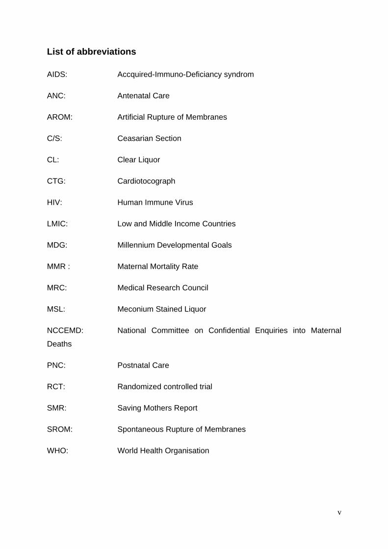
List of abbreviations
AIDS: Accquired-Immuno-Deficiancy syndrom
ANC: Antenatal Care
AROM: Artificial Rupture of Membranes
C/S: Ceasarian Section
CL: Clear Liquor
CTG: Cardiotocograph
HIV: Human Immune Virus
LMIC: Low and Middle Income Countries
MDG: Millennium Developmental Goals
MMR : Maternal Mortality Rate
MRC: Medical Research Council
MSL: Meconium Stained Liquor
NCCEMD: National Committee on Confidential Enquiries into Maternal
Deaths
PNC: Postnatal Care
RCT: Randomized controlled trial
SMR: Saving Mothers Report
SROM: Spontaneous Rupture of Membranes
WHO: World Health Organisation
vi
Table of contents
Declaration .................................................................................................................................. i
Dedication .................................................................................................................................. ii
Acknowledgements .................................................................................................................. iii
Abstract ..................................................................................................................................... iv
List of abbreviations .................................................................................................................. v
Table of contents ....................................................................................................................... vi
List of annexures ...................................................................................................................... xii
List of figures ......................................................................................................................... xiii
List of tables ............................................................................................................................ xvi
Chapter One ............................................................................................................................... 1
Introduction to the study ............................................................................................................ 1
1.1. Background of the study ................................................................................................. 1
1.2. Problem statement ........................................................................................................... 5
1.3. Purpose of the study ........................................................................................................ 7
1.4. Research objectives ......................................................................................................... 7
1.5. Research questions .......................................................................................................... 7
1.6. Significance of the study ................................................................................................. 8
1.7. Operational definitions .................................................................................................... 8
1.7.1. Partograph ................................................................................................................... 8
1.7.2. Partogram .................................................................................................................... 8
1.7.3. Midwives ..................................................................................................................... 9
1.7.4. Obstetrician ................................................................................................................. 9
1.7.5. Labour ......................................................................................................................... 9
1.7.6. Maternal death ........................................................................................................... 10
1.7.7. Neonatal death ........................................................................................................... 10

vii
1.7.8. Stillbirth ..................................................................................................................... 10
1.7.9. Perinatal mortality ..................................................................................................... 10
1.7.10. Records ...................................................................................................................... 10
1.8. Chapters outline ............................................................................................................ 11
1.9. Conclusion .................................................................................................................... 12
Chapter two .............................................................................................................................. 13
Literature review ...................................................................................................................... 13
2.1. Introduction ................................................................................................................... 13
2.2. Maternal and newborn mortality rate ............................................................................ 13
2.3. Importance of partogragh ............................................................................................. 16
2.4. The utilization of partograph ........................................................................................ 19
2.5. Challenges in the utilization of the partograph ............................................................. 23
2.6. Theoretical framework guiding the research study ....................................................... 25
2.6.1 Structure .......................................................................................................................... 25
2.6.2 Process ............................................................................................................................ 26
2.6.3 Outcome .......................................................................................................................... 27
2.7. Eight principles of Tessier‘s Model and application of those relevant to this study: ... 28
2.7.1. Unique Identification ................................................................................................. 29
2.7.2. Accuracy .................................................................................................................... 29
2.7.3. Confidentiality ........................................................................................................... 30
2.7.4. Completeness ............................................................................................................ 30
2.7.5. Timeliness ................................................................................................................. 30
2.7.6. Accountability ........................................................................................................... 31
2.7.7. Legibility ................................................................................................................... 31
2.7.8. Resources .................................................................................................................. 31
2.8. Conclusion .................................................................................................................... 32
Chapter Three........................................................................................................................... 33
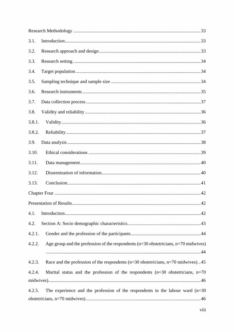
viii
Research Methodology ............................................................................................................ 33
3.1. Introduction ................................................................................................................... 33
3.2. Research approach and design ...................................................................................... 33
3.3. Research setting ............................................................................................................ 34
3.4. Target population .......................................................................................................... 34
3.5. Sampling technique and sample size ............................................................................ 34
3.6. Research instruments .................................................................................................... 35
3.7. Data collection process ................................................................................................. 37
3.8. Validity and reliability .................................................................................................. 36
3.8.1. Validity ...................................................................................................................... 36
3.8.2. Reliability .................................................................................................................. 37
3.9. Data analysis ................................................................................................................. 38
3.10. Ethical considerations ............................................................................................... 39
3.11. Data management ...................................................................................................... 40
3.12. Dissemination of information .................................................................................... 40
3.13. Conclusion ................................................................................................................. 41
Chapter Four ............................................................................................................................ 42
Presentation of Results ............................................................................................................. 42
4.1. Introduction ................................................................................................................... 42
4.2. Section A: Socio demographic characteristics .............................................................. 43
4.2.1. Gender and the profession of the participants ........................................................... 44
4.2.2. Age group and the profession of the respondents (n=30 obstetricians, n=70 midwives)
................................................................................................................................... 44
4.2.3. Race and the profession of the respondents (n=30 obstetricians, n=70 midwives) .. 45
4.2.4. Marital status and the profession of the respondents (n=30 obstetricians, n=70
midwives) ................................................................................................................................. 46
4.2.5. The experience and the profession of the respondents in the labour ward (n=30
obstetricians, n=70 midwives) ................................................................................................. 46
ix
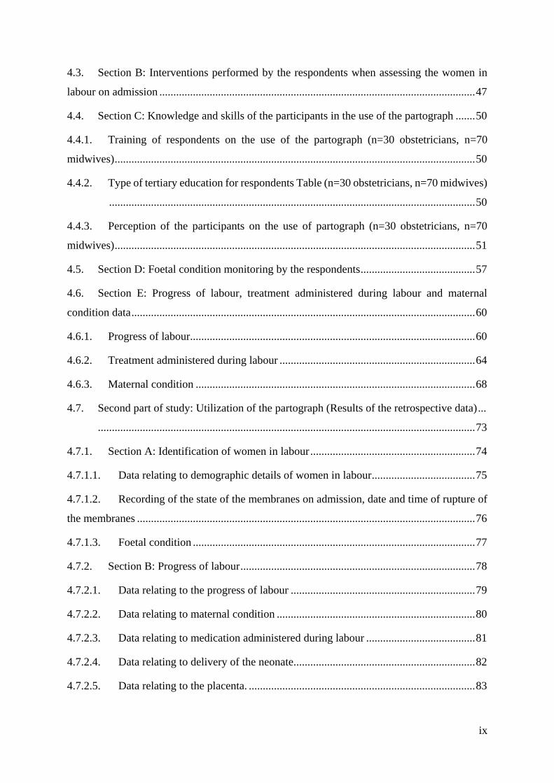
4.3. Section B: Interventions performed by the respondents when assessing the women in
labour on admission ................................................................................................................. 47
4.4. Section C: Knowledge and skills of the participants in the use of the partograph ....... 50
4.4.1. Training of respondents on the use of the partograph (n=30 obstetricians, n=70
midwives) ................................................................................................................................. 50
4.4.2. Type of tertiary education for respondents Table (n=30 obstetricians, n=70 midwives)
................................................................................................................................... 50
4.4.3. Perception of the participants on the use of partograph (n=30 obstetricians, n=70
midwives) ................................................................................................................................. 51
4.5. Section D: Foetal condition monitoring by the respondents ......................................... 57
4.6. Section E: Progress of labour, treatment administered during labour and maternal
condition data ........................................................................................................................... 60
4.6.1. Progress of labour ...................................................................................................... 60
4.6.2. Treatment administered during labour ...................................................................... 64
4.6.3. Maternal condition .................................................................................................... 68
4.7. Second part of study: Utilization of the partograph (Results of the retrospective data) ...
....................................................................................................................................... 73
4.7.1. Section A: Identification of women in labour ........................................................... 74
4.7.1.1. Data relating to demographic details of women in labour ..................................... 75
4.7.1.2. Recording of the state of the membranes on admission, date and time of rupture of
the membranes ......................................................................................................................... 76
4.7.1.3. Foetal condition ..................................................................................................... 77
4.7.2. Section B: Progress of labour .................................................................................... 78
4.7.2.1. Data relating to the progress of labour .................................................................. 79
4.7.2.2. Data relating to maternal condition ....................................................................... 80
4.7.2.3. Data relating to medication administered during labour ....................................... 81
4.7.2.4. Data relating to delivery of the neonate. ................................................................ 82
4.7.2.5. Data relating to the placenta. ................................................................................. 83

x
4.7.2.6. Data relating to condition of the infant at birth ..................................................... 83
4.8. Conclusion .................................................................................................................... 85
Chapter Five ............................................................................................................................. 86
Discussion, conclusion, recommendations, of the study ......................................................... 86
5.1. Introduction ................................................................................................................... 86
5.2. Midwives’ and obstetricians’ knowledge of the use of partograph in monitoring progress
of labour. .................................................................................................................................. 86
5.2.1. Source of Knowledge and skills to use the partograph ............................................. 86
5.2.2. Assessment of women in labour on admission to labour ward. ................................ 87
5.2.3. Monitoring of the foetal condition ............................................................................ 88
5.2.3.1. Foetal Heart Rate ................................................................................................... 88
5.2.3.2. State of membranes and liquor .............................................................................. 89
5.2.3.3. Caput and moulding............................................................................................... 89
5.2.3.4. The progress of labour ........................................................................................... 90
5.2.3.5. Administration of medication during labour ......................................................... 91
5.2.3.6. Maternal condition ................................................................................................. 91
5.3. Factors that influence the use of the partograph in the labour ward ............................. 92
5.3.1. Factors that facilitate the use of partograph in the labour ward ................................ 92
5.3.2. Factors that hinder the use of the partograph in the labour ward. ............................. 93
5.4. Utilization of the partograph by midwives and obstetricians in the labour ward
(retrospective part of the study) ............................................................................................... 95
5.4.1. Documentation/recording of the partograph ............................................................. 95
5.4.1.1. Presentation of demographic data is the partograpgh ............................................ 95
5.4.1.2. The state of the membranes on admission ............................................................. 96
5.4.1.3. The foetal condition ............................................................................................... 96
5.4.1.4. The progress of labour ........................................................................................... 97
5.4.1.5. Administration of treatment during labour ............................................................ 97
5.4.1.6. Maternal condition ................................................................................................. 97
xi
5.4.1.7. Recording of post-delivery information ................................................................ 98
5.5. Training of midwives and obstetricians ...................................................................... 100
5.6. Recommendations ....................................................................................................... 101
5.6.1. In-service training for health care providers ........................................................... 101
5.6.2. Education and curriculum for training of midwifery and medical students. ........... 102
5.6.3. Policy-makers, management and government ........................................................ 102
5.6.4. Further research ....................................................................................................... 103
5.7. Limitations of the study .............................................................................................. 103
5.8. Conclusion .................................................................................................................. 104
References .............................................................................................................................. 105
Annexures .............................................................................................................................. 113
xii
List of annexures
Annexure 1: Information Sheet .............................................................................................. 113
Annexure 2: Consent to Participate in Research Project ....................................................... 114
Annexure 3: Questionnaire .................................................................................................... 115
Annexure 4: Checklist for partograms for retrospective part of the study ............................ 120
Annexure 5: Request for permission to conduct research study ............................................ 126
Annexure 6: Request for permission to conduct a research study ......................................... 127
Annexure 7: Request for permission to conduct a research study ......................................... 128
Annexure 8: Letter of approval from DoH ............................................................................ 129
Annexure 9: Letter of approval from PMMH ........................................................................ 130
Annexure 10: UKZN Ethical Clearance ................................................................................ 131
xiii
List of figures
Figure 2.1: A diagrammatic illustration of the theoretical framework guiding the study ....... 28
Figure 2.2: Eight principles of Tessier’s model (Tessier, 2003).............................................. 29
Figure 4.1: The study outline ............................................................................................................ 42
Figure 4.2: The gender of the respondents (n=30 obstetricians, n=70 midwives) .................... 44
Figure 4.3: Comparison of the different age groups of obstetricians versus midwives (n=30
obstetricians, n=70 midwives) .......................................................................................................... 45
Figure 4.4: The number of respondents from the different racial groups (n=30 obstetricians,
n=70 midwives) .................................................................................................................................. 45
Figure 4.5: Marital status and the profession of the respondents (n=30 obstetricians, n=70
midwives) ............................................................................................................................................ 46
Figure 4.6: The experience and the profession of the respondents in the labour ward (n=30
obstetricians, n=70 midwives) .......................................................................................................... 47
Figure 4.7: Responses of obstetricians and midwives on assessment of women in labour on
admission (n=30 obstetricians, n=70 midwives) ............................................................................ 49
Figure 4.8: Response of obstetricians and midwives to the systematic use of the partograph
(n=30 obstetricians, n=70 midwives) .............................................................................................. 52
Figure 4.9: Response to the significant use of the partograph in the labour ward (n=30
obstetricians, n=70 midwives) .......................................................................................................... 53
Figure 4.10: Response to the partograph as a tool for decision-making in the labour ward (n=30
obstetricians, n=70 midwives) .......................................................................................................... 53
Figure 4.11: Responses to the partograph as a tool to monitor the progress of labour (n=30
obstetricians, n=70 midwives) .......................................................................................................... 54
Figure 4.12: Responses to the partograph as a tool to detect complications during labour (n=30
obstetricians, n=70 midwives) .......................................................................................................... 55
Figure 4.13: Responses to the partograph as a tool that improves management of labour (n=30
obstetricians, n=70 midwives) .......................................................................................................... 55
Figure 4.14: Responses to the view that the partograph is difficult to use in the labour ward
(n=30 obstetricians, n=70 midwives) .............................................................................................. 56
Figure 4.15: Monitoring of the foetal heart rate in the active phase of labour ........................... 58
Figure 4.16: Time intervals for monitoring the state of membranes and liquor in the active
phase of labour (n=30 obstetricians, n=70 midwives) .................................................................. 59
xiv
Figure 4.17: Time intervals for monitoring of caput and moulding in the active phase of labour
(n=30 obstetricians, n=70 midwives) .............................................................................................. 59
Figure 4.18: Views of obstetricians and midwives regarding time interval monitoring of
cervical dilatation during the active phase of labour (n=30 obstetricians, n=70 midwives) .... 61
Figure 4.19: Views of obstetricians and midwives regarding time interval of monitoring of
cervical effacement during the active phase of labour (n=30 obstetricians, n=70 midwives) . 62
Figure 4.20: Views of obstetricians and midwives regarding the time interval of monitoring of
descent of the foetal head during the active phase of labour (n=30 obstetricians, n=70
midwives) ............................................................................................................................................ 62
Figure 4.21: Views of obstetricians and midwives regarding the time interval of monitoring
uterine contractions during the active phase of labour (n=30 obstetricians, n=70 midwives) . 63
Figure 4.22: Views of obstetricians and midwives regarding administration of intravenous
fluids, only if necessary (obstetricians n=30, midwives n=70) .................................................... 65
Figure 4.23: Views of obstetricians and midwives regarding the use of oxytocic drugs for
augmentation of labour in cases of poor progress (obstetrician’s n=30, midwives n=70) ....... 66
Figure 4.24: Views of obstetricians and midwives regarding analgesics, e.g. pethidine or
entonox inhalation administered for pain relief (obstetricians n=30, midwives n=70) ............ 67
Figure 4.25: Views of respondents that antibiotics may be administered in cases of prolonged
rupture of the membranes (obstetricians’ n=30, midwives n=70) ............................................... 68
Figure 4.26: Time interval for blood pressure and pulse rate monitoring in the active phase of
labour (obstetricians’ n=30, midwives n=70) ................................................................................. 69
Figure 4.27: Time interval for temperature observation throughout labour (obstetricians n=30,
midwives n=70) .................................................................................................................................. 70
Figure 4.28: Time interval for checking urinalysis and urine output during the active phase of
labour (obstetricians n=30, midwives n=70) .................................................................................. 70
Figure 4.29: Factors that facilitate the use of partograph in the labour ward. ............................ 71
Figure 4.30: Factors that hinder the use of partograph in the labour ward. ................................ 72
Figure 4.31: Recording of name and surname, age, gravid and parity on the partograms
(n=100). ............................................................................................................................................... 75
Figure 4.32: Recording of the state of membranes on admission, date and time of rupture of the
membranes on partograms (n=100) ................................................................................................. 76
Figure 4.33: Recording of foetal heart rate, liquor, caput and moulding on partograms (n=100)
.............................................................................................................................................................. 77
xv
Figure 4.34: Recording of data regarding cervical dilatation and effacement, level of foetal
head and uterine contractions on the partographs (n=100) ........................................................... 80
Figure 4.35: Recording of temperature, pulse, blood pressure and urine output on partograms
(n=100) ................................................................................................................................................ 81
Figure 4.36: Recording of the type of medication, dosage, time of administration and mode of
administration on the partograms (n=100) ...................................................................................... 82
Figure 4.37: Recording of data relating to delivery of the neonate on the partogram (n=100)82
Figure 4.38: Recording of the date and time of expulsion of the placenta, the state of the
placenta and membranes after delivery (n=100) ............................................................................ 83
Figure 4.39: Recording of neonatal outcome, Apgar score, gender, birth weight and whether a
quick examination of the newborn was recorded on the partograms (n=100) ........................... 84
xvi
List of tables
Table 3.1: Content validity ...................................................................................................... 37
Table 4.1: General overview of the demographic data relating to all obstetricians and midwives
(n= 100) .................................................................................................................................... 43
Table 4.2: The responses of respondents on the assessment of women in labour on admission
to the labour ward (n=30 obstetricians, n=70 midwives) ........................................................ 48
Table 4.3: Perception of the participants on the use of partograph (n=30 obstetricians, n=70
midwives) ................................................................................................................................. 51
Table 4.4: Monitoring of the foetal condition at different time intervals (n=30 obstetricians,
n=70 midwives ......................................................................................................................... 57
Table 4.5: Views of obstetricians and midwives on timely intervals for monitoring the progress
of labour ................................................................................................................................... 60
Table 4.6: Views of obstetricians and midwives regarding treatment administered during labour
(n=30 obstetricians, n=70 midwives) ....................................................................................... 64
Table 4.7: Shows the views of the respondents about time interval of monitoring blood
pressure, pulse, temperature, urinalysis and urine output (obstetricians n=30, midwives n=70)
.................................................................................................................................................. 68
Table 4.8: General overview of findings obtained from the old 100 partograms regarding
demographic details, the state of membranes and the foetal condition in women who delivered
in September, 2010 (n=100) .................................................................................................... 74
Table 4.9: The general overview of recording of information in the progress of labour, the
maternal condition, medication administered during labour, delivery of the placenta and
membranes, as well as the condition of the infant at birth, as recorded on the partograms
(n=100) ..................................................................................................................................... 79
Table 4. 10: Proportions of partograms with records of parameters of labour (n=100 audited
records) .................................................................................................................................... 84
1
Chapter One
Introduction of the study
1.1. Background
Globally it is of paramount importance that all pregnant women in labour are monitored
by midwives utilizing a partograph so that a live baby is delivered (Mathibe-Neke,
Lebeko and Motupa, 2013; Shokane, 2011). A partograph is a pre-printed paper that
provides a visual display of recorded observations carried out on mother and foetus
during labour. It is universally used as part of safe motherhood initiative for improving
labour management and reducing maternal and foetal morbidity and mortality (Souza,
Oladapo, Bohren, Mugerwa, Fawole, Moscovici et al., 2015; Khonje, 2012). The
partograph is used to identify abnormal labours which are the cause of problems that
lead to morbidity and mortality (Yisma, Dessalegn, Astatkie and Fesseha, 2013a).
However, most parameters on the partograph are not monitored and most health care
workers do not document their findings on the partograph after reviewing a woman in
labour. Midwives seem not to utilize the partograph correctly when monitoring women
in labour (Shokane, 2011). Hence the progress of labour may not be closely monitored
or labour monitoring may not translate into actions required when need arise (Khonje,
2012).
It is immense problem that deaths of women from any cause associated with and or
aggravated by the pregnancy or its management i.e. throughout the duration period of
pregnancy (gestation), child birth (delivery) as well as after birth (postpartum period)
continue to persist in the 21st century. Globally, in 2015, the maternal mortality rate
(MMR) stabilized around 216 deaths per 100 000 live births while total estimate for
maternal deaths was 300 000 (WHO and UNICEF, 2015; Hogan, Foreman, Naghavi,
Ahn, Wang, Makela et al., 2010). According to the WHO and UNICEF (2015) there are
stark disparities in service accessibility and utilization; for instance; women in sub-
Saharan Africa have a 1:31 lifetime risk of dying as a consequence of childbirth,
compared with a 1:4300 risk in high income countries, explaining the spatial sorting of
the burden of maternal mortality (Sakala, 2011).
2
Notably, the difference in the (MMR) between less economically developed (LED)
countries and high income countries provides strong indication that maternal deaths
are after all preventable (UNICEF, 2009; Ozumba and Nwogu-Ikojo, 2008; De Lange,
Budde, Heard, Tucker, Kennare and Dekker, 2008; Alves, 2007). It is with this view
that the United Nations (UN) under the auspices of Millennium Development Goal
number 5 (MDG 5) aimed to facilitate the reduction of the MMR which would be
reduced by 49 per cent from its 1990 level by 2015 (United Nations, 2015).
Furthermore, United Nations (2015) states that the majority of neonatal deaths
worldwide are caused by preterm birth complications (35 per cent), complications
during labour and delivery (24 per cent) and sepsis (15 per cent). In sub-Saharan
Africa and Southern Asia many deaths are also due to preventable infectious
diseases. Many neonatal deaths could be avoided with simple, cost-effective and high-
impact interventions that address the needs of women and newborns across the
continuum of care, with an emphasis on care around the time of birth. However,
analysis shows that too many newborns and mothers miss out on these key
interventions (United Nations, 2015).
In spite of substantial efforts in many Sub-Saharan countries to reduce the maternal
mortality ratio over recent decades, country-specific decline has been largely marginal,
in fact, the MMR even is stalling in some countries/regions, such as Eritrea, Guinea
Bissau and the Central African Republic where the rate of decline has been under 2
per cent over a 25-year period from 1990-2015. The adverse effect of HIV on women's
health in sub-Saharan Africa appears to be an important contributing factor to poor
progress towards the millennium developmental goals (MDG) (Wilmoth, Mathers, Say
and Mills, 2010; Hogan et al., 2010).
According to Wakgari, Amano, Berta and Tessema (2015) obstructed labour
accounted for 8% of maternal deaths, hence the reported incidence of obstructed
labour expected to reach 20% in developing countries. Additionally, in Ethiopia
obstructed or prolonged labour accounted for 13% of maternal deaths (Wakgari et al.,
2015). The women who experience obstructed labour usually suffer from postpartum
hemorrhage, uterine rupture, puerperal sepsis and obstetric fistula. Furthermore, it is
highly associated with birth trauma, birth asphyxia, stillbirths, neonatal sepsis and
3
neonatal deaths (de Wet, 2016; Nakimuli, Chazara, Byamugisha, Elliott, Kaleebu,
Mirembe et al., 2014; Prata, Passano, Sreenivas and Gerdts, 2010).
Therefore, prevention of prolonged and obstructed labour by using partograph during
labour is a key intervention in the reduction of maternal and perinatal morbidity and
mortality (Wakgari et al., 2015). The partograph provides a framework for assessing
maternal and foetal condition and labour progress during Systematic monitoring of
labour is doubly beneficial in that it secures the well-being of the pregnant mother and
leverages it for in improving the foetal condition but altogether saving the lives of both
(Shokane, 2011). With the premise that prolonged labour is a common cause of death
for delivering mothers, universal uptake of partograph (i.e. a form for the registry of
vital statistics on the progress of labour process) would detect birth complications
timeously and refer women to relevant prevention and treatment procedures (Bazirete,
2014). In other words, the partograph seeks to illustrate, in the form of a pictorial
overview labour, to signal midwives and obstetricians whether there are deviations in
maternal and foetal well-being and the labour progress in general. In addition, a
secondary benefit is that accurate imputation of birth related data facilitates smoother
dissemination within the strata of health personnel attending to mothers in labour,
increasing the odds of replicating the literacy for its use in a hospital (Bazirete, 2014).
The literature indicates that the use of partogram assist in deducing significantly
maternal and perinatal mortality rate. According to Yisma et al. (2013a) a partograph
is one of the valuable appropriate technologies in use for improved monitoring of
labour progress, maternal and foetal wellbeing. In addition, the partograph enables
midwives and obstetricians to record their examination findings on a standardized
form, which generates a pictorial overview of labour progress, maternal and foetal
condition, which allows for early identification and diagnosis of pathological labour
(Masika, Katongole and Govule, 2015; Yisma et al., 2013a). Accordingly, it captures;
measures of cervical effacement, dilatation, descent of the foetal head and
contractions, foetal heart rate, maternal pulse, blood pressure and temperature, urine
output and volume as well as type of intravenous infusions and medication given
(Mathibe-Neke et al., 2013). The use of partograph is critical in preventing maternal
and perinatal morbidity and mortality and therefore has applicability in developed as
well as developing world settings (Yisma, Dessalegn, Astatkie and Fesseha, 2013b).

4
In diverse regions in Sub-Saharan Africa, the effective utilization of partograph during
births is fraught by skills deficiency and a general lack will to adopt it fully (Yisma et
al., 2013a). In a study examining completion of the modified WHO partograph during
labour in public health institutions in Ethiopia finds evidence for poor management of
labour, particularly associated with inappropriate completion of the tool. To counteract
this, they recommend pre-service and regular on-job training for health workers on the
correct completion of the partograph (Yisma et al., 2013a).
Elsewhere in Kenya, students undertaking training to be midwives were more likely to
face technical difficulties completing partograph when practicing in the labour ward
environment (Lavender, Omoni, Lee, Wakasiaka, Watiti and Mathai, 2011). These
results reveal that partograph training and implementation are not adequately taught
during formal training, increasing the need for re-orientation exercises and support for
students in labour wards. Another interesting finding in Lavender et al. (2011) is that
student midwife training was not likely to be implemented into practice unless the more
qualified health workers supports their learning. This further reinforces the assertion
that partograph will continue to be somewhat trivially regarded in the labour ward
unless senior health professionals (midwives and obstetricians) lead by example,
suggesting the need for further research to explore the views of obstetricians and
qualified midwives on partograph use. A study in Ghana thus questions current
strategies for implementing partograph by suggesting the need to further investigate
reasons surrounding the limited use of partograph even among skilled birth attendants
(Gans-Lartey, O'Brien, Gyekye and Schopflocher, 2013).
In the context of South Africa, in the review of the Millennium Development Goals
(MDG) in 2013, South Africa was still listed as one of the countries making poor
progress towards reducing maternal and perinatal mortality. South Africa, despite of
being the single high-income nation among in a largely developing Sub-Saharan
Africa, surprisingly high rates of maternal mortality persist (Department of Health,
2015b). Presently, the maternal mortality rate is 269 deaths per 100,000 live births, a
standing glaringly higher than the rate of 38 which the national government committed
by 2015, the termination on the MDGs. Yet, it is an unobjectionable view by patrons
of maternal healthcare that up to 60% of maternal deaths in South Africa are in-fact

5
avoidable indicating mostly poor quality of care during the antenatal, intrapartum and
postnatal period (Department of Health, 2015b).
Locally, the reason for this failure has been attributed to a combination of leading
cause (Lloyd and De Witt, 2013). These causes include intrapartum asphyxia
complications which can be monitored with widespread use of partograph during birth.
For instance, between 2010-2011, deaths induced by intrapartum asphyxia were
reported to be linked to healthcare-worker-related factors in 44% of the cases
according to the Saving Babies 2010-2011 report (NCCEMD, 2013). Five of these
determinants implicating the healthcare worker were: (i) fetal distress monitored but
neglected; (ii) fetal distress not monitored and not detected; (iii) no intervention for
prolonged second stage labour; (iv) delay in referring the patient and (v) delay in calling
for expert assistance (NCCEMD, 2013; Lloyd and De Witt, 2013).
In south Africa midwives and obstetricians face challenges in the utilisation of the
partograph to monitor the progress of labour. It is important that midwives should use
the partograph when monitoring pregnant women in labour so that complications
identified early during monitoring of labour can be attended to timeously by both the
midwife and the attending obstetricians (Mathibe-Neke et al., 2013; Shokane, 2011).
Despite the call by the National Department of Health, Mathibe-Neke (2009)
highlighted that midwives use the partograph incorrectly and inappropriately.
Failure of midwives to utilise the partograph correctly and appropriately when
monitoring women during intrapartum constitutes sub-standard care (Mianda, 2010;
NCCEMD, 2006). This viewpoint is supported by Opiah, Ofi, Essien and Monjok (2012)
who highlights that the number of pregnant women who suffer complications is likely
to be reduced significantly through utilisation of a partograph to monitor women in
labour.
1.2. Problem statement
Globally it is of paramount importance that all pregnant women in labour are monitored
by midwives utilizing a partograph so that a live baby is delivered. Midwives seem not
to utilize the partograph correctly when monitoring women in labour (Shokane, 2011).
Although a considerable amount of experience and information on the use of the
6
partograph has been accumulated in the past forty years, it seems not to be effectively
used in many developing countries (Adesola, Adekemi and Audu, 2014; Mathibe-Neke
et al., 2013). Proper monitoring of women during labour and continuous accurate
recording on the partograph are critical not only in intervening appropriately, but also
for the purpose of minimizing unnecessary interventions (Mathibe-Neke et al., 2013).
The literature indicates that the tragedy of maternal mortality in sub-Saharan Africa is
that, despite its recognition as a major public health issue, maternal mortality figures
continue to rise, in spite of the apparent commitment by stake-holders. The majority
of maternal and perinatal deaths and complications could be prevented by cost-
effective and affordable health interventions like the use of the partograph as a tool for
monitoring the progress of labour and prevent morbidity and improve neonatal
outcome (Bazirete, 2014; Fawole, Adekanle and Hunyinbo, 2010).
Though several literatures demonstrated good knowledge about the partograph
among midwives and obstetricians, several factors still hinder appropriate utilization
of the partograph such as lack of detailed knowledge of the partograph, non-availability
of the partograph, poor staff numbers, and inadequate training are factors that work
against the effective utilization of the partograph in the study facility (Asibong, Okokon,
Agan, Oku, Opiah, Essien et al., 2014). Furthermore a gross deficiencies have been
highlighted regarding knowledge about normal characteristics during labour (Fawole
et al., 2010). Midwives frequently do not utilize the partograph appropriately when
monitoring pregnant women in labour by not plotting or incomplete plotting and not
analyzing nor interpreting the findings (Shokane, 2011). With regards to the knowledge
of different components of the partogram, Yisma et al. (2013a) in a cross-sectional
quantitative study to assess knowledge and utilization of partograph among obstetric
care givers in public health institutions of Addis Ababa, affirms that knowledge of the
function of both alert line and action line were poor.
In diverse regions in Sub-Saharan Africa, the effective utilization of partograph during
births is fraught by skills deficiency and a general lack will to adopt it fully (Yisma et
al., 2013a). Although the partograph has been in use in many provinces including
KwaZulu-Natal for over 25 years, its adoption and adaptation has been shown to be
poor, and despite the training at undergraduate level, and in service training there is
still a high maternal and peri-natal mortality rate associated with incorrect use of

7
partograph in regional hospitals in South Africa. Monitoring labour in a systematic way
with use of the partograph; is advocated even in low income countries, to prevent
possible child birth complications. Early detection and early management of
complications reduces maternal and perinatal mortality and morbidity (Mathibe-Neke
et al., 2013; Khonje, 2012; Barros, Bhutta, Batra, Hansen, Victora and Rubens, 2010).
Thus this study tends to explore and describe the knowledge, attitudes and utilization
of the partograph by midwives and obstetricians in a regional hospital in the eThekwini
district.
1.3. Purpose of the study
The purpose of this study was to explore and describe the knowledge, attitudes and
utilization of the partograph by midwives and obstetricians in a regional hospital in the
eThekwini district.
1.4. Research objectives
To determine the midwives' and obstetricians' knowledge of the
partograph in monitoring the progress of labour.
To describe the utilization of the partograph by midwives and
obstetricians in the labour ward.
To identify factors that influence the utilization of the partograph to
monitor the progress of labour.
1.5. Research questions
What is the midwives’ and obstetricians’ knowledge regarding the use of
the partograph?
How do midwives and obstetricians utilize f the partograph in the labour
ward?
What factors influence partograph use by midwives and obstetricians in
8
the labour ward?
1.6. Significance of the study
The findings from this study may assist in awakening the importance of the appropriate
utilization of the partograph. More important than this, the researcher will share
findings with the Department of Health (DOH) and if required recommend ongoing in-
service training on the correct usage of the partograph.
The participation in this study would allow the midwives and obstetricians to share
their knowledge, attitude and the utilization of the partograph as well as the challenges
they face while using the partogram.
The findings from this study would trigger further research in the utilization of the
partograph, and necessity to put more emphasis on continuous development of
midwives and obstetricians in terms of proper utilization of the partograph.
1.7. Operational definitions
1.7.1. Partograph
A partograph is a graphical presentation of a woman’s progress of labour. Once the
woman has true signs of labour, the midwife initiates the use of the partograph to
record her findings of maternal, foetal condition, progress of labour and treatment
administered to a woman (Mathibe-Neke et al., 2013). In this study, the term
'partograph' will refer to a blank or unplotted labour graph as a tool that is used by
midwives and obstetricians to record their findings during labour so as to realize when
intervention is necessary to prevent complications.
1.7.2. Partogram
A partogram is a printed format that provides a pictorial overview of what was plotted
or recorded in the progress of labour, the purpose of which is to alert midwives and
obstetricians to any maternal and foetal problem (Lavender, Alfirevic and Walkinshaw,

9
2006). In this study, the term 'partogram' will refer to a record of completed or plotted
information by midwives and obstetricians to monitor the progress of labour.
1.7.3. Midwives
The International Confederation of Midwives (ICM) (ICM, 2005) defines the midwife
as: A person who has been regularly admitted to a midwifery educational programme,
duly recognized in the country in which it is located, has successfully completed the
prescribed course of studies in midwifery and has acquired the requisite qualifications
to be registered and/or legally licensed to practice midwifery. She may practice in
hospital, clinics, health units, domiciliary conditions or any other service (ICM, 2005).
In this study, the term 'midwife' will refer to a registered midwife (male or female) who
provides prenatal care to expectant mothers, conducts delivery of the babies and
provides postpartum care to mothers and their infants.
1.7.4. Obstetrician
The obstetrician is a medical practitioner who specializes in the care of pregnant
women from the time of conception through delivery and the period following delivery,
that is, the postpartum period (Sellers, 1995). In this study, an obstetrician is a medical
officer who takes full responsibility (specialized) for the woman during pregnancy,
labour and the puerperium period.
1.7.5. Labour
Labour is the process which involves the progressive dilatation and effacement or
shortening of the cervix. This process is always accompanied by increasingly painful
uterine contractions, descent of the foetal head and occasional spontaneous rupture
of the membranes (Buhimschi, Buhimschi, Malinow, Saade, Garfield and Weiner,
2003). In this study, 'labour' will refer to uterine contractions that result in progressive
dilatation and effacement of the cervix.
10
1.7.6. Maternal death
The International Classification of Diseases Injuries and Causes of Death (10th
Revision) defines maternal deaths as: The death of a woman while pregnant or within
42 days of termination of pregnancy, from any cause related to or aggravated by the
pregnancy or its management, but not from accidental or incidental causes (NCCEMD,
2006). In this study, 'maternal death' will refer to the death of a woman during or shortly
after pregnancy, intrapartum and puerperium period which is within 6 weeks or 42
days after delivery.
1.7.7. Neonatal death
This is the death of a live born infant within the first 28 days of life. Death of a live born
infant within the first 7 days of life is referred to as early neonatal death and death of
a live born infant extending from 7-28 days is a late neonatal death (Nannan,
Dorrington, Laubscher, Zinyakatira, Prinsloo, Darikwa et al., 2012). In this study,
'neonatal death' refers to the death of a live born infant within the first 28 days of life.
1.7.8. Stillbirth
A stillbirth is a viable foetus which has shown no signs of life after its complete birth,
that is, no heart beat and respiration (Sellers, 1995). In this study, a 'stillbirth' is a viable
infant that is born dead.
1.7.9. Perinatal mortality
This refers to a viable infant which is born dead (stillbirth), plus to infants who are born
alive but die within seven days after delivery (NCCEMD, 2006). In this study, 'perinatal
mortality' is death occurring during late pregnancy, (at 26 weeks completed gestation
and above) during childbirth and up to seven completed days of life.
1.7.10. Records
This refers to documentation of all necessary information that gives a clear picture of
11
what took place (Keenan, Yakel, Tschannen and Mandeville, 2008). In this study,
'records' means the complete and legible pertinent assessment data such as vital
signs, patients' complaints, who was notified and what interventions were carried out.
1.8. Chapters outline
Chapter One: Introduction and background
Chapter one presents the background of the study, the problem statement, the
purpose to the study, objectives and research questions, significance to the study and
operational definitions.
Chapter two: Literature review
Chapter two reviews the existing literature on partograph use, describes the different
features of partographs used for monitoring the management of labour, and presents
the conceptual framework used in the study. In addition, a review of the relevant
literature and previous research studied on partograph use, in relation to the maternal
and neonatal outcome is conducted.
Chapter Three: Methodology
Chapter three outlines the research approach, the research design, the study
instruments, the research setting, the target population, the ethical considerations, and
the study limitations. This takes the form of a prospective questionnaire-based survey
and a retrospective chart review study.
Chapter Four: presentation of results
Chapter four presents the results of the study and are presented in tables and graphs.
The presentation of the results of the study is divided into the following two main
categories. Both the results from prospective questionnaire and Retrospective chart
review are presented in this chapter.
Chapter Five: Discussion, conclusion of the study
This chapter discuss about the major findings and provide conclusion and the
recommendations.

12
1.9. Conclusion
This chapter covered the introduction and the background to the study, the problem
statement, the purpose to the study, objectives and research questions, significance
to the study and operational definitions. And the following chapter covers the
literature underpinning this study.
13
Chapter two
Literature review
2.1. Introduction
This chapter summarises the previous studies conducted on the use of the partograph.
Grove, Burns and Gray (2013) state that literature review includes both theoretical and
empirical sources that document the current knowledge of the problem. These authors
continued that the theoretical component consists of theories, models and conceptual
frameworks while the empirical component consists of sources from various studies
published in journals, books and theses. Therefore, the literature for the present study
was gleaned from published journal articles, textbooks, published reports, newsletters
and internet search on partogram. The literature in this study was organized into the
following headings: Maternal and new-born mortality rate, importance of the
partograph, utilization of the partograph, challenges in the utilization of the partograph
and the theoretical framework guiding the study. The following data bases were used:
Google scholar, google, pub med, science direct.
Key words: Partograph, partogram, perinatal care, delivery ward.
2.2. Maternal and newborn mortality rate
Maternal mortality is unacceptably high. About 830 women die from pregnancy- or
childbirth-related complications around the world every day (WHO, 2015). By the end
of 2015, roughly 303 000 women will have died during and following pregnancy and
childbirth. Almost all of these deaths occurred in low-resource settings, and most could
have been prevented (WHO, 2015). It is tragic that maternal deaths occur during the
natural process of pregnancy, child birth and the postpartum period. Globally, each
year there are at least 3.2 million stillborn babies, 4 million neonatal deaths and more
than half a million maternal deaths as a result of complications associated with
pregnancy and childbirth (Hogan et al., 2010; WHO, 2006; WHO, 2005). More than
half the maternal deaths take place within one day of birth (Time, 2016; UNICEF,
2009). Efforts to reduce deaths among women from complications related to
14
pregnancy and childbirth have been less successful than other areas of human
development, with the result that having a child remains among the most serious
health risks for women (UNICEF, 2009).
Maternal survival has significantly improved since the adoption of the MDGs. The
maternal mortality ratio dropped by 45 per cent worldwide between 1990 and 2013,
from 380 maternal deaths per 100,000 live births to 210. Many developing regions
have made steady progress in improving maternal health, including the regions with
the highest maternal mortality ratios (United Nations, 2015). For example, in Southern
Asia the maternal mortality ratio declined by 64 per cent between 1990 and 2013, and
in sub-Saharan Africa it fell by 49 per cent. Despite this progress, every day hundreds
of women die during pregnancy or from childbirth-related complications (United
Nations, 2015).
Profound inequalities in access to and use of reproductive health services persist
within and across regions. The high number of maternal deaths in some areas of the
world reflects inequities in access to health services, and highlights the gap between
rich and poor. Almost all maternal deaths (99%) occur in developing countries. More
than half of these deaths occur in sub-Saharan Africa and almost one third occur in
South Asia. More than half of maternal deaths occur in fragile and humanitarian
settings (WHO, 2015).
In the developing regions, there is a 31 percentage-point gap between urban and rural
areas in the coverage of births attended by skilled health personnel, but even this large
disparity masks the range of inequalities among regions. The largest difference
between rural and urban coverage is found in Central Africa, at 52 percentage points.
In contrast, Eastern Asia has no gap; 100 per cent of births are attended by skilled
health personnel in both urban and rural settings (United Nations, 2015). In 2013, most
of these deaths were in the developing regions, where the maternal mortality ratio is
about 14 times higher than in the developed regions. Globally, there were an estimated
289,000 maternal deaths in 2013, equivalent to about 800 women dying each day.
Maternal deaths are concentrated in sub-Saharan Africa and Southern Asia, which
together accounted for 86 per cent of such deaths globally in 2013 (United Nations,
2015). Most of these deaths are preventable (United Nations, 2015; De Lange et al.,
2008; Ozumba and Nwogu-Ikojo, 2008; Alves, 2007).
15
Based on data from 2003–2009, haemorrhage was the cause of the greatest number
of maternal deaths. It accounted for more than 27 per cent of maternal deaths in the
developing regions and approximately 16 per cent in the developed regions. Other
major complications include infections, high blood pressure during pregnancy,
complications from delivery and unsafe abortion. Proven health-care interventions can
prevent or manage these complications, including antenatal care in pregnancy, skilled
care during childbirth and care and support in the weeks after childbirth (United
Nations, 2015).
According to the World Health Organization (2005) women in sub-Saharan Africa have
a 1:31 risk of dying as a consequence of childbirth, compared with a 1:4300 risk in
high income countries (WHO, 2010). Maternal mortality (MM) is an indicator of health
services in a country and is, essentially, a problem of low and middle income countries
(LMIC). In spite of the efforts in many sub-Saharan countries to reduce the maternal
mortality ratio over recent decades, it shows no evidence of decline; in fact, the MMR
even remained the same in some countries/regions, such as sub-Saharan Africa. The
adverse effect of HIV on women's health in sub-Saharan Africa appears to be an
important contributing factor to poor progress towards the millennium developmental
goals (MDG) (Hogan et al., 2010; Wilmoth et al., 2010).
A key strategy for reducing maternal morbidity and mortality is ensuring that every
birth occurs with the assistance of skilled health personnel, meaning a medical doctor,
nurse or midwife. Progress in raising the proportion of births delivered with skilled
attendance has been modest over the course of the MDG time frame, reflecting lack
of universal access to care (United Nations, 2015). Globally, the proportion of
deliveries attended by skilled health personnel increased from 59 per cent around
1990 to 71 per cent around 2014. Yet this leaves more than one in four babies and
their mothers without access to crucial medical care during childbirth. Wide disparities
are found among regions in the coverage of skilled attendance at birth. Coverage
ranges from universal in Eastern Asia and nearly universal (96 per cent) in the
Caucasus and Central Asia to a low of about 52 per cent in sub-Saharan Africa and
Southern Asia. These two regions have the highest rates of maternal and newborn
mortality in the world (United Nations, 2015).
16
Variation in stillbirth rates across high-income countries and large equity gaps within
high-income countries persist. If all high-income countries achieved stillbirth rates
equal to the best performing countries, 19 439 late gestation (28 weeks or more)
stillbirths could have been avoided in 2015 (Flenady, Wojcieszek, Middleton, Ellwood,
Erwich, Coory et al., 2016).
The proportion of unexplained stillbirths is high and can be addressed through
improvements in data collection, investigation, and classification, and with a better
understanding of causal pathways. Substandard care contributes to 20–30% of all
stillbirths and the contribution is even higher for late gestation intrapartum stillbirths.
National perinatal mortality audit programmes need to be implemented in all high-
income countries (Flenady et al., 2016). The need to reduce stigma and fatalism
related to stillbirth and to improve bereavement care are also clear, persisting priorities
for action. In high-income countries, a woman living under adverse socioeconomic
circumstances has twice the risk of having a stillborn child when compared to her more
advantaged counterparts. Programmes at community and country level need to
improve health in disadvantaged families to address these inequities (Flenady et al.,
2016). Skilled care before, during and after childbirth can save the lives of women and
newborn babies (WHO, 2015).
2.3. Importance of partogragh
Partogram is a universal tool for monitoring of labour (Ogwang, Karyabakabo and
Rutebemberwa, 2009). Yisma et al. (2013a) after conducting a cross-sectional
quantitative study in Ethiopia reported that, the use of the partograph is a well-known
best practice for quality monitoring of labour and subsequent prevention of obstructed
and prolonged labour. Various studies have revealed that using the partograph can be
highly effective in reducing complications from prolonged labour for the mother and
the newborn (Bazirete, 2014; Fawole et al., 2010). The labour graph has also been
shown to be effective in preventing prolonged labour, in reducing operative
interventions, and in improving the neonatal outcome. Proper utilization of the labour
graph therefore results in the reduction of maternal mortality and morbidity owing to
obstructed labour which leads to the prolonged labour, augmented labour, uterine
rupture, caesarian sections and intrapartum foetal death (Shokane, 2011).
17
Literature in the recent past align itself with the notion that partograph facilitates certain
decision-making with positive outcomes for the survival of mothers as well as the
foetus. For instance notes that even in resource-poor countries like Ethiopia, utilization
of the partograph is conceived to be important in assisting midwives and obstetricians
making timely decisions and facilitating better care, thereby averting life threatening
complications (Yisma et al., 2013b). Conceptualizing maternal mortality through
lenses of disaster risk reduction (Sharmin et al 2014; Lakshmidevi et al., 2012),
highlights the predictive attributes of the partograph by noting that it encourages easy
and early recognition of poor progress in labour, fittingly rendering it an early warning
system. Other desirable attributes identified include the ability to significantly reduce
the workload of record-keeping i.e. unlike the traditional way which largely kept
delivery related data and information under mutually exclusive files (Abebe et al.,
2012:13).
Opiah et al. (2012) used a structured questionnaire administered to 165 midwives and
reported that 84% of the midwives knew what the labour graph was, and that 92.7%
indicated that the use of the labour graph reduces maternal and child mortality. About
50.6% of the midwives from Study site 1 and 98.8% from Study site 2 indicated that it
was routinely utilized in their centers. Fahdhy and Chongsuvivatwong (2005)
compared the use of midwives trained to employ the partograph versus standard
midwifery care without the partograph. Seventy-one of three hundred and four labours
plotted on the partograph progressed beyond the alert line. The study reported
significant decreases in obstructed labour, oxytocin use and Apgar scores less than 7
at 1 minute, but reductions were non-significant. A study on the assessment of labour
graph use during labour conducted in Dar es Salam reported a 70% use, but with only
2% of the labour graph fulfilling the standard monitoring of the foetal heart rate
(Ogwang et al., 2009). Studies conducted elsewhere in Africa have reported poor
labour graph utilization in lower health facilities (Fawole et al., 2010; Oladapo, Daniel
and Olatunji, 2006) and a high utilization rate in a tertiary hospitals (Fawole and
Fadare, 2007).
Women monitored with the two-hour action line required more interventions, like more
oxytocin augmentation, without improving maternal and foetal outcomes, than with the
four-hour action line. The two-hour action line has been criticised for increasing
18
unnecessary interventions. Although the four-hour action line was recommended, it is
argued that four hours is too long and that dangerous omissions may occur which can
lead to maternal or foetal death (Fawole and Fadare, 2007).
According to Fawole and Fadare (2007) improper use of the partograph during labour
was reported to have resulted in missed opportunities to diagnose timely cephalo-
pelvic disproportion, mal presentations, foetal abnormalities and other causes of
obstructed labour. Obstructed labour led to 8% of the maternal deaths worldwide,
11.3% of maternal deaths in Bangladesh, 26.2% in a study of a community based in
Uganda, 45.5% in a hospital-based study in Ethiopia, and 36% in Malawi, concluding
that a careful assessment of engagement and descent of the presenting part is
important to prevent the occurrence of obstructed labour. The partograph, when used
correctly, is effective, and helps to ensure careful monitoring of the woman in labour,
to recognise and respond to complications in a timely manner, to avoid unnecessary
interventions, thus preventing maternal and neonatal morbidity or mortality (Fawole
and Fadare, 2007).
Partograph have proved to be useful in reducing both maternal and perinatal mortality.
While the labour graph is a very valuable tool for managing labour, it fails to identify
risk factors present before labour started (Hofmeyr, Haws, Bergström, Lee, Okong,
Darmstadt et al., 2009). Hofmeyr et al. (2009) further highlights that, in LMIC settings,
labour graph use is recommended for monitoring all women in labour and can serve
as a guide for timely referral to comprehensive emergency obstetric care facilities.
Research shows that use of the partograph to manage labour results in better
outcomes for mother and baby by increasing timely and appropriate interventions
including C/S deliveries when medically necessary or absolutely indicated, and by
avoiding unnecessary C/S deliveries when labour is not prolonged. Labour graphs are
not needed if a C/S delivery is conducted before labour, or if a woman arrives in a
critical condition (Hofmeyr et al., 2009).
Use of the labour graph for decision-making during routine labour can be encouraged
with further instruction for providers in the importance and use of labour graphs, along
with consistent supervision, feedback and periodic refresher training. Involving a broad
range of stakeholders, including pre-service educators, professional organizations,
19
clinical supervisors and hospital administrators can help create the enabling
environment needed to implement and sustain the change (Hofmeyr et al., 2009).
2.4. The utilization of partograph
The first WHO partograph or ‘Composite partograph’, covers a latent phase of labour
of up to 8 hours and an active phase beginning when the cervical dilatation reaches 3
cm. The active phase is provided with an alert line and an action line, drawn 4 hours
apart on the partograph as aids to monitoring labor. This partograph is based on the
principle that during active labor, the rate of cervical dilation should not be slower than
1 cm/hour. A lag time of 4 hours between slowing of labour and the need for
intervention is unlikely to compromise the foetus or the mother and avoids
unnecessary intervention. Since a prolonged latent phase is relatively infrequent and
not usually associated with poor perinatal outcome, the usefulness of recording the
latent phase of labour in the partograph has been questioned (Tayade and Jadhao,
2012). Moreover, differentiating the latent phase from false labour being difficult,
diagnosis is often made in retrospect (Mathai, 2009). To alleviate these
disadvantages, a WHO ‘Modified Partograph’ was introduced by removing the latent
phase and considering the beginning of active phase at 4 cm dilatation of cervix
instead of 3 cm. There were some other minor changes which include considering two
squares in 1 hour instead of one square in 1 hour in cervical dilatation curve (Tayade
and Jadhao, 2012; Mathews, Rajaratnam, George and Mathai, 2007).
The partograph provides a framework for assessing maternal and foetal condition and
labour progress during labour. Maternal condition is monitored to assess the well-
being of the mother. If the mother’s well-being is compromised, certainly the foetal
condition will also be compromised and labour may not be allowed to continue, to save
the lives of both (Khonje, 2012). Maternal condition is observed through the checking
of blood pressure which helps to detect pre-eclampsia and eclampsia. The pulse rate
is observed to detect dehydration during labour. Temperature checking helps to
identify raised temperature which indicates sepsis. Urine output is checked to exclude
proteinuria and dehydration, but also to keep the bladder empty. A full urinary bladder
obstructs foetal head descent (Khonje, 2012).
20
Foetal condition is monitored to assess the well-being of the foetus. If foetal condition
is compromised, even if the mother is healthy, normal labour may also be discontinued
by an intervention to save the life of the baby. In addition, foetal heart-rate monitoring
is assumed to identify babies at risk of intra-uterine hypoxia leading to intra-uterine
death (Khonje, 2012).
The state of the membranes indicates the risk of the foetus and the mother to
ascending infections in the case of prolonged rupture. The state of the colour of liquor
can indicate whether the foetal condition is compromised or not, in the case of
meconium staining of liquor, the midwife, in agreement with the obstetrician, would
decide on the method of delivery (Khonje, 2012). During labour, assessment of the
membranes and liquor can assist in evaluating foetal wellbeing, and is documented
on the partograph using the universal abbreviations, e.g. (MI) membranes intact, (CL)
clear liquor, (MSL) meconium stained liquor, (SROM) spontaneous rupture of
membranes and (ARO) artificial rupture of membranes. Membranes should not be
ruptured artificially unless the cervix is fully effaced and is 3cm and more dilated and
there are regular uterine contractions. This should be accompanied by a reasonable
descent of the foetal head (Nolte, 2008:21). The presence of meconium in liquor may
be an indication of foetal distress resulting from intrauterine hypoxia of the newborn;
therefore, continuous foetal heart monitoring using the CTG may be required (Louis,
Sundaram, Mukhopadhyay, Dutta and Kumar, 2014; Mundhra and Agarwal, 2013;
Ross, 2005). Labour progress is captured through monitoring cervical dilatation which
indicates whether labour is precipitated, normal or prolonged. Precipitated and
prolonged labours harbor the potential risks of postpartum haemorrhage (Neilson,
Lavender, Quenby and Wray, 2003).
Meaningful interpretation of the cervical dilatation is aided by alerts and action lines
on the graph. An alert line is a graphic line drawn from 4cm to 10cm dilatation. The
role of the alert line is to separate normal labour from abnormal labour. The crossing
of the alert line is associated with cephalo-pelvic disproportion and foetal distress,
which increase the need for intra-partum resuscitation, whereas an action line is a
graphic line drawn two hours to the right of the alert line and represents poor progress
of labour. Poor and prolonged labours are a potential source of maternal and foetal
distress, maternal dehydration, exhaustion and uterine rupture (Neilson et al., 2003).
21
The partogram is a practical graph for medico-legal evaluation in the case of
contentiousness (Leanza, Leanza and Monte, 2011).
A randomized study was conducted on 434 women in Mexico in 1966 to test for the
effectiveness of the utilization of the partograph during labour using Friedman’s
partograph and a non-graphical descriptive chart. The women were randomized to
either Friedman’s partograph or a non-graphical descriptive chart. The study revealed
that those who were not put on the partograph had more operative deliveries and more
babies with low apgar scores at 5 minutes (Shokane, 2011). Another study conducted
in Karachi by Javed, Bhutta and Shoaib (2007) tested the role of the partograph in
preventing prolonged labour. The objective of the study was to determine the effect of
the partograph on the frequency of prolonged labour, augmentation of labour,
operative deliveries and appropriate interventions based on the partograph to reduce
maternal and perinatal complications. A case-controlled prospective and interventional
study on 1000 women in labour was carried out in the obstetric units of Jinna
Postgraduate Medical Centre, Karachi. Five hundred (500) women were studied
before and after the introduction of the partograph. The results showed that there was
a reduction in both the duration of labour and the number of augmented labour and
vaginal examinations. It was concluded that by using the partograph to monitor
pregnant women in labour reduced the frequency of prolonged labour, augmented
labour, postpartum haemorrhage, ruptured uterus, puerperal sepsis and perinatal
morbidity and mortality rates. The aim of the utilization of partograph is to empower
midwives with plotting, analysis and interpretation skills when monitoring pregnant
women in labour (Shokane, 2011; Javed et al., 2007).
Furthermore, Lavender et al. (2006) support that if progress of labour crossed the
action line, a diagnosis of prolonged labour was made and managed according to
protocol. The results of this study showed that the use of the 4-hour action line
partograph improved the maternal and neonatal outcomes.
The utilization of partograph as a tool for intrapartum monitoring by midwives in sub
Saharan Africa is still a challenge, a notion supported by the study conducted in South
West Nigeria by Fawole, Hunyinbo and Adekanle (2008) who found that a partograph
is commonly not used to monitor Nigerian women in labour since knowledge about the
partograph is poor. Furthermore, the authors concluded that the maternal mortality
22
rate in Nigeria is a major public health issue and continues to rise since a partograph
is not effectively used as a tool for monitoring labour. Hence, Dangal (2007) stated
that the partograph as a tool serves as an initial warning system assisting in early
decision on transfer actions and ongoing evaluation of the effect of midwifery
interventions. Therefore, if utilized effectively, the partograph can reduce
complications from prolonged labour which might be puerperal sepsis, uterine rupture,
and postpartum haemorrhage. Nakkazi and Asis (2001) also pointed out that the
utilization of the partograph to monitor pregnant women in labour saves the women’s
lives by ensuring that labour is closely monitored and life-threatening complications
such as obstructed labour are identified and treated. However, despite the WHO
recommendation, the partograph is still not widely used in Uganda and in other
developing countries. Nakkazi and Asis (2001) further indicates that midwives often
feel that completing the partograph is an additional time-consuming task, and they do
not always understand how the utilization of the partograph to monitor pregnant
women in labour can be life-saving.
This tool is now applied worldwide to monitor labour progress, foetal and maternal
wellbeing. The underlying issue is to prevent obstructed labour through early detection
of abnormal progress of labour and initiation of appropriate clinical responses
(Windrim, Seaward, Hodnett, Akoury, Kingdom, Salenieks et al., 2007).
Since its inception, the lay-out of the labour graph has undergone changes, especially
in the presence or absence of a latent phase and the shape of the grid, with the result
that there are about 12 versions of the original labour graph which makes universal
implementation very difficult (Van Bogaert, 2004).
The findings from a study conducted in South Africa by Singh (2013) revealed that
100% of participants were familiar with the partograph. This response is expected,
since participants would have used a partograph at some stage of their training or
during their work experience. A significant number of participants (97%) also indicated
that partographs were available in the labour wards. Since the newer designs of the
maternity carrier cards are more like booklets and include a partograph, it is always
available (Singh, 2013). A few clients who were foreigners had cards without a
partograph, and that situation posed a challenge. To overcome this challenge, the
foreign clients were usually given South African maternity cards with their notes

23
clipped in. The same situation applied to clients that came in as non-clinic case or from
private practitioners (Singh, 2013).
2.5. Challenges in the utilization of the partograph
The literature indicates that factors still hinder appropriate utilization of the partograph
such as lack of detailed knowledge of the partograph, non-availability of the
partograph, poor staff numbers, and inadequate training are factors that work against
the effective utilization of the partograph in the study facility (Asibong et al., 2014).
Furthermore a gross deficiencies have been highlighted regarding knowledge about
normal characteristics during labour (Fawole et al., 2010).
Midwives frequently do not utilize the partograph appropriately when monitoring
pregnant women in labour by not plotting or incomplete plotting and not analyzing nor
interpreting the findings (Shokane, 2011).With regards to the knowledge of different
components of the partogram, Yisma et al. (2013a) in a cross-sectional quantitative
study to assess knowledge and utilization of partograph among obstetric care givers
in public health institutions of Addis Ababa, affirms that knowledge of the function of
both alert line and action line were poor.
In a study conducted by Bazirete (2014) several challenges regarding the utilization of
the partograph, were pointed out, shortage of skilled birth attendants. There was also
reveal shortage of staff per shift in labour ward. The majority of respondents (45%)
claimed that they worked as 1 person per shift, 33.6 % work at two, 20.6% work at 3
and the minority represented by 0.8% confirm that they work at 4 per shift.
The findings from a study conducted by Opiah et al. (2012) indicated factors affecting
the utilization of the labour graph were: the non-availability of the labour graph,
(30.3%) a shortage of staff, (19.4%) little or no knowledge of the use of the labour
graph (22.2%) and 8.6 percent indicated that it was time-consuming. A significant
relationship existed between the knowledge of the partograph and its utilization, and
between midwives' years of experience and its utilization. However, this study also
showed that, despite midwives' good knowledge of the partograph, there was poor
utilization of labour monitoring in both study sites (Opiah et al., 2012).
Findings from a questionnaire-based survey conducted in Ogun State, Nigeria,
24
indicated that, out of 216 midwives who were aware of the partograph, 36 (16,7%),
119 (55,5%) and 61 (28, 2%) demonstrated poor, fair and good levels of knowledge,
respectively. Only 39 (9,8%) of the midwives routinely employed the partograph for
labour management, and almost half of them had a poor level of knowledge (Oladapo
et al., 2006).
The finding from a study conducted by Mathibe-Neke et al. (2013), several factors
were reported to hinder the proper use of the partograph. The factors that were cited
by respondents as contributing to the inefficient use of partograph were: shortage of
midwives (65%), which seemed to be a major concern that also limits an opportunity
to attend in-service training as reflected by 38% of respondents who never attended
in-service; the shortage of partographs, the increased number of students to be
facilitated in partograph use, lack of understanding the skill of recording, lack of
commitment by midwives and ignorance. The outcome of a multi-Centre clinical trial
undertaken in the Pretoria Academic Complex indicated that it was not possible to
have a midwife for each woman due to midwives’ shortage, and a midwife was
allocated to care for two to three women. Other cited factors were discrepancies
between midwives and obstetricians regarding per vaginal examination findings, early
plotting before the woman is in an active stage of labour and transfers or referrals from
other hospitals (Mathibe-Neke et al., 2013).
In areas of the world with high rates of home delivery, stillbirths are prevalent, but they
are difficult to distinguish from early neonatal deaths(Carlo, Goudar, Jehan, Chomba,
Tshefu, Garces et al., 2010; Stanton, Lawn, Rahman, Wilczynska-Ketende and Hill,
2006; Lawn, Shibuya and Stein, 2005). Country neonatal and perinatal mortality rates
cover a wide range with obvious differences and similarities. However, analyzing and
comparing mortality rates between countries is also fraught with pitfalls, as minor
differences or similarities may be the result of real distinctions in mortality levels, or
may be due to diverging definitions and reporting systems, sources of data, or levels
of accuracy and completeness (WHO, 2006). Therefore, examining both stillbirths and
early neonatal deaths is important in an evaluation of perinatal programs that are
designed to reduce mortality (Carlo et al., 2010).
A study conducted in South Africa by Singh (2013) demonstrated that although a
significant majority of participants declared that the partograph was ‘always’ used,
25
there were those participants that detailed some reasons for non-use of the
partograph. Clients that came in ‘fully dilated’ or ‘head on the perineum’ were indicated
by the greater number of responses. Some of the participants indicated ‘early
labour/latent labour’ for non-use of the partograph during labour.
Challenges such as Staff shortages, busy departments and limited resources were
also listed as reasons for non-usage of the partograph. Researchers from other
studies also indicated that busy departments, staff shortage and limited resources
played a huge role in the under-utilization of the partograph (Singh, 2013; Opiah et al.,
2012; Nyamtema, Urassa, Massawe, Massawe, Lindmark and Van Roosmalen,
2008). Furthermore, lack of time as a reason for not completing partographs has been
reported in various studies (Singh, 2013; Fatusi, Makinde, Adeyemi, Orji and
Onwudiegwu, 2008). Having stated above the factors for non-usage of the partograph,
one can conclude that it leads to sub-optimal use of the partograph and affects the
quality of the use of the partograph during labour. This could hinder early detection of
complications and timely intervention that are the most important activities to prevent
perinatal mortality and morbidity (Yisma et al., 2013a; Singh, 2013; Nyamtema et al.,
2008; Fatusi et al., 2008).
2.6. Theoretical framework guiding the research study
The utilization of the partograph in this study will be assessed to evaluate the quality
of care provided to women during labour by midwives and obstetricians, according to
three major approaches to the evaluation of quality of care identified and simplified by
the dimensions’ model provided by Donabedian (1980). The Donabedian (1980)
model for evaluating quality of care comprising the dimension of structure, process
and outcome provides a suitable conceptual framework with which to consider the
utilization of the partograph.
2.6.1 Structure
According to Donabedian (1980) structure means the relatively stable characteristics
of the physical and organizational setting in which care takes place, of the providers
of care, of the equipment and resources they have at their disposal. In addition, this
refers to the administrative organization including management, economic
26
organization such as financing and environmental conditions (Donabedian, 1966).
This includes the number, distribution and quality of health professionals, the
availability of equipment and the physical aspects of the facilities required to facilitate
service delivery and quality health care.
Structure, therefore, is based on assumptions that, when certain conditions are
satisfied, good care is more likely to be provided because it influences the kind of care
that is provided (Donabedian, 1980). Hence structure is necessary for the provision of
quality midwifery and obstetric care. In this study, the researcher has taken into
consideration two aspects of the framework, that is, structure and process. Structure
refers to human resources (midwives and obstetricians) in particular, years of
experience in the labour ward and knowledge of the pantograph, while process refers
to the execution of duties which, in this study, comprises the monitoring of the progress
of labour through use of the partograph.
2.6.2 Process
Process is the primary focus of this study since it is the most direct route to an
assessment of the quality of care rendered to women in labour. In addition, process is
a set of activities carried out by the midwives and obstetricians to meet the set
standards, and to facilitate the actions and procedures carried out to ensure optimal
quality care to all woman in labour (Donabedian, 1980).
According to Donabedian (1980) process refers to the skills used to provide health
care that could include preventive, diagnostic, therapeutic and rehabilitative service,
as well as the procedures and activities required to deliver health care by providers
and the support system (Donabedian, 1980). The process of care is the care itself,
and is measured in this study only by all events recorded in the records and may reflect
the standard of care. In this study, the process of care refers to the implementation
which is the actual utilization of the partograph, whereby monitoring and recording of
foetal and maternal condition, progress of labour and treatment given during labour
are conducted for early identification of risk factors as well as possible complications,
and therefore execution of appropriate management timeously.
27
2.6.3 Outcome
Outcome means a change in a woman in labour’s current and future health status that
can be attributed to received health care and its effectiveness (Donabedian, 1980). In
addition, outcome can be assessed in terms of the woman's health, well-being and
degree of satisfaction, which indicates that the health care provided was indeed based
on the assessed needs of the woman in labour and her newborn baby (Donabedian,
1980).
The outcome component has limitations, in that the woman in labour’s health status
and/or result does not depend on direct care received, but, apart from care, also on
other influencing factors that need to be considered, such as social and emotional
factors, input from the body mechanisms, the woman in labour’s own contribution,
medication and her family’s contribution. In this study, the term 'outcome' will refer to
the women’s response to care rendered, that is, health status, well-being and degree
of satisfaction.
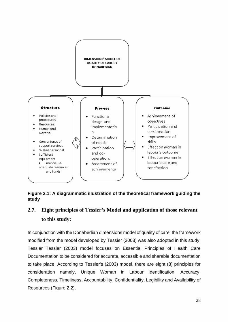
28
Figure 2.1: A diagrammatic illustration of the theoretical framework guiding the
study
2.7. Eight principles of Tessier’s Model and application of those relevant
to this study:
In conjunction with the Donabedian dimensions model of quality of care, the framework
modified from the model developed by Tessier (2003) was also adopted in this study.
Tessier Tessier (2003) model focuses on Essential Principles of Health Care
Documentation to be considered for accurate, accessible and sharable documentation
to take place. According to Tessier's (2003) model, there are eight (8) principles for
consideration namely, Unique Woman in Labour Identification, Accuracy,
Completeness, Timeliness, Accountability, Confidentiality, Legibility and Availability of
Resources (Figure 2.2).
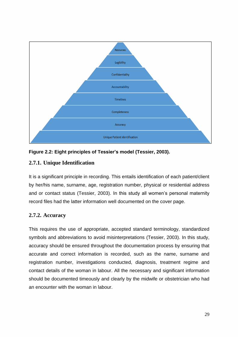
29
Figure 2.2: Eight principles of Tessier’s model (Tessier, 2003).
2.7.1. Unique Identification
It is a significant principle in recording. This entails identification of each patient/client
by her/his name, surname, age, registration number, physical or residential address
and or contact status (Tessier, 2003). In this study all women’s personal maternity
record files had the latter information well documented on the cover page.
2.7.2. Accuracy
This requires the use of appropriate, accepted standard terminology, standardized
symbols and abbreviations to avoid misinterpretations (Tessier, 2003). In this study,
accuracy should be ensured throughout the documentation process by ensuring that
accurate and correct information is recorded, such as the name, surname and
registration number, investigations conducted, diagnosis, treatment regime and
contact details of the woman in labour. All the necessary and significant information
should be documented timeously and clearly by the midwife or obstetrician who had
an encounter with the woman in labour.
30
2.7.3. Confidentiality
Policies are generally in place to control confidentiality and security and there are
movements towards enhancing their enforcement through audit trails (Tessier, 2003).
In this study confidentiality of women in labour’s records was ensured by keeping them
under lock and key where they can only be accessed by midwives and obstetricians.
2.7.4. Completeness
Completeness of the information recorded should meet institutional policy or other
requirements for specific reports (Tessier, 2003). Complete documentation in this
study means that the information recorded in the partograph has been recorded
legibly, accurately with a written date, time, observations concerning progress of
labour, condition of the foetus and woman in labour, treatment rendered and midwife's
or obstetrician's signature. All observations and assessments conducted by midwives
and obstetricians should be completely recorded without any gaps to ensure
consistency.
2.7.5. Timeliness
It is the principle that facilitates health care documentation during or immediately after
an event has occurred, while one’s memory can still remember everything about what
happened (Tessier, 2003). A record of the date and time of the phenomenon is of vital
significance to this study since it alerts midwives and obstetricians as to when did an
assessment and intervention occur. This will assist in cases of occurrence of
complications, e.g. cephalo-pelvic disproportion.
However, the application of the principle of timeliness was inconvenient to apply to the
second part of this study that focuses on auditing of the information documented in the
old partograms because it would be difficult to prove that documented data was done
timeously. The researcher could only rely on the date and time as written in the old
partograms.
31
2.7.6. Accountability
Tessier (2003) summarised the basic principles and emphasised the importance of
documentation being shared among all members of the clinical team. She further
highlighted that teamwork means that there is a recognition that good patient care
requires the skills of many people, which implies an understanding and respect for the
contributions of everyone, and that there is a commitment to share ideas, information,
responsibilities and accountability. Therefore, there should be a willingness to share
responsibility for all aspects of patient care. This includes the sharing of responsibility
in terms of identifying problems, defining solutions and taking action to resolve
problems of all kinds.
There must also be shared accountability, for ensuring that performance is accurately
reported to all parties who have a right to know. All staff should take the initiative to fix
problems, since all problems are shared, and everyone should have the attitude that
it is appropriate for them to attend to problems which may arise. No-one should have
the view that any issue is somebody else’s problem (Tessier, 2003). In this study,
midwives and obstetricians, therefore remain responsible and accountable for their
documentation with regard to progress for women in labour and their endorsed
signatures should be legible.
2.7.7. Legibility
All recorded information on patients/clients records should be legible, that is, clearly
written and readable by everybody because ambiguity of information recorded may
impact on the plan or decision which management decides to institute thus causing
delay and further complications (Tessier, 2003). In this study, information recorded in
partograms was expected to be clearly outlined from the time of admission up until
delivery of the woman
2.7.8. Resources
Ultimately, for documentation to be achieved there should be availability of resources
in the workplace (Tessier, 2003). Material resources such as maternity case records
with partographs, cardiotocographs (CTGs), dynamaps, fetoscopes, resuscitation

32
equipment, the necessary stationary and adequate personnel, such as midwives and
obstetricians are vital in this study.
2.8. Conclusion
This chapter covered the literature review related to the importance and the utilization
of the partograph, challenges encountered in the utilization of the partograph as well
as the theoretical framework underpinning this study. The following chapter covers the
research methodology underpinning the study.
33
Chapter Three
Research Methodology
3.1. Introduction
Research methodology is seen as the glue that holds the study together because it
gives structure to the research project and shows how the major parts of the research
project work together to address the research question under study (Trochim and
Donnelly, 2007). This chapter therefore outlines the process and steps that were
followed to achieve the research objectives of this study. The outline of this chapter
therefore includes the research design, the research setting, the target population,
inclusion criteria, sampling technique, sample size, research equipment, the data
collection procedure, validity and reliability, ethical issues, data analysis and the
dissemination of information.
3.2. Research approach and design
A quantitative approach was used in this study. Quantitative research is a formal,
objective, rigorous, systematic process for generating information about the world
(Grove, Burns and Gray, 2014). It is the investigation of phenomena that can be
precisely measured and quantified involving a rigorous and controlled design (Polit
and Beck, 2008). The quantitative approach was chosen because it is systematic and
the researcher was able to progress logically through a series of steps.
A cross sectional, descriptive, exploratory and non-experimental design was used in
this study. Application of both descriptive and exploratory design through a quantitative
approach allowed the researcher to explore and describe the phenomena. Descriptive
research is the exploration and description of phenomena in real life situations (Grove
et al., 2014). This study explores and describe the knowledge, attitudes and utilization
of the partograph by midwives and obstetricians in a regional hospital in the eThekwini
district.
34
3.3. Research setting
The study was conducted in a labour ward at a Regional Hospital in the eThekwini
District. It is a 1200-bed health facility with a 34-bed labour ward which serves the
surrounding area, up to and including part of the Eastern Cape. This hospital is staffed
by midwives and obstetricians who are medical doctors working in the maternity
department (consisting of consultant obstetricians, medical officers, interns and
registrars on training).
3.4. Target population
The population is the entire aggregation of cases which the researcher is interested in
(Polit and Beck, 2004). The target population for this study comprised the midwives
and obstetricians working in the maternity admission room and labour ward in a
Regional Hospital in the EThekwini District. There were 101 midwives and 48
obstetricians on day and night duty in the maternity section respectively. According to
statistics, there were approximately 1010 deliveries per month in the labour ward from
January to December 2010. The questionnaires were directed at all midwives,
irrespective of whether they had specialized in midwifery (Advanced midwifery and
Neonatal Nursing Science) or not, and all obstetricians including interns who were
currently working in maternity admission room and labour ward.
The second target population of this study involved the sampling and selection of
partograms, completed by midwives and obstetricians for women who had delivered
per normal vaginal delivery over a period of one month (September 2010) as records.
3.5. Sampling technique and sample size
Sampling is the process used by the researcher to select the sample from the true
population in order to obtain information regarding a phenomenon in a way that
represents the population of interest (Brink, Van Der Walt and Van Rensburg, 2006).
Probability sampling will be used in this study. Probability sampling is generally used
in quantitative studies and involves the simple random selection of elements (Polit and
Beck, 2004).
35
Simple random sampling was used to select 70 midwives and 30 obstetricians who
took part in this study. In this instance, each midwife and obstetrician was listed
separately, and therefore had an equal chance of being included in the sample. This
was conducted by placing numbers in a bowl and drawing them out one at a time. It
helped the researcher to overcome variables and problems that might otherwise bias
the study.
The data obtained from the birth register of the Regional hospital revealed that there
were 1100 deliveries during the month of September, 2010. Every tenth woman’s file
was obtained from medical records and was used as a source of information on
previously used partograms for the retrospective section of the study. Selecting the
partographs was done by selecting the partograph randomly as it was done for the
midwives and obstetricians.
Inclusion criteria: The midwives and obstetricians who had three months and more
experience of having worked in the labour ward were recruited to be part of this study,
as well as partograms as records for women who only delivered per normal vaginal
delivery during September 2010.
3.6. Research instrument and data collection process
3.6.1. Research instruments
In this study, two types of instruments were used, namely self-administered
questionnaires and a checklist that was used to audit information that was written in
the old partograph. A self-administered questionnaire that was used to collect data
from midwives and obstetricians in this study was the questionnaire developed by the
researcher using the literature, and in consultation with the research supervisor. A
research instrument consists of a series of questions and other prompts for the
purpose of gathering information from respondents (Brink et al., 2006).
The questionnaire used in this study was developed from the literature was used to
collect data and the questions were coded for the midwives and obstetricians, and
were hand-delivered to the respondents with the request that they complete the
questionnaire on the same day and return the document to the researcher on the day
36
of data collection. The questionnaire that was used to collect data from Midwives and
Obstetricians is divided into five sections, which are, Section A: Demographic data,
Section B: Assessment on admission, Section C: Training on partograph use, Section
D: Foetal condition, Section E: Progress of labour data. All questions were closed-
ended questions.
The researcher also reviewed the partograms of women who delivered in September
2009. The second instrument for data collection was the checklist. It was developed
by the researcher in consultation with the research supervisor adopting the principle
of completeness that incorporated other Tier’s Model principles of ensuring that all the
information in the old partograms was written accurately, legible or clearly, timeously
meaning that date and time was written.
3.6.2. Validity and reliability
Validity and reliability are major criteria for assessing the instruments' quality and
adequacy (Polit and Beck, 2004).
3.6.2.1. Validity
Validity is defined as the degree to which an instrument measures what is supposed
to measure (Polit and Beck, 2004). The instrument was based on the evaluation of the
utilization of the partograph in a labour ward at a Regional Hospital in the EThekwini
District. Content validity will be used as a means of ensuring the validity of the data
collection instrument. The content of the self-reported questionnaire will be based on
the objectives of the study. Content validity is defined as an assessment of how well
the instrument represents all the components of the variable to be measured (Polit
and Beck, 2004).
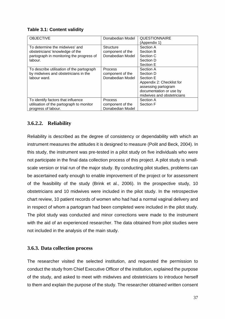
37
Table 3.1: Content validity
OBJECTIVE Donabedian Model QUESTIONNAIRE (Appendix 1}
To determine the midwives' and obstetricians' knowledge of the partograph in monitoring the progress of labour.
Structure component of the Donabedian Model
Section A Section B Section C Section D Section E
To describe utilisation of the partograph by midwives and obstetricians in the labour ward.
Process component of the Donabedian Model
Section A Section D Section E Appendix 2: Checklist for assessing partogram documentation or use by midwives and obstetricians
To identify factors that influence utilisation of the partograph to monitor progress of labour.
Process component of the Donabedian Model
Section A Section F
3.6.2.2. Reliability
Reliability is described as the degree of consistency or dependability with which an
instrument measures the attitudes it is designed to measure (Polit and Beck, 2004). In
this study, the instrument was pre-tested in a pilot study on five individuals who were
not participate in the final data collection process of this project. A pilot study is small-
scale version or trial run of the major study. By conducting pilot studies, problems can
be ascertained early enough to enable improvement of the project or for assessment
of the feasibility of the study (Brink et al., 2006). In the prospective study, 10
obstetricians and 10 midwives were included in the pilot study. In the retrospective
chart review, 10 patient records of women who had had a normal vaginal delivery and
in respect of whom a partogram had been completed were included in the pilot study.
The pilot study was conducted and minor corrections were made to the instrument
with the aid of an experienced researcher. The data obtained from pilot studies were
not included in the analysis of the main study.
3.6.3. Data collection process
The researcher visited the selected institution, and requested the permission to
conduct the study from Chief Executive Officer of the institution, explained the purpose
of the study, and asked to meet with midwives and obstetricians to introduce herself
to them and explain the purpose of the study. The researcher obtained written consent
38
and administered the questionnaires to those who had agreed and signed consent to
participate in the study.
The questionnaire was handed out personally with the advantage of having an
improved response rate (Brink et al., 2006). The researcher was available for the
participant’s questions or when they needed some clarification regarding the
questionnaire.
A self-administered questionnaire, was used to collect data from 70 midwives and 30
obstetricians. The questionnaires were hand-delivered to the respondents with a
request that they complete the questionnaire on the same day and return it to the
researcher on the day of data collection. The data was collected over a period of two
months, namely October and November, 2011.
For women’s records: There were 1100 deliveries in September 2010. Every 10th file
from all the files of women who delivered vaginally in September 2010 on the list was
sampled, until one hundred (100) partograms had been obtained, which is nine per
cent (9%) of the entire number of partograms that were reviewed. Women’s records
(partograms) were listed randomly to prevent biases and to ensure that the sample
selection was truly representative of the partograms of women who delivered during
the month of September 2010.
Confidentiality and anonymity was preserved during data collection and throughout
the research study. Information gathered will not be held any longer than necessary
as data on computers. It will also be destroyed after five years, once the report and
dissemination of results by means of presentation at conferences and publication, has
been completed. The data was collected over a period of two months during October
and November in 2011.
3.7. Data analysis
The data was analyzed using the Statistical Package for Social Science (SPSS)
Version 21, in the form of descriptive statistics with the use of frequency tables,
percentages and cross-tabulations.
39
In respect of the assessment of the partograms, these were divided into complete and
incomplete 'filling-in' using a modified scoring system of the 25-point score of the
labour management checklist (Farrell and Pattinson, 2005). The modified system that
was used included the labour section of the University of Pretoria's labour
management checklist. This checklist was modified by combining the recording and
plotting of cervical dilatation four-hourly and two-hourly in latent and active phases
respectively into one point.
A checklist consisting of 10 parameters was drawn up from this 25-point score. Our
modified scoring system therefore consisted of 10 points and consisted of the 10
clinical parameters (risk factors, foetal heart rate, state of liquor, caput and moulding,
uterine contractions, cervical dilatation, level of the foetal head above the pelvic brim,
blood pressure and heart rate, temperature and urine output, intravenous fluids and
drugs). Where parameters were partly assessed or partly documented, a half-mark
was allocated. Complete partograms were defined as those having at least 7.5 out of
the 10 (75%) of all parameters correctly assessed. Incomplete partograms were those
with less than 7.5 out of the 10 (75%) parameters correctly assessed (Farrell and
Pattinson, 2005).
3.8. Ethical considerations
Ethical clearance was sought and secured from the University of KwaZulu-Natal Ethics
Committee and permission was secured from the KwaZulu-Natal Department of
Health to conduct this study. In addition, permission from the Chief Executive Officer
of the institution where the study was conducted was obtained.
The eight principles of Tessier’s Model (2003) and application of their relevant to this
study was considered which included: Unique Woman in Labour Identification,
Accuracy, Completeness, Timeliness, Accountability, Confidentiality, Legibility and
Availability of Resources (Figure 2.2).
The participants' human rights were protected and it was ensured that the information
they would provide for this study was treated with the utmost privacy and would not be
shared nor disclosed to unauthorized people.
40
Consent was sought from respondents after the purpose of the study had been
explained to them. It was explained that their participation was voluntary, and that, as
such, they could decide to participate or withdraw from the study without the risk of
incurring any penalties. Their privacy and confidentiality was maintained throughout
the study, and anonymity was ensured by coding the questionnaires and by not
seeking any identifying information.
Women’s records, which in this study comprised of partograms, were not disclosed to
anyone not providing direct care to women in labour. Permission to review women’s
records was sought from the Chief Executive Officer of the Regional Hospital where
the study was conducted. Confidentiality and anonymity was preserved throughout the
research study.
The women's records database was anonymized, that is, women’s records were
assigned a number rather than identifying them by name. Information gathered will not
be held any longer than necessary by means of data on computers. This will also be
destroyed after five years once the report and dissemination of the results, through
presentation of these at conferences and publication of results, has been completed.
3.9. Data management
Data collected in this study will be stored to a private computer with an access code
known only by the researcher was used to store the data, and hard copies of the data
were kept under lock and key and will be destroyed by fire after a period of five years.
3.10. Dissemination of information
The results of the study will be disseminated in the form of a hard copy to the Regional
Hospital and to the KZN Health Department for future use. Oral presentations of the
results will be presented at conferences and will be submitted for publication in peer-
reviewed professional journals, or in a variety of outlets on the internet. Anonymity and
confidentiality will be maintained during the dissemination of information.

41
3.11. Conclusion
This chapter covered research methodology of the study, research approach, the
design, the setting, population, sampling and sample size. The instrument was
described and the validity with regard to the research objective was attended to. The
data collection procedure, analysis, management, and dissemination of the findings.
The next chapter will cover the presentation of the results.
.
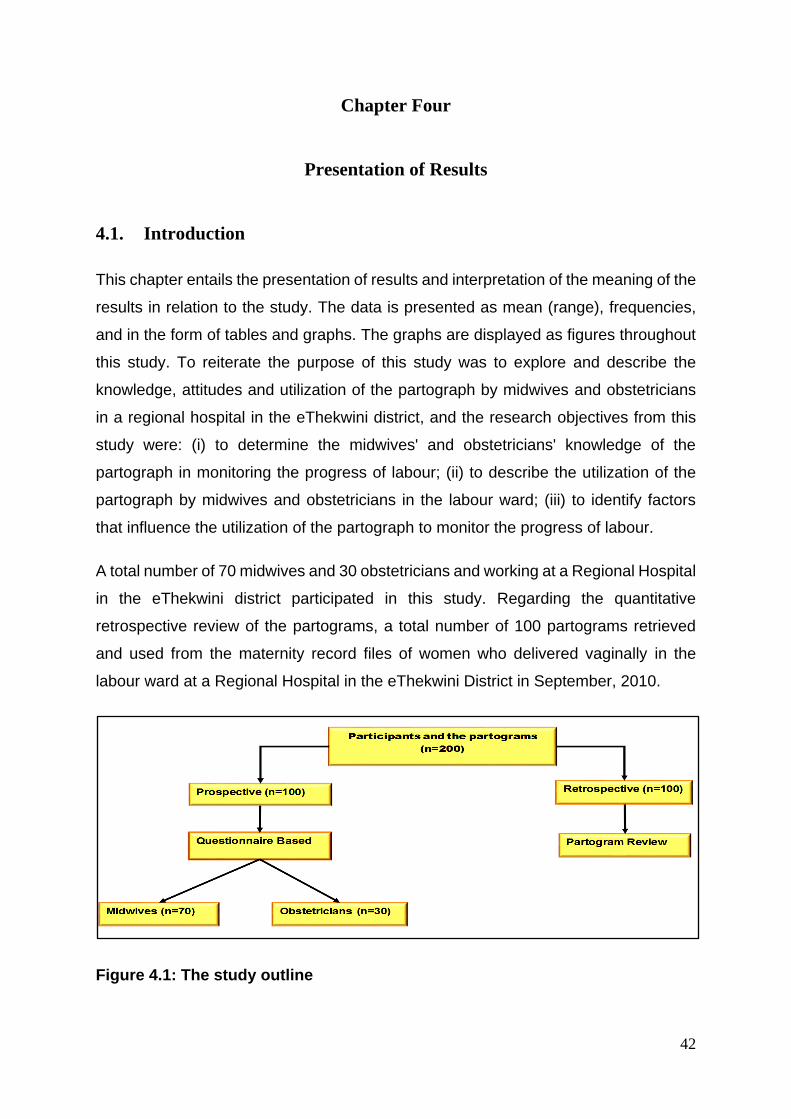
42
Chapter Four
Presentation of Results
4.1. Introduction
This chapter entails the presentation of results and interpretation of the meaning of the
results in relation to the study. The data is presented as mean (range), frequencies,
and in the form of tables and graphs. The graphs are displayed as figures throughout
this study. To reiterate the purpose of this study was to explore and describe the
knowledge, attitudes and utilization of the partograph by midwives and obstetricians
in a regional hospital in the eThekwini district, and the research objectives from this
study were: (i) to determine the midwives' and obstetricians' knowledge of the
partograph in monitoring the progress of labour; (ii) to describe the utilization of the
partograph by midwives and obstetricians in the labour ward; (iii) to identify factors
that influence the utilization of the partograph to monitor the progress of labour.
A total number of 70 midwives and 30 obstetricians and working at a Regional Hospital
in the eThekwini district participated in this study. Regarding the quantitative
retrospective review of the partograms, a total number of 100 partograms retrieved
and used from the maternity record files of women who delivered vaginally in the
labour ward at a Regional Hospital in the eThekwini District in September, 2010.
Figure 4.1: The study outline
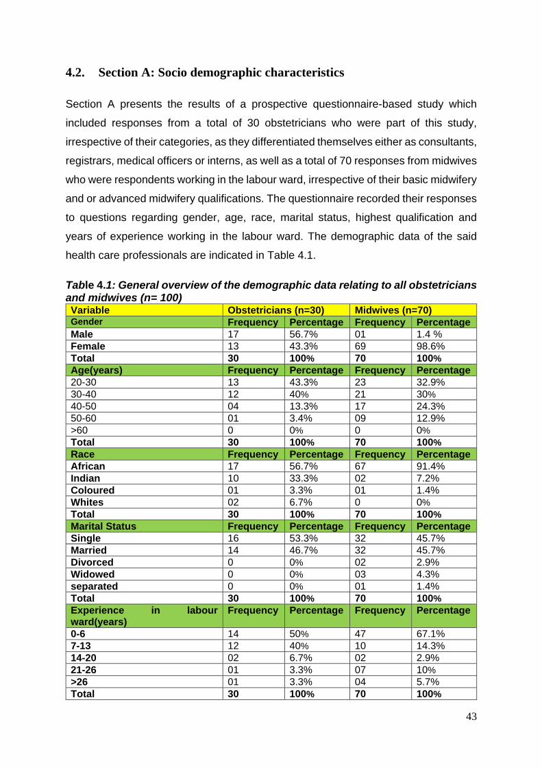
43
4.2. Section A: Socio demographic characteristics
Section A presents the results of a prospective questionnaire-based study which
included responses from a total of 30 obstetricians who were part of this study,
irrespective of their categories, as they differentiated themselves either as consultants,
registrars, medical officers or interns, as well as a total of 70 responses from midwives
who were respondents working in the labour ward, irrespective of their basic midwifery
and or advanced midwifery qualifications. The questionnaire recorded their responses
to questions regarding gender, age, race, marital status, highest qualification and
years of experience working in the labour ward. The demographic data of the said
health care professionals are indicated in Table 4.1.
Table 4.1: General overview of the demographic data relating to all obstetricians and midwives (n= 100) Variable Obstetricians (n=30) Midwives (n=70) Gender Frequency Percentage Frequency Percentage
Male 17 56.7% 01 1.4 %
Female 13 43.3% 69 98.6%
Total 30 100% 70 100%
Age(years) Frequency Percentage Frequency Percentage
20-30 13 43.3% 23 32.9%
30-40 12 40% 21 30%
40-50 04 13.3% 17 24.3%
50-60 01 3.4% 09 12.9%
>60 0 0% 0 0%
Total 30 100% 70 100%
Race Frequency Percentage Frequency Percentage
African 17 56.7% 67 91.4%
Indian 10 33.3% 02 7.2%
Coloured 01 3.3% 01 1.4%
Whites 02 6.7% 0 0%
Total 30 100% 70 100%
Marital Status Frequency Percentage Frequency Percentage
Single 16 53.3% 32 45.7%
Married 14 46.7% 32 45.7%
Divorced 0 0% 02 2.9%
Widowed 0 0% 03 4.3%
separated 0 0% 01 1.4%
Total 30 100% 70 100%
Experience in labour ward(years)
Frequency Percentage Frequency Percentage
0-6 14 50% 47 67.1%
7-13 12 40% 10 14.3%
14-20 02 6.7% 02 2.9%
21-26 01 3.3% 07 10%
>26 01 3.3% 04 5.7%
Total 30 100% 70 100%
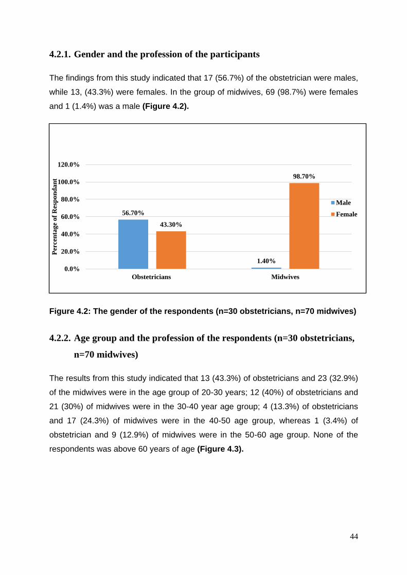
44
4.2.1. Gender and the profession of the participants
The findings from this study indicated that 17 (56.7%) of the obstetrician were males,
while 13, (43.3%) were females. In the group of midwives, 69 (98.7%) were females
and 1 (1.4%) was a male (Figure 4.2).
Figure 4.2: The gender of the respondents (n=30 obstetricians, n=70 midwives)
4.2.2. Age group and the profession of the respondents (n=30 obstetricians,
n=70 midwives)
The results from this study indicated that 13 (43.3%) of obstetricians and 23 (32.9%)
of the midwives were in the age group of 20-30 years; 12 (40%) of obstetricians and
21 (30%) of midwives were in the 30-40 year age group; 4 (13.3%) of obstetricians
and 17 (24.3%) of midwives were in the 40-50 age group, whereas 1 (3.4%) of
obstetrician and 9 (12.9%) of midwives were in the 50-60 age group. None of the
respondents was above 60 years of age (Figure 4.3).
56.70%
1.40%
43.30%
98.70%
0.0%
20.0%
40.0%
60.0%
80.0%
100.0%
120.0%
Obstetricians Midwives
Per
cen
tag
e o
f R
esp
on
da
nt
Male
Female
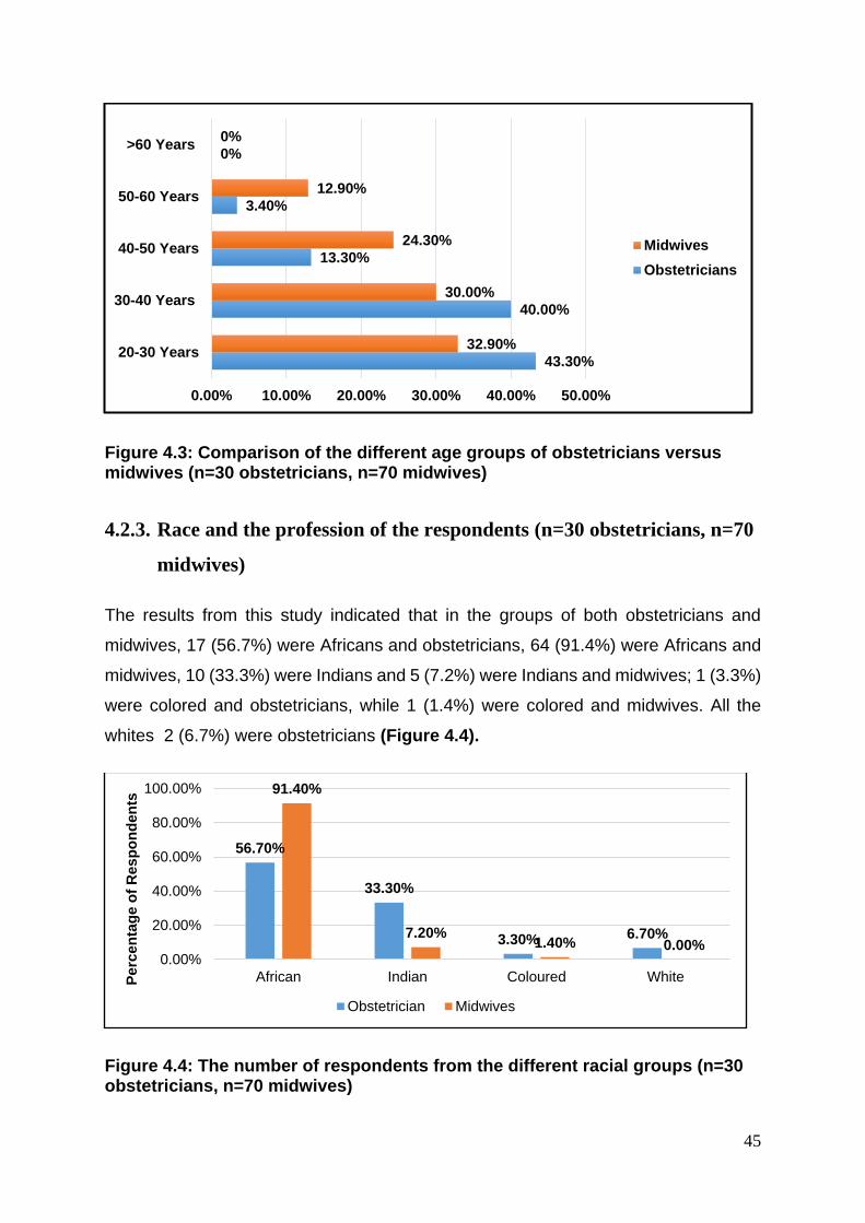
45
Figure 4.3: Comparison of the different age groups of obstetricians versus midwives (n=30 obstetricians, n=70 midwives)
4.2.3. Race and the profession of the respondents (n=30 obstetricians, n=70
midwives)
The results from this study indicated that in the groups of both obstetricians and
midwives, 17 (56.7%) were Africans and obstetricians, 64 (91.4%) were Africans and
midwives, 10 (33.3%) were Indians and 5 (7.2%) were Indians and midwives; 1 (3.3%)
were colored and obstetricians, while 1 (1.4%) were colored and midwives. All the
whites 2 (6.7%) were obstetricians (Figure 4.4).
Figure 4.4: The number of respondents from the different racial groups (n=30 obstetricians, n=70 midwives)
43.30%
40.00%
13.30%
3.40%
0%
32.90%
30.00%
24.30%
12.90%
0%
0.00% 10.00% 20.00% 30.00% 40.00% 50.00%
20-30 Years
30-40 Years
40-50 Years
50-60 Years
>60 Years
Midwives
Obstetricians
56.70%
33.30%
3.30% 6.70%
91.40%
7.20%1.40% 0.00%
0.00%
20.00%
40.00%
60.00%
80.00%
100.00%
African Indian Coloured WhitePe
rce
nta
ge
of
Re
sp
on
de
nts
Obstetrician Midwives
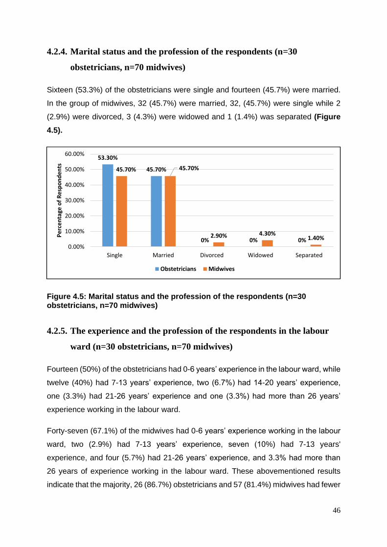
46
4.2.4. Marital status and the profession of the respondents (n=30
obstetricians, n=70 midwives)
Sixteen (53.3%) of the obstetricians were single and fourteen (45.7%) were married.
In the group of midwives, 32 (45.7%) were married, 32, (45.7%) were single while 2
(2.9%) were divorced, 3 (4.3%) were widowed and 1 (1.4%) was separated (Figure
4.5).
Figure 4.5: Marital status and the profession of the respondents (n=30 obstetricians, n=70 midwives)
4.2.5. The experience and the profession of the respondents in the labour
ward (n=30 obstetricians, n=70 midwives)
Fourteen (50%) of the obstetricians had 0-6 years’ experience in the labour ward, while
twelve (40%) had 7-13 years’ experience, two (6.7%) had 14-20 years’ experience,
one (3.3%) had 21-26 years’ experience and one (3.3%) had more than 26 years’
experience working in the labour ward.
Forty-seven (67.1%) of the midwives had 0-6 years’ experience working in the labour
ward, two (2.9%) had 7-13 years’ experience, seven (10%) had 7-13 years'
experience, and four (5.7%) had 21-26 years’ experience, and 3.3% had more than
26 years of experience working in the labour ward. These abovementioned results
indicate that the majority, 26 (86.7%) obstetricians and 57 (81.4%) midwives had fewer
53.30%
45.70%
0% 0% 0%
45.70% 45.70%
2.90% 4.30%1.40%
0.00%
10.00%
20.00%
30.00%
40.00%
50.00%
60.00%
Single Married Divorced Widowed Separated
Pe
rce
nta
ge o
f R
esp
on
de
nts
Obstetricians Midwives
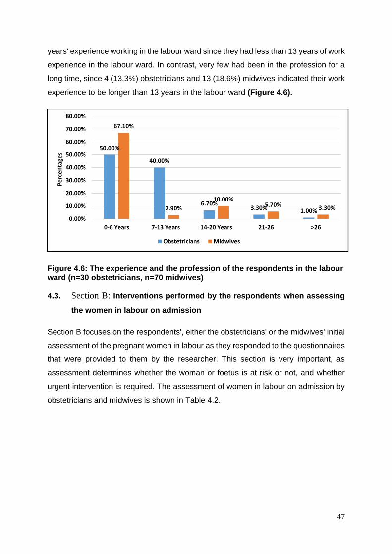
47
years' experience working in the labour ward since they had less than 13 years of work
experience in the labour ward. In contrast, very few had been in the profession for a
long time, since 4 (13.3%) obstetricians and 13 (18.6%) midwives indicated their work
experience to be longer than 13 years in the labour ward (Figure 4.6).
Figure 4.6: The experience and the profession of the respondents in the labour ward (n=30 obstetricians, n=70 midwives)
4.3. Section B: Interventions performed by the respondents when assessing
the women in labour on admission
Section B focuses on the respondents', either the obstetricians' or the midwives' initial
assessment of the pregnant women in labour as they responded to the questionnaires
that were provided to them by the researcher. This section is very important, as
assessment determines whether the woman or foetus is at risk or not, and whether
urgent intervention is required. The assessment of women in labour on admission by
obstetricians and midwives is shown in Table 4.2.
50.00%
40.00%
6.70%3.30% 1.00%
67.10%
2.90%
10.00%5.70% 3.30%
0.00%
10.00%
20.00%
30.00%
40.00%
50.00%
60.00%
70.00%
80.00%
0-6 Years 7-13 Years 14-20 Years 21-26 >26
Pe
rce
nta
ges
Obstetricians Midwives
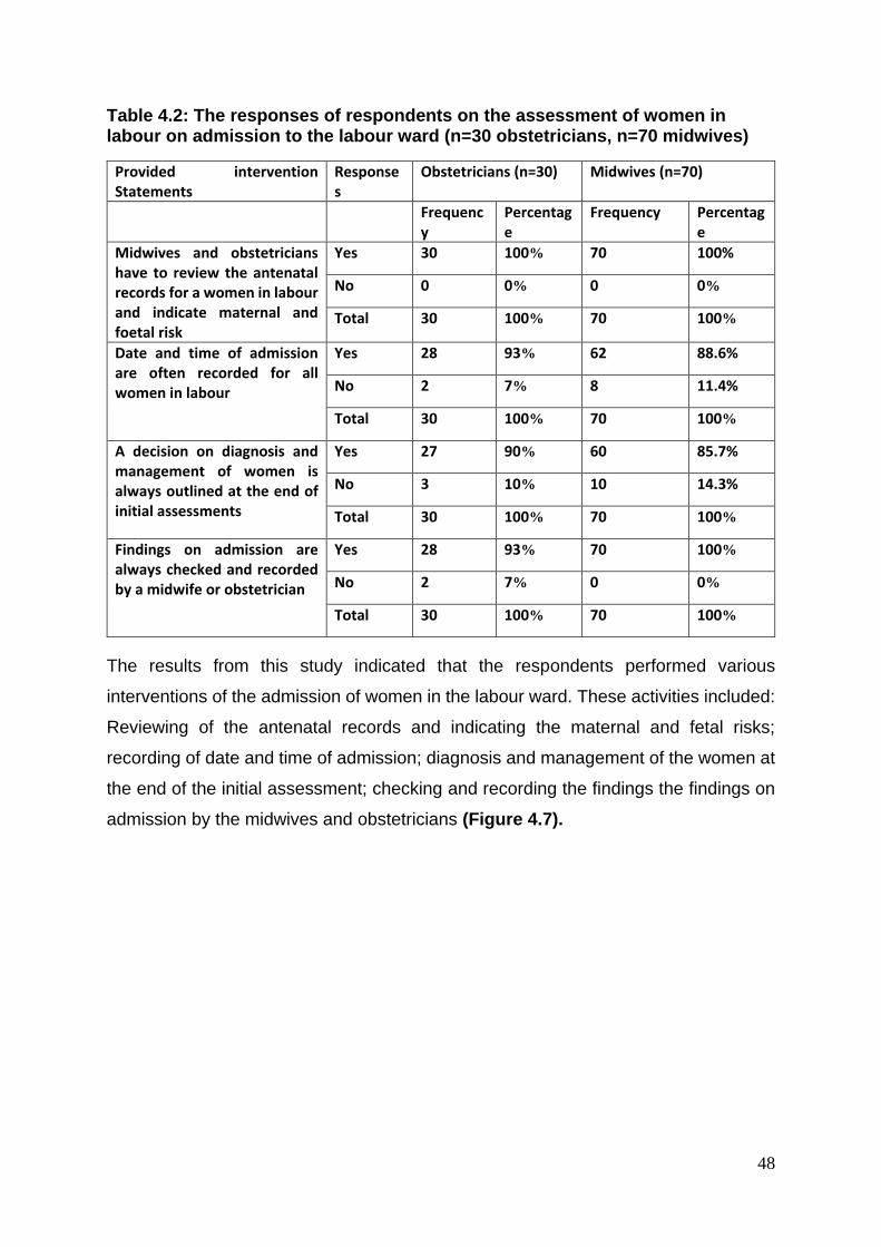
48
Table 4.2: The responses of respondents on the assessment of women in labour on admission to the labour ward (n=30 obstetricians, n=70 midwives)
Provided intervention Statements
Responses
Obstetricians (n=30) Midwives (n=70)
Frequency
Percentage
Frequency Percentage
Midwives and obstetricians have to review the antenatal records for a women in labour and indicate maternal and foetal risk
Yes 30 100% 70 100%
No 0 0% 0 0%
Total 30 100% 70 100%
Date and time of admission are often recorded for all women in labour
Yes 28 93% 62 88.6%
No 2 7% 8 11.4%
Total 30 100% 70 100%
A decision on diagnosis and management of women is always outlined at the end of initial assessments
Yes 27 90% 60 85.7%
No 3 10% 10 14.3%
Total 30 100% 70 100%
Findings on admission are always checked and recorded by a midwife or obstetrician
Yes 28 93% 70 100%
No 2 7% 0 0%
Total 30 100% 70 100%
The results from this study indicated that the respondents performed various
interventions of the admission of women in the labour ward. These activities included:
Reviewing of the antenatal records and indicating the maternal and fetal risks;
recording of date and time of admission; diagnosis and management of the women at
the end of the initial assessment; checking and recording the findings the findings on
admission by the midwives and obstetricians (Figure 4.7).
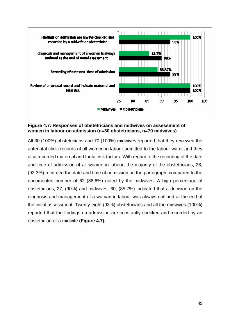
49
Figure 4.7: Responses of obstetricians and midwives on assessment of women in labour on admission (n=30 obstetricians, n=70 midwives)
All 30 (100%) obstetricians and 70 (100%) midwives reported that they reviewed the
antenatal clinic records of all women in labour admitted to the labour ward, and they
also recorded maternal and foetal risk factors. With regard to the recording of the date
and time of admission of all women in labour, the majority of the obstetricians, 28,
(93.3%) recorded the date and time of admission on the partograph, compared to the
documented number of 62 (88.6%) noted by the midwives. A high percentage of
obstetricians, 27, (90%) and midwives, 60, (85.7%) indicated that a decision on the
diagnosis and management of a woman in labour was always outlined at the end of
the initial assessment. Twenty-eight (93%) obstetricians and all the midwives (100%)
reported that the findings on admission are constantly checked and recorded by an
obstetrician or a midwife (Figure 4.7).
50
4.4. Section C: Knowledge and skills of the participants in the use of the
partograph
4.4.1. Training of respondents on the use of the partograph (n=30
obstetricians, n=70 midwives)
All 30 (100%) obstetricians and 70 (100%) midwives involved in the study indicated
that they had been trained in the use of partograph.
4.4.2. Type of tertiary education for respondents Table (n=30 obstetricians,
n=70 midwives)
When it comes to the type of tertiary education where respondents received their
training in the use of the partograph, all 30 (100%) obstetricians were trained in the
use of the partograph at medical school. In contrast, midwives reported that they
received their training in the use of the partograph in different tertiary education
institutions. Forty-two (60%) midwives indicated they were trained in the use of the
partograph at Nursing Colleges and twenty-one (30 0%) at the School of Nursing at
the University of KwaZulu-Natal. Seven (10%) midwives reported that they were
trained in the use of the partograph when they were already practicing as qualified
midwives during in-service training. The responses of 63 (90%) midwives revealed
that they were trained during their tertiary education at Universities and Colleges
respectively. This was the majority, as only 7 (10%) midwives indicated that they were
trained when they were already practicing as qualified midwives during in-service
training.
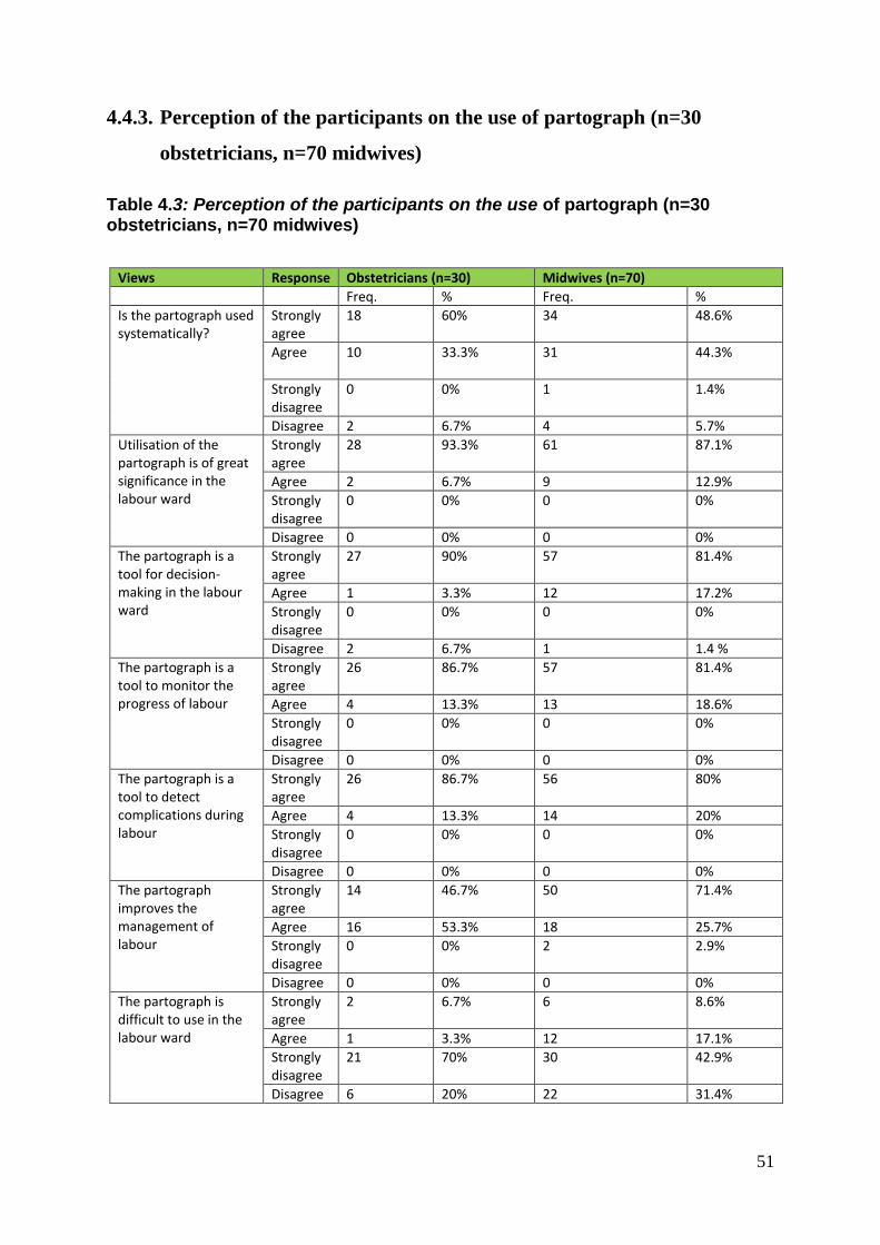
51
4.4.3. Perception of the participants on the use of partograph (n=30
obstetricians, n=70 midwives)
Table 4.3: Perception of the participants on the use of partograph (n=30 obstetricians, n=70 midwives)
Views Response Obstetricians (n=30) Midwives (n=70)
Freq. % Freq. %
Is the partograph used systematically?
Strongly agree
18 60% 34 48.6%
Agree 10 33.3%
31 44.3%
Strongly disagree
0 0%
1 1.4%
Disagree 2 6.7% 4 5.7%
Utilisation of the partograph is of great significance in the labour ward
Strongly agree
28 93.3% 61 87.1%
Agree 2 6.7% 9 12.9%
Strongly disagree
0 0%
0 0%
Disagree 0 0% 0 0%
The partograph is a tool for decision-making in the labour ward
Strongly agree
27 90% 57 81.4%
Agree 1 3.3% 12 17.2%
Strongly disagree
0 0% 0 0%
Disagree 2 6.7% 1 1.4 %
The partograph is a tool to monitor the progress of labour
Strongly agree
26 86.7% 57 81.4%
Agree 4 13.3% 13 18.6%
Strongly disagree
0 0% 0 0%
Disagree 0 0% 0 0%
The partograph is a tool to detect complications during labour
Strongly agree
26 86.7% 56 80%
Agree 4 13.3% 14 20%
Strongly disagree
0 0% 0 0%
Disagree 0 0% 0 0%
The partograph improves the management of labour
Strongly agree
14 46.7% 50 71.4%
Agree 16 53.3% 18 25.7%
Strongly disagree
0 0% 2 2.9%
Disagree 0 0% 0 0%
The partograph is difficult to use in the labour ward
Strongly agree
2 6.7% 6 8.6%
Agree 1 3.3% 12 17.1%
Strongly disagree
21 70% 30 42.9%
Disagree 6 20% 22 31.4%
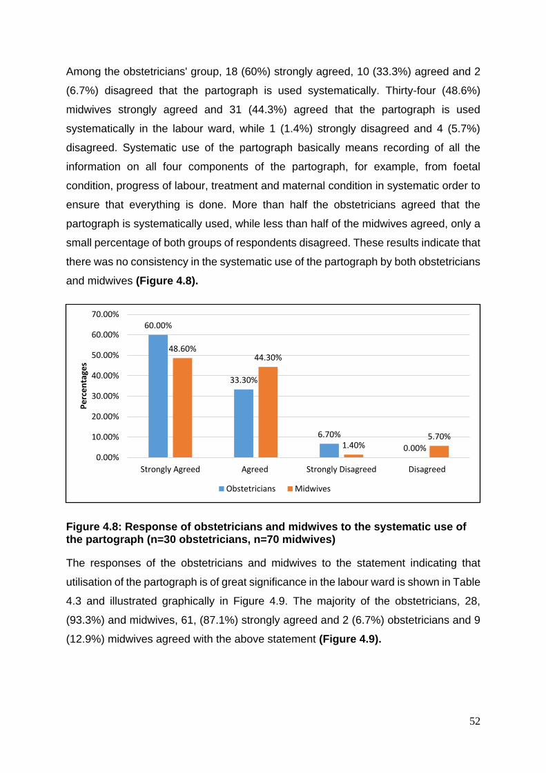
52
Among the obstetricians' group, 18 (60%) strongly agreed, 10 (33.3%) agreed and 2
(6.7%) disagreed that the partograph is used systematically. Thirty-four (48.6%)
midwives strongly agreed and 31 (44.3%) agreed that the partograph is used
systematically in the labour ward, while 1 (1.4%) strongly disagreed and 4 (5.7%)
disagreed. Systematic use of the partograph basically means recording of all the
information on all four components of the partograph, for example, from foetal
condition, progress of labour, treatment and maternal condition in systematic order to
ensure that everything is done. More than half the obstetricians agreed that the
partograph is systematically used, while less than half of the midwives agreed, only a
small percentage of both groups of respondents disagreed. These results indicate that
there was no consistency in the systematic use of the partograph by both obstetricians
and midwives (Figure 4.8).
Figure 4.8: Response of obstetricians and midwives to the systematic use of the partograph (n=30 obstetricians, n=70 midwives)
The responses of the obstetricians and midwives to the statement indicating that
utilisation of the partograph is of great significance in the labour ward is shown in Table
4.3 and illustrated graphically in Figure 4.9. The majority of the obstetricians, 28,
(93.3%) and midwives, 61, (87.1%) strongly agreed and 2 (6.7%) obstetricians and 9
(12.9%) midwives agreed with the above statement (Figure 4.9).
60.00%
33.30%
6.70%
0.00%
48.60%44.30%
1.40%5.70%
0.00%
10.00%
20.00%
30.00%
40.00%
50.00%
60.00%
70.00%
Strongly Agreed Agreed Strongly Disagreed Disagreed
Pe
rce
nta
ges
Obstetricians Midwives
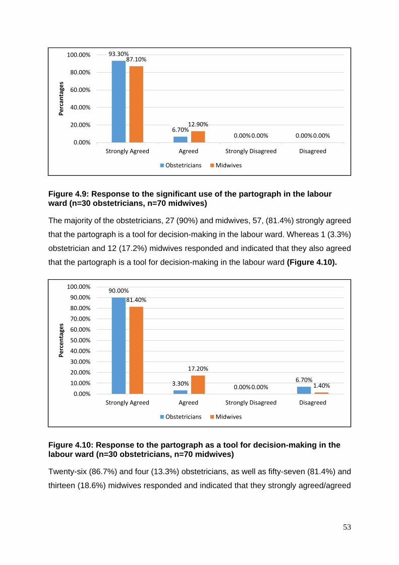
53
Figure 4.9: Response to the significant use of the partograph in the labour ward (n=30 obstetricians, n=70 midwives)
The majority of the obstetricians, 27 (90%) and midwives, 57, (81.4%) strongly agreed
that the partograph is a tool for decision-making in the labour ward. Whereas 1 (3.3%)
obstetrician and 12 (17.2%) midwives responded and indicated that they also agreed
that the partograph is a tool for decision-making in the labour ward (Figure 4.10).
Figure 4.10: Response to the partograph as a tool for decision-making in the labour ward (n=30 obstetricians, n=70 midwives)
Twenty-six (86.7%) and four (13.3%) obstetricians, as well as fifty-seven (81.4%) and
thirteen (18.6%) midwives responded and indicated that they strongly agreed/agreed
93.30%
6.70%0.00% 0.00%
87.10%
12.90%
0.00% 0.00%0.00%
20.00%
40.00%
60.00%
80.00%
100.00%
Strongly Agreed Agreed Strongly Disagreed Disagreed
Pe
rcan
tage
s
Obstetricians Midwives
90.00%
3.30%0.00%
6.70%
81.40%
17.20%
0.00% 1.40%0.00%
10.00%
20.00%
30.00%
40.00%
50.00%
60.00%
70.00%
80.00%
90.00%
100.00%
Strongly Agreed Agreed Strongly Disagreed Disagreed
Pe
rce
nta
ges
Obstetricians Midwives
54
that the partograph is a tool used to monitor the progress of labour, whereas no one
from either group disagreed (Figure 4.11).
Figure 4.11: Responses to the partograph as a tool to monitor the progress of labour (n=30 obstetricians, n=70 midwives)
Twenty-six (86.7%) obstetricians strongly agreed and the remaining four (13%)
obstetricians agreed that the partograph is a tool to detect complications during labour,
whereas fifty-six (80%) midwives strongly agreed and fourteen (20%) midwives agreed
that the partograph is a tool to detect complications during labour. None of the
respondents disagreed with the above statement. The responses for both obstetricians
and midwives were excellent, which is a clear indication that both groups had
knowledge of the partograph. However, complications could occur during the
antepartum, intrapartum and postpartum period, therefore, making use of the
partograph during labour assists in the early identification of complications and the
prompt management thereof. The response to this statement is displayed in Figure
4.12.
86.70%81.40%
0.00% 0.00%
13.30%18.60%
0.00% 0.00%0.00%
10.00%
20.00%
30.00%
40.00%
50.00%
60.00%
70.00%
80.00%
90.00%
100.00%
Strongly Agreed Agreed Strongly Disagreed Disagreed
Pe
rce
nta
ges
Obstetricians Midwives
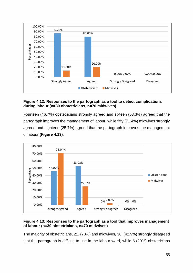
55
Figure 4.12: Responses to the partograph as a tool to detect complications during labour (n=30 obstetricians, n=70 midwives)
Fourteen (46.7%) obstetricians strongly agreed and sixteen (53.3%) agreed that the
partograph improves the management of labour, while fifty (71.4%) midwives strongly
agreed and eighteen (25.7%) agreed that the partograph improves the management
of labour (Figure 4.13).
Figure 4.13: Responses to the partograph as a tool that improves management of labour (n=30 obstetricians, n=70 midwives)
The majority of obstetricians, 21, (70%) and midwives, 30, (42.9%) strongly disagreed
that the partograph is difficult to use in the labour ward, while 6 (20%) obstetricians
86.70%80.00%
0.00% 0.00%
13.00%20.00%
0.00% 0.00%0.00%
10.00%
20.00%
30.00%
40.00%
50.00%
60.00%
70.00%
80.00%
90.00%
100.00%
Strongly Agreed Agreed Strongly Disagreed Disagreed
Pe
rce
nta
ges
Obstetricians Midwives
46.07%
53.03%
0% 0%
71.04%
25.07%
2.09% 0%0.00%
10.00%
20.00%
30.00%
40.00%
50.00%
60.00%
70.00%
80.00%
Strongly Agreed Agreed Strongly disagreed Disagreed
Pe
rce
nta
ge
Obstericians
Midwives
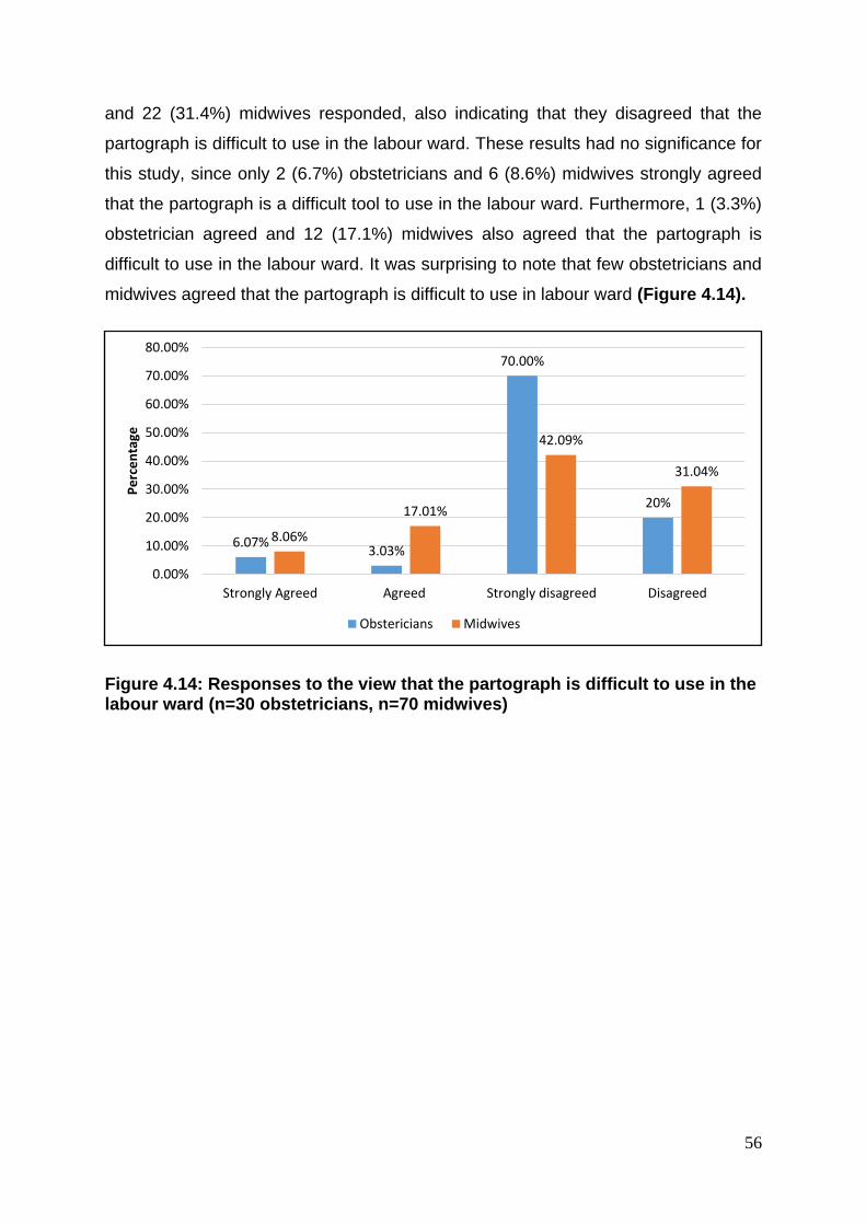
56
and 22 (31.4%) midwives responded, also indicating that they disagreed that the
partograph is difficult to use in the labour ward. These results had no significance for
this study, since only 2 (6.7%) obstetricians and 6 (8.6%) midwives strongly agreed
that the partograph is a difficult tool to use in the labour ward. Furthermore, 1 (3.3%)
obstetrician agreed and 12 (17.1%) midwives also agreed that the partograph is
difficult to use in the labour ward. It was surprising to note that few obstetricians and
midwives agreed that the partograph is difficult to use in labour ward (Figure 4.14).
Figure 4.14: Responses to the view that the partograph is difficult to use in the labour ward (n=30 obstetricians, n=70 midwives)
6.07%3.03%
70.00%
20%
8.06%
17.01%
42.09%
31.04%
0.00%
10.00%
20.00%
30.00%
40.00%
50.00%
60.00%
70.00%
80.00%
Strongly Agreed Agreed Strongly disagreed Disagreed
Pe
rce
nta
ge
Obstericians Midwives
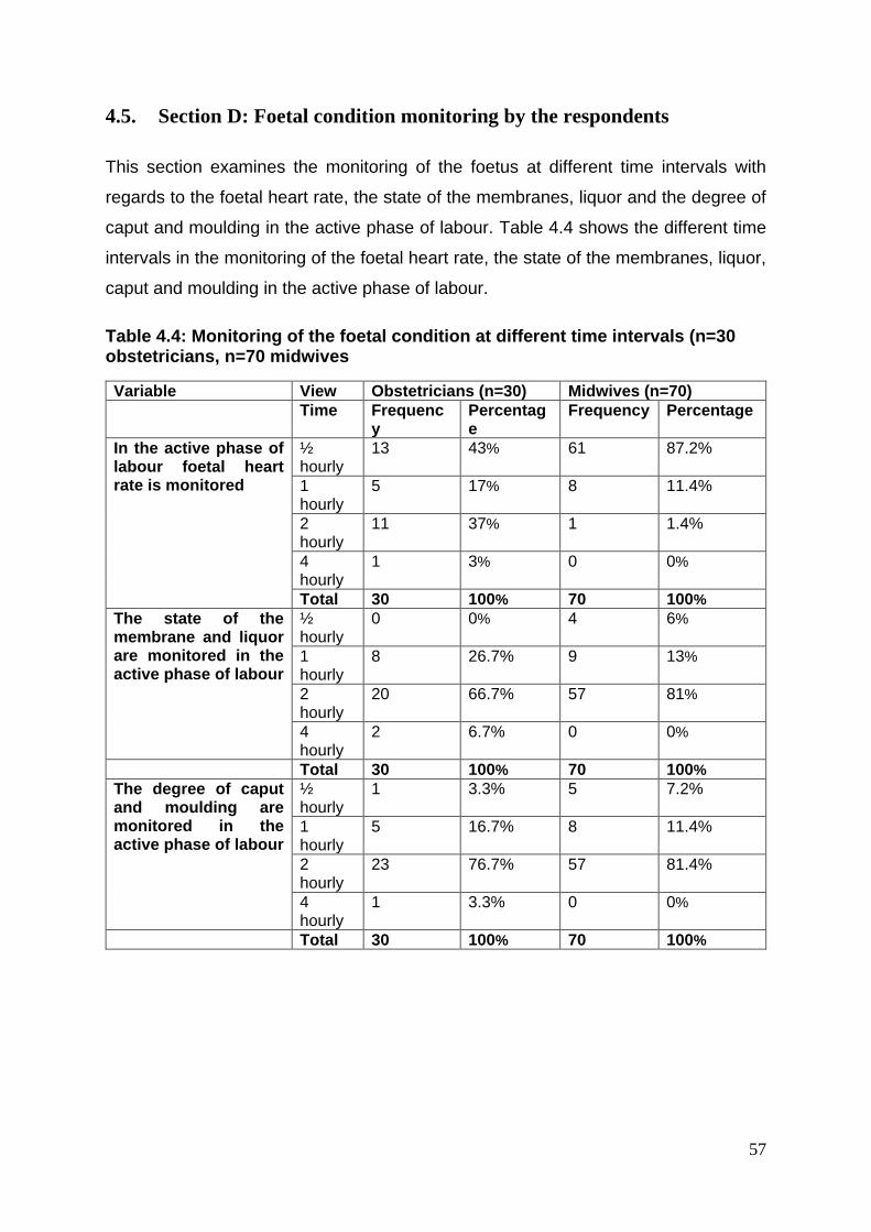
57
4.5. Section D: Foetal condition monitoring by the respondents
This section examines the monitoring of the foetus at different time intervals with
regards to the foetal heart rate, the state of the membranes, liquor and the degree of
caput and moulding in the active phase of labour. Table 4.4 shows the different time
intervals in the monitoring of the foetal heart rate, the state of the membranes, liquor,
caput and moulding in the active phase of labour.
Table 4.4: Monitoring of the foetal condition at different time intervals (n=30 obstetricians, n=70 midwives
Variable View Obstetricians (n=30) Midwives (n=70) Time Frequenc
y
Percentage
Frequency Percentage
In the active phase of labour foetal heart rate is monitored
½ hourly
13 43% 61 87.2%
1 hourly
5 17% 8 11.4%
2 hourly
11 37% 1 1.4%
4 hourly
1 3% 0 0%
Total 30 100% 70 100%
The state of the membrane and liquor are monitored in the active phase of labour
½ hourly
0 0% 4 6%
1 hourly
8 26.7% 9 13%
2 hourly
20 66.7% 57 81%
4 hourly
2 6.7% 0 0%
Total 30 100% 70 100%
The degree of caput and moulding are monitored in the active phase of labour
½ hourly
1 3.3% 5 7.2%
1 hourly
5 16.7% 8 11.4%
2 hourly
23 76.7% 57 81.4%
4 hourly
1 3.3% 0 0%
Total 30 100% 70 100%
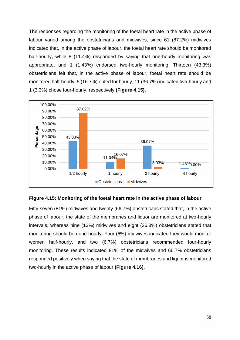
58
The responses regarding the monitoring of the foetal heart rate in the active phase of
labour varied among the obstetricians and midwives, since 61 (87.2%) midwives
indicated that, in the active phase of labour, the foetal heart rate should be monitored
half-hourly, while 8 (11.4%) responded by saying that one-hourly monitoring was
appropriate, and 1 (1.43%) endorsed two-hourly monitoring. Thirteen (43.3%)
obstetricians felt that, in the active phase of labour, foetal heart rate should be
monitored half-hourly, 5 (16.7%) opted for hourly, 11 (36.7%) indicated two-hourly and
1 (3.3%) chose four-hourly, respectively (Figure 4.15).
Figure 4.15: Monitoring of the foetal heart rate in the active phase of labour
Fifty-seven (81%) midwives and twenty (66.7%) obstetricans stated that, in the active
phase of labour, the state of the membranes and liquor are monitored at two-hourly
intervals, whereas nine (13%) midwives and eight (26.8%) obstetricians stated that
monitoring should be done hourly. Four (6%) midwives indicated they would monitor
women half-hourly, and two (6.7%) obstetricians recommended four-hourly
monitoring. These results indicated 81% of the midwives and 66.7% obstetricians
responded positively when saying that the state of membranes and liquor is monitored
two-hourly in the active phase of labour (Figure 4.16).
43.03%
11.04%
36.07%
1.43%
87.02%
16.07%
3.03%0.00%
0.00%
10.00%
20.00%
30.00%
40.00%
50.00%
60.00%
70.00%
80.00%
90.00%
100.00%
1/2 hourly 1 hourly 2 hourly 4 hourly
Pe
rce
nta
ge
Obstetricians Midwives
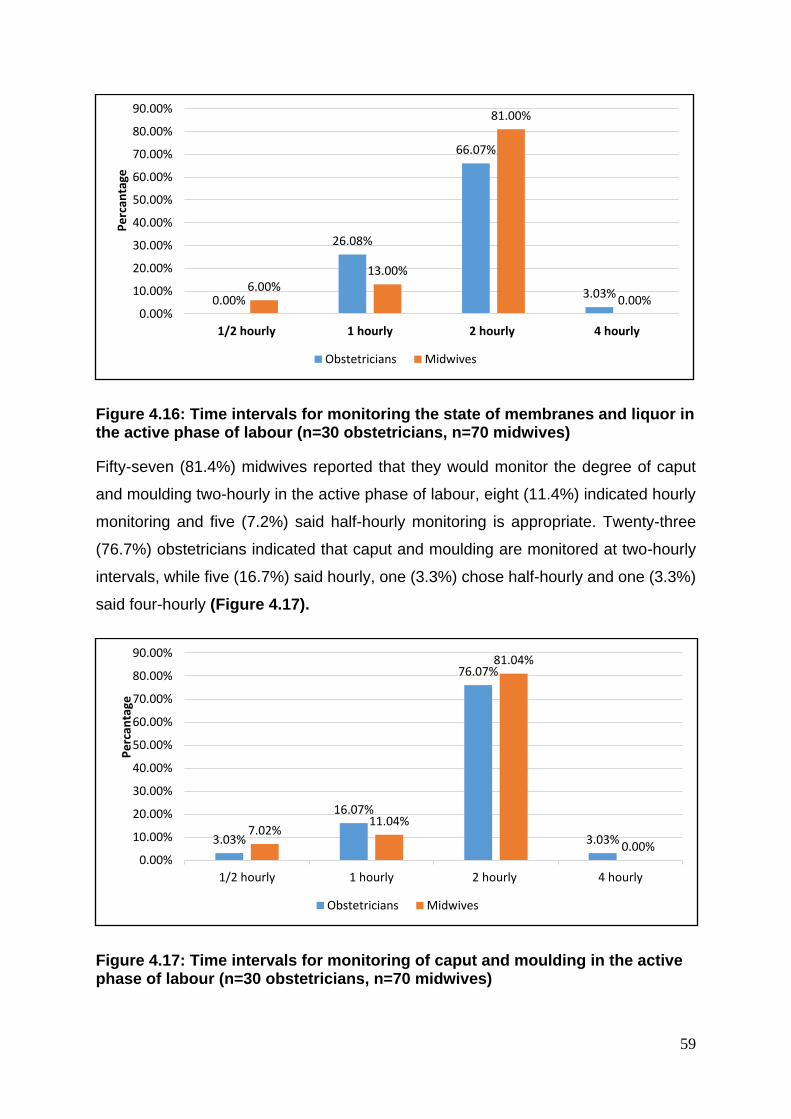
59
Figure 4.16: Time intervals for monitoring the state of membranes and liquor in the active phase of labour (n=30 obstetricians, n=70 midwives)
Fifty-seven (81.4%) midwives reported that they would monitor the degree of caput
and moulding two-hourly in the active phase of labour, eight (11.4%) indicated hourly
monitoring and five (7.2%) said half-hourly monitoring is appropriate. Twenty-three
(76.7%) obstetricians indicated that caput and moulding are monitored at two-hourly
intervals, while five (16.7%) said hourly, one (3.3%) chose half-hourly and one (3.3%)
said four-hourly (Figure 4.17).
Figure 4.17: Time intervals for monitoring of caput and moulding in the active phase of labour (n=30 obstetricians, n=70 midwives)
0.00%
26.08%
66.07%
3.03%6.00%
13.00%
81.00%
0.00%0.00%
10.00%
20.00%
30.00%
40.00%
50.00%
60.00%
70.00%
80.00%
90.00%
1/2 hourly 1 hourly 2 hourly 4 hourly
Pe
rcan
tage
Obstetricians Midwives
3.03%
16.07%
76.07%
3.03%7.02%
11.04%
81.04%
0.00%0.00%
10.00%
20.00%
30.00%
40.00%
50.00%
60.00%
70.00%
80.00%
90.00%
1/2 hourly 1 hourly 2 hourly 4 hourly
Pe
rcan
tage
Obstetricians Midwives
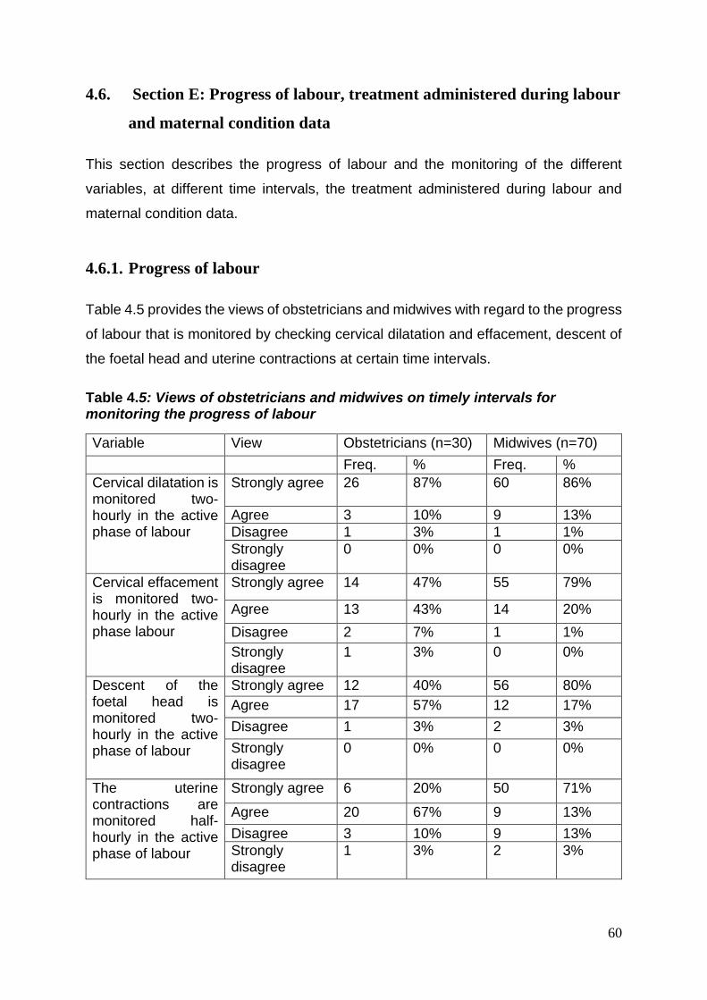
60
4.6. Section E: Progress of labour, treatment administered during labour
and maternal condition data
This section describes the progress of labour and the monitoring of the different
variables, at different time intervals, the treatment administered during labour and
maternal condition data.
4.6.1. Progress of labour
Table 4.5 provides the views of obstetricians and midwives with regard to the progress
of labour that is monitored by checking cervical dilatation and effacement, descent of
the foetal head and uterine contractions at certain time intervals.
Table 4.5: Views of obstetricians and midwives on timely intervals for monitoring the progress of labour
Variable View Obstetricians (n=30) Midwives (n=70)
Freq. % Freq. %
Cervical dilatation is monitored two-hourly in the active phase of labour
Strongly agree 26 87% 60 86%
Agree 3 10% 9 13%
Disagree 1 3% 1 1%
Strongly disagree
0 0% 0 0%
Cervical effacement is monitored two-hourly in the active phase labour
Strongly agree 14 47% 55 79%
Agree 13 43% 14 20%
Disagree 2 7% 1 1%
Strongly disagree
1 3% 0 0%
Descent of the foetal head is monitored two-hourly in the active phase of labour
Strongly agree 12 40% 56 80%
Agree 17 57% 12 17%
Disagree 1 3% 2 3%
Strongly disagree
0 0% 0 0%
The uterine contractions are monitored half-hourly in the active phase of labour
Strongly agree 6 20% 50 71%
Agree 20 67% 9 13%
Disagree 3 10% 9 13%
Strongly disagree
1 3% 2 3%
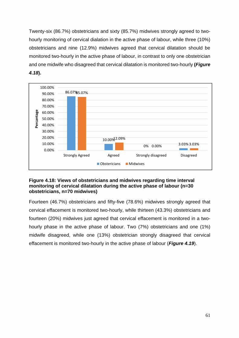
61
Twenty-six (86.7%) obstetricians and sixty (85.7%) midwives strongly agreed to two-
hourly monitoring of cervical dialation in the active phase of labour, while three (10%)
obstetricians and nine (12.9%) midwives agreed that cervical dilatation should be
monitored two-hourly in the active phase of labour, in contrast to only one obstetrician
and one midwife who disagreed that cervical dilatation is monitored two-hourly (Figure
4.18).
Figure 4.18: Views of obstetricians and midwives regarding time interval monitoring of cervical dilatation during the active phase of labour (n=30 obstetricians, n=70 midwives)
Fourteen (46.7%) obstetricians and fifty-five (78.6%) midwives strongly agreed that
cervical effacement is monitored two-hourly, while thirteen (43.3%) obstetricians and
fourteen (20%) midwives just agreed that cervical effacement is monitored in a two-
hourly phase in the active phase of labour. Two (7%) obstetricians and one (1%)
midwife disagreed, while one (13%) obstetrician strongly disagreed that cervical
effacement is monitored two-hourly in the active phase of labour (Figure 4.19).
86.07%
10.00%
0% 3.03%
85.07%
12.09%
0.00% 3.03%
0.00%
10.00%
20.00%
30.00%
40.00%
50.00%
60.00%
70.00%
80.00%
90.00%
100.00%
Strongly Agreed Agreed Strongly disagreed Disagreed
Pe
rcan
tage
Obstericians Midwives
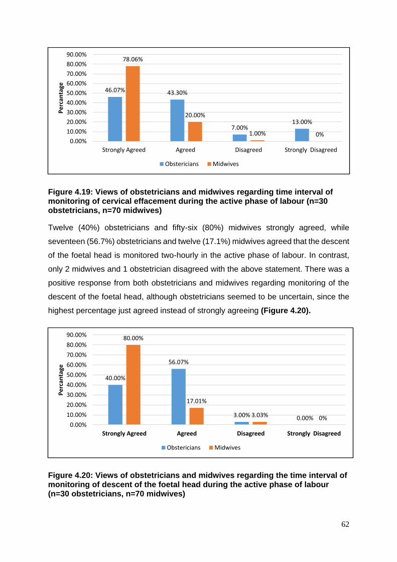
62
Figure 4.19: Views of obstetricians and midwives regarding time interval of monitoring of cervical effacement during the active phase of labour (n=30 obstetricians, n=70 midwives)
Twelve (40%) obstetricians and fifty-six (80%) midwives strongly agreed, while
seventeen (56.7%) obstetricians and twelve (17.1%) midwives agreed that the descent
of the foetal head is monitored two-hourly in the active phase of labour. In contrast,
only 2 midwives and 1 obstetrician disagreed with the above statement. There was a
positive response from both obstetricians and midwives regarding monitoring of the
descent of the foetal head, although obstetricians seemed to be uncertain, since the
highest percentage just agreed instead of strongly agreeing (Figure 4.20).
Figure 4.20: Views of obstetricians and midwives regarding the time interval of monitoring of descent of the foetal head during the active phase of labour (n=30 obstetricians, n=70 midwives)
46.07% 43.30%
7.00%13.00%
78.06%
20.00%
1.00% 0%0.00%
10.00%
20.00%
30.00%
40.00%
50.00%
60.00%
70.00%
80.00%
90.00%
Strongly Agreed Agreed Disagreed Strongly Disagreed
Pe
rcan
tage
Obstericians Midwives
40.00%
56.07%
3.00% 0.00%
80.00%
17.01%
3.03% 0%0.00%
10.00%
20.00%
30.00%
40.00%
50.00%
60.00%
70.00%
80.00%
90.00%
Strongly Agreed Agreed Disagreed Strongly Disagreed
Pe
rcan
tage
Obstericians Midwives
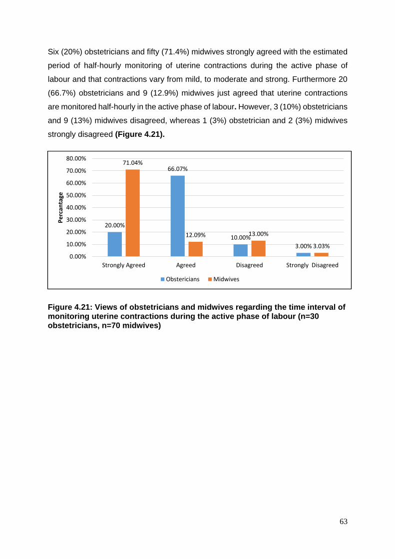
63
Six (20%) obstetricians and fifty (71.4%) midwives strongly agreed with the estimated
period of half-hourly monitoring of uterine contractions during the active phase of
labour and that contractions vary from mild, to moderate and strong. Furthermore 20
(66.7%) obstetricians and 9 (12.9%) midwives just agreed that uterine contractions
are monitored half-hourly in the active phase of labour. However, 3 (10%) obstetricians
and 9 (13%) midwives disagreed, whereas 1 (3%) obstetrician and 2 (3%) midwives
strongly disagreed (Figure 4.21).
Figure 4.21: Views of obstetricians and midwives regarding the time interval of monitoring uterine contractions during the active phase of labour (n=30 obstetricians, n=70 midwives)
20.00%
66.07%
10.00%
3.00%
71.04%
12.09% 13.00%
3.03%
0.00%
10.00%
20.00%
30.00%
40.00%
50.00%
60.00%
70.00%
80.00%
Strongly Agreed Agreed Disagreed Strongly Disagreed
Pe
rcan
tage
Obstericians Midwives
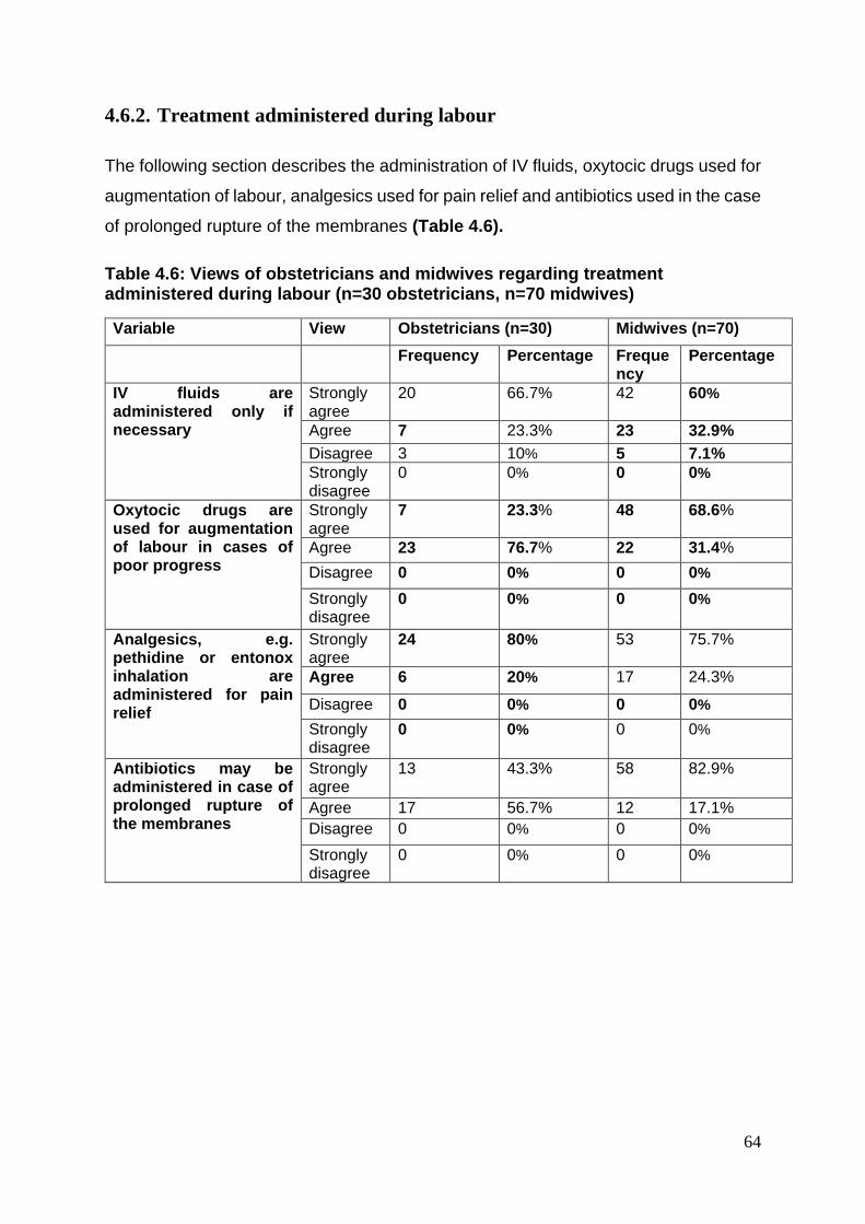
64
4.6.2. Treatment administered during labour
The following section describes the administration of IV fluids, oxytocic drugs used for
augmentation of labour, analgesics used for pain relief and antibiotics used in the case
of prolonged rupture of the membranes (Table 4.6).
Table 4.6: Views of obstetricians and midwives regarding treatment administered during labour (n=30 obstetricians, n=70 midwives)
Variable View Obstetricians (n=30) Midwives (n=70)
Frequency Percentage Frequency
Percentage
IV fluids are administered only if necessary
Strongly agree
20 66.7% 42 60%
Agree 7 23.3% 23 32.9%
Disagree 3 10% 5 7.1%
Strongly disagree
0 0% 0 0%
Oxytocic drugs are used for augmentation of labour in cases of poor progress
Strongly agree
7 23.3% 48 68.6%
Agree 23 76.7% 22 31.4%
Disagree 0 0% 0 0%
Strongly disagree
0 0% 0 0%
Analgesics, e.g. pethidine or entonox inhalation are administered for pain relief
Strongly agree
24 80% 53 75.7%
Agree 6 20% 17 24.3%
Disagree 0 0% 0 0%
Strongly disagree
0 0% 0 0%
Antibiotics may be administered in case of prolonged rupture of the membranes
Strongly agree
13 43.3% 58 82.9%
Agree 17 56.7% 12 17.1%
Disagree 0 0% 0 0%
Strongly disagree
0 0% 0 0%
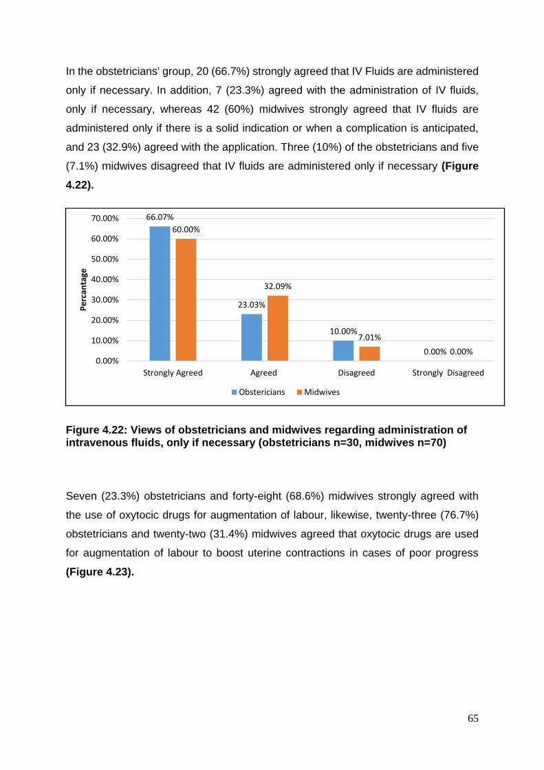
65
In the obstetricians' group, 20 (66.7%) strongly agreed that IV Fluids are administered
only if necessary. In addition, 7 (23.3%) agreed with the administration of IV fluids,
only if necessary, whereas 42 (60%) midwives strongly agreed that IV fluids are
administered only if there is a solid indication or when a complication is anticipated,
and 23 (32.9%) agreed with the application. Three (10%) of the obstetricians and five
(7.1%) midwives disagreed that IV fluids are administered only if necessary (Figure
4.22).
Figure 4.22: Views of obstetricians and midwives regarding administration of intravenous fluids, only if necessary (obstetricians n=30, midwives n=70)
Seven (23.3%) obstetricians and forty-eight (68.6%) midwives strongly agreed with
the use of oxytocic drugs for augmentation of labour, likewise, twenty-three (76.7%)
obstetricians and twenty-two (31.4%) midwives agreed that oxytocic drugs are used
for augmentation of labour to boost uterine contractions in cases of poor progress
(Figure 4.23).
66.07%
23.03%
10.00%
0.00%
60.00%
32.09%
7.01%
0.00%0.00%
10.00%
20.00%
30.00%
40.00%
50.00%
60.00%
70.00%
Strongly Agreed Agreed Disagreed Strongly Disagreed
Pe
rcan
tage
Obstericians Midwives
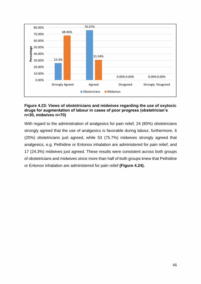
66
Figure 4.23: Views of obstetricians and midwives regarding the use of oxytocic drugs for augmentation of labour in cases of poor progress (obstetrician’s n=30, midwives n=70)
With regard to the administration of analgesics for pain relief, 24 (80%) obstetricians
strongly agreed that the use of analgesics is favorable during labour, furthermore, 6
(20%) obstetricians just agreed, while 53 (75.7%) midwives strongly agreed that
analgesics, e.g. Pethidine or Entonox inhalation are administered for pain relief, and
17 (24.3%) midwives just agreed. These results were consistent across both groups
of obstetricians and midwives since more than half of both groups knew that Pethidine
or Entonox inhalation are administered for pain relief (Figure 4.24).
23.3%
76.07%
0.00% 0.00%
68.06%
31.04%
0.00% 0.00%0.00%
10.00%
20.00%
30.00%
40.00%
50.00%
60.00%
70.00%
80.00%
Strongly Agreed Agreed Disagreed Strongly Disagreed
Pe
rcan
tage
Obstericians Midwives
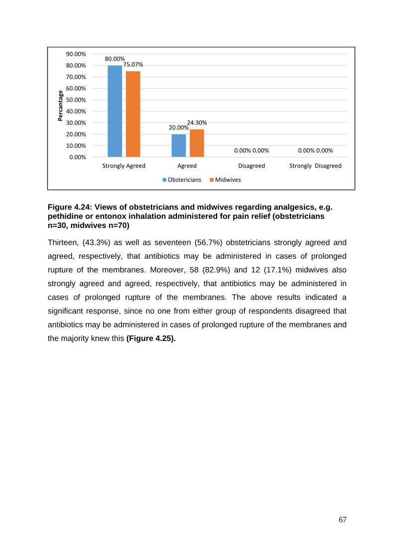
67
Figure 4.24: Views of obstetricians and midwives regarding analgesics, e.g. pethidine or entonox inhalation administered for pain relief (obstetricians n=30, midwives n=70)
Thirteen, (43.3%) as well as seventeen (56.7%) obstetricians strongly agreed and
agreed, respectively, that antibiotics may be administered in cases of prolonged
rupture of the membranes. Moreover, 58 (82.9%) and 12 (17.1%) midwives also
strongly agreed and agreed, respectively, that antibiotics may be administered in
cases of prolonged rupture of the membranes. The above results indicated a
significant response, since no one from either group of respondents disagreed that
antibiotics may be administered in cases of prolonged rupture of the membranes and
the majority knew this (Figure 4.25).
80.00%
20.00%
0.00% 0.00%
75.07%
24.30%
0.00% 0.00%0.00%
10.00%
20.00%
30.00%
40.00%
50.00%
60.00%
70.00%
80.00%
90.00%
Strongly Agreed Agreed Disagreed Strongly Disagreed
Pe
rcan
tage
Obstericians Midwives
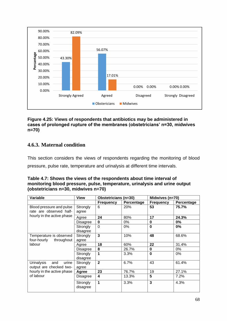
68
Figure 4.25: Views of respondents that antibiotics may be administered in cases of prolonged rupture of the membranes (obstetricians’ n=30, midwives n=70)
4.6.3. Maternal condition
This section considers the views of respondents regarding the monitoring of blood
pressure, pulse rate, temperature and urinalysis at different time intervals.
Table 4.7: Shows the views of the respondents about time interval of monitoring blood pressure, pulse, temperature, urinalysis and urine output (obstetricians n=30, midwives n=70)
Variable View Obstetricians (n=30) Midwives (n=70)
Frequency Percentage Frequency Percentage
Blood pressure and pulse rate are observed half-hourly in the active phase
Strongly agree
6 20% 53 75.7%
Agree 24 80% 17 24.3%
Disagree 0 0% 0 0%
Strongly disagree
0 0% 0 0%
Temperature is observed four-hourly throughout labour
Strongly agree
3 10% 48 68.6%
Agree 18 60% 22 31.4%
Disagree 8 26.7% 0 0%
Strongly disagree
1 3.3% 0 0%
Urinalysis and urine output are checked two-hourly in the active phase of labour
Strongly agree
2 6.7% 43 61.4%
Agree 23 76.7% 19 27.1%
Disagree 4 13.3% 5 7.2%
Strongly disagree
1 3.3% 3 4.3%
43.30%
56.07%
0.00% 0.00%
82.09%
17.01%
0.00% 0.00%0.00%
10.00%
20.00%
30.00%
40.00%
50.00%
60.00%
70.00%
80.00%
90.00%
Strongly Agreed Agreed Disagreed Strongly Disagreed
Pe
rcan
tage
Obstericians Midwives
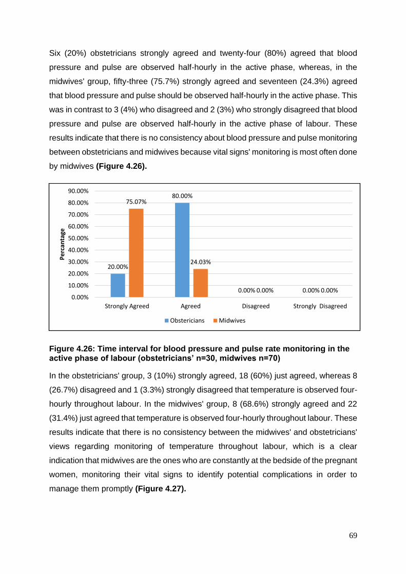
69
Six (20%) obstetricians strongly agreed and twenty-four (80%) agreed that blood
pressure and pulse are observed half-hourly in the active phase, whereas, in the
midwives' group, fifty-three (75.7%) strongly agreed and seventeen (24.3%) agreed
that blood pressure and pulse should be observed half-hourly in the active phase. This
was in contrast to 3 (4%) who disagreed and 2 (3%) who strongly disagreed that blood
pressure and pulse are observed half-hourly in the active phase of labour. These
results indicate that there is no consistency about blood pressure and pulse monitoring
between obstetricians and midwives because vital signs' monitoring is most often done
by midwives (Figure 4.26).
Figure 4.26: Time interval for blood pressure and pulse rate monitoring in the active phase of labour (obstetricians’ n=30, midwives n=70)
In the obstetricians' group, 3 (10%) strongly agreed, 18 (60%) just agreed, whereas 8
(26.7%) disagreed and 1 (3.3%) strongly disagreed that temperature is observed four-
hourly throughout labour. In the midwives' group, 8 (68.6%) strongly agreed and 22
(31.4%) just agreed that temperature is observed four-hourly throughout labour. These
results indicate that there is no consistency between the midwives' and obstetricians'
views regarding monitoring of temperature throughout labour, which is a clear
indication that midwives are the ones who are constantly at the bedside of the pregnant
women, monitoring their vital signs to identify potential complications in order to
manage them promptly (Figure 4.27).
20.00%
80.00%
0.00% 0.00%
75.07%
24.03%
0.00% 0.00%0.00%
10.00%
20.00%
30.00%
40.00%
50.00%
60.00%
70.00%
80.00%
90.00%
Strongly Agreed Agreed Disagreed Strongly Disagreed
Pe
rcan
tage
Obstericians Midwives
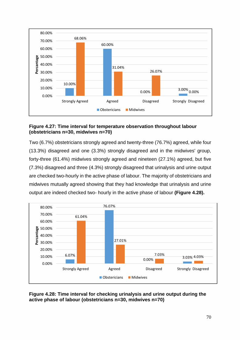
70
Figure 4.27: Time interval for temperature observation throughout labour (obstetricians n=30, midwives n=70)
Two (6.7%) obstetricians strongly agreed and twenty-three (76.7%) agreed, while four
(13.3%) disagreed and one (3.3%) strongly disagreed and in the midwives' group,
forty-three (61.4%) midwives strongly agreed and nineteen (27.1%) agreed, but five
(7.3%) disagreed and three (4.3%) strongly disagreed that urinalysis and urine output
are checked two-hourly in the active phase of labour. The majority of obstetricians and
midwives mutually agreed showing that they had knowledge that urinalysis and urine
output are indeed checked two- hourly in the active phase of labour (Figure 4.28).
Figure 4.28: Time interval for checking urinalysis and urine output during the active phase of labour (obstetricians n=30, midwives n=70)
10.00%
60.00%
0.00%3.00%
68.06%
31.04%26.07%
0.00%0.00%
10.00%
20.00%
30.00%
40.00%
50.00%
60.00%
70.00%
80.00%
Strongly Agreed Agreed Disagreed Strongly Disagreed
Pe
rcan
tage
Obstericians Midwives
6.07%
76.07%
0.00%3.03%
61.04%
27.01%
7.03%4.03%
0.00%
10.00%
20.00%
30.00%
40.00%
50.00%
60.00%
70.00%
80.00%
Strongly Agreed Agreed Disagreed Strongly Disagreed
Pe
rcan
tage
Obstericians Midwives
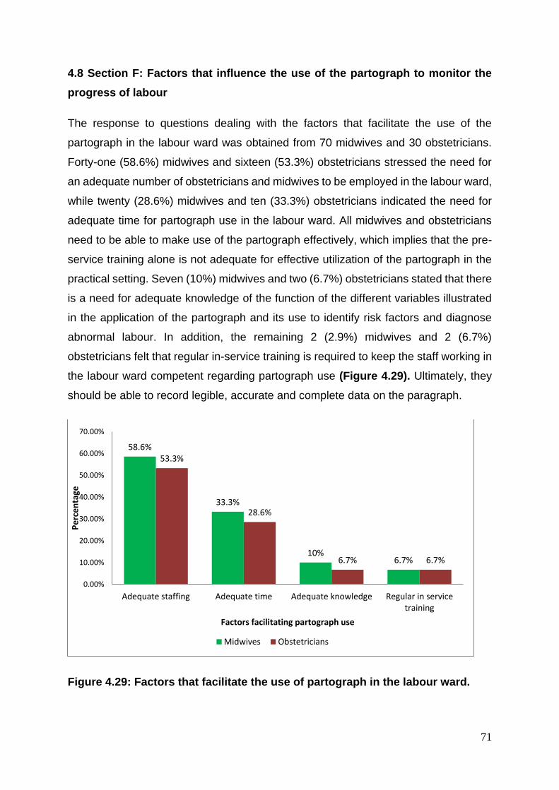
71
4.8 Section F: Factors that influence the use of the partograph to monitor the
progress of labour
The response to questions dealing with the factors that facilitate the use of the
partograph in the labour ward was obtained from 70 midwives and 30 obstetricians.
Forty-one (58.6%) midwives and sixteen (53.3%) obstetricians stressed the need for
an adequate number of obstetricians and midwives to be employed in the labour ward,
while twenty (28.6%) midwives and ten (33.3%) obstetricians indicated the need for
adequate time for partograph use in the labour ward. All midwives and obstetricians
need to be able to make use of the partograph effectively, which implies that the pre-
service training alone is not adequate for effective utilization of the partograph in the
practical setting. Seven (10%) midwives and two (6.7%) obstetricians stated that there
is a need for adequate knowledge of the function of the different variables illustrated
in the application of the partograph and its use to identify risk factors and diagnose
abnormal labour. In addition, the remaining 2 (2.9%) midwives and 2 (6.7%)
obstetricians felt that regular in-service training is required to keep the staff working in
the labour ward competent regarding partograph use (Figure 4.29). Ultimately, they
should be able to record legible, accurate and complete data on the paragraph.
Figure 4.29: Factors that facilitate the use of partograph in the labour ward.
58.6%
33.3%
10%6.7%
53.3%
28.6%
6.7% 6.7%
0.00%
10.00%
20.00%
30.00%
40.00%
50.00%
60.00%
70.00%
Adequate staffing Adequate time Adequate knowledge Regular in servicetraining
Pe
rce
nta
ge
Factors facilitating partograph use
Midwives Obstetricians
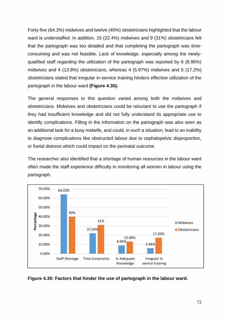
72
Forty-five (64.3%) midwives and twelve (40%) obstetricians highlighted that the labour
ward is understaffed. In addition, 15 (22.4%) midwives and 9 (31%) obstetricians felt
that the partograph was too detailed and that completing the partograph was time-
consuming and was not feasible. Lack of knowledge, especially among the newly-
qualified staff regarding the utilization of the partograph was reported by 6 (8.96%)
midwives and 4 (13.8%) obstetricians, whereas 4 (5.97%) midwives and 5 (17.2%)
obstetricians stated that irregular in-service training hinders effective utilization of the
partograph in the labour ward (Figure 4.30).
The general responses to this question varied among both the midwives and
obstetricians. Midwives and obstetricians could be reluctant to use the partograph if
they had insufficient knowledge and did not fully understand its appropriate use to
identify complications. Filling in the information on the partograph was also seen as
an additional task for a busy midwife, and could, in such a situation, lead to an inability
to diagnose complications like obstructed labour due to cephalopelvic disproportion,
or foetal distress which could impact on the perinatal outcome.
The researcher also identified that a shortage of human resources in the labour ward
often made the staff experience difficulty in monitoring all women in labour using the
partograph.
Figure 4.30: Factors that hinder the use of partograph in the labour ward.
64.03%
22.04%
8.96%5.96%
40%
31%
13.08%17.20%
0.00%
10.00%
20.00%
30.00%
40.00%
50.00%
60.00%
70.00%
Staff Shortage Time Constraints In AdequateKnowledge
Irregular Inservice training
Pe
rcan
tage
Midwives
Obstetricians
73
4.7. Second part of study: Utilization of the partograph (Results of
the retrospective data)
The second part of this study presents quantitative retrospective results obtained in
October and November 2011 through an audit of data on partograms that had
previously been recorded by obstetricians and midwives working in the labour wards.
These partograms were obtained from the maternity case records of 100 women who
had previously delivered over a one-month period, in September, 2010. The second
part of the study consists of two sections. Section A dealt with the recording of the
identification of women in labour (name, registration number, age, gravid and parity),
data relating to the state of the membranes on admission (date and time of rupture of
the membranes).
Section B related to the recording of the progress of labour, (dilatation and effacement
of the cervix, level of foetal head, and uterine contractions) data relating to medications
administered during labour, (type of medication, dosage, time and mode of
administration) data relating to maternal condition, (temperature, pulse, blood
pressure and urinalysis) and data relating to delivery (date, time and mode of delivery,
name of midwife or doctor who conducted the delivery, whether or not it was an
assisted delivery, whether the woman sustained a 1st or 2nd degree tear, whether an
episiotomy was performed and the possible complications thereof).
The audited partograms were assessed in terms of either being recorded based on
completeness or not recorded based on incompleteness of the information
documented on them. The factors which reflected completeness of information in this
study were whether information documented was legibly or clearly written, accurate
and date and time written, with all information documented on all components of the
partogram from the time when the active phase of the first stage of labour was reached
until delivery of the baby or not completely recorded.
Generally, it was observed that the information in some audited partograms was
complete as it was completely recorded since all the components of the audited
partograms were well documented. However, in some partograms information was
found to be incompletely recorded because information written in them was
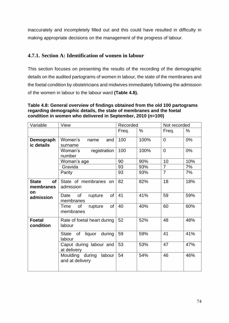
74
inaccurately and incompletely filled out and this could have resulted in difficulty in
making appropriate decisions on the management of the progress of labour.
4.7.1. Section A: Identification of women in labour
This section focuses on presenting the results of the recording of the demographic
details on the audited partograms of women in labour, the state of the membranes and
the foetal condition by obstetricians and midwives immediately following the admission
of the women in labour to the labour ward (Table 4.8).
Table 4.8: General overview of findings obtained from the old 100 partograms regarding demographic details, the state of membranes and the foetal condition in women who delivered in September, 2010 (n=100)
Variable View Recorded Not recorded
Freq. % Freq. %
Demographic details
Women’s name and surname
100 100% 0 0%
Woman’s registration number
100 100% 0 0%
Woman’s age 90 90% 10 10%
Gravida 93 93% 7 7%
Parity 93 93% 7 7%
State of membranes on admission
State of membranes on admission
82 82% 18 18%
Date of rupture of membranes
41 41% 59 59%
Time of rupture of membranes
40 40% 60 60%
Foetal condition
Rate of foetal heart during labour
52 52% 48 48%
State of liquor during labour
59 59% 41 41%
Caput during labour and at delivery
53 53% 47 47%
Moulding during labour and at delivery
54 54% 46 46%
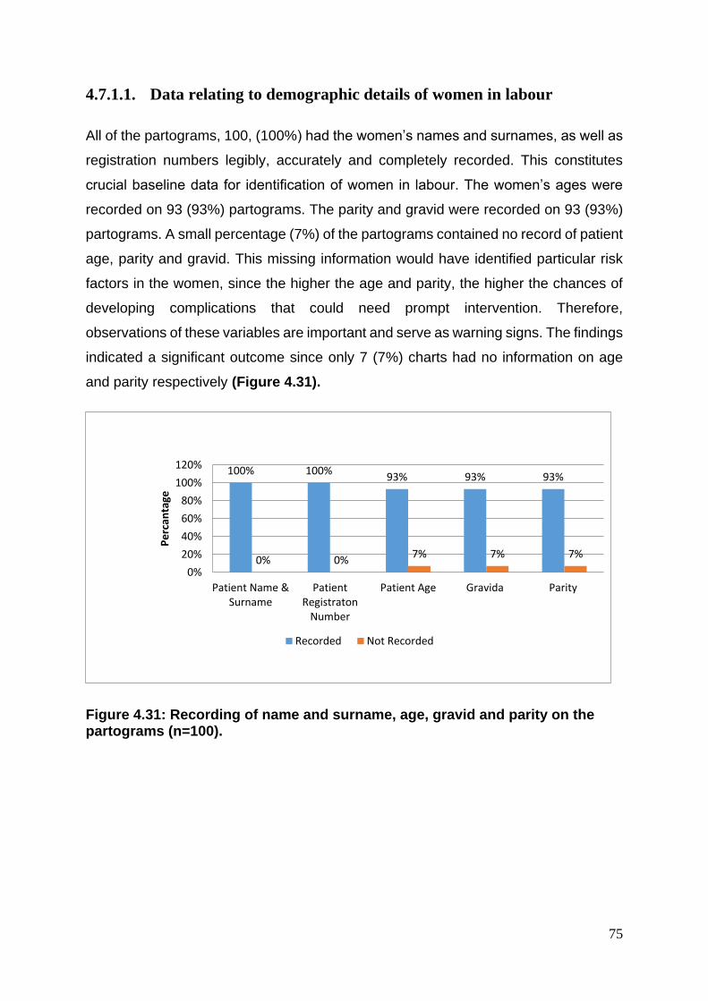
75
4.7.1.1. Data relating to demographic details of women in labour
All of the partograms, 100, (100%) had the women’s names and surnames, as well as
registration numbers legibly, accurately and completely recorded. This constitutes
crucial baseline data for identification of women in labour. The women’s ages were
recorded on 93 (93%) partograms. The parity and gravid were recorded on 93 (93%)
partograms. A small percentage (7%) of the partograms contained no record of patient
age, parity and gravid. This missing information would have identified particular risk
factors in the women, since the higher the age and parity, the higher the chances of
developing complications that could need prompt intervention. Therefore,
observations of these variables are important and serve as warning signs. The findings
indicated a significant outcome since only 7 (7%) charts had no information on age
and parity respectively (Figure 4.31).
Figure 4.31: Recording of name and surname, age, gravid and parity on the partograms (n=100).
100% 100%93% 93% 93%
0% 0%7% 7% 7%
0%
20%
40%
60%
80%
100%
120%
Patient Name &Surname
PatientRegistraton
Number
Patient Age Gravida Parity
Pe
rcan
tage
Recorded Not Recorded
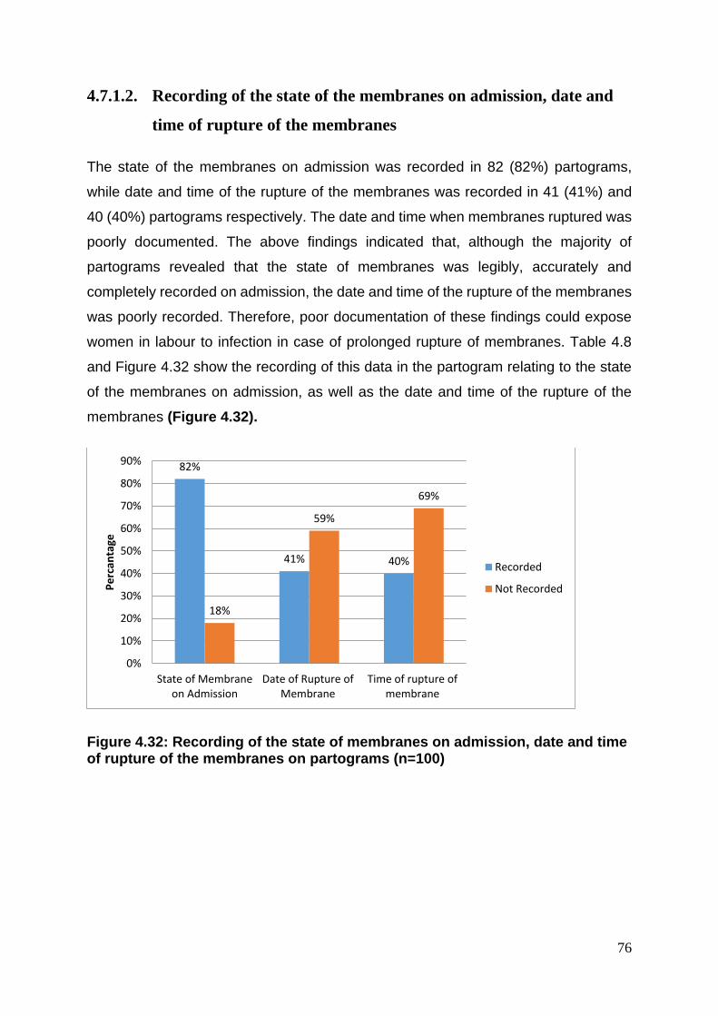
76
4.7.1.2. Recording of the state of the membranes on admission, date and
time of rupture of the membranes
The state of the membranes on admission was recorded in 82 (82%) partograms,
while date and time of the rupture of the membranes was recorded in 41 (41%) and
40 (40%) partograms respectively. The date and time when membranes ruptured was
poorly documented. The above findings indicated that, although the majority of
partograms revealed that the state of membranes was legibly, accurately and
completely recorded on admission, the date and time of the rupture of the membranes
was poorly recorded. Therefore, poor documentation of these findings could expose
women in labour to infection in case of prolonged rupture of membranes. Table 4.8
and Figure 4.32 show the recording of this data in the partogram relating to the state
of the membranes on admission, as well as the date and time of the rupture of the
membranes (Figure 4.32).
Figure 4.32: Recording of the state of membranes on admission, date and time of rupture of the membranes on partograms (n=100)
82%
41% 40%
18%
59%
69%
0%
10%
20%
30%
40%
50%
60%
70%
80%
90%
State of Membraneon Admission
Date of Rupture ofMembrane
Time of rupture ofmembrane
Pe
rcan
tage
Recorded
Not Recorded
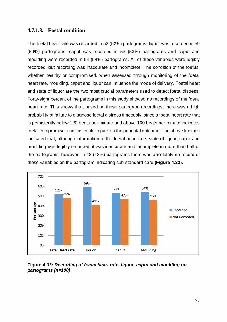
77
4.7.1.3. Foetal condition
The foetal heart rate was recorded in 52 (52%) partograms, liquor was recorded in 59
(59%) partograms, caput was recorded in 53 (53%) partograms and caput and
moulding were recorded in 54 (54%) partograms. All of these variables were legibly
recorded, but recording was inaccurate and incomplete. The condition of the foetus,
whether healthy or compromised, when assessed through monitoring of the foetal
heart rate, moulding, caput and liquor can influence the mode of delivery. Foetal heart
and state of liquor are the two most crucial parameters used to detect foetal distress.
Forty-eight percent of the partograms in this study showed no recordings of the foetal
heart rate. This shows that, based on these partogram recordings, there was a high
probability of failure to diagnose foetal distress timeously, since a foetal heart rate that
is persistently below 120 beats per minute and above 160 beats per minute indicates
foetal compromise, and this could impact on the perinatal outcome. The above findings
indicated that, although information of the foetal heart rate, state of liquor, caput and
moulding was legibly recorded, it was inaccurate and incomplete in more than half of
the partograms, however, in 48 (48%) partograms there was absolutely no record of
these variables on the partogram indicating sub-standard care (Figure 4.33).
Figure 4.33: Recording of foetal heart rate, liquor, caput and moulding on partograms (n=100)
52%
59%
53% 54%
48%
41%
47% 46%
0%
10%
20%
30%
40%
50%
60%
70%
Fetal Heart rate liquor Caput Moulding
Pe
rcan
tage
Recorded
Not Recorded
78
4.7.2. Section B: Progress of labour
This section observed the recording of the progress of labour with regard to dilatation
and effacement of the cervix; level/descent of head and uterine contractions on the
audited partograms of 100 women who had normal vaginal delivery in September
2010. The maternal condition included the recording of the temperature, the pulse, the
blood pressure and urinalysis on the partograms. With regard to the administration of
medication during labour, the recording on the partograms of the type of medication,
dosage, time and mode of administration was also assessed.
The recording of the time and date of delivery, the mode of delivery, the name of the
doctor or midwife who conducted the delivery, whether the delivery was assisted or
not, the occurrence of a first or second degree tear or whether an episiotomy was
performed and if any complications occurred was checked on the partograms. With
regards to the placenta, the recording of the date and time of expulsion of the placenta
and membranes, as well as the state of the placenta and membranes was checked.
The recording of the condition of the infant at birth on the partograms was assessed,
with regard to whether the infant was born alive, which is determined by use of the
Apgar score at 1 minute and at 5 minutes of delivery, the birth weight, the gender and
a quick examination of the new born to exclude any congenital defects.
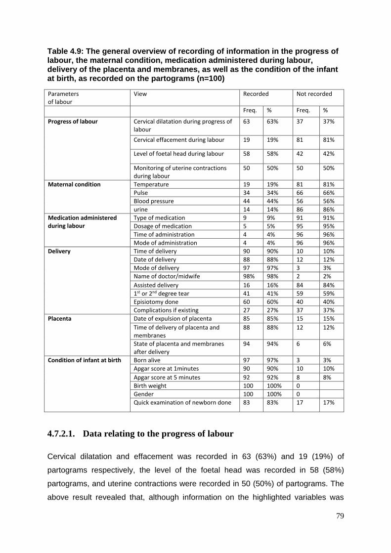
79
Table 4.9: The general overview of recording of information in the progress of labour, the maternal condition, medication administered during labour, delivery of the placenta and membranes, as well as the condition of the infant at birth, as recorded on the partograms (n=100)
Parameters of labour
View Recorded Not recorded
Freq. % Freq. %
Progress of labour
Cervical dilatation during progress of labour
63 63% 37 37%
Cervical effacement during labour 19 19% 81 81%
Level of foetal head during labour 58 58% 42 42%
Monitoring of uterine contractions during labour
50 50% 50 50%
Maternal condition Temperature 19 19% 81 81%
Pulse 34 34% 66 66%
Blood pressure 44 44% 56 56%
urine 14 14% 86 86%
Medication administered during labour
Type of medication 9 9% 91 91%
Dosage of medication 5 5% 95 95%
Time of administration 4 4% 96 96%
Mode of administration 4 4% 96 96%
Delivery Time of delivery 90 90% 10 10%
Date of delivery 88 88% 12 12%
Mode of delivery 97 97% 3 3%
Name of doctor/midwife 98% 98% 2 2%
Assisted delivery 16 16% 84 84%
1st or 2nd degree tear 41 41% 59 59%
Episiotomy done 60 60% 40 40%
Complications if existing 27 27% 37 37%
Placenta
Date of expulsion of placenta 85 85% 15 15%
Time of delivery of placenta and membranes
88 88% 12 12%
State of placenta and membranes after delivery
94 94% 6 6%
Condition of infant at birth
Born alive 97 97% 3 3%
Apgar score at 1minutes 90 90% 10 10%
Apgar score at 5 minutes 92 92% 8 8%
Birth weight 100 100% 0
Gender 100 100% 0
Quick examination of newborn done 83 83% 17 17%
4.7.2.1. Data relating to the progress of labour
Cervical dilatation and effacement was recorded in 63 (63%) and 19 (19%) of
partograms respectively, the level of the foetal head was recorded in 58 (58%)
partograms, and uterine contractions were recorded in 50 (50%) of partograms. The
above result revealed that, although information on the highlighted variables was
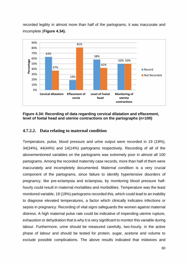
80
recorded legibly in almost more than half of the partograms, it was inaccurate and
incomplete (Figure 4.34).
Figure 4.34: Recording of data regarding cervical dilatation and effacement, level of foetal head and uterine contractions on the partographs (n=100)
4.7.2.2. Data relating to maternal condition
Temperature, pulse, blood pressure and urine output were recorded in 19 (19%),
34(34%), 44(44%) and 14(14%) partograms respectively. Recording of all of the
abovementioned variables on the partograms was extremely poor in almost all 100
partograms. Among the recorded maternity case records, more than half of them were
inaccurately and incompletely documented. Maternal condition is a very crucial
component of the partograms, since failure to identify hypertensive disorders of
pregnancy, like pre-eclampsia and eclampsia, by monitoring blood pressure half-
hourly could result in maternal mortalities and morbidities. Temperature was the least
monitored variable, 19 (19%) partograms recorded this, which could lead to an inability
to diagnose elevated temperatures, a factor which clinically indicates infections or
sepsis in pregnancy. Recording of vital signs safeguards the women against maternal
distress. A high maternal pulse rate could be indicative of impending uterine rupture,
exhaustion or dehydration that is why it is very significant to monitor this variable during
labour. Furthermore, urine should be measured carefully, two-hourly, in the active
phase of labour and should be tested for protein, sugar, acetone and volume to
exclude possible complications. The above results indicated that midwives and
63%
19%
58%
50%
37%
81%
42%
50%
0%
10%
20%
30%
40%
50%
60%
70%
80%
90%
Cervical dilatation Effacement ofcervix
Level of Foetalhead
Monitoring ofuterine
contractions
Pe
rcan
tage
Record
Not Recorded
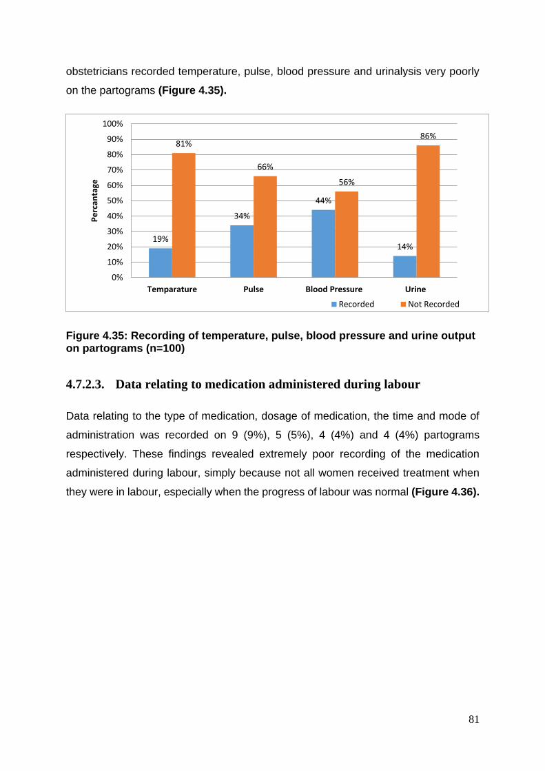
81
obstetricians recorded temperature, pulse, blood pressure and urinalysis very poorly
on the partograms (Figure 4.35).
Figure 4.35: Recording of temperature, pulse, blood pressure and urine output on partograms (n=100)
4.7.2.3. Data relating to medication administered during labour
Data relating to the type of medication, dosage of medication, the time and mode of
administration was recorded on 9 (9%), 5 (5%), 4 (4%) and 4 (4%) partograms
respectively. These findings revealed extremely poor recording of the medication
administered during labour, simply because not all women received treatment when
they were in labour, especially when the progress of labour was normal (Figure 4.36).
19%
34%
44%
14%
81%
66%
56%
86%
0%
10%
20%
30%
40%
50%
60%
70%
80%
90%
100%
Temparature Pulse Blood Pressure Urine
Pe
rcan
tage
Recorded Not Recorded
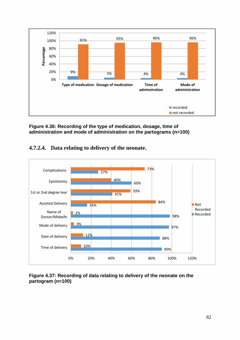
82
Figure 4.36: Recording of the type of medication, dosage, time of administration and mode of administration on the partograms (n=100)
4.7.2.4. Data relating to delivery of the neonate.
Figure 4.37: Recording of data relating to delivery of the neonate on the partogram (n=100)
9% 5% 4% 4%
91% 95% 96% 96%
0%
20%
40%
60%
80%
100%
120%
Type of medication Dosage of medication Time ofadminstration
Mode ofadministration
Pe
rcan
tage
recorded
not recorded
90%
88%
97%
98%
16%
41%
60%
27%
10%
12%
3%
2%
84%
59%
40%
73%
0% 20% 40% 60% 80% 100% 120%
Time of delivery
Date of delivery
Mode of delivery
Name ofDoctor/Midwife
Assisted Delivery
1st or 2nd degree tear
Epislotomy
Complications
NotRecordedRecorded
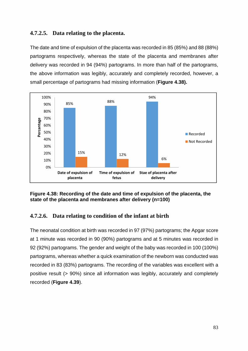
83
4.7.2.5. Data relating to the placenta.
The date and time of expulsion of the placenta was recorded in 85 (85%) and 88 (88%)
partograms respectively, whereas the state of the placenta and membranes after
delivery was recorded in 94 (94%) partograms. In more than half of the partograms,
the above information was legibly, accurately and completely recorded, however, a
small percentage of partograms had missing information (Figure 4.38).
Figure 4.38: Recording of the date and time of expulsion of the placenta, the state of the placenta and membranes after delivery (n=100)
4.7.2.6. Data relating to condition of the infant at birth
The neonatal condition at birth was recorded in 97 (97%) partograms; the Apgar score
at 1 minute was recorded in 90 (90%) partograms and at 5 minutes was recorded in
92 (92%) partograms. The gender and weight of the baby was recorded in 100 (100%)
partograms, whereas whether a quick examination of the newborn was conducted was
recorded in 83 (83%) partograms. The recording of the variables was excellent with a
positive result (> 90%) since all information was legibly, accurately and completely
recorded (Figure 4.39).
85%88%
94%
15%12%
6%
0%
10%
20%
30%
40%
50%
60%
70%
80%
90%
100%
Date of expulsion ofplacenta
Time of expulsion offetus
Stae of placenta afterdelivery
Pe
rcan
tage
Recorded
Not Recorded
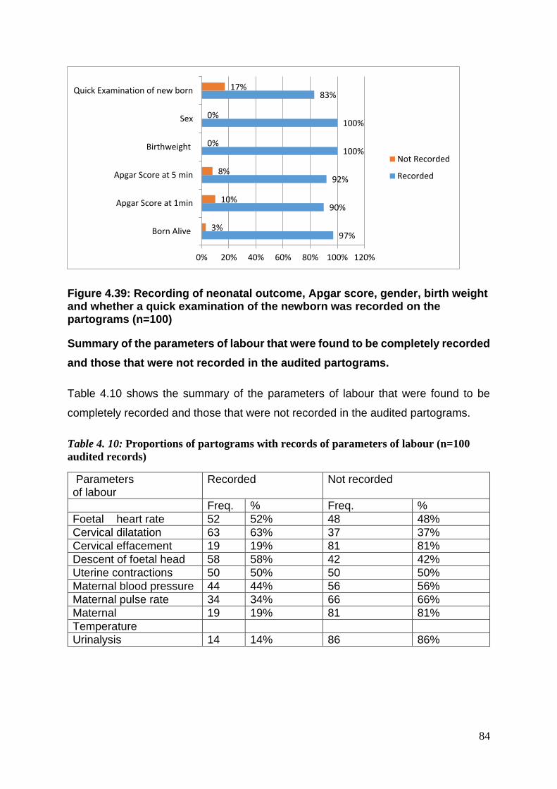
84
Figure 4.39: Recording of neonatal outcome, Apgar score, gender, birth weight and whether a quick examination of the newborn was recorded on the partograms (n=100)
Summary of the parameters of labour that were found to be completely recorded
and those that were not recorded in the audited partograms.
Table 4.10 shows the summary of the parameters of labour that were found to be
completely recorded and those that were not recorded in the audited partograms.
Table 4. 10: Proportions of partograms with records of parameters of labour (n=100
audited records)
Parameters of labour
Recorded Not recorded
Freq. % Freq. %
Foetal heart rate 52 52% 48 48%
Cervical dilatation 63 63% 37 37%
Cervical effacement 19 19% 81 81%
Descent of foetal head 58 58% 42 42%
Uterine contractions 50 50% 50 50%
Maternal blood pressure 44 44% 56 56%
Maternal pulse rate 34 34% 66 66%
Maternal 19 19% 81 81%
Temperature
Urinalysis 14 14% 86 86%
97%
90%
92%
100%
100%
83%
3%
10%
8%
0%
0%
17%
0% 20% 40% 60% 80% 100% 120%
Born Alive
Apgar Score at 1min
Apgar Score at 5 min
Birthweight
Sex
Quick Examination of new born
Not Recorded
Recorded
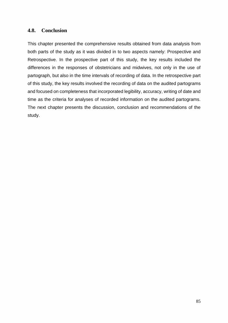
85
4.8. Conclusion
This chapter presented the comprehensive results obtained from data analysis from
both parts of the study as it was divided in to two aspects namely: Prospective and
Retrospective. In the prospective part of this study, the key results included the
differences in the responses of obstetricians and midwives, not only in the use of
partograph, but also in the time intervals of recording of data. In the retrospective part
of this study, the key results involved the recording of data on the audited partograms
and focused on completeness that incorporated legibility, accuracy, writing of date and
time as the criteria for analyses of recorded information on the audited partograms.
The next chapter presents the discussion, conclusion and recommendations of the
study.
86
Chapter Five
Discussion, conclusion, recommendations, of the study
5.1. Introduction
In this chapter the major results of the study will be discussed starting with the major
results of the prospective part of the study, followed by those of the retrospective part
of the study, in line with the research objectives of the study. Reiterate the purpose of
this study was to explore and describe the knowledge, attitudes and utilization of the
partograph by midwives and obstetricians in a regional hospital in the eThekwini
district. Furthermore, the research objectives were: (i) to determine the midwives' and
obstetricians' knowledge of the partograph in monitoring the progress of labour; (ii) to
describe the utilization of the partograph by midwives and obstetricians in the labour
ward; (iii) to identify factors that influence utilization of the partograph to monitor the
progress of labour.
In this chapter the current results are compared with results from previous studies with
respect to similarities and differences. The researcher further illustrated the limitations
of the study, together with recommendations for obstetricians and midwives and
conclusions based on the major results of this study.
5.2. Midwives’ and obstetricians’ knowledge of the use of partograph in
monitoring progress of labour.
5.2.1. Source of Knowledge and skills to use the partograph
The findings of this study indicated that all obstetricians (100%) and midwives (100%)
involved in the study indicated that they had been trained in the use of partograph.
Furthermore, it was found that 100% of obstetricians were trained in the use of the
partograph at medical school. In contrast, the responses of 63 (90%) midwives
revealed that they were trained during their tertiary education at Universities and
Colleges respectively. This was the majority, as only 7 (10%) midwives indicated that
87
they were trained when they were already practicing as qualified midwives during in-
service training.
The source of knowledge on partogram was identified in the study conducted by
Oladapo et al. (2006). The authors highlight that Life Saving Skill training workshop
was reported as the primary source of knowledge by one-third of those aware of the
partograph and these individuals generally had fair and good levels of knowledge.
Opiah et al. (2012) in a cross-sectional study to assess knowledge and utilization of
the partograph among midwives in two tertiary health facilities in the Niger Delta
Region of Nigeria, 86.2% of respondents had partograph training while in school of
midwifery, 13% said they were not trained in the midwifery school. The training of
midwives on the use of the partograph as well as periodic workshops and seminars
should be seen as vital to ensure the safety of women in labour. Further research on
the outcome of an incomplete or wrongly recorded partograph is recommended
(Mathibe-Neke et al., 2013).
5.2.2. Assessment of women in labour on admission to labour ward.
The results from this study indicated that 100% of obstetricians and 100% of midwives
reviewed the antenatal clinic records of all women in labour admitted to the labour
ward of this hospital and they also indicated that they identified and wrote maternal
and foetal risk factors. The majority of the obstetricians, (93%) and midwives (88.6%)
stated that they documented the date and time of admission, the date and time when
the membranes ruptured, as well as the date and time when labour started on the
partographs. The results of this study revealed that the respondents’ assessment of
the antenatal clinic records and identification of maternal and foetal risk factors were
done as expected from trained and practising midwives and obstetricians.
In this study, the results demonstrated that a high percentage of obstetricians (90%)
and midwives (85.7%) indicated that a decision as to the diagnosis and management
of a woman is always outlined at the end of the initial assessment. Twenty-eight (93%)
of the obstetricians and seventy (100%) midwives stated that the findings on
admission are always checked and recorded by a midwife or an obstetrician.
88
Similarly, to the findings from this study, in a study conducted by Mathibe-Neke et al.
(2013), it was found that twenty-five respondents (74%) indicated that the partograph
is important in the management of labour as it helps in monitoring the progress of
labour. Seven (21%) indicated that it is important for maternal wellbeing, and further
specified that it is important for both maternal and fetal wellbeing. Furthermore, in the
same study by Mathibe-Neke et al. (2013), it was noted that fifteen (44%) stated that
it assists with detecting complications allowing them to intervene accordingly. Eighteen
midwives (53%) specified that the partograph should be initiated during the active
phase of the first stage of labour. Some of the responses were non-specific for
example, all stages of labour, first stage to discharge, first stage to the fourth stage
(Mathibe-Neke et al., 2013).. Four respondents stated that the partograph is initiated
during the latent phase of the first stage of labour which is a recommended guideline
but only if the woman is experiencing true labour (Department of Health, 2015a;
Mathibe-Neke et al., 2013).
5.2.3. Monitoring of the foetal condition
5.2.3.1. Foetal Heart Rate
The majority 61(87.1%) of the midwives response was that, in the active phase of
labour, the foetal heart rate should be monitored half-hourly, 8 (11.4%) midwives said
one-hourly and 1 (1.4%) midwife opted for two-hourly, while only 13 (43%)
obstetricians felt that, in the active phase of labour, the foetal heart rate should be
measured half-hourly, 5 (16.7%) indicated one-hourly, 11 (36.7%) said two-hourly and
1 (3.3%) opted for four-hourly. These findings indicated that midwives are the ones
who are almost always at the bedside of the women in labour doing observations and
all other assessments. This also affirms the findings in previous studies that reported
that the expirienced midwife develops an extra sixth sense in her assessment of the
progress of labour , and of the maternal and fetal condition (Sellers, 1995).
89
5.2.3.2. State of membranes and liquor
More than half 57 (81.4%) midwives, and 20 (66.7%) obstetricians stated that, in the
active phase of labour, the state of the membranes and liquor is monitored two-hourly
which is the most appropriate response, since according to the Guidelines for
Maternity Care in South Africa (Department of Health, 2015a) frequent vaginal
examinations may introduce infection. It is therefore possible that complications
detrimental to the woman and the foetus could be promptly identified. In support of the
results of this study, Nolte (2008) also stated that the amniotic fluid should be recorded
regarding whether the membranes are intact, clear or meconium-stained. In addition,
Pettersson, Svensson and Christensson (2000) reported that the condition of the
amniotic fluid is an important indicator for early detection of foetal asphyxia.
5.2.3.3. Caput and moulding
Fifty-seven (81.4%) midwives reported they would monitor the degree of caput and
moulding in the active phase of labour two-hourly; eight (11.4%) indicated hourly and
five (7.1%) responded half-hourly. Twenty-two (73.3%) obstetricians indicated that
they would monitor the degree of caput and moulding in the active phase of labour at
two-hourly intervals, six (20%) hourly, one (3.3%) opted for half-hourly and one (3.3%)
said four-hourly. These results indicated that, with monitoring of the degree of caput
and moulding in the active phase of labour, the two-hourly interval monitoring was the
most appropriate intervention to be done by the respondents.Similarly to the two hourly
monitoring of membranes and liquor which according to the Guidelines for Maternity
Care in South Africa (Department of Health, 2015a) introduce infection if they are
frequently done. However, Nolte (2008) agreed that moulding is an indication of how
well the maternal pelvis can accommodate the foetal head.
Although the results of this study showed that less than 50% of the obstetricians
indicated that they monitored and plotted the foetal status during the active phase of
labour half hourly but more than 80% of the midwives responded the same way. The
midwives’ response concurs with The Fourth Report on Confidential Enquiries into
Maternal Deaths (NCCEMD), (2006:36) which requires that the foetal heart rate be
monitored half-hourly in the active phase of labour. Nolte (2008) reiterates that the
90
foetal heart rate should be assessed and recorded on the partograph every 30 minutes
during the active phase of labour. Therefore, half-hourly monitoring of foetal status
would assist the respondents to be able diagnose complications such as foetal distress
due to intrapartum hypoxia and to act upon them promptly to prevent perinatal
mortalities as it is required by the protocol of the Department of Health (2007). In
contrast, Tucker (2004) reported that the frequency of assessment of the foetal heart
rate should be guided by the risk status of both the woman in labour and the foetus. If
both the woman and the foetus are classified as high risk, the foetal heart rate should
be checked half-hourly in the active phase of labour (Shokane, 2011; Tucker, 2004).
5.2.3.4. The progress of labour
The majority of the obstetricians, 26 (86.7%) and midwives, 60 (85.7%) strongly
agreed/agreed that cervical dilatation, effacement and descent of the foetal head are
recorded two-hourly in the active phase of labour. These results were supported by
the South African maternity guideline from Department of Health (2007) where it is
stated that cervical dilatation, and effacement and descent of the foetal head are
recorded four-hourly in the latent phase and two-hourly in the active phase of labour.
Less than 50% of respondents, though, disagreed with this statement. Regarding
uterine contractions, the majority of obstetricians, 20 (66.7%) and midwives, 50
(71.4%) strongly agreed/agreed that uterine contractions are measured at half-hourly
intervals in the active phase of labour, although less than 50% respondents from both
groups disagreed probably because it is not always possible to monitor contractions
half –hourly especially if a midwife is looking after two or more women in labour.
In addition, the majority of obstetricians, 28, (93.3%) and midwives 65 (92.9%) also
strongly agreed/agreed that the partograph is systematically used in the labour ward,
hence the WHO (2010: 4) has also approved the universal application of the
partograph for the management of labour. Again, the majority of the obstetricians, 28
(93.3%) and midwives, 69 (98.6%) strongly agreed/agreed that the partograph is a
tool for decision-making in the labour ward, which is supported by the WHO (2010) in
its report stating that the partograph is a necessary tool in the management of labour
and recommending its universal application in labour wards. All the obstetricians, 30
(100%) and midwives, 70 (100%) strongly agreed/agreed that the partograph is a tool
91
used to monitor the progress of labour, and 30 (100%) obstetricians and 70 (100%)
midwives, respectively, strongly agreed/agreed that the partograph is a tool to detect
complications during labour which is still in line with recommendations of the (WHO,
2010).
Literature emphasizes that the partograph improves the management of labour as
recommended by the guidelines for maternity care in South Africa (Department of
Health, 2015a). Hence, all obstetricians, 30 (100%) and the majority of midwives, 68
(97.1%) strongly agreed/agreed that the partograph improves the management of
labour, whereas only 3 (10%) obstetricians and 18 (25.7%) midwives strongly
disagreed/disagreed that the partograph is difficult to use in the labour ward which
probably could be those who were newly employed and had never practiced in labour
ward.
5.2.3.5. Administration of medication during labour
According to the South African Maternity Guidelines from Department of Health
(2007), intravenous fluids are administered only if necessary, whereas oxytocic drugs
are used for the augmentation of labour in cases of poor progress and analgesics, e.g.
pethidine or entonox gas inhalation are drugs of choice administered for pain relief
during labour, lastly antibiotics may be administered in cases of prolonged rupture of
the membranes. Similarly, most of the obstetricians, 20 (66.7%) and midwives, 42
(60%) strongly agreed/agreed that intravenous fluids are administered only if
necessary, and that oxytocic drugs are used for the augmentation of labour in cases
of poor progress,and also that analgesics, e.g. pethidine or entonox inhalation are
administered for pain relief whilst antibiotics may be administered in cases of
prolonged rupture of the membranes, but less than 50% respondents from both groups
disagreed to this statement.
5.2.3.6. Maternal condition
According to the South African Maternity Guidelines from Department of Health
(2015a) blood pressure and pulse rate are observed half-hourly in the active phase of
labour, while temperature is observed four-hourly throughout labour, and urinalysis
92
and urine output are checked two-hourly in the active phase of labour. The majority of
the obstetricians, 24 (80%) and midwives, 53 (75.7%) strongly agreed/agreed that
these parameters are measured according to the guidelines, however, only a few of
the respondents disagreed to these interventions. These findings have identified a
serious gap since monitoring of vital signs alerts the midwife of potential complications
like imminent eclampsia if there is acute rise of blood pressure and or maternal
exhaustion if there are more leucocytes in urine.
5.3. Factors that influence the use of the partograph in the labour ward
5.3.1. Factors that facilitate the use of partograph in the labour ward
The response to factors that facilitate the use of partograph in labour ward was
obtained from 70 midwives and 30 obstetricians. Forty-one (58.6%) of the midwives
and sixteen (53.3%) obstetricians stressed the need for a full complement of staff in
the labour ward, while twenty (28.6%) midwives and ten (33.3%) obstetricians
indicated that there is a need for frequent in-service training for both midwives and
obstetricians, so that they could be able to make use of the partograph effectively. In
support of these findings the pre-service training alone was not sufficient for effective
utilization of the partograph in the practical area Yisma et al. (2013a). Seven (10%)
midwives and two (6.7%) obstetricians highlighted that there is need for equipping
both groups with knowledge of the function of the different variables listed on the
partograph and its use to predict abnormal labour. In addition, the remaining 2 (2.9%)
midwives and 2 (6.7%) obstetricians felt that if the work load in the labour ward could
be alleviated by employing more staff, this could ease work pressure, and therefore
midwives and obstetricians would be able to record data on the paragraph completely.
These results were also supported by Thopola (2002) where he stated that midwives
expressed exhaustion, general body pains and insomnia because they were short-
staffed.
93
5.3.2. Factors that hinder the use of the partograph in the labour ward.
The majority, 96 (96 0%) of the respondents highlighted that there are several factors
which are known to hinder the use of the partograph to monitor the progress of labour
and that these include little or lack of knowledge of partograph use, too much detail to
fill in, time-consuming, shortage of staff, obstetricians perceived partograph use as a
task for midwives. The response to this question was variable from both the midwives
and obstetricians, however, the obstetricians portrayed a negative attitude towards the
use of partograph since they perceived it the task for midwives. Adesola et al. (2014)
in support of the results of this study reported the grouping of the midwives attitudes
showed 54.5% had positive attitude while 45.5% had negative attitude towards
partograph use.
Forty-five (64.3%) midwives and twelve (40%) obstetricians highlighted that the labour
ward was understaffed. In addition, 15 (22.4%) midwives and 9 (31%) obstetricians
felt that the partograph was too detailed and that completing the partograph was time-
consuming and it was not practical to use. The lack of knowledge on the use of the
partograph was also mentioned by 6 (8.96%) midwives and 4 (13.8%) obstetricians,
whereas 4 (5.97%) midwives and 5 (17.2%) obstetricians indicated that inadequate in-
service training hindered the use of the partograph in the labour ward. Maputle (2005)
supports some of these results that, due to shortage of midwives, it is not easy to
spend quality time with all women in labour. This also affirms the findings in previous
studies that midwives expressed exhaustion, general body pains and insomnia
because they were short-staffed (Thopola, 2002). The shortage of nurses in South
Africa impacts negatively on the delivery of healthcare services in the country (Mateus,
Allen-Ile and Iwu, 2014).
The shortage of nurses is a chronic problem in South Africa. Many hospitals in the
rural areas and small towns do not have adequate health personnel. Yet there are
many South African nurses who work overseas (Mateus et al., 2014). Alonso-Garbayo
and Maben (2009) note that a global shortage of healthcare workers has become a
major concern for both developing and developed countries, especially since the
government of South Africa wishes to eliminate mortality among children during birth,
rising vaccine exposure, and epidemics such as HIV/AIDS (Mateus et al., 2014;
Alonso-Garbayo and Maben, 2009).
94
Bimenyimana, Poggenpoel, Myburgh and Van Niekerk (2009) highlighted that
shortage of staff resulted in a high workload and this led to job dissatisfaction,
absenteeism and a high staff turn-over. Dolamo (2009) reiterated that an estimated
13,496 nurse’s work in developed countries and that shortage of staff also contributed
to nurses overwork and this resulted in tiredness and job dissatisfaction.
Generally, completing the partograph was seen as an additional task for a health care
professional working in a busy labour ward hence they could not be motivated to
complete the partograph. However, in the study conducted by Mohammad and Fahdhy
and Chongsuvivatwong (2005) it was observed that while midwives had received
training on the utilisation of the partograph, they were not documenting their findings
because they complained that the partograph required too many details to be
completed. Mafalo (2003) further reported that many nurses including midwives are
also dying of the human immunodeficiency virus (HIV) and acquired immunodeficiency
syndrome (AIDS) in this country, and this also impacts negatively on health care
providers. According to Mafalo (2003) the population of South Africa grows by 14% a
year, while the nursing profession grows only by 10% and this results in a gradual
shortage of fully-qualified nursing staff.
This clearly indicated the need for in-service refresher training which midwives and
obstetricians should periodically undergo on the use of the partograph, especially the
newly-qualified midwives and obstetricians, while those with more work experience (in
terms of duration) should mentor the newly-employed staff. However, there is also a
need for very urgent steps to improve the knowledge of obstetric care-givers on
partograph use through workshops and seminars in order to maximize knowledge and
proper utilization of the partograph.
Yisma et al. (2013a) stated in his study that the midwives cited the following reasons
for not routinely using the partograph; little or no knowledge of the partograph, too
much detail to fill in, time-consuming, lack of an adequate number of personnel and
lack of in-service training. Nakkazi and Asis (2001) further added that midwives often
felt that completing the partograph was an additional time-consuming task, and they
did not always understand how the utilisation of the partograph to monitor pregnant
women in labour could be life-saving.
95
5.4. Utilization of the partograph by midwives and obstetricians in the labour
ward (retrospective part of the study)
5.4.1. Documentation/recording of the partograph
In this study it was discovered that all women were monitored using the partogram.
However, most of the parameters had been recorded at least once in the partogram.
It was also observed that the documentation in 19 (19%) partograms was illegible,
inaccurate, and incomplete as such, did not provide any valuable information. Twenty-
two (22%) of the partograms were recorded legibly, but inaccurately and incompletely.
The partograms that had inaccurate and incomplete information could have been
difficult to record for the health care professionals who were newly-qualified and
uncertain as to what to record.
According to a study by Mulondo (2007) that reviewed maternity case records, it was
discovered that some midwives were incompetent in recording the progress of labour
and the correct plotting of the partograph. The findings from a study conducted in
Ethiopia by Yisma et al. (2013a) revealed high proportions of unrecorded parameters
of labour on the modified WHO partograph and substandard monitoring of the
progress of labour among public health institutions in Addis Ababa, Ethiopia. Lack of
records for the descent of the presenting part in 84%, moulding in 86.7% and the foetal
heart rate in 41.1% of the studied partographs indicates poor documentation, and
perhaps monitoring and supervision of labour.
5.4.1.1. Presentation of demographic data is the partograpgh
With the chart audit, the monitoring of demographic details in respect to personal
particulars for identification; the women’s names and surnames 100 (100%) and
registration numbers 100 (100%) were the most legibly, accurately and completely
recorded parameters on the partogram. A small percentage 7 (7%) of the partograms
had no record of the patients' ages, gravid and parity. This missing information would
have identified particular risks factors and thus prompt intervention could have been
implemented to curb serious complications. According to Neilson et al. (2003) and
Mathai (2009) women with advanced age and higher parity have greater chances of
96
developing complications and these women require close monitoring throughout their
pregnancy. Therefore, the recording of these observations is important and would act
as warning signs for unforeseen complications. The age, parity and gravid may have
a direct effect on the labour outcomes (Mathai, 2009; Neilson et al., 2003).
5.4.1.2. The state of the membranes on admission
This study revealed significant results, since, on admission, the documentation of the
state of the membranes was recorded legibly, accurately and completely in more than
80 (80%) of the partograms, although the date and time of rupture of the membranes
was only recorded in 40 (40%) of the partograms. Nolte (2008) confirmed in his study
that the amniotic fluid should be recorded regarding whether the membranes are
intact, clear or meconium-stained .In addition, Pettersson et al. (2000) reported that
the condition of the amniotic fluid is an important indicator for early detection of foetal
asphyxia.
5.4.1.3. The foetal condition
The foetal heart rate in 52 (52%), state of liquor in 59 (59%), degree of caput in 53
(53%) and degree of moulding in 54 (54%) partograms was poorly recorded such that,
although the recording was legible, but it was inaccurately and incompletely
documented. However, literature in previous studies emphasizes that the expert,
expirienced midwife develops an extra sixth sense in her assessment of the progress
of labour , and of the maternal and fetal condition (Sellers, 1995). According to
Department of Health (2015a) effective monitoring of the foetal conditions, for low risk
labour, listen to the fetal heart with, ideally, a hand-held Doppler device, or a fetal or
normal stethoscope, before and immediately after contractions. CTG is used for high
risk labour only and must be available in all hospitals. CTG monitors are not
recommended for intrapartum use in CHCs. After CTG interpretation, write a note
about the findings in the woman’s notes, so that a record of the CTG is still available
even if the CTG tracing is lost. All CTG tracings must be kept safely in the woman’s
file and be stored with the file after delivery (Department of Health, 2015a).
97
5.4.1.4. The progress of labour
Of the 100 partograms that were reviewed, cervical dilatation, effacement of the cervix,
level of the foetal head and uterine contractions were recorded in 63 (63%) and 19
(19%), 58 (58%) and 50 (50%) of the partograms respectively. These findings
indicated, therefore, that although some of the respondents had knowledge of the
utilization of the partograph, but information was recorded inaccurately and
incompletely which could probably be due to shortage of staff and high workload.
These results were supported by the South African Maternity Guideline from
Department of Health (2007) where it is stated that cervical dilatation, effacement and
descent of the foetal head are recorded four-hourly in the latent phase and two-hourly
in the active phase of labour.
5.4.1.5. Administration of treatment during labour
Data relating to type of medication, dosage, time and mode of administration was
recorded legibly, accurately, but incompletely in 9 (9%); 5 (5%); 4 (4%) and 4 (4%)
partograms respectively. According to the South African Maternity Guidelines from
Department of Health (2007), intravenous fluids are administered only if necessary,
whereas oxytocic drugs are used for the augmentation of labour in cases of poor
progress and analgesics, e.g. pethidine or entonox gas inhalation are drugs of choice
administered for pain relief during labour, lastly antibiotics may be administered in
cases of prolonged rupture of the membranes. These results revealed sub-standard
care on the part of the midwives since they are the ones who administer medication
after it has been ordered by the obstetrician.
5.4.1.6. Maternal condition
Maternal condition is monitored and recorded in the last component of the partograph.
In this study, temperature was not recorded in 81 (81%) partograms, pulse in 66 (66%),
blood pressure in 54 (54%) and urine output in 86 (86%). These variables were poorly
recorded since more than half the parameters were neither inaccurate nor incomplete,
but were not recorded at all. These findings identified a serious gap since monitoring
of vital signs alerts the midwife of potential complications like imminent eclampsia if
98
there is acute rise of blood pressure and or maternal exhaustion if there are more
leucocytes in urine.
A study conducted by Delvaux, Aké-Tano, Gohou-Kouassi, Bosso, Collin and
Ronsmans (2007) on 229 women, affirms that blood pressure was measured in less
than half, 115 (50%) of the women and the partograms were completed in only 5% of
the women in labour. Nyamtema et al. (2008) confirmed in his study that temperature,
pulse and blood pressure were not recorded in 70%, 76% and 47% of the partographs,
respectively. It is, therefore, clear that monitoring of the maternal condition is sub-
standard in low and middle income countries.
In a retrospective study on previously completed partograms, Basu, Hoosain, Leballo,
Leistner, Masango, Mercer et al. (2009) reiterated that the blood pressure, pulse and
temperature were documented completely in 50 women, (45%) 29 (26%) and 41
(37%), respectively, and were not recorded in 29 (26%) 40 (36%) and 42 (38%)
women, and infrequently recorded in the remainder.
5.4.1.7. Recording of post-delivery information
Post-delivery results revealed a significant and tremendous outcome since, in more
than half of the partograms, this component was legibly, accurately and completely
recorded. The recording of the date and time of delivery was recorded in 90 (90%)
partograms, the mode of delivery in 90 (97%) partograms and the name of the midwife
or doctor who conducted the delivery was recorded in 98 (98%) partograms
respectively. The date and time of the expulsion of the placenta was recorded in 85
(85%) and 88 (88%) partograms, whereas the state of the placenta and membranes
after delivery was recorded in 94 (94%).
The neonatal condition at birth was recorded in 97 (97%) partograms, Apgar scores
at 1 minute were recorded in 90 (90%); Apgar scores at 5 minutes were recorded in
92 (92%); whereas details of gender and weight of the baby were recorded in 100
(100%).In addition, quick examination of the newborn was recorded in 83 (83%)
partograms. However, indication of whether the delivery was assisted or not was
recorded completely only in 16 (16%) partograms probably because complicated
deliveries like breech or shoulder dystocia are very rare since they are normally
99
diagnosed during antenatal period and delivered per ceasarian section. Details
relating to a 1st or 2nd degree tear were recorded in 41 (41%) and record of whether
an episiotomy was performed was shown in 60, (60%) while complications like post-
partum heamorrhage were recorded in 27 (27%) partograms respectively since far
less than 50% women present with postpartum heamorrhage after delivery.
A recent analysis of maternal and perinatal deaths in the intrapartum period showed
that sub-standard care and a lack of basic obstetric knowledge and skills was
responsible for these deaths (Ameh and van den Broek, 2015). Ogwang et al. (2009)
reported that, in their study, 1170 (69.9%) partograms were used in a total of 1674
deliveries, while Kim, Tappis, Zainullah, Ansari, Evans, Bartlett et al. (2012) stated that
only 107 (62%) partograms were used in 173 deliveries and that only 30 (28%)
deliveries were managed by a partogram, while information on partograms used was
inaccurate, incomplete and missing in 66 (38%) of the cases. Information on parity
was missing in 39 (23%) of the cases, indications relating to caesarean section were
missing in 15 (9%) cases, information on anaesthetic use was missing in 10 (6%)
cases, foetal outcomes were missing in 5 (3%) of the cases, and emergency
classification was missing in 4 (2%) of the cases. Information on cervical dilatation,
cervical effacement; level of foetal head and uterine contractions was missing in 37%,
81%, 42% and 50% of the partograms respectively. Temperature, pulse, blood
pressure and urine estimation was unrecorded in 81%, 66%, 56% and 86% of the
partograms respectively. With regard to the state of the membranes on admission, the
date and time of the rupture of the membranes was missing in 59% and 60% of the
partograms respectively. The state of liquor, caput and moulding during labour and
delivery was missing in 41%, 47% and 46% of the partograms respectively. With
regard to medication administered during labour, the type, dosage, time of
administration and mode of administration was not recorded in 91%, 95%, 96% and
96% of the partograms respectively. The information relating to delivery, the state of
the placenta and the condition of the newborn was well recorded, except for details of
assisted deliveries, 1st or 2nd degree tears, episiotomies and complications.
A study conducted by Khonje (2012) demonstrated that not documenting or forgetting
to document after carrying out observations as was expressed, was a form of
negligence. In some cases, health workers used shortcuts and could not carry out
100
observations, this was also negligence. These behaviours might have led to wrong
decisions. Lack of complete information can make normal labours to become
abnormal labours. This might have increased the C/S rate. Laziness among health
workers made them not maximise the time to assess and document on the partograph.
Laziness also contributed to the partial filling in of the partograph describing it to be
detailed. It is the researcher’s observation that whenever people settle in routines, the
initial value of an activity or procedure is usually lost. So is the use of partograph at
the units that it has become a routine activity that is no longer valued; and
documentation may be looked at as not important rather than an extra work (Barros et
al., 2010).
5.5. Training of midwives and obstetricians
Although all the midwives and obstetricians in this study were trained in the use of
partograph, but it was surprising that most of the parameters in the partograms were
inaccurately and incompletely recorded. The obstetricians perceived partograph use
as a task for midwives. The response to this question was variable from both the
midwives and obstetricians, however, the obstetricians portrayed a negative attitude
towards the use of partograph since they perceived it the task for midwives. Adesola
et al. (2014) in support of the results of this study reported the grouping of the
midwives’ attitudes showed 54.5% had positive attitude while 45 .5% had negative
attitude towards partograph use.
A questionnaire-based study involving 60 midwifery students showed that all the
students were adequately assisted theoretically and minimally in the clinical setting,
which might have implications for the poor utilization of the partograph when they are
registered midwives. In addition, the students indicated that the partograph was mostly
used by registered midwives, 33, (55.4%) followed by doctors, 22 (36.1%) and
midwifery students, 5 (8, 4%). This reflected inadequate exposure of midwifery
students to the use of the partograph in a clinical setting (Mathibe-Neke, 2009).
There is, therefore, a need to standardize training programs, especially on practical
issues, e.g. clinical procedures, including use and interpretation of the partograph, as
well as the incorporation of a system of mentorship in the labour ward that will ensure
101
that midwives and obstetricians are adequately up skilled. It is possible that the
monitoring of progress of labour was more intense in high-risk women which could
account for those partograms that were incomplete.
5.6. Recommendations
In view of the results presented in chapter four in this study the researcher will present
the following recommendations for consideration by the relevant authorities of
government and professional stake-holders.
5.6.1. In-service training for health care providers
There is widespread concern regarding the correct and efficient use of the partograph
for the monitoring of the progress of labour. Given the high maternal and perinatal
morbidity and mortality, several interventions have been identified as important for
curbing this high maternal mortality rate. One of these interventions includes the early
detection of the abnormal progress of labour through the use of the partograph, as
recommended by the (NCCEMD, 2012). In addition, the NCCEMD (2007) reported
that, of the women who died from pregnancy-related sepsis, 163 deaths were related
to complications resulting from obstructed and prolonged labour and that the lack of
and or improper use of the partogram was probably a contributing and avoidable factor
in some of these deaths.
Midwives and obstetricians working in the labour wards, therefore need to constantly
up skill their knowledge and obtain information in order to practice and provide high
quality care to women in labour, therefore in-service training to strengthen and
upgrade the knowledge and competency of qualified health personnel, namely
midwives and obstetricians is thus imperative.
The emphasis on the use of partograph is for early detection of deviations from normal
and thus preventing them from becoming complications. Hence, this shows the
significance of carrying out accurate and regular observations so that the midwives
and obstetricians know when labour is deviating from the norm so as to institute prompt
intervention. Each component of the partograph requires legible, accurate and
102
complete information, since each part complements each other in the effective
utilisation of the pantograph (Mathai, 2009).
5.6.2. Education and curriculum for training of midwifery and medical
students.
There is a dire need to discuss the results of this study with the staff of nursing schools
and medical schools so that standardization of teaching at tertiary level in the use of
the partograph is done. This will be in line with the (NCCEMD, 2007) recommendations
as suggested by the Essential Steps in the Management of Obstetric Emergencies
(ESMOE) Board, which started an initiative to standardize the use of the partograph
by midwives and medical students in South Africa a long time ago to ensure quality
service delivery thus saving lives of mothers and babies.
5.6.3. Policy-makers, management and government
The Government, including management has to ensure that policies pertaining to
partograph use are known, displayed on bulletin boards and regular partogram audits
are implemented in labour wards of all health facilities.
The results of this study demonstrate, that the partograph is not being used properly,
and that the National Committee for the Confidential Enquiries into Maternal Deaths
(NCCEMD) (2012) recommendations of performing quality improvement regarding
proper use of the partograph may not be taking place on a regular basis in our
hospitals. Therefore, improper use of the partograph and poor interpretation may be
prevalent. Government, therefore has to provide support for the training of
obstetricians and midwives in ESMOE, by informing the Chief Executive Officer (CEO)
that they are required to ensure that training that focuses on emergency obstetric
simulation training exercises is taking place at least monthly in all maternity wards of
their institutions.
In addition, one way of monitoring and evaluating recommendations is to have regular
clinical audits to establish where problems lie, and then to institute regular in-service
training of health professionals and students at the bedside in the form of emergency

103
obstetric simulation training exercises with case scenarios. The Government has
responsibility also to ensure that staffing and equipment norms are established for
each level and for every health facility providing health care services to pregnant
women to prevent human and material resources shortages.
5.6.4. Further research
Similar research studies should be conducted on a larger scale to explore the
knowledge and use of the partograph by midwives and obstetricians in both private
and public health facilities in different districts and provinces in South Africa and other
African countries. In addition, a study focusing on the direct effect of incomplete
recording of the partograph on maternal and child mortality and morbidity is essential
and needs to be conducted.
5.7. Limitations of the study
The limitations of this study included the following:
Firstly, as this study is confined to midwives and obstetricians working in one
public health facility, the results may not be generalizable or applicable to
those working in other public health facilities or private health facilities.
The other limitation of the study was the small sample size which could render
estimates unstable and associations between dependent and independent
variables undetectable.
A further limitation of this study was that it did not assess the maternal and
foetal outcome of completely and incompletely documented partograms.
Limited experience in conducting a research project presents a limitation to the
study as this was the first research project to be conducted by the researcher.
Time constrains was one of the major limitations of conducting this study as
the researcher conducted this study as a part time student as she was
permanently employed by the Department of Health. This resulted to a delay
in completing report writing of this study.
104
5.8. Conclusion
The results of this study not only confirm the impression that the partograph is not
always entirely completed, but also that the views and attitudes of midwives and
obstetricians differ. The results of this study may be understandable in a district
hospital given the shortage of medical staff, but what is of extreme concern is that this
is occurring in a regional hospital. It is, however, also possible that obstetricians and
midwives recorded their findings elsewhere in the maternity-case records rather than
on the partograph. However, this defeats the purpose of having the partograph and
interpreting the information in a holistic manner.
South Africa has maternal and child mortality rates that are among the highest in the
world and ever since the issue of maternal mortality was recognized as a major public
health problem, the unbooked women with prolonged and obstructed labour have
been recognized as a major risk factor for maternal deaths. Therefore, more attention
has to be given to improving knowledge and skills around managing obstetric
emergencies. This study revealed high proportions of unrecorded parameters of
labour on the partographs and sub-standard monitoring of the progress of labour which
is an example of the general lack of uniform standards in the care of women in labour,
contributing to poor maternal and neonatal outcomes.
The conclusion made based on the results of this study, therefore is that poor
recording of the partographs might be one of the factors contributing to maternal and
perinatal morbidities and mortalities in various institutions. It is evident, therefore, that
despite the wide use of the partograph in the labour ward of the study institution, the
completion, interpretation and eventual interventions are still deficient. Given these
results, the researcher is calling upon all policymakers and health professional
associations to recognise this issue of improper partograph use as a national
emergency and to map out effective short-term solutions to ensure that partograph is
used properly to save lives of mothers and babies.
105
References
ADESOLA, O., ADEKEMI, O. & AUDU, O. 2014. Partograph utilization at three levels of
health care delivery services in Ile-Ife, Nigeria. International Journal of Caring
Sciences, 7, 678-691.
ALONSO-GARBAYO, Á. & MABEN, J. 2009. Internationally recruited nurses from India
and the Philippines in the United Kingdom: the decision to emigrate. Human Resources
for Health, 7, 1-11.
ALVES, S. V. 2007. Maternal mortality in Pernambuco, Brazil: What has changed in ten years?
Reproductive Health Matters, 15, 134-144.
AMEH, C. A. & VAN DEN BROEK, N. 2015. Making It Happen: Training health-care
providers in emergency obstetric and newborn care. Best Practice & Research Clinical
Obstetrics & Gynaecology, 29, 1077-1091.
ASIBONG, U., OKOKON, I. B., AGAN, T. U., OKU, A., OPIAH, M., ESSIEN, E. J. &
MONJOK, E. 2014. The use of the partograph in labor monitoring: a cross-sectional
study among obstetric caregivers in General Hospital, Calabar, Cross River State,
Nigeria. International Journal of Women's Health, 6, 873-887.
BARROS, F. C., BHUTTA, Z. A., BATRA, M., HANSEN, T. N., VICTORA, C. G. &
RUBENS, C. E. 2010. Global report on preterm birth and stillbirth: Evidence for
effectiveness of interventions. BMC Pregnancy and Childbirth, 10, 1.
BASU, J. K., HOOSAIN, S., LEBALLO, G., LEISTNER, E., MASANGO, D., MERCER, M.,
MOHAPI, M., PETKAR, S. & TSHIOVHE, N. 2009. The partogram: A missed
opportunity. SAMJ: South African Medical Journal, 99, 578-578.
BAZIRETE, O. 2014. Utilization of partogram among nurses and midwives in Rwamagana
health facilities in the eastern province of Rwanda. University of the Western Cape.
BIMENYIMANA, E., POGGENPOEL, M., MYBURGH, C. & VAN NIEKERK, V. 2009.
The lived experience by psychiatric nurses of aggression and violence from patients in
a Gauteng psychiatric institution. Curationis, 32, 4-13.
BRINK, H., VAN DER WALT, C. & VAN RENSBURG, G. 2006. Fundamentals of Research
Methodology for Health Care Professionals, Cape Town, Juta & Co.
BUHIMSCHI, C. S., BUHIMSCHI, I. A., MALINOW, A. M., SAADE, G. R., GARFIELD,
R. E. & WEINER, C. P. 2003. The forces of labour. Fetal and Maternal Medicine
Review, 14, 273-307.
CARLO, W. A., GOUDAR, S. S., JEHAN, I., CHOMBA, E., TSHEFU, A., GARCES, A.,
SAILAJANANDAN, P., ALTHABE, F., MCCLURE, E. M. & DERMAN, R. J. 2010.
Newborn-care training and perinatal mortality in developing countries. New England
Journal of Medicine, 362, 614-623.
DANGAL, G. 2007. Preventing prolonged labor by using partograph. The Internet Journal of
Gynecology and Obstetrics, 7, 1-9.
106
DE LANGE, T. E., BUDDE, M. P., HEARD, A. R., TUCKER, G., KENNARE, R. &
DEKKER, G. A. 2008. Avoidable risk factors in perinatal deaths: A perinatal audit in
South Australia. Australian and New Zealand Journal of Obstetrics and Gynaecology,
48, 50-57.
DE WET, N. 2016. Pregnancy and death: An examination of pregnancy-related deaths among
adolescents in South Africa. S Afr J Child Health, 10, 151-155.
DELVAUX, T., AKÉ-TANO, O., GOHOU-KOUASSI, V., BOSSO, P., COLLIN, S. &
RONSMANS, C. 2007. Quality of normal delivery care in Côte d'Ivoire [Online].
Available: https://www.ncbi.nlm.nih.gov/pubmed/17982945 [Accessed 12th
November 2016].
DEPARTMENT OF HEALTH 2007. Guidelines for Maternity Care in South Africa: A Manual
for Clinics, Community Health Centres and District Hospitals, Cape Town, Republic
of South Africa, Department of Health.
DEPARTMENT OF HEALTH 2015a. Guidelines for Maternity Care in South Africa: A
Manual for Clinics, Community Health Centres and District Hospitals, Pretoria, South
Africa, Department of Health.
DEPARTMENT OF HEALTH. 2015b. Saving Mothers 2011-2013: Sixth report on the
Confidential Enquiries into Maternal Deaths in South Africa [Online]. Available:
http://www.kznhealth.gov.za/mcwh/Maternal/Saving-Mothers-2011-2013-short-
report.pdf [Accessed 16th October 2014].
DOLAMO, B. 2009. The impact of health care facilities on the nursing morale (inhuman work
environment). Nursing Update, 33, 30-33.
DONABEDIAN, A. 1966. Evaluating the quality of medical care. Milbank Mem Fund, 44,
166-206.
DONABEDIAN, A. 1980. Exploration in Quality Assessment and Monitoring: The Definition
of Quality and approaches to its assessment, Ann Arbor, Health Adminstration Press.
FAHDHY, M. & CHONGSUVIVATWONG, V. 2005. Evaluation of World Health
Organization partograph implementation by midwives for maternity home birth in
Medan, Indonesia. Midwifery, 21, 301-310.
FARRELL, E.-M. & PATTINSON, B. 2005. Intrapartum Care in South Africa: Review and
Guidelines [Online]. Available: http://www.ppip.co.za/wp-
content/uploads/Intrapartum-Care.pdf [Accessed 3 November 2016].
FATUSI, A., MAKINDE, O., ADEYEMI, A., ORJI, E. & ONWUDIEGWU, U. 2008.
Evaluation of health workers' training in use of the partogram. International Journal of
Gynecology & Obstetrics, 100, 41-44.
FAWOLE, A., ADEKANLE, D. & HUNYINBO, K. 2010. Utilization of the partograph in
primary health care facilities in southwestern Nigeria. Nigerian Journal of Clinical
Practice, 13, 200-204.
107
FAWOLE, A. & FADARE, O. 2007. Audit of use of the partograph at the University College
Hospital, Ibadan. African Journal of Medicine and Medical Sciences, 36, 273-278.
FAWOLE, A., HUNYINBO, K. & ADEKANLE, D. 2008. Knowledge and Utilization of the
Partograph among obstetric care givers in South West Nigeria. African Journal of
Reproductive Health, 12, 22-29.
FLENADY, V., WOJCIESZEK, A. M., MIDDLETON, P., ELLWOOD, D., ERWICH, J. J.,
COORY, M., KHONG, T. Y., SILVER, R. M., SMITH, G. C. & BOYLE, F. M. 2016.
Stillbirths: recall to action in high-income countries. The Lancet, 387, 691-702.
GANS-LARTEY, F., O'BRIEN, B. A., GYEKYE, F. O. & SCHOPFLOCHER, D. 2013. The
relationship between the use of the partograph and birth outcomes at Korle-Bu teaching
hospital. Midwifery, 29, 461-467.
GROVE, S. K., BURNS, N. & GRAY, J. R. 2013. The Practice of Nursing Research:
Appraisal, Synthesis, and Generation of Evidence, China, Elsevier Saunders.
GROVE, S. K., BURNS, N. & GRAY, J. R. 2014. Understanding Nursing Research: Building
an Evidence-Based Practice, New York, Elsevier Health Sciences.
HOFMEYR, G. J., HAWS, R. A., BERGSTRÖM, S., LEE, A. C., OKONG, P.,
DARMSTADT, G. L., MULLANY, L. C., OO, E. K. S. & LAWN, J. E. 2009. Obstetric
care in low-resource settings: what, who, and how to overcome challenges to scale up?
International Journal of Gynecology & Obstetrics, 107, S21-S45.
HOGAN, M. C., FOREMAN, K. J., NAGHAVI, M., AHN, S. Y., WANG, M., MAKELA, S.
M., LOPEZ, A. D., LOZANO, R. & MURRAY, C. J. 2010. Maternal mortality for 181
countries, 1980–2008: a systematic analysis of progress towards Millennium
Development Goal 5. The lancet, 375, 1609-1623.
ICM 2005. Promoting the Health of Mothers and Newborns during Birth and the Postnatal
Period, Brisbane, ICM.
JAVED, I., BHUTTA, S. & SHOAIB, T. 2007. Role of partogram in preventing prolonged
labour. J Pak Med Assoc, 57, 408-811.
KEENAN, G. M., YAKEL, E., TSCHANNEN, D. & MANDEVILLE, M. 2008.
Documentation and the nurse care planning process [Online]. Available:
https://www.ncbi.nlm.nih.gov/books/NBK2674/?report=reader [Accessed 19 October
2016].
KHONJE, M. 2012. Use and documentation of Partograph in urban hospitals in Lilongwe-
Malawi: health workers perspective: A cross sectional study on use and documentation
of partograph and factors that prevent optimal utilization of the partograph:
perspectives of health workers at Bwaila and Ethel Mutharika Maternity Units in
Lilongwe-Malawi. Master of Philosophy (MPhil) in International Community Health
Dissertation, University of Oslo.
KIM, Y.-M., TAPPIS, H., ZAINULLAH, P., ANSARI, N., EVANS, C., BARTLETT, L.,
ZAKA, N. & ZECK, W. 2012. Quality of caesarean delivery services and
108
documentation in first-line referral facilities in Afghanistan: A chart review. BMC
pregnancy and childbirth, 12, 1-11.
LAVENDER, T., ALFIREVIC, Z. & WALKINSHAW, S. 2006. Effect of different partogram
action lines on birth outcomes: A randomized controlled trial. Obstetrics &
Gynecology, 108, 295-302.
LAVENDER, T., OMONI, G., LEE, K., WAKASIAKA, S., WATITI, J. & MATHAI, M.
2011. Students' experiences of using the partograph in Kenyan labour wards. African
Journal of Midwifery & Women's Health, 5, 117 - 122.
LAWN, J., SHIBUYA, K. & STEIN, C. 2005. No cry at birth: global estimates of intrapartum
stillbirths and intrapartum-related neonatal deaths. Bulletin of the World Health
Organization, 83, 409-417.
LEANZA, V., LEANZA, G. & MONTE, S. 2011. A didactic protocol for labour and delivery:
the partogram. Minerva Ginecologica, 63, 325-332.
LLOYD, L. G. & DE WITT, T. 2013. Neonatal mortality in South Africa: How are we doing
and can we do better? SAMJ: South African Medical Journal, 103, 518-519.
LOUIS, D., SUNDARAM, V., MUKHOPADHYAY, K., DUTTA, S. & KUMAR, P. 2014.
Predictors of mortality in neonates with meconium aspiration syndrome. Indian
Pediatrics, 51, 637-640.
MAFALO, E. 2003. Shortage of nurses in South Africa an impact to the health service and
nursing. Nursing Update, 26, 38-41.
MAPUTLE, M. 2005. Mothers' Experiences of Labour in a Tertiary Care Hospital, Auckland
Park, Rand Afrikaans University.
MASIKA, M. A., KATONGOLE, S. P. & GOVULE, P. 2015. Improving partograph
documentation and use by health workers of bwera hospital: A process improvement
research. International Journal of Nursing and Health Science, 2, 37-45.
MATEUS, A. D., ALLEN-ILE, C. & IWU, C. G. 2014. Skills shortage in South Africa:
Interrogating the repertoire of discussions. Mediterranean Journal of Social Sciences,
5, 63-73.
MATHAI, M. 2009. The partograph for the prevention of obstructed labor. Clinical obstetrics
and gynecology, 52, 256-269.
MATHEWS, J., RAJARATNAM, A., GEORGE, A. & MATHAI, M. 2007. Comparison of
two World Health Organization partographs. International Journal of Gynecology &
Obstetrics, 96, 147-150.
MATHIBE-NEKE, J., LEBEKO, F. & MOTUPA, B. 2013. The partograph: A labour
management tool or a midwifery record? International Journal of Nursing and
Midwifery, 5, 145-153.
MATHIBE-NEKE, J. M. 2009. Facilitation of midwifery students regarding utilisation of a
partograph. Africa Journal of Nursing and Midwifery, 11, 34-47.
109
MIANDA, S. 2010. Measuring Skilled Attendance in the uThungulu District, KwaZulu-Natal,
in 2008. Master in Public Health Dissertation, University of KwaZulu-Natal.
MULONDO, S. 2007. Competence of Midwives Regarding Prevention of Low Apgar score
Among Neonates, Unpublished dissertation, University of Venda.
MUNDHRA, R. & AGARWAL, M. 2013. Fetal outcome in meconium stained deliveries.
Journal of Clinical And Diagnostic Research (JCDR), 7, 2874.
NAKIMULI, A., CHAZARA, O., BYAMUGISHA, J., ELLIOTT, A. M., KALEEBU, P.,
MIREMBE, F. & MOFFETT, A. 2014. Pregnancy, parturition and preeclampsia in
women of African ancestry. American Journal Of Obstetrics and Gynecology, 210,
510-520.
NAKKAZI, S. & ASIS, A. 2001. Beyond the call of duty. Midwives work to improve care in
Ugandan Hospital [Online]. Available:
www.planetwire.org/files.fcgi/3443_MDugmid-Sp01e.pdf [Accessed 10 November
2016].
NANNAN, N., DORRINGTON, R., LAUBSCHER, R., ZINYAKATIRA, N., PRINSLOO,
M., DARIKWA, T., MATZOPOULOS, R. & BRADSHAW, D. 2012. Under-5
mortality statistics in South Africa: Shedding light on trend and causes [Online].
Available: http://www.mrc.ac.za/bod/MortalityStatisticsSA.pdf. [Accessed 19 October
2016].
NCCEMD 2006. Saving Mothers Third Report on Confidential Enquiries into Maternal Deaths
in South Africa. 2002–2004, Pretoria, Government Printer.
NCCEMD 2007. Saving Mothers: Fourth Report on Confidential Enquiries into Maternal
Deaths in South Africa, 2005-2007, Pretoria, South Africa, Department of Health.
NCCEMD 2012. Saving Mothers: Fifth Report on Confidential Enquiries into Maternal
Deaths in South Africa 2008-2010, Pretoria, South Africa, Department of Health.
NCCEMD 2013. Saving Babies 2010-2011: Eighth Report on Perinatal Care in South Africa,
Pretoria, South Africa, Department of Health.
NEILSON, J., LAVENDER, T., QUENBY, S. & WRAY, S. 2003. Obstructed labour Reducing
maternal death and disability during pregnancy. British Medical Bulletin, 67, 191-204.
NOLTE, A. 2008. The Partograph and How to Assess Labour, Pretoria, Juta.
NYAMTEMA, A., URASSA, D., MASSAWE, S., MASSAWE, A., LINDMARK, G. & VAN
ROOSMALEN, J. 2008. Partogram use in the Dar es Salaam perinatal care study.
International Journal of Gynecology & Obstetrics, 100, 37-40.
OGWANG, S., KARYABAKABO, Z. & RUTEBEMBERWA, E. 2009. Assessment of
partogram use during labour in Rujumbura health Sub district, Rukungiri district,
Uganda. African Health Sciences [Online], 9. Available:
https://www.ncbi.nlm.nih.gov/pmc/articles/PMC2890990/ [Accessed 10th November].
110
OLADAPO, O., DANIEL, O. & OLATUNJI, A. 2006. Knowledge and use of the partograph
among healthcare personnel at the peripheral maternity centres in Nigeria. Journal of
Obstetrics and Gynaecology, 26, 538-541.
OPIAH, M. M., OFI, A. B., ESSIEN, E. J. & MONJOK, E. 2012. Knowledge and utilization
of the partograph among midwives in the Niger Delta Region of Nigeria. African
journal of reproductive health, 16, 125-132.
OZUMBA, B. & NWOGU-IKOJO, E. 2008. Avoidable maternal mortality in Enugu, Nigeria.
Public Health, 122, 354-360.
PETTERSSON, K. O., SVENSSON, M.-L. & CHRISTENSSON, K. 2000. Evaluation of an
adapted model of the World Health Organization partograph used by Angolan
midwives in a peripheral delivery unit. Midwifery, 16, 82-88.
POLIT, D. F. & BECK, C. T. 2004. Nursing Research: Principles and Methods, Philadelphia,
J.B. Lippincott Williams and Wilkins.
POLIT, D. F. & BECK, C. T. 2008. Nursing Research: Generating and Assessing Evidence
for Nursing Practice, New york, Lippincott Williams and Wilkins.
PRATA, N., PASSANO, P., SREENIVAS, A. & GERDTS, C. E. 2010. Maternal mortality in
developing countries: challenges in scaling-up priority interventions. Women's Health,
6, 311-327.
ROSS, M. G. 2005. Meconium aspiration syndrome—more than intrapartum meconium. New
England Journal of Medicine, 353, 946-948.
SAKALA, M. 2011. Assessment of the barriers to the utilization of antenatal care services in
Kazungula district, Zambia [Online]. Available:
http://etd.uwc.ac.za/xmlui/handle/11394/3606 [Accessed 20th November 2016].
SELLERS, P. M. 1995. Midwifery: A Textbook and Reference Book for Midwives in South
Africa, Cape Town, South Africa, Creda Press.
SHOKANE, M. A. 2011. The utilization of the partograph by midwives in Lebowakgomo and
Zebediela level 1 hospitals in the Capricorn District of the Limpopo Province, South
Africa. University of Limpopo (Turfloop Campus).
SINGH, R. 2013. A description of the utilisation of the partograph by midwives in the public
hospitals in the Umgungundlovu district, KwaZulu-Natal. Durban University of
Technology.
SOUZA, J. P., OLADAPO, O. T., BOHREN, M. A., MUGERWA, K., FAWOLE, B.,
MOSCOVICI, L., ALVES, D., PERDONA, G., OLIVEIRA-CIABATI, L. & VOGEL,
J. P. 2015. The development of a Simplified, Effective, Labour Monitoring-to-Action
(SELMA) tool for Better Outcomes in Labour Difficulty (BOLD): study protocol.
Reproductive health, 12, 1.
STANTON, C., LAWN, J. E., RAHMAN, H., WILCZYNSKA-KETENDE, K. & HILL, K.
2006. Stillbirth rates: delivering estimates in 190 countries. The Lancet, 367, 1487-
1494.
111
TAYADE, S. & JADHAO, P. 2012. The impact of use of modified WHO partograph on
maternal and perinatal outcome. International Journal of Biomedical and Advance
Research, 3, 256-262.
TESSIER, C. 2003. The essentials of healthcare documentation patient safety and the quality
of care can be improved with accurate, accessible, shareable health information.
Healthcare Informatics, 20, 87-92.
THOPOLA, M. 2002. The Experience of Midwives Delivering Babies of HIV Positive Mothers,
Rand Afrikaans University, Unpublished thesis.
TIME, V. M. 2016. Women's Social and Legal Issues in African Current Affairs: Lifting the
Barriers, Lexington Books.
TROCHIM, W. M. & DONNELLY, J. P. 2007. Research Methods Knowledge Base, Mason,
OH, Atomic Dog.
TUCKER, S. 2004. Fetal Monitoring and Assessment, United States of America, Elsevier.
UNICEF 2009. The State of the World’s Children 2009: Unite Forchildren Maternal and
Newborn Health, New York, United Nations Children’s Fund (UNICEF).
UNITED NATIONS. 2015. The Millennium Development Goals Report 2015 [Online].
Available:
http://www.un.org/millenniumgoals/2015_MDG_Report/pdf/MDG%202015%20rev
%20(July%201).pdf [Accessed 12 November 2016].
VAN BOGAERT, L. 2004. The multigravid partogram: Should it be customised? Journal of
Obstetrics and Gynaecology, 24, 881-885.
WAKGARI, N., AMANO, A., BERTA, M. & TESSEMA, G. A. 2015. Partograph utilization
and associated factors among obstetric care providers in North Shoa Zone, Central
Ethiopia: a cross sectional study. African Health Sciences, 15, 552-559.
WHO 2005. Making Every Mother and Child Count, Geneva, WHO.
WHO. 2006. Neonatal and perinatal mortality: Country, regional and global estimates
[Online]. Available:
http://apps.who.int/iris/bitstream/10665/43444/1/9241563206_eng.pdf [Accessed 23
November 2016].
WHO 2010. Progress Report: Reproductive Health Strategy to Accelerate Progress Towards
the Attainment of International Development Goals and Targets, Geneva, WHO.
WHO. 2015. Maternal mortality [Online]. Available:
http://www.who.int/mediacentre/factsheets/fs348/en/ [Accessed 15th November
2015].
WHO & UNICEF 2015. Trends in maternal mortality: 1990-2015: Estimates from WHO,
UNICEF, UNFPA, World Bank Group and the United Nations Population Division,
Geneva, World Health Organization.
112
WILMOTH, J., MATHERS, C., SAY, L. & MILLS, S. 2010. Maternal deaths drop by one-
third from 1990 to 2008: a United Nations analysis. Bulletin of the World Health
Organization, 88, 718-718a.
WINDRIM, R., SEAWARD, P. G., HODNETT, E., AKOURY, H., KINGDOM, J.,
SALENIEKS, M. E., FALLAH, S. & RYAN, G. 2007. A randomized controlled trial
of a bedside partogram in the active management of primiparous labour. Journal of
Obstetrics and Gynaecology Canada, 29, 27-34.
YISMA, E., DESSALEGN, B., ASTATKIE, A. & FESSEHA, N. 2013a. Completion of the
modified World Health Organization (WHO) partograph during labour in public health
institutions of Addis Ababa, Ethiopia. Reproductive health [Online], 10. Available:
https://reproductive-health-journal.biomedcentral.com/articles/10.1186/1742-4755-
10-23 [Accessed 20th November].
YISMA, E., DESSALEGN, B., ASTATKIE, A. & FESSEHA, N. 2013b. Knowledge and
utilization of partograph among obstetric care givers in public health institutions of
Addis Ababa, Ethiopia. BMC pregnancy and childbirth, 13, 1-9.
113
Annexures
Annexure 1: Information Sheet
Date : 16 April 2010 Name of Student : Mrs. L. Msomi Student No : 207519164 Contact No : 082 475 4373 Name of Supervisor : Dr. Sisana Majeke Contact No : 031 260 3317 Name of Department : Nursing Name of Institution : University of KwaZulu-Natal Dear Respondent I am doing a research as part of the requirements for the Master's Degree. Title of the Research: Exploring knowledge, attitudes and utilization of the partograph by midwives and obstetricians in a regional hospital in the eThekwini District. Purpose of the Study: The purpose of this study is to explore and describe the knowledge, attitudes and utilization of the partograph by midwives and obstetricians in the labour ward at a regional hospital in the eThekwini District. Description of the Procedure: Your participation is requested as you are representative of the target population under study. As part of the research process, you will be required to fill out a questionnaire. It will take you about 15-20 minutes to complete the questionnaire. Ethical considerations Please be advised that your identity and information will be treated with utmost confidentiality. Please feel free to ask any questions you may have so that you are clear about what is expected of you. Please note that:
You are free not to participate.
You are free to withdraw at any stage without any compulsion.
Your name will not be used, nor will you be identified with any comment made when the data is published.
There will be no risk attached to your participation. Advantages to you as a respondent: The findings of the study will be made available to you. You are requested to participate in this project due to your experience and expertise, therefore kindly provide the most honest answers to the questions provided. Your input is needed solely for research purposes. The research findings shall be made available on request. Thank you Researcher: Mrs. L. Msomi

114
Annexure 2: Consent to Participate in Research Project
CONSENT TO PARTICIPATE IN RESEARCH PROJECT TITLE: Exploring knowledge, attitudes and utilization of the partograph by midwives and obstetricians in a regional hospital in the eThekwini District. I………………………………………………………………………… (Your full name) hereby confirm that I understand the contents of this document and the nature of the research project and I consent to participating in the research project. I understand that PARTICIPATION IS VOLUNTARY and I am at liberty to withdraw from the project at any time, should I so desire. ……………………………………………………………………………………………… SIGNATURE OF PARTICIPANT ……….. DATE ………………..
SIGNATURE OF WITNESS …………… DATE ……………….

115
Annexure 3: Questionnaire
The research topic: “Exploring knowledge, attitudes and utilization of the partograph by midwives and obstetricians in a regional hospital in eThekwini District". Dear Respondent You are kindly requested to participate in this project due to your experience and expertise. Please provide the most honest answers to the questions in the spaces provided. Your input is needed solely for research purposes. The research findings shall be made available on request. Your input will be of great value and is highly esteemed. Yours faithfully ……………………… Lungile Audrey Msomi Researcher SECTION A : DEMOGRAPHY 1. Gender 2. Age
20-30
30-40
40-50
50-60
60+
3. Race 4. Marital status
5. Higher qualification
Male
Female
African
Indian
Coloured
Whites
Single
Married
Divorced
Widowed
Separated
Obstetrician
Midwives

116
6. Years of experience in a labour ward SECTION B : ASSESSMENT ON ADMISSION DURING LABOUR
1. Midwives and obstetricians have to review the Antenatal Clinic records of a woman in labour and indicate maternal and foetal risk factors.
Yes No
2. The date and time of admission of all woman in labour are often recorded.
3. A decision on the diagnosis and management of a woman in labour is always outlined at the end of the initial assessment.
4. Findings on admission are always checked and recorded by a midwife or obstetrician.
SECTION C: TRAINING ON PARTOGRAPH USE 1. Were you trained in the use of the partograph? 2. Place of training in the use of the partograph
Further questions on partograph use during training of respondents
SA A SD D
3. Is the partograph used systematically? 1 2 3 4
4. Utilization of the partograph is of great significance in the labour ward.
1 2 3 4
5. The partograph is a tool for decision-making in the labour ward.
1 2 3 4
0 – 6yrs
7 – 13yrs
14 – 20yrs
21—26yrs
26yrs and above
Yes
No
College
Medical School
School of nursing (University)

117
6. The partograph is a tool used to monitor the progress of labour.
1 2 3 4
7. The partograph is a tool to detect complications during labour.
1 2 3 4
8. The partograph improves the management during labour.
1 2 3 4
9. The partograph is difficult to use in the labour ward.
1 2 3 4
Key: SA = Strongly Agree
A = Agree SD = Strongly disagree D = Disagree
SECTION D: FOETALCONDITION DURING LABOUR
1. Indicate by an X on the relevant answer
Monitoring of foetal condition 1/2hourly hourly two hourly
four hourly
1. In the active phase of labour, foetal heart rate is monitored.
1 2 3 4
2. The state of the membranes and liquor are monitored in the active phase of labour.
1 2 3 4
3. The degree of caput and moulding are monitored in the active phase of labour.
1 2 3 4
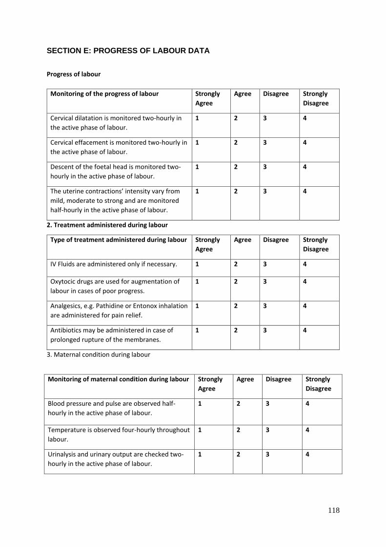
118
SECTION E: PROGRESS OF LABOUR DATA
Progress of labour
2. Treatment administered during labour
3. Maternal condition during labour
Monitoring of the progress of labour Strongly
Agree
Agree Disagree Strongly
Disagree
Cervical dilatation is monitored two-hourly in
the active phase of labour.
1 2 3 4
Cervical effacement is monitored two-hourly in
the active phase of labour.
1 2 3 4
Descent of the foetal head is monitored two-
hourly in the active phase of labour.
1 2 3 4
The uterine contractions’ intensity vary from
mild, moderate to strong and are monitored
half-hourly in the active phase of labour.
1 2 3 4
Type of treatment administered during labour Strongly
Agree
Agree Disagree Strongly
Disagree
IV Fluids are administered only if necessary. 1 2 3 4
Oxytocic drugs are used for augmentation of
labour in cases of poor progress.
1 2 3 4
Analgesics, e.g. Pathidine or Entonox inhalation
are administered for pain relief.
1 2 3 4
Antibiotics may be administered in case of
prolonged rupture of the membranes.
1 2 3 4
Monitoring of maternal condition during labour Strongly
Agree
Agree Disagree Strongly
Disagree
Blood pressure and pulse are observed half-
hourly in the active phase of labour.
1 2 3 4
Temperature is observed four-hourly throughout
labour.
1 2 3 4
Urinalysis and urinary output are checked two-
hourly in the active phase of labour.
1 2 3 4

119
SECTION F: FACTORS INFLUENCING THE USE OF A PARTOGRAPH BY MIDWIVES AND OBSTETRICIANS TO MONITOR PROGRESS OF LABOUR IN THE LABOUR WARD. 1.What are the factors that facilitate the use of a partograph to monitor the progress of labour in the labour ward? 1.1 List any factor: ……………………………………………………………………………………………………………………………………………………………………………………………………………………………………………………………………………………………………………………………………………………………………………………………… 1. What are the factors that hinder the use of a partograph in monitoring the progress
of labour in the labour ward?
1.1 List any factor: ………………………………………………………………………………………………………………………………………………………………………………………………………………………………………………………………………………………………………………………………………………………………………………………………………………………………………………………………………………………………

120
Annexure 4: Checklist for partograms for retrospective part of the study
According to Tessier (2003:89) there were eight essential principles of health care documentation, namely, unique identification accuracy,completeness, timeliness, confidentiality, accountability, legibility and resources. Tessier (2003:89) principle of completeness was used as criteria to be fulfilled for each variable to be considered as fully recorded. In this study completeness incorporated other Tessier’s principles of ensuring that all the information in the old partograms was written accurately, legible or clearly, timeously meaning that date and time was written. The principle of timeous in terms of proving that assessment and interventions were done in time could not be considered since this part of the study was retrospective in nature. In contract the information documented in the partograms that was classified as not recorded was found to be incomplete. As the information documented in those partograms was found to be either illegible, inaccurate and or date and time not written on audited partograms thus not meeting Tessier’s principle of completeness as used in this study. SECTION A: IDENTIFICATION OF WOMAN IN LABOUR
1. Data relating to identification of woman in labour (Indicate by a tick or X where applicable)
Data relating to state of the membranes on admission
Recorded, Not recorded
Woman in labour's name and surname
Completely
Incomplete
Woman in labour's registration number
Completely Incomplete
Woman in labour's age
Completely Incomplete
Gravida Completely Incomplete
Parity Completely
Recorded Not recorded
State of the membranes on admission
Completely
Incomplete
Date of rupture of the membranes
Completely Incomplete
Time of rupture of the membranes
Completely Incomplete
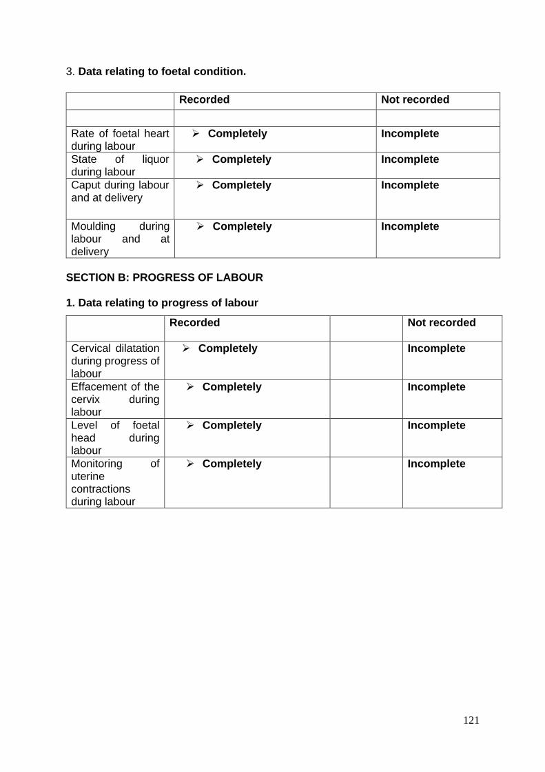
121
3. Data relating to foetal condition.
SECTION B: PROGRESS OF LABOUR 1. Data relating to progress of labour
Recorded Not recorded
Rate of foetal heart during labour
Completely
Incomplete
State of liquor during labour
Completely Incomplete
Caput during labour and at delivery
Completely Incomplete
Moulding during labour and at delivery
Completely Incomplete
Recorded
Not recorded
Cervical dilatation during progress of labour
Completely
Incomplete
Effacement of the cervix during labour
Completely
Incomplete
Level of foetal head during labour
Completely
Incomplete
Monitoring of uterine contractions during labour
Completely
Incomplete
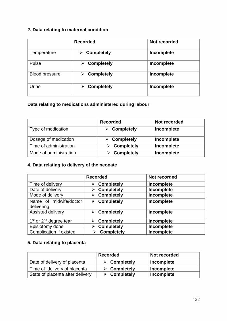
122
2. Data relating to maternal condition
Data relating to medications administered during labour
4. Data relating to delivery of the neonate
5. Data relating to placenta
Recorded
Not recorded
Temperature Completely
Incomplete
Pulse Completely
Incomplete
Blood pressure Completely
Incomplete
Urine Completely
Incomplete
Recorded Not recorded
Type of medication Completely Incomplete
Dosage of medication Completely Incomplete
Time of administration Completely Incomplete
Mode of administration Completely Incomplete
Recorded Not recorded
Time of delivery Completely Incomplete
Date of delivery Completely Incomplete
Mode of delivery Completely Incomplete
Name of midwife/doctor delivering
Completely Incomplete
Assisted delivery Completely Incomplete
1st or 2nd degree tear Completely Incomplete
Episiotomy done Completely Incomplete
Complication if existed Completely Incomplete
Recorded Not recorded
Date of delivery of placenta Completely Incomplete
Time of delivery of placenta Completely Incomplete
State of placenta after delivery Completely Incomplete

123
6. Data relating to condition of neonate at birth
Recorded Not recorded
Born alive Completely Incomplete
Apgar score at 1 min Completely Incomplete
Apgar score at 5 min Completely Incomplete
Birth weight Completely Incomplete
Gender Completely Incomplete
Quick examination of newborn done Completely Incomplete

124
SECTION B: Format of the obstetric/ gyne: ANC RECORDS

125
SECTION C: Format of the Obstetric/ Gyne: LABOUR RECORDS

126
Annexure 5: Request for permission to conduct research study
Faculty of Health Science
School of Nursing Howard College, 4000 08 June 2010
The Chief Executive Officer Prince Mshiyeni Memorial Hospital Private Bag X07 Mobeni 4060 Dear Sir or Madam, A REQUEST FOR PERMISSION TO CONDUCT A RESEARCH STUDY I, Lungile Audrey Msomi, employed at the abovementioned institution, wish to request permission to conduct a research study at this institution. I am a student registered for: Course-work Master's Degree in Maternal and Child Health at the University of KwaZulu Natal. I intend doing a research project in fulfillment of the academic requirement as indicated above. The research topic is: "Exploring knowledge, attitudes and utilization of the partograph by midwives and obstetricians in a regional hospital in the eThekwini District. The purpose of the study is to explore and describe the knowledge, attitudes and utilization of the partograph by midwives and obstetricians in the labour ward. Copies of the research proposal, the letter requesting permission to conduct research from the KwaZulu-Natal Department of Health, ethical approval from the University of KwaZulu-Natal research ethics' committee and the information document to respondents will be enclosed for your consideration. Your kind consideration of my request will be highly appreciated. Yours faithfully L. A. Msomi. Cell phone no.: 0824754373 CC: The Ethics Committee University of Kwa-Zulu Natal. Research Supervisor: Mrs S. Majeke. University of Kwa-Zulu Natal Howard College Campus, School of Nursing

127
Annexure 6: Request for permission to conduct a research study
Faculty of Health Sciences School of Nursing Howard College 08 June 2010
The Head of Department KwaZulu - Natal Department of Health Private Bag 95051 4000 Dear Sir or Madam, REQUEST FOR PERMISSION TO CONDUCT A RESEARCH STUDY I, Lungile Audrey Msomi, employed at the abovementioned institution, wish to request permission to conduct a research study at this institution. I am a student registered for: Course-work Master's Degree in Maternal and Child Health at the University of KwaZulu Natal. I intend doing a research project in fulfillment of the academic requirement as indicated above. The research topic is: "Exploring knowledge, attitudes and utilization of the partograph by midwives and obstetricians in a regional hospital in the eThekwini District. The purpose of the study is to explore and describe the knowledge, attitudes and utilization of the partograph by midwives and obstetricians in the labour ward. Copies of the research proposal, the letter requesting permission to conduct research from the KwaZulu-Natal Department of Health, ethical approval from the University of KwaZulu-Natal research ethics' committee and the information document to respondents will be enclosed for your consideration. Your kind consideration of my request will be highly appreciated. Yours faithfully L. A. Msomi. Cell phone no.: 0824754373 CC: The Ethics Committee University of Kwa-Zulu Natal. Research Supervisor: Mrs S. Majeke University of Kwa-Zulu Natal Howard College Campus, School of Nursing.

128
Annexure 7: Request for permission to conduct a research study
Faculty of Health Science
School of Nursing Howard College, 4000
08 June 2010 The Chief Executive Officer Prince Mshiyeni Memorial Hospital Private Bag X07 Mobeni 4060 Dear Sir or Madam, A REQUEST FOR PERMISSION TO CONDUCT A RESEARCH STUDY I, Lungile Audrey Msomi, employed at the abovementioned institution, wish to request permission to conduct a research study at this institution. I am a student registered for: Course-work Master's Degree in Maternal and Child Health at the University of KwaZulu Natal. I intend doing a research project in fulfillment of the academic requirement as indicated above. The research topic is: "Exploring knowledge, attitudes and utilization of the partograph by midwives and obstetricians in a regional hospital in the eThekwini District. The purpose of the study is to explore and describe the knowledge, attitudes and utilization of the partograph by midwives and obstetricians in the labour ward. Copies of the research proposal, the letter requesting permission to conduct research from the KwaZulu-Natal Department of Health, ethical approval from the University of KwaZulu-Natal research ethics' committee and the information document to respondents will be enclosed for your consideration. Your kind consideration of my request will be highly appreciated. Yours faithfully L. A. Msomi. Cell phone no.: 0824754373 CC: The Ethics Committee University of Kwa-Zulu Natal. Research Supervisor: Mrs S. Majeke. University of Kwa-Zulu Natal Howard College Campus, School of Nursing.

129
Annexure 8: Letter of approval from DoH

130
Annexure 9: Letter of approval from PMMH

131
Annexure 10: UKZN Ethical Clearance