Embed Size (px)
Citation preview
HAL Id: hal-03379698https://hal-cnrs.archives-ouvertes.fr/hal-03379698
Submitted on 15 Oct 2021
HAL is a multi-disciplinary open accessarchive for the deposit and dissemination of sci-entific research documents, whether they are pub-lished or not. The documents may come fromteaching and research institutions in France orabroad, or from public or private research centers.
L’archive ouverte pluridisciplinaire HAL, estdestinée au dépôt et à la diffusion de documentsscientifiques de niveau recherche, publiés ou non,émanant des établissements d’enseignement et derecherche français ou étrangers, des laboratoirespublics ou privés.
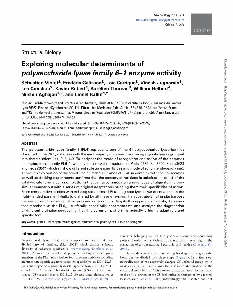
Exploring molecular determinants of polysaccharidelyase family 6–1 enzyme activity
Sébastien Violot, Frédéric Galisson, Loïc Carrique, Vinesh Jugnarain, LéaConchou, Xavier Robert, Aurélien Thureau, William Helbert, Nushin
Aghajari, Lionel Ballut
To cite this version:Sébastien Violot, Frédéric Galisson, Loïc Carrique, Vinesh Jugnarain, Léa Conchou, et al.. Exploringmolecular determinants of polysaccharide lyase family 6–1 enzyme activity. Glycobiology, OxfordUniversity Press (OUP), 2021, �10.1093/glycob/cwab073�. �hal-03379698�
1© The Author(s) 2021. Published by Oxford University Press. All rights reserved. For permissions, please e-mail: [email protected]
Glycobiology, 2021, 1–14https://doi.org/10.1093/glycob/cwab073
Original Article
Structural Biology
Exploring molecular determinants of
polysaccharide lyase family 6–1 enzyme activity
Sébastien Violot2, Frédéric Galisson2, Loïc Carrique2, Vinesh Jugnarain2,
Léa Conchou2, Xavier Robert2, Aurélien Thureau3, William Helbert4,
Nushin Aghajari1,2, and Lionel Ballut1,2
2Molecular Microbiology and Structural Biochemistry, UMR 5086, CNRS Université de Lyon, 7 passage du Vercors,Lyon 69367, France, 3Synchrotron SOLEIL, L’Orme des Merisiers, Saint Aubin, BP 48 91192 Gif-sur-Yvette, France,and 4Centre de Recherches sur les Macromolécules Végétales (CERMAV), CNRS and Grenoble Alpes Université,BP53, 38000 Grenoble Cedex 9, France1To whom correspondence should be addressed: Tel: +33-(0)4-72-72-26-34/+33-(0)4-72-72-26-33;Fax: +33-(0)4-72-72-26-04; e-mails: [email protected]; [email protected]
Received 15 April 2021; Revised 24 June 2021; Editorial Decision 6 July 2021; Accepted 7 July 2021
Abstract
The polysaccharide lyase family 6 (PL6) represents one of the 41 polysaccharide lyase families
classified in the CAZy database with the vast majority of its members being alginate lyases grouped
into three subfamilies, PL6_1–3. To decipher the mode of recognition and action of the enzymes
belonging to subfamily PL6_1, we solved the crystal structures of Pedsa0632, Patl3640, Pedsa3628
and Pedsa3807, which all show different substrate specificities and mode of action (endo-/exolyase).
Thorough exploration of the structures of Pedsa0632 and Patl3640 in complex with their substrates
as well as docking experiments confirms that the conserved residues in subsites −1 to +3 of the
catalytic site form a common platform that can accommodate various types of alginate in a very
similar manner but with a series of original adaptations bringing them their specificities of action.
From comparative studies with existing structures of PL6_1 alginate lyases, we observe that in the
right-handed parallel β-helix fold shared by all these enzymes, the substrate-binding site harbors
the same overall conserved structures and organization. Despite this apparent similarity, it appears
that members of the PL6_1 subfamily specifically accommodate and catalyze the degradation
of different alginates suggesting that this common platform is actually a highly adaptable and
specific tool.
Key words: protein–carbohydrates recognition, structure of alginate lyases, surface-binding site
Introduction
Polysaccharide lyases (PLs) are a group of enzymes (EC 4.2.2.-)divided into 41 families, (May 2021) which display a broaddiversity of substrate specificities (www.cazy.org; Lombard et al.2014). Among this variety of polysaccharide-specific enzymes,members of the PL6 family harbor four different activities includingmannuronate-specific alginate lyases (M-specific lyases, EC 4.2.2.3),guluronate-specific alginate lyases (G-specific lyases, EC 4.2.2.11),chondroitin B lyases (chondroitin sulfate (CS)- and dermatansulfate (DS)-specific lyases, EC 4.2.2.19) and oligo-alginate lyases(EC 4.2.2.26) (Garron and Cygler 2010; Mathieu et al. 2016).
Enzymes belonging to this family cleave uronic acid–containingpolysaccharides via a β-elimination mechanism resulting in theformation of an unsaturated hexuronic acid residue (Zhu and Yin2015).
The catalytic mechanism underlying breakage of the glycosidicbond can be divided into three steps (Figure 1). In a first step,neutralization of the negatively charged C6 carboxyl group by, inmost cases, a Ca2+ ion allows the resonance stabilization of theenolate thereby formed. This enolate formation causes the reductionof the pKa α-proton on the C5, facilitating its abstraction by a generalbase catalyst (Xu et al. 2017). Interestingly, this first step does not
Dow
nloaded from https://academ
ic.oup.com/glycob/advance-article/doi/10.1093/glycob/cw
ab073/6318796 by guest on 15 October 2021

2 S. Violot et al.
Fig. 1. Schematic illustrating the cleavage modes of endo- and exotype PL6: (A) poly α-L-guluronate, poly β-D-mannuronate and mixed poly α-L-guluronate
and poly β-D-mannuronate (B) β-elimination mechanism on a polyM substrate. The uronate neutralizer (Ca2+, Na2+ or water) is depicted as an orange sphere,
and acid and base as a red and blue sphere, respectively. (C) Endolyase activity observed in Pedsa0632. (D) Exolyase activity observed in Patl3640 and AlyGC.
Subsites are numbered from −1 to +3 and other potential subsites are shown in parentheses. Final products �, �GG or �GGG for Pedsa0632 and � for Patl3640
and AlyGC are underlined.
seem to be strictly Ca2+ dependent: In some enzymes, calcium couldbe replaced by other divalent cations such as Mn2+ or by a watermolecule (Mathieu et al. 2016; Lyu et al. 2019). In the last step, β-elimination of the 4-O-glycosidic bond results in the concomitantformation of an unsaturated C4-C5 bond within the hexuronic acidwhen a transfer of electrons from the carboxyl group occurs. Thisresults in the formation of an oligosaccharide with a 4-deoxy-l-erythro-hex-4-eno-pyranosyluronic acid at the nonreducing terminalend. During this very last step, the leaving group must be protonatedby a side chain acting as a general acid. Depending on the substrate,proton abstraction can occur either in a syn configuration, when theC5 proton and the glycosidic oxygen of the bond are situated on thesame side of the sugar ring, like for M/G-M bond–specific lyases, or inan anticonfiguration when the groups are placed on opposite sides ofthe sugar ring, as for G/M-G specific lyases and chondroitin B lyases(Lombard et al. 2010).
Except for chondroitin B lyases, other members of the PL6family are alginate lyases. Alginate is a linear polysaccharide andthe main constituent of the brown algae cell wall. It is composed ofmannuronic acid (β-d-mannuronate, M-residues) and its C5 epimerguluronic acid (α-l-guluronate, G-residues); both of which arrange
into different blocks of polyM (M-blocks), polyG (G-blocks) andpolyMG heteropolymer (MG-blocks) (Figure 1A) (Haug et al. 1967;Aarstad et al. 2012). In this context, PL6 family members harboractivities toward M-blocks, G-blocks, but also MG-blocks (Garronand Cygler 2010). Alginate lyases frequently have broad substratespecificity (Mathieu et al. 2016) and can cleave more than onetype of alginate (e.g. polyG plus polyM or polyG plus polyMG) (Liet al. 2019). The polysaccharide lyase family PL6 is subdivided intothree subfamilies, PL6_1, PL6_2 and PL6_3, according to sequencesimilarity (Mathieu et al. 2016). Chondroitin B lyases, polyG, polyMand polyMG alginate lyases are members of PL6_1. In contrasthereto, PL6_2 and PL6_3 contain essentially polyMG lyases.
Regarding 3D structures, PL6 members adopt the same right-handed parallel β-helix fold formed by three β-sheets. This wasfirst observed for the single-domain (one β-helix) chondroitin Blyase from Pedobacter heparinus DSM 2366 (PDB entry 1OFL) andmore recently within the single-domain AlyF from Vibrio splen-didus OU2 (PDB entries 5Z9T, 6ITG, 6A40 and 7BZ0), the single-domain BCelPL6 from Bacteroides cellulosilyticus CRE21 (PDBentry 6QPS) and the two-domain enzymes (two β-helices) AlyGCfrom Paraglaciecola chatamensis S18K6T (PDB entry 5GKQ) (Huang
Dow
nloaded from https://academ
ic.oup.com/glycob/advance-article/doi/10.1093/glycob/cw
ab073/6318796 by guest on 15 October 2021
Exploring molecular determinants of polysaccharide lyase family 6–1 enzyme activity 3
et al. 1999; Xu et al. 2017; Lyu et al. 2019; Stender et al. 2019; Zhanget al., 2021). On one side of the β-helix, a β-sheet forms a groove(chondroitinase B, AlyGC, BCelPL6) or a pocket (AlyF) delimitatingthe binding site in which at least six subsites can be described(subsites −3 to +3). Between subsites −1 and +1, two conservedresidues, respectively, a lysine and an arginine, are identified as thecatalytic residues in PL6 enzymes (Xu et al. 2017; Stender et al.2019). In addition, the Ca2+ ion located between subsites +1 and +2is substituted by a water molecule in AlyF, whereas a Ca2+ ion isbound in the G6 bound structure. The O-C4 glycosidic bond cleavagealways occurs between subsites −1 and +1 with two different typesof activity: i) endolyase activity (AlyF or BCelPL6) or ii) exolyaseactivity (AlyGC). Enzymes in PL6_1 display both endo- and exolyaseactivities as for Pedsa0632 from Pedobacter saltans or Mase04135from Alteromonas macleodii (Mathieu et al. 2016).
Altogether, PL6 family members interact with at least four dif-ferent substrates (M-blocks, G-blocks, MG-blocks and CS/DS) in aspecific manner despite sharing common fold and catalytic residues.This fact held together with their ability to cleave substrates, either inan endo- or exolyase mode, raises the questions of how these enzymesdiscriminate between substrates and how they cleave either in themiddle or at the nonreducing end of the polysaccharide chain.
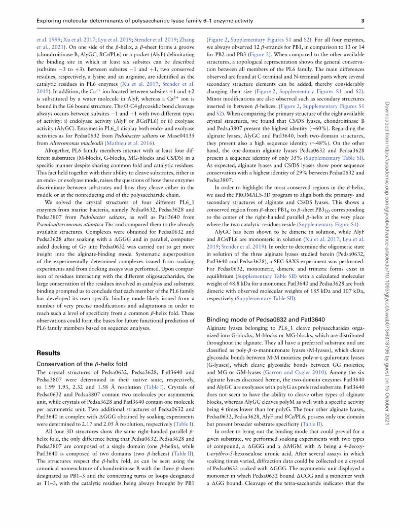
We solved the crystal structures of four different PL6_1enzymes from marine bacteria, namely Pedsa0632, Pedsa3628 andPedsa3807 from Pedobacter saltans, as well as Patl3640 fromPseudoalteromonas atlantica T6c and compared them to the alreadyavailable structures. Complexes were obtained for Pedsa0632 andPedsa3628 after soaking with a �GGG and in parallel, computer-aided docking of G7 into Pedsa0632 was carried out to get moreinsight into the alginate-binding mode. Systematic superpositionof the experimentally determined complexes issued from soakingexperiments and from docking assays was performed. Upon compar-ison of residues interacting with the different oligosaccharides, thelarge conservation of the residues involved in catalysis and substratebinding prompted us to conclude that each member of the PL6 familyhas developed its own specific binding mode likely issued from anumber of very precise modifications and adaptations in order toreach such a level of specificity from a common β-helix fold. Theseobservations could form the bases for future functional prediction ofPL6 family members based on sequence analyses.
Results
Conservation of the β-helix fold
The crystal structures of Pedsa0632, Pedsa3628, Patl3640 andPedsa3807 were determined in their native state, respectively,to 1.99 1.93, 2.32 and 1.58 Å resolution (Table I). Crystals ofPedsa0632 and Pedsa3807 contain two molecules per asymmetricunit, while crystals of Pedsa3628 and Patl3640 contain one moleculeper asymmetric unit. Two additional structures of Pedsa0632 andPatl3640 in complex with �GGG obtained by soaking experimentswere determined to 2.17 and 2.05 Å resolution, respectively (Table I).
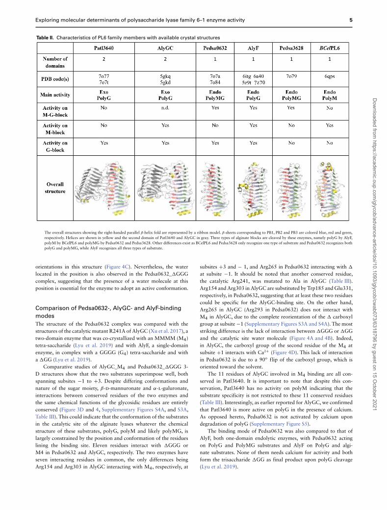
All four 3D structures show the same right-handed parallel β-helix fold, the only difference being that Pedsa0632, Pedsa3628 andPedsa3807 are composed of a single domain (one β-helix), whilePatl3640 is composed of two domains (two β-helices) (Table II).The structures respect the β-helix fold, as can be seen using thecanonical nomenclature of chondroitinase B with the three β-sheetsdesignated as PB1–3 and the connecting turns or loops designatedas T1–3, with the catalytic residues being always brought by PB1
(Figure 2, Supplementary Figures S1 and S2). For all four enzymes,we always observed 12 β-strands for PB1, in comparison to 13 or 14for PB2 and PB3 (Figure 2). When compared to the other availablestructures, a topological representation shows the general conserva-tion between all members of the PL6 family. The main differencesobserved are found at C-terminal and N-terminal parts where severalsecondary structure elements can be added, thereby considerablychanging their size (Figure 2, Supplementary Figures S1 and S2).Minor modifications are also observed such as secondary structuresinserted in between β-helices. (Figure 2, Supplementary Figures S1and S2). When comparing the primary structure of the eight availablecrystal structures, we found that CS/DS lyases, chondroitinase Band Pedsa3807 present the highest identity (∼60%). Regarding thealginate lyases, AlyGC and Patl3640, both two-domain structures,they present also a high sequence identity (∼48%). On the otherhand, the one-domain alginate lyases Pedsa0632 and Pedsa3628present a sequence identity of only 35% (Supplementary Table SI).As expected, alginate lyases and CS/DS lyases show poor sequenceconservation with a highest identity of 29% between Pedsa0632 andPedsa3807.
In order to highlight the most conserved regions in the β-helix,we used the PROMALS-3D program to align both the primary- andsecondary structures of alginate and CS/DS lyases. This shows aconserved region from β-sheet PB16 to β-sheet PB310 correspondingto the center of the right-handed parallel β-helix at the very placewhere the two catalytic residues reside (Supplementary Figure S1).
AlyGC has been shown to be dimeric in solution, while AlyFand BCelPL6 are monomeric in solution (Xu et al. 2017; Lyu et al.2019; Stender et al. 2019). In order to determine the oligomeric statein solution of the three alginate lyases studied herein (Pedsa0632,Patl3640 and Pedsa3628), a SEC-SAXS experiment was performed.For Pedsa0632, monomeric, dimeric and trimeric forms exist inequilibrium (Supplementary Table SII) with a calculated molecularweight of 48.8 kDa for a monomer. Patl3640 and Pedsa3628 are bothdimeric with observed molecular weights of 185 kDa and 107 kDa,respectively (Supplementary Table SII).
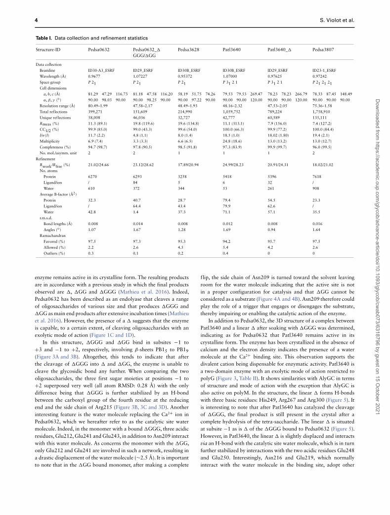
Binding mode of Pedsa0632 and Patl3640
Alginate lyases belonging to PL6_1 cleave polysaccharides orga-nized into G-blocks, M-blocks or MG-blocks, which are distributedthroughout the alginate. They all have a preferred substrate and areclassified as poly-β-d-mannuronate lyases (M-lyases), which cleaveglycosidic bonds between M-M moieties; poly-α-l-guluronate lyases(G-lyases), which cleave glycosidic bonds between GG moieties;and MG or GM-lyases (Garron and Cygler 2010). Among the sixalginate lyases discussed herein, the two-domain enzymes Patl3640and AlyGC are exolyases with polyG as preferred substrate. Patl3640does not seem to have the ability to cleave other types of alginateblocks, whereas AlyGC cleaves polyM as well with a specific activitybeing 4 times lower than for polyG. The four other alginate lyases,Pedsa0632, Pedsa3628, AlyF and BCelPL6, possess only one domainbut present broader substrate specificity (Table II).
In order to bring out the binding mode that could prevail for agiven substrate, we performed soaking experiments with two typesof compound, a �GGG and a �MGM with � being a 4-deoxy-l-erythro-5-hexoseulose uronic acid. After several assays in whichsoaking times varied, diffraction data could be collected on a crystalof Pedsa0632 soaked with �GGG. The asymmetric unit displayed amonomer in which Pedsa0632 bound �GGG and a monomer witha �GG bound. Cleavage of the tetra-saccharide indicates that the
Dow
nloaded from https://academ
ic.oup.com/glycob/advance-article/doi/10.1093/glycob/cw
ab073/6318796 by guest on 15 October 2021
4 S. Violot et al.
Table I. Data collection and refinement statistics
Structure-ID Pedsa0632 Pedsa0632_�
GGG/�GGPedsa3628 Patl3640 Patl3640_� Pedsa3807
Data collection
Beamline ID30-A3_ESRF ID29_ESRF ID30B_ESRF ID30B_ESRF ID29_ESRF ID23-1_ESRF
Wavelength (Å) 0.9677 1.07227 0.95372 1.07000 0.97625 0.97242
Space group P 21 P 21 P 21 P 31 2 1 P 31 2 1 P 21 21 21Cell dimensions
a, b, c (Å) 81.29 47.29 116.73 81.18 47.58 116.20 58.19 51.75 74.26 79.53 79.53 269.47 78.23 78.23 266.79 78.33 87.45 148.49
α, β, γ (◦) 90.00 98.03 90.00 90.00 98.25 90.00 90.00 97.22 90.00 90.00 90.00 120.00 90.00 90.00 120.00 90.00 90.00 90.00
Resolution range (Å) 80.49–1.99 47.58–2.17 48.49–1.93 48.16–2.32 47.53–2.05 75.36–1.58
Total reflections 399,271 151,609 214,990 1,059,752 789,224 1,758,910
Unique reflections 58,008 46,036 32,727 42,777 60,589 135,111
Rmeas (%) 11.3 (89.1) 19.8 (119.6) 19.6 (134.8) 11.1 (313.1) 7.9 (156.0) 7.4 (127.2)
CC1/2 (%) 99.9 (85.0) 99.0 (43.3) 99.6 (54.0) 100.0 (66.3) 99.9 (77.2) 100.0 (84.4)
I/σ (I) 11.7 (2.2) 4.8 (1.1) 8.0 (1.4) 18.5 (1.0) 18.02 (1.80) 19.4 (2.1)
Multiplicity 6.9 (7.4) 3.3 (3.3) 6.6 (6.5) 24.8 (18.6) 13.0 (13.2) 13.0 (12.7)
Completeness (%) 94.7 (98.7) 97.8 (90.5) 98.5 (91.8) 97.3 (83.9) 99.9 (99.7) 96.0 (99.5)
No. mol./asymm. unit 2 2 1 1 1 2
Refinement
Rwork/Rfree (%) 21.02/24.66 23.12/28.62 17.89/20.94 24.99/28.23 20.91/24.31 18.02/21.02
No. atoms
Protein 6270 6293 3258 5418 5396 7618
Ligand/ion / 84 5 6 32 /
Water 610 372 344 53 261 908
Average B-factor (Å2)
Protein 32.3 40.7 28.7 79.4 54.5 23.3
Ligand/ion / 64.4 43.4 79.9 62.6 /
Water 42.8 1.4 37.3 71.1 57.1 35.5
r.m.s.d.
Bond lengths (Å) 0.008 0.014 0.008 0.012 0.008 0.016
Angles (◦) 1.07 1.67 1.28 1.69 0.94 1.64
Ramachandran
Favored (%) 97.5 97.3 95.3 94.2 95.7 97.5
Allowed (%) 2.2 2.6 4.5 5.4 4.2 2.6
Outliers (%) 0.3 0.1 0.2 0.4 0 0
enzyme remains active in its crystalline form. The resulting productsare in accordance with a previous study in which the final productsobserved are �, �GG and �GGG (Mathieu et al. 2016). Indeed,Pedsa0632 has been described as an endolyase that cleaves a rangeof oligosaccharides of various size and that produces �GGG and�GG as main end products after extensive incubation times (Mathieuet al. 2016). However, the presence of a � suggests that the enzymeis capable, to a certain extent, of cleaving oligosaccharides with anexolytic mode of action (Figure 1C and 1D).
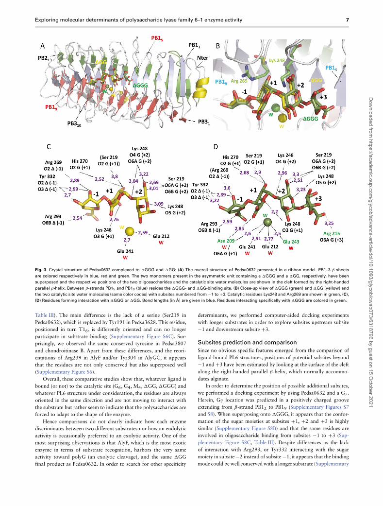
In this structure, �GGG and �GG bind in subsites −1 to+3 and −1 to +2, respectively, involving β-sheets PB15 to PB19(Figure 3A and 3B). Altogether, this tends to indicate that afterthe cleavage of �GGG into � and �GG, the enzyme is unable tocleave the glycosidic bond any further. When comparing the twooligosaccharides, the three first sugar moieties at positions −1 to+2 superposed very well (all atom RMSD: 0.28 Å) with the onlydifference being that �GGG is further stabilized by an H-bondbetween the carboxyl group of the fourth residue at the reducingend and the side chain of Arg215 (Figure 3B, 3C and 3D). Anotherinteresting feature is the water molecule replacing the Ca2+ ion inPedsa0632, which we hereafter refer to as the catalytic site watermolecule. Indeed, in the monomer with a bound �GGG, three acidicresidues, Glu212, Glu241 and Glu243, in addition to Asn209 interactwith this water molecule. As concerns the monomer with the �GG,only Glu212 and Glu241 are involved in such a network, resulting ina drastic displacement of the water molecule (∼2.5 Å). It is importantto note that in the �GG bound monomer, after making a complete
flip, the side chain of Asn209 is turned toward the solvent leavingroom for the water molecule indicating that the active site is notin a proper configuration for catalysis and that �GG cannot beconsidered as a substrate (Figure 4A and 4B). Asn209 therefore couldplay the role of a trigger that engages or disengages the substrate,thereby impairing or enabling the catalytic action of the enzyme.
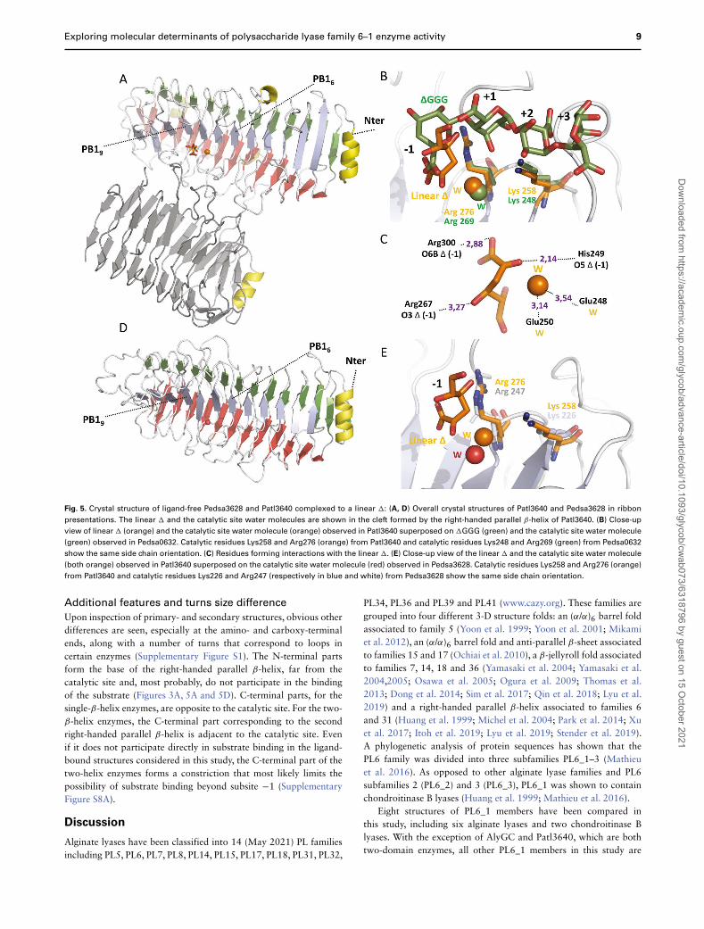
In addition to Pedsa0632, the 3D structure of a complex betweenPatl3640 and a linear � after soaking with �GGG was determined,indicating as for Pedsa0632 that Patl3640 remains active in itscrystalline form. The enzyme has been crystallized in the absence ofcalcium and the electron density indicates the presence of a watermolecule at the Ca2+ binding site. This observation supports thedivalent cation being dispensable for enzymatic activity. Patl3640 isa two-domain enzyme with an exolytic mode of action restricted topolyG (Figure 3, Table II). It shows similarities with AlyGC in termsof structure and mode of action with the exception that AlyGC isalso active on polyM. In the structure, the linear � forms H-bondswith three basic residues: His249, Arg267 and Arg300 (Figure 5). Itis interesting to note that after Patl3640 has catalyzed the cleavageof �GGG, the final product is still present in the crystal after acomplete hydrolysis of the tetra-saccharide. The linear � is situatedat subsite −1 as is � of the �GGG bound to Pedsa0632 (Figure 5).However, in Patl3640, the linear � is slightly displaced and interactsvia an H-bond with the catalytic site water molecule, which is in turnfurther stabilized by interactions with the two acidic residues Glu248and Glu250. Interestingly, Asn216 and Glu219, which normallyinteract with the water molecule in the binding site, adopt other
Dow
nloaded from https://academ
ic.oup.com/glycob/advance-article/doi/10.1093/glycob/cw
ab073/6318796 by guest on 15 October 2021
Exploring molecular determinants of polysaccharide lyase family 6–1 enzyme activity 5
Table II. Characteristics of PL6 family members with available crystal structures
The overall structures showing the right-handed parallel β-helix fold are represented by a ribbon model. β-sheets corresponding to PB1, PB2 and PB3 are colored blue, red and green,respectively. Helices are shown in yellow and the second domain of Patl3640 and AlyGC in gray. Three types of alginate blocks are cleaved by these enzymes, namely polyG by AlyF,polyM by BCelPL6 and polyMG by Pedsa0632 and Pedsa3628. Other differences exist as BCelPL6 and Pedsa3628 only recognize one type of substrate and Pedsa0632 recognizes bothpolyG and polyMG, while AlyF recognizes all three types of substrate.
orientations in this structure (Figure 4C). Nevertheless, the waterlocated in the position is also observed in the Pedsa0632_�GGGcomplex, suggesting that the presence of a water molecule at thisposition is essential for the enzyme to adopt an active conformation.
Comparison of Pedsa0632-, AlyGC- and AlyF-binding
modes
The structure of the Pedsa0632 complex was compared with thestructures of the catalytic mutant R241A of AlyGC (Xu et al. 2017), atwo-domain enzyme that was co-crystallized with an MMMM (M4)tetra-saccharide (Lyu et al. 2019) and with AlyF, a single-domainenzyme, in complex with a GGGG (G4) tetra-saccharide and witha �GG (Lyu et al. 2019).
Comparative studies of AlyGC_M4 and Pedsa0632_�GGG 3-D structures show that the two substrates superimpose well, bothspanning subsites −1 to +3. Despite differing conformations andnature of the sugar moiety, β-d-mannuronate and α-l-guluronate,interactions between conserved residues of the two enzymes andthe same chemical functions of the glycosidic residues are entirelyconserved (Figure 3D and 4, Supplementary Figures S4A, and S3A,Table III). This could indicate that the conformation of the substratesin the catalytic site of the alginate lyases whatever the chemicalstructure of these substrates, polyG, polyM and likely polyMG, islargely constrained by the position and conformation of the residueslining the binding site. Eleven residues interact with �GGG orM4 in Pedsa0632 and AlyGC, respectively. The two enzymes haveseven interacting residues in common, the only differences beingArg154 and Arg303 in AlyGC interacting with M4, respectively, at
subsites +3 and − 1, and Arg265 in Pedsa0632 interacting with �
at subsite −1. It should be noted that another conserved residue,the catalytic Arg241, was mutated to Ala in AlyGC (Table III).Arg154 and Arg303 in AlyGC are substituted by Trp185 and Glu331,respectively, in Pedsa0632, suggesting that at least these two residuescould be specific for the AlyGC-binding site. On the other hand,Arg265 in AlyGC (Arg293 in Pedsa0632) does not interact withM4 in AlyGC, due to the complete reorientation of the � carboxylgroup at subsite −1 (Supplementary Figures S3A and S4A). The moststriking difference is the lack of interaction between �GGG or �GGand the catalytic site water molecule (Figure 4A and 4B). Indeed,in AlyGC, the carboxyl group of the second residue of the M4 atsubsite +1 interacts with Ca2+ (Figure 4D). This lack of interactionin Pedsa0632 is due to a 90◦ flip of the carboxyl group, which isoriented toward the solvent.
The 11 residues of AlyGC involved in M4 binding are all con-served in Patl3640. It is important to note that despite this con-servation, Patl3640 has no activity on polyM indicating that thesubstrate specificity is not restricted to these 11 conserved residues(Table III). Interestingly, as earlier reported for AlyGC, we confirmedthat Patl3640 is more active on polyG in the presence of calcium.As opposed hereto, Pedsa0632 is not activated by calcium upondegradation of polyG (Supplementary Figure S5).
The binding mode of Pedsa0632 was also compared to that ofAlyF, both one-domain endolytic enzymes, with Pedsa0632 actingon PolyG and PolyMG substrates and AlyF on PolyG and algi-nate substrates. None of them needs calcium for activity and bothform the trisaccharide �GG as final product upon polyG cleavage(Lyu et al. 2019).
Dow
nloaded from https://academ
ic.oup.com/glycob/advance-article/doi/10.1093/glycob/cw
ab073/6318796 by guest on 15 October 2021
6 S. Violot et al.
Fig. 2. Topological representation of PL6 structures: relative spatial positions and orientations of the secondary structure elements of PL6 family members show
a right-handed parallel β-helix fold made of three β-sheets designated as PB1 (blue), PB2 (red) and PB3 (green). β-strands are numbered from 1 (e.g. PB11) to X
(e.g. PB1x), X being the last β-strand, with the exception of β-sheet PB2, which starts at PB20. Other β-strands (e.g. β41) are numbered relative to their position
in the primary structure. Sizes and relative positions of secondary structures (β-sheets and helices (yellow)) are respected. Turns between β-sheets designated
as T1–3 (black lines), are used as connectors between secondary structures without size consideration. (A) Pedsa0632, (B) Pedsa3628 and (C) Pedsa3807 are
single-domain PL6 family members. (D) Patl3640 is a two-domain PL6 family member. The second domain (gray) shows the same β-helix fold. Red stars indicate
catalytic residues. Secondary structures insertions: (A) In Pedsa0632, two helices (helix 218–222 and helix 312–316) are present in turn T16 and turn T310.(B) In
Pedsa3628, helix 394–398 is introduced in turn T112 and a small β-sheet composed of two β-strands (311–313, 319–321) is inserted in turn T110. (C) In Pedsa3807,
two helices (helix 101–105 and helix 352–364) are inserted within turn T33 and turn T111.
When superposing �GGG from Pedsa0632 on G4 from AlyF itcan be observed that both substrates span subsites −1 to +3, with G4being slightly translated by ∼1 Å toward subsite +3 (SupplementaryFigure S3B).
More strikingly, when comparing the �GG binding modes, trisac-charides bind to subsites −1 to +2 and subsites +1 to +3 forPedsa0632 and AlyF, respectively (Supplementary Figure S3C). AlyFinteracts with 17 (G6), 16 (G4) and 12 (�GG) residues, respec-tively, (Table III) in addition to the four residues interacting with thecatalytic site water (G4) or binding the catalytic site Na+ (�GG)(Figure 4E and 4F). As for AlyGC, in addition to these four aminoacid residues, a carboxyl group from ligands G4 or �GG interactswith the catalytic site water molecule or the catalytic site Na+,respectively (Figure 4E and 4F).
Residue conservation in the alginate-binding site
Analyses of crystal structures of the available PL6 enzymes com-plexed to a substrate reveal the existence of a common platformmade of 11 residues: two catalytic residues, five residues interacting
with the substrate and four residues interacting with the catalytic sitewater, Na+ or Ca2+. In AlyF, this platform displays a higher numberof interactions when binding G6, G4 or �GG (Table III). The sidechain orientation of the two catalytic residues in a given enzymesuperposes well with counterparts in all other enzymes, whether inligand-free or a ligand-bound form (Figures 3B, 5B and 5E, Supple-mentary Figure S3).
The same observation is made when considering the four residuesthat bind to Ca2+, Na+ or water in the catalytic site of the differentenzymes. Indeed, apart from noninteracting residues Asn209 in thestructure of Pedsa0632 complexed to a �GG, and Asn216 in thestructure of Patl3640 complexed to a linear �, each of the otherresidues is similarly positioned (Figure 4). Regarding the substrate-binding residues, Arg215, Ser219, His270, Arg293 and Tyr332 inPedsa0632 and their counterparts in the other enzymes (Table III),only slight differences can be spotted. Gln377 in AlyF and Ser248 inPedsa3628 replace the conserved tyrosine and histidine, respectively.However, these two residues are both able to form H-bonds asobserved in other enzymes (Supplementary Figure S6A and S6C,
Dow
nloaded from https://academ
ic.oup.com/glycob/advance-article/doi/10.1093/glycob/cw
ab073/6318796 by guest on 15 October 2021
Exploring molecular determinants of polysaccharide lyase family 6–1 enzyme activity 7
Fig. 3. Crystal structure of Pedsa0632 complexed to �GGG and �GG: (A) The overall structure of Pedsa0632 presented in a ribbon model. PB1–3 β-sheets
are colored respectively in blue, red and green. The two monomers present in the asymmetric unit containing a �GGG and a �GG, respectively, have been
superposed and the respective positions of the two oligosaccharides and the catalytic site water molecules are shown in the cleft formed by the right-handed
parallel β-helix. Between β-strands PB15 and PB19 (blue) resides the �GGG- and �GG-binding site. (B) Close-up view of �GGG (green) and �GG (yellow) and
the two catalytic site water molecules (same color codes) with subsites numbered from −1 to +3. Catalytic residues Lys248 and Arg269 are shown in green. (C),
(D) Residues forming interaction with �GGG or �GG. Bond lengths (in Å) are given in blue. Residues interacting specifically with �GGG are colored in green.
Table III). The main difference is the lack of a serine (Ser219 inPedsa0632), which is replaced by Tyr191 in Pedsa3628. This residue,positioned in turn T16, is differently oriented and can no longerparticipate in substrate binding (Supplementary Figure S6C). Sur-prisingly, we observed the same conserved tyrosine in Pedsa3807and chondroitinase B. Apart from these differences, and the reori-entations of Arg239 in AlyF and/or Tyr304 in AlyGC, it appearsthat the residues are not only conserved but also superposed well(Supplementary Figure S6).
Overall, these comparative studies show that, whatever ligand isbound (or not) to the catalytic site (G6, G4, M4, �GG, �GGG) andwhatever PL6 structure under consideration, the residues are alwaysoriented in the same direction and are not moving to interact withthe substrate but rather seem to indicate that the polysaccharides areforced to adapt to the shape of the enzyme.
Hence comparisons do not clearly indicate how each enzymediscriminates between two different substrates nor how an endolyticactivity is occasionally preferred to an exolytic activity. One of themost surprising observations is that AlyF, which is the most exoticenzyme in terms of substrate recognition, harbors the very sameactivity toward polyG (an exolytic cleavage), and the same �GGfinal product as Pedsa0632. In order to search for other specificity
determinants, we performed computer-aided docking experimentswith longer substrates in order to explore subsites upstream subsite−1 and downstream subsite +3.
Subsites prediction and comparison
Since no obvious specific features emerged from the comparison ofligand-bound PL6 structures, positions of potential subsites beyond−1 and +3 have been estimated by looking at the surface of the cleftalong the right-handed parallel β-helix, which normally accommo-dates alginate.
In order to determine the position of possible additional subsites,we performed a docking experiment by using Pedsa0632 and a G7.Herein, G7 location was predicted in a positively charged grooveextending from β-strand PB12 to PB19 (Supplementary Figures S7and S8). When superposing onto �GGG, it appears that the confor-mation of the sugar moieties at subsites +1, +2 and +3 is highlysimilar (Supplementary Figure S8B) and that the same residues areinvolved in oligosaccharide binding from subsites −1 to +3 (Sup-plementary Figure S8C, Table III). Despite differences as the lackof interaction with Arg293, or Tyr332 interacting with the sugarmoiety in subsite −2 instead of subsite −1, it appears that the bindingmode could be well conserved with a longer substrate (Supplementary
Dow
nloaded from https://academ
ic.oup.com/glycob/advance-article/doi/10.1093/glycob/cw
ab073/6318796 by guest on 15 October 2021
8 S. Violot et al.
Fig. 4. Close-up view of PL6 catalytic sites with residues involved in the binding of catalytic site water/Ca2+/Na+: (A) Pedsa0632 complexed to �GG. (B) Pedsa0632
complexed to �GGG. (C) Patl3640 complexed to a linear �. (D) AlyGC complexed to MMMM (pdb entry 5GKQ) (E) AlyF complexed to GGGG (pdb entry 6A40). (F)
AlyF complexed to �GG (pdb entry 5Z9T). For convenience, only the oligosaccharide residues present in subsites −1 and +1 are shown. (A, B) Arrows indicate
the position of the oligosaccharide residues carboxylic function turned toward the solvent. (D, E, F) Dotted arrows indicate the position of the oligosaccharide
residues carboxylic function interacting with Ca2+, water or Na+.
Figure S8C, Table III). Unexpectedly, only few additional interactionswere observed with G7 in +5 in which Lys123 (T33) and Asp125(PB13) interact with the sugar moiety at subsite +5, and no apparentinteractions in subsite +4 (Supplementary Figure S8C). Lys123 ispoorly conserved and is only present in Pedsa3628 (Lys88), which,surprisingly, does not show any activity toward polyG, thus sug-gesting that this basic residue could interact with polyMG as well(Supplementary Figure S1). Asp125 is strictly conserved in BCelPL6and functionally conserved in AlyGC and Patl3640 in which it issubstituted by a glutamate. Once again, this residue does not seemto be specific for PolyG activity as BCelPL6 only cleaves PolyM.
At the nonreducing end of the substrate, three new interactionsare found. In addition to Tyr332 (T110) mentioned above, Lys361(T111) and Arg362 (T111) may interact with the sugar moiety atsubsite −2 (Supplementary Figure S7C). Tyr332 is well conservedin all six alginate lyases from a functional point of view as it isreplaced by a glutamine only in AlyF. Interestingly, a histidine isfound in both chondroitinases (Supplementary Figure S1) therebysuggesting that the possibility of making electrostatic interactionsin subsite −2 is essential. This residue is found both in exo- andendolyases and can therefore not be considered as a determinantof one type of activity or the other. As opposed to Tyr332,Lys361 is poorly conserved and replaced by Glu332 and Glu367in AlyGC and Patl3640, respectively, and Gln358 in Pedsa3628.Based on primary and secondary structure alignment performedwith PROMALS3D, Lys361 is substituted by Arg363 in BCelPL6and by Gly416 in AlyF (Supplementary Figure S1). RegardingArg362, it is conserved in AlyGC (Arg333), Patl3640 (Arg368) andPedsa3628 (Arg359) and replaced by Gly364 in BCelPL6 and Ile417in AlyF.
Altogether, this first analysis seems to be not sufficient to appre-hend the molecular determinants of substrate recognition. To get a
better view of the roles of residues present in the binding cleft, wethoroughly inspected each enzyme structure and selected all residuesfor which the side chain is oriented in a manner that it can interactwith the substrate. Alignment of the selected residues spanning sub-sites −2 to +5 was performed (Supplementary Figure S9). A WebL-ogo representation (Crooks et al. 2004) confirmed the existence of aconserved platform of 13 residues hereof 10 being strictly conservedand the lack of conserved residues at subsites +4 and +5 (Figure 6).No common features observed in this study could be attributed tomolecular determinants of substrate specificity with certainty, and itseems that each enzyme has its own recognition mode regardless ofthe substrate specificity. Interestingly, this observation is confirmedby the recent structure of AlyF in complex with a G6 extending fromsubsite +3 to −3 (Table III; Zhang et al. 2021). Herein, the residuesinteracting at subsite −3 are not conserved in any other PL6 structuredescribed.
To further test the validity of this observation, the primarystructure of six additional enzymes (all PL6_1 members) was includedin the alignment. These two-domain enzymes display exolyase activ-ity on polyG and endolyase activity on polyMG. Once again, aWebLogo representation showed a good conservation of the commonplatform and very low conservation at subsites −2, +4 and +5. Ifconsidering the enzymes with polyG exolyase activity, an aromaticresidue (subsite +4) as well as an asparagine (subsite −1) seem to beconserved. However, residues at these two positions are also presentin AlyF, which has endolyase activity (Figure 6). As for enzymes withpolyMG endolyase activity, no obvious determinants of specificitycould be identified, and only a single specific threonine is shared bythe two enzymes having polyG endolyase activity. Finally, BCelPL6,which is the only enzyme with a polyM endolyase activity in thisstudy, possesses three residues that seem to be specific: a glutamine,a threonine and a serine (subsite +3 and − 1) (Figure 6).
Dow
nloaded from https://academ
ic.oup.com/glycob/advance-article/doi/10.1093/glycob/cw
ab073/6318796 by guest on 15 October 2021
Exploring molecular determinants of polysaccharide lyase family 6–1 enzyme activity 9
Fig. 5. Crystal structure of ligand-free Pedsa3628 and Patl3640 complexed to a linear �: (A, D) Overall crystal structures of Patl3640 and Pedsa3628 in ribbon
presentations. The linear � and the catalytic site water molecules are shown in the cleft formed by the right-handed parallel β-helix of Patl3640. (B) Close-up
view of linear � (orange) and the catalytic site water molecule (orange) observed in Patl3640 superposed on �GGG (green) and the catalytic site water molecule
(green) observed in Pedsa0632. Catalytic residues Lys258 and Arg276 (orange) from Patl3640 and catalytic residues Lys248 and Arg269 (green) from Pedsa0632
show the same side chain orientation. (C) Residues forming interactions with the linear �. (E) Close-up view of the linear � and the catalytic site water molecule
(both orange) observed in Patl3640 superposed on the catalytic site water molecule (red) observed in Pedsa3628. Catalytic residues Lys258 and Arg276 (orange)
from Patl3640 and catalytic residues Lys226 and Arg247 (respectively in blue and white) from Pedsa3628 show the same side chain orientation.
Additional features and turns size difference
Upon inspection of primary- and secondary structures, obvious otherdifferences are seen, especially at the amino- and carboxy-terminalends, along with a number of turns that correspond to loops incertain enzymes (Supplementary Figure S1). The N-terminal partsform the base of the right-handed parallel β-helix, far from thecatalytic site and, most probably, do not participate in the bindingof the substrate (Figures 3A, 5A and 5D). C-terminal parts, for thesingle-β-helix enzymes, are opposite to the catalytic site. For the two-β-helix enzymes, the C-terminal part corresponding to the secondright-handed parallel β-helix is adjacent to the catalytic site. Evenif it does not participate directly in substrate binding in the ligand-bound structures considered in this study, the C-terminal part of thetwo-helix enzymes forms a constriction that most likely limits thepossibility of substrate binding beyond subsite −1 (SupplementaryFigure S8A).
Discussion
Alginate lyases have been classified into 14 (May 2021) PL familiesincluding PL5, PL6, PL7, PL8, PL14, PL15, PL17, PL18, PL31, PL32,
PL34, PL36 and PL39 and PL41 (www.cazy.org). These families aregrouped into four different 3-D structure folds: an (α/α)6 barrel foldassociated to family 5 (Yoon et al. 1999; Yoon et al. 2001; Mikamiet al. 2012), an (α/α)6 barrel fold and anti-parallel β-sheet associatedto families 15 and 17 (Ochiai et al. 2010), a β-jellyroll fold associatedto families 7, 14, 18 and 36 (Yamasaki et al. 2004; Yamasaki et al.2004,2005; Osawa et al. 2005; Ogura et al. 2009; Thomas et al.2013; Dong et al. 2014; Sim et al. 2017; Qin et al. 2018; Lyu et al.2019) and a right-handed parallel β-helix associated to families 6and 31 (Huang et al. 1999; Michel et al. 2004; Park et al. 2014; Xuet al. 2017; Itoh et al. 2019; Lyu et al. 2019; Stender et al. 2019).A phylogenetic analysis of protein sequences has shown that thePL6 family was divided into three subfamilies PL6_1–3 (Mathieuet al. 2016). As opposed to other alginate lyase families and PL6subfamilies 2 (PL6_2) and 3 (PL6_3), PL6_1 was shown to containchondroitinase B lyases (Huang et al. 1999; Mathieu et al. 2016).
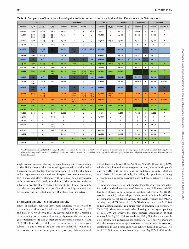
Eight structures of PL6_1 members have been compared inthis study, including six alginate lyases and two chondroitinase Blyases. With the exception of AlyGC and Patl3640, which are bothtwo-domain enzymes, all other PL6_1 members in this study are
Dow
nloaded from https://academ
ic.oup.com/glycob/advance-article/doi/10.1093/glycob/cw
ab073/6318796 by guest on 15 October 2021
10 S. Violot et al.
Table III. Comparison of interactions involving the residues present in the catalytic site of the different available PL6 structures
Catalytic residues are highlighted in orange. Residues involved in the binding to water/Ca2+ /Na+ present in the catalytic site are highlighted in blue (water: vertical hatching, Ca2+ :oblique hatching, Na+ : horizontal hatching). Conserved residues involved in the binding to an oligosaccharide are highlighted in blue and green. Residues not strictly conserved arehatched in green.
single-domain enzymes sharing the same binding site correspondingto the PB1 β-sheet of the conserved right-handed parallel β-helix.This catalytic site displays four subsites from −1 to +3 with a lysineand an arginine as catalytic residues. Despite these common features,PL6_1 members cleave alginates with an endo- or an exoactivity,with or without Ca2+ and, in addition to the respective preferredsubstrates, are also able to cleave other substrates like e.g. Pedsa0632that cleaves polyMG but also polyG with an endolyase activity, orAlyGC cleaving polyG but also polyM with an exolyase activity.
Endolyase activity vs. exolyase activity
Endo- or exolyase activities have been suggested to be related tothe number of domains (Stender et al. 2019). Indeed, for AlyGCand Patl3640, we observe that the second helix in the C-terminalcorresponding to the second domain partly covers the binding sitecorresponding to the PB1 β-sheet (Supplementary Figures S8A andS8B). This limits the possibility for the substrate to extend beyondsubsite −1 and seems to be also true for Pedsa0631, which is atwo-domain enzyme with exolyase activity on polyG (Mathieu et al.
2016). However, Mase04135, Patl3659, Nonul2381 and Celly0294,which are all two-domain enzymes as well, cleave both polyGand polyMG with an exo- and an endolyase activity (Mathieuet al. 2016). More surprisingly, FsAlyPL6, also predicted as beinga two-domain enzyme, possesses only endolyase activity (Li et al.2019).
Another characteristic that could potentially be an exolyase activ-ity marker is the dimeric state of these enzymes. Full-length AlyGChas been shown to be a dimer in solution, whereas a �CTD (C-terminal domain) variant adopts a monomer in solution. In addition,as compared to full-length AlyGC, the �CTD variant lost 94.3%activity toward PG (Xu et al. 2017). We demonstrated that Patl3640(a two-domain enzyme) is a dimer form in solution (SupplementaryTable SII). More interestingly, when looking at the crystal packingof Patl3640, we observe the same dimeric organization as thatobserved for AlyGC. Unfortunately, for FsAlyPL6, there is no avail-able information concerning its oligomeric state but we speculatethat this enzyme may adopt a monomeric organization in solution,explaining its unexpected endolyase activity. Regarding AlyGC (Xuet al. 2017), it was shown that a large loop (Arg627-His638) of the
Dow
nloaded from https://academ
ic.oup.com/glycob/advance-article/doi/10.1093/glycob/cw
ab073/6318796 by guest on 15 October 2021
Exploring molecular determinants of polysaccharide lyase family 6–1 enzyme activity 11
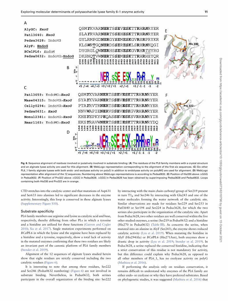
Fig. 6. Sequence alignment of residues involved or putatively involved in substrate binding: (A) The residues of the PL6 family members with a crystal structure
and an alginate lyase activity are used for this alignment. (B) WebLogo representation corresponding to the alignment of the first six sequences. (C) Six other
PL6_1 family alginate lyases with both endo- and exolyase activity on polyG in addition to endolyase activity on polyMG are used for alignment. (D) WebLogo
representation after alignment of the 12 sequences. Numbering above WebLogo representations is according to Pedsa0632. (E) Position of His330 above �GGG
in Pedsa0632. (F) Position of Pro322 above �GGG in Pedsa3628. �GGG in Pedsa3628 has been obtained by superimposing Pedsa3628 and Pedsa0632. Loops
containing both His330 and Pro322 are in orange.
CTD stretches into the catalytic center and that mutations of Asp631and Ser633 into alanines led to significant decreases in the enzymeactivity. Interestingly, this loop is conserved in these alginate lyases(Supplementary Figure S10).
Substrate specificity
PL6 family members use arginine and lysine as catalytic acid and base,respectively, thereby differing from other PLs in which a tyrosineand a histidine are utilized for these functions (Garron and Cygler2010; Xu et al. 2017). Single mutation experiments performed onBCelPL6 in which the lysine and the arginine have been replaced bya histidine and a tyrosine, respectively, show a total lack of activityin the mutated enzymes confirming that these two residues are likelyan invariant part of the canonic platform of PL6 family members(Stender et al. 2019).
Alignment of the 12 sequences of alginate lyases studied hereinshow that eight residues are strictly conserved including the twocatalytic residues (Figure 6).
It is interesting to note that among these residues, Ser222and Ser246 (Pedsa0632 numbering) (Figure 6) are not involved insubstrate binding. Nevertheless, in Pedsa0632, both serinesparticipate in the overall organization of the binding site: Ser222
by interacting with the main chain carbonyl group of Ser219 presentin turn T16 and Ser246 by interacting with Glu243 and one of thewater molecules forming the water network of the catalytic site.Similar observations are made for residues Ser229 and Ser253 inPatl3640 or Ser194 and Ser224 in Pedsa3628, for which the twoserines also participate in the organization of the catalytic site. Apartfrom Pedsa3628, two other residues are well conserved within the fiveother studied enzymes, a serine (Ser219 in Pedsa0632) and a histidine(His270 in Pedsa0632) (Table III). As concerns the serine, whenmutated into an alanine in AlyF (Ser243), the enzyme shows reducedcatalytic activity (Lyu et al. 2019). When mutating the histidine inAlyF (His294Ala) or BCelPL6 (His271Asn), both enzymes show adrastic drop in activity (Lyu et al. 2019; Stender et al. 2019). InPedsa3628, a serine replaced the conserved histidine, indicating thata strict conservation of this residue is not mandatory for activity,but this difference could explain why Pedsa3628, as opposed toall other members of PL6_1, has no exolyase activity on polyG(Mathieu et al. 2016).
If performing the analysis only on residues conservation, itremains difficult to understand why enzymes of the PL6 family areeither endo- or exolyase or why they have preferred substrates. Basedon phylogenetic studies, it was suggested (Mathieu et al. 2016) that
Dow
nloaded from https://academ
ic.oup.com/glycob/advance-article/doi/10.1093/glycob/cw
ab073/6318796 by guest on 15 October 2021
12 S. Violot et al.
PL6 family members could have evolved from strict endo-polyMGlyases to exo-polyG lyase via intermediates in which enzymes couldhave both endo- and exoactivities. Furthermore, they observed thatwhen looking at the end-products of polyMG lyases, a G residue isalways present at subsite +1, suggesting that the shift from MGto GG substrate moieties required remodeling of subsite −1 toaccommodate a G-residue instead of an M-residue.
The only strict endo-MG in PL6_1 is Pedsa3628. The otherenzymes, with the exception of BCelPL6, all have polyG as substratewith exo- or endoactivities (Figure 6). When focusing on subsite−1, it can be observed that the hydrophobic Pro322 in Pedsa3628is systematically replaced by a polar residue in the other enzymes(Figure 6). These polar residues do not directly interact with thesubstrate at subsite −1 but are part of a conserved loop that capsthe catalytic site as illustrated by His330 in Pedsa0632 (Figure 6E).In the presence of proline, this loop in Pedsa3628 does not cover thecatalytic site anymore (Figure 6F).
Despite a thorough examination of the sequences and 3D struc-tures available, only a few links between a given activity and a givengroup of residues or structural features have been established. Alto-gether, we suggest that from a common ancestral structure, severalsolutions have been found to adapt to the immediate environment.We conclude that, except for the enzymes showing a high degreeof similarity like AlyGC and Patl3640, prediction of a substratespecificity based on the sequence comparison alone seems, at thispoint, precarious. In order to decipher how the PL6 family membersspecifically cleave their substrates, additional structure–activity stud-ies are needed.
Materials and methods
Sequence alignments
Alignment profiles were generated using ESPript (ESPript - http://espript.ibcp.fr; Gouet et al. 2003). Primary- and secondary structurealignments were performed with PROMALS3D (Pei et al. 2008).
Expression and purification
The plasmids carrying the different constructs were transformed intoEscherichia coli BL21-CodonPlus (DE3)-RIL cells. A single colonywas inoculated into 10 mL of Luria Broth containing 25 μg mL−1
of kanamycin and grown overnight at 37◦C. The overnight culturewas added to 2 L of LB with antibiotics and grown at 37◦C untilthe OD600 reached ∼0.6, hereafter induced with 0.2 mM IPTG(isopropyl-β-d-thiogalactopyranoside) and grown for further 18 hat 20◦C. The cells were pelleted by centrifugation at 5000g, 20 minat 20◦C and stored at −20◦C.
The pellet was resuspended in lysis buffer (20 mM Tris–HClpH 8.0, 150 mM NaCl - buffer A) and disrupted using a microflu-idizer (3 cycles at 15,000 psi). The lysate was centrifuged at 10,000g,30 min at 4◦C and the supernatant was filtered using a 0.45 μmcutoff filter before injection onto a 5 mL HisTrap FF crude using anÄKTA purifier (GE Healthcare). The column was washed with BufferA containing 2 M NaCl followed by an elution in buffer A containing500 mM imidazole. After 12% SDS-PAGE (SDS-polyacrylamidegel electrophoresis) analysis, fractions with relatively pure proteinwere pooled and concentrated (appropriated cutoff Amicon Ultra-15centrifugal filter units (EMD Millipore)). Further purification by size-exclusion chromatography using a Superdex 200 10/300 GL column(GE Healthcare) was done in buffer A. Aliquots of the purifiedprotein were liquid nitrogen flash frozen and stored at −80◦C.
Crystallization
Crystallization conditions screening was carried out at 292 K (vapor-diffusion in sitting-drops), using commercially available crystalliza-tion kits. For screening, a Mosquito® crystallization robot (SPTLabtech Ltd.) was employed using two protein/crystallization agentratios (200 nL + 200 nL and 300 nL + 100 nL drops equilibratedagainst 70 μL in MRC Crystallization Plates (Molecular Dimen-sions)). Proteins were concentrated to 10–40 mg mL−1 in 20 mMTris–HCl pH 8.0, 150 mM NaCl buffer. Once the crystallizationconditions were established, a scale-up was performed in hangingdrops mixing 2 μL protein solution with 2 μL reservoir solution(or 3 μL protein and 1 μL reservoir solution) equilibrated against500 μL reservoir solution in 24-well plates. Crystals of Pedsa0632grew in 0.2 M K sulfate, 20% (w/v) PEG 3350, while complexeswere obtained in 0.2 M ammonium chloride, 20% (w/v) PEG 3350by co-crystallizing the protein with 10 mM of �GGG. Crystals ofPedsa3628 were obtained in 0.2 M K phosphate, 20% (w/v) PEG3350. Crystals of Patl3640 complexed with � were obtained in 0.1 MNa citrate pH 5.6, 2.0 M ammonium sulfate by co-crystallizing theprotein with 10 mM of �GGG. �MGM and �GGG were obtainedby enzymatic degradation of PolyG and PolyMG by Pedsa0632. Theywere purified on a semipreparative size-exclusion chromatographysystem and the structures were confirmed by NMR (Mathieu et al.2016). The ligand-free form was crystallized in 0.1 M Mg acetatepH 5.6, 0.1 M Na nitrate, 8% (w/v) PEG 10000, 0.15 mM CYMAL-7. As concerns Pedsa3807, the crystallization conditions were 0.2 Mtri-Li citrate, 20% (w/v) PEG 3350. Crystals were cryoprotected byadding 15% (v/v) ethylene glycol to initial conditions.
Data collection, structure determination and
refinement
X-ray diffraction data were collected at 100 K at the European Syn-chrotron Radiation Facility (Table I). Data were integrated and scaledwith XDS (Kabsch 2010). Data collection statistics are compiled inTable I.
The crystal structures were solved by molecular replacement withthe program Phaser (Grayling 2014), using the structure of AlyF (PDBentry 6ITG, Lyu et al. 2019), for Pedsa0632 and Pedsa3628, using thestructure of AlyGC (PDB entry 5GKD, Xu et al. 2017) for Patl3640and using the structure of chondroitinase B (PDB entry 1OFM,Michel et al. 2004) for Pedsa3807 as starting models. Cycles ofmaximum-likelihood refinement using the program “phenix.refine”and keeping apart 5% of the reflections for cross-validation, wereinterspersed with manual corrections of the models using COOT(Emsley et al. 2010). Refinement statistics are presented in Table I.
Molecular docking
Docking of substrates into the PL crystal structures were performedusing AutoDock Vina embedded in PyRx 0.8 (Trott and Olson 2010;Dallakyan and Olson 2015).
Briefly, the protein structures were prepared for docking byremoving unwanted water molecules and bound ligands and byadding polar hydrogens atoms using Discovery Studio Visualizer. Thesame program was used to build the substrates molecules as PDBfiles and for energy minimization. PyRx was used for convertingall molecules to AutoDock Ligand format (PDBQT). The 3D gridbox for molecular docking simulation was obtained using Autodocktools. The Grid box was centered to cover the active site and allessential residues. The docking results were analyzed by comparing
Dow
nloaded from https://academ
ic.oup.com/glycob/advance-article/doi/10.1093/glycob/cw
ab073/6318796 by guest on 15 October 2021
Exploring molecular determinants of polysaccharide lyase family 6–1 enzyme activity 13
the binding interactions and binding energies between substratemolecules and PL enzymes.
Small angle X-ray scattering
Protein samples were centrifuged prior to measuring their concentra-tion with a Nanodrop spectrophotometer (Thermo). SAXS data werecollected in line at the beamline SWING at the synchrotron SOLEIL(David and Perez 2009) after elution on an analytical BioSec-3 (3 mmparticle size, 300 Å pore size) column from Agilent, maintained at288 K, equilibrated in 50 mM Tris–HCl pH 8.0, 150 mM NaCl andoperated at a flow rate of 0.3 mL min−1. Individual SAXS framesof 990 ms were collected at a sample-to-detector distance of 2 m,accessing a q range of 0.007 to 0.5 Å (λ = 1.03 Å). All frameswere normalized to the intensity of the transmitted beam, radiallyaveraged and background-subtracted using the program Foxtrot.Data were then processed using the HPLC-SAXS module of US-SOMO (Brookes et al. 2016). Then, data were analyzed using theATSAS 3.0.3 software suite (Manalastas-Cantos et al. 2021). P(r)functions were computed from the scattering curves by an indirecttransform method in GNOM (Svergun 1992).
Degradation kinetics
Alginate substrates PolyG (FG = 0.95, DPn = 20) were prepared fromLaminaria hyperborea according to Haug et al (Haug et al. 1967).Enzymatic assays were carried out by incubating 150 μL of alginate(0.2% w/v in 50 mM Tris–HCl pH 8, with or without 0.2 mM CaCl2)with 10 μM of purified enzyme at 25◦C. The production of reducingends was measured using the ferricyanide method at different timesof the reaction. Aliquots of 40 μL were transferred to a 200 μLferricyanide solution (4.55 mM K3[Fe(CN)6], 225 mM Na2CO3,5 mM NaOH), which stopped the enzymatic reaction. The solutionwas heated to 100◦C for 10 min and, after cooling, the absorbance of100 μL of sample was measured at 415 nm with a microplate reader(TECAN M200).
Comparative studies of 3D structures
Crystal structures were compared with existing structures in theProtein Data Bank at Rutgers, RCSB, using the DALI server.
Figure rendering
Figures of 3D structures were drawn with PyMol (Schrödinger, http://pymol.org).
Data availability
Coordinates and structure factors have been deposited in theProtein Data Bank under accession codes 7O79 (Pedsa3628),7O78 (Pedsa3807), 7O7A (Pedsa0632), 7O77 (Patl3640), 7O84(Pedsa0632/�GGG) and 7O7T (Patl3640/�), respectively. Otherdata are available from the corresponding authors upon reasonablerequest.
Supplementary data
Supplementary data for this article are available at Glycobiologyonline.
Acknowledgments
Technical support from staff on beamlines MX and FIP (both EuropeanSynchrotron Radiation Facility), as well as on beamlines PX3 (Swiss Light
Source, Switzerland) and SWING (Soleil, France) is gratefully acknowledged.We are also grateful for assistance from V. Gueguen-Chaignon and F. Delolmeof the Protein Science Facility of SFR Biosciences Lyon (UAR3444/US8).
Author contributions
Conceptualization L.B., S.V.; formal analysis, L.B., S.V.; investigationS.V., L.B., F.G., L.Ca., A.T., X.R., V.J., L.Co.; supervision, L.B., N.A.,S.V.; validation L.B., S.V., N.A., W.H.; writing L.B., S.V., N.A.
Conflict of interest statement
The authors declare no competing interests.
Abbreviations
CS, chondroitin sulfate; CTD, C-terminal domain; �, 4-deoxy-l-erythro-hex-4-eno-pyranosyluronic acid; DS, dermatan sulfate;G, guluronate; HPLC, high-performance liquid chromatography;IPTG, isopropyl-β-d-thiogalactopyranoside; M, mannuronate; PEG,polyethylene glycol; PL, polysaccharide lyase; SAXS, small angle X-ray scattering; SDS-PAGE, SDS-polyacrylamide gel electrophoresis;Tris, tris(hydroxymethyl)aminomethane.
References
Aarstad OA, Tøndervik A, Sletta H, Skjåk-Bræk G. 2012. Alginate sequencing:An analysis of block distribution in alginates using specific alginatedegrading enzymes. Biomacromolecules. 13(1):106–116.
Brookes E, Vachette P, Rocco M, Pérez J. 2016. US-SOMO HPLC-SAXSmodule: Dealing with capillary fouling, and extraction of pure com-ponent patterns from poorly resolved SEC-SAXS data. J Appl Cryst.49(5):1827–1841.
Crooks GE, Hon G, Chandonia JM, Brenner SE. 2004. WebLogo: A sequencelogo generator. Genome Res. 14(6):1188–1190.
Dallakyan S, Olson AJ. 2015. Small-molecule library screening by docking withPyRx. Methods Mol. 1263:243–50.
David G, Perez J. 2009. Combined sampler robot and high-performance liquidchromatography: A fully automated system for biological small-angle X-ray scattering experiments at the Synchrotron SOLEIL SWING beamline.J Appl Cryst. 42(5):892–900.
Dong S, Wei TD, Chen XL, Li CY, Wang P, Xie BB, Qin QL, Zhang XZ,Pang XH, Zhou BC, et al. 2014. Molecular insight into the role of the N-terminal extension in the maturation, substrate recognition, and catalysisof a bacterial alginate lyase from polysaccharide lyase family 18. J BiolChem. 289(43):29558–29569.
Emsley P, Lohkamp B, Scott WG, Cowtan K. 2010. Features and developmentof Coot. Acta Crystallogr D Biol Crystallogr. 66(4):486–501.
Garron ML, Cygler M. 2010. Structural and mechanistic classifica-tion of uronic acid-containing polysaccharide lyases. Glycobiology.20(12):1547–1573.
Gouet P, Robert X, Courcelle E. 2003. ESPript/ENDscript: Extracting and ren-dering sequence and 3D information from atomic structures of proteins.Nucl. Acids Res. 31(13):3320–3323.
Grayling MJ. 2014. phaseR: An R package for phase plane analysis ofautonomous ODE systems. The R Journal. 6(2):43–51.
Haug A, Larsen B, Smidsrod O. 1967. Studies on sequence of uronic acidresidues in alginic acid. Acta Chem Scand. 21:691–704.
Huang W, Matte A, Li Y, Kim YS, Linhardt RJ, Su H, Cygler M. 1999. Crystalstructure of chondroitinase B from Flavobacterium Heparinum and itscomplex with a disaccharide product at 1.7 Å resolution. J Mol Biol.294(5):1257–1269.
Itoh T, Nakagawa E, Yoda M, Nakaichi A, Hibi T, Kimoto H. 2019. Structuraland biochemical characterisation of a novel alginate lyase from Paenibacil-lus sp. str. FPU-7. Sci Rep. 9(1):14870.
Dow
nloaded from https://academ
ic.oup.com/glycob/advance-article/doi/10.1093/glycob/cw
ab073/6318796 by guest on 15 October 2021
14 S. Violot et al.
Kabsch W. 2010. XDS. Acta Crystallogr D Biol Crystallogr. 66(2):125–132.Li Q, Hu F, Zhu B, Sun Y, Yao Z. 2019. Biochemical characterization and
elucidation of action pattern of a novel polysaccharide lyase 6 familyalginate lyase from marine bacterium Flammeovirga sp. NJ-04. MarDrugs. 17(6):323.
Lombard V, Bernard T, Rancurel C, Brumer H, Coutinho PM, Henrissat B.2010. A hierarchical classification of polysaccharide lyases for glycoge-nomics. Biochem J. 432(3):437–444.
Lombard V, Golaconda Ramulu H, Drula E, Coutinho PM, Henrissat B. 2014.The carbohydrate-active enzymes database (CAZy) in 2013. Nucleic AcidsRes. 42(Database issue):D490–D495.
Lyu Q, Zhang K, Shi Y, Li W, Diao X, Liu W. 2019. Structural insightsinto a novel Ca2+-independent PL-6 alginate lyase from Vibrio OU02identify the possible subsites responsible for product distribution. BiochimBiophys Acta Gen Subj. 1863(7):1167–1176.
Manalastas-Cantos K, Konarev PV, Hajizadeh NR, Kikhney AG, PetoukhovMV, Molodenskiy DS, Panjkovich A, Mertens HDT, Gruzinov A, BorgesC, et al. 2021. ATSAS 3.0: Expanded functionality and new tools for small-angle scattering data analysis. J Appl Cryst. 54(1):343–355.
Mathieu S, Henrissat B, Labre F, Skjåk-Bræk G, Helbert W. 2016. Func-tional exploration of the polysaccharide lyase family PL6. PLoS One.11(7):e0159415.
Michel G, Pojasek K, Li Y, Sulea T, Linhardt RJ, Raman R, Prabhakar V,Sasisekharan R, Cygler M. 2004. The structure of chondroitin B lyasecomplexed with glycosaminoglycan oligosaccharides unravels a calcium-dependent catalytic machinery. J Biol Chem. 279(31):32882–32896.
Mikami B, Ban M, Suzuki S, Yoon HJ, Miyake O, Yamasaki M, Ogura K,Maruyama Y, Hashimoto W, Murata K. 2012. Induced-fit motion of a lidloop involved in catalysis in alginate lyase A1-III. Acta Crystallogr D BiolCrystallogr. 68(9):1207–1216.
Ochiai A, Yamasaki M, Mikami B, Hashimoto W, Murata K. 2010. Crystalstructure of exotype alginate lyase Atu3025 from Agrobacterium Tume-faciens. J Biol Chem. 285(32):24519–24528.
Ogura K, Yamasaki M, Yamada T, Mikami B, Hashimoto W, Murata K. 2009.Crystal structure of family 14 polysaccharide lyase with pH-dependentmodes of action. J Biol Chem. 284(51):35572–35579.
Osawa T, Matsubara Y, Muramatsu T, Kimura M, Kakuta Y. 2005. Crystalstructure of the alginate (poly alpha-l-guluronate) lyase from Corynebac-terium sp. at 1.2 Å resolution. J Mol Biol. 345(5):1111–1118.
Park D, Jagtap S, Nair SK. 2014. Structure of a PL17 family alginate lyasedemonstrates functional similarities among exotype depolymerases. J BiolChem. 289(12):8645–8655.
Pei J, Kim BH, Grishin NV. 2008. PROMALS3D: A tool for multiple sequenceand structure alignment. Nucleic Acids Res. 36(7):2295–2300.
Qin M, Miyakawa T, Inoue A, Nishiyama R, Nakamura A, AsanoA, Ojima T, Tanokura M. 2018. Structural basis for control-
ling the enzymatic properties of polymannuronate preferred alginatelyase FlAlyA from the PL-7 family. Chem Commun (Camb). 54(5):555–558.
Sim PF, Furusawa G, Teh AH. 2017. Functional and structural studies of amultidomain alginate lyase from Persicobacter sp. CCB-QB2. Sci Rep.7(1):13656–13656.
Stender EGP, Andersen CD, Fredslund F, Holck J, Solberg A, Teze D, PetersGHJ, Christensen BE, Aachmann FL, Welner DH, et al. 2019. Structuraland functional aspects of mannuronic acid-specific PL6 alginate lyasefrom the human gut microbe Bacteroides cellulosilyticus. J Biol Chem.294(47):17915–17930.
Svergun DI. 1992. Determination of the regularization parameter in indirect-transform methods using perceptual criteria. J Appl Cryst. 25(4):495–503.
Thomas F, Lundqvist LCE, Jam M, Jeudy A, Barbeyron T, Sandstrom C,Michel G, Czjzek M. 2013. Comparative characterization of two marinealginate lyases from zobellia galactanivorans reveals distinct modes ofaction and exquisite adaptation to their natural substrate. J Biol Chem.288(32):23021–23037.
Trott O, Olson, AJ. 2010. AutoDock vina: Improving the speed and accuracyof docking with a new scoring function, efficient optimization, andmultithreading. J Comput Chem. 31:455–461.
Xu F, Dong F, Wang P, Cao HY, Li CY, Li PY, Pang XH, Zhang YZ, Chen XL.2017. Novel molecular insights into the catalytic mechanism of marinebacterial alginate lyase AlyGC from polysaccharide lyase family 6. J BiolChem. 292(11):4457–4468.
Yamasaki M, Moriwaki S, Miyake O, Hashimoto W, Murata K, MikamiB. 2004. Structure and function of a hypothetical Pseudomonasaeruginosa protein PA1167 classified into family PL-7: A novelalginate lyase with a beta-sandwich fold. J Biol Chem. 279(30):31863–31872.
Yamasaki M, Ogura K, Hashimoto W, Mikami B, Murata K. 2005. A structuralbasis for depolymerization of alginate by polysaccharide lyase family-7. JMol Biol. 352(1):11–21.
Yoon HJ, Mikami B, Hashimoto W, Murata K. 1999. Crystal structure ofalginate lyase A1-III from Sphingomonas species A1 at 1.78 Å resolution.J Mol Biol. 290(2):505–514.
Yoon HJ, Hashimoto W, Miyake O, Murata K, Mikami B. 2001. Crystalstructure of alginate lyase A1-III complexed with trisaccharide productat 2.0 Å resolution. J Mol Biol. 307(1):9–16.
Zhang K, Liu T, Liu W, Lyu Q. 2021. Structural insights into the substrate-binding cleft of AlyF reveal the first long-chain alginate-binding mode.Acta Crystallogr D Struct Biol. 77(3):336–346.
Zhu B, Yin H. 2015. Alginate lyase: Review of major sources and classification,properties, structure-function analysis and applications. Bioengineered.6(3):125–131.
Dow
nloaded from https://academ
ic.oup.com/glycob/advance-article/doi/10.1093/glycob/cw
ab073/6318796 by guest on 15 October 2021