Embed Size (px)
Citation preview
Exploring the ability of chlorophyll b to bind to the CP43P proteininduced under iron deprivation in
a mutant of Synechocystis PCC 6803 containing the cao gene
James Duncana, Thomas Bibbya, Ayumi Tanakab, James Barbera;�
aWolfson Laboratories, Department of Biological Sciences, South Kensington Campus, Imperial College, London SW7 2AZ, UKbInstitute of Low Temperature Science, Hokkaido University, Sapporo 060-0819, Japan
Received 11 February 2003; revised 21 March 2003; accepted 21 March 2003
First published online 3 April 2003
Edited by Stuart Ferguson
Abstract Cyanobacteria, unlike plants and green algae, do notcontain chlorophyll (Chl) b. This is because of the absence of thecao gene which encodes the enzyme that catalyses a two stepoxygenation of chlorophyllide a to chlorophyllide b. Recently,however, the cao gene of higher plants was engineered intoSynechocystis PCC 6803 leading to Chl b synthesis in thiscyanobacterium [Satoh et al., J. Biol. Chem. 276 (2001)4293^4297]. Here we use this same cao-plus mutant to showthat Chl b can bind to the CP43PP protein, expressed in cellsexposed to low iron levels, which normally binds Chl a only.In so doing CP43PP is changed to a Chl a/Chl b-binding proteinand in this respect resembles the closely related Chl a/Chlb-binding Pcb protein of prochlorophytes (green oxyphotobac-teria). The results emphasise the possibility of using an in vitrosystem to elucidate factors which control the binding of thesetwo di¡erent forms of chlorophylls to the six transmembranehelical light-harvesting proteins of oxygenic photosynthetic or-ganisms.6 2003 Federation of European Biochemical Societies. Pub-lished by Elsevier Science B.V. All rights reserved.
Key words: Synechocystis PCC 6803; Photosystem II;cao gene
1. Introduction
Chlorophyll b (Chl b) is a ubiquitous accessory pigment inthe chloroplasts of land plants and green algae and is boundto three transmembrane helical proteins encoded by cab genes[1]. According to the endosymbiotic theory [2], cyanobacteriaare the ancestors of plant and algal chloroplasts [3] but para-doxically they do not contain Chl b but instead employ phy-cobiliproteins as accessory pigments [4]. The discovery, how-ever, of a cyanobacterial-like organism which contained Chl bseemed to provide evidence for the missing link [5]. These Chlb-binding green oxyphotobacteria, known as prochlorophytes,like plants and green algae use this form of Chl to harvestlight energy and transfer it to their reaction centres [6]. Sur-
prisingly, gene sequencing showed that the Chl b-binding pro-teins of prochlorophytes are not encoded by cab genes but bypcb genes [7]. These genes encode six transmembrane helicalproteins which are homologous with a cyanobacterial proteinexpressed only when the cells are exposed to low levels ofiron. This protein, known as CP43P because of its structuralhomology with the photosystem II (PSII) protein CP43 [8], isencoded by the iron-stress-induced isiA gene and, unlike thePcb proteins, binds only Chl a. A possible reason for thedi¡erence between the CP43P protein of cyanobacteria andPcb proteins of prochlorophytes is that cyanobacteria lackthe chlorophyll(ide) a oxygenase cao gene [9]. The enzymeencoded by this gene catalyses the conversion of chlorophyl-lide a to chlorophyllide b [9]. Recently the cao gene of higherplants has been engineered into cyanobacteria and its expres-sion has resulted in the conversion of some Chl a to Chl b[10,11]. Here we explore the ability of the Pcb-like CP43Pprotein to bind Chl b in a cao-plus mutant of the cyano-bacterium Synechocystis PCC 6803 [10]. In so doing we aredeveloping an experimental approach to gain a better under-standing of the pigment-binding properties of the six trans-membrane helical light-harvesting proteins which form asuperfamily including the light-harvesting systems of PSII(CP43, CP47) and photosystem I (PSI) (N-terminal domainof PsaA and PsaB) reaction centres as well as the Pcb andCP43P proteins [6,7].
2. Materials and methods
2.1. Growth conditionsStudies were conducted on CP43P-PSI and PSI preparations iso-
lated from Synechocystis sp. PCC 6803 either having a histidine tagattached to the C-terminus of the PSII protein CP47 [12,13] or havingthe cao gene of Arabidopsis thaliana engineered into its genome [10].Cells of both mutants were grown photoheterotrophically in mineralmedium BG11 containing kanamycin (His-tagged mutant), chloram-phenicol (cao-plus mutant) and glucose at 30‡C and 70 WE/m2/s illu-mination. Iron-stressed cultures were obtained by growing cells ofeither mutant in the same BG11 medium but lacking iron-containingcompounds. Cultures were harvested after 3 days and in the case ofiron-starved cultures, the cells had a characteristic blue shift in theirlong wavelength absorption band of about 8 nm compared with cellsgrown normally.
Thylakoid membranes were isolated using a procedure reportedpreviously [12,13]. The isolated thylakoids (1 mg Chl/ml) were solu-bilised with 1% n-dodecyl L-D-maltoside (DM) at 4‡C for 10 min andcentrifuged at 45 000 rpm using a Beckman Ti70 rotor. In the case ofthe His-tagged mutant, the supernatant was passed through a Ni2þ
a⁄nity column resulting in the PSII fraction being bound to the
0014-5793 / 03 / $22.00 B 2003 Federation of European Biochemical Societies. Published by Elsevier Science B.V. All rights reserved.doi:10.1016/S0014-5793(03)00323-5
*Corresponding author. Fax: (44)-20-7594 5267.E-mail address: [email protected] (J. Barber).
Abbreviations: Chl, chlorophyll ; DM, n-dodecyl L-D-maltoside; EM,electron microscopy; HPLC, high pressure liquid chromatography;PSII, photosystem II; PSI, photosystem I; SDS^PAGE, sodium do-decyl sulphate^polyacrylamide gel electrophoresis
FEBS 27165 11-4-03
FEBS 27165 FEBS Letters 541 (2003) 171^175
column and the PSI fraction localised to the elutant. The PSI-enrichedfraction was then layered on the top of a continuous sucrose gradientprepared according to the freeze^thaw method given in Hankamer etal. [14]. For the cao-plus mutant no prior separation of PSII and PSIwas made and the supernatant obtained after DM treatment andcentrifugation was layered onto the same continuous sucrose gra-dients. Both were subjected to 12 h of centrifugation at 26 000 rpmusing a Beckman SW28 rotor.
2.2. Biochemical characterisationSodium dodecyl sulphate^polyacrylamide gel electrophoresis (SDS^
PAGE) was performed as reported previously [12]. Optical absorptionspectra were measured at room temperature using a Shimadzu MPS2000 spectrometer. Steady-state £uorescence spectra were obtainedusing a Perkin Elmer LS50 at 77 K and measured with an excitationwavelength of 440 nm. Fluorescence excitation spectra were measuredover the appropriate spectral ranges for the emission being detected at77 K. Pigments were extracted into acetone and after recording theirabsorption and emission spectra, subjected to quantitative high pres-sure liquid chromatography (HPLC) following the methodology givenin Zheleva et al. [15].
2.3. Electron microscopy (EM) and image processingPreparations were negatively stained with 2% uranyl acetate on
glow-discharged carbon-evaporated grids and imaged using a PhilipsCM 100 electron microscope at 80 kV. The magni¢cation was cali-brated as being 51 500U. Eight electron micrographs were taken foreach preparation and subsequently calculated to have the ¢rst minimaof their contrast transfer functions to be in the range of 17^24 AP .Electron micrographs were digitised using a Leafscan 45 densitometerset at a step size of 10 Wm. Single particle data sets were obtained byinteractively selecting all possible particles from the micrographs. Allsubsequent processing was performed within the IMAGIC-5 softwareenvironment [16,17]. The single particle images were coarsened by afactor of 2 resulting in a sampling frequency of 3.88 AP per pixel on thespecimen scale. Reference-free alignment coupled with multivariatestatistical analysis [18] was used to classify each data set in order toidentify initial class averages. These were then used for iterative re¢ne-ment, resulting in the improved class averages.
2.4. ModellingCo-ordinate data sets were obtained from the RCSB Data bank
(www.rcsb.org) under the entry codes for 1JBO (PSI 2.5 AP structure[19] and 1FE1 (PSII 3.8 AP structure) [20]. These structural modelswere visualised using the program Swiss-PDB viewer (Glaxo-Well-come Experimental Research) [21] and overlaid at the same scaleonto the calculated single particle projection maps. The carbon-Kbackbone for the transmembrane helices of the CP43 subunit wasextracted from the 1FE1 co-ordinates and modeled into each subunitof the ring surrounding the PSI trimer, according to the centre ofmass observed for each of the 18 subunits within the ring.
3. Results
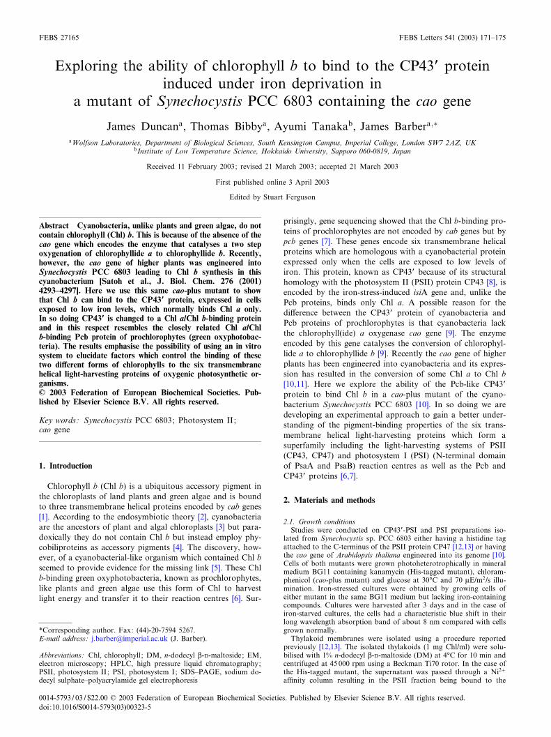
Thylakoid membranes were isolated from the cao-plus mu-tant grown in the presence of iron and in iron-de¢cient media.Fig. 1 shows the absorption spectra of the isolated membraneswhere it can be seen that iron deprivation induces a blue shiftof the far-red absorption maximum by 8 nm similar to thatreported previously for the wild type [12,13]. There are, how-ever, no obvious absorption features indicative of the presenceof Chl b in either spectrum. In order to check whether Chl bhad been synthesised in the cao-plus mutant, Chl was ex-tracted and subjected to HPLC analysis. Fig. 2a shows the
0
0.1
0.2
0.3
0.4
0.5
0.6
0.7
0.8
0.9
1
370 420 470 520 570 620 670
Wavelength (nm)
Ab
so
rba
nc
e (
a.u
)
+Fe
-Fe
67
5
68
3
Fig. 1. Room temperature absorption spectra of thylakoid membranes isolated from the cao mutant of Synechocystis showing a characteristicblue shift in long-wavelength absorption maximum in response to iron deprivation. Thylakoid membranes were suspended in 50 mM MES pH6.0, 0.5 M betaine, 5 mM CaCl2, 5 mM MgCl2 at 10 WM Chl.
Fig. 2. HPLC elution pro¢les of Chl extracted from thylakoid mem-branes of cells of (a) the cao mutant and (b) the mutant of Synecho-cystis with His-tagged Chl-binding proteins grown in the presenceof iron.
FEBS 27165 11-4-03
J. Duncan et al./FEBS Letters 541 (2003) 171^175172
HPLC trace of the Chl extracted from thylakoid membranesof cao-plus mutant grown in the presence of iron. Using Chl aand Chl b standards it was shown that the smaller peak cor-responds to Chl b and the larger to Chl a. This was con¢rmedby conducting a similar analysis on the thylakoid membranes
of Synechocystis cells with a His tag attached to the carboxy-terminus of the PSII Chl a-binding protein, CP47 [13]. Thesecells contained only Chl a (Fig. 2b). Calibrations with the Chla and Chl b standards indicated that the Chl b level variedbetween di¡erent cultures and ranged from about 4 to 7% ofthe total Chl. A similar level was detected in thylakoid mem-branes isolated from cao-plus cells grown under iron-free con-ditions. Slightly higher amounts of Chl b could accumulateunder iron-rich conditions, but as reported previously [10] thelevel was dependent on the growth rate of the culture, whichin the case of the iron-free condition was less vigorous.
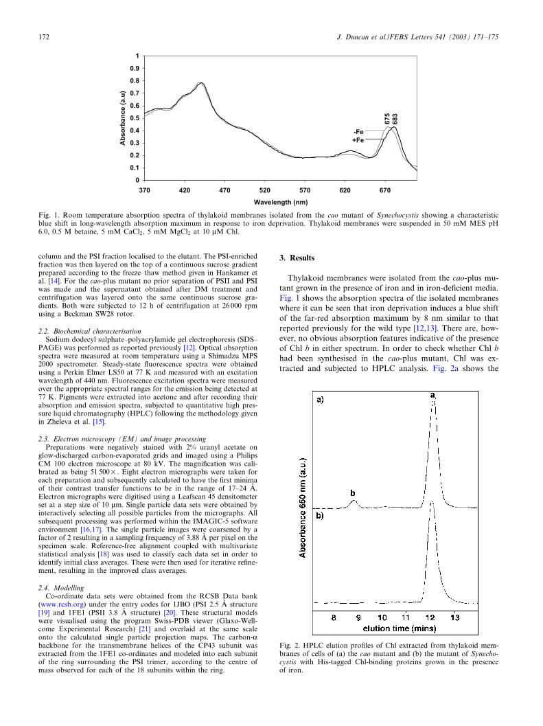
In order to explore whether Chl b was located in the CP43Pprotein and therefore in the CP43P-PSI supercomplex reportedpreviously [12,13], we solubilised the membranes with DMand separated the resulting complexes by sucrose density gra-dient centrifugation. Fig. 3 shows the pro¢les obtained withthe cao-plus mutant. The cells grown in the presence of irongave four bands, A, B, C and D, while the minus-iron con-dition induced an additional heavy band, E. Inspection ofthese bands by absorption and emission spectroscopy, SDS^PAGE and EM indicated that band A from the plus-iron cellsconsisted of free carotenoid while the corresponding band inthe minus-iron cells contained CP43P as well as free carot-enoids. In both cases band B contained PSII and PSI com-plexes in their monomeric states. Band C was mainly PSII
Fig. 3. Sucrose density-banding pro¢le of products derived fromDM solubilisation of isolated thylakoids from cells of the cao mu-tant grown in the presence (+Fe) or absence of iron (3Fe). +Fe:A = carotenoid, B = PSI/PSII monomers, C = PSII dimers, D = PSItrimers. 3Fe: A = carotenoid/CP43P, B = PSI/PSII monomers,C = PSII dimers, D = PSI trimers, E = CP43P-PSI supercomplex.
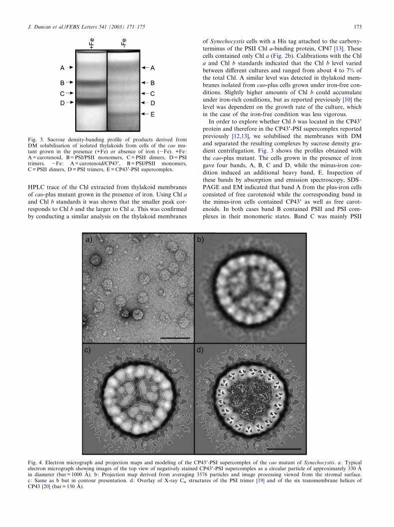
Fig. 4. Electron micrograph and projection maps and modeling of the CP43P-PSI supercomplex of the cao mutant of Synechocystis. a: Typicalelectron micrograph showing images of the top view of negatively stained CP43P-PSI supercomplex as a circular particle of approximately 330 APin diameter (bar = 1000 AP ). b: Projection map derived from averaging 3576 particles and image processing viewed from the stromal surface.c: Same as b but in contour presentation. d: Overlay of X-ray CK structures of the PSI trimer [19] and of the six transmembrane helices ofCP43 [20] (bar = 150 AP ).
FEBS 27165 11-4-03
J. Duncan et al./FEBS Letters 541 (2003) 171^175 173
dimers while band D, which was signi¢cantly reduced in theiron-depleted cells compared to normal cells, containedmainly PSI trimers. Band E, observed only when the cellswere grown in minus-iron medium, consisted almost entirelyof the CP43P-PSI supercomplex. Images of this complex areshown in Fig. 4a and the averaged top views shown in Fig. 4b.The latter is also shown in Fig. 4c as a contour map with theoverlay of X-ray data in Fig. 4d. It can be seen that the 2.5 AP
X-ray structure of the PSI trimer of Synechococcus elongatusdetermined by Jordan et al. [19] is accommodated in the cen-tral region of the projection map. Similarly, the overlaying ofthe transmembrane helices of CP43, obtained by X-ray dif-fraction [20], is consistent with the presence of 18 copies of theCP43P protein in the ring surrounding the PSI reaction centretrimer. At the resolution of the projection map it seems thatthis CP43P-PSI supercomplex is identical to that isolated pre-viously from Synechocystis with a His tag [12,13] and fromwild type Synechococcus [22]. The absorption and £uorescenceproperties were also comparable with those reported previ-ously [12,13] indicating that the CP43P antenna was function-ally associated with the reaction centre.
To determine whether Chl b in the supercomplex waspresent, HPLC was conducted following the same procedureas for thylakoid membranes. In this particular experiment thepercentage of Chl b compared to the total Chl was in theregion of 3.7 for the CP43P-PSI supercomplex, comparablewith the level determined for the thylakoids from whichthey were isolated (Table 1). Using mild DM treatment theisolated supercomplexes were dissociated into their compo-nents, CP43P and PSI trimers, followed by pigment analyses.As Table 1 shows, the percentage of Chl b remained constantin CP43P and the PSI trimer.
4. Discussion
The formation of the 18-mer CP43P subunit antenna ringaround the PSI reaction centre trimer is probably a strategy tocompensate for the reduction in the levels of phycobiliproteinsand a decrease in the PSI/PSII ratio under iron-limiting con-ditions [8]. Normally CP43P contains only Chl a, and based onthe recent X-ray analyses of CP43 [23] there are probably 13molecules bound to each copy of the protein. Therefore the234 Chl a molecules contained in the 18-mer CP43P antennaring increase the light-harvesting capacity of PSI by 81%,given that the trimer binds 288 Chl a molecules, accordingto the recently determined X-ray structure of PSI [19]. Herewe show that the cao-plus mutant also forms the 18-merCP43P antenna ring under iron stress conditions but in thiscase contains Chl b as well as Chl a. The level of Chl b is,however, relatively low and was found to be dependent on thestage of growth of the culture as previously noted [10]. Higherlevels of Chl b have been obtained in a PSI-less cao-plus mu-
tant when the lhcb gene of pea was also engineered into Syn-echocystis along with the Arabidopsis cao gene [11].
The binding of Chl b as well as Chl a to CP43P in the cao-plus Synechocystis mutant is of interest because of the simi-larity of CP43P to the Chl a/Chl b Pcb-binding proteins ofprochlorophytes [7] in which an 18-mer Pcb antenna ringhas been detected [24]. The level of Chl b can also be relativelylow (6^20%) [25^27] in prochlorophytes, such as Prochlorondidemni, Prochlorothrix hollandica and certain strains of Pro-chlorooccus marinus, like MED4. However, in the low light-adapted Prochlorococcus strain SS120 the Chl b level can be ashigh as 66%, which is more comparable with the levels insome of the Cab proteins that act as light-harvesting systemsfor PSII [1]. Other Cab proteins, however, have signi¢cantlylower levels of Chl b [1] and the factors which control thedi¡erent levels of Chl b in light-harvesting proteins are un-known. The cao-plus cyanobacterial mutant used here o¡ersan in vivo experimental system to investigate these di¡erences.
References
[1] Green, B.R. and Durnfold, D.G. (1996) Annu. Rev. Plant Phys-iol. Plant Mol. Biol. 47, 685^714.
[2] Margulis, L. (1981) Symbiosis in Cell Evolution, Freeman, SanFrancisco, CA.
[3] Bhattacharya, D. and Medlin, L. (1998) Plant Physiol. 116, 9^15.[4] Sidler, W. (1994) in: Molecular Biology of Cyanobacteria (Bry-
ant, D.A., Ed.), pp. 139^216, Kluwer Academic, Dordrecht.[5] Lewin, R.A. (1976) Nature 261, 697^698.[6] Partensky, F., Hess, W.R. and Vaulot, D. (1999) Microbiol. Mol.
Biol. Rev. 63, 106^127.[7] La Roche, J., van der Staay, G.W., Partensky, F., Ducret, A.,
Aebersold, R., Li, R., Golden, S.S., Hiller, R.G., Wrench, P.M.and Larkum, A.W. (1996) Proc. Natl. Acad. Sci. USA 93, 15244^15248.
[8] Strauss, N.A. (1994) in: Molecular Biology of Cyanobacteria(Bryant, D.A., Ed.), pp. 731^750, Kluwer Academic, Dordrecht.
[9] Oster, U., Tanaka, R., Tanaka, A. and Rudiger, W. (2000) PlantJ. 21, 305^310.
[10] Satoh, S., Ikeuchi, M., Mimuro, M. and Tanaka, A. (2001)J. Biol. Chem. 276, 4293^4297.
[11] Xu, H., Vavilin, D. and Vermaas, W. (2001) Proc. Natl. Acad.Sci. USA 98, 14168^14173.
[12] Bibby, T.S., Nield, J. and Barber, J. (2001) J. Biol. Chem. 276,43246^43252.
[13] Bibby, T.S., Nield, J. and Barber, J. (2001) Nature 412, 743^745.[14] Hankamer, B., Nield, J., Zheleva, D., Boekema, E.J., Jansson, S.
and Barber, J. (1997) Eur. J. Biochem. 243, 422^429.[15] Zheleva, D., Hankamer, B. and Barber, J. (1996) Biochemistry
35, 15074^15079.[16] van Heel, M., Harauz, G. and Orlova, E.V. (1996) J. Struct. Biol.
116, 17^24.[17] van Heel, M., Gowen, B., Matedeen, R., Orlova, E.V., Finn, R.,
Pape, T., Cohen, D., Stark, H., Schmidt, R., Schatz, M. andPatwardhan, A. (2000) Q. Rev. Biophys. 33, 307^369.
[18] Sherman, M., Soejima, T., Chui, W. and van Heel, M. (1998)Ultramicroscopy 74, 179^199.
[19] Jordan, P., Fromme, P., Witt, H.T., Klukas, O., Saenger, W. andKraus, N. (2001) Nature 411, 909^916.
Table 1Chl b levels in cao mutant of Synechocystis grown in minus-iron culture medium
Photosynthetic fraction Chl b (%)
Thylakoids from iron-de¢cient cells 3.6CP43P-PSI supercomplex 3.7Free CP43P dissociated from the CP43P-PSI supercomplex 3.6PSI trimers dissociated from the CP43P-PSI supercomplex 3.6
FEBS 27165 11-4-03
J. Duncan et al./FEBS Letters 541 (2003) 171^175174
[20] Zouni, A., Witt, H.T., Kern, J., Fromme, P., Krauss, N.,Saenger, W. and Orth, P. (2001) Nature 409, 739^743.
[21] Guex, N. and Peitsch, M.C. (1997) Electrophoresis 18, 2714^2723.
[22] Boekema, E.J., Hi¡ney, A., Yakushevska, A.E., Piotrowski, M.,Keegstra, W., Berry, S., Michel, K.-P., Pistorius, E.K. andKruip, J. (2001) Nature 412, 745^748.
[23] Kamiya, N. and Shen, J.R. (2003) Proc. Natl. Acad. Sci. USA100, 98^102.
[24] Bibby, T.S., Nield, J., Partensky, F. and Barber, J. (2001) Nature413, 590.
[25] Matthijs, H.C.P., van der Staay, G.W.M., van Amerongen, H.,van Grondelle, R. and Garab, G. (1989) Biochim. Biophys. Acta975, 185^187.
[26] Lewin, R.A. and Withers, N.W. (1975) Nature 256, 735^737.
[27] Garczarek, L., van der Staay, G.W.M., Thomas, J.C. and Par-tensky, F. (1998) Photosynth. Res. 56, 131^141.
FEBS 27165 11-4-03
J. Duncan et al./FEBS Letters 541 (2003) 171^175 175