Embed Size (px)
Citation preview
1
Exploring the Limits of Dative Boratrane Bonding: Iron as a Strong Lewis Base in Low-Valent Non-Heme Iron-Nitrosyl Complexes Hai T. Dong1; Matthew J. Chalkley2; Paul H. Oyala2; Jiyong Zhao3; E. Ercan Alp3; Michael Y. Hu3; Jonas C. Peters2,*; Nicolai Lehnert1,* 1 Department of Chemistry and Department of Biophysics, University of Michigan, Ann Arbor, Michigan 48109-1055, United States
2Department of Chemistry and Chemical Engineering, California Institute of Technology, Pasadena, California 91125, United States 3Advanced Photon Source (APS), Argonne National Laboratory (ANL), Argonne, Illinois 60439, United States
Abstract. We previously reported the synthesis and preliminary characterization of a unique series of low-spin (ls) {FeNO}8-10 complexes supported by an ambiphilic trisphosphineborane ligand, [Fe(TPB)(NO)]+/0/. Herein, we use advanced spectroscopic techniques and density functional theory (DFT) calculations to extract detailed information as to how the bonding changes across the redox series. We find that, despite the highly reduced nature of these complexes, they feature an NO+ ligand throughout with strong Fe-NO -backbonding and essentially closed-shell electronic structures of their FeNO units. This is enabled by an Fe-B interaction that is present throughout the series. In particular, the most reduced [Fe(TPB)(NO)]complex, an example of a ls-{FeNO}10 species, features a true reverse dative Fe→B bond where the Fe center acts as a strong Lewis-base. Hence, this complex is in fact electronically similar to the ls-{FeNO}8 system, with two additional electrons “stored” on site in an Fe-B single bond. The outlier in this series is the ls-{FeNO}9 complex, due to spin polarization (quantified by pulse EPR spectroscopy), which weakens the Fe-NO bond. These data are further contextualized by comparison with a related N2 complex, [Fe(TPB)(N2)]−, which is a key intermediate in Fe(TPB)-catalyzed N2 fixation. Our present study finds that the Fe→B interaction is key for storing the electrons needed to achieve a highly reduced state in these systems, and highlights the pitfalls associated with using geometric parameters to try to evaluate reverse dative interactions, a finding with broader implications to the study of transition metal complexes with boratrane and related ligands.
2
1. Introduction
Heme and non-heme iron-nitrosyl units are highly prevalent in biology, and (bio)inorganic
chemists have pondered their electronic structures and reactivity patterns for decades to better
understand these systems. In particular, heme-nitrosyls are relevant in NO sensing, transport and
as intermediates in nitrogen-cycle enzymes,1-12 whereas non-heme iron centers are particularly
relevant in bacterial NO reductases (NORs).13-16 Transition metal nitrosyl (M-NO) complexes
represent some of the earliest recognized examples of redox non-innocence, leading to the
development of the Enemark-Feltham notation, {MNO}n, which classifies a M-NO complex by
its total number of valence electrons n (= metal(d) + NO(π*) electrons).17 In this regard, NO can
coordinate to metals in three different oxidation states (NO+/0/). In the case of non-heme iron
enzymes, for example, binding of •NO to the Fe(II) form generates high-spin (hs) {FeNO}7
adducts, which, in general, have FeIII-NO type electronic structures.18-20 Due to their highly
covalent Fe-NO bonds, these complexes are usually stable and unreactive (with NORs being
potential exceptions), but capable of undergoing reduction at mild potentials to form very reactive
hs-{FeNO}8 complexes.21-24 The latter species have been shown to undergo a number of different
reactions, including N-N coupling to form N2O,25-27 disproportionation to form dinitrosyl iron
complexes (DNICs),28 and protonation to generate HNO.29 Recent findings by Balkus and
coworkers show that non-heme iron enzymes are also involved in biosynthetic pathways of natural
products containing the N-nitroso group, with a hs-{FeNO}6 intermediate potentially involved in
this reaction.30 Understanding how the electron distribution effects the reactivity and stability of
Fe-NO complexes is of critical importance to develop a better understanding of their many roles
both in signaling and energy-transducing reactions in biology.
3
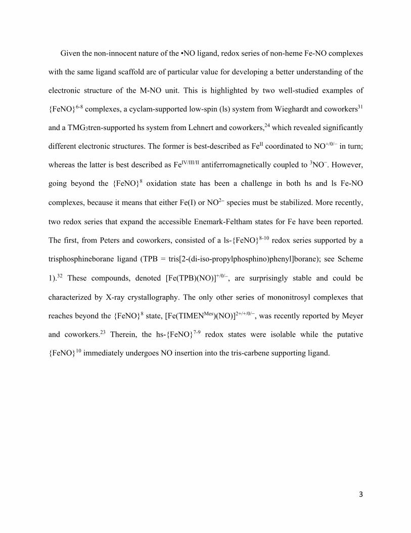
Given the non-innocent nature of the •NO ligand, redox series of non-heme Fe-NO complexes
with the same ligand scaffold are of particular value for developing a better understanding of the
electronic structure of the M-NO unit. This is highlighted by two well-studied examples of
{FeNO}6-8 complexes, a cyclam-supported low-spin (ls) system from Wieghardt and coworkers31
and a TMG3tren-supported hs system from Lehnert and coworkers,24 which revealed significantly
different electronic structures. The former is best-described as FeII coordinated to NO+/0/− in turn;
whereas the latter is best described as FeIV/III/II antiferromagnetically coupled to 3NO−. However,
going beyond the {FeNO}8 oxidation state has been a challenge in both hs and ls Fe-NO
complexes, because it means that either Fe(I) or NO2 species must be stabilized. More recently,
two redox series that expand the accessible Enemark-Feltham states for Fe have been reported.
The first, from Peters and coworkers, consisted of a ls-{FeNO}8-10 redox series supported by a
trisphosphineborane ligand (TPB = tris[2-(di-iso-propylphosphino)phenyl]borane); see Scheme
1).32 These compounds, denoted [Fe(TPB)(NO)]+/0/, are surprisingly stable and could be
characterized by X-ray crystallography. The only other series of mononitrosyl complexes that
reaches beyond the {FeNO}8 state, [Fe(TIMENMes)(NO)]2+/+/0/−, was recently reported by Meyer
and coworkers.23 Therein, the hs-{FeNO}7-9 redox states were isolable while the putative
{FeNO}10 immediately undergoes NO insertion into the tris-carbene supporting ligand.
4
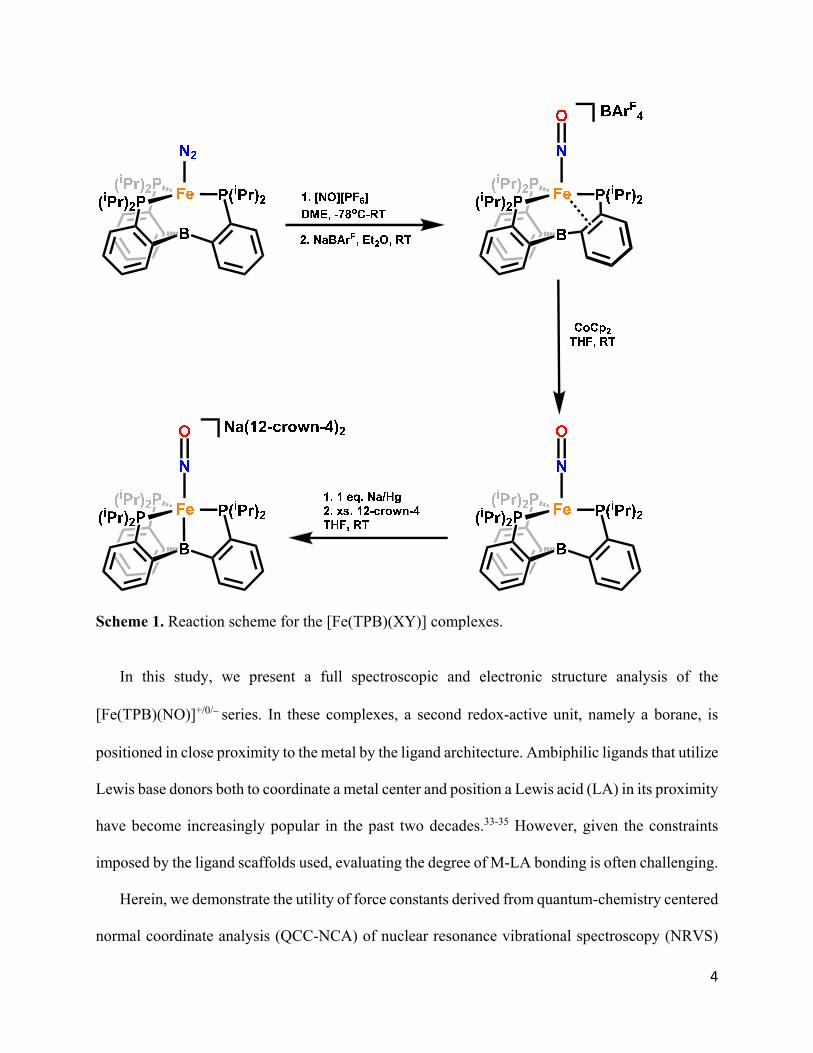
Scheme 1. Reaction scheme for the [Fe(TPB)(XY)] complexes.
In this study, we present a full spectroscopic and electronic structure analysis of the
[Fe(TPB)(NO)]+/0/series. In these complexes, a second redox-active unit, namely a borane, is
positioned in close proximity to the metal by the ligand architecture. Ambiphilic ligands that utilize
Lewis base donors both to coordinate a metal center and position a Lewis acid (LA) in its proximity
have become increasingly popular in the past two decades.33-35 However, given the constraints
imposed by the ligand scaffolds used, evaluating the degree of M-LA bonding is often challenging.
Herein, we demonstrate the utility of force constants derived from quantum-chemistry centered
normal coordinate analysis (QCC-NCA) of nuclear resonance vibrational spectroscopy (NRVS)
5
data in deconvoluting the electronic structure and bonding at Fe in a highly covalent ligand sphere
comprised of nitrosyl, boratrane, and phosphine ligands. We find that, despite their low formal Fe
redox states, an NO+ redox state with strong Fe-NO π-bonds is maintained throughout the redox
series. This is made possible because of the high degree of structural and electronic flexibility in
the TPB ligand, demonstrated via the breaking of an η4-BCCP donor interaction present in the
most oxidized complex, and formation of a reverse dative Fe→B bond in the most reduced
complex. Similarly, a reverse dative Fe→B bond has also been identified in the structurally related
[Fe(TPB)(N2)]− complex by NRVS, underscoring the relevance of this interaction in promoting
small molecule functionalization (i.e., N2 fixation).36 These conclusions are corroborated by
continuous wave and pulse electron paramagnetic resonance spectroscopy (EPR) and density
functional theory (DFT) calculations.
2. Experimental Section
All complexes including 57Fe complexes were prepared as previously reported and obtained as
pure compounds, as determined by Mössbauer and IR spectroscopy.32 Efforts to label the
complexes with 15NO were largely unsuccessful. However, trace amounts of the ls-{FeNO}9
complex [Fe(TPB)(15NO)], sufficient for pulse EPR measurements, could be obtained via reaction
of [Fe(TPB)(N2)] with [TBA][15NO2] followed by extraction by pentane and filtration through
celite. This reaction is not well-defined but proved technically useful. We suspect reducing
equivalents come from degradation of the iron-phosphine system, accounting for the poor mass
balance of the reaction. All efforts that we made to improve this synthesis were unsuccessful.
NRVS measurements. Nuclear resonance vibrational spectroscopy (NRVS) data were
obtained as described previously3 at beamline 3-ID at the Advanced Photon Source (APS) at
6
Argonne National Laboratory. Samples were loaded in copper sample holders with lucite lids.
During data collection, samples were maintained at cryogenic temperatures using a liquid helium-
cooled cryostat. Spectra of solid samples were recorded from 0 to +90 meV in 0.25 meV steps.
Multiple scans were taken, normalized to the intensity of the incident beam, and added together to
achieve adequate signal to noise ratios; the final spectra represent averages between 6 and 10 scans.
The program Phoenix4 was used to convert the raw NRVS data to the vibrational density of states
(VDOS).
Pulse EPR measurements for the ls-{FeNO}9 complex. All pulse X-band (ν ≈ 9.7 GHz) EPR
and electron nuclear double resonance (ENDOR) experiments were performed using a Bruker
(Billerica, MA) ELEXSYS E580 pulse EPR spectrometer equipped with a Bruker MD-4 resonator.
Temperature control for experiments at 7 K was achieved using an ER 4118HV-CF5-L Flexline
Cryogen-Free VT cryostat manufactured by ColdEdge (Allentown, PA), while ENDOR
experiments at 5 K were performed using an Oxford Instruments CF935 helium flow cryostat. An
Oxford Instruments Mercury ITC was used for temperature regulation with both cryostats.
X-band Electron spin-echo detected field swept spectra (ESE-EPR) were acquired using the 2-
pulse Hahn echo sequence ( 𝜏 𝜋 𝜏 𝑒𝑐ℎ𝑜 , while the magnetic field was varied. The
“CW-EPR like” 1st derivative spectrum was generated by use of the pseudomodulation function in
EasySpin, an EPR simulation toolbox for use with Matlab.37,38
Pulse X-band ENDOR was acquired using the Davies pulse sequence (𝜋 𝑇 𝜋
𝑇 𝜋/2 – 𝜏 – 𝜋 – echo), where 𝑇 is the delay between mw pulses and RF pulses, 𝜋 is the
length of the RF pulse and the RF frequency is randomly sampled during each pulse sequence.
X-band Hyperfine sublevel correlation (HYSCORE) spectra were acquired using the 4-pulse
sequence (𝜋/2 𝜏 𝜋/2 𝑡 𝜋 –𝑡 – 𝜋/2 – echo), where 𝜏 is a fixed delay, while 𝑡 and 𝑡
7
are independently incremented by Δ𝑡 and Δ𝑡 , respectively. The time domain data was baseline-
corrected (third-order polynomial) to eliminate the exponential decay in the echo intensity,
apodized with a Hamming window function, zero-filled to eight-fold points, and fast Fourier-
transformed to yield the 2-dimensional frequency domain. The intensity of this FT data was plotted
as a series of contours on a logarithmic scale, in colors ranging from blue to red in increasing
intensity.
EPR Simulations. Simulations of all EPR data were achieved using the EasySpin simulation
toolbox (release 5.2.28) with Matlab 2019a.37 For more details of these simulations, we refer
readers to the SI.
DFT Calculations using Gaussian 09 and Normal Coordinate Analysis. Geometry
optimization of the ls-{FeNO}8-10 complexes was carried out using the BP86 and B3LYP
functionals with the TZVP basis set, using both closed shell and broken symmetry wavefunctions
(see text). All calculations were performed using the program Gaussian 09.39 Subsequent
frequency calculations on the optimized structures show no imaginary frequencies, indicating that
true energy minima were obtained. The DFT-calculated force constants in Cartesian coordinates
were extracted from the Gaussian output files and transformed into internal coordinates using a
modified version of the program Redong. Modified normal coordinate analysis (NCA) programs
based on QCPE 576 were used for the subsequent fitting of the experimental NRVS data. The
fitting was performed by adjusting a minimal set of force constants (in the spirit of the QCC-NCA
approach)40 to reproduce the vibrations of the Fe-N-O units in the ls-{FeNO}8-10 series of
complexes (see text).
DFT Calculations using ORCA 4.0. The Gaussian-optimized structures of the ls-{FeNO}8-10
complexes were used for following single-point calculations (BP86/TZVP) with ORCA 4.0 to
8
predict Mössbauer and EPR parameters, and to further analyze the electronic structures of the
complexes. This includes the use of unrestricted corresponding orbitals (UCOs) for the ls-
{FeNO}9 complex.41
3. Results and Analysis
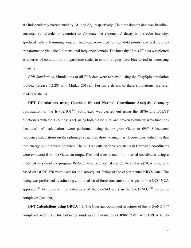
3.1. Nuclear Resonance Vibrational Spectroscopy (NRVS) for the ls-{FeNO}8-10 Series
The Fe-NO bonding in the ls-{FeNO}8-10 series is evaluated and analyzed herein based on NRVS
measurements (see Figure 1). NRVS is a vibrational technique that selectively detects vibrations
that involve the 57Fe center, making it well-suited for the identification of Fe-ligand stretching and
bending modes. The experimental NRVS data of the ls-{FeNO}8 complex reveal an intense band
at 610 cm-1 and weaker signals at 537 and 540 cm-1. The feature at 610 cm-1 is assigned to the Fe-
NO stretch (see below), whereas those at 537 and 540 cm-1 are in the correct range for Fe-N-O
bending modes. With an Fe-NO stretch of 610 cm-1, this complex has one of the strongest transition
metal-NO bonds observed to this date and the strongest for an iron compound,42 surpassing even
ls-{FeNO}6 complexes in hemes (with typical Fe-NO stretching frequencies around 590 cm-1).43,44
In IR spectroscopy, the N-O stretch of this complex is observed at 1745 cm-1. The NRVS data of
the ls-{FeNO}10 complex are remarkably similar to those of the ls-{FeNO}8 species described
above. In particular, its Fe-NO stretch is observed as the most intense signal at 602 cm-1, with the
weaker features at 525 and 543 cm-1 again associated with Fe-N-O bending modes (see Figure 1).
The N-O bond of this complex is the weakest (and most activated) in the series, with an N-O
stretching frequency of 1568 cm-1 as determined by IR spectroscopy.
9
Figure 1. Experimental NRVS VDOS data of the ls-{FeNO}8 complex [Fe(TPB)(NO)](BArF
4) (purple), the ls-{FeNO}9 complex [Fe(TPB)(NO)] (brown) and the ls-{FeNO}10 complex [Na(12-crown-4)2][Fe(TPB)(NO)] (red) vs QCC-NCA fits (black).
The intense, high-energy NRVS feature of the ls-{FeNO}9 species, observed at 583 cm-1, is
again assigned to the Fe-NO stretch. This mode is significantly shifted compared to 610 cm-1 (
= -27 cm-1) and 602 cm-1 ( = -19 cm-1) in the other two complexes, respectively, which, as we
will show below, is due to spin polarization. The Fe-N-O bending modes are similarly shifted as
well (506 and 522 cm-1, see Figure 1). The N-O stretch of this complex is located at 1667 cm-1.
In summary, comparison of the Fe-NO and N-O stretching frequencies along the ls-{FeNO}8-
10 series does not reveal a consistent trend. In a simple -backbonding model (between the Fe-d
10
and NO(*) orbitals), we would anticipate that concomitant with the observed stepwise weakening
of the N-O bond along the ls-{FeNO}8-10 series there would be a stepwise strengthening of the Fe-
NO bond. Instead, for the ls-{FeNO}8/9 pair, both the Fe-NO and N-O stretching frequencies (and
bond strengths) decrease in the ls-{FeNO}9 compound. This trend is then reversed in the ls-
{FeNO}9/10 pair (now showing a pattern that would be in line with an increase in -backbonding
upon reduction), creating a discontinuity in the observed behavior. Thus, it is clear that a more
detailed analysis, one that considers all available experimental data supported by electronic
structure calculations, is necessary.
DFT Calibration for the ls-{FeNO}8-10 Series. In our previous report, the ls-{FeNO}8 and ls-
{FeNO}10 complexes were described as closed shell systems, on the basis of their diamagnetic
ground states (from multinuclear nuclear magnetic resonance (NMR) spectroscopy). Alternatively,
diamagnetic ground states could also arise from strong antiferromagnetic coupling between a hs
iron center and a triplet NO ligand, which is often observed for non-heme Fe-NO complexes.24
Furthermore, a recent interrogation of a related redox series, [Fe(TPB)(NNMe2)]+/0/−, by
experiment and theory revealed antiferromagnetic coupling between the Fe center and a hydrazyl
radical anion, [NNMe2]•− in some redox states.45 Therefore, we decided to re-evaluate whether the
ground states of the [Fe(TPB)(NO)]+/0/ complexes are best described by closed shell (CS) or
broken-symmetry (BS) wave functions. As in previous work, we applied both the gradient-
corrected functional BP86 and the hybrid functional B3LYP, now in combination with the TZVP
basis set, for these calculations.46-50 While BP86 has previously been shown to be a reliable
functional in predicting geometric structures and spectroscopic properties of iron-nitrosyl
complexes, B3LYP tends to underestimate the covalency of the Fe-N-O moiety.51,52 However,
hybrid functionals like B3LYP with a higher percentage of Hartree-Fock exchange often allow for
11
the geometry optimization of BS states in strongly spin-coupled systems, which is difficult with
gradient-corrected functionals like BP86.
To our surprise, the structural features derived from X-ray crystallography were well-
reproduced by both the CS and BS state in B3LYP calculations on the ls-{FeNO}8 complex (see
Table S1). For example, the N-O bond length only deviates by 0.01 Å for both states
(1.16/1.17/1.17 Å for exp/CS/BS). Similarly, the Fe-NO bond distance shows very good agreement
with the experimental data, with just 0.01-0.02 Å deviation for both states (1.66/1.65/1.68 Å for
exp/CS/BS). Both calculations show moderate agreement with the experimental Fe-B bond
distance (2.31/2.37/2.37 Å for exp/CS/BS). Finally, the BS state shows better agreement with the
experimental data for the Fe-N-O angle (176/172/175o for exp/CS/BS). Thus, although purely
structural comparisons do not distinguish between a CS or BS electronic structure for the ls-
{FeNO}8 complex, the accuracy of the predicted NRVS spectra is dramatically different, as shown
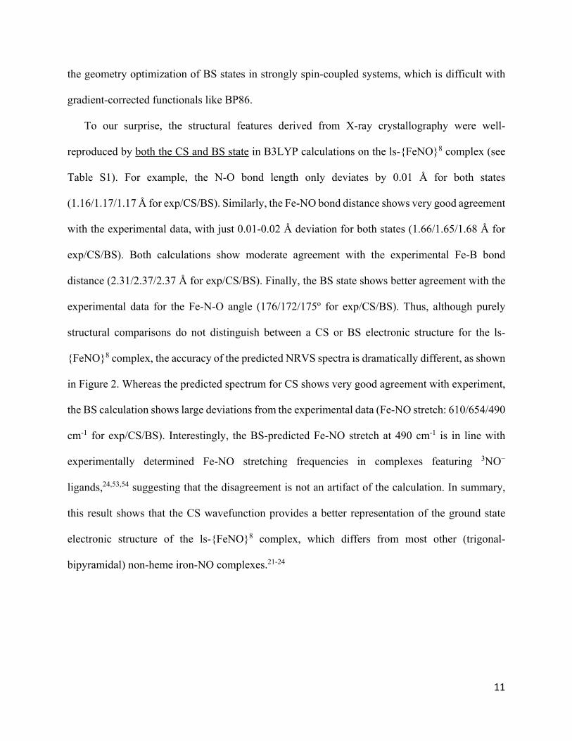
in Figure 2. Whereas the predicted spectrum for CS shows very good agreement with experiment,
the BS calculation shows large deviations from the experimental data (Fe-NO stretch: 610/654/490
cm-1 for exp/CS/BS). Interestingly, the BS-predicted Fe-NO stretch at 490 cm-1 is in line with
experimentally determined Fe-NO stretching frequencies in complexes featuring 3NO−
ligands,24,53,54 suggesting that the disagreement is not an artifact of the calculation. In summary,
this result shows that the CS wavefunction provides a better representation of the ground state
electronic structure of the ls-{FeNO}8 complex, which differs from most other (trigonal-
bipyramidal) non-heme iron-NO complexes.21-24
12
Figure 2. Experimental NRVS VDOS data of the ls-{FeNO}8 complex (top) in comparison with the spectra generated by closed-shell (middle) and broken-symmetry (bottom) calculations, using the indicated functionals together with the TZVP basis set.
Comparing CS solutions calculated with both B3LYP and BP86, we find that the BP86
functional not only better reproduces the vibrational and structural data for the ls-{FeNO}8-10
series, but is also able to accurately predict the isomer shift (δ) and quadrupole splitting (|Δeq|)
derived from Mössbauer spectroscopy and the hyperfine parameters derived from pulse EPR
spectroscopy (Table 1). Thus, we confirm that a CS, highly covalent description of the ground
state in the [Fe(TPB)(NO)]+/0/ complexes is most appropriate.
13
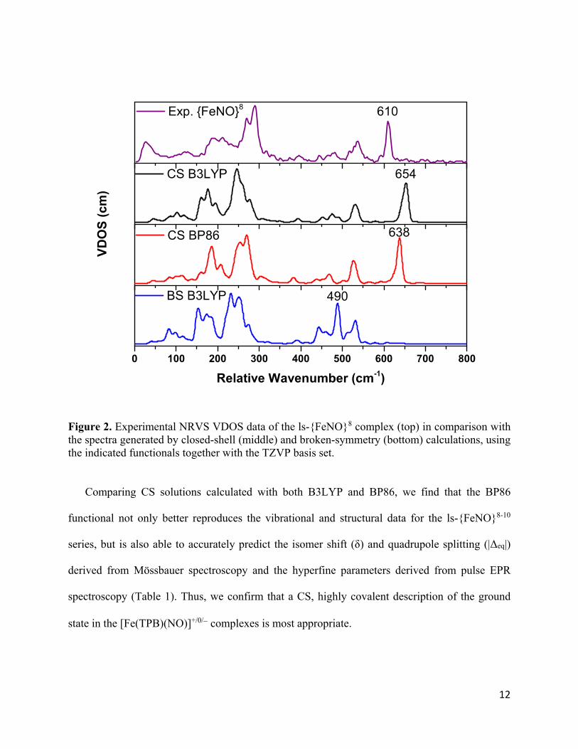
The BP86-optimized structures show very good agreement with the crystal structures of all
three compounds, as further demonstrated by the structural overlays in Figure 3. The ls-{FeNO}8
complex has a distinct distorted trigonal-bipyramidal geometry, where one of the P-Fe-P angles in
the trigonal plane is expanded to 154o allowing for an unusual intramolecular 4-BCCP interaction.
Both of these features are well reproduced in the DFT optimized structure. As the compound is
reduced to the ls-{FeNO}9 state, the complex becomes more symmetric (closer to an actual
trigonal-bipyramidal geometry), and the unusually large P-Fe-P angle decreases from 154o to 126o.
The ls-{FeNO}10 complex is the most symmetric with only about 1o difference between the three
P-Fe-P angles.
Figure 3. Overlay of crystal structures (blue) and the BP86/TZVP-optimized structures (yellow) of the ls-{FeNO}8-10 series, [Fe(TPB)(NO)]+/0/, showing excellent agreement between the DFT-predictions and the experimental structures.
The BP86 calculations reproduce the vibrational properties of the ls-{FeNO}8-10 complexes,
especially the Fe-NO and N-O stretching frequencies, quite well with respect to experimental data
(Figures S1). Importantly, the calculations capture the lack of a correlation between the change in
Fe-NO and N-O stretching frequencies along the ls-{FeNO}8-10 series (see Table 1). Thus, we use
these calculations as the basis to further analyze the NRVS data and refine the force constants of
ls‐{FeNO}8 ls‐{FeNO}9 ls‐{FeNO}10

14
the Fe-N-O units in the three complexes. In this way, we further address the question of whether
the reduction along the ls-{FeNO}8-10 series is metal- or NO-based.
Table 1. Experimental structural and spectroscopic data versus computational results for the series of ls-{FeNO}8-10 complexes.
QCC-NCA for the ls-{FeNO}8-10 Series. To obtain simulations of the NRVS data of the ls-
{FeNO}8-10 complexes and determine high-quality (experimental) force constants for their Fe-N-
O units, a quantum-chemistry centered normal coordinate analysis (QCC-NCA) was
performed.45,55 This process allows us to correct the DFT-calculated force constants, vibrational
ls-{FeNO}8 ls-{FeNO}9 ls-{FeNO}10 Exp. BP86 Exp. BP86 Exp. BP86
Geometric Parameters (Å and degrees) d(NO) 1.16 1.18 1.19 1.19 1.22 1.21
d(FeO) 1.66 1.66 1.67 1.66 1.65 1.65 d(FeB) 2.31 2.32 2.45 2.42 2.45 2.46 FeNO 176 174 176 176 179 180 d(FeP) 2.28 2.33 2.28 2.30 2.21 2.24 d(FeP) 2.28 2.33 2.30 2.32 2.21 2.24 d(FeP) 2.29 2.31 2.35 2.37 2.23 2.24 P-Fe-P 100 99 106 107 115 116 P-Fe-P 101 101 111 110 116 116 P-Fe-P 154 154 126 126 116 116
Spectroscopic Parameters: Vibrational (cm-1, mdyn/Å and mdyn•Å) (FeNO) 610 638 583 621 602 633 (N-O) 1745 1751 1667 1692 1568 1607
lb(Fe-N-O) 540/537 531/525 522/506 516/508 543/525 561/535
f(Fe-NO) 4.53 4.95 4.15 4.80 4.45 5.07
f(N-O) 12.5 12.4 11.3 11.5 9.79 10.1
f(Fe-B) 0.51 0.51 0.42 0.42 1.56 1.56
Spectroscopic Parameters: Mössbauer (mm/s)
0.24 0.30 0.26 0.25 0.17 0.20
EQ 1.50 1.43 0.91 0.81 1.62 1.53 Spectroscopic Parameters: Pulse EPR (MHz)
A(14N) - - -6.0, -8.3, 3.8
-0.8, -5.1, 8.8
- -
A(11B) - - 14.7, 14.7, 18.0
-15.2, -15.6, -20.3
- -
15
frequencies and NRVS intensities by fitting the experimental NRVS data, starting from the DFT-
predicted force field. In this way, we obtain high-quality force constants for the modes of interest
that afford detailed insight into the changes in Fe-NO and N-O bonding along the ls-{FeNO}8-10
series, independent of potential vibrational (mode) mixing. In the spirit of the QCC-NCA
approach,55 only the small number of force constants relevant to the Fe-N-O unit are varied, while
the DFT-predicted force constants of the [Fe(TPB)] frame are kept unchanged.
For the ls-{FeNO}8 complex, the Fe-NO force constant was corrected from the calculated
value of 4.95 to 4.53 mdyn/Å to fit the Fe-NO stretch at 610 cm-1 (DFT-calculated value: 638 cm-
1). Since the Fe-N-O unit is close to linear, the Fe-N-O unit has two linear bending vibrations,
which are assigned to the modes at 537 and 540 cm-1 in the NRVS data, with force constants of
0.41 and 0.57 mdyn•Å. The relatively high anisotropy of the two linear bends is consistent with
the strong deviation from trigonal symmetry in the FeP3 plane. The experimental N-O force
constant of 12.5 mdyn/Å is close to the initial, DFT-calculated value. Vibrational assignments are
listed in Table 2, and the experimental and QCC-NCA simulated NRVS data are compared in
Figure 1. All force constants that were fit are listed in Table 3.
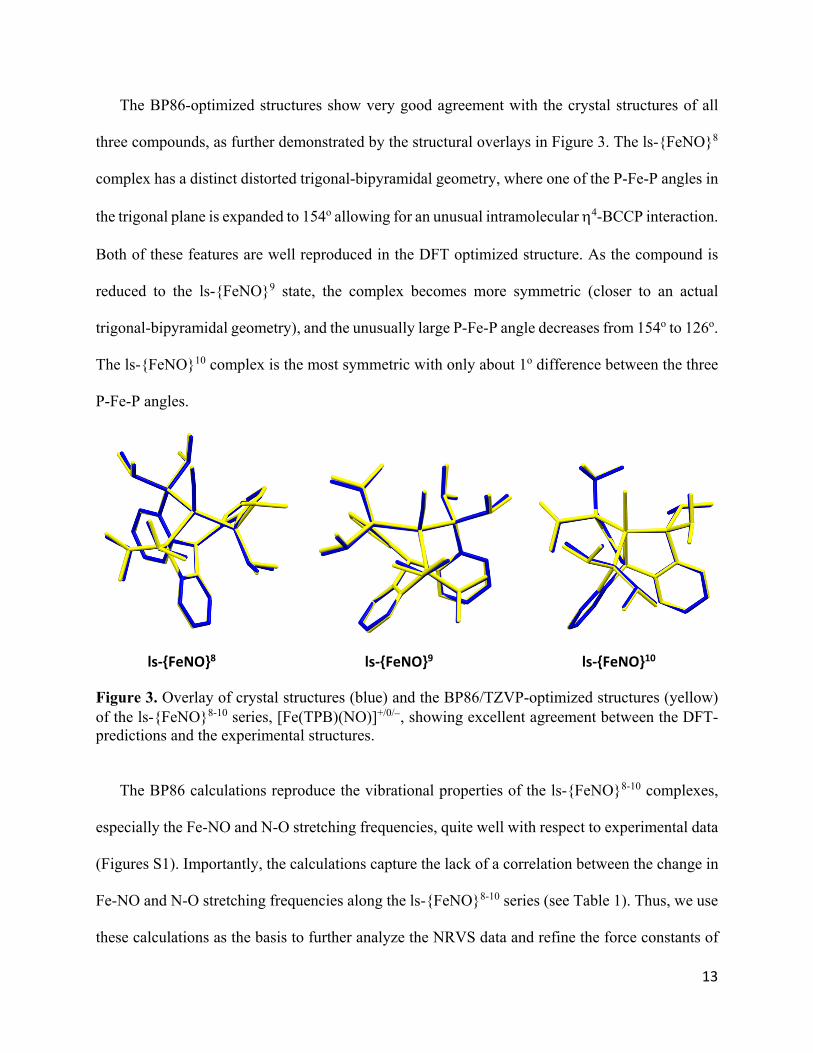
Table 2. Experimental NRVS data vs. QCC-NCA simulation results (in cm-1) and vibrational assignments for the ls-{FeNO}8-10 series.
The same process was applied to the ls-{FeNO}9 and ls-{FeNO}10 compounds, and the
resulting QCC-NCA simulated NRVS data are compared to experiment in Figure 1. Vibrational
assignments are provided in Table 2, and key force constants of the ls-{FeNO}8-10 series are listed
ls-{FeNO}8 ls-{FeNO}9 ls-{FeNO}10
Exp. QCC-NCA Exp. QCC-NCA Exp. QCC-NCA
(FeN) 610 610 583 583 602 602 (N-O) 1745 1745 1667 1667 1568 1568
(Fe-N-O) 537 535 506 (500) 506 (504) 525 536 (Fe-N-O) 540 544 522 527 543 570
16
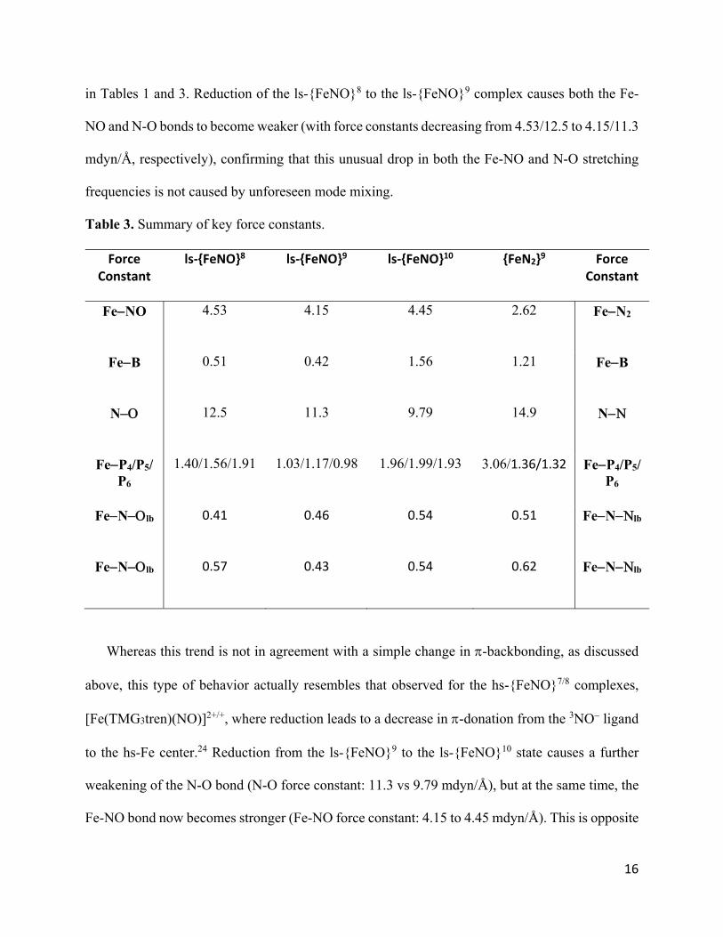
in Tables 1 and 3. Reduction of the ls-{FeNO}8 to the ls-{FeNO}9 complex causes both the Fe-
NO and N-O bonds to become weaker (with force constants decreasing from 4.53/12.5 to 4.15/11.3
mdyn/Å, respectively), confirming that this unusual drop in both the Fe-NO and N-O stretching
frequencies is not caused by unforeseen mode mixing.
Table 3. Summary of key force constants.
Force Constant
ls‐{FeNO}8 ls‐{FeNO}9 ls‐{FeNO}10 {FeN2}9 Force Constant
FeNO 4.53 4.15 4.45 2.62 FeN2
FeB 0.51 0.42 1.56 1.21 FeB
N 12.5 11.3 9.79 14.9 N
FeP4/P5/P6
1.40/1.56/1.91 1.03/1.17/0.98 1.96/1.99/1.93 3.06/1.36/1.32 FeP4/P5/P6
FeNlb 0.41 0.46 0.54 0.51 FeNlb
FeNlb 0.57 0.43 0.54 0.62 FeNlb
Whereas this trend is not in agreement with a simple change in -backbonding, as discussed
above, this type of behavior actually resembles that observed for the hs-{FeNO}7/8 complexes,
[Fe(TMG3tren)(NO)]2+/+, where reduction leads to a decrease in -donation from the 3NO ligand
to the hs-Fe center.24 Reduction from the ls-{FeNO}9 to the ls-{FeNO}10 state causes a further
weakening of the N-O bond (N-O force constant: 11.3 vs 9.79 mdyn/Å), but at the same time, the
Fe-NO bond now becomes stronger (Fe-NO force constant: 4.15 to 4.45 mdyn/Å). This is opposite
17
to the trend observed for the ls-{FeNO}8/9 pair, but in agreement with the trends derived from the
vibrational frequencies (see above).
A distinct Fe-B stretching mode is not observed in the experimental NRVS data. Because of
this, we were unable to optimize the corresponding Fe-B force constants via the QCC-NCA
process, and Table 3 lists the DFT-calculated Fe-B force constants. Nonetheless, the close
agreement between the DFT-predicted and the experimental force constants gives us confidence
that the Fe-B force constants are accurate (±10%).
In the ls-{FeNO}8 and ls-{FeNO}9 complexes, the Fe-B interaction is relatively weak, with a
calculated force constant of ~0.5 mdyn/Å. Reduction to the ls-{FeNO}10 state then causes a
remarkable increase in the Fe-B bond strength, with the Fe-B force constant increasing to 1.56
mdyn/Å. The data thus suggest that an Fe-B single bond forms in the ls-{FeNO}10 state via a
reverse dative bond with the Fe center serving as a Lewis base, donating a pair of electrons to the
borane Lewis acid. This clearly shows that dz2 is doubly occupied in the ls-{FeNO}10 state.
Relatedly, a dative B−→Cu bond has previously been identified computationally and
spectroscopically in [Cu(TPB)]−, 56 and Fe-B flexibility has been implicated as a key feature in
stabilizing Fe across formal redox states.57,58
3.2. Pulse EPR Measurements of the ls-{FeNO}9 Complex
The ls-{FeNO}9 complex [Fe(TPB)(NO)] has an St = 1/2 ground state and is therefore EPR active.
As previously reported, this complex displays an axial EPR signal (g = 1.99, 1.99, 2.45; see Figure
S4) with a large gz value (2.45). This is consistent with the approximate trigonal-bipyramidal
geometry of the complex and an electronic structure in which the electron hole is mostly located
in the xy-plane (with the Fe-NO vector corresponding to the z-axis) and on the metal center
18
(directly indicated by the large g shift). This leads to strong 2nd order spin-orbit coupling in the z
direction. Indeed, similar axial EPR spectra with large gz shifts have been measured for a number
of TPB and P3Si (features Si in place of B) complexes with similar electronic structures (i.e., eg
3
ground states).59 As these complexes vary primarily in the identity of their axial ligand,
information about that Fe-L interaction can be extracted from the g-anisotropy. This is further
analyzed in the Discussion section, in direct comparison to the isoelectronic N2-adduct
[Fe(TPB)(N2)].
Interestingly, if we include all (P3E)Fe-L complexes (E = B in TPB, Si) with an eg
3 ground state
for which an X-ray structure and EPR spectrum has been measured, we find a strong linear
correlation between Δgz and the Fe–P distance (R2 = 0.92). This suggests that the covalency of the
Fe-P bond and/or the out-of-plane displacement of the Fe center might play a key role in
determining Δgz. Furthermore, we find that the Fe center in [Fe(TPB)(NO)] has a Δgz that lies
between those found for formally FeI and FeIII ions in (P3E)Fe-L complexes. Given the vibrational
and computational data are consistent with an NO+ ligand state and thus the Fe is formally Fe−I,
this demonstrates the tremendous ability of a covalently bonded NO+ ligand to accept electron
density.
Analysis of X-band hyperfine sublevel correlation (HYSCORE) spectroscopy acquired on
samples prepared with natural abundance (14N) and 15N labeled NO bound (see Figure 4) allowed
us to accurately determine relatively weak hyperfine coupling constants to the coordinated
14/15N(O) and 11B centers, providing further insight into the electron spin distribution in the
complex. The observed coupling to the 14N nucleus is largely axial consistent with the axial g-
tensor observed in the CW EPR measurements. Simulation of the 15N HYSCORE data allowed for
determination of the nitrogen hyperfine coupling tensor as A(15N) = [8.4, 11.6, -5.4] MHz,
19
independent of any influence from the nuclear quadrupole interaction present in the natural
abundance data due to the presence of the 𝐼 = 1 14N nucleus. Accounting for the relative
gyromagnetic ratios of 14/15N (γ14N/γ15N = -0.7129) the 14N hyperfine coupling tensor is A(14N) =
[-6.0, -8.3, 3.8] MHz, which can be decomposed into an isotropic component aiso(14N) = -3.5 MHz
and an anisotropic component of T(14N) = [−2.5, −4.8, 7.3] MHz. The small aiso value indicates
that minimal spin (estimated: 0.002 e−) is an a1-type orbital (s or pz) with most spin (estimated:
0.065 e−) in the e-symmetric px and py set. These results would be consistent with a spin
polarization mechanism that transfers electron density from the dxy/dx2-y2 orbitals into the px/py
orbitals of the NO ligand. The total spin density of −0.07 e− on the N atom is consistent with the
DFT predictions for a CS state. Comparison of these hyperfine parameters with those similarly
extracted for [Fe(TPB)(NNMe2)]+/− further supports the CS rather than a BS electronic ground
state for the ls-{FeNO}9 complex.
20
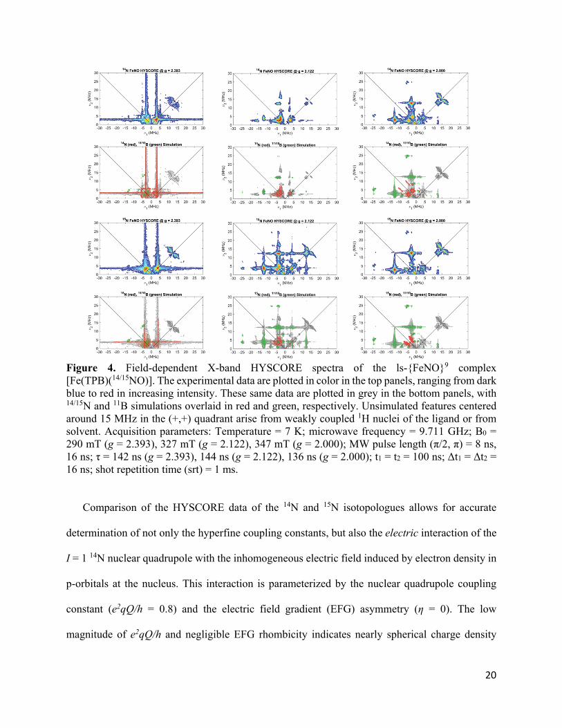
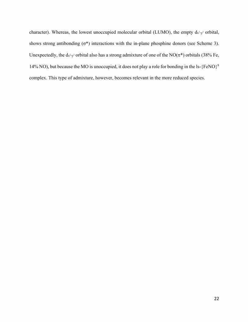
Figure 4. Field-dependent X-band HYSCORE spectra of the ls-{FeNO}9 complex [Fe(TPB)(14/15NO)]. The experimental data are plotted in color in the top panels, ranging from dark blue to red in increasing intensity. These same data are plotted in grey in the bottom panels, with 14/15N and 11B simulations overlaid in red and green, respectively. Unsimulated features centered around 15 MHz in the (+,+) quadrant arise from weakly coupled 1H nuclei of the ligand or from solvent. Acquisition parameters: Temperature = 7 K; microwave frequency = 9.711 GHz; B0 = 290 mT (g = 2.393), 327 mT (g = 2.122), 347 mT (g = 2.000); MW pulse length (π/2, π) = 8 ns, 16 ns; τ = 142 ns (g = 2.393), 144 ns (g = 2.122), 136 ns (g = 2.000); t1 = t2 = 100 ns; Δt1 = Δt2 = 16 ns; shot repetition time (srt) = 1 ms.
Comparison of the HYSCORE data of the 14N and 15N isotopologues allows for accurate
determination of not only the hyperfine coupling constants, but also the electric interaction of the
I = 1 14N nuclear quadrupole with the inhomogeneous electric field induced by electron density in
p-orbitals at the nucleus. This interaction is parameterized by the nuclear quadrupole coupling
constant (e2qQ/h = 0.8) and the electric field gradient (EFG) asymmetry (η = 0). The low
magnitude of e2qQ/h and negligible EFG rhombicity indicates nearly spherical charge density
21
about the nitrogen nucleus in this complex, in agreement with the linear Fe-N-O unit and equal
spin distribution in the px and py orbitals.
The hyperfine coupling to boron with A(11B) = [14.7, 14.7, 18.0] MHz can be decomposed
into aiso(11B) = 15. 8 MHz and a small anisotropic contribution of T(11B) = [−1.1, −1.1, 2.2] MHz.
These data indicate that significantly less electron density is on that ligand (0.006 e− in a1 type
orbitals and 0.017 e− in e-type orbitals) and are consistent with the DFT results. We interpret these
results as being consistent with the lack of available orbitals of appropriate symmetry to accept
electron density from the xy-plane via spin polarization. X-band ENDOR experiments to
determine the hyperfine coupling to 31P of the phosphine ligands are best modeled with a single
class of fairly isotropic coupling constants, A(31P) = [82, 70, 70] MHz, which corresponds to
aiso(31P) = 74 MHz and an anisotropic component of T(31P) = [8, −4, −4] MHz. The large hyperfine
coupling to the 31P centers again supports the idea that the electron hole is mostly located in the
xy-plane.
3.3. Electronic Structure Analysis
The ls-{FeNO}8 Complex has eight valence electrons, as indicated by the Enemark-Feltham
index, and, as discussed above, the complex has a closed-shell singlet ground state, which means
that of the total seven valence MOs (5 Fe(d) + 2 NO(*) orbitals), four valence MOs are doubly
occupied, and three are empty. The MOs themselves are strongly mixed, and Scheme 2 represents
a simplified version of the bonding scheme. Here, the Fe-N-O unit corresponds to the molecular
z-axis. The strong distortion away from C3 symmetry towards a T-shaped geometry in the FeP3
plane, characterized by a large P-Fe-P angle (154°), causes a large energy splitting between the dxy
and dx2-y2 orbitals of 1.97 eV, as indicated in Scheme 3. In this geometry, the lower energy orbital,
dxy (HOMO-1), is essentially -nonbonding with respect to the phosphine ligands (80% Fe
22
character). Whereas, the lowest unoccupied molecular orbital (LUMO), the empty dx2-y2 orbital,
shows strong antibonding (σ*) interactions with the in-plane phosphine donors (see Scheme 3).
Unexpectedly, the dx2-y2 orbital also has a strong admixture of one of the NO(*) orbitals (38% Fe,
14% NO), but because the MO is unoccupied, it does not play a role for bonding in the ls-{FeNO}8
complex. This type of admixture, however, becomes relevant in the more reduced species.
23
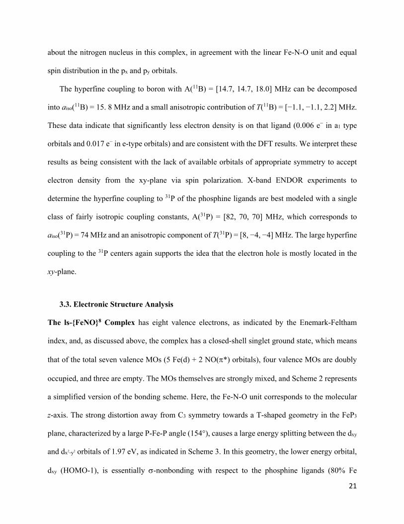
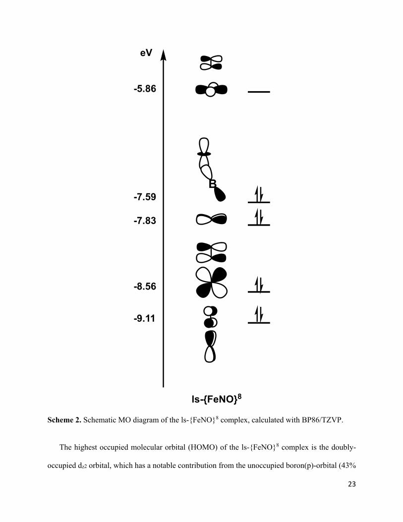
Scheme 2. Schematic MO diagram of the ls-{FeNO}8 complex, calculated with BP86/TZVP.
The highest occupied molecular orbital (HOMO) of the ls-{FeNO}8 complex is the doubly-
occupied dz2 orbital, which has a notable contribution from the unoccupied boron(p)-orbital (43%
B
ls-{FeNO}8
-5.86
-7.59
-7.83
-8.56
-9.11
eV
24
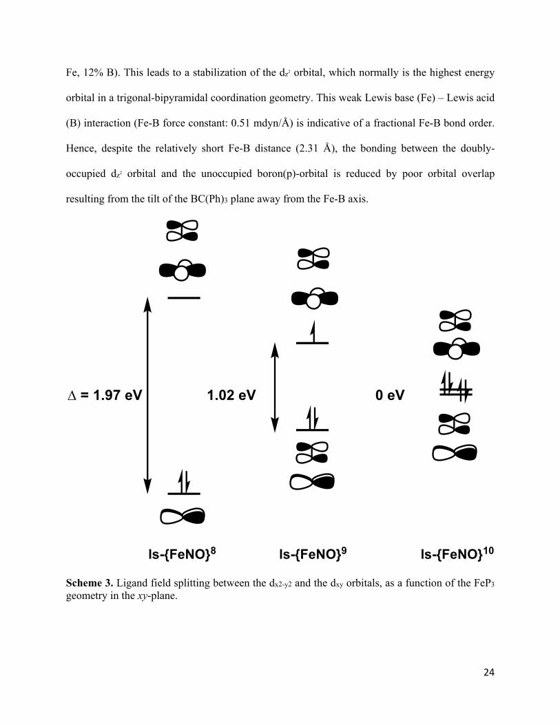
Fe, 12% B). This leads to a stabilization of the dz2 orbital, which normally is the highest energy
orbital in a trigonal-bipyramidal coordination geometry. This weak Lewis base (Fe) – Lewis acid
(B) interaction (Fe-B force constant: 0.51 mdyn/Å) is indicative of a fractional Fe-B bond order.
Hence, despite the relatively short Fe-B distance (2.31 Å), the bonding between the doubly-
occupied dz2 orbital and the unoccupied boron(p)-orbital is reduced by poor orbital overlap
resulting from the tilt of the BC(Ph)3 plane away from the Fe-B axis.
Scheme 3. Ligand field splitting between the dx2-y2 and the dxy orbitals, as a function of the FeP3 geometry in the xy-plane.
= 1.97 eV 1.02 eV 0 eV
ls-{FeNO}8 ls-{FeNO}9 ls-{FeNO}10
25
The lowest-lying valence orbitals are the doubly-occupied, Fe-NO -bonding combinations of
the dxz_*x and dyz_*y orbitals (HOMO-2 and HOMO-3). These bonds are very covalent, with
about 60% Fe(d) and 30% NO(*) contribution.
Based on this analysis, and assigning MOs to the atom or group with the dominant charge
contribution, the ls-{FeNO}8 complex can formally be assigned an Fe(0)-NO+ type electronic
structure with all 8 valence electrons originating primarily from the Fe center, and two very strong
-backbonds with the NO+ ligand (consistent with the large Fe-NO force constant of 4.53
mdyn/Å). The presence of an NO+ ligand explains the absence of spin polarization in this system.
This is similar to six-coordinate ls-{FeNO}6 complexes in hemes, which have been shown to have
a closed-shell Fe(II)-NO+ type ground state with no spin polarization.48,60 In this sense, the FeNO
unit in the ls-{FeNO}8 complex could be considered an electronic analog to that of heme ls-
{FeNO}6 complexes, where two additional electrons of the Fe center are stabilized by the dz2B(p)
interaction. This becomes more evident in the ls-{FeNO}10 system (see below).
Finally, the crystal structure of the ls-{FeNO}8 complex reveals a unique -bond between the
iron center and the C=C bond of one of the aromatic benzene rings. This interaction is unique in
the ls-{FeNO}8 complex and explains the observed, significant contributions of phenyl orbitals to
the valence MOs in this complex, which complicates the analysis. However, this interaction does
not affect the FeNO moiety significantly.
The ls-{FeNO}9 Complex has an EPR-active St = 1/2 ground state, which provides additional
spectroscopic handles to further interrogate its ground state electronic structure. Due to spin-
polarization effects, the - and -spin covalencies differ in the ls-{FeNO}9 complex, which
complicates the analysis of its electronic structure. As we might expect based on its more C3-
symmetric structure, reduction of the ls-{FeNO}8 complex results in an orbital ordering more
26
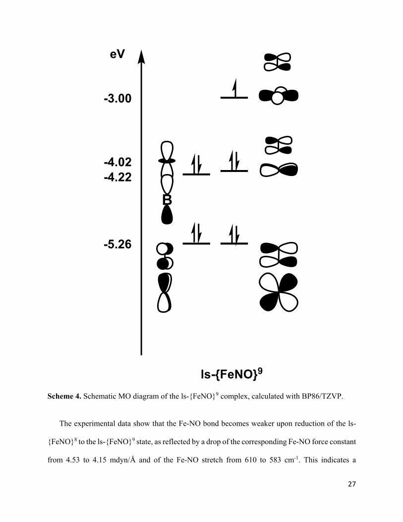
similar to that of a canonical trigonal bipyramid (see Scheme 3). The SOMO of the ls-{FeNO}9
complex is the dx2-y2 orbital, as indicated in Scheme 4, pointing towards an iron-based reduction
(in agreement with the EPR results). Because of this, the Fe-P covalency in the xy-plane is reduced,
and the energy splitting between the dx2-y2 and dxy orbitals decreases to 1.02 eV. Accordingly, the
dxy orbital is now higher in energy than the dz2 orbital, and becomes the SOMO-1. The two lowest
energy valence orbitals remain the Fe-NO π-bonding interactions, which again correspond to the
bonding combinations of the dxz and dyz orbitals and the NO(π*x/y) orbitals. Finally, the dz2 orbital
is again lowered in energy by the Fe-B interaction. Scheme 4 shows the resulting bonding scheme
of the ls-{FeNO}9 complex, which points towards an unusual Fe(−I)-NO+ type ground state.
27
Scheme 4. Schematic MO diagram of the ls-{FeNO}9 complex, calculated with BP86/TZVP.
The experimental data show that the Fe-NO bond becomes weaker upon reduction of the ls-
{FeNO}8 to the ls-{FeNO}9 state, as reflected by a drop of the corresponding Fe-NO force constant
from 4.53 to 4.15 mdyn/Å and of the Fe-NO stretch from 610 to 583 cm-1. This indicates a
B
ls-{FeNO}9
-3.00
-4.02-4.22
-5.26
eV
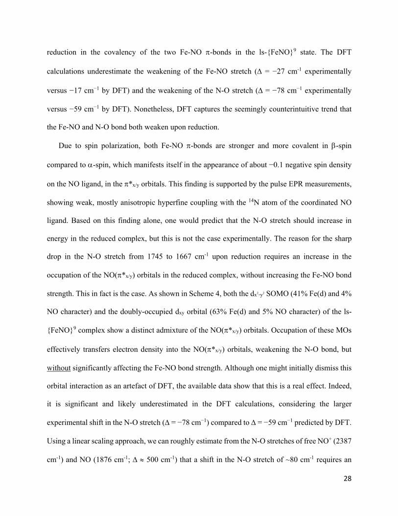
28
reduction in the covalency of the two Fe-NO -bonds in the ls-{FeNO}9 state. The DFT
calculations underestimate the weakening of the Fe-NO stretch ( = −27 cm-1 experimentally
versus −17 cm−1 by DFT) and the weakening of the N-O stretch ( = −78 cm−1 experimentally
versus −59 cm−1 by DFT). Nonetheless, DFT captures the seemingly counterintuitive trend that
the Fe-NO and N-O bond both weaken upon reduction.
Due to spin polarization, both Fe-NO -bonds are stronger and more covalent in -spin
compared to -spin, which manifests itself in the appearance of about −0.1 negative spin density
on the NO ligand, in the *x/y orbitals. This finding is supported by the pulse EPR measurements,
showing weak, mostly anisotropic hyperfine coupling with the 14N atom of the coordinated NO
ligand. Based on this finding alone, one would predict that the N-O stretch should increase in
energy in the reduced complex, but this is not the case experimentally. The reason for the sharp
drop in the N-O stretch from 1745 to 1667 cm-1 upon reduction requires an increase in the
occupation of the NO(*x/y) orbitals in the reduced complex, without increasing the Fe-NO bond
strength. This in fact is the case. As shown in Scheme 4, both the dx2-y2 SOMO (41% Fe(d) and 4%
NO character) and the doubly-occupied dxy orbital (63% Fe(d) and 5% NO character) of the ls-
{FeNO}9 complex show a distinct admixture of the NO(*x/y) orbitals. Occupation of these MOs
effectively transfers electron density into the NO(*x/y) orbitals, weakening the N-O bond, but
without significantly affecting the Fe-NO bond strength. Although one might initially dismiss this
orbital interaction as an artefact of DFT, the available data show that this is a real effect. Indeed,
it is significant and likely underestimated in the DFT calculations, considering the larger
experimental shift in the N-O stretch ( = −78 cm−1) compared to = −59 cm−1 predicted by DFT.
Using a linear scaling approach, we can roughly estimate from the N-O stretches of free NO+ (2387
cm-1) and NO (1876 cm-1; 500 cm-1) that a shift in the N-O stretch of ~80 cm-1 requires an
29
increase in the occupation of the NO(*x/y) orbitals by 0.16 electrons (assuming similar electronic
structures), which is slightly underestimated in the calculations (Loewdin charges for NO: ls-
{FeNO}8: +0.02; ls-{FeNO}9: -0.11, Δ(e−) = 0.13).
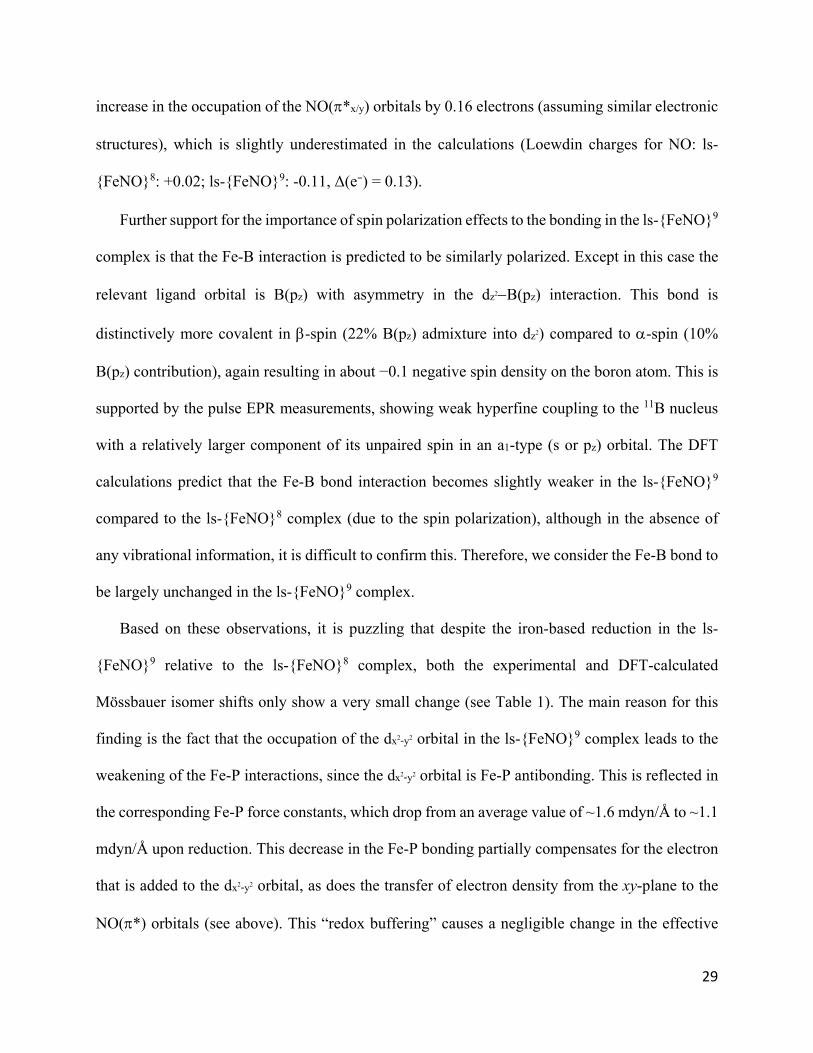
Further support for the importance of spin polarization effects to the bonding in the ls-{FeNO}9
complex is that the Fe-B interaction is predicted to be similarly polarized. Except in this case the
relevant ligand orbital is B(pz) with asymmetry in the dz2B(pz) interaction. This bond is
distinctively more covalent in -spin (22% B(pz) admixture into dz2) compared to -spin (10%
B(pz) contribution), again resulting in about −0.1 negative spin density on the boron atom. This is
supported by the pulse EPR measurements, showing weak hyperfine coupling to the 11B nucleus
with a relatively larger component of its unpaired spin in an a1-type (s or pz) orbital. The DFT
calculations predict that the Fe-B bond interaction becomes slightly weaker in the ls-{FeNO}9
compared to the ls-{FeNO}8 complex (due to the spin polarization), although in the absence of
any vibrational information, it is difficult to confirm this. Therefore, we consider the Fe-B bond to
be largely unchanged in the ls-{FeNO}9 complex.
Based on these observations, it is puzzling that despite the iron-based reduction in the ls-
{FeNO}9 relative to the ls-{FeNO}8 complex, both the experimental and DFT-calculated
Mössbauer isomer shifts only show a very small change (see Table 1). The main reason for this
finding is the fact that the occupation of the dx2-y2 orbital in the ls-{FeNO}9 complex leads to the
weakening of the Fe-P interactions, since the dx2-y2 orbital is Fe-P antibonding. This is reflected in
the corresponding Fe-P force constants, which drop from an average value of ~1.6 mdyn/Å to ~1.1
mdyn/Å upon reduction. This decrease in the Fe-P bonding partially compensates for the electron
that is added to the dx2-y2 orbital, as does the transfer of electron density from the xy-plane to the
NO(*) orbitals (see above). This “redox buffering” causes a negligible change in the effective
30
nuclear charge of the iron center upon reduction, and minimizes the change in the Mössbauer
isomer shift.
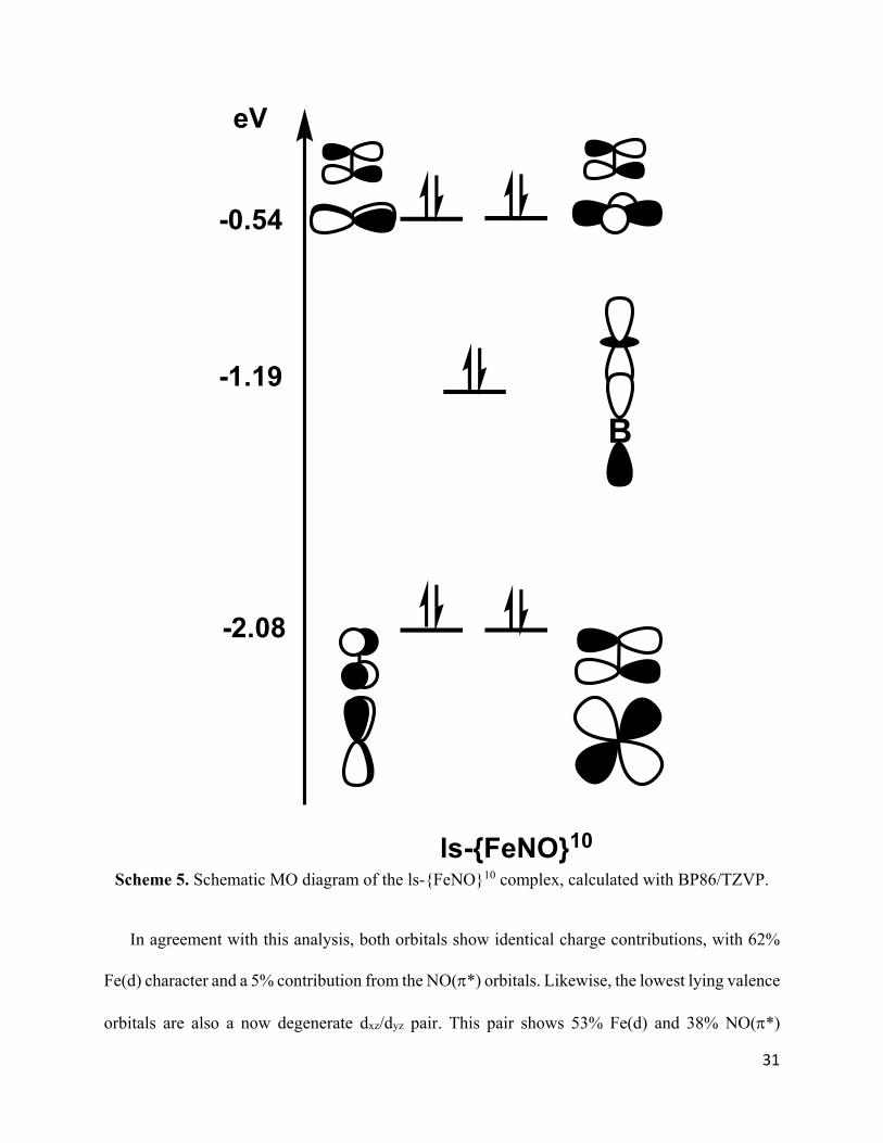
The ls-{FeNO}10 Complex is completely diamagnetic with a CS ground state, as shown in
Scheme 5. Compared to the ls-{FeNO}9 complex, the extra electron is located in the dx2-y2 orbital,
completing the d10 shell of the iron center. Therefore, once again, the reduction is iron-centered.
As a consequence of the now [dxy,dx2-y2]4 electron configuration, the ls-{FeNO}10 complex adapts
an almost perfect trigonal symmetry of the FeP3 unit, causing the dxy and dx2-y2 orbitals to form a
degenerate set (Scheme 3).
31
Scheme 5. Schematic MO diagram of the ls-{FeNO}10 complex, calculated with BP86/TZVP.
In agreement with this analysis, both orbitals show identical charge contributions, with 62%
Fe(d) character and a 5% contribution from the NO(*) orbitals. Likewise, the lowest lying valence
orbitals are also a now degenerate dxz/dyz pair. This pair shows 53% Fe(d) and 38% NO(*)
B
ls-{FeNO}10
-0.54
-1.19
-2.08
eV

32
contributions, indicating the presence of a very covalent Fe-NO bond, similar to that in the ls-
{FeNO}8 complex (60% Fe and 30% NO). Indeed, the similar orbital contributions of the
corresponding dxz_*x and dyz_*y bonding pairs and the similar Fe-NO force constants of 4.53
and 4.45 mdyn/Å are strongly suggestive of similar Fe-NO bonding interactions in the ls-{FeNO}8
and ls-{FeNO}10 complexes. Nonetheless, the N-O stretching frequency in the ls-{FeNO}10
complex is 177 cm−1 lower than in the ls-{FeNO}8 complex, and the N-O force constant is reduced
by about 2.7 mdyn/Å. As discussed for the ls-{FeNO}9 compound, this is best explained not by
increased Fe-NO π-backbonding but rather by the transfer of electron density from the xy-plane
into the NO(π*) orbitals. Indeed, in the ls-{FeNO}10 complex, the dxy/dx2-y2 pair contains 5%
NO(π*) character each, as indicated in Scheme 5. Once again, this likely represents a lower bound
on the magnitude of this effect, given the reduction in the N-O stretching frequency (exp = −99
cm−1 vs DFT = −85 cm−1 compared to ls-{FeNO}9) is underestimated in the calculations.
Due to the formal d10 configuration, the Fe center becomes unusually low-valent (Fe(−II)) in
the ls-{FeNO}10 complex. However, this charge accumulation on the Fe center is largely
compensated by a dramatic strengthening of the Fe-B interaction, indicated by the increase in the
Fe-B force constant to 1.56 mdyn/Å, which corresponds to the formation of an Fe-B single bond.
Here, the iron center becomes a Lewis base and donates one electron pair, located in the doubly-
occupied dz2 orbital, to the boron center, which therefore functions as a Lewis acid in the ls-
{FeNO}10 complex. This mechanism is key to the stabilization of the ls-{FeNO}10 system.
Because of the formation of a full Fe-B single bond, the dz2 orbital drops in energy after reduction
and is now located significantly below the dxy/dx2-y2 degenerate pair. Orbital analysis further reveals
that the corresponding (bonding) MO has 35% Fe(d) and 23% B(pz) charge contributions (the rest
is ligand contribution), in agreement with a very covalent Fe-B interaction. Thus, the ls-{FeNO}10
33
complex has an Fe(-II)-NO+ type electronic structure, but with the electron pair in the dz2 orbital
being strongly stabilized by donation to the boron Lewis acid. In this sense, the ls-{FeNO}10
complex contains two non-innocent ligands and could be designated as ls-{BFeNO}10.
Curiously, the ls-{FeNO}10 complex has the Fe center with the most positive effective nuclear
charge, based on the Mössbauer isomer shift. We attribute the positive isomer shift of the complex
relative to the ls-{FeNO}9 system to (a) the newly formed Fe-B single bond, which reduces the
electron density on the Fe center, and (b) the onset of Fe-P backbonding. Our observations
emphasize the uniqueness of the TPB coligand scaffold and its ability to stabilize extremely low-
valent metal centers through an adjustable interaction between the metal center and the empty pz
orbital of boron. Surprisingly, the effect on the N-O bond strength observed for the ls-{FeNO}8
and ls-{FeNO}10 pair is not so much due to changes in the Fe-NO -bond itself, but due to a
secondary effect, i.e. the admixture of NO(*) character into the dx2-y2/dxy orbital pair as discussed
above.
4. Discussion
In previously characterized redox series of Fe-NO complexes, Mössbauer spectroscopy has
been a key tool for understanding the redox state of the Fe center. In the cyclam-ac supported Fe-
NO series from Wieghardt and coworkers, the change in isomer shift (δ) across redox states is
linear (Δδ ~ 0.2 mm/s per redox state), which has been interpreted in terms of NO-centered redox
changes, dramatically affecting the Fe-NO bond and, in turn, the isomer shift.11,31 In the TMG3tren
supported Fe-NO series from Lehnert and coworkers even larger changes in the isomer shift are
observed (Δδ ~ 0.4 mm/s per redox state), which, in combination with other findings, was taken
as evidence of Fe-centered redox changes.24,61 More recently, Meyer’s hs-{FeNO}7-9 series with
the TIMENMes coligand has also been shown to follow metal-centered reductions, with changes in
34
isomer shift of Δδ ~ 0.2 mm/s.23
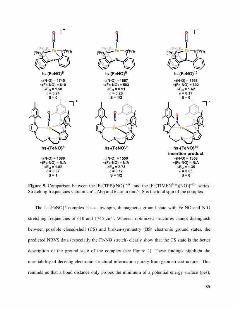
A direct comparison between the [Fe(TPB)(NO)]+/0/ and the [Fe(TIMENMes)(NO)]+/0/
complexes in Figure 5 highlights the stark contrast in stability and reactivity of these low-valent
FeNO systems.62 In addition, the FeNO redox series studied here presents a notable difference to
the previously reported examples in that the Mössbauer isomer shift does not trend linearly with
the redox state of the complex, and the complete range spans less than 0.1 mm/s (0.17-0.26).32
This small overall range speaks to a consistent effective nuclear charge at Fe across our redox
series. Similar observations have been reported in a recent study by Moore et al. on a bimetallic
Fe-Ti system. In this case, redox-induced changes of the effective nuclear charge at Fe are buffered
by the Lewis-acidic Ti center. Thus, changes in the covalency of the Fe-Ti interaction minimize
changes in the isomer shift across the redox series.63 The main objective of this study was therefore
to interrogate the electronic structural changes of the FeNO unit in our [Fe(TPB)(NO)]+/0/ series,
and to identify the origins of the “redox-buffering”. For this purpose, we used different
spectroscopic methods, especially NRVS and pulse EPR, coupled to DFT calculations.
35
Figure 5. Comparison between the [Fe(TPB)(NO)]+/0/ and the [Fe(TIMENMes)(NO)]+/0/ series. Stretching frequencies are in cm-1, EQ and are in mm/s. S is the total spin of the complex.
The ls-{FeNO}8 complex has a low-spin, diamagnetic ground state with Fe-NO and N-O
stretching frequencies of 610 and 1745 cm-1. Whereas optimized structures cannot distinguish
between possible closed-shell (CS) and broken-symmetry (BS) electronic ground states, the
predicted NRVS data (especially the Fe-NO stretch) clearly show that the CS state is the better
description of the ground state of the complex (see Figure 2). These findings highlight the
unreliability of deriving electronic structural information purely from geometric structures. This
reminds us that a bond distance only probes the minimum of a potential energy surface (pes),
(iPr)2P Fe
B
P(iPr)2(iPr)2P
N
O-
(iPr)2P Fe
B
P(iPr)2(iPr)2P
N
O
(iPr)2P Fe
B
P(iPr)2(iPr)2P
N
O+
Fe
N
O
N
N
N
N
N
N
N
hs-{FeNO}8
+
(N-O) = 1686(Fe-NO) = N/AEQ = 1.82= 0.37
S = 1
Fe
N
O
N
N
N
N
N
N
N
(N-O) = 1550(Fe-NO) = N/AEQ = 2.73= 0.17S = 1/2
hs-{FeNO}9
Fe
N
O
N
N
N
N
N
N
N
-
(N-O) = 1358(Fe-NO) = N/AEQ = 1.35= 0.05
S = 0
hs-{FeNO}10
insertion product
ls-{FeNO}8 ls-{FeNO}9 ls-{FeNO}10
(N-O) = 1745(Fe-NO) = 610EQ = 1.50= 0.24
S = 0
(N-O) = 1667(Fe-NO) = 583EQ = 0.91= 0.26S = 1/2
(N-O) = 1568(Fe-NO) = 602EQ = 1.62= 0.17
S = 0
36
whereas a vibrational frequency probes the curvature of the pes around the energy minimum,
which is a much more sensitive gauge for electronic structure and the strength of a bond. Hence,
vibrational data (especially stretching frequencies, in the absence of significant mode-mixing)
provide a superior measure of bond strength. The electronic structure of the ls-{FeNO}8 complex
is best described as Fe(0)-NO+, with two strong, highly covalent Fe-NO -backbonds (see below).
The dz2 orbital is doubly occupied and undergoes a weak but distinct interaction with the boron
center, and the LUMO of the complex is the dx2-y2 orbital.
Upon one-electron reduction, the dx2-y2 orbital becomes singly occupied, leading to an St = 1/2
ground state in the ls-{FeNO}9 complex. The resulting spin-polarization (directly visible as
hyperfine coupling interactions as measured by pulse EPR methods) perturbs both the Fe-NO and
Fe-B interactions, which become weaker. This is reflected by a drop in the Fe-NO stretching
frequency to 583 cm-1. In the Fe-NO -backbonding picture, this should lead to an increase in the
N-O stretch, but this is counteracted by further occupation of the NO(*) orbitals via unusual
mixing with the dx2-y2 and dxy orbitals, which causes the N-O stretch to drop to 1667 cm-1.
Lastly, reduction to the diamagnetic ls-{FeNO}10 state leads to the double occupation of the
dx2-y2 orbital. The strength of Fe-NO bond is restored, evident from an increase in the Fe-NO stretch
to 602 cm-1. This increase in Fe-NO -backbonding (compared to the ls-{FeNO}9 complex) as
well as the further occupation of the NO(*) orbitals (via mixing with the dxy/dx2-y2 pair) causes a
significant drop in the N-O stretch to 1568 cm-1. Based on all of these observations, we conclude
that the Fe-NO -bonds are essentially unchanged along the ls-{FeNO}8-10 series.
Counterintuitively, the “weak link” in this series is actually the ls-{FeNO}9 complex, due to spin
polarization. Importantly, this significant effect of spin polarization on a metal-ligand bond is often
proposed but can rarely be directly observed, as in the NO complexes described in this paper.

37
The Fen-NO+ (n = 0, -1, -2) electronic structure descriptions for our ls-{FeNO}8-10 complexes
include a very strong -backbond, so from a charge perspective the complexes are on average best
described as Fen+1-NO(neutral). Considering that the occupied dxz and dyz orbitals involved in -
backbonding have roughly 30 - 35% NO(*) character, in line with the low N-O stretching
frequencies of the series, the Fen-NO+ description is certainly pushed to an extreme here, especially
in the ls-{FeNO}10 complex, where the charges are estimated around Fe-0.6-NO-0.4. Nevertheless,
besides applying the IUPAC rule (“the winner takes it all”), we also believe that the Fen-
NO+/strong -backbond description has merit and is the most accurate representation of the
electronic structure of the complexes. The two strong -backbonds lead to the transfer of roughly
the same amount of - and -spin electron density back from Fen to the NO+ ligand (in all
complexes), leading to charge accumulation on the NO ligand, without generating any spin (hence,
atypically, the ligand is NO(neutral), but not NO•). Thus, this does not correspond to an actual
electron transfer, as an electron has a charge and a spin but rather is an effect of metal-ligand
covalency. If an actual electron transfer were to happen, the electronic structure would change to
an open shell (BS) ground state like Fen+1-NO• or Fen+2-NO, where the spin(s) of the NO• (S =
1/2) or 3NO (S =1) ligand would likely couple antiferromagnetically to the unpaired electrons of
the iron center to which the ligand is directly bound. However, as we demonstrate in this paper,
such broken symmetry descriptions are not in agreement with the experimental vibrational
(NRVS) data, and can therefore be ruled out. This finding is further supported by the pulse EPR
data, showing only small 14N hyperfine coupling constants in the ls-{FeNO}9 complex. This
difference is not semantic, as our previous work on ferric heme-nitrosyls has shown that the closed-
shell Fen-NO+/strong -backbond versus open shell Fen+1-NO• ground states lead to different
electronic properties and Fe-NO/N-O bond strengths of the complexes.48
38
We further explored the reactivity of our ls-{FeNO}8-10 complexes with X-type ligands (such
as F−), to confirm the NO+ character. However, no reaction was observed. In reactions with more
strongly reducing nucleophiles, such as sources of Me− and H−, we only ever observed reductive
chemistry.
It is notable and worth emphasizing that although the dz2 orbital of Fe is doubly occupied
throughout the redox series, only the ls-{FeNO}10 complex has a strong Fe-B single bond. Thus,
iron only adopts a high degree of Lewis base character upon reduction to formal Fe(−II), not at
Fe(0). Through this reverse dative Fe→B bond, the redox non-innocent tri(aryl)borane subunit of
the TPB ligand system can de facto serve as a redox buffer or electron reservoir by storing two
electrons on site (with minimal effect on the Fe-XY bond of an axially coordinated diatomic). In
this way, the Fe(TPB) platform shifts the accessible redox states of the complex down by 2, and
the anionic complex can be best described as ls-{BFeNO}10. Thus, although the electron density
at Fe is similar in the cationic and anionic complexes, the NO ligand is far more activated due to
the NO(*) admixture into the dxy/dx2-y2 pair. In comparison, the only other known, stable ls-
{FeNO}10 complex is Hieber’s anion, [Fe(CO)3(NO)].64-66 In this case, the three strongly -
backbonding CO ligands take on the role of the boron Lewis acid, and allow for the stabilization
of the highly reduced iron center in this unusual compound.
We suggest that Fe→B bond formation should be an important mechanism for storing electrons
that can facilitate small molecule functionalization steps, such as axial ligand protonations that
oxidize the metal. Such a role has previously been articulated in the context of N2 fixation catalysis
mediated by Fe(TPB).57,67 However, the key intermediate prior to N2 functionalization,
[Fe(TPB)(N2)]− (or {FeN2}9 in analogy to the Enemark-Feltham notation), is isoelectronic to the
39
ls-{FeNO}9 complex, and hence might not be expected to have a signficant Fe→B bond. Both
complexes can be described as Fe(-I) systems with bound N2 and NO+ ligands, respectively.
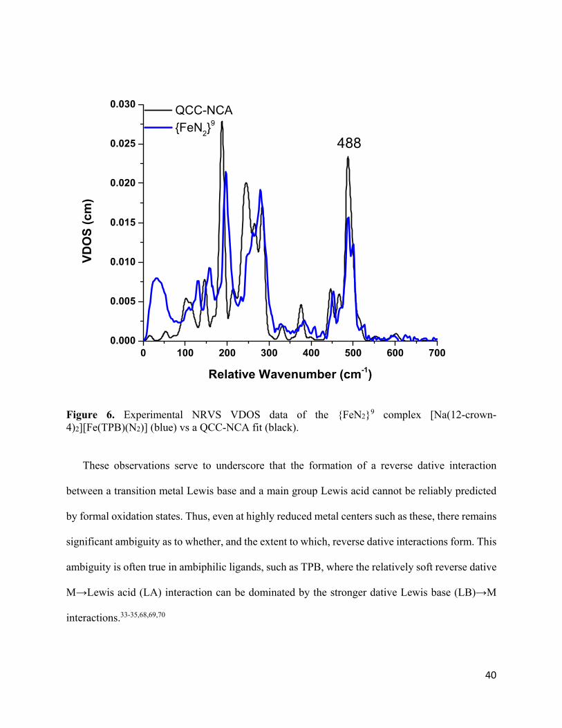
We therefore evaluated the {FeN2}9 complex by NRVS coupled to QCC-NCA analysis to
determine the extent of an Fe→B interaction (Figure 6). The {FeN2}9 species shows a much
weaker Fe-N bond compared to the ls-{FeNO}9 complex, with the Fe-NN stretch observed at 488
cm-1 (corresponding to an Fe-N force constant of 2.62 mdyn/Å, compared to 4.15 mdyn/Å for ls-
{FeNO}9; see Table 3). In turn, a significantly stronger Fe-B interaction is observed in the {FeN2}9
complex (Fe-B force constant 1.21 vs. 0.42 mdyn/Å). Thus, the formally Fe(-I) center is much less
stabilized by N2 than by NO+, consistent with their relative π-accepting abilities. Accordingly, in
the N2 complex, formation of an Fe-B -bond already occurs at the d9 state. These data provide
further support for the hypothesis that Fe-B bonding is critical for achieving productive small
molecule functionalization, including N2 fixation, in this system.57,67
40
Figure 6. Experimental NRVS VDOS data of the {FeN2}9 complex [Na(12-crown-4)2][Fe(TPB)(N2)] (blue) vs a QCC-NCA fit (black).
These observations serve to underscore that the formation of a reverse dative interaction
between a transition metal Lewis base and a main group Lewis acid cannot be reliably predicted
by formal oxidation states. Thus, even at highly reduced metal centers such as these, there remains
significant ambiguity as to whether, and the extent to which, reverse dative interactions form. This
ambiguity is often true in ambiphilic ligands, such as TPB, where the relatively soft reverse dative
M→Lewis acid (LA) interaction can be dominated by the stronger dative Lewis base (LB)→M
interactions.33-35,68,69,70
41
Although the presence of Fe-B interactions in the ls-{FeNO}8-10/{FeN2}9 complexes cannot be
directly observed in the NRVS data, internal calibration of the DFT predicted Fe-B force constants
using the experimentally validated Fe-N and Fe-P interactions provides significant confidence in
the theoretical predictions. Furthermore, the formation of an Fe-B bond in the ls-{FeNO}10
complex is supported by the significant upfield shift of the 11B NMR chemical shift relative to the
ls-{FeNO}8 species (19.9 ppm vs 36.6 ppm).32 These predictions run counter to the expectations
based on a simple geometric analysis and led us to evaluate how predicted Fe-B force constants
correlate with more typically used geometric measures of M→LA bonding, the M-LA distance
and the degree of pyramidalization at the LA.35
In the ls-{FeNO}8-10 series, the Fe-B distance is by far the shortest in ls-{FeNO}8 and is
identical, within error, in the ls-{FeNO}9/10 congeners. Nonetheless, the ls-{FeNO}10 complex has
a significantly larger Fe-B force constant (1.56/0.41/0.52 for ls-{FeNO}10/9/8; see Table 3). The
short Fe-B distance in ls-{FeNO}8 is a result of the aforementioned η4-BCCP→Fe interaction, a
reminder that even in highly related complexes the M-LA distance can be a poor measure of the
M→LA bonding.
Similarly, although both [Fe(TPB)(N2)] (fFe-B = 1.21 mdyn/Å) and [Fe(TPB)(NO)] (fFe-B =
1.56 mdyn/Å) feature significant pyramidalization at boron (Σ(<CBC) = 332.0°, and 331.0°), an
identical degree of pyramidalization is also observed in [Fe(TPB)(NNMe2)]− Σ(<CBC) = 332.1°);
nonetheless, the latter features a much weaker Fe-B bond (fFe-B = 0.44 mdyn/Å).45 Just as structural
comparisons were insufficient to differentiate between CS and BS wavefunctions, they are
insufficient for evaluating the Fe→B interaction. While other spectroscopic techniques, such as
NMR and pulse EPR, can provide insight into M→LA bonding, vibrational spectroscopy provides
a powerful tool to directly interrogate such interactions without the limitations of spin selection
42
rules. In combination with theoretical methods, this enables a thorough mapping of the degree of
M→LA bonding.
Since both the ls-{FeNO}9 and {FeN2}9 complexes have paramagnetic St =1/2 ground states,
further comparisons on their electronic structures can be made using EPR spectroscopy. Based on
this work and previous DFT studies, the SOMO of both complexes is the dx2-y2 orbital, with a d9
valence electron configuration.36 This situation is analogous to tetragonal Cu(II) complexes, and
one might therefore expect a large gz value to originate from spin-orbit coupling (SOC) in the z-
direction between the ground state and the dxy excited state. This is in fact the case, but
interestingly, the g-tensor of the NO+ complex (g = 1.99, 1.99, 2.45) is significantly more axial
(larger Δgz) than that of the N2 complex (g = 2.04, 2.04, 2.31). Based on the usual 2nd order SOC
formalism,71,72 the larger Δgz shift of the ls-{FeNO}9 complex can result from three possibilities:
(a) a distinctly larger spin-orbit coupling constant (which is unlikely), (b) a smaller covalency
factor for the dx2-y2 and dxy orbitals, or (c) a reduction in the energy splitting between the dx2-y2 and
dxy orbitals.
From the crystal structures, we observe a greater out-of-plane shift for the Fe center in the NO+
complex, which could reduce the dx2-y2/dxy energy splitting and, in this way, increase the g shift.
However, this possibility is not supported by the DFT calculations, which show a very similar
energy gap between the dx2-y2 and the dxy orbital (1.02 vs. 0.96 eV).73 On the other hand, the DFT
calculations point to substantially different orbital covalencies for dx2-y2 in these complexes (50%
dx2-y2 character in the N2 compared to 63% in the NO+ complex). Using these numbers and starting
from gz = 2.45 in the ls-{FeNO}9 complex, the gz value for the {FeN2}9 complex would be
predicted to be 2.35, in very good agreement with experiment. Based on this result, we conclude
43
that the differential covalency of the dx2-y2 orbital is to a large degree responsible for the difference
in gz values between these complexes.
In summary, the EPR data further support the notion of an approximate d9 ground state in the
ls-{FeNO}9 and {FeN2}9 complexes, where the larger gz shift in the former complex is due to the
much stronger Fe-NO compared to the Fe-NN bond (evident from the corresponding stretching
frequencies), affecting the metal-ligand covalencies in the xy-plane.
5. Conclusions
The electronic descriptions developed here for the ls-{FeNO}8-10 series are in agreement with all
available spectroscopic data, and emphasize the special role of the TPB ligand in allowing for the
storage of two electrons in the Fe-B bond, enabling the Fe(TPB) complex to reach a very low
oxidation state while allowing for the utilization of these two extra electrons for reductive catalysis.
This complements the more conventional approach in small molecule model chemistry of storing
electrons in the * orbitals of supporting ligands with extended -systems. A prominent example
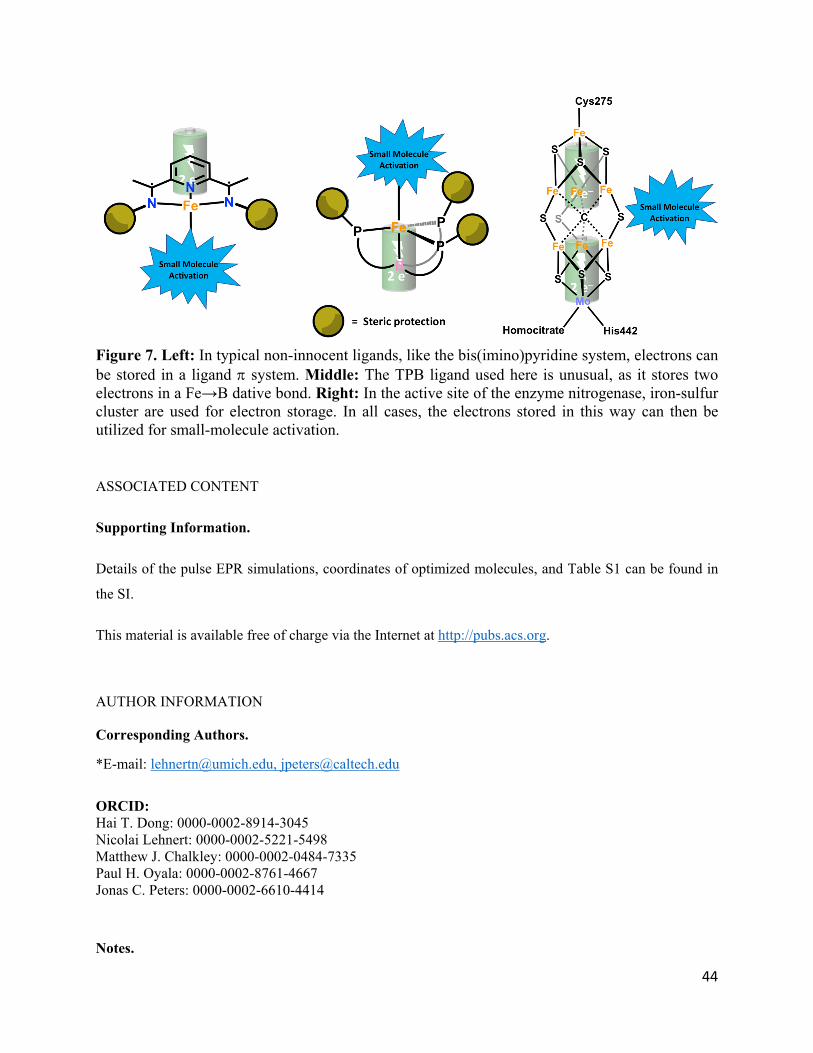
for this approach is the bis(imino)pyridine ligand platform, shown in Figure 7, left.74,75 These
approaches are reminiscent of that used by Nature, in which larger metalloclusters, such as Fe-S
cluster, are electron-loaded before activating small molecules. Prominent examples of this strategy
include the nitrogenase and CO dehydrogenase enzymes (see Figure 7, right).76-79 While in the
case of Fe(TPB) a very low formal oxidation state at Fe must be reached in order for the borane to
adopt this special role, tuning of M→LA interactions potentially provides a route to small molecule
activation under milder conditions.
44
Figure 7. Left: In typical non-innocent ligands, like the bis(imino)pyridine system, electrons can be stored in a ligand system. Middle: The TPB ligand used here is unusual, as it stores two electrons in a Fe→B dative bond. Right: In the active site of the enzyme nitrogenase, iron-sulfur cluster are used for electron storage. In all cases, the electrons stored in this way can then be utilized for small-molecule activation. ASSOCIATED CONTENT
Supporting Information.
Details of the pulse EPR simulations, coordinates of optimized molecules, and Table S1 can be found in
the SI.
This material is available free of charge via the Internet at http://pubs.acs.org.
AUTHOR INFORMATION
Corresponding Authors.
*E-mail: [email protected], [email protected]
ORCID: Hai T. Dong: 0000-0002-8914-3045 Nicolai Lehnert: 0000-0002-5221-5498 Matthew J. Chalkley: 0000-0002-0484-7335 Paul H. Oyala: 0000-0002-8761-4667 Jonas C. Peters: 0000-0002-6610-4414
Notes.
45
The authors declare no competing financial interest.
ACKNOWLEDGMENTS
This work was supported by grants from the National Science Foundation (CHE-1608331 and CHE-
2002855 to NL) and the National Institutes of Health (GM 070757 to JCP). HTD acknowledges support
from the Eastman Summer Research Fellowship and the Robert W. Parry Scholarship. MJC
acknowledges support from the Resnick Sustainability Institute at Caltech. The Caltech EPR facility was
supported by the Dow Next Generation Educator Fund.
46
References 1. Ignarro, L. Nitric Oxide: Biology and Pathobiology; Academic Press: San Diego, 2000. 2. Lehnert, N.; Berto, T. C.; Galinato, M. G. I.; Goodrich, L. E. In The Role of Heme‐Nitrosyls in the Biosynthesis, Transport, Sensing, and Detoxification of Nitric Oxide (NO) in Biological Systems: Enzymes and Model Complexes; Kadish, K. M., Smith, K. M., Guilard, R., Eds.; World Scientific: New Jersey, 2011; Vol. 14,, p 1‐247 3. Wink, D. A.; Mitchell, J. B. Chemical biology of nitric oxide: insights into regulatory, cytotoxic, and cytoprotective mechanisms of nitric oxide. Free Rad. Biol. Med. 1998, 25, 434‐456. 4. Bykov, D.; Neese, F. Six‐Electron Reduction of Nitrite to Ammonia by Cytochrome c Nitrite Reductase: Insights from Density Functional Theory Studies. Inorg. Chem. 2015, 54, 9303‐9316. 5. Ferousi, C.; Majer, S. H.; DiMucci, I. M.; Lancaster, K. M. Biological and Bioinspired Inorganic N–N Bond‐Forming Reactions. Chem. Rev. 2020. 6. Fields, S. Global Nitrogen: Cycling out of Control. Environ Health Perspect. 2004, 112, A556‐A563. 7. Lancaster, K. M.; Caranto, J. D.; Majer, S. H.; Smith, M. A. Alternative Bioenergy: Updates to and Challenges in Nitrification Metalloenzymology. Joule 2018, 2, 421‐441. 8. Lehnert, N.; Dong, H. T.; Harland, J. B.; Hunt, A. P.; White, C. J. Reversing Nitrogen Fixation. Nat. Rev. Chem. 2018, 2, 278‐289. 9. Averill, B. A. Dissimilatory Nitrite and Nitric Oxide Reductases. Chem. Rev. 1996, 96, 2951‐2964. 10. Moura, I.; Moura, J. J. G. Structural Aspects of Denitrifying Enzymes. Curr. Opin. Chem. Biol. 2001, 5, 168‐175. 11. Speelman, A.; Lehnert, N. Heme versus Non‐Heme Iron‐Nitroxyl {FeN(H)O}8 Complexes: Electronic Structure and Biologically Relevant Reactivity. Acc. Chem. Res. 2014, 47, 1106‐1116. 12. Wasser, I. M.; de Vries, S.; Moënne‐Loccoz, P.; Schröder, I.; Karlin, K. D. Nitric Oxide in Biological Denitrification: Fe/Cu Metalloenzymes and Metal Complex NOx Redox Chemistry. Chem. Rev. 2002, 102, 1201‐1234. 13. Hayashi, T.; Caranto, J. D.; Wampler, D. A.; Kurtz, D. M., Jr.; Moënne‐Loccoz, P. Insights into the Nitric Oxide Reductase Mechanism of Flavodiiron Proteins from a Flavin‐Free Enzyme. Biochemistry 2010, 49, 7040−7049. 14. Silaghi‐Dumitrescu, R.; Coulter, E. D.; Das, A.; Ljungdahl, L. G.; Jameson, G. N. L.; Huynh, B. H.; Kurtz, D. M., Jr. A flavodiiron protein and high molecular weight rubredoxin from Moorella thermoacetica with nitric oxide reductase activity. Biochemistry 2003, 42, 2806‐2815. 15. Khatua, S.; Majumdar, A. Flavodiiron Nitric Oxide Reductases: Recent Developments in the Mechanistic Study and Model Chemistry for the Catalytic Reduction of NO. J. Inorg. Biochem. 2015, 142, 145‐153. 16. Lehnert, N.; Fujisawa, K.; Camarena, S.; Dong, H. T.; White, C. J. Activation of Non‐Heme Iron‐Nitrosyl Complexes: Turning up the Heat. ACS Catal. 2019, 9, 10499‐10518. 17. Enemark, J. H.; Feltham, R. D. Principles of Structure, Bonding, and Reactivity for Metal Nitrosyl Complexes. Coord. Chem. Rev. 1974, 13, 339‐406.
47
18. Caranto, J. D.; Weitz, A.; Giri, N.; Hendrich, M. P.; Kurtz, D. M. J. A Diferrous‐Dinitrosyl Intermediate in the N2O‐Generating Pathway of a Deflavinated Flavo‐Diiron Protein. Biochemistry 2014, 53, 5631–5637. 19. Caranto, J. D.; Weitz, A.; Hendrich, M. P.; Kurtz, D. M., Jr. The Nitric Oxide Reductase Mechanism of a Flavo‐Diiron Protein: Identification of Active‐Site Intermediates and Products. J. Am. Chem. Soc. 2014, 136, 7981–7992. 20. Van Stappen, C.; Lehnert, N. Mechanism of N‐N Bond Formation by Transition Metal‐Nitrosyl Complexes: Modeling Flavodiiron Nitric Oxide Reductases. Inorg. Chem. 2018, 57, 4252‐4269. 21. Confer, A. M.; McQuilken, A. C.; Matsumura, H.; Moënne‐Loccoz, P.; Goldberg, D. P. A Nonheme, High‐Spin {FeNO}8 Complex that Spontaneously Generates N2O. J. Am. Chem. Soc. 2017, 139, 10621‐10624. 22. Fujisawa, K.; Soma, S.; Kurihara, H.; Ohta, A.; Dong, H. T.; Minakawa, Y.; Zhao, J.; Alp, E. E.; Hu, M. Y.; Lehnert, N. Stable Ferrous Mononitroxyl {FeNO}8 Complex with a Hindered Hydrotris(pyrazolyl)borate Coligand: Structure, Spectroscopic Characterization, and Reactivity Toward NO and O2. Inorg. Chem. 2019, 58, 4059‐4062. 23. Keilwerth, M.; Hohenberger, J.; Heinemann, F. W.; Sutter, J. r.; Scheurer, A.; Fang, H.; Bill, E.; Neese, F.; Ye, S.; Meyer, K. A Series of Iron Nitrosyl Complexes {Fe–NO}6–9 and a Fleeting {Fe–NO}10 Intermediate en Route to a Metalacyclic Iron Nitrosoalkane. J. Am. Chem. Soc. 2019, 141, 17217‐17235. 24. Speelman, A. L.; White, C. J.; Zhang, B.; Alp, E. E.; Zhao, J.; Hu, M.; Krebs, C.; Penner‐Hahn, J.; Lehnert, N. Non‐heme High‐Spin {FeNO}6‐8 Complexes: One Ligand Platform Can Do It All. J. Am. Chem. Soc. 2018, 140, 11341‐11359. 25. Dong, H. T.; White, C. J.; Zhang, B.; Krebs, C.; Lehnert, N. Non‐Heme Diiron Model Complexes Can Mediate Direct NO Reduction: Mechanistic Insight into Flavodiiron NO Reductases. J. Am. Chem. Soc. 2018, 140, 13429‐13440. 26. Zheng, S.; Berto, T. C.; Dahl, E. W.; Hoffman, M. B.; Speelman, A. L.; Lehnert, N. The Functional Model Complex [Fe2(BPMP)(OPr)(NO)2](BPh4)2 Provides Insight into the Mechanism of Flavodiiron NO Reductases. J. Am. Chem. Soc. 2013, 135, 4902−4905. 27. Jana, M.; Pal, N.; White, C. J.; Kupper, C.; Meyer, F.; Lehnert, N.; Majumdar, A. Functional Mononitrosyl Diiron(II) Complex Mediates the Reduction of NO to N2O with Relevance for Flavodiiron NO Reductases. J. Am. Chem. Soc. 2017, 140, 14380‐14383. 28. Dong, H. T.; Speelman, A. L.; Kozemchak, C. E.; Sil, D.; Krebs, C.; Lehnert, N. The Fe2(NO)2 Diamond Core: A Unique Structural Motif in Non‐Heme Iron‐NO Chemistry. Angew. Chem. Int. Ed. 2019, 131, 17859‐17863. 29. Ye, S.; Price, J. C.; Barr, E. W.; Green, M. T.; Bollinger, J. M.; Krebs, C.; Neese, F. Cryoreduction of the NO‐Adduct of Taurine:α‐Ketoglutarate Dioxygenase (TauD) Yields an Elusive {FeNO}8 Species. J. Am. Chem. Soc. 2010, 132, 4739‐4751. 30. Ng, T. L.; Rohac, R.; Mitchell, A. J.; Boal, A. K.; Balskus, E. P. An N‐nitrosating Metalloenzyme Constructs the Pharmacophore of Streptozotocin. Nature 2019, 566, 94‐99. 31. Serres, R. G.; Grapperhaus, C. A.; Bothe, E.; Bill, E.; Weyhermüller, T.; Neese, F.; Wieghardt, K. Structural, Spectroscopic, and Computational Study of an Octahedral, Non‐Heme {Fe‐NO}6‐8 Series: [Fe(NO)(cyclam‐ac)]2+/+/0. J. Am. Chem. Soc. 2004, 126, 5138‐5153.
48
32. Chalkley, M. J.; Peters, J. C. A Triad of Highly Reduced, Linear Iron Nitrosyl Complexes: {FeNO}8‐10. Angew. Chem. Int. Ed. 2016, 55, 11995‐11998. 33. Jones, J. S.; Gabbaï, F. P. Coordination‐ and Redox‐Noninnocent Behavior of Ambiphilic Ligands Containing Antimony. Acc. Chem. Res. 2016, 49, 857‐867. 34. Braunschweig, H.; Dewhurst, R. D. Transition metals as Lewis bases: “Z‐type” boron ligands and metal‐to‐boron dative bonding. Dalton Trans. 2011, 40, 549‐558. 35. Bouhadir, G.; Bourissou, D. In The Chemical Bond III: 100 years old and getting stronger; Mingos, D. M. P., Ed.; Springer International Publishing: Cham, 2017, p 141‐201. 36. Vyas, N.; Kumar, A.; Ojha, A. K.; Grover, A. Electronic structure of iron dinitrogen complex [(TPB)FeN2]2−/1−/0: correlation to Mössbauer parameters. RSC Adv. 2020, 10, 7948‐7955. 37. Stoll, S.; Schweiger, A. EasySpin, a comprehensive software package for spectral simulation and analysis in EPR. J. Magn. Reson. 2006, 178, 42‐55. 38. Hyde, J. S.; Pasenkiewicz‐Gierula, M.; Jesmanowicz, A.; Antholine, W. E. Pseudo field modulation in EPR spectroscopy. Appl. Magn. Reson. 1990, 1, 483. 39. Frisch, M. J.; Trucks, G. W.; Schlegel, H. B.; Scuseria, G. E.; Robb, M. A.; Cheeseman, J. R.; Scalmani, G.; Barone, V.; Mennucci, B.; Petersson, G. A.; Nakatsuji, H.; Caricato, M.; Li, X.; Hratchian, H. P.; Izmaylov, A. F.; Bloino, J.; Zheng, G.; Sonnenberg, J. L.; Hada, M.; Ehara, M.; Toyota, K.; Fukuda, R.; Hasegawa, J.; Ishida, M.; Nakajima, T.; Honda, Y.; Kitao, O.; Nakai, H.; Vreven, T.; Montgomery, J. A.; Peralta, J. E.; Ogliaro, F.; Bearpark, M.; Heyd, J. J.; Brothers, E.; Kudin, K. N.; Staroverov, V. N.; Kobayashi, R.; Normand, J.; Raghavachari, K.; Rendell, A.; Burant, J. C.; Iyengar, S. S.; Tomasi, J.; Cossi, M.; Rega, N.; Millam, J. M.; Klene, M.; Knox, J. E.; Cross, J. B.; Bakken, V.; Adamo, C.; Jaramillo, J.; Gomperts, R.; Stratmann, R. E.; Yazyev, O.; Austin, A. J.; Cammi, R.; Pomelli, C.; Ochterski, J. W.; Martin, R. L.; Morokuma, K.; Zakrzewski, V. G.; Voth, G. A.; Salvador, P.; Dannenberg, J. J.; Dapprich, S.; Daniels, A. D.; Farkas; Foresman, J. B.; Ortiz, J. V.; Cioslowski, J.; Fox, D. J. Gaussian 09, Revision B.01. Wallingford CT 2009. 40. Praneeth, V. K. K.; Näther, C.; Peters, G.; Lehnert, N. Spectroscopic Properties and Electronic Structure of Five‐ and Six‐Coordinate Iron(II) Porphyrin NO Complexes: Effect of the Axial N‐Donor Ligand. Inorg. Chem. 2006, 45, 2795‐2811. 41. Neese, F. The ORCA program system. Wiley Interdisciplinary Reviews: Computational Molecular Science 2011, 2, 73‐78. 42. Mingos, D. M. P. Nitrosyl Complexes in Inorganic Chemistry, Biochemistry and Medicine II; Springer‐Verlag Berlin Heidelberg, 2014; Vol. 154. 43. McQuarters, A. B.; Kampf, J. W.; Alp, E. E.; Hu, M.; Zhao, J.; Lehnert, N. Ferric Heme‐Nitrosyl Complexes: Kinetically Robust or Unstable Intermediates? Inorg. Chem. 2017, 56, 10513‐10528. 44. Soldatova, A. V.; Ibrahim, M.; Olson, J. S.; Czernuszewicz, R. S.; Spiro, T. G. New Light on NO Bonding in Fe(III) Heme Proteins from Resonance Raman Spectroscopy and DFT Modeling. J. Am. Chem. Soc. 2010, 132, 4614‐4625. 45. Thompson, N. B.; Oyala, P. H.; Dong, H. T.; Chalkley, M. J.; Zhao, J.; Alp, E. E.; Hu, M.; Lehnert, N.; Peters, J. C. Electronic Structures of an [Fe(NNR2)]+/0/– Redox Series: Ligand Noninnocence and Implications for Catalytic Nitrogen Fixation. Inorg. Chem. 2019, 58, 3535‐3549.
49
46. Lehnert, N.; Galinato, M. G. I.; Paulat, F.; Richter‐Addo, G. B.; Sturhahn, W.; Xu, N.; Zhao, J. Nuclear Resonance Vibrational Spectroscopy applied to [Fe(OEP)(NO)]: the Vibrational Assignments of Five‐Coordinate Ferrous Heme Nitrosyls and Implications for Electronic Structure. Inorg. Chem. 2010, 49, 4133‐4148. 47. Goodrich, L. E.; Paulat, F.; Praneeth, V. K. K.; Lehnert, N. Electronic Structure and Reactivity of Heme‐Nitrosyls and Its Significance for Nitric Oxide Sensing, Transport, and Catalysis in Biological Systems. Inorg. Chem. 2010, 49, 6293–6316. 48. Praneeth, V. K. K.; Paulat, F.; Berto, T. C.; DeBeer George, S.; Näther, C.; Sulok, C. D.; Lehnert, N. Electronic Structure of Six‐Coordinate Iron(III)‐Porphyrin NO Adducts: the Elusive Iron(III)‐NO(radical) State and Its Influence on the Properties of these Complexes. J. Am. Chem. Soc. 2008, 130, 15288‐15303. 49. Soma, S.; Van Stappen, C.; Kiss, M.; Szilagyi, R. K.; Lehnert, N.; Fujisawa, K. Distorted Tetrahedral Nickel-Nitrosyl Complexes: Spectroscopic Characterization and Electronic Structure. J. Biol. Inorg. Chem. 2016, 21, 757‐775. 50. Hunt, A. P.; Lehnert, N. The Thiolate Trans Effect in Heme {FeNO}6 Complexes and Beyond: Insight into the Nature of the Push Effect. Inorg. Chem. 2019, 58, 11317‐11332. 51. Fujisawa, K.; Soma, S.; Kurihara, H.; Dong, H. T.; Bilodeau, M.; Lehnert, N. A cobalt‐nitrosyl complex with a hindered hydrotris(pyrazolyl)borate coligand: detailed electronic structure, and reactivity towards dioxygen. Dalton Trans. 2017, 46, 13273‐13289. 52. Van Stappen, C.; Lehnert, N. Mechanism of N–N Bond Formation by Transition Metal–Nitrosyl Complexes: Modeling Flavodiiron Nitric Oxide Reductases. Inorg. Chem. 2018, 57, 4252‐4269. 53. Berto, T. C.; Hoffman, M. B.; Murata, Y.; Landenberger, K. B.; Alp, E. E.; Zhao, J.; Lehnert, N. Structural and Electronic Characterization of Non‐Heme Fe(II)‐Nitrosyls as Biomimetic Models of the FeB Center of Bacterial Nitric Oxide Reductase (NorBC). J. Am. Chem. Soc. 2011, 133, 16714–16717. 54. Berto, T. C.; Speelman, A.; Zheng, S.; Lehnert, N. Mono‐ and Dinuclear Non‐Heme Iron‐Nitrosyl Complexes: Models for Key Intermediates in Bacterial Nitric Oxide Reductases. Coord. Chem. Rev. 2013, 257, 244‐259. 55. Lehnert, N. In Computational Inorganic and Bioinorganic Chemistry; John Wiley & Sons, Chichester, UK: Encyclopedia of Inorganic Chemistry, 2009; Vol. 2009, p 123‐140. 56. Moret, M.‐E.; Zhang, L.; Peters, J. C. A Polar Copper–Boron One‐Electron σ‐Bond. J. Am. Chem. Soc. 2013, 135, 3792‐3795. 57. Thompson, N. B.; Green, M. T.; Peters, J. C. Nitrogen Fixation via a Terminal Fe(IV) Nitride. J. Am. Chem. Soc. 2017, 139, 15312‐15315. 58. Moret, M.‐E.; Peters, J. C. N2 Functionalization at Iron Metallaboratranes. J. Am. Chem. Soc. 2011, 133, 18118‐18121. 59. Gunderson, W. A.; Suess, D. L. M.; Fong, H.; Wang, X.; Hoffmann, C. M.; Cutsail Iii, G. E.; Peters, J. C.; Hoffman, B. M. Free H2 Rotation vs Jahn–Teller Constraints in the Nonclassical Trigonal (TPB)Co–H2 Complex. J. Am. Chem. Soc. 2014, 136, 14998‐15009. 60. McQuarters, A. B.; Kampf, J.; Alp, E. E.; Hu, M. Y.; Zhao, J.; Lehnert, N. Ferric Heme‐Nitrosyl Complexes: Kinetically Robust or Unstable Intermediates? Inorg. Chem. 2017, 56, 10513‐10528.
50
61. Speelman, A.; Zhang, B.; Krebs, C.; Lehnert, N. Structural and Spectroscopic Characterization of a High‐Spin {FeNO}6 Complex with an Iron(IV)‐NO‐ Electronic Structure. Angew. Chem. Int. Ed. 2016, 55, 6685‐6688.
62. In particular, while the putative {FeNO}10 complex, [Fe(TIMENMes)(NO)], immediately undergoes N insertion into one of the iron carbene bonds of the NHC coligand, the ls‐{FeNO}10
complex, [Fe(TPB)(NO)], is stable. This is in agreement with the findings from the NRVS data,
which show that the [Fe(TPB)(NO)]+/0/ complexes all have very strong Fe‐NO bonds, with corresponding Fe‐NO stretching frequencies in the 600 cm‐1 range, and corresponding Fe‐NO force constants > 4 mdyn/Å. . 63. Moore, J. T.; Chatterjee, S.; Tarrago, M.; Clouston, L. J.; Sproules, S.; Bill, E.; Bernales, V.; Gagliardi, L.; Ye, S.; Lancaster, K. M.; Lu, C. C. Enhanced Fe‐Centered Redox Flexibility in Fe–Ti Heterobimetallic Complexes. Inorg. Chem. 2019, 58, 6199‐6214. 64. Burkhardt, L.; Vukadinovic, Y.; Nowakowski, M.; Kalinko, A.; Rudolph, J.; Carlsson, P.‐A.; Jacob, C. R.; Bauer, M. Electronic Structure of the Hieber Anion [Fe(CO)3(NO)]− Revisited by X‐ray Emission and Absorption Spectroscopy. Inorg. Chem. 2020, 59, 3551‐3561. 65. Hieber, W.; Beutner, K. Notizen: Über Nitrosyltricarbonylferrat(‐I), [Fe(CO)3NO]⊖. Zeitschrift für Naturforschung B 1960, 15, 323‐324. 66. Klein, J. E. M. N.; Miehlich, B.; Holzwarth, M. S.; Bauer, M.; Milek, M.; Khusniyarov, M. M.; Knizia, G.; Werner, H.‐J.; Plietker, B. The Electronic Ground State of [Fe(CO)3(NO)]−: A Spectroscopic and Theoretical Study. Angew. Chem. Int. Ed. 2014, 53, 1790‐1794. 67. Anderson, J. S.; Rittle, J.; Peters, J. C. Catalytic conversion of nitrogen to ammonia by an iron model complex. Nature 2013, 501, 84‐87. 68. Hill, A. F. An Unambiguous Electron‐Counting Notation for Metallaboratranes. Organometallics 2006, 25, 4741‐4743. 69. Parkin, G. A Simple Description of the Bonding in Transition‐Metal Borane Complexes. Organometallics 2006, 25, 4744‐4747. 70. Bouhadir, G. B., D. In Ligand Design in Metal Chemistry; Vol. 2016, p 237‐269. 71. Both complexes show strongly Jahn‐Teller (JT) distorted ground states which quenches residual orbital angular momentum and annihilates in‐state SOC. Based on an in‐depth analysis on pseudo‐JT coupling in trigonal Mo‐based d1 systems, 72 it was shown that in such cases second‐order SOC is sufficient to analyze the EPR g tensor. 72. Sharma, A.; Roemelt, M.; Reithofer, M.; Schrock, R. R.; Hoffman, B. M.; Neese, F. EPR/ENDOR and Theoretical Study of the Jahn–Teller‐Active [HIPTN3N]MoVL Complexes (L = N–, NH). Inorg. Chem. 2017, 56, 6906‐6919. 73. Based on this finding, one would expect similar excitation energies for the dxy excited state (dx
2‐y2 dxy excitation) in both complexes.
74. Delle Chiaie, K. R.; Biernesser, A. B.; Ortuño, M. A.; Dereli, B.; Iovan, D. A.; Wilding, M. J. T.; Li, B.; Cramer, C. J.; Byers, J. A. The role of ligand redox non‐innocence in ring‐opening polymerization reactions catalysed by bis(imino)pyridine iron alkoxide complexes. Dalton Trans. 2017, 46, 12971‐12980. 75. Stieber, S. C. E.; Milsmann, C.; Hoyt, J. M.; Turner, Z. R.; Finkelstein, K. D.; Wieghardt, K.; DeBeer, S.; Chirik, P. J. Bis(imino)pyridine Iron Dinitrogen Compounds Revisited: Differences in Electronic Structure Between Four‐ and Five‐Coordinate Derivatives. Inorg. Chem. 2012, 51, 3770‐3785.
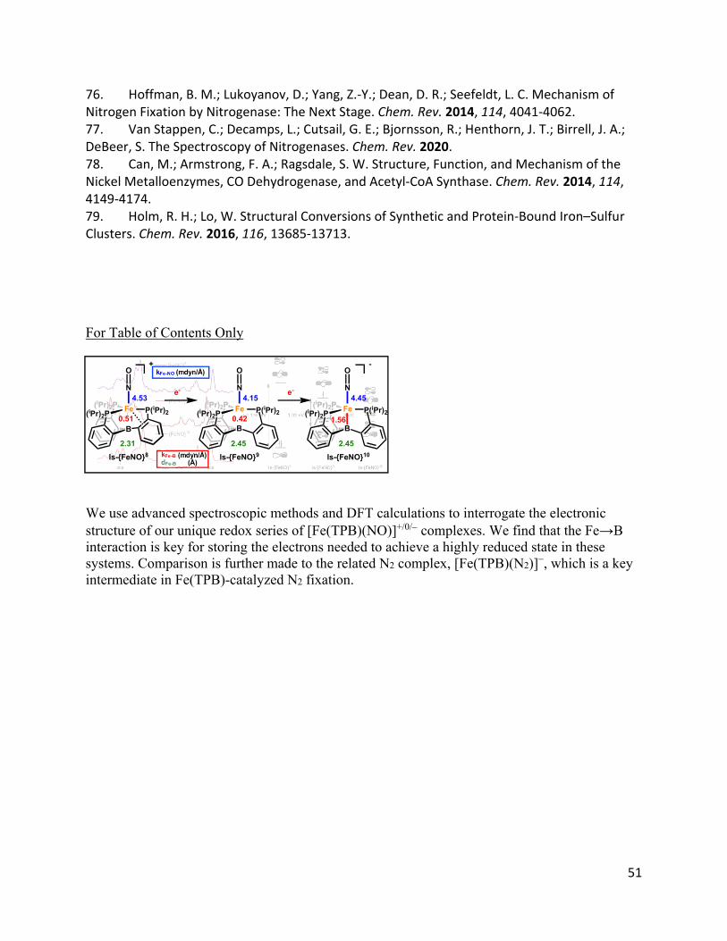
51
76. Hoffman, B. M.; Lukoyanov, D.; Yang, Z.‐Y.; Dean, D. R.; Seefeldt, L. C. Mechanism of Nitrogen Fixation by Nitrogenase: The Next Stage. Chem. Rev. 2014, 114, 4041‐4062. 77. Van Stappen, C.; Decamps, L.; Cutsail, G. E.; Bjornsson, R.; Henthorn, J. T.; Birrell, J. A.; DeBeer, S. The Spectroscopy of Nitrogenases. Chem. Rev. 2020. 78. Can, M.; Armstrong, F. A.; Ragsdale, S. W. Structure, Function, and Mechanism of the Nickel Metalloenzymes, CO Dehydrogenase, and Acetyl‐CoA Synthase. Chem. Rev. 2014, 114, 4149‐4174. 79. Holm, R. H.; Lo, W. Structural Conversions of Synthetic and Protein‐Bound Iron–Sulfur Clusters. Chem. Rev. 2016, 116, 13685‐13713. For Table of Contents Only We use advanced spectroscopic methods and DFT calculations to interrogate the electronic structure of our unique redox series of [Fe(TPB)(NO)]+/0/ complexes. We find that the Fe→B interaction is key for storing the electrons needed to achieve a highly reduced state in these systems. Comparison is further made to the related N2 complex, [Fe(TPB)(N2)]−, which is a key intermediate in Fe(TPB)-catalyzed N2 fixation.
k Fe-N O
(m d yn /Å )
k Fe -B ( Å) d Fe -B
(md yn / Å)
(iPr)2P Fe
B
P(iPr)2(iPr)2P
N
O
1.56
4.45
-
(iPr)2P Fe
B
P(iPr)2(iPr)2P
N
O
0.42
4.15(iPr)2P Fe
B
P(iPr)2(iPr)2P
N
O
0.51
4.53
+
e- e-
ls-{FeNO}8 ls-{FeNO}9 ls-{FeNO}10
2.31 2.45 2.45