Embed Size (px)
Citation preview

REVIEW FOR THE 100TH ANNIVERSARY
Exploring the phytoplasmas, plant pathogenic bacteria
Kensaku Maejima • Kenro Oshima •
Shigetou Namba
Received: 3 September 2013 / Accepted: 11 December 2013 / Published online: 18 March 2014
� The Author(s) 2014. This article is published with open access at Springerlink.com
Abstract Phytoplasmas are plant pathogenic bacteria
associated with devastating damage to over 700 plant
species worldwide. It is agriculturally important to identify
factors involved in their pathogenicity and to discover
effective measures to control phytoplasma diseases.
Despite their economic importance, phytoplasmas remain
the most poorly characterized plant pathogens, primarily
because efforts at in vitro culture, gene delivery, and
mutagenesis have been unsuccessful. However, recent
molecular studies have revealed unique biological features
of phytoplasmas. This review summarizes the history and
recent progress in phytoplasma research, focusing on (1)
the discovery of phytoplasmas, (2) molecular classification
of phytoplasmas, (3) diagnosis of phytoplasma diseases, (4)
reductive evolution of the genomes, (5) characteristic fea-
tures of the plasmids, (6) molecular mechanisms of insect
transmissibility, and (7) virulence factors involved in their
unique symptoms.
Keywords Diagnosis � Genome � Phytoplasma � Insect
transmission � Molecular classification � Virulence factors
Introduction
Phytoplasmas are plant pathogenic bacteria in the class
Mollicutes and are formally called mycoplasma-like
organisms (MLOs) (Doi et al. 1967). They are transmitted
by insect vectors (leafhoppers, planthoppers, and psyllids)
and infect hundreds of plant species worldwide, including
many economically important crops, fruit trees, and orna-
mental plants (Hogenhout et al. 2008; Oshima et al. 2013).
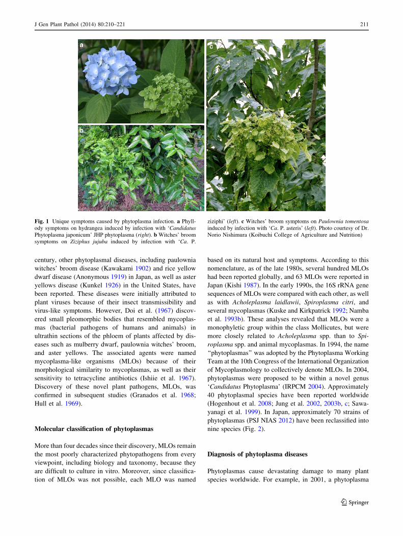
Infected plants show a wide range of symptoms including
stunting, yellowing, witches’ broom (development of
numerous tiny shoot branches with small leaves), phyllody
(formation of leaf-like tissues instead of flowers), vires-
cence (greening of floral organs), proliferation (growth of
shoots from floral organs), purple top (reddening of leaves
and stems), and phloem necrosis (Fig. 1). Phytoplasma
infection is often fatal and causes devastating damage to
global agricultural production. Due not only to difficulties
in culturing phytoplasmas in vitro and in manipulating them
genetically but also to their recalcitrant biological proper-
ties such as restriction to phloem cells, nontransmissibility
by manual inoculation, and high vector specificity, these
organisms are one of the most poorly characterized groups
of plant pathogens. However, recent molecular studies have
revealed interesting features of phytoplasmas. This review
summarizes the history and recent progress in phytoplasma
research, especially in Japan.
Phytoplasmal diseases and discovery of the pathogen
Phytoplasmal diseases have been observed globally. In
Japan, mulberry dwarf disease has caused severe damage to
mulberry plants, the sole source of food for silkworms,
since the Tokugawa period (1603–1868) (Okuda 1972). In
1890, Shirai (the first president of the Phytopathological
Society of Japan and the first professor of plant pathology
at the University of Tokyo) reported that this disease was
transmissible by grafting, although the causal agent
remained unknown (Shirai 1890). Since the early twentieth
K. Maejima � K. Oshima � S. Namba (&)
Department of Agricultural and Environmental Biology,
Graduate School of Agricultural and Life Sciences,
The University of Tokyo, 1-1-1 Yayoi, Bunkyo-ku,
Tokyo 113-8657, Japan
e-mail: [email protected]
123
J Gen Plant Pathol (2014) 80:210–221
DOI 10.1007/s10327-014-0512-8
century, other phytoplasmal diseases, including paulownia
witches’ broom disease (Kawakami 1902) and rice yellow
dwarf disease (Anonymous 1919) in Japan, as well as aster
yellows disease (Kunkel 1926) in the United States, have
been reported. These diseases were initially attributed to
plant viruses because of their insect transmissibility and
virus-like symptoms. However, Doi et al. (1967) discov-
ered small pleomorphic bodies that resembled mycoplas-
mas (bacterial pathogens of humans and animals) in
ultrathin sections of the phloem of plants affected by dis-
eases such as mulberry dwarf, paulownia witches’ broom,
and aster yellows. The associated agents were named
mycoplasma-like organisms (MLOs) because of their
morphological similarity to mycoplasmas, as well as their
sensitivity to tetracycline antibiotics (Ishiie et al. 1967).
Discovery of these novel plant pathogens, MLOs, was
confirmed in subsequent studies (Granados et al. 1968;
Hull et al. 1969).
Molecular classification of phytoplasmas
More than four decades since their discovery, MLOs remain
the most poorly characterized phytopathogens from every
viewpoint, including biology and taxonomy, because they
are difficult to culture in vitro. Moreover, since classifica-
tion of MLOs was not possible, each MLO was named
based on its natural host and symptoms. According to this
nomenclature, as of the late 1980s, several hundred MLOs
had been reported globally, and 63 MLOs were reported in
Japan (Kishi 1987). In the early 1990s, the 16S rRNA gene
sequences of MLOs were compared with each other, as well
as with Acholeplasma laidlawii, Spiroplasma citri, and
several mycoplasmas (Kuske and Kirkpatrick 1992; Namba
et al. 1993b). These analyses revealed that MLOs were a
monophyletic group within the class Mollicutes, but were
more closely related to Acholeplasma spp. than to Spi-
roplasma spp. and animal mycoplasmas. In 1994, the name
‘‘phytoplasmas’’ was adopted by the Phytoplasma Working
Team at the 10th Congress of the International Organization
of Mycoplasmology to collectively denote MLOs. In 2004,
phytoplasmas were proposed to be within a novel genus
‘Candidatus Phytoplasma’ (IRPCM 2004). Approximately
40 phytoplasmal species have been reported worldwide
(Hogenhout et al. 2008; Jung et al. 2002, 2003b, c; Sawa-
yanagi et al. 1999). In Japan, approximately 70 strains of
phytoplasmas (PSJ NIAS 2012) have been reclassified into
nine species (Fig. 2).
Diagnosis of phytoplasma diseases
Phytoplasmas cause devastating damage to many plant
species worldwide. For example, in 2001, a phytoplasma
Fig. 1 Unique symptoms caused by phytoplasma infection. a Phyll-
ody symptoms on hydrangea induced by infection with ‘Candidatus
Phytoplasma japonicum’ JHP phytoplasma (right). b Witches’ broom
symptoms on Ziziphus jujuba induced by infection with ‘Ca. P.
ziziphi’ (left). c Witches’ broom symptoms on Paulownia tomentosa
induced by infection with ‘Ca. P. asteris’ (left). Photo courtesy of Dr.
Norio Nishimura (Koibuchi College of Agriculture and Nutrition)
J Gen Plant Pathol (2014) 80:210–221 211
123

outbreak in apple trees caused losses of about €100 million
in Italy and €25 million in Germany (Strauss 2009). Lethal
yellowing of palm has killed millions of coconut palm trees
in the Caribbean over the past 40 years (Brown et al.
2006). In Japan, phytoplasmas also cause serious economic
losses to agricultural production.
‘Candidatus Phytoplasma asteris’ PaWB strain (PaWB)
is the causal agent of paulownia witches’ broom disease, an
important paulownia tree disease. Once infected with
PaWB, tree vigor is reduced, occasionally followed by
dieback. Until 1960, more than 1 million PaWB-infected
trees were found in Japan, excluding Hokkaido Prefecture
genu
s ‘C
andi
datu
sP
hyto
plas
ma’
SoyST: Ca. P. costaricanum (HQ225630)BVGY (AY083605)
THP: Ca. P. lycopersici (EF199549)RhY (AB738740)MD (AB693124)
OY (AP006628 r001)OAY (M30790)
PaWB (AB693131)AYWB (CP000061 r01)
CbY (AF495882)JHP: Ca. P. japonicum (AB010425)
StrawY: Ca. P. fragariae (HM104662)BY: Ca. P. convolvuli (JN833705)
PPT: Ca. P. americanum (DQ174122)AUSGY: Ca. P. australiense (AM422018 16S rRNA)STOL: Ca. P. solani (AF248959)
SCYLP: Ca. P. graminis (AY725228)PAY: Ca. P. caricae (AY725234)
BWB: Ca. P. rhamni (X76431)SCWB: Ca. P. tamaricis (FJ432664)SpaWB: Ca. P. spartii (X92869)
ESFY: Ca. P. prunorum (AJ542544)AP: Ca. P. mali (AJ542541)
PD: Ca. P. pyri (AJ542543)HibWB: Ca. P. brasiliense (AF147708)
WBDL: Ca. P. aurantifolia (U15442)SPLL (AJ289193)
WX: Ca. P. pruni (L04682)AlmWB: Ca. P. phoenicium (AF515636)
PPWB (U18763)VILL (Y15866)
CWB: Ca. P. omanense (EF666051)BGWL: Ca. P. cynodontis (AJ550984)
RYD: Ca. P. oryzae (D12581)CnWB: Ca. P. castaneae (AB054986)
PinP: Ca. P. pini (AJ310849)LDN: Ca. P. cocosnigeriae* (Y14175)
LD: Ca. P. cocostanzaniae* (X80117)LY: Ca. P. palmae* (U18747)LfWB: Ca. P. luffae* (AF086621)
StLL (Y17055)MaPV: Ca. P. malaysianum (EU371934)
CP: Ca. P. trifolii (AY390261)PassWB: Ca. P. sudamericanum (GU292081)
AshY: Ca. P. fraxini (AF092209)BltWB: Ca. P. balanitae (AB689678)
JWB: Ca. P. ziziphi (AF305240)EY: Ca. P. ulmi (X68376)
FD: Ca. P. vitis (AY197643)Acholeplasma laidlawii (M23932)
100
100
89100
98
100
98
8996
100
96
94
100
99
89
96
91
8489
98
85
87
99
99
89
82
100
100
91
0.01
Ca. P. asteris
Fig. 2 Phylogenetic relationship of phytoplasmas. A phylogenetic
tree was constructed using the neighbor-joining method (Saitou and
Nei 1987) with 16S rRNA gene sequences from phytoplasmas and
acholeplasma (outgroup). Numbers on the branches are the bootstrap
values (only values [80 % are shown). GenBank accession numbers
are in parentheses. Asterisks indicate the proposed provisional names
(IRPCM 2004). Scale bar indicates the number of nucleotide
substitutions per site. Species found in Japan are in bold type.
AlmWB almond witches’ broom, AP apple proliferation, AshY ash
yellows, AUSGY Australian grapevine yellows, AY-WB aster yellows
phytoplasma strain witches’ broom, BGWL Bermuda grass white leaf,
BltWB Balanites triflora witches’ broom, BVGY Buckland Valley
grapevine yellows, BWB buckthorn witches’ broom, BY bindweed
yellows, Ca. P. Candidatus Phytoplasma, CbY chinaberry yellows,
CnWB chestnut witches’ broom, CP clover proliferation, CWB cassia
witches’ broom, ESFY European stone fruit yellows, EY elm yellows,
FD flavescence doree of grapevine, HibWB hibiscus witches’ broom,
JHP Japanese hydrangea phyllody, JWB jujube witches’ broom, LD
coconut lethal yellowing, substrain Tanzanian lethal disease, LDN
coconut lethal yellowing, substrain Nigerian Awka disease, LY
coconut lethal yellowing, LfWB loofah witches’ broom, MaPV
Malaysian periwinkle virescence, MD mulberry dwarf, OAY oeno-
thera aster yellows, OY onion yellows, PassWB passion fruit witches’
broom, PaWB paulownia witches’ broom, PAY papaya, PD pear
decline, PinP pine phytoplasma, PPT potato purple top wilt, PPWB
Caribbean pigeon pea witches’ broom, RhY rhus yellows, RYD rice
yellow dwarf, SCWB salt cedar witches’ broom, SCYLP sugarcane
yellow leaf syndrome, SoyST soybean stunt, SpaWB spartium
witches’ broom, SPLL sweet potato little leaf, STOL stolbur, StrawY
strawberry yellows, THP ‘Hoja de perejil’ (parsley leaf) of tomato,
ViLL vigna little leaf, WBDL witches’ broom disease of lime, WX
western X-disease
212 J Gen Plant Pathol (2014) 80:210–221
123

and the Tohoku region (Ito 1960), and the disease later
spread to almost every plantation in the Tohoku region
(Nakamura et al. 1998). Paulownia witches’ broom disease
also occurs in China (Hiruki 1999), where it has affected
880,000 ha of plantations (Yue et al. 2008). ‘Candidatus
Phytoplasma oryzae’ RYD strain (RYD) is the causal agent
of rice yellow dwarf disease, a serious rice disease in many
regions of Asia. Early infection with RYD results in a
nearly 80 % yield loss. In the 1960s, rice yellow dwarf
disease occurred in 10–50 % of rice growing areas of Japan
and caused serious economic losses in many prefectures
(Komori 1966; Sameshima 1967). In other cases, sweet
potato witches’ broom disease threatened the food supply
in the Ryukyu Islands in the 1950s and 1960s (Nakamori
and Maezato 1968; Shinkai 1964), and hydrangea phyllody
disease remains a destructive disease in hydrangea culti-
vation (Kanehira et al. 1996; Sawayanagi et al. 1999;
Takinami et al. 2013).
There is no effective way to control phytoplasma dis-
eases; thus, early detection and removal of infected plants
are critical to prevent the spread of disease. Until the early
1980s, phytoplasma diseases were diagnosed by transmis-
sion electron microscopic (TEM) observation because of
their small size and the difficulty of culturing phytoplasmas
in vitro. However, that approach requires expensive TEM
equipment and time to prepare ultrathin sections of phloem
tissue. In the 1980s, simple diagnostic techniques for
phytoplasma diseases, such as direct fluorescence detection
(DFD) (Namba et al. 1981) and DAPI staining (Hiruki and
da Rocha 1986), were developed, both of which utilize
florescent microscopy. The DFD method detects autofluo-
rescence of necrotic phloem cells, and DAPI detects phy-
toplasmal DNA. Enzyme-linked immunosorbent assay
(ELISA), the most prevalent method to diagnose viral
diseases, was rarely used to detect phytoplasmal diseases in
the 1980s, except for a few successful cases (Lin and Chen
1985), because it was difficult to prepare a phytoplasma-
specific antibody at that time due to difficulties in purifying
phytoplasmal cells.
Around 1990, advances in molecular biology enabled
direct detection of phytoplasmal DNA by DNA–DNA
hybridization (Kirkpatrick et al. 1987) and the polymerase
chain reaction (PCR) (Deng and Hiruki 1991). PCR
amplification of the 16S rRNA genes of phytoplasmas (Lee
et al. 1993; Namba et al. 1993a; Schneider et al. 1995) has
become the gold standard in the diagnosis of phytoplasmal
diseases. PCR has also been applied to analyze phytopl-
asmal localization and dynamics in planta (Nakashima and
Hayashi 1995b; Sahashi et al. 1995; Wei et al. 2004b).
Recently, loop-mediated isothermal amplification (LAMP)
(Notomi et al. 2000) has also been used to detect several
phytoplasmas and is expected to become a rapid and reli-
able field-diagnostic system for phytoplasma diseases
(Sugawara et al. 2012; Tomlinson et al. 2010). LAMP is
more sensitive and rapid than PCR amplification and does
not require DNA purification or special equipment such as
a thermal cycler. The first LAMP-based detection kit for
phytoplasmas has been commercially available since 2011
in Japan (http://nippongene-analysis.com/phytoplasma-fs.
htm). Application of the kit for on-site diagnosis will aid in
the early control of phytoplasmal diseases.
Reductive evolution of the genomes
Biological and molecular characteristics of phytoplasmas
are still quite unclear because of the difficulty in culturing
them in vitro. However, in the late 1990s, several genome
projects were initiated globally to explore the genomic
features of phytoplasmas. At the same time, several mild-
pathogenicity mutants (Shiomi et al. 1998; Uehara et al.
1999) and a non-insect-transmissible mutant (Oshima et al.
2001b) were identified in Japan. Among these mutant
strains, the ‘Ca. P. asteris’ OY-M strain (OY-M) was
thought to be suitable for whole genome analysis because
the chromosome size of OY-M (ca. 870 kbp) was much
smaller than that of the ‘Ca. P. asteris’ OY-W strain
(OY-W; ca. 1,000 kbp) (Oshima et al. 2001b). In parallel
with the OY-M genome project, the draft genomic
sequences of OY-W were obtained (Oshima et al. 2002;
Miyata et al. 2002b), revealing phytoplasma codon usage
(Miyata et al. 2002a), catalytic activity assays of phytopl-
asmal proteins (Miyata et al. 2003), and the presence of
two rRNA operons (Jung et al. 2003a). In 2004, the com-
plete genomic sequence of OY-M was determined (Oshima
et al. 2004). To date, the complete genomic sequences of
three other phytoplasmas have been reported (Bai et al.
2006; Tran-Nguyen et al. 2008; Kube et al. 2008).
Although phytoplasma genomes contain genes for basic
cellular functions such as DNA replication, transcription,
translation, and protein translocation, they lack genes for
amino acid biosynthesis, fatty acid biosynthesis, the tri-
carboxylic acid cycle, and oxidative phosphorylation, just
as in the mycoplasma genome (Razin et al. 1998). Phy-
toplasma genomes, however, encode even fewer metabol-
ically functional proteins than do mycoplasma genomes,
which were previously thought to have the minimum
possible gene set (Mushegian and Koonin 1996). For
example, phytoplasma genomes lack the pentose phosphate
pathway genes and genes encoding F1Fo-type ATP syn-
thase. Therefore, ATP synthesis in phytoplasmas is
strongly dependent on glycolysis instead of ATP synthase
(Oshima et al. 2007). Interestingly, phytoplasmas harbor
multiple copies of transporter-related genes not found in
mycoplasmas. These genomic features suggest that phy-
toplasmas are highly dependent on metabolic compounds
J Gen Plant Pathol (2014) 80:210–221 213
123

Table 1 Summary of insect vectors of phytoplasma diseases in Japan
Disease name Host plant Causal phytoplasma
species
Insect vector References
Anemone witches’ broom Anemone coronaria Ca. P. asteris Macrosteles striifrons Namba (1996)
Buplever yellows Bupleurum falcatum Ca. P. asteris Macrosteles striifrons Namba (1996)
Carrot yellows Daucus carota Ca. P. pruni Ophiola flavopicta Namba (1996)
Celery yellows Apium graveolens Ca. P. pruni Ophiola flavopicta Namba (1996)
China aster yellows Callistephus chinensis Ca. P. pruni Ophiola flavopicta Namba (1996)
Cineraria witches’ broom Senecio 9 hybridus Ca. P. asteris Macrosteles striifrons Namba (1996)
Cosmos yellows Cosmos bipinnatus Ca. P. asteris Macrosteles striifrons Wakibe et al. (1995)
Cryptotaenia japonica witches’
broom
Cryptotaenia japonica Ca. P. asteris Macrosteles striifrons Namba (1996)
Hishimonus sellatus Nishimura et al.
(1998)
Hishimonoides
sellatiformis
Nishimura et al.
(2004)
Garland chrysanthemum
witches’ broom
Chrysanthemum coronarium var.
spatiosum
Ca. P. asteris Macrosteles striifrons Shiomi and Sugiura
(1983)
Gentian witches’ broom Gentiana scabra var. buergeri Ca. P. pruni Ophiola flavopicta Namba (1996)
Ca. P. asteris Macrosteles striifrons Tanaka et al. (2006)
Geranium witches’ broom Pelargonium 9 hortorum Ca. P. pruni Ophiola flavopicta Namba (1996)
Glaucidium witches’ broom Glaucidium palmatum Ca. P. pruni Ophiola flavopicta Tanaka et al. (1998)
Hovenia witches’ broom Hovenia tomentella Ca. P. asteris Hishimonus sellatus Kusunoki et al.
(2002)
Iceland poppy yellows Papaver nudicaule Ca. P. asteris Macrosteles striifrons Namba (1996)
Jujube witches’ broom Zizyphus jujuba Ca. P. ziziphi Hishimonus sellatus Kusunoki et al.
(2002)
Legume witches’ broom (plants in the family Fabaceae) Ca. P. aurantifolia Orosius orientalis Namba (1996)
Marguerite yellows Argyranthemum frutescens Ca. P. asteris Macrosteles striifrons Namba (1996)
Mulberry dwarf Morus spp. Ca. P. asteris Hishimonus sellatus Namba (1996)
Hishimonoides
sellatiformis
Namba (1996)
Tautoneura mori Jiang et al. (2005)
Onion yellows Allium cepa Ca. P. asteris Macrosteles striifrons Namba (1996)
Hishimonus sellatus Nishimura et al.
(1998)
Hishimonoides
sellatiformis
Nishimura et al.
(2004)
Potato witches’ broom Solanum tuberosum Ca. P. pruni Ophiola flavopicta Namba (1996)
Rhus yellows Rhus javanica Ca. P. asteris Hishimonus sellatus Tanaka et al. (2000)
Rice yellow dwarf Oryza sativa Ca. P. oryzae Nephotettix cincticeps Namba (1996)
Nephotettix
nigropictus
Namba (1996)
Nephotettix virescens Namba (1996)
Rocket larkspur witches’ broom Consolida ambigua Ca. P. asteris Macrosteles striifrons Tanaka et al. (2007)
Statice witches’ broom Limonium spp. Ca. P. asteris Macrosteles striifrons Shiomi et al. (1999)
Strawberry witches’ broom Fragaria 9 ananassa Ca. P. asteris Macrosteles striifrons Namba (1996)
Sweet potato witches’ broom Ipomoea batatas Ca. P. aurantifolia Orosius ryukyuensis Namba (1996)
Tomato yellows Lycopersicon esculentum Ca. P. pruni Ophiola flavopicta Namba (1996)
Ca. P. asteris Macrosteles striifrons Kato et al. (1988)
Tsuwabuki witches’ broom Farfugium japonicum Ca. P. pruni Ophiola flavopicta Namba (1996)
Udo dwarf Aralia cordata Ca. P. pruni Ophiola flavopicta Namba (1996)
Water dropwort yellows Oenanthe javanica Ca. P. asteris Macrosteles striifrons Shiomi and Sugiura
(1983)
214 J Gen Plant Pathol (2014) 80:210–221
123

from their hosts (Oshima et al. 2013). Phytoplasma genomes
contain the sodA gene, which encodes superoxide dismutase
that can inactivate reactive oxygen species (ROS) (Miura
et al. 2012). Plants deploy a broad range of defenses during
infection by various pathogens. The oxidative burst, which
produces ROS, is one of the earliest events in the plant
defense response. Since the genomes of mycoplasmas do not
contain this gene, the presence of sodA may defend phy-
toplasmas against the unique threat of ROS released by the
plant cell. Phytoplasma genomes also contain clusters of
repeated gene sequences called potential mobile units
(PMUs) (Bai et al. 2006), which consist of similar genes
organized in a conserved order (Arashida et al. 2008a; Jo-
mantiene and Davis 2006). A PMU in ‘Ca. P asteris’ AY-WB
strain (AY-WB) exists as both linear chromosomal and cir-
cular extrachromosomal elements (Toruno et al. 2010), sug-
gesting the ability to transpose within the genome.
Characteristic features of the plasmids
In general, a phytoplasma genome consists of one chromo-
some and several small plasmids (Firrao et al. 2007). Phy-
toplasmal plasmids were cloned in the late 1980s (Davis
et al. 1988; Nakashima et al. 1991) and have often been used
as targets of DNA hybridization to compare phytoplasma
strains (Nakashima and Hayashi 1995a). Complete nucleo-
tide sequence analyses of the plasmids revealed that they
may be products of DNA recombination between the phy-
toplasmal plasmids (Nishigawa et al. 2002a) and that each
plasmid encodes a replication initiation protein (Rep)
involved in rolling-circle replication, as well as several other
unknown proteins (Nakashima and Hayashi 1997; Kuboy-
ama et al. 1998; Nishigawa et al. 2003). Interestingly, the
phytoplasmal Reps are similar to the Reps encoded by
bacterial plasmids and to geminivirus (Nakashima and
Hayashi 1997; Nishigawa et al. 2001) and circovirus Reps
(Oshima et al. 2001a), suggestive of evolutionary relation-
ships between phytoplasmas and viruses.
The functions of other genes in the phytoplasmal plas-
mids remain unknown. However, some genes are apparently
related to adaptation to the insect host. For example, ‘Ca. P.
asteris’ OY-NIM strain (OY-NIM), a non-insect-transmis-
sible derivative line of OY-M, lacked the ability to express
the ORF3 gene encoded in plasmids (Nishigawa et al.
2002b; Ishii et al. 2009a). Sequence analysis of the plasmids
over 10 years revealed that one of the plasmids was grad-
ually lost from OY-NIM and finally disappeared during the
maintenance of OY-NIM in plant tissue culture (Ishii et al.
2009b). These results suggested that the plasmid is not
essential for phytoplasmal viability in a plant host, but may
be a key element for adaption to an insect host.
Molecular mechanisms of insect transmissibility
Phytoplasmas are transmitted by specific insect species in a
persistent manner. It is important to understand the molec-
ular mechanisms underlying these strict pathogen–insect
specificities in order to control phytoplasmal diseases. Many
insect vectors involved in phytoplasma diseases have been
identified in Japan (Table 1). Building on knowledge about
the insect vectors, recent studies on phytoplasmal localiza-
tion in nonvector insects revealed that the phytoplasma–
insect specificity is determined by the ability to pass through
several physical barriers, including the midgut and salivary
gland (Nakajima et al. 2002, 2009).
Since phytoplasmas are endocellular parasites that lack a
cell wall, their membrane proteins and secreted proteins
function directly in the host cell. In many phytoplasmas, a
subset of membrane proteins (usually referred to as
immunodominant membrane proteins) accounts for a major
portion of the total cellular membrane proteins (Kakizawa
et al. 2006b). Immunodominant membrane proteins were
classified into three types: immunodominant membrane
protein (Imp) (Kakizawa et al. 2009), antigenic membrane
protein (Amp) (Kakizawa et al. 2004), and immunodomi-
nant membrane protein A (IdpA) (Neriya et al. 2011),
Table 1 continued
Disease name Host plant Causal phytoplasma
species
Insect vector References
Welsh onion yellows Allium fistulosum Ca. P. asteris Macrosteles striifrons Shiomi et al. (1996)
White lace flower yellows Ammi majus Ca. P. asteris Macrosteles striifrons Namba (1996)
Chestnut yellows Castanea spp. Ca. P. castaneae Unknown –
Elaeocarpus yellows Elaeocarpus sylvestris var.
ellipticus
Ca. P. luffae Unknown –
Hydrangea phyllody Hydrangea spp. Ca. P. asteris Unknown –
Ca. P. japonicum Unknown –
Paulownia witches’ broom Paulownia tomentosa Ca. P. asteris Unknown –
J Gen Plant Pathol (2014) 80:210–221 215
123

which are not orthologues. In both OY-W and OY-M, Amp
was a major surface membrane protein (Kakizawa et al.
2009). Cloning of amp genes from several phytoplasma
strains showed that Amp proteins are under positive
selection, and positively selected amino acids can be found
in the central hydrophilic domain (Kakizawa et al. 2006a).
In addition, the Amp protein forms a complex with insect
microfilaments (Suzuki et al. 2006). Interestingly, the for-
mation of Amp–microfilament complexes was correlated
with the phytoplasma-transmitting capability of leafhop-
pers, suggesting that the interaction between Amp and
insect microfilaments may play a major role in determining
the transmissibility of phytoplasmas (Suzuki et al. 2006).
The interaction between Amp and actin (a component of
microfilament), as well as the ATP synthase b subunit of
insect vectors, has also been observed for the CYP strain of
‘Ca. P. asteris’ (Galetto et al. 2011).
Moreover, a phytoplasmal microarray analysis of OY-M
revealed that expression of approximately one-third of the
genes is affected during host switching between plant and
insect, suggesting that the phytoplasma alters its gene
expression in response to its host (Fig. 3) and may use trans-
porters, membrane proteins, secreted proteins, and metabolic
enzymes in a host-specific manner (Oshima et al. 2011).
Virulence factors involved in unique symptoms
Phytoplasmas cause dramatic changes in plant develop-
ment (Arashida et al. 2008b); however, the molecular
Fig. 3 Global patterns of gene regulation in phytoplasma associated with host switching between plant and insect (modified from Oshima et al.
2011). Genes upregulated in the plant host are in green; those upregulated in the insect host are in red
216 J Gen Plant Pathol (2014) 80:210–221
123

mechanisms underlying their pathogenicity remain unclear.
The phytoplasma genome lacks homologs of the type III
secretion system, which are essential for the virulence of
most phytopathogenic bacteria (Abramovitch et al. 2006),
and for toxins produced by Pseudomonas spp., cell wall
maceration enzymes of Pectobacterium spp., and the type
IV secretion system of Rhizobium (Agrobacterium) spp.
(Oshima et al. 2004). On the other hand, phytoplasmas have
no cell walls and reside within host cells, and their secreted
proteins are believed to directly function in the host plant or
insect cells. The genes encoding SecA, SecY, and SecE,
required for protein translocation in Escherichia coli
(Economou 1999), were identified in strain OY-M of ‘Ca. P.
asteris’ (Kakizawa et al. 2001, 2004) and in three other
phytoplasma genomes (Bai et al. 2006; Kube et al. 2008;
Tran-Nguyen et al. 2008). In addition, SecA protein was
expressed in plants infected with five respective phytopl-
asma species (Wei et al. 2004a). These results suggest that
the Sec system is widely conserved among phytoplasmas.
Homologs of the virulence genes of other phytopatho-
genic bacteria were not identified in the phytoplasma
genomes. Therefore, phytoplasmal proteins secreted via the
Sec system are candidate virulence factors that induce
morphological changes. In 2009, a secreted protein
(TENGU) was identified from OY-M as the first phytopl-
asmal virulence factor (Hoshi et al. 2009). TENGU induces
witches’ broom and dwarfism, which are typical of phy-
toplasma infection in Nicotiana benthamiana and Arabi-
dopsis thaliana. TENGU homologs from several
phytoplasma strains induce similar symptoms (Sugawara
et al. 2013). Microarray analysis of TENGU-transgenic
Arabidopsis plants revealed that TENGU inhibits auxin-
related pathways, thereby affecting plant development
(Fig. 4) (Hoshi et al. 2009). More recently, TENGU was
found to be processed in planta into a small functional
peptide, similar to the proteolytic processing of plant
endogenous peptide signals (Sugawara et al. 2013). As for
phyllody symptoms, a previous study has shown the
involvement of the floral homeotic MADS-domain tran-
scription factors of the floral ABCE model (floral quartet
model) (Himeno et al. 2011). Recently, PHYL1 and SAP54
were found to be homologous secreted proteins that induce
phyllody in floral organs of Arabidopsis thaliana (Ma-
cLean et al. 2011; Maejima et al. 2014). PHYL1 actually
interacts with and degrades these MADS-domain tran-
scription factors (Maejima et al. 2014). PHYL1 gene was
genetically and functionally conserved among other phy-
toplasma strains and species. Therefore, PHYL1 and its
homologs were designated as members of the phyllody-
inducing gene family ‘‘phyllogen.’’ SAP11, a secreted
protein from AY-WB, contains eukaryotic nuclear locali-
zation signals and localizes in plant cell nuclei (Bai et al.
2009). SAP11 downregulates JA synthesis and increases
the fecundity of insect vectors (Sugio et al. 2011). In
addition to these secreted proteins, the consumption of
plant metabolites by phytoplasmas may be associated with
virulence. The duplicated glycolytic genes in OY-W are
likely responsible for the severe symptoms (Oshima et al.
2007). Moreover, in the most recent study, activation of the
host anthocyanin biosynthesis pathway in response to
phytoplasma infection is responsible for purple top symp-
toms and is associated with a reduction of leaf cell death
(Himeno et al. 2014). Through all these studies, the
molecular mechanisms of phytoplasma symptoms are
becoming clear. On the other hand, the biological signifi-
cance of the unique symptoms, which is also an interesting
topic, remains largely unknown, and awaits further study.
Concluding remarks
Although phytoplasmas remain the most poorly charac-
terized phytopathogens, recent studies have identified vir-
ulence factors that induce typical phytoplasma disease
TENGU (aa 1-12)
in planta processing
parenchyma
SP (32 aa)
TENGU preprotein
38 aa
phytoplasma cell
TENGU
secretion
phloem
38 aa transportation to adjacent tissues
12 aa
38 aa
apical buds
transportation to meristem tissues
TENGU (aa 1–12)
12 aa
witches’ broom (tengu-su) symptoms
down-regulation of auxin-related genes
Sec system
Fig. 4 Hypothetical mechanism of witches’ broom (tengu-su) symp-
toms induced by a phytoplasmal virulence factor, TENGU. TENGU,
a small peptide secreted from the phytoplasma, is translated as a
70-amino acid preprotein with a signal peptide (SP) of 32 amino acids
(aa) at its N terminus. The C-terminal 38 aa of TENGU preprotein are
secreted into plant phloem via the Sec system, a bacterial conserved
protein translocation system on the cellular membrane, where the
N-terminal signal peptide is cleaved. TENGU is transported from
phloem into parenchyma and apical buds. During the transportation,
TENGU is proteolytically processed into the active form consisting of
N-terminus 12 aa. The active form of TENGU inhibits auxin-related
pathways, resulting in induction of witches’ broom (tengu-su)
symptoms
J Gen Plant Pathol (2014) 80:210–221 217
123

symptoms and have characterized the unique reductive
evolution of the genome. Phytoplasma-related diseases are
expected to increase because the warming global climate is
advantageous to the cold-sensitive vectors of the phytopl-
asmas. Therefore, pest control and detection of phytopl-
asmas are important. Further analysis of phytoplasmas at
the molecular level will increase our understanding of these
economically important and biologically fascinating
microorganisms.
Open Access This article is distributed under the terms of the
Creative Commons Attribution License which permits any use, dis-
tribution, and reproduction in any medium, provided the original
author(s) and the source are credited.
References
Abramovitch RB, Anderson JC, Martin GB (2006) Bacterial elicita-
tion and evasion of plant innate immunity. Nat Rev Mol Cell
Biol 7:601–611
Anonymous (1919) Annu Rep Kochi Agric Res Inst: 62
Arashida R, Kakizawa S, Hoshi A, Ishii Y, Jung H-Y, Kagiwada S,
Yamaji Y, Oshima K, Namba S (2008a) Heterogeneic dynamics
of the structures of multiple gene clusters in two pathogeneti-
cally different lines originating from the same phytoplasma.
DNA Cell Biol 27:209–217
Arashida R, Kakizawa S, Ishii Y, Hoshi A, Jung H-Y, Kagiwada S,
Yamaji Y, Oshima K, Namba S (2008b) Cloning and character-
ization of the antigenic membrane protein (Amp) gene and in situ
detection of Amp from malformed flowers infected with Japanese
hydrangea phyllody phytoplasma. Phytopathology 98:769–775
Bai X, Zhang J, Ewing A, Miller SA, Radek A, Schevchenko DV,
Tsukerman K, Walunas T, Lapidus A, Campbell JW, Hogenhout
SA (2006) Living with genome instability: the adaptation of
phytoplasmas to diverse environments of their insect and plant
hosts. J Bacteriol 188:3682–3696
Bai X, Correa VR, Toruno TY, Ammar E-D, Kamoun S, Hogenhout
SA (2009) AY-WB phytoplasma secretes a protein that targets
plant cell nuclei. Mol Plant Microbe Interact 22:18–30
Brown SE, Been BO, McLaughlin WA (2006) Detection and
variability of the lethal yellowing group (16Sr IV) phytoplasmas
in the Cedusa sp. (Hemiptera: Auchenorrhyncha: Derbidae) in
Jamaica. Ann Appl Biol 149:53–62
Davis MJ, Tsai JH, Cox RL, McDaniel LL, Harrison NA (1988)
Cloning of chromosomal and extrachromosomal DNA of the
mycoplasmalike organism that causes maize bushy stunt disease.
Mol Plant Microbe Interact 1:295–302
Deng S, Hiruki C (1991) Amplification of 16S rRNA genes from
culturable and nonculturable mollicutes. J Microbiol Methods
14:53–61
Doi Y, Teranaka M, Yora K, Asuyama H (1967) Mycoplasma- or
PLT group-like microorganisms found in the phloem elements of
plants infected with mulberry dwarf, potato witches’ broom,
aster yellows or paulownia witches’ broom (in Japanese with
English summary). Ann Phytopath Soc Japan 33:259–266
Economou A (1999) Following the leader: bacterial protein export
through the Sec pathway. Trends Microbiol 7:315–320
Firrao G, Garcia-Chapa M, Marzachı C (2007) Phytoplasmas:
genetics, diagnosis and relationships with the plant and insect
host. Front Biosci 12:1353–1375
Galetto L, Bosco D, Balestrini R, Genre A, Fletcher J, Marzachı C
(2011) The major antigenic membrane protein of ‘‘Candidatus
Phytoplasma asteris’’ selectively interacts with ATP synthase
and actin of leafhopper vectors. PLoS ONE 6:e22571
Granados RR, Maramorosch K, Shikata E (1968) Mycoplasma:
suspected etiologic agent of corn stunt. Proc Natl Acad Sci USA
60:841–844
Himeno M, Neriya Y, Minato N, Miura C, Sugawara K, Ishii Y,
Yamaji Y, Kakizawa S, Oshima K, Namba S (2011) Unique
morphological changes in plant pathogenic phytoplasma-
infected petunia flowers are related to transcriptional regulation
of floral homeotic genes in an organ-specific manner. Plant J
67:971–979
Himeno M, Kitazawa Y, Yoshida T, Maejima K, Yamaji Y, Oshima
K, Namba S (2014) Purple top symptoms are associated with
reduction of leaf cell death in phytoplasma-infected plants. Sci
Rep 4:4111
Hiruki C (1999) Paulownia witches’-broom disease important in East
Asia. Acta Hort 496:63–68
Hiruki C, da Rocha A (1986) Histochemical diagnosis of mycoplasma
infections in Catharanthus roseus by means of a fluorescent
DNA-binding agent, 4,6-diamidino-2-phenylindole-2HCl
(DAPI). Can J Plant Pathol 8:185–188
Hogenhout SA, Oshima K, Ammar E-D, Kakizawa S, Kingdom HN,
Namba S (2008) Phytoplasmas: bacteria that manipulate plants
and insects. Mol Plant Pathol 9:403–423
Hoshi A, Oshima K, Kakizawa S, Ishii Y, Ozeki J, Hashimoto M,
Komatsu K, Kagiwada S, Yamaji Y, Namba S (2009) A unique
virulence factor for proliferation and dwarfism in plants iden-
tified from a phytopathogenic bacterium. Proc Natl Acad Sci
USA 106:6416–6421
Hull R, Home RW, Nayar RM (1969) Mycoplasma-like bodies
associated with sandal spike disease. Nature 224:1121–1122
IRPCM (2004) ‘Candidatus Phytoplasma’, a taxon for the wall-less,
non-helical prokaryotes that colonize plant phloem and insects.
Int J Syst Evol Microbiol 54:1243–1255
Ishii Y, Kakizawa S, Hoshi A, Maejima K, Kagiwada S, Yamaji Y,
Oshima K, Namba S (2009a) In the non-insect-transmissible line
of onion yellows phytoplasma (OY-NIM), the plasmid-encoded
transmembrane protein ORF3 lacks the major promoter region.
Microbiology 155:2058–2067
Ishii Y, Oshima K, Kakizawa S, Hoshi A, Maejima K, Kagiwada S,
Yamaji Y, Namba S (2009b) Process of reductive evolution
during 10 years in plasmids of a non-insect-transmissible
phytoplasma. Gene 446:51–57
Ishiie T, Doi Y, Yora K, Asuyama H (1967) Suppressive effects of
antibiotics of tetracycline group on symptom development of
mulberry dwarf disease. Ann Phytopath Soc Japan 33:267–275
Ito K (1960) Witches’ broom of paulownia trees (in Japanese). Forest
Pests News 9:221–225
Jiang H, Saiki T, Watanabe K, Kawakita H, Sato M (2005) Possible
vector insect of mulberry dwarf phytoplasma, Tautoneura mori
Matsumura. J Gen Plant Pathol 71:370–372
Jomantiene R, Davis RE (2006) Clusters of diverse genes existing as
multiple, sequence-variable mosaics in a phytoplasma genome.
FEMS Microbiol Lett 255:59–65
Jung H-Y, Sawayanagi T, Kakizawa S, Nishigawa H, Miyata S,
Oshima K, Ugaki M, Lee J-T, Hibi T, Namba S (2002)‘Candidatus Phytoplasma castaneae’, a novel phytoplasma taxon
associated with chestnut witches’ broom disease. Int J Syst Evol
Microbiol 52:1543–1549
Jung H-Y, Miyata S, Oshima K, Kakizawa S, Nishigawa H, Wei W,
Suzuki S, Ugaki M, Hibi T, Namba S (2003a) First complete
nucleotide sequence and heterologous gene organization of the
two rRNA operons in the phytoplasma genome. DNA Cell Biol
22:209–215
Jung H-Y, Sawayanagi T, Kakizawa S, Nishigawa H, Wei W, Oshima
K, Miyata S, Ugaki M, Hibi T, Namba S (2003b) ‘Candidatus
218 J Gen Plant Pathol (2014) 80:210–221
123

Phytoplasma ziziphi’, a novel phytoplasma taxon associated with
jujube witches’-broom disease. Int J Syst Evol Microbiol
53:1037–1041
Jung H-Y, Sawayanagi T, Wongkaew P, Kakizawa S, Nishigawa H,
Wei W, Oshima K, Miyata S, Ugaki M, Hibi T, Namba S
(2003c) ‘Candidatus Phytoplasma oryzae’, a novel phytoplasma
taxon associated with rice yellow dwarf disease. Int J Syst Evol
Microbiol 53:1925–1929
Kakizawa S, Oshima K, Kuboyama T, Nishigawa H, Jung H-Y,
Sawayanagi T, Tsuchizaki T, Miyata S, Ugaki M, Namba S
(2001) Cloning and expression analysis of Phytoplasma protein
translocation genes. Mol Plant Microbe Interact 14:1043–1050
Kakizawa S, Oshima K, Nishigawa H, Jung H-Y, Wei W, Suzuki S,
Tanaka M, Miyata S, Ugaki M, Namba S (2004) Secretion of
immunodominant membrane protein from onion yellows phy-
toplasma through the Sec protein-translocation system in Esch-
erichia coli. Microbiology 150:135–142
Kakizawa S, Oshima K, Jung H-Y, Suzuki S, Nishigawa H, Arashida
R, Miyata S, Ugaki M, Kishino H, Namba S (2006a) Positive
selection acting on a surface membrane protein of the plant
pathogenic phytoplasmas. J Bacteriol 188:3424–3428
Kakizawa S, Oshima K, Namba S (2006b) Diversity and functional
importance of phytoplasma membrane proteins. Trends Micro-
biol 14:254–256
Kakizawa S, Oshima K, Ishii Y, Hoshi A, Maejima K, Jung H-Y,
Yamaji Y, Namba S (2009) Cloning of immunodominant
membrane protein genes of phytoplasmas and their in planta
expression. FEMS Microbiol Lett 293:92–101
Kanehira T, Horikoshi N, Yamakita Y, Shinohara M (1996)
Occurrence of hydrangea phyllody in Japan and detection of
the causal phytoplasma (in Japanese with English summary).
Ann Phytopathol Soc Jpn 62:537–540
Kato S, Shiomi T, Wakibe H, Iwanami S (1988) Tomato yellows
transmitted by the leafhopper vector, Macrosteles orientalis
Virbaste (in Japanese with English summary). Ann Phytopath
Soc Japan 54:220–223
Kawakami T (1902) On the hexenbesen of Paulownia tomentosa (in
Japanese). Shokwabo, Tokyo
Kirkpatrick BC, Stenger DC, Morris TJ, Purcell AH (1987) Cloning
and detection of DNA from a nonculturable plant pathogenic
mycoplasma-like organism. Science 238:197–200
Kishi K (1987) The world of loupe in plant pathology (in Japanese).
Ann Phytopath Soc Japan 53:275–278
Komori N (1966) Occurrence and control of rice yellow dwarf disease
in Ibaraki Prefecture (in Japanese). Plant Prot 20:285–288
Kube M, Schneider B, Kuhl H, Dandekar T, Heitmann K, Migdoll
AM, Reinhardt R, Seemuller E (2008) The linear chromosome of
the plant-pathogenic mycoplasma ‘Candidatus Phytoplasma
mali’. BMC Genom 9:306
Kuboyama T, Huang C–C, Lu X, Sawayanagi T, Kanazawa T,
Kagami T, Matsuda I, Tsuchizaki T, Namba S (1998) A plasmid
isolated from phytopathogenic onion yellows phytoplasma and
its heterogeneity in the pathogenic phytoplasma mutant. Mol
Plant Microbe Interact 11:1031–1037
Kunkel LO (1926) Studies on aster yellows. Am J Bot 13:646–705
Kuske CR, Kirkpatrick BC (1992) Phylogenetic relationships between
the western aster yellows mycoplasmalike organism and other
prokaryotes established by 16S rRNA gene sequence. Int J Syst
Bacteriol 42:226–233
Kusunoki M, Shiomi T, Kobayashi M, Okudaira T, Ohashi A, Nohira
T (2002) A leafhopper (Hishimonus sellatus) transmits phylo-
genetically distant phytoplasmas: rhus yellows and hovenia
witches’ broom phytoplasma. J Gen Plant Pathol 68:147–154
Lee I-M, Hammond RW, Davis RE, Gundersen DE (1993) Universal
amplification and analysis of pathogen 16S rDNA for
classification and identification of mycoplasmalike organisms.
Phytopathology 83:834–842
Lin CP, Chen TA (1985) Monoclonal antibodies against the aster
yellows agent. Science 227:1233–1235
MacLean AM, Sugio A, Makarova OV, Findlay KC, Grieve VM,
Toth R, Nicolaisen M, Hogenhout SA (2011) Phytoplasma
effector SAP54 induces indeterminate leaf-like flower develop-
ment in Arabidopsis plants. Plant Physiol 157:831–841
Maejima K, Iwai R, Himeno M, Komatsu K, Kitazawa Y, Fujita N,
Ishikawa K, Fukuoka M, Minato N, Yamaji Y, Oshima K,
Namba S (2014) Recognition of floral homeotic MADS-domain
transcription factors by a phytoplasmal effector, phyllogen,
induces phyllody. Plant J. doi:10.1111/tpj.12495
Miura C, Sugawara K, Neriya Y, Minato N, Keima T, Himeno M,
Maejima K, Komatsu K, Yamaji Y, Oshima K, Namba S (2012)
Functional characterization and gene expression profiling of
superoxide dismutase from plant pathogenic phytoplasma. Gene
510:107–112
Miyata S, Furuki K, Oshima K, Sawayanagi T, Nishigawa H,
Kakizawa S, Jung H-Y, Ugaki M, Namba S (2002a) Complete
nucleotide sequence of the S10-spc operon of phytoplasma: gene
organization and genetic code resemble those of Bacillus
subtilis. DNA Cell Biol 21:527–534
Miyata S, Furuki K, Sawayanagi T, Oshima K, Kuboyama T,
Tsuchizaki T, Ugaki M, Namba S (2002b) Gene arrangement
and sequence of str operon of phytoplasma resemble those of
Bacillus more than those of Mycoplasma. J Gen Plant Pathol
68:62–67
Miyata S, Oshima K, Kakizawa S, Nishigawa H, Jung H-Y,
Kuboyama T, Ugaki M, Namba S (2003) Two different
thymidylate kinase gene homologues, including one that has
catalytic activity, are encoded in the onion yellows phytoplasma
genome. Microbiology 149:2243–2250
Mushegian AR, Koonin EV (1996) A minimal gene set for cellular
life derived by comparison of complete bacterial genomes. Proc
Natl Acad Sci USA 93:10268–10273
Nakajima S, Nishimura N, Matsuda I, Shiomi T, Namba S, Tsuchizaki T
(2002) Detection of mulberry dwarf and onion yellows phytopl-
asmas by PCR from vector insects and nonvector insects (in
Japanese with English summary). Jpn J Phytopathol 68:39–42
Nakajima S, Nishimura N, Jung H-Y, Kakizawa S, Fujisawa I, Namba
S, Tsuchizaki T (2009) Movement of onion yellows phytoplasma
and Cryptotaenia japonica witches’ broom phytoplasma in the
nonvector insect Nephotettix cincticeps (in Japanese with
English summary). Jpn J Phtopathol 75:29–34
Nakamori K, Maezato T (1968) Control of sweet potato witches’
broom disease in Okinawa (in Japanese). Plant Prot 22:19–24
Nakamura H, Ohgake S, Sahashi N, Yoshioka N, Kubono T,
Takahashi T (1998) Seasonal variation of paulownia witches’-
broom phytoplasma in paulownia trees and distribution of the
disease in the Tohoku district of Japan. J Forest Res 3:39–42
Nakashima K, Hayashi T (1995a) Extrachromosomal DNAs of rice
yellow dwarf and sugarcane white leaf phytoplasmas. Ann
Phytopathol Soc Jpn 61:456–462
Nakashima K, Hayashi T (1995b) Multiplication and distribution of
rice yellow dwarf phytoplasma in infected tissues of rice and
green rice leafhopper Nephotettix cincticeps. Ann Phytopathol
Soc Jpn 61:451–455
Nakashima K, Hayashi T (1997) Sequence analysis of extrachromo-
somal DNA of sugarcane white leaf phytoplasma. Ann Phyto-
pathol Soc Jpn 63:21–25
Nakashima K, Kato S, Iwanami S, Murata N (1991) Cloning and
detection of chromosomal and extrachromosomal DNA from
mycoplasmalike organisms that cause yellow dwarf disease of
rice. Appl Environ Microbiol 57:3570–3575
J Gen Plant Pathol (2014) 80:210–221 219
123

Namba S (1996) Taxonomy of phytoplasmas (in Japanese). Plant Prot
50:152–156
Namba S, Yamashita S, Doi Y, Yora K (1981) Direct fluorescence
detection method (DFD method) for diagnosing yellow-type
virus diseases and mycoplasma diseases of plants (in Japanese
with English summary). Ann Phytopath Soc Japan 47:258–263
Namba S, Kato S, Iwanami S, Oyaizu H, Shiozawa H, Tsuchizaki T
(1993a) Detection and differentiation of plant-pathogenic my-
coplasmalike organisms using polymerase chain reaction. Phy-
topathology 83:786–791
Namba S, Oyaizu H, Kato S, Iwanami S, Tsuchizaki T (1993b)
Phylogenetic diversity of phytopathogenic mycoplasmalike
organisms. Int J Syst Bacteriol 43:461–467
Neriya Y, Sugawara K, Maejima K, Hashimoto M, Komatsu K,
Minato N, Miura C, Kakizawa S, Yamaji Y, Oshima K, Namba S
(2011) Cloning, expression analysis, and sequence diversity of
genes encoding two different immunodominant membrane
proteins in poinsettia branch-inducing phytoplasma (PoiBI).
FEMS Microbiol Lett 324:38–47
Nishigawa H, Miyata S, Oshima K, Sawayanagi T, Komoto A,
Kuboyama T, Matsuda I, Tsuchizaki T, Namba S (2001) In
planta expression of a protein encoded by the extrachromosomal
DNA of a phytoplasma and related to geminivirus replication
proteins. Microbiology 147:507–513
Nishigawa H, Oshima K, Kakizawa S, Jung H-Y, Kuboyama T,
Miyata S, Ugaki M, Namba S (2002a) Evidence of intermolec-
ular recombination between extrachromosomal DNAs in phy-
toplasma: a trigger for the biological diversity of phytoplasma?
Microbiology 148:1389–1396
Nishigawa H, Oshima K, Kakizawa S, Jung H-Y, Kuboyama T,
Miyata S, Ugaki M, Namba S (2002b) A plasmid from a non-
insect-transmissible line of a phytoplasma lacks two open
reading frames that exist in the plasmid from the wild-type line.
Gene 298:195–201
Nishigawa H, Oshima K, Miyata S, Ugaki M, Namba S (2003)
Complete set of extrachromosomal DNAs from three pathogenic
lines of onion yellows phytoplasma and use of PCR to
differentiate each line. J Gen Plant Pathol 69:194–198
Nishimura N, Nakajima S, Sawayanagi T, Namba S, Shiomi T,
Matsuda I, Tsuchizaki T (1998) Transmission of Cryptotaenia
japonica witches’ broom and onion yellows phytoplasmas by
Hishimonus sellatus Uhler. Ann Phytopathol Soc Jpn
64:474–477
Nishimura N, Nakajima S, Kawakita H, Sato M, Namba S, Fujisawa I,
Tsuchizaki T (2004) Transmission of Cryptotaenia japonica
witches’ broom and onion yellows by Hishimonoides sellatifor-
mis (in Japanese with English summary). Jpn J Phytopathol
70:22–25
Notomi T, Okayama H, Masubuchi H, Yonekawa T, Watanabe K,
Amino N, Hase T (2000) Loop-mediated isothermal amplifica-
tion of DNA. Nucleic Acids Res 28:e63
Okuda S (1972) Phytoplasmal diseases in Japan (in Japanese). Plant
Prot 26:180–183
Oshima K, Kakizawa S, Nishigawa H, Kuboyama T, Miyata S, Ugaki
M, Namba S (2001a) A plasmid of phytoplasma encodes a
unique replication protein having both plasmid- and virus-like
domains: clue to viral ancestry or result of virus/plasmid
recombination? Virology 285:270–277
Oshima K, Shiomi T, Kuboyama T, Sawayanagi T, Nishigawa H,
Kakizawa S, Miyata S, Ugaki M, Namba S (2001b) Isolation and
characterization of derivative lines of the onion yellows
phytoplasma that do not cause stunting or phloem hyperplasia.
Phytopathology 91:1024–1029
Oshima K, Miyata S, Sawayanagi T, Kakizawa S, Nishigawa H, Jung
H-Y, Furuki K, Yanazaki M, Suzuki S, Wei W, Kuboyama T,
Ugaki M, Namba S (2002) Minimal set of metabolic pathways
suggested from the genome of onion yellows phytoplasma. J Gen
Plant Pathol 68:225–236
Oshima K, Kakizawa S, Nishigawa H, Jung H-Y, Wei W, Suzuki S,
Arashida R, Nakata D, Miyata S, Ugaki M, Namba S (2004)
Reductive evolution suggested from the complete genome
sequence of a plant-pathogenic phytoplasma. Nat Genet
36:27–29
Oshima K, Kakizawa S, Arashida R, Ishii Y, Hoshi A, Hayashi Y,
Kagiwada S, Namba S (2007) Presence of two glycolytic gene
clusters in a severe pathogenic line of Candidatus Phytoplasma
asteris. Mol Plant Pathol 8:481–489
Oshima K, Ishii Y, Kakizawa S, Sugawara K, Neriya Y, Himeno M,
Minato N, Miura C, Shiraishi T, Yamaji Y, Namba S (2011)
Dramatic transcriptional changes in an intracellular parasite
enable host switching between plant and insect. PLoS ONE
6:e23242
Oshima K, Maejima K, Namba S (2013) Genomic and evolutionary
aspects of phytoplasmas. Front Microbiol 4:230
PSJ NIAS (ed) (2012) Common names of plant diseases in Japan, 2nd
edn (in Japanese). Phase out, Aichi
Razin S, Yogev D, Naot Y (1998) Molecular biology and pathoge-
nicity of mycoplasmas. Microbiol Mol Biol Rev 62:1094–1156
Sahashi N, Nakamura H, Yoshikawa N, Kubono T, Shoji T,
Takahashi T (1995) Distribution and seasonal variation in
detection of phytoplasma in bark phloem tissues of single
paulownia trees infected with witches’ broom. Ann Phytopathol
Soc Jpn 61:481–484
Saitou N, Nei M (1987) The neighbor-joining method: a new method
for reconstructing phylogenetic trees. Mol Biol Evol 4:406–425
Sameshima T (1967) Occurrence and control of rice yellow dwarf
disease in Miyazaki Prefecture (in Japanese). Plant Prot
21:47–50
Sawayanagi T, Horikoshi N, Kanehira T, Shinohara M, Bertaccini A,
Cousin M-T, Hiruki C, Namba S (1999) ‘Candidatus Phytopl-
asma japonicum’, a new phytoplasma taxon associated with
Japanese hydrangea phyllody. Int J Syst Bacteriol 49:1275–1285
Schneider B, Seemuller E, Smart CD, Kirkpatrick BC (1995)
Phylogenetic classification of plant pathogenic mycoplasma-like
organisms or phytoplasmas. In: Razin S, Tully JG (eds)
Molecular and diagnostic procedures in mycoplasmology. Aca-
demic Press, San Diego, pp 369–380
Shinkai A (1964) Transmission of sweet potato witches’ broom
disease by Orosius ryukyuensis (in Japanese). Plant Prot
18:259–262
Shiomi T, Sugiura M (1983) Water dropwort yellows and chrysan-
themum witches’ broom occurred in Ishikawa Prefecture (in
Japanese with English summary). Ann Phytopath Soc Japan
49:367–370
Shiomi T, Tanaka M, Wakiya H, Zenbayashi R (1996) Occurrence of
welsh onion yellows (in Japanese with English summary). Ann
Phytopathol Soc Jpn 62:258–260
Shiomi T, Tanaka M, Sawayanagi T, Yamamoto S, Tsuchizaki T,
Namba S (1998) A symptomatic mutant of onion yellows
phytoplasma derived from a greenhouse-maintained isolate (in
Japanese with English summary). Ann Phytopathol Soc Jpn
64:501–505
Shiomi T, Tanaka M, Uematsu S, Wakibe H, Nakamura H (1999)
Statice witches’ broom, a Macrosteles striifrons-borne phytopl-
asma disease (in Japanese with English summary). Ann Phyto-
pathol Soc Jpn 65:87–90
Shirai M (1890) The cause of mulberry dwarf disease (in Japanese).
Jpn Silk Culturist 32:1–2
Strauss E (2009) Phytoplasma research begins to bloom. Science
325:388–390
Sugawara K, Himeno M, Keima T, Kitazawa Y, Maejima K, Oshima
K, Namba S (2012) Rapid and reliable detection of phytoplasma
220 J Gen Plant Pathol (2014) 80:210–221
123

by loop-mediated isothermal amplification targeting a house-
keeping gene. J Gen Plant Pathol 78:389–397
Sugawara K, Honma Y, Komatsu K, Himeno M, Oshima K, Namba S
(2013) The alteration of plant morphology by small peptides
released from the proteolytic processing of the bacterial peptide
TENGU. Plant Physiol 162:2005–2014
Sugio A, Kingdom HN, MacLean AM, Grieve VM, Hogenhout SA
(2011) Phytoplasma protein effector SAP11 enhances insect
vector reproduction by manipulating plant development and
defense hormone biosynthesis. Proc Natl Acad Sci USA
108:E1254–E1263
Suzuki S, Oshima K, Kakizawa S, Arashida R, Jung H-Y, Yamaji Y,
Nishigawa H, Ugaki M, Namba S (2006) Interaction between the
membrane protein of a pathogen and insect microfilament
complex determines insect–vector specificity. Proc Natl Acad
Sci USA 103:4252–4257
Takinami Y, Maejima K, Takahashi A, Keima T, Shiraishi T, Okano
Y, Komatsu K, Oshima K, Namba S (2013) First report of
‘Candidatus Phytoplasma asteris’ infecting hydrangea showing
phyllody in Japan. J Gen Plant Pathol 79:209–213
Tanaka M, Shiomi T, Tairako K, Uehara T, Matsuda I (1998)
Occurrence of glaucidium witches’ broom (in Japanese with
English summary). Ann Phytopathol Soc Jpn 64:205–207
Tanaka M, Osada S, Matsuda I (2000) Transmission of rhus (Rhus
javanica L.) yellows by Hishimonus sellatus and host range of
the causal phytoplasma. J Gen Plant Pathol 66:323–326
Tanaka M, Kodama K, Chiba K, Iwadate Y, Usugi T (2006)
Occurrence of gentian witches’ broom caused by ‘Candidatus
Phytoplasma asteris’ (in Japanese with English summary). Jpn J
Phytopathol 72:191–194
Tanaka M, Tanina K, Kasuyama S, Usugi T (2007) Occurrence of
rocket larkspur witches’ broom caused by ‘‘Candidatus Phy-
toplasma asteris’’ in Japan. J Gen Plant Pathol 73:286–289
Tomlinson JA, Boonham N, Dickinson M (2010) Development and
evaluation of a one-hour DNA extraction and loop-mediated
isothermal amplification assay for rapid detection of phytoplas-
mas. Plant Pathol 59:465–471
Toruno TY, Music MS, Simi S, Nicolaisen M, Hogenhout SA (2010)
Phytoplasma PMU1 exists as linear chromosomal and circular
extrachromosomal elements and has enhanced expression in
insect vectors compared with plant hosts. Mol Microbiol
77:1406–1415
Tran-Nguyen LTT, Kube M, Schneider B, Reinhardt R, Gibb KS
(2008) Comparative genome analysis of ‘‘Candidatus Phytopl-
asma australiense’’ (subgroup tuf-Australia I; rp-A) and ‘‘Ca.
Phytoplasma asteris’’ strains OY-M and AY-WB. J Bacteriol
190:3979–3991
Uehara T, Tanaka M, Shiomi T, Namba S, Tsuchizaki T, Matsuda I
(1999) Histopathological studies on two symptom types of
phytoplasma associated with lettuce yellows. Ann Phytopathol
Soc Jpn 65:465–469
Wakibe H, Shiomi T, Tanaka M (1995) Cosmos yellows disease
transmitted by Macrosteles striifrons in Saga Prefecture (in Japanese
with English summary). Proc Assoc Plant Prot Kyushu 41:51–54
Wei W, Kakizawa S, Jung H-Y, Suzuki S, Tanaka M, Nishigawa H,
Miyata S, Oshima K, Ugaki M, Hibi T, Namba S (2004a) An
antibody against the SecA membrane protein of one phytoplas-
ma reacts with those of phylogenetically different phytoplasmas.
Phytopathology 94:683–686
Wei W, Kakizawa S, Suzuki S, Jung H-Y, Nishigawa H, Miyata S,
Oshima K, Ugaki M, Hibi T, Namba S (2004b) In planta
dynamic analysis of onion yellows phytoplasma using localized
inoculation by insect transmission. Phytopathology 94:244–250
Yue HN, Wu YF, Shi YZ, Wu KK, Li YR (2008) First report of
paulownia witches’-broom phytoplasma in China. Plant Dis
92:1134
J Gen Plant Pathol (2014) 80:210–221 221
123