Embed Size (px)
Citation preview
Expression and Detrimental Role of HematopoieticProstaglandin D Synthase in Spinal CordContusion Injury
ADRIANA REDENSEK,1 KHIZR I. RATHORE,1 JENNIFER L. BERARD,1 RUBEN L�OPEZ-VALES,1
LEIGH ANNE SWAYNE,2 STEFFANY A.L. BENNETT,2 IKUKO MOHRI,3 MASAKO TANIIKE,3
YOSHIHIRO URADE,3 AND SAMUEL DAVID1*1Center for Research in Neuroscience, Research Institute of the McGill University Health Center, Montreal,Quebec, Canada H3G 1A42Neural Regeneration Laboratory, Department. of Biochemistry, Microbiology, and Immunology, University of Ottawa,ON, Canada3Department of Molecular Behavioral Biology, Osaka Bioscience Institute, Suita, Osaka 565-0874, Japan
KEY WORDSspinal cord injury; inflammation; prostaglandins
ABSTRACTProstaglandin D2 (PGD2) is a potent inflammatory mediator,which is implicated in both the initiation and resolution ofinflammation in peripheral non-neural tissues. Its role in thecentral nervous system has not been fully elucidated. Spinalcord injury (SCI) is associated with an acute inflammatoryresponse, which contributes to secondary tissue damage thatworsens functional loss. We show here, with the use of hema-topoietic prostaglandin D synthase (HPGDS) deficient miceand a HPGDS selective inhibitor (HQL-79), that PGD2 playsa detrimental role after SCI. We also show that HPGDS isexpressed in macrophages in the injured mouse spinal cordand contributes to the increase in PGD2 in the contusedspinal cord. HPGDS2/2 mice also show reduced secondarytissue damage and reduced expression of the proinflamma-tory chemokine CXCL10 as well as an increase in IL-6 andTGFb-1 expression in the injured spinal cord. This wasaccompanied by a reduction in the expression of the micro-glia/macrophage activation marker Mac-2 and an increase inthe antioxidant metallothionein III. Importantly, HPGDSdeficient mice exhibit significantly better locomotor recoveryafter spinal cord contusion injury than wild-type (Wt) mice.In addition, systemically administered HPGDS inhibitor(HQL-79) also enhanced locomotor recovery after SCI in Wtmice. These data suggest that PGD2 generated via HPGDShas detrimental effects after SCI and that blocking the activ-ity of this enzyme can be beneficial. VVC 2011 Wiley-Liss, Inc.
INTRODUCTION
Prostaglandin D2 (PGD2) is a potent inflammatorymediator produced by two synthases, hematopoieticprostaglandin D synthase (HPGDS), and lipocalin-typeprostaglandin D synthase (L-PGDS). Arachidonic acidassociated with membrane phospholipids is cleaved byphospholipase A2 and converted to a prostanoid precur-sor by the cycloxygenases-1/2 (COX-1/2). The prostanoidprecursor, prostaglandin H2, can then be converted byHPGDS or L-PGDS to PGD2. In the central nervous sys-tem (CNS), L-PGDS is expressed by oligodendrocytes,
meningeal cells, and the choroid plexus (Beuckmannet al., 2000; Urade et al., 1993). In contrast, HPGDS isexpressed by certain immune cells of hematopoietic ori-gin (Lewis et al., 1982; Mohri et al., 2003; Ujihara et al.,1988; Urade et al., 1989, 1990). PGD2 can induce theproduction of IL-2, IL-4, IL-5, and IL-13 in Th2 cells(Tanaka et al., 2004) and act as a chemoattractant forTh2 cells, eosinophils, and basophils (Hirai et al., 2001),which has led to studies on the role of PGD2 andHPGDS in allergic immune responses (Kostenis andUlven, 2006). Various studies have demonstrated thatPGD2 can also promote the resolution of inflammation(Angeli et al., 2004; Matsuoka et al., 2000; Rajakariaret al., 2007; Spik et al., 2005). As PGD2 can act througha variety of mechanisms, its role as either a pro or anti-inflammatory mediator remains to be fully elucidated.PGD2 is very abundant in the cerebrospinal fluid, likelyderived from the choroid plexus and meninges; and inthe normal CNS plays a role in the regulation of thesleep-wake cycle (Hayaishi and Urade, 2002). Recentstudies suggest that PGD2 generated via HPGDS playsa role in CNS inflammatory responses. HPGDS isexpressed in activated microglia surrounding senileplaques in Alzheimer’s brains (Mohri et al., 2007) and isdetrimental in the mouse model of human globoid cellleukodystrophy (Mohri et al., 2006). PGD2 also mediatesneuronal damage in vitro via microglia (Bate et al.,2006). However, PGD2 may also be beneficial in modelsof CNS ischemia (Liu et al., 2009; Saleem et al., 2007;Taniguchi et al., 2007).
Inflammation after spinal cord injury (SCI) contrib-utes to secondary tissue damage and functional loss
Additional Supporting Information may be found in the online version of thisarticle.
Grant sponsor: Wings for Life Spinal Cord Research Foundation (Austria); Grantsponsor: Canadian Institutes of Health Research (CIHR); Grant number: MOP-89999.
*Correspondence to: Samuel David, Center for Research in Neuroscience, McGillUniversity Health Center Research Institute, Livingston Hall, Room L7-210, 1650Cedar Ave., Montreal, Qu�ebec, Canada H3G 1A4. E-mail: [email protected]
Received 22 July 2010; Accepted 30 November 2010
DOI 10.1002/glia.21128
Published online 3 February 2011 in Wiley Online Library (wileyonlinelibrary.com).
GLIA 59:603–614 (2011)
VVC 2011 Wiley-Liss, Inc.
(Donnelly and Popovich, 2008; Dumont et al., 2001;Kwon et al., 2004). Inhibition of COX-2, the enzymeimmediately upstream of HPGDS, results in someimprovement in functional recovery, reduced lesion size,and an increase in viable tissue in mild forms of SCI(Faden et al., 1988; Hains et al., 2001; Lopez-Valeset al., 2006; O’Banion et al., 2002; Resnick et al., 1998).As COX enzymes give rise to both pro- and anti-inflam-matory mediators, maximal therapeutic benefits wouldbe expected to arise from targeting only the proinflam-matory mediators generated downstream of COX. Wetherefore assessed the potential role of PGD2 produced byHPGDS in inflammation-induced secondary damage trig-gered after spinal cord contusion injury in mice. We showhere that PGD2 produced from HPGDS after SCI is proin-flammatory and contributes to secondary damage andgreater locomotor deficits and may be a possible target fortherapeutic intervention for the treatment of acute SCI.
MATERIALS AND METHODSSpinal Cord Contusion Injury
The HPGDS2/2 and HPGDS1/1 mice (littermate con-trols) on a C57BL/6 background were generated as previ-ously described (Mohri et al., 2006). Adult female mice(18–22 g body weight) were anesthetized with ketamine/xylazine/acepromazine (50/5/1 mg/kg). A partial laminec-tomy was done at the 11th thoracic level to expose thespinal cord, and a contusion injury was performed asdescribed previously (Ghasemlou et al., 2005). Briefly,adjacent vertebrae to the laminectomy were immobilizedwith modified serrated Adson forceps (Fine Science Tools)and the spinal cord contused with the Infinite Horizonsspinal cord impactor (Precision Scientific Instrumenta-tion). Moderate type of contusion injuries were made aswe have described before (Ghasemlou et al., 2005) withdisplacements of the spinal cord tissue at the time ofimpact ranging between 400 and 600 lm (n 5 6 for eachgroup). Because of the difficulty in obtaining sufficientnumbers of knockout mice born at the same time, theseexperiments were done in two separate small groups andthe data pooled. Locomotor analysis was performed usingthe Basso Mouse Scale (BMS) (Basso et al., 2006), whichis a nine-point scale that was designed for evaluating loco-motor control after contusion injuries in mice. For thisanalysis, mice were scored by two observers trained atthe Basso laboratory at Ohio State University and theconsensus score taken. The BMS scoring as well as thesubsequent histological analysis were all performed blind.C57BL/6 mice (Charles River Canada) showed no differen-ces in locomotor recovery after SCI when compared toHPGDS1/1 littermate controls.
In another set of experiments, female C57BL/6 mice(18–22 g body weight) were treated daily with subcuta-neous injections with 4-benzhydryloxy-(1) {3-(1H-tetra-zol-5-yl)-propyl}piperidine (HQL-79; Cayman Chemicals),a selective small molecule inhibitor of HPGDS (Aritakeet al., 2006). Five mice were used in each group but onefrom the HQL-79 group died a few days after surgery
(n 5 4 HQL-79 and n 5 5 for vehicle controls). HQL-79was suspended in 0.5% methylcellulose and the treat-ments started 1 h after SCI for 28 days at a dose of 50mg/kg body weight. This dose and subcutaneous route ofadministration was previously reported to be effectivein a model of CNS demyelination (Mohri et al., 2006).Control mice were treated similarly with vehicle. HQL-79 has also been shown to have protective effects intransient cerebral ischemia (Liu et al., 2009).
All procedures were approved by the McGill Univer-sity Animal Care Committee and followed the guidelinesof the Canadian Council on Animal Care.
Reverse Transcriptase Polymerase Chain Reaction
Spinal cord contusion injuries were made in adultC57BL/6 mice as described above. A 4 mm length ofspinal cord centered on the lesion was collected on days1, 3, 7, 14, 21, and 28 after injury (n 5 3 for each timepoint). This tissue was homogenized in QIAzol reagent(Qiagen) and total RNA extracted using the RNeasyLipid Mini Tissue Kit (Qiagen). The RNA concentrationswere determined by spectrophotometry, and 1 lg of RNAwas converted to cDNA using the Omniscript RT Kit(Qiagen) according to the manufacturer’s protocol. Semi-quantitative PCR was performed using HSTaq MasterMix (Qiagen). Primers and conditions for HPGDS andL-PGDS were the same as that used in a previous publi-cation (Mohri et al., 2006). PCR products were separatedon a 2% agarose gel, visualized by ethidium bromidestaining, and densitometric analysis carried out usingImageQuant 5.0 (Molecular Dynamics). Each time pointwas compared to na€ıve uninjured spinal cord and nor-malized to peptidylprolyl isomerase A (PPIA).
Quantitative Real-Time PCR
Contused spinal cord tissue from HPGDS2/2 and wildtype (Wt) controls were collected as stated above on days1, 3, 14, and 28 after injury (n 5 3 for each time point).Total RNA was extracted in a similar manner as forRT-PCR. Following this, 0.5 lg of RNA was converted tocDNA using the Stratascript RT set (Stratagene) accord-ing to the manufacturers’ protocol. Quantitative real-time PCRs were performed using the Brilliant SYBRGreen QPCR Master Mix and MX4000 (Stratagene).Gene-specific primers were designed using PrimerQuest(Integrated DNA Technology). The sequence-specific pri-mers used were as follows:
� TGF b1 forward, 50-TGGAGCTGGTGAAACGGAAG-30;� TGF b1 reverse, 50-ACAGGATCTGGCCACGGAT-30;� Mac-2 forward, 50-TGTGTGCCTTAGGAGTGGGAAA
CT-30;� Mac-2 reverse, 50-AGAACACTTGCCTAGCAGTCACGA-30;� Metallothionein III forward, 50-TGTGAGAAGTGTG-
CAAGGACTGT-30
� Metallothionein III reverse, 50-TTTACATAGGCTGTGTGGGAGGG-30
604 REDENSEK ET AL.
GLIA
� TNF-a forward, 50-AGACCCTCACACTCAGATCATCTTC-30
� TNF-a reverse, 50-CCTCCACTTGGTGGTTTGCT-30
� IL-1b forward, 50-GCTTCAGGCAGGCAGTATCACT-30
� IL-1b reverse, 50-CACGGGAAAGACACAGGTAGCT-30
� Glyceraldehydes 3-phosphate dehydrogenase (GAPDH)forward, 50-TCAACAGCAACTCCCACTCTTCCA-30;
� GAPDH reverse, 50-ACCCTGTTGCTGTAGCCGTATT-CA-30.
Annealing temperature was 60�C for all primer sets.Each time point was compared to uninjured controls andnormalized to GAPDH.
Immunofluorescence, Immunohistochemical,and Histological Staining
Under deep anesthesia (ketamine/xylazine/aceproma-zine [50/5/1 mg/kg]), mice were perfused transcardiallywith 0.1 M phosphate buffer (PB) followed by 4% para-formaldehyde in 0.1 M PB. A 1 cm length of spinal cordcentered on the lesion was removed and postfixed in thesame fixative and processed for cryostat sectioning(12 lm). Tissue sections were incubated with 1% bovineserum albumin and 0.1% Triton-x-100 in PBS to blocknonspecific binding. Sections were subsequently washedand incubated overnight at 4�C with the following pri-mary antibodies: rabbit anti-HPGDS (1:500; CaymanChemical) or rabbit anti-L-PGDS (1:2,000; CaymanChemical). Differential cell types were identified withthe following antibodies: rat Mac-1 antibody (1:200;Serotec, for macrophages/microglia), rat Mac-2 antibody(1:2; supernatant from Mac-2 producing hybridoma), ratanti-CD3 (1:100; BD Bioscience, for T cells), rat anti-B220 (1:100; BD Bioscience, for B cells), mouse anti-APC (1:50; Calbiochem, for oligodendrocytes), mouseanti-NeuN (1:50; Chemicon, for neurons), and rabbitanti-GFAP (1:500, Dako, for astrocytes). Serotonergicinnervation was assessed using rabbit anti 5-HT(1:5,000; Sigma Aldrich). Tissue sections were subse-quently washed and incubated for 1 h at room tempera-ture with the following secondary antibodies: AlexaFluor 488 goat anti-rabbit IgG (1:400 for GFAP staining,1:600 for all other incubations; Invitrogen) and eitherAlexa Fluor 594 donkey anti-rat IgG (1:200 for Mac-1,T cell, and B cell staining, 1:600 for all other incuba-tions; Invitrogen) or rhodamine conjugated goat anti-mouse IgG (1:500; Jackson ImmunoResearch). Myelinwas visualized by staining with Luxol Fast Blue (LFB;Fisher) as described previously (Ghasemlou et al., 2005).For neuronal counts, tissue sections were stained withcresyl violet (Sigma-Aldrich) for 10 min at room temper-ature followed by dehydration through ascending alco-hols and Hemo-De (Thermo Fisher Scientific).
Quantification of Histological Results
Histological quantification was performed from spinalcord cross-sections obtained from mice 28 days after
SCI. All images were captured with a QImaging Retiga1300C camera and viewed using a Zeiss Axioskop2 Plusmicroscope and quantification done using BioQuantNova Prime image analysis system (BioQuant ImageAnalysis Corp.). Tissue sparing was calculated by man-ually outlining the GFAP stained area in cross-sections.Myelin sparing was assessed by calculating the LFB-stained area in the dorsal column. Neuronal survivalwas assessed by counting neuronal profiles in the ven-tral horn below the level of the central canal in tissuesections stained with cresyl violet. Serotonergic innerva-tion was quantified by calculating the area occupied byserotonergic axons in a fixed area of the ventral hornat a distance of 1,000 lm caudal to the lesion site. Allanalysis was carried out blind.
Cytokine Protein Expression
Contusion injuries were done in adult HPGDS2/2 andWt mice (n 5 4) and a 4 mm length of spinal cord cen-tered on the lesion collected at 12 h after surgery andsnap-frozen. The tissue was homogenized in TissueExtraction Reagent I (Invitrogen), and protein concen-tration was determined using the DC Protein Assay(Bio-Rad). Samples were concentrated using MicroConcentrifugation filters (Millipore) and the protein con-centration re-determined. All samples were diluted to3.7 lg/lL to ensure equal amounts of protein. Theprotein levels of 20 cytokines and chemokines (FGF,GM-CSF, IFN-g, IL-1a, IL-1b, IL-2, IL-4, IL-5, IL-6, IL-10, IL-12p40/p70, IL-13, IL-17, IP-10, CXCL1/KC, CCL2/MCP-1, MIG, CCL3/MIP-1a, TNF-a, and VEGF) werethen analyzed using the BioSource Mouse Cytokine20-PlexMultiplex Bead Immunoassay (Invitrogen) on aLuminex-100LS system (Luminex Corp.) as per manufac-turers’ protocol. Results were analyzed using Beadviewmultiplex data analysis software (UpState Biotechnology).
PGD2 Enzyme Immunoassay
Contusion injuries were done in HPGDS2/2 and Wtmice (n 5 4) and 4 mm length of spinal cord centered onthe lesion collected on 1, 5, and 14 days after injury andsnap-frozen. Total lipids were extracted using a modifiedBligh and Dyer method (Bonin et al. 2004) and prosta-glandin D2 (PGD2) levels analyzed using a PGD2 EnzymeImmunoassay (EIA) Kit (Cayman Chemical) as permanufacturer’s protocol with the following modifications:to control for extraction efficiency, tissue was spiked witha synthetic internal standard [187.5 ng C13:0 lysophos-phatidylcholine (LPC)] added at the time of lipid extrac-tion. Concentrations of C13:0 LPC were determined byhigh-performance liquid chromatography electrosprayionization mass spectrometry (LC-ESI-MS). Variation inextraction efficiency between samples was less than 2%.For PGD2 quantification, samples were analyzed in tripli-cate and concentrations assessed in comparison with astandard curve at three different dilutions to ensure a
605EXPRESSION AND ROLE OF HEMATOPOIETIC PGD SYNTHASE
GLIA
linear response. Assays were conducted in replicate for atotal of 18 measurements per time point averaged to giveone data point per animal (n 5 4/condition/time point).Data are expressed as pg/milligram of tissue wet weightat time of extraction.
Statistical Analyses
The data are shown as mean 6 SEM. The RT-PCRwas analyzed by one-way ANOVA with post hoc Dun-nett’s test. The EIA data were analyzed by two-wayANOVA with post hoc Bonferroni test. The BMS dataand histological assessments were performed by usingtwo-way repeated measures ANOVA with post hocTukey’s test for multiple comparisons. Differences wereconsidered significant at P < 0.05.
RESULTSHPGDS Is Upregulated in Microglia/Macrophages
After SCI
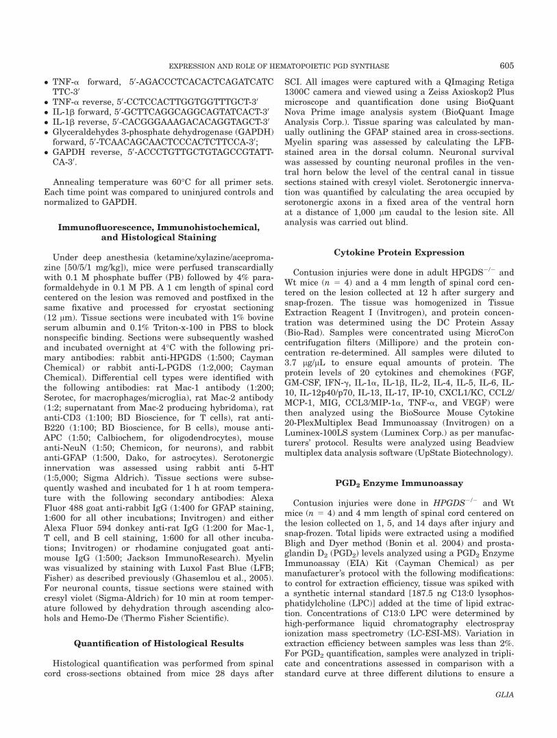
We first assessed the changes in expression and local-ization of the two PGD2 synthases [L-PGDS and hema-topoietic prostaglandin D synthase (HPGDS)] in theuninjured and injured spinal cord of adult C57BL/6 miceat several time-points after contusion injury. HPGDSmRNA levels begin to show an increase at 3 days andare significantly increased between seven and eightfoldat 7 and 14 days after SCI and decrease at later times(Fig. 1A). In contrast, the mRNA levels of L-PGDS didnot change after SCI (Fig. 1A). Double immunofluores-cence labeling showed that HPGDS is expressed inMac11 cells (Fig. 1B–G), a marker that is highlyexpressed by activated microglia and macrophages ofhematogenous origin. Activated microglia/macrophageshave been shown to reach their peak numbers at 7 daysafter contusion injury (Sroga et al., 2003). Double immu-nofluorescence labeling of L-PGDS in na€ıve and injuredspinal cord showed L-PGDS was localized to oligoden-drocytes as previously reported (Urade et al., 1985a,1985b) with no discernible differences in staining beforeand after SCI (data not shown).
HPGDS Is Responsible for the Increase in PGD2
After SCI
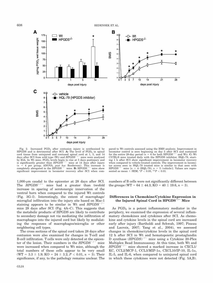
We next examined the changes in the levels of prosta-glandin D2 (PGD2) after SCI and, in particular, assessedthe contribution of hematopoietic prostaglandin D syn-thase (HPGDS) to the increase in PGD2 after SCI. Thiswas done by quantifying PGD2 in spinal cord tissue at1, 5, and 14 days after injury in Wt and HPGDS2/2
mice using a competitive EIA approach (n 5 4 for eachgroup). Comparisons of the differences in the PGD2 lev-els in Wt and HPGDS2/2 mice would also reveal therelative contributions of HPGDS and L-PGDS to PGD2
production. The EIA results showed a striking 3-fold
increase in PGD2 levels at 14 days after contusion injury(Fig. 2A). Interestingly, this increase in PGD2 was com-pletely abrogated in the HPGDS2/2 mice. These resultstherefore suggest that the increase in PGD2 in theinjured spinal cord is produced via HPGDS and notL-PGDS. It is possible that the assay is not sensitiveenough to detect small changes in PGD2 that is likely tooccur earlier than day 14 after injury, as PGD2 is verylabile. This may account for why changes in locomotorrecovery and cytokine expression are detected at earliertime points (see below).
HPGDS Mediated Production of PGD2
After SCI Is Detrimental
The experiments described earlier indicate that theincreased level of PGD2 in the spinal cord after contu-sion injury is mainly attributable to hematopoietic pros-taglandin D synthase (HPGDS), which is expressed bymicroglia/macrophages located in the lesion epicenter. Todetermine what role PGD2 plays after SCI, spinal cordcontusion injuries were done in HPGDS1/1 andHPGDS2/2 mice, and locomotor recovery assessed usingthe BMS analysis. Significant improvement in locomotorcontrol was observed in HPGDS2/2 mice when com-pared to Wt controls, starting from 5 days after injuryand continuing until day 28. The HPGDS2/2 micereached an average maximal BMS score of 4.5, which indi-cates stepping with both hind limbs, while HPGDS1/1
mice reached an average score of three, which indicatesthat the mice can only place their hind limb paws in thecorrect placement, with or without the ability to bearweight (Fig. 2B). The significant improvement seen in theHPGDS2/2 indicates a detrimental role for PGD2 afterSCI. These results were further confirmed in Wt micetreated with a selective inhibitor of HPGDS (HQL-79)after SCI (Fig. 2C). C57BL/6 mice that were given dailysubcutaneous injections of HQL-79 starting 1 h after SCIfor 28 days showed a significant improvement in locomotorrecovery as judged by the BMS analysis when comparedto the vehicle treated controls. The earliest time point atwhich statistically significant differences in locomotor con-trol were observed between HQL-79 and vehicle-treatedmice occurred at day 10 after SCI. This improvement is5 days later than that seen in HPGDS2/2 mice. This dif-ference between HQL-79 and HPGDS2/2 mice may haveto do with the concentration and frequency of the inhibitorused. Additional experiments will need to be done to opti-mize the conditions of the inhibitor treatment. Similar tothe HPGDS2/2 mice, Wt mice given HQL-79 reached anaverage score of 5 by 28 days after injury, which indicatesfrequent stepping with both hind limbs. Wt mice givenvehicle performed similar to HPGDS1/1 mice and wereonly able to place both hind limbs but unable to step fre-quently by day 28. These additional experiments with theHPGDS inhibitor confirmed that PGD2 produced viaHPGDS after SCI is detrimental.
606 REDENSEK ET AL.
GLIA
Secondary Tissue Damage After SCI IsReduced in HPGDS2/2 Mice
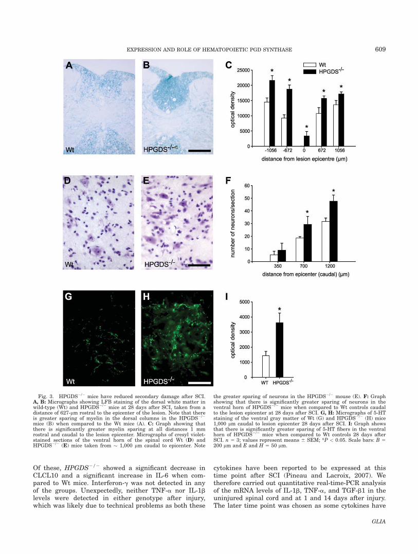
Because HPGDS2/2 mice showed improved locomotorrecovery, we next examined whether the lack of HPGDSalso had an impact on secondary tissue damage. Toassess this, the extent of myelin loss after SCI was exam-ined by staining cross sections of the spinal cord withLFB. HPGDS2/2 mice showed significantly greater spar-ing of myelin when compared to Wt mice (Fig. 3A,B).HPGDS2/2 mice showed greater myelin sparing at theepicenter and at distances of up to 1 mm rostral andcaudal to the injury (Fig. 3C). Images of spinal cord crosssections stained for GFAP at different distances from the
lesion epicenter are shown in Supporting InformationFig. 1 and display the extent of the moderate injury.Mice treated with the HPGDS inhibitor also showedgreater myelin sparing at some regions of the injured spi-nal cord with a trend to an increase in other regions (seeSupp. Info. Fig. 2). We also assessed the number of sur-viving neurons in the ventral gray matter of the spinalcord at varying distances from the epicenter of the lesion.HPGDS2/2 mice had significantly more neurons caudalto the epicenter when compared to HPGDS1/1 mice (Fig.3D–F). Serotonergic axons that descend from the raphenuclei in the brainstem and innervate the ventral hornare required for locomotor control. We therefore assessedthe serotonergic innervation in the ventral gray matter
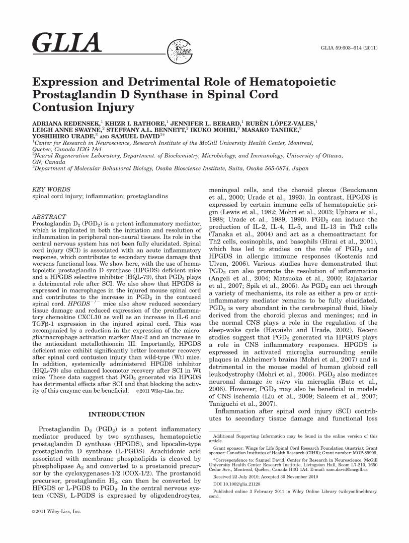
Fig. 1. HPGDS expression is increased after spinal cord contusioninjury. The mRNA expression levels of the two PGD2 synthases (HPGDSand L-PGDS) in the spinal cord were analyzed at several time points aftercontusion injury. A: Quantification of relative mRNA fold increases overlevels in uninjured spinal cord tissue; values normalized to PPIA. HPGDSexpression peaks at 7 and 14 days after injury and remains high for up to28 days. L-PGDS mRNA levels do not change after SCI. B: Low magnifi-cation image of a spinal cord cross-section stained for GFAP at 7 dayspostinjury to show the lesion architecture. This section is at a distanceof 100 lm caudal to the lesion epicenter. Nuclei are labeled with DAPI.
C–H: Double immunofluorescence labeling of HPGDS and Mac-1, 7 daysafter SCI of a section adjacent to that shown in B. C–E: Low magnifica-tion image of the entire cross-section of the spinal cord showing labelingfor HPGDS (C), Mac-1 (D), and the merged image showing HPGDS,Mac-1, and nuclear staining with DAPI (E). F–H: Higher magnification ofthe area outlined in the dashed line in panel C, showing HPGDS labeling(F), Mac-1 (G), and merged image showing HPGDS, Mac-1, and DAPIstaining (H). Note that HPGDS is expressed in Mac-11 macrophages/microglia (arrows). Scale bars: E 5 500 lm, H 5 50 lm (inset 5 20 lm). n5 3 for all analyses. Values represent mean 6 SEM; *P < 0.05.
607EXPRESSION AND ROLE OF HEMATOPOIETIC PGD SYNTHASE
GLIA
1,000-lm caudal to the epicenter at 28 days after SCI.The HPGDS2/2 mice had a greater than twofoldincrease in sparing of serotonergic innervation of theventral horn when compared to the injured Wt controls(Fig. 3G–I). Interestingly, the extent of macrophage/microglial infiltration into the injury site based on Mac-1staining appears to be similar in Wt and HPGDS2/2
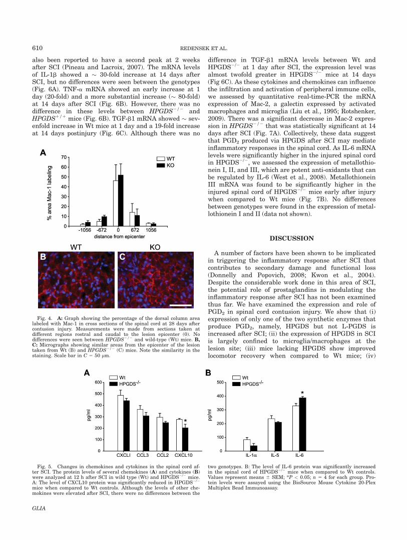
mice 28 days after SCI (Fig. 4A–C). This suggests thatthe metabolic products of HPGDS are likely to contributeto secondary damage not via mediating the infiltration ofmacrophages into the injured cord but likely by modulat-ing the responses of macrophages/microglia or otherneighboring cell types.
The cross sections of the spinal cord taken 28 days aftercontusion were also examined for changes in T-cell andB-cell infiltration. T cells were only detected at the epicen-ter of the lesion. Their numbers in the HPGDS2/2 micewere increased when compared to Wt mice, although thetotal numbers of these cells appear to be very small(WT 5 3.3 6 1.9; KO 5 24 6 3.2; P < 0.01, n 5 3). Theirsignificance, if any, to the pathology remains unclear. The
numbers of B cells were not significantly different betweenthe groups (WT 5 646 44.3; KO5 406 19.6, n5 3).
Differences in Chemokine/Cytokine Expression inthe Injured Spinal Cord in HPGDS2/2 Mice
As PGD2 is a potent inflammatory mediator in theperiphery, we examined the expression of several inflam-matory chemokines and cytokines after SCI. As chemo-kine and cytokine levels in the spinal cord are increasedearly after injury (Bartholdi and Schwab, 1997; Pineauand Lacroix, 2007; Yang et al., 2004), we assessedchanges in chemokine/cytokine levels in the spinal cord12 h after SCI in Wt and hematopoietic prostaglandinD synthase (HPGDS)2/2 mice using a Cytokine 20-PlexMultiplex Bead Immunoassay. At this time, both Wt andHPGDS2/2 mice showed a marked increase in CXCL1/KC, CCL2/MCP-1, CCL3/MIP-1a, CXCL10/IP-10, IL-1a,IL-5, and IL-6, when compared to uninjured spinal cordin which these cytokines were not detected (Fig. 5A,B).
Fig. 2. Increased PGD2 after contusion injury is synthesized byHPGDS and is detrimental after SCI. A: The level of PGD2 in spinalcord tissue from uninjured and contused spinal cord at 1, 5, and 14days after SCI from wild type (Wt) and HPGDS2/2 mice were analyzedby EIA. In Wt mice, PGD2 levels begin to rise at 5 days postinjury andis significantly greater than HPGDS2/2 mice at 14 days after injury(n 5 4 per group; ANOVA, post hoc Bonferroni). This increase iscompletely abrogated in the HPGDS2/2 mice. B: HPGDS2/2 mice showsignificant improvement in locomotor recovery after SCI when com-
pared to Wt controls assessed using the BMS analysis. Improvement inlocomotor control is seen beginning on day 5 after SCI and sustainedfor the entire 28-day period (n 5 6 for both HPGDS2/2 and Wt). C: WtC57BL/6 mice treated daily with the HPGDS inhibitor, HQL-79, start-ing 1 h after SCI show significant improvement in locomotor recoverywhen compared to vehicle-treated controls. The improvement in locomo-tor scores seen in HQL-79 treated mice is similar to that seen withHPGDS2/2 mice [n 5 4 (HQL-79); n 5 5 (vehicle)]. Values are repre-sented as mean 6 SEM; *P < 0.05, **P < 0.01.
608 REDENSEK ET AL.
GLIA
Of these, HPGDS2/2 showed a significant decrease inCLCL10 and a significant increase in IL-6 when com-pared to Wt mice. Interferon-g was not detected in anyof the groups. Unexpectedly, neither TNF-a nor IL-1blevels were detected in either genotype after injury,which was likely due to technical problems as both these
cytokines have been reported to be expressed at thistime point after SCI (Pineau and Lacroix, 2007). Wetherefore carried out quantitative real-time-PCR analysisof the mRNA levels of IL-1b, TNF-a, and TGF-b1 in theuninjured spinal cord and at 1 and 14 days after injury.The later time point was chosen as some cytokines have
Fig. 3. HPGDS2/2 mice have reduced secondary damage after SCI.A, B: Micrographs showing LFB staining of the dorsal white matter inwild-type (Wt) and HPGDS2/2 mice at 28 days after SCI, taken from adistance of 627-lm rostral to the epicenter of the lesion. Note that thereis greater sparing of myelin in the dorsal columns in the HPGDS2/2
mice (B) when compared to the Wt mice (A). C: Graph showing thatthere is significantly greater myelin sparing at all distances 1 mmrostral and caudal to the lesion epicenter. Micrographs of cresyl violet-stained sections of the ventral horn of the spinal cord Wt (D) andHPGDS2/2 (E) mice taken from � 1,000 lm caudal to epicenter. Note
the greater sparing of neurons in the HPGDS2/2 mouse (E). F: Graphshowing that there is significantly greater sparing of neurons in theventral horn of HPGDS2/2 mice when compared to Wt controls caudalto the lesion epicenter at 28 days after SCI. G, H: Micrographs of 5-HTstaining of the ventral gray matter of Wt (G) and HPGDS2/2 (H) mice1,000 lm caudal to lesion epicenter 28 days after SCI. I: Graph showsthat there is significantly greater sparing of 5-HT fibers in the ventralhorn of HPGDS2/2 mice when compared to Wt controls 28 days afterSCI. n 5 3; values represent means 6 SEM; *P < 0.05. Scale bars: B 5200 lm and E and H 5 50 lm.
609EXPRESSION AND ROLE OF HEMATOPOIETIC PGD SYNTHASE
GLIA
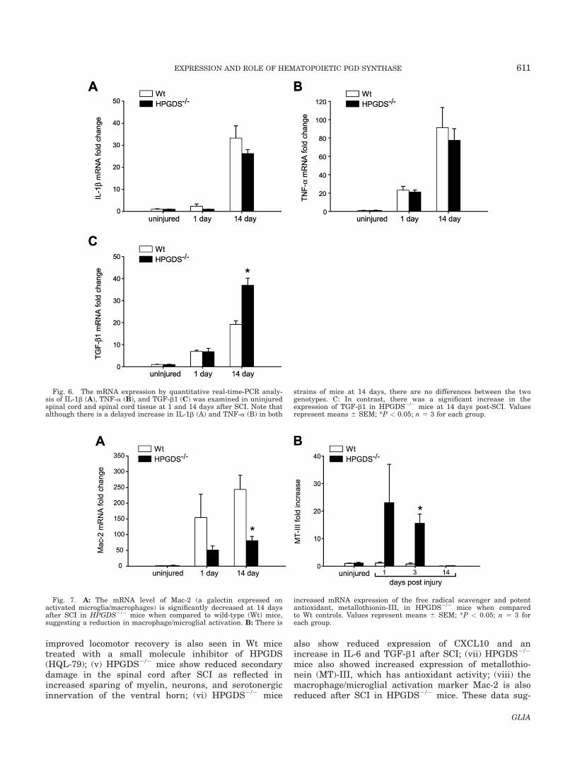
also been reported to have a second peak at 2 weeksafter SCI (Pineau and Lacroix, 2007). The mRNA levelsof IL-1b showed a � 30-fold increase at 14 days afterSCI, but no differences were seen between the genotypes(Fig. 6A). TNF-a mRNA showed an early increase at 1day (20-fold) and a more substantial increase (� 80-fold)at 14 days after SCI (Fig. 6B). However, there was nodifference in these levels between HPGDS2/2 andHPGDS1/1 mice (Fig. 6B). TGF-b1 mRNA showed � sev-enfold increase in Wt mice at 1 day and a 19-fold increaseat 14 days postinjury (Fig. 6C). Although there was no
difference in TGF-b1 mRNA levels between Wt andHPGDS2/2 at 1 day after SCI, the expression level wasalmost twofold greater in HPGDS2/2 mice at 14 days(Fig 6C). As these cytokines and chemokines can influencethe infiltration and activation of peripheral immune cells,we assessed by quantitative real-time-PCR the mRNAexpression of Mac-2, a galectin expressed by activatedmacrophages and microglia (Liu et al., 1995; Rotshenker,2009). There was a significant decrease in Mac-2 expres-sion in HPGDS2/2 that was statistically significant at 14days after SCI (Fig. 7A). Collectively, these data suggestthat PGD2 produced via HPGDS after SCI may mediateinflammatory responses in the spinal cord. As IL-6 mRNAlevels were significantly higher in the injured spinal cordin HPGDS2/2, we assessed the expression of metallothio-nein I, II, and III, which are potent anti-oxidants that canbe regulated by IL-6 (West et al., 2008). MetallothioneinIII mRNA was found to be significantly higher in theinjured spinal cord of HPGDS2/2 mice early after injurywhen compared to Wt mice (Fig. 7B). No differencesbetween genotypes were found in the expression of metal-lothionein I and II (data not shown).
DISCUSSION
A number of factors have been shown to be implicatedin triggering the inflammatory response after SCI thatcontributes to secondary damage and functional loss(Donnelly and Popovich, 2008; Kwon et al., 2004).Despite the considerable work done in this area of SCI,the potential role of prostaglandins in modulating theinflammatory response after SCI has not been examinedthus far. We have examined the expression and role ofPGD2 in spinal cord contusion injury. We show that (i)expression of only one of the two synthetic enzymes thatproduce PGD2, namely, HPGDS but not L-PGDS isincreased after SCI; (ii) the expression of HPGDS in SCIis largely confined to microglia/macrophages at thelesion site; (iii) mice lacking HPGDS show improvedlocomotor recovery when compared to Wt mice; (iv)
Fig. 4. A: Graph showing the percentage of the dorsal column arealabeled with Mac-1 in cross sections of the spinal cord at 28 days aftercontusion injury. Measurements were made from sections taken atdifferent regions rostral and caudal to the lesion epicenter (0). Nodifferences were seen between HPGDS2/2 and wild-type (Wt) mice. B,C: Micrographs showing similar areas from the epicenter of the lesiontaken from Wt (B) and HPGDS2/2 (C) mice. Note the similarity in thestaining. Scale bar in C 5 50 lm.
Fig. 5. Changes in chemokines and cytokines in the spinal cord af-ter SCI. The protein levels of several chemokines (A) and cytokines (B)were analyzed at 12 h after SCI in wild type (Wt) and HPGDS2/2 mice.A: The level of CXCL10 protein was significantly reduced in HPGDS2/2
mice when compared to Wt controls. Although the levels of other che-mokines were elevated after SCI, there were no differences between the
two genotypes. B: The level of IL-6 protein was significantly increasedin the spinal cord of HPGDS2/2 mice when compared to Wt controls.Values represent means 6 SEM; *P < 0.05; n 5 4 for each group. Pro-tein levels were assayed using the BioSource Mouse Cytokine 20-PlexMultiplex Bead Immunoassay.
610 REDENSEK ET AL.
GLIA
improved locomotor recovery is also seen in Wt micetreated with a small molecule inhibitor of HPGDS(HQL-79); (v) HPGDS2/2 mice show reduced secondarydamage in the spinal cord after SCI as reflected inincreased sparing of myelin, neurons, and serotonergicinnervation of the ventral horn; (vi) HPGDS2/2 mice
also show reduced expression of CXCL10 and anincrease in IL-6 and TGF-b1 after SCI; (vii) HPGDS2/2
mice also showed increased expression of metallothio-nein (MT)-III, which has antioxidant activity; (viii) themacrophage/microglial activation marker Mac-2 is alsoreduced after SCI in HPGDS2/2 mice. These data sug-
Fig. 6. The mRNA expression by quantitative real-time-PCR analy-sis of IL-1b (A), TNF-a (B), and TGF-b1 (C) was examined in uninjuredspinal cord and spinal cord tissue at 1 and 14 days after SCI. Note thatalthough there is a delayed increase in IL-1b (A) and TNF-a (B) in both
strains of mice at 14 days, there are no differences between the twogenotypes. C: In contrast, there was a significant increase in theexpression of TGF-b1 in HPGDS2/2 mice at 14 days post-SCI. Valuesrepresent means 6 SEM; *P < 0.05; n 5 3 for each group.
Fig. 7. A: The mRNA level of Mac-2 (a galectin expressed onactivated microglia/macrophages) is significantly decreased at 14 daysafter SCI in HPGDS2/2 mice when compared to wild-type (Wt) mice,suggesting a reduction in macrophage/microglial activation. B: There is
increased mRNA expression of the free radical scavenger and potentantioxidant, metallothionin-III, in HPGDS2/2 mice when comparedto Wt controls. Values represent means 6 SEM; *P < 0.05; n 5 3 foreach group.
611EXPRESSION AND ROLE OF HEMATOPOIETIC PGD SYNTHASE
GLIA
gest that PGD2 produced by macrophages after SCI hasdetrimental effects, which can be blocked by treatmentwith a small molecule HPGDS inhibitor.
The reduction of secondary damage and the locomotorimprovement after SCI in HPGDS2/2 mice indicates apotential role for PGD2 in the proinflammatory responsefollowing SCI. In support of this, we observed a signifi-cant decrease in the expression of the macrophagemarker Mac-2 in the HPGDS2/2 mice after SCI. PGD2
has previously been shown to mediate microglial activa-tion in the twitcher mouse mutant (Mohri et al., 2006), amodel of human Krabbes disease. In those experiments,the double twitcher/HPGDS2/2 mice showed reducedmicroglial activation and signs of reduced inflammationin the CNS as well as a reduction in iNOS expression(Mohri et al., 2006). Although we found a reduction inthe macrophage activation marker Mac-2, we did not seedifferences in iNOS mRNA expression on days 1, 3, or14 after SCI in HPGDS2/2 and Wt mice (data notshown). Furthermore, the twitcher/HPGDS2/2 miceshowed no differences in the level of IL-6, in contrast tothe increase we have seen in HPGDS2/2 mice early af-ter SCI. Although both models highlight PGD2 as aproinflammatory mediator in the CNS, the mechanismsof action appear to be different in these two models. Inaddition to the changes in Mac-2, the expression of thechemokine CXCL10 was also significantly reduced in theHPGDS2/2 mice after spinal cord contusion injury. De-spite the reduction in this cytokine, T cells, which werefound only at the epicenter of the lesion, appeared to beincreased in the HPGDS2/2 mice, although the totalnumber of these cells remained very low. The reason forthis is not clear at present. However, other studies haveshown that CXCL10 can mediate effects independent ofits T-cell chemoattractant properties. For instance, theinhibition of CXCL10 using a function-blocking antibodyin a dorsal spinal cord hemisection lesioning model inrats was found not only to reduce macrophage numbersbut also increase angiogenesis (Glaser et al., 2004) andneuroprotection (Glaser et al., 2006). The decrease inCXCL10 in HPGDS2/2 seen in our work may thereforemediate beneficial effects on angiogenesis after SCI.
Interestingly, IL-6 and TGF-b1 were significantlyincreased in HPGDS2/2 mice after SCI. Some studieshave suggested that IL-6 has proinflammatory effectsafter SCI (Okada et al., 2004), while a number of otherstudies indicate that it may have anti-inflammatoryand/or protective effects after SCI, for example, IL-6plays an important role in peripheral nerve regenerationand is required for regenerating dorsal column axons(Cafferty et al., 2004) as well as promoting axonal regen-eration in the presence of myelin inhibitors in SCI (Caoet al., 2006; Hannila and Filbin, 2008). Increased levelsof IL-6 have also been found after spinal cord contusioninjury in transgenic mice lacking active NF-jB (Bram-billa et al., 2005), as well as in Wt mice injected with15d-PGJ2, a metabolite of PGD2 (Kerr et al., 2008) inwhich secondary damage was reduced and accompaniedby improvement in locomotor recovery. IL-6 has alsobeen found to be neuroprotective when administered to
rodents, which are exposed to excitotoxic insults or is-chemia (Allan and Rothwell, 2001). IL-6 has also beenlinked to the expression of MT, free radical scavengers,and antioxidant proteins (West et al., 2008). We foundincreased expression of MT-III in HPGDS2/2 mice afterSCI, which could also contribute to reducing secondarydamage. The increase in IL-6 found in the HPGDS2/2
mice may therefore have a positive impact on tissue pro-tection after spinal cord contusion injury and couldpotentially impact either indirectly or directly on theincreased serotonergic innervation after SCI in theHPGDS2/2 mice. We also found that TGF-b1 expressionwas increased significantly in HPGDS2/2 when com-pared to Wt mice at 14 days after SCI. TGF-b1 is a plei-otropic cytokine, which is expressed in the injured spinalcord (McTigue et al., 2000). We did not find differencesin expression of GFAP or laminin by qRT-PCR or chon-droitin sulfate proteoglycan by immunostaining in theinjured spinal cord of HPGDS2/2 and Wt mice (datanot shown). However, this cytokine exhibits a variety ofanti-inflammatory properties (Lefer et al., 1990; Perrellaet al., 1994) that could contribute to the reduction insecondary damage seen after SCI.
Our laboratory has previously reported that dailyinjections of low doses of 15d-PGJ2, a dehydration prod-uct of PGD2, has beneficial effects after SCI, whilehigher doses are detrimental (Kerr et al., 2008). Similarbeneficial and detrimental effects of 15d-PGJ2 were alsoseen in experimental autoimmune encephalomyelitis(Diab et al., 2002). In the present study, we show thatreducing PGD2 produced by HPGDS in hematogenousmacrophages in HPGDS2/2 mice after SCI results inbeneficial effects in terms of histopathology and locomo-tor recovery. In these HPGDS2/2 mice, the unaffectedL-PGDS may produce low basal levels of PGD2 that canget nonenzymatically converted to the lower protectiveconcentrations of 15d-PGD2 that would add to the bene-ficial effects seen. Our results also suggest that PGD2
produced via HPGDS in macrophages either directly orindirectly mediate inflammatory responses that contrib-ute to secondary tissue damage after SCI. Future workneeds to focus on delineating the downstream receptormechanisms, as well as other possible beneficial mecha-nisms, which might result from blocking HPGDS. As theinhibition of PGD2 using the small molecule inhibitor(HQL-79) was also effective in improving locomotor func-tion after SCI, HPGDS could be a target for therapeuticintervention in the treatment of acute SCI.
ACKNOWLEDGMENTS
AR was a recipient of a studentship from the CIHRTraining Program in Neuroinflammation. LAS was arecipient of a Vision 2010/Ontario Ministry of ResearchInnovation post-doctoral fellowship. The authors thankHiba Kazak, Ourania Tsatas, and Claude Lachance fortechnical help, Ashleigh McLean for critical reading ofthis manuscript, and Margaret Attiwell for help withthe illustrations.
612 REDENSEK ET AL.
GLIA
REFERENCES
Allan SM, Rothwell NJ. 2001. Cytokines and acute neurodegeneration.Nat Rev Neurosci 2:734–744.
Angeli V, Staumont D, Charbonnier AS, Hammad H, Gosset P, Picha-vant M, Lambrecht BN, Capron M, Dombrowicz D, Trottein F. 2004.Activation of the D prostanoid receptor 1 regulates immune and skinallergic responses. J Immunol 172:3822–3829.
Aritake K, Kado Y, Inoue T, Miyano M, Urade Y. 2006. Structural andfunctional characterization of HQL-79, an orally selective inhibitor ofhuman hematopoietic prostaglandin D synthase. J Biol Chem 281:15277–15286.
Bartholdi D, Schwab ME. 1997. Expression of pro-inflammatory cyto-kine and chemokine mRNA upon experimental spinal cord injury inmouse: An in situ hybridization study. Eur J Neurosci 9:1422–1438.
Basso DM, Fisher LC, Anderson AJ, Jakeman LB, McTigue DM, Popo-vich PG. 2006. Basso Mouse Scale for locomotion detects differencesin recovery after spinal cord injury in five common mouse strains.J Neurotrauma 23:635–659.
Bate C, Kempster S, Williams A. 2006. Prostaglandin D2 mediates neu-ronal damage by amyloid-b or prions which activates microglial cells.Neuropharmacology 50:229–237.
Beuckmann CT, Lazarus M, Gerashchenko D, Mizoguchi A, Nomura S,Mohri I, Uesugi A, Kaneko T, Mizuno N, Hayaishi O, Urade Y. 2000.Cellular localization of lipocalin-type prostaglandin D synthase(b-trace) in the central nervous system of the adult rat. J CompNeurol 428:62–78.
Bonin F, Ryan SD, Migahed L, Mo F, Lallier J, Franks DJ, Arai H,Bennett SA. 2004. Anti-apoptotic actions of the platelet-activatingfactor acetylhydrolase I alpha2 catalytic subunit. J Biol Chem 279:52425–52436.
Brambilla R, Bracchi-Ricard V, Hu WH, Frydel B, Bramwell A,Karmally S, Green EJ, Bethea JR. 2005. Inhibition of astroglialnuclear factor jB reduces inflammation and improves functionalrecovery after spinal cord injury. J Exp Med 202:145–156.
Cafferty WB, Gardiner NJ, Das P, Qiu J, McMahon SB, Thompson SW.2004. Conditioning injury-induced spinal axon regeneration fails ininterleukin-6 knock-out mice. J Neurosci 24:4432–4443.
Cao Z, Gao Y, Bryson JB, Hou J, Chaudhry N, Siddiq M, Martinez J,Spencer T, Carmel J, Hart RB, Filbin MT. 2006. The cytokineinterleukin-6 is sufficient but not necessary to mimic the peripheralconditioning lesion effect on axonal growth. J Neurosci 26:5565–5573.
Diab A, Deng C, Smith JD, Hussain RZ, Phanavanh B, Lovett-Racke AE,Drew PD, Racke MK. 2002. Peroxisome proliferator-activated receptor-gamma agonist 15-deoxy-D12,14-prostaglandin J2 ameliorates experi-mental autoimmune encephalomyelitis. J Immunol 168:2508–2515.
Donnelly DJ, Popovich PG. 2008. Inflammation and its role in neuro-protection, axonal regeneration and functional recovery after spinalcord injury. Exp Neurol 209:378–388.
Dumont RJ, Okonkwo DO, Verma S, Hurlbert RJ, Boulos PT, EllegalaDB, Dumont AS. 2001. Acute spinal cord injury, Part 1: Pathophysio-logic mechanisms. Clin Neuropharmacol 24:254–264.
Faden AI, Lemke M, Demediuk P. 1988. Effects of BW755C, a mixedcyclo-oxygenase-lipoxygenase inhibitor, following traumatic spinalcord injury in rats. Brain Res 463:63–68.
Ghasemlou N, Kerr BJ, David S. 2005. Tissue displacement and impactforce are important contributors to outcome after spinal cord contu-sion injury. Exp Neurol 196:9–17.
Glaser J, Gonzalez R, Perreau VM, Cotman CW, Keirstead HS. 2004.Neutralization of the chemokine CXCL10 enhances tissue sparing andangiogenesis following spinal cord injury. J Neurosci Res 77:701–708.
Glaser J, Gonzalez R, Sadr E, Keirstead HS. 2006. Neutralization ofthe chemokine CXCL10 reduces apoptosis and increases axon sprout-ing after spinal cord injury. J Neurosci Res 84:724–734.
Hains BC, Yucra JA, Hulsebosch CE. 2001. Reduction of pathologicaland behavioral deficits following spinal cord contusion injury withthe selective cyclooxygenase-2 inhibitor NS-398. J Neurotrauma18:409–423.
Hannila SS, Filbin MT. 2008. The role of cyclic AMP signaling in pro-moting axonal regeneration after spinal cord injury. Exp Neurol209:321–332.
Hayaishi O, Urade Y. 2002. Prostaglandin D2 in sleep-wake regulation:Recent progress and perspectives. Neuroscientist 8:12–15.
Hirai H, Tanaka K, Yoshie O, Ogawa K, Kenmotsu K, Takamori Y,Ichimasa M, Sugamura K, Nakamura M, Takano S, Nagata K. 2001.Prostaglandin D2 selectively induces chemotaxis in T helper type 2cells, eosinophils, and basophils via seven-transmembrane receptorCRTH2. J Exp Med 193:255–261.
Kerr BJ, Girolami EI, Ghasemlou N, Jeong SY, David S. 2008. Theprotective effects of 15-deoxy-delta-(12,14)-prostaglandin J2 in spinalcord injury. Glia 56:436–448.
Kostenis E, Ulven T. 2006. Emerging roles of DP, CRTH2 in allergicinflammation. Trends Mol Med 12:148–158.
Kwon BK, Tetzlaff W, Grauer JN, Beiner J, Vaccaro AR. 2004. Patho-physiology and pharmacologic treatment of acute spinal cord injury.Spine J 4:451–464.
Lefer AM, Tsao P, Aoki N, Palladino MA Jr. 1990. Mediation of cardio-protection by transforming growth factor-b. Science 249:61–64.
Lewis RA, Soter NA, Diamond PT, Austen KF, Oates JA, Roberts LJ II.1982. Prostaglandin D2 generation after activation of rat and humanmast cells with anti-IgE. J Immunol 129:1627–1631.
Liu FT, Hsu DK, Zuberi RI, Kuwabara I, Chi EY, Henderson WR Jr. 1995.Expression and function of galectin-3, a b-galactoside-binding lectin, inhuman monocytes and macrophages. Am J Pathol 147:1016–1028.
Liu M, Eguchi N, Yamasaki Y, Urade Y, Hattori N, Urabe T. 2009. Pro-tective role of hematopoietic prostaglandin D synthase in transientfocal cerebral ischemia in mice. Neuroscience 163:296–307.
Lopez-Vales R, Garcia-Alias G, Guzman-Lenis MS, Fores J, Casas C,Navarro X, Verdu E. 2006. Effects of COX-2 and iNOS inhibitorsalone or in combination with olfactory ensheathing cell grafts afterspinal cord injury. Spine 31:1100–1106.
Matsuoka T, Hirata M, Tanaka H, Takahashi Y, Murata T, KabashimaK, Sugimoto Y, Kobayashi T, Ushikubi F, Aze Y, Eguchi N, Urade Y,Yoshida N, Kimura K, Mizoguchi A, Honda Y, Nagai H, Narumiya S.2000. Prostaglandin D2 as a mediator of allergic asthma. Science287:2013–2017.
McTigue DM, Popovich PG, Morgan TE, Stokes BT. 2000. Localizationof transforming growth factor-beta1 and receptor mRNA after experi-mental spinal cord injury. Exp Neurol 163:220–230.
Mohri I, Eguchi N, Suzuki K, Urade Y, Taniike M. 2003. Hematopoieticprostaglandin D synthase is expressed in microglia in the developingpostnatal mouse brain. Glia 42:263–274.
Mohri I, Kadoyama K, Kanekiyo T, Sato Y, Kagitani-Shimono K, SaitoY, Suzuki K, Kudo T, Takeda M, Urade Y, Murayama S, Tanike M.2007. Hematopoietic prostaglandin D synthase and DP1 receptor areselectively upregulated in microglia and astrocytes within senileplaques from human patients and in a mouse model of Alzheimerdisease. J Neuropathol Exp Neurol 66:469–480.
Mohri I, Taniike M, Taniguchi H, Kanekiyo T, Aritake K, Inui T,Fukumoto N, Eguchi N, Kushi A, Sasai H, Kanaoka Y, Ozono K,Narumiya S, Suzuki K, Urade Y. 2006. Prostaglandin D2-mediatedmicroglia/astrocyte interaction enhances astrogliosis and demyelin-ation in twitcher. J Neurosci 26:4383–4393.
O’Banion MK, Kyrkanides S, Olschowka JA. 2002. Selective inhibitionof cyclooxygenase-2 attenuates expression of inflammation-relatedgenes in CNS injury. Adv Exp Med Biol 507:155–160.
Okada S, Nakamura M, Mikami Y, Shimazaki T, Mihara M, Ohsugi Y,Iwamoto Y, Yoshizaki K, Kishimoto T, Toyama Y Okano H. 2004.Blockade of interleukin-6 receptor suppresses reactive astrogliosisand ameliorates functional recovery in experimental spinal cordinjury. J Neurosci Res 76:265–276.
Perrella MA, Yoshizumi M, Fen Z, Tsai JC, Hsieh CM, Kourembanas S,Lee ME. 1994. Transforming growth factor-b 1, but not dexametha-sone, down-regulates nitric-oxide synthase mRNA after its inductionby interleukin-1 beta in rat smooth muscle cells. J Biol Chem269:14595–14600.
Pineau I, Lacroix S. 2007. Proinflammatory cytokine synthesis in theinjured mouse spinal cord: Multiphasic expression pattern and identi-fication of the cell types involved. J Comp Neurol 500:267–285.
Rajakariar R, Hilliard M, Lawrence T, Trivedi S, Colville-Nash P,Bellingan G, Fitzgerald D, Yaqoob MM, Gilroy DW. 2007. Hematopoi-etic prostaglandin D2 synthase controls the onset and resolution ofacute inflammation through PGD2 and 15-deoxyD12–14 PGJ2. ProcNatl Acad Sci USA 104:20979–20984.
Resnick DK, Graham SH, Dixon CE, Marion DW. 1998. Role of cy-clooxygenase 2 in acute spinal cord injury. J Neurotrauma 15:1005–1013.
Rotshenker S. 2009. The Role of Galectin-3/MAC-2 in the Activation ofthe Innate-Immune Function of Phagocytosis in Microglia in Injuryand Disease. J Mol Neurosci.
Saleem S, Zhuang H, de Brum-Fernandes AJ, Maruyama T, NarumiyaS, Dore S. 2007. PGD(2) DP1 receptor protects brain from ischemia-reperfusion injury. Eur J Neurosci 26:73–78.
Spik I, Brenuchon C, Angeli V, Staumont D, Fleury S, Capron M,Trottein F, Dombrowicz D. 2005. Activation of the prostaglandin D2receptor DP2/CRTH2 increases allergic inflammation in mouse.J Immunol 174:3703–3708.
Sroga JM, Jones TB, Kigerl KA, McGaughy VM, Popovich PG. 2003.Rats and mice exhibit distinct inflammatory reactions after spinalcord injury. J Comp Neurol 462:223–240.
Tanaka K, Hirai H, Takano S, Nakamura M, Nagata K. 2004. Effects ofprostaglandin D2 on helper T cell functions. Biochem Biophys ResCommun 316:1009–1014.
613EXPRESSION AND ROLE OF HEMATOPOIETIC PGD SYNTHASE
GLIA
Taniguchi H, Mohri I, Okabe-Arahori H, Aritake K, Wada K, KanekiyoT, Narumiya S, Nakayama M, Ozono K, Urade Y, et al. 2007. Prosta-glandin D2 protects neonatal mouse brain from hypoxic ischemicinjury. J Neurosci 27:4303–4312.
Ujihara M, Horiguchi Y, Ikai K, Urade Y. 1988. Characterization anddistribution of prostaglandin D synthetase in rat skin. J InvestDermatol 90:448–451.
Urade Y, Fujimoto N, Hayaishi O. 1985a. Purification and characterization ofrat brain prostaglandin D synthetase. J Biol Chem 260:12410–12415.
Urade Y, Kaneko T, Fujimoto N, Watanabe Y, Mizuno N, Hayaishi O.1985b. Purification, characterization, and immunohistochemistry ofrat brain prostaglandin D synthetase. Adv Prostaglandin Thrombox-ane Leukot Res 15:549–551.
Urade Y, Kitahama K, Ohishi H, Kaneko T, Mizuno N, Hayaishi O.1993. Dominant expression of mRNA for prostaglandin D synthase inleptomeninges, choroid plexus, and oligodendrocytes of the adult ratbrain. Proc Natl Acad Sci USA 90:9070–9074.
Urade Y, Ujihara M, Horiguchi Y, Igarashi M, Nagata A, Ikai K,Hayaishi O. 1990. Mast cells contain spleen-type prostaglandin Dsynthetase. J Biol Chem 265:371–375.
Urade Y, Ujihara M, Horiguchi Y, Ikai K, Hayaishi O. 1989. The majorsource of endogenous prostaglandin D2 production is likely antigen-presenting cells. Localization of glutathione-requiring prostaglandinD synthetase in histiocytes, dendritic, and Kupffer cells in variousrat tissues. J Immunol 143:2982–2989.
West AK, Hidalgo J, Eddins D, Levin ED, Aschner M. 2008. Metallo-thionein in the central nervous system: Roles in protection, regenera-tion and cognition. Neurotoxicology 29:489–503.
Yang L, Blumbergs PC, Jones NR, Manavis J, Sarvestani GT, GhabrielMN. 2004. Early expression and cellular localization of proinflamma-tory cytokines interleukin-1beta, interleukin-6, and tumor necrosisfactor-alpha in human traumatic spinal cord injury. Spine 29:966–971.
614 REDENSEK ET AL.
GLIA