Embed Size (px)
Citation preview
Plant Physiol. (1988) 86, 137-1420032-0889/88/86/01 37/06/$01.00/0
Expression of Amyloplast and Chloroplast DNA in Suspension-Cultured Cells of Sycamore (Acerpseudoplatanus L.)1
Received for publication April 23, 1987 and in revised form September 18, 1987
JARUNYA NGERNPRASIRTSIRI2, DAVID MACHEREL3, HIROKAZU KOBAYASHI, AND TAKASHI AKAZAWA*Research Institutefor Biochemical Regulation, School ofAgriculture (J.N., D.M., T.A.) and RadioisotopeCenter (H.K.), Nagoya University, Chikusa, Nagoya 464, Japan
ABSTRACT
Green mutant cells of sycamore (Acer pseudoplatus L.), which hadbeen selected by mutagenic treatment of the white wild type, growphotoheterotrophically in auxin-depleted culture medium. In contrast tothe wild-type ceUls, mutant cells exhibit photosynthetic Or-evolutionactivity dwuing their growth coincident with inceses of (a) chlorophyll,(b) protein, and (c) ribulose-1,5-bisphosphate (RuBP) carboxylase activ-ity. Functionally competent chloroplasts were isolated from the greenceUls. Mecanism(s) governing gene expression of amyloplast DNA inthe heterotrophically grown white cells were compared with those of thechloroplst DNA isolated from the mutant ceUls. We have demonstratedin both amyloplast and chloroplast DNAs the presence of sequenceshomologous to the maize chloroplast genes for photosynthesis, ncludithe large sabunt of ribulose 1,5-bisphosphate carboxylase/oxygenase(RuBisCO) (rbcL), the 32 kDa Qs protein (PG32) (psbA), the apoproteinof P700 (paaA) and subunits of CF, (MpA, atpB, and atpE). However,employing either enzyme assays or immunological techniques, RuBisCOand CF, cannot be detected in the white wild type cells. Northern blothybridization of the RNA from the white cells showed high levels oftnipts for the 16S rRNA gene and low level of transripts for psbA;based on comparison with results obtained using the green mutant cells,we propose that the amyloplast genome is mostly inactive except for the16S rRNA gene andpsbA which is presumably regulated at the transcrip-tional level.
In view of the fact that amyloplasts are the sites of starchsynthesis in storage organs such as seeds and roots (21), it isimperative to examine the structure and function of the amylo-plastgenome and to elucidate the regulatory mechanism(s) whichcontrol its expression (10). It is frequently postulated that amy-loplasts and chloroplasts are ontogenically related (9), althoughneither the functional nor the structural nature of the formerorganelle has been substantively characterized in comparisonwith that of the latter which has been studied by numerous
' Supported by the grants from the Mnistry ofEducation, Science andCulture (Monbusho) of Japan. This is the paper No. 70 in the seriesStructure and Function of Chloroplast Proteins.
2Recipient of the Fellowship provided by the Hitachi ScholarshipFoundation (Tokyo), 1986. Permanent address: Department ofBiochem-istry, Faculty of Science, Chulalongkorn University, Bangkok 10400,Thailand.
3 Recipient of the Postdoctoral Fellowship provided by the JapanSociety for the Promotion of Science (JSPS) under the Japan-Francescientist exchange program (1986). Current address: CENG, Grenoble,Cedex, France.
investigators employing molecular biological techniques (7). Onecan surmise that there may exist closely related genetic machi-neries in these two different types of plastids, and in the case ofpotato the essentially identical restriction patterns were obtainedbetween chloroplast (leaf) and amyloplast (root) DNAs (23). It iswell recognized that upon illumination, proplastids or etioplastsin some plant tissues such as potato tuber and etiolated wheatseedlings are transformed into the Chl-containing chloroplasts.Many biochemical investigations have been performed with thissystem (6, 7, 22). In recent years, extensive research has beenundertaken to elucidate the molecular mechanism(s) of devel-opment and differentiation of another class of plastid, chromo-plasts, during the period oftomato fruit growth and maturation.In these studies, the mode of plastid gene expression during fruitdevelopment has been examined in comparison with that oper-ating in chloroplasts (2, 3, 19, 20).The suspension-cultured cells of sycamore (Acer pseudopla-
tanus L.) originally derived from nonphotosynthetic cambiumcells, are heterotrophic and contain mainly one type of differ-entiated plastid (amyloplasts), but they are unable to transformto the photosynthetically active cells upon illumination. Sincethe structure and the function of amyloplasts and chloroplastsare clearly distinct, mechanism(s) underlying the expression ofthe amyloplast genome and its regulation are evidently of im-portance and value to determine the nature of genes specific tothis unique organelle. The presence in the amyloplast DNA ofseveral homologous sequences to genes for photosynthesis e.g.,rbcL (large subunit of RuBisCO),4 psbA (32kDa QB protein,PG32), atpA (a-subunit of CF,), atpB (,B-subunit of CF,), atpE(e-subunit of CF,), and psaA (apoprotein of P700), and 16SrDNA, was previously reported (15). However, in a subsequentinvestigation (16), it was found that most ofthe amyloplast DNAwas not transcribed in the sycamore cells.The ultimate goal of our present investigation is to elucidate
the mechanism(s) governing the gene expression in amyloplastsby taking advantage of the availability of the green mutant typesycamore cells which had been selected from the white wild typecells ofsycamore after chemical mutagenesis (12). Transcription-ally active chloroplast genomes isolated from the mutant cell linehave been employed as the control.
MATERIALS AND METHODS
Suspension Culture of Sycamore Cells. White wild-type syca-more cells were grown in the liquid medium as previouslydescribed by Bligny (4). Mutant green sycamore cells which wereoriginally selected by Lescure (12) and maintained in the CNRS
4Abbreviations: RuBP, ribulose-1,5-biophosphate; RuBisCO, ribu-lose-1,5-bisphosphate carboxylase/oxygenase; CF,, coupling factor ofchloroplasts; PG32, 32 kDa QBprotein (Figs. 5 and 6).
137
ChIorophylI(jg/m o
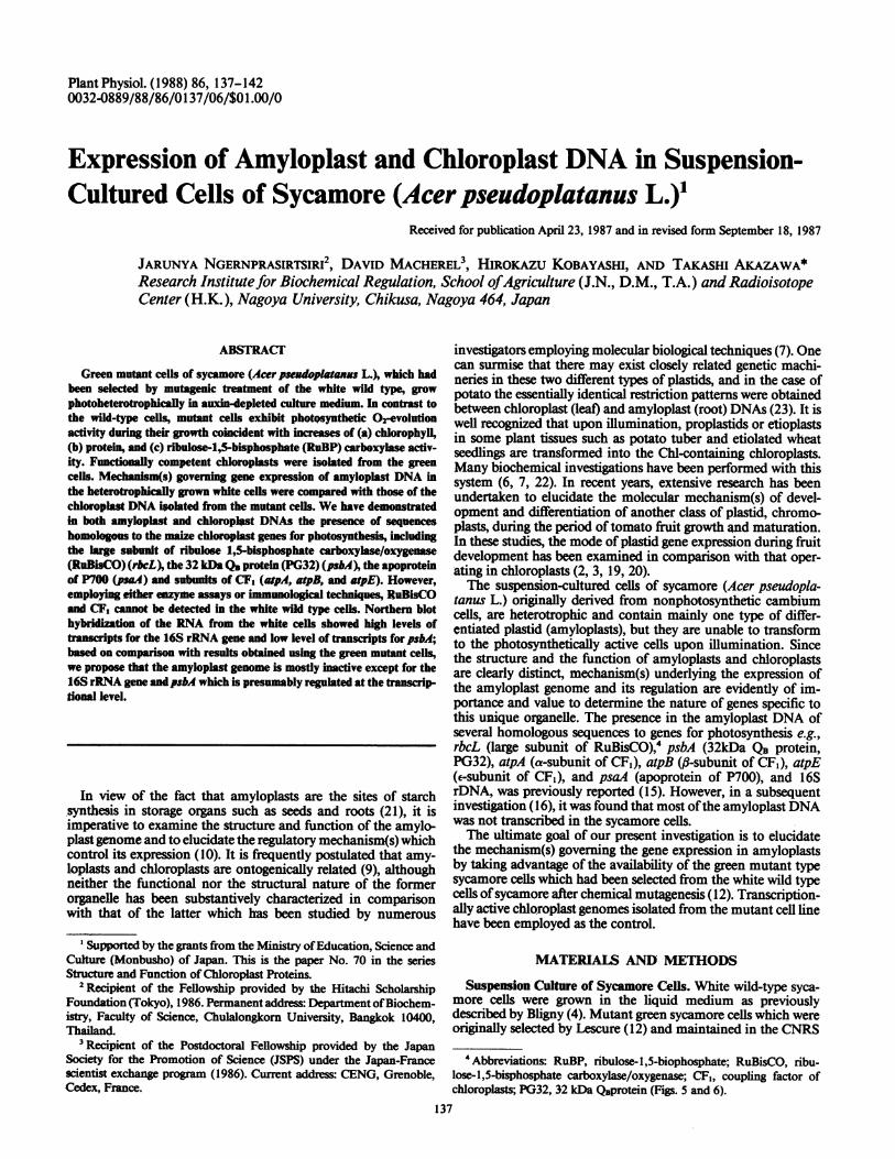
FIG. 1. loslation profile ofintact chloroplasts from green mutant cellsof sycamore. Chloroplasts of the green mutant cells were isolated fromthe ruptured sycamore protoplasts by the linear gradient of Percollaccording to the method of Takabe et al. (29). The Chl content andRuBPcarboxylase activity were measured as described in text.
laboratories of Marseille (France) were provided to us by Dr.Peaud-Lenoel. They were grown photoheterotrophically in theculture-medium depleted of 2,4-D according to the descriptionof Lescure (12). They were cultured under the continuous ilu-mination of fluorescent light (2000 lux, 25C).Enzyme Assay. About 2 g (fresh weight) of sycamore cells,
freshly harvested at the exponential growth stage, were suspendedin approximately 2 ml of sonicating buffer consisting of 50 mMHEPES in KOH (pH 7.5, 4C), 5 mM DTT, 1 mM EDTA, and0.3 M KCG. The suspension was sonicated twice at 200 W for 10min at OC in a Kubota Insonator Model 200M, and thencentrifuged at 94,000g for 1 h. The supematant was fractionatedwith (NH4)2SO4 solution and the 25 to 50% saturation precipitatewas dissolved in a minimal volume of the sonicating buffer andpassed through a Sephadex G-25 column, which was preequili-brated with the sonicating buffer as described above. RuBPcarboxylase (EC 4.1.1.39) activity was measured as described byNishimura et al (18).
Analytical Methods. Chl content in the sycamore cells wasdetermined according to Arnon (1) after extraction in 80%acetone. Protein content was determined by the spectrophoto-metric method of Lowry et al. (14) using BSA as a standard.Immunolgil Bloffing Analysis of Proteins. The crude extract
of sycamore cells after sonication was first subjected to SDS-PAGE (10-20% linear gradient) and electrophoretically trans-
_- C
-a
C.0C.
1c
E c---
c0
Cl0
.-
C
'a0L.S
-
'..0.0.2
z:aS
Plant Physiol. Vol. 86, 1988. 1. .
(A) Cells
green mutant
).2 -
wild0 t -0 -
0 5 10 15 20
(c) Chloroplasts
15-
10
5-
00 5 10 15 20
Time (min)
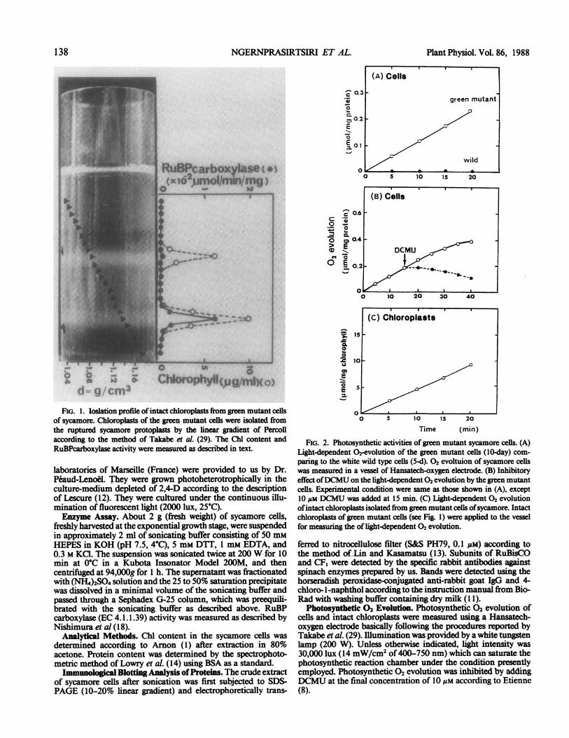
FIG. 2. Photosynthetic activities of green mutant sycamore cells. (A)Light-dependent 02-evolution of the green mutant cells (10-day) com-
paring to the white wild type cells (5-d). 02 evoltuion of sycamore cellswas measured in a vessel of Hansatech-oxygen electrode. (B) Inhibitoryeffect ofDCMU on the light-dependent 02 evolution by the green mutantcells. Experimental condition were same as those shown in (A), except10 um DCMU was added at 15 min. (C) Light-dependent 02 evolution
ofintact chloroplasts isolated from green mutant cells ofsycamore. Intactchloroplasts of green mutant cells (see Fig. 1) were applied to the vesselfor measuring the of light-dependent 02 evolution.
ferred to nitrocellulose filter (S&S PH79, 0.1 ,uM) according tothe method of Lin and Kasamatsu (13). Subunits of RuBisCOand CF1 were detected by the specific rabbit antibodies againstspinach enzymes prepared by us. Bands were detected using thehorseradish peroxidase-conjugated anti-rabbit goat IgG and 4-
chloro- I-naphthol according to the instruction manual from Bio-Rad with washing buffer containing dry milk (11).
Photosynthetic 02 Evolution. Photosynthetic 02 evolution ofcells and intact chloroplasts were measured using a Hansatech-oxygen electrode basically following the procedures reported byTakabe et al. (29). Illumination was provided by a white tungstenlamp (200 W). Unless otherwise indicated, light intensity was30,000 lux (14 mW/cm2 of 400-750 nm) which can saturate thephotosynthetic reaction chamber under the condition presentlyemployed. Photosynthetic 02 evolution was inhibited by addingDCMU at the final concentration of 10 uM according to Etienne(8).
138 NGERNPRASIRTSIRI ET AL.
RuBPcarboxylase ( uo( lB l rfx f I
.0'- 7
OD
d 3
--W
.- -k
I
EXPRESSION OF AMYLOPLAST AND CHLOROPLAST DNA
0.8
0L-- 0.6
E 0E
_0
X 0.20
L--
._-1
E
C)
xoco
0~mt-
m
:
cr-
Q20
0.15-
o051-
3 5 10 15 20
Age (day)
Amyloplasts and Chloroplasts Preparation. Protoplasts wereprepared following the method as described previously by Mach-erel et al. (15) using the freshly harvested white sycamore cells atthe exponential growth stage (5-d). Amyloplasts and mitochon-dria free from other organelles were prepared according to theprocedure reported by Macherel et al. (16).
Protoplasts ofmutant green cells, harvested at the exponentialgrowth stage (10-d), were prepared basically following the pro-cedure described by Macherel et al. (15), except for the 45-minincubation with 2% (w/v) Cellulase Onozuka RIO (Yakult Co.Ltd., Japan), 0.2% (w/v) Pectolyase Y23 (Seishin Co. Ltd.,Japan), and 0.2% (w/v) Macerozyme RIO (Yakult Co. Ltd.,Japan). The resulting protoplasts were then ruptured by passagethrough a syringe and chloroplasts were prepared according tothe procedure of Sugiura et al. (28) for DNA extraction andTakabe et al. (29) for the purpose of assaying photosyntheticactivities.DNA and Cellular RNA Preparation. Amyloplast DNA and
mitochondrial DNA were prepared as described by Macherel etal. (16). Chloroplast DNA was prepared according to Sugiura etal. (28) after a slight modification by a second centrifugation ina CsCl gradient containing bisbenzimide Hoeschst 33258 (Sigma)as an intercalating agent for the double strand DNA. The extrac-tion oftotal cellularRNA was performed following the procedurereported by Macherel et al. (16).
Characterization of DNA and RNA. DNA was labeled radio-actively with [a-32P]-dCTP in vitro by using the Klenow fiagmentof the Escherichia coli DNA polymerase I and primers of Oli-golabelling Kit (Pharmacia). The digestion ofDNA by restrictionendonucleases and subsequent agarose gel electrophoresis of thedigested fragments were performed using the conventional tech-niques. Purified cellular RNA was denatured by glyoxal andelectrophoresed on agarose gel run in 10 mm Na phosphate (pH7.0). The transfer of nucleic acids to GeneScreen (New EnglandNuclear) membranes and the hybridization conditions for North-em and Southern blot analyses were carried out according to theinstruction manual ofNew England Nuclear (24). The radioac-tive bands on GeneScreen were detected by radioautography.
Plasmids containing the maize chloroplast genes; rbcL (larggesubunit of RuBisCO), psbA (32 kDa QB protein,PG32), atpA (a-subunit ofCF,), atpB (,B-subunit ofCF,), atpE (e-subunit ofCF,),psaA (apoprotein of P700), and 16S rDNA were kindly providedto us by Dr. Bogorad (5).
RESULTSThe suspension-cultured cells of green mutant sycamore grow
photoheterotrophically in the culture medium lacking 2,4-D and
204
15 Ecm
10 >C.00
X.)o
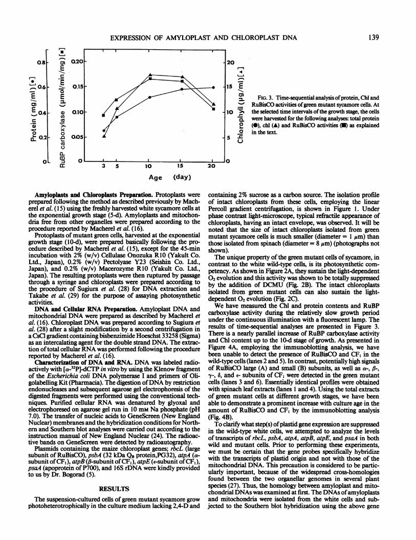
FIG. 3. Time-sequential analysis ofprotein, Chl andRuBisCO activities ofgreen mutant sycamore cells. Atthe selected time intervals ofthe growth stage, the cellswere harvested for the following analyses: total protein(@), chl (A) and RuBisCO activities (U) as explainedin the text.
v . I I I I I0
containing 2% sucrose as a carbon source. The isolation profileof intact chloroplasts from these cells, employing the linearPercoll gradient centrifugation, is shown in Figure 1. Underphase contrast light-microscope, typical refractile appearance ofchloroplasts, having an intact envelope, was observed. It will benoted that the size of intact chloroplasts isolated from greenmutant sycamore cells is much smaller (diameter = 1 Mm) thanthose isolated from spinach (diameter = 8 Mm) (photographs notshown).The unique property of the green mutant cells of sycamore, in
contrast to the white wild-type cells, is its photosynthetic com-petency. As shown in Figure 2A, they sustain the light-dependent02 evolution and this activity was shown to be totally suppressedby the addition of DCMU (Fig. 2B). The intact chloroplastsisolated from green mutant cells can also sustain the light-dependent 02 evolution (Fig. 2C).We have measured the Chl and protein contents and RuBP
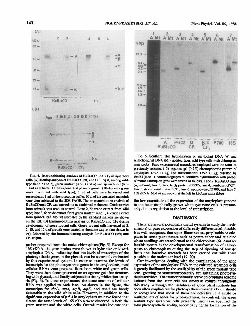
carboxylase activity during the relatively slow growth periodunder the continuous illumination with a fluorescent lamp. Theresults of time-sequential analyses are presented in Figure 3.There is a nearly paallel increase of RuBP carboxylase activityand Chl content up to the l0-d stage of growth. As presented inFigure 4A, employing the immunoblotting analysis, we havebeen unable to detect the presence of RuBisCO and CF1 in thewild-type cells (lanes 2 and 5). In contrast, potentially high signalsof RuBisCO large (A) and small (B) subunits, as well as a-, -,a-, 6, and e- subunits of CF1 were detected in the green mutantcells (lanes 3 and 6). Essentially identical profiles were obtainedwith spinach leaf extracts (lanes 1 and 4). Using the total extractsof green mutant cells at different growth stages, we have beenable to demonstrate a prominent increase with culture age in theamount of RuBisCO and CF, by the immunoblotting analysis(Fig. 4B).To clarify what step(s) ofplastid gene expression are suppressed
in the wild-type white cells, we attempted to analyze the levelsof transcripts of rbcL, psbA, atpA, atpB, atpE, and psaA in bothwild and mutant cells. Prior to performing these experiments,we must be certain that the gene probes specifically hybridizewith the transcripts of plastid origin and not with those of themitochondrial DNA. This precaution is considered to be partic-ularly important, because of the widespread cross-homologiesfound between the two organellar genomes in several plantspecies (27). Thus, the homology between amyloplast and mito-chondrial DNAs was examined at first. The DNAs ofamyloplastsand mitochondria were isolated from the white cells and sub-jected to the Southern blot hybridization using the above gene
I I I I I
139
I I
a I
NGERNPRASIRTSIRI ET AL.
3k. tb)p)
-; (I*^.Rp
-A
'3I'21 2
5.15.0>'4 3-.3. 5-o
20 o
2.0-1.9-
i, 6...
3 .
RuB,sCO CF .
0 -.
B)
Age 15 10 5 3 3 5 1015 (day)
A- - a
1 2 ~~ ~~ ~~ ~~~~~~~~~~~~~34 5 bA Mt A Mt A Mt A Mt A Mt A Mt A Mt
a._
A, PGt a831F87(iP It.,
RuBisCO CF1
FIG. 4. Immunoblotting analysis of RuBisCO tnd CF, in sycamorecells. (A) Blotting analysis ofRuBisCO (left) and CF, (right) among wild-type (lane 2 and 5), green mutant (lane 3 and 6) and spinach leaf (lane1 and 4) extracts. At the exponential phase ofgrowth (10-day with greenmutant and 5-d with wild type), 5 ml of cells were harvested andsuspended in 1 ml ofthe sonicating buffer, 20 Ml ofthe sonicated materialswere then subjected to the SDS-PAGE. The immunoblotting analysis ofRuBisCO and CF, was carried out as explained in the text. Crude extractfrom spinach was used as control. Lane 2, 5: crude extract from wildtype; lane 3, 6: crude extract from green mutant; lane 1, 4: crude extractfrom spinach leaf. Mol wt estimated by the standard markers are shownon the left. (B) Immunoblotting analysis of RuBisCO and CF, duringdevelopment of green mutant cells. Green mutant cells harvested at 3,5, 10, and 15 d ofgrowth were treated in the same way as that shown in(A), followed by the immunoblotting analysis for RuBisCO (left) andCF, (right).
probes prepared from the maize chloroplasts (Fig. 5). Except for16S rDNA, the gene probes were shown to hybridize only withamyloplast DNA, indicating that the levels of transcripts of thephotosynthetic genes in the plastids can be accurately estimatedby this experimental system. In order to examine the levels oftranscripts for the photosynthetic genes in the amyloplasts, totalcellular RNAs were prepared from both white and green cells.They were then electrophoresed on an agarose gel after denatur-ing with glyoxal, and finally subjected to the hybridization analy-sis (Fig. 6). In these experiments, the same amount of cellularRNA was applied to each lane. As shown in the figure, thetranscripts for rbcL, atpA, atpB, atpE, and psaA are barelydetectable in the wild white cells. However, in addition to thesignificant expression ofpsbA in amyloplasts we have found thatalmost the same levels of 16S rRNA were observed in both thegreen mutant and the white cells. Overall results indicate that
FIG. 5. Southern blot hybridization of amyloplast DNA (A) andmitochondrial DNA (Mt) isolated from wild type cells with chloroplastgene probe. Basic experimental procedures employed were the same aspreviously reported (15). Agarose gel (0.7%) electrophoretic pattern ofamyloplast DNA (1 ug) and mitochondrial DNA (1 ug) digested byEcoRI (lane 1). Autoradiographs of Southern hybridization with probesofmaize chloroplast gene were shwon as follows. Lane 2, RuBisCO lage(A) subunit; lane 3, 32-kDa QB protein (PG32); lane 4, a-subunit ofCF,;lane 5, ,8- and E-subunits of CF,; lane 6, apoprotein of P700; and lane 7,16S rRNA. Mol wt are shown at the left in kilobase pairs (kbp).
the low magnitude of the expression of the amyloplast genomein the heterotrophically grown white sycamore cells is presum-ably due to regulation at the level of transcription.
DISCUSSIONThere are several potentially useful systems to study the mech-
anism(s) of gene expression of differently differentiated plastids.It is well recognized that upon illumination, proplastids or etio-plasts in some plant tissues such as potato tuber and etiolatedwheat seedlings are transformed to the chloroplasts (6). Anotherfeasible system is the developmental transformation of chloro-plasts to chromoplasts during the step of ripening of tomatofruits and intensive research has been carried out with theseplastids at the molecular level (19, 20).Our investigation dealing with the examination of the gene
expression ofthe amyloplast DNA in the cultured sycamore cellsis greatly facilitated by the availability of the green mutant typecells, growing photoheterotrophically yet sustaining photosyn-thetic activities. The transcriptionally active chloroplastsgenomeisolated from the latter cells has served as control throughoutthis study. Although the usefulness of green plant mutants hasbeen often emphasized for photosynthesis research (17), it shouldbe recognized that most of them are defective in a single ormultiple sets of genes for photosynthesis. In contrast, the greenmutant type sycamore cells presently used have acquired thetotal photosynthetic ability, accompanying the formation of the
140 Plant Physiol. Vol. 86, 1988
EXPRESSION OF AMYLOPLAST AND CHLOROPLAST DNA
1 2 3 4 5 6 7 8sWm m WM WM WMWM WM
:*"
0.-
A PG32 a I,£ P700 16SRuBisCO CF1 CF,
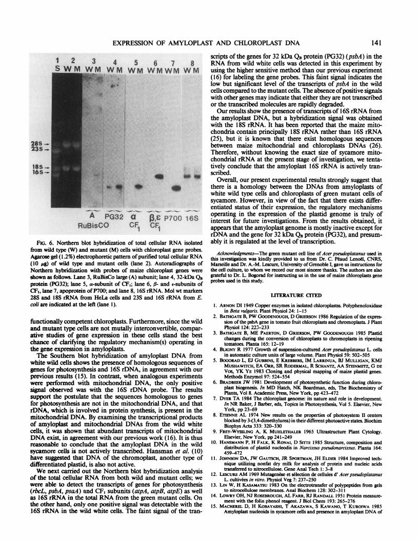
FiG. 6. Northern blot hybridization of total cellular RNA isolatedfrom wild type (W) and mutant (M) cells with chloroplast gene probes.Agarose gel (1.2%) electrophoretic pattern of purified total cellular RNA(10 gg) of wild type and mutant cells (lane 2). Autoradiographs ofNorthern hybridization with probes of maize chloroplast genes were
shown as follows. Lane 3, RuBisCo large (A) subunit; lane 4, 32-kDa QBprotein (PG32); lane 5, a-subunit of CFI; lane 6, ,@- and esubunits ofCF1, lane 7, apoprotein of P700; and lane 8, 16S rRNA. Mol wt markers28S and 18S rRNA from HeLa cells and 23S and 16S rRNA from E.coli are indicated at the left (lane 1).
functionally competent chloroplasts. Furthermore, since the wildand mutant type cells are not mutally interconvertible, compar-ative studies of gene expression in these cells stand the bestchance of clarifying the regulatory mechanism(s) operating inthe gene expression in amyloplasts.The Southern blot hybridization of amyloplast DNA from
white wild cells shows the presence of homologous sequences ofgenes for photosynthesis and 16S rDNA, in agreement with ourprevious results (15). In contrast, when analogous experimentswere performed with mitochondrial DNA, the only positivesignal observed was with the 16S rDNA probe. The resultssupport the postulate that the sequences homologous to genesfor photosynthesis are not in the mitochondrial DNA, and thatrDNA, which is involved in protein synthesis, is present in themitochondrial DNA. By examining the transcriptional productsof amylopiast and mitochondrial DNAs from the wild whitecells, it was shown that abundant transcripts of mitochondrialDNA exist, in agreement with our previous work (16). It is thusreasonable to conclude that the amyloplast DNA in the wildsycamore cells is not actively transcribed. Hansman et al. (10)have sus that DNA of the chromoplast, another type ofdifferentiated plastid, is also not active.We next carried out the Northern blot hybridization analysis
of the total cellular RNA from both wild and mutant cells; wewere able to detect the transcripts of genes for photosynthesis(rbcL, psbA, psaA) and CF, subunits (atpA, atpB, atpE) as wellas 16S rRNA in the total RNA from the green mutant cells. Onthe other hand, only one positive signal was detectable with the16S rRNA in the wild white cells. The faint signal of the tran-
scripts of the genes for 32 kDa QB protein (PG32) (psbA) in theRNA from wild white cells was detected in this experiment byusing the higher sensitive method than our previous experiment(16) for labeling the gene probes. This faint signal indicates thelow but significant level of the transcripts of psbA in the wildcells compared to the mutant cells. The absence ofpositive signalswith other genes may indicate that either they are not transcribedor the transcribed molecules are rapidly degraded.Our results show the presence of transcripts of 16S rRNA from
the amyloplast DNA, but a hybridization signal was obtainedwith the 18S rRNA. It has been reported that the maize mito-chondria contain principally 18S rRNA rather than 16S rRNA(25), but it is known that there exist homologous sequencesbetween maize mitochondrial and chloroplasts DNAs (26).Therefore, without knowing the exact size of sycamore mito-chondrial rRNA at the present stage of investigation, we tenta-tively conclude that the amyloplast 16S rRNA is actively tran-scribed.
Overall, our present experimental results strongly suggest thatthere is a homology between the DNAs from amyloplasts ofwhite wild type cells and chloroplasts of green mutant cells ofsycamore. However, in view of the fact that there exists differ-entiated status of their expression, the regulatory mechanismsoperating in the expression of the plastid genome is truly ofinterest for future investigations. From the results obtained, itappears that the amyloplast genome is mostly inactive except forrDNA and the gene for 32 kDa QB protein (PG32), and presum-ably it is regulated at the level of transcription.
Acknowledgments-The green mutant cell line of Acer pseudoplatanus used inthis investigation was kindly provided to us from Dr. C. Peaud Lenoel, CNRS,Marseille and Dr. A.-M. Lescure, University ofGrenoble I, gave us instructions forthe cell culture, to whom we record our most sincere thanks. The authors are alsograteful to Dr. L. Bogorad for instructing us in the use of maize chloroplasts geneprobes used in this study.
LITERATURE CI TED
1. ARNON DI 1949 Copper enzymes in isolated chloroplastss. Polyphenoloxidasein Beta vulgaris. Plant Physiol 24: 1-15
2. BATHGATE B, PW GOODENOUGH, D GRIERSON 1986 Regulation of the expres-sion of the psbA gene in tomato fruit chloroplasts and chromoplasts. J PlantPhysiol 124: 223-233
3. BATHGATE B, ME PAERTON, D GRIERSON, PW GOODENOUGH 1985 Plastidchanges during the conversion of chloroplasts to chromoplasts in ripeningtomatoes. Planta 165: 12-19
4. BLIGNY R 1977 Growth of suspension-cultured Acer pseudoplatanus L. cellsin automatic culture units of large volume. Plant Physiol 59: 502-505
5. BOGORAD L, EJ GUBBINS, E KREBBERS, IM LARRINUA, BJ MULLIGAN, KMJMUSHAWITCH, EA ORR, SR RODERMAL, R SCHANTZ, AA STEINMETZ, G DEVos, YK YE 1983 Cloning and physical mapping of maize plastid genes.Methods Enzymol 97: 524-554
6. BRADBEER JW 1981 Development of photosynthetic function during chloro-plast biogenesis. In MD Hatch, NK Boardman, eds, The Biochemistry ofPlants, Vol 8. Academic Press, New York, pp 423-472
7. DYER TA 1984 The chloroplast genome: its nature and role in development.In NR Baker, J Barber, eds, Topics in Photosynthesis, Vol 5. Elsevier, NewYork, pp 23-69
8. ETIENNE AL 1974 New results on the properties of photosystem II centersblocked by 3-(3,4-dimethylurea) in their different photoactive states. BiochimBiophys Acta 333: 320-330
9. FREY-WYSSLING A, K MUHLETHALER 1965 Ultrastructure Plant Cytology.Elsevier, New York, pp 241-249
10. HANSMANN P, H FALK, K RONAI, D SrrrE 1985 Structure, composition anddistribution of plastid nucleodis in Narcissus pseudonarcissus. Planta 164:459-472
1 1. JOHNSON DA, JW GAurscH, JR SPORTMAN, JH ELDER 1984 Improved tech-nique utilizing nonfat dry milk for analysis of protein and nucleic acidstransferred to nitrocellulose. Gene Anal Tech 1: 3-8
12. LEscuRE AM 1969 Mutagenese et selection de cellules d' Acer pseudoplatanusL. cultivees in vitro. Physiol Veg 7: 237-250
13. LIN W, H KASAMATSU 1983 On the electrotransfer of polypeptides from gelsto nitrocellulose membranes. Anal Biochem 128: 302-311
14. LOWRY OH, NJ ROSEBROUGH, AL FARR, RJ RANDALL 1951 Protein measure-ment with the folin phenol reagent. J Biol Chem 193: 265-276
15. MACHEREL D, H KOBAYASHI, T AKAZAWA, S KAWANO, T KuROIWA 1985Amyloplast nucleoids in sycamore cells and presence in amyloplast DNA of
28S523S -
18S -16S-.
141
142 NGERNPRASIRTSIRI ET AL. Plant Physiol. Vol. 86, 1988
homologous sequences to chloroplasts genes. Biochem Biophys Res Com- etioplst isolated from dark-grown wheat (Triticum aestivum L.). Plantmun 133: 140-146 Physiol 76: 1041-1046
16. MACHEREL D, H KOBAYASHI, E VALLE, T AKAZAWA 1986 Expression of 23. Sco)r NS, MJ TYMMS, JV POSsINGHAM 1984 Plastid-DNA levels in theamyloplast DNA in suspension-cultured cells of sycamore (Acer pseudopla- different tissue of potato. Planta 161: 12-19tanus L.). FEBS Lett 201: 315-320 24. SOUTHERN EM 1975 Detection of specific sequences among DNA fragments
17. MILEs D 1982 The use of mutations to probe photosynthesis in higher plants. separated by gel electrophoress. J Mol Biol 98: 503-517In M Edelman, RB Hallick, N-H Chua, eds, Methods in Chloroplast Molec- 25. STmN DB, TA DYER, DM LONSDALE 1982 Organization ofthe mitochondrialular Biology. Elsevier, Amsterdam, pp 75-107 ribosomal RNA genes of maize. Nucleic Acid Res 10: 3333-3340
18. NISHIMURA M, T TAKABE, T SUGIYAMA, T AK.ZWA 1973 Structure and 26. SmN DB, DM LONSDALE 1982 Mitochondrial and chloroplast genomes offuRAtioof chloroplaKA T SUGIYAMADissoaxat of spinach leaf ribulose 27 maz have a 12-kilobase DNA sequence in common. Nature 299: 698-702function of chloropast proteins. XIX. Dissociation ofspinachleafribuloe 27. STEN DB, JB PALMER 1984 Extensive and widespread homologies between1,5-diphosphate carboxylase by.p-mercuribenzoate.Biochem 74: 945954 mitochondrial DNA and chloroplast DNA in plants. Proc Natl Acad Sci19. PIEcHuu.A B, KRC IMLAY, W GRuissEm 1985 Plastid gene expression during USA 81: 1946-1950fruit ripening in tomato. Plant Mol Biol 5: 373-384 28. SuGIuRA M, S Kuzuo, N ZAITA, M KUSUDA, M KUMANO 1986 aone bank
20. PlBcHULLA B, E PICHERSHY, AR CAsHABORE, W GRUisSEM 1986 Expression of the tobacco (Nicotiana tabacum) chloroplast genome as a set of overlap-of nuclear and plastid genes for photosynthesis-specific proteins during ping restriction endonuclease fragments: mapping of eleven ribosomal pro-tomato fruit development and ripening. Plant Mol Biol 7: 367-376 tein genes. Plant Science 44: 211-216
21. PRm J 1982 Regulation of the biosynthesis and degradation of starch. Annu 29. TAKABE T, M NISHIMURA, T AKAZAWA 1979 Isolation of intact chloplastsRev Plant Physiol 33: 431-454 from spinach leafby centrifugation in gradients ofthe modified silica percoll.
22. SANDELINS AS, E SELSTAM 1984 Lclization of galactolipid biosynthesis in Agric Biol Chem 43: 2137-2142


![The Molecular Machinery of Chloroplast Division1[OPEN] · Update on Chloroplast Division The Molecular Machinery of Chloroplast Division1[OPEN] Cheng Chen,a Joshua S. MacCready,b](https://img.pdfslide.net/doc/110x75/6025c6a7c7a5a154ca59f7b1/the-molecular-machinery-of-chloroplast-division1open-update-on-chloroplast-division.jpg)
![Amyloplast-Localized SUBSTANDARD STARCH GRAIN4 Protein ... · Amyloplast-Localized SUBSTANDARD STARCH GRAIN4 Protein Influences the Size of Starch Grains in Rice Endosperm1[W] Ryo](https://img.pdfslide.net/doc/110x75/5e0d7b5872e93a29d062d359/amyloplast-localized-substandard-starch-grain4-protein-amyloplast-localized.jpg)