Embed Size (px)
Citation preview
Xiang Qun SHI
EXPRESSION OF CCR2 IN BOTH RESIDENT AND
BONE MARROW-DERIVED MICROGLIA PLAYS A
CRITICAL ROLE IN NEUROPATHIC PAIN
Mémoire présenté à la Faculté des études supérieures de l'Université Laval
dans le cadre du programme de Maîtrise en Neurobiologie pour l'obtention du grade de maître es sciences (M.Sc)
FACULTE DE MEDECINE UNIVERSITÉ LAVAL
QUÉBEC
2010
© Xiang Qun SHI, 2010

RESUME
La douleur neuropathique suite à une lésion d'un nerf périphérique est une condition
répandue pour laquelle aucun traitement efficace n'est disponible. Sans nier le rôle
joué par les neurones au niveau du système nerveux central (SNC), des données
récentes ont démontré la participation active des cellules gliales dans le
développement de la douleur neuropathique. En effet, des études ont montré un rôle
critique des cellules gliales dans l'initiation et la maintenance de l'hypersensibilité liée
à la douleur. Cependant, les origines de ces cellules gliales activées et les
déclencheurs de leur activation n'ont pas encore été élucidés. Nous avons démontré
dans cette étude que suite à une blessure au nerf périphérique, causée par la ligation
partielle du nerf sciatique, en plus des l'activation de microglies résidentes du SNC,
les cellules hématogènes, macrophages/monocytes, infiltrent la moelle épiniere,
prolifèrent et se différencient en microglies ramifiées. La signalisation entre la
chimiokine "monocyte chemoattractant protein-1" (MCP-1, CCL2) et son récepteur
CCR2 est critique dans l'activation des cellules microglials de la moelle épiniere. En
effet, l'injection intrathécale de MCP-1 exogène entraine l'activation des microglies
chez des souris sauvages, mais pas chez des souris déficientes en CCR2. De plus, un
traitement avec un anticorps neutralisant dirigé contre MCP-1 a empêché l'infiltration
de cellules microglials dérivées de la moelle osseuse (MDMO) dans la moelle
épiniere après le dommage du nerf sciatique. En utilisant les souris CCR2 knock-out-
sélectif dans la microglie résidente ou la MDMO, nous avons constaté que, bien que
des souris CCR2 knock-out-total n'ont pas développé l'activation microgliale ni
l'allodynie mécanique, l'expression de CCR2 soit dans les microglies résidentes ou
dans les MDMO est suffisante pour le développement d'allodynie mécanique. Ainsi,
pour soulager de façon efficace la douleur neuropathique, il faut viser non seulement
la microglie résidente du SNC, mais aussi la microglie provenant de la circulation
sanguine. Ces découvertes ouvrent la porte pour une nouvelle stratégie thérapeutique:
on peut profiter de la capacité naturelle des cellules dérivées de la moelle osseuse qui

s'infiltrent sélectivement des régions affectées du SNC en utilisant ces cellules
comme le véhicule pour la livraison de médicaments afin de contrôler
l'hypersensibilité et la douleur chronique.

ABSTRACT
Neuropathic pain resulting from damage to or dysfunction of peripheral nerves is not
well understood and difficult to treat. Although CNS hyperexcitability is a critical
component, recent findings challenge the neuron-centric view of neuropathic pain
etiology and pathology. Indeed, glial cells were shown to play an active role in the
initiation and maintenance of pain hypersensitivity. However, the origins of these cells
and the triggers that induce their activation have yet to be elucidated. Here we show
that, after peripheral nerve injury induced by a partial ligation of the sciatic nerve, in
addition to activation of CNS resident microglia, hematogenous
macrophage/monocytes infiltrate the spinal cord, proliferate, and differentiate into
microglia. Signaling from chemokine monocyte chemoattractant protein-1 (MCP-1,
CCL2) to its receptor CCR2 is critical in the spinal microglial activation. Indeed,
intrathecal injection of MCP-1 caused activation of microglia in wild-type but not in
CCR2-deficient mice. Furthermore, treatment with an MCP-1 neutralizing antibody
prevented bone marrow-derived microglia (BMDM) infiltration into the spinal cord
after nerve injury. In addition, using selective knock-out of CCR2 in resident
microglia or BMDM, we found that, although total CCR2 knock-out mice did not
develop microglial activation or mechanical allodynia, CCR2 expression in either
resident microglia or BMDM is sufficient for the development of mechanical
allodynia. Thus, to effectively relieve neuropathic pain, both CNS resident microglia
and blood-borne macrophages need to be targeted. These findings open the door for a
novel therapeutic strategy: to take advantage of the natural ability of bone marrow-
derived cells to infiltrate selectively affected CNS regions by using these cells as
vehicle for targeted drug delivery to inhibit hypersensitivity and chronic pain.

ACKNOWLEDGEMENT
First and foremost, I thank my director and supervisor Dr. Ji Zhang for accepting me
in her laboratory and guiding me during my studies. Her responsibility, great
scientific talent, kindness, precise manner and spirit in research helped me to face the
world of neuroscience. I have learned so many skills about how to use the different
instruments in the best way and how to correctly work in laboratory. I have been
taught how to analyze the experimental data and how to do the research work in
neuroscience. That makes me obtain more scientific knowledge and skills.
I also thank all the teachers who taught me: Dr. Yves De Koninck, Dr. Armen
Saghatelyan, Dr, Martin Deschênes, Dr. Igor Timofeev, Dr. Charles Capaday, Dr.
Didier Mouginot I learned from them the theory of neuroscience and some
updates in the field that provided me with the knowledge and correct concept in my
research.
In the mean time, I send my thanks to our collaborators Dr. Serge Rivest, Dr. Jeffrey
S. Mogil and Dr Yves De Koninck who helped us a lot in our research.
I will remember the Centre de recherche de l'Université Laval for the rest of my life.
There, I began my studies in neuroscience. I also met there many colleagues and
friends who have given me much help in my studies and life. There, I gradually learn

about the life of Canadians, which helped me and will continue to help me to better
integrate within the Canadian society. I thank God to give me this chance to know
Stefania, Hakima, Rémy, Sirisha, Rose, Walter, Catherine, Oane and Daniel...I will
deeply keep all these memories in my heart.
I finally thank my wife Min and my little daughter for their support, comprehension
and encouragement.

MATERIAL TABLE
RÉSUMÉ 2
ABSTRACT , 4
ACKNOWLEDGEMENT 5
MATERIAL TABLE 7
LIST OF ABBREVIATION 9
CHAPITRE I GENERAL INTRODUCTION 10
1.1 Neuropathic pain 10
1.2 Microglia and macrophages 11
1.3 Activated Microglia and Neuropathic Pain.. 15
1.4 MCP-1/CCR2 17
1.5 Research Problems 20
1.6 Animal model and behavioral testing 20
1.7 Presentation 23
CHAPITRE II EXPRESSION OF CCR2 IN BOTH RESIDENT AND BONE
MARROW-DERIVED MICROGLIA PLAYS A CRITICAL ROLE IN
NEUROPATHIC PAIN 24
2.1 ABSTRACT 24

8
2.2 INTRODUCTION 25
2.3 MATERIALS AND METHODS 28
2.4 RESULTS 36
2.5 DISCUSSION 52
2.6 ACKNOWLEDGEMENTS 59
2.7 REFERENCES 59
CHAPITRE III CONCLUSION
CONCLUSION 66 REFERENCES FOR INTRODUCTION AND CONCLUSION 68

LIST OF ABBREVIATIONS
CCR2
MCP-1
BMDM
BrdU
GFAP
GFP
Iba-1
DH
VH
P2X4
TLR4
Chemokine chemoattractant receptor 2
Monocyte chemoattractant protein-1
Bone marrow-derived microglia
Bromodeoxy uridine
Glial fibrillary acidic protein
Green fluorescent protein
Ionized calcium-binding adaptor molecule
Dorsal horn
Ventral horn
Purinergic receptor-4
Toll like receptor-4

10
Chapter I
GENERAL INTRODUCTION
1.1 Neuropathic pain
Pain is defined as an unpleasant sensory and emotional experience associated
with actual or potential tissue damage (Merskey H and Bogduk N, 1994). Pain
pathways begin with sensory neurons ofthe dorsal root ganglia (DRG) and trigeminal
ganglion, which constitute the first relay. Primary sensory neurons send their axons
peripherally to the body surface, muscles, and viscera, and centrally to enter the
ascending pathways via pain transmission neurons in the spinal cord or brainstem
trigeminal nuclei that carry information to the brain.
Neuropathic pain is a complex and chronic pain state that usually is accompanied
by damage or dysfunction of peripheral and/or central neuronal pathways after injury,
cancer, diabetes, or infection. It is characterized by spontaneous and/or abnormal
stimulus-evoked pain, such as allodynia (pain sensation evoked by normally
innocuous stimuli) and/or hyperalgesia (increased pain intensity evoked by normally
painful stimuli). The pathophysiological processes underlying the etiology of
neuropathic pain involve molecular and cellular changes in neuronal plasticity and
anatomical reorganization at various levels of the peripheral and central nervous
systems (Campbell and Meyer, 2006). Recent findings have highlighted the active
involvement of glial cells in the pathogenesis of nerve injury-induced neuropathic

11
pain and uncover new targets for potential painkilling drugs (Marchand et al, 2005;
Tsuda et al, 2005)
Neuropathie pain, caused by various central and peripheral nervous system
disorders, is especially problematic because of its severity, chronicity and resistance
to simple analgesics. Millions of people worldwide suffer from this disorder.
Unfortunately, many forms of neuropathic pain cannot be adequately treated using
conventional analgesics (Sindrup and Jensen, 1999); these can only be managed
partially with antidepressants and antiepileptics with various levels of success
(Watson, 2000). The field of neuropathic pain research and drug discovery is still in
the early stages of development, with many unmet goals. In the coming years, several
advances are expected in the basic and clinical sciences of neuropathic pain, which
will provide new and improved therapies for patients who continue to experience this
disabling condition.
1.2 Macrophage and microglia
Macrophages are cells within tissues that originate from monocytes.
Macrophages are versatile cells that play many roles. As scavengers, they rid the
body of worn-out cells and other debris. After digesting a pathogen, macrophages
will present antigens of the pathogen to T cells, a crucial event in the initiation of
immune responses. As secretory cells, monocytes and macrophages are vital to the
regulation of immune responses and the development of inflammation; they produce
12
an amazing array of powerful chemical substances including enzymes, complement
proteins, and regulatory factors such as interleukin (IL)-1. Immediately after nerve
injury, resident macrophages and monocytes from the peripheral blood rush to the
lesion site and massively infiltrate damaged nerves and the DRGs on the ipsilateral
side, eventually surrounding cell bodies from injured sensory neurons. The immune
response in the periphery is driven by invasion of immune cells and orchestrated with
local satellite and Schwann's cells through cytokines/chemokines network (Scholz
and Woolf, 2007). Pro-inflammatory cytokines, including IL-lp, TNF-a, and IL-6
can directly modulate neuronal activity and elicit spontaneous action potential
discharges (Wolf et al., 2006).
Microglia are considered as being the resident macrophages of the brain and
spinal cord. They make up 5-15% ofthe total glial population in the CNS. They
become activated at early stage in response to injury, infections, ischemia, brain
tumors and neurodegeneration. Activated microglia are characterized by a specific
morphology, proliferation, increased expression of cell surface markers or receptors
and changes in functional activities, such as migration to areas of damage,
phagocytosis, and production/release of pro-inflammatory substances (Gehrmann et
al., 1995). There are many different microglial activation states, with various
components expressed with different time-courses and intensities that are dependent
on the stimulus that triggers activation. Functional and morphological changes are
not time-locked, one can be detected in the absence ofthe other (Raivich et al., 1999).

13
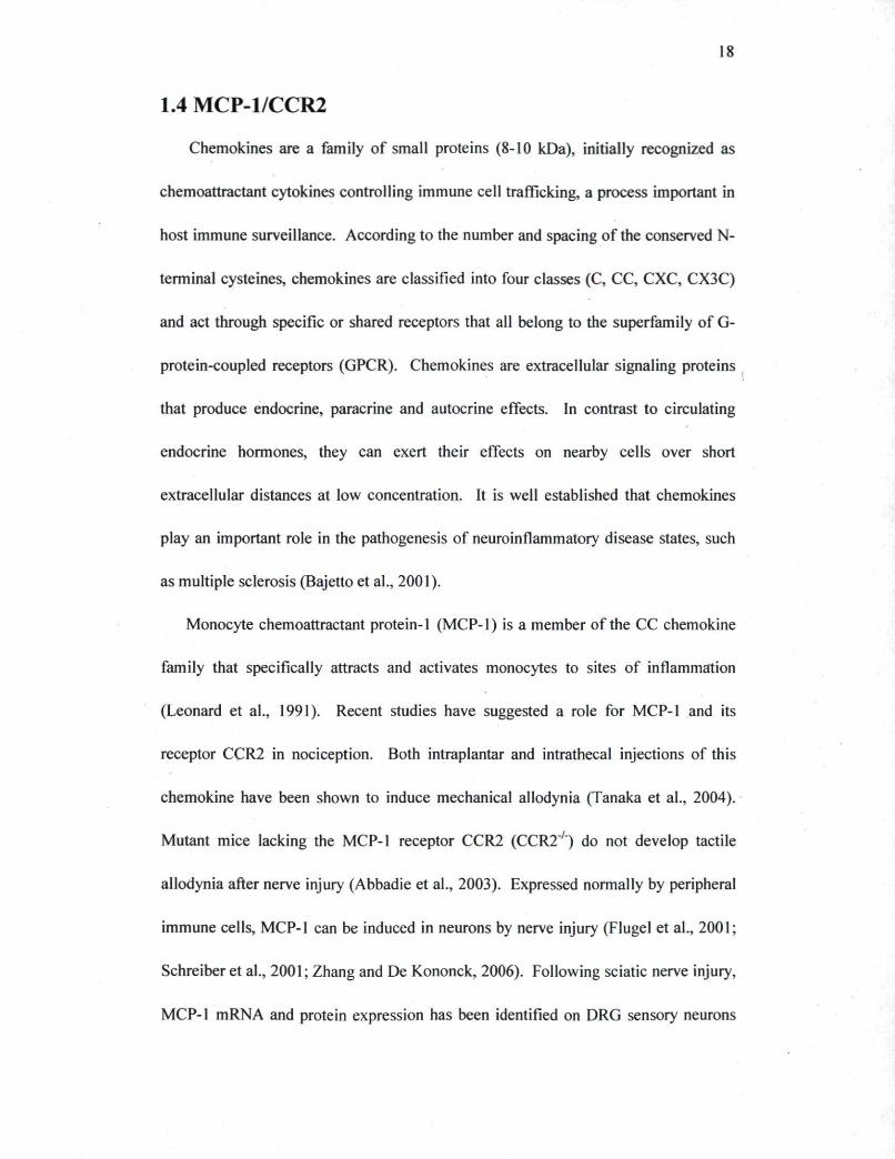
Blood vessel Endothelial cells
CGRP, substance P, bradykinin, nitric oxide
Injured axon
Macrophages and mast cells release prostaglandins and the cytokines IL-1, IL-6, IL-18.TNFandLIF
Mast cell
Chemokines (CCL2. CCL3)
T lymphocyte
Chemokine receptors
(CCR2, CCR1 and CCR5)
Figure 1.2-(A) Macrophages and Schwann cells produce matrix metalloproteases that degrade the blood-nerve barrier. Macrophages and mast cells release prostaglandins and the cytokines IL-lfi IL-6, IL-18, TNF and LIF. TNF has an autocrine effect on macrophages that is mediated through TNFR1 activation and enhances cytokine synthesis and release. TNF also promotes further macrophage infiltration.
Different from other glial cells, microglia originate from monocytic mesodermal
cells and also derive from the infiltration of hematopoietic cells during the early
development of the CNS. The normal CNS is characterized by two major monocyte-
related populations: highly ramified, resting microglia inside the CNS parenchyma
and perivascular macrophages on the luminal side of brain vessel-associated basal
membranes. During development and shortly after birth, monocyte precursor cells
populate the brain and thereby give rise to microglia. The renewal of microglia in the

14
adult CNS is traditionally thought to occur exclusively via proliferation of resident
differentiated cells. However, recent evidence suggests that, under certain
circumstances, microglia can also come from a second population (macrophages).
These macrophages are elongated and located around blood vessels. Perivascular
macrophages are gradually replenished by circulating monocytes. Thus, severe
inflammation could lead monocytes to infiltrate the brain through the blood-brain
barrier. Many ofthe hematognous monocytes invading CNS parenchyma will later
ramify and gradually transform into microglia-like cells. Inflammatory stimuli may
also lead to rapid activation of resident microglia, which may transform into
phagocytic macrophages in the presence of cellular debris. Thus, both populations of
macrophages oftmacrophages-resident microglia and hematogenous macrophages-
monocytes) may contribute to the removal of debris.
Microglia act as the first and main form of active immune defense in the CNS.
Microglia are distributed throughout the brain and spinal cord and are constantly
moving and analyzing the CNS for damaged neurons, plaques, and infectious
agents(Nimmerjahn, science,2005). The brain and spinal cord are considered
"immune privileged" organs in the sense that they are separated from the rest of the
body by a series of endothelial cells, interconnected by complex tight junction, known
as the blood-brain barrier (BBB) and blood-spinal cord barrier(BSB), respectively.
One of the roles of the BBB/BSB is to prevent most infections microbes from
reaching the vulnerable nervous tissue. In the case in which infectious agents are
directly introduced into the CNS, microglia must react quickly by initiating

15
inflammation and destroying infectious agents before they cause damage to neural
tissue. Microglias are extremely sensitive to even small pathological changes
occurring in the CNS. Microglia activation could be neuroprotective or neurotoxic,
which depends partially on the microenvironment.
1.3 Activated Microglia and Neuropathic Pain
Until very recently, chronic pain, such as neuropathic pain, was thought to be
mediated only through the dysfunction of neurons. However, recent studies have
shown that neuroimmune alterations may also contribute to pain after injury to the
nervous system. The fact that peripheral nerve injury can induce spinal
microglial/astrocytic activation has been demonstrated in several chronic neuropathic
pain models (Colburn et al., 1999; Zhang et al ., 2003; Fu et al., 1999). Activated
microglia release pain-enhancing substances such as pro-inflammatory cytokines,
nitric oxide (NO), prostaglandins (PGs), and excitatory amino acids (EAA)
(Hashizume et al., 2000), that excite spinal pain responsive neurons either directly or
indirectly, and promote the release of other transmitters that can act on nociceptive
neurons (Watkins et al., 2003). Several drugs that disrupt glia signaling by targeting
glial activation (Milligan et al., 2003; Raghavendra et al., 2003), inhibiting the
synthesis of cytokines, blocking pro-inflammatory cytokine receptors (Sweitzer et al.,
2001), or disrupting pro-inflammatory cytokine signaling pathway (Sweitzer et al.,

16
2004) have successfully controlled enhanced nociceptive states in animal models. All
these data strongly suggest that spinal cord glia are important pain modulators.
At the junction between incoming central neurons from DRG sensory reunions
and cell bodies from secondary ascending neurons, microglia patrol the spinal cord
environment and are ready to intervene if necessary, like following an injury.
Nerve injury induced-stereotypic microglial activation is characterized by a
striking increase of Iba-1 ir on the ipsilateral side DH and VH. Iba-1 (ionized binding
adaptor protein-1) is a marker specific for microglia (Fig. 1.2). On the contralateral
side, the resting microglia have long, ramified processes, and they are well spaced
from one another. On the ipsilateral side, the affected microglia show an increase in
the size of the cell body, a thickening of proximal processes and a decrease in the
ramification of distal branches; they appear to be more densely packed together.
Temporally, microglia respond to nerve injury very early. The increase of OX-42 ir
(another marker for microglia) started both in VH and DH at day 3 post-injury,
increased significantly at day 7, then peaked at day 14, a time at which activated
microglia were condensed around sensory nerve terminals in the dorsal horn and
around motoneuron cell bodies in the ventral horn. From day 22, the intensity of OX-
42-ir declined. On day 150, intensity of OX-42ir was similar to that on the
contralateral side, although some activated microglia were also present on the
contralateral side (Zhang and DeKoninck, 2006). Microglial activation is a graded
process of acquisition of many functions: not only the morphological transformation

17
from ramified to rod like or ameboid shape, Microglial activation also results in
temporal changes in gene expression. The acquired functions could include cell
proliferation, migration, phagocytosis, regulation of antigen-presenting cell
capabilities, up-regulation of cell surface receptors, and secretion of pro-inflammatory
mediators: cytokines, chemokines, PGE2, secretion of anti-inflammatory cytokines.
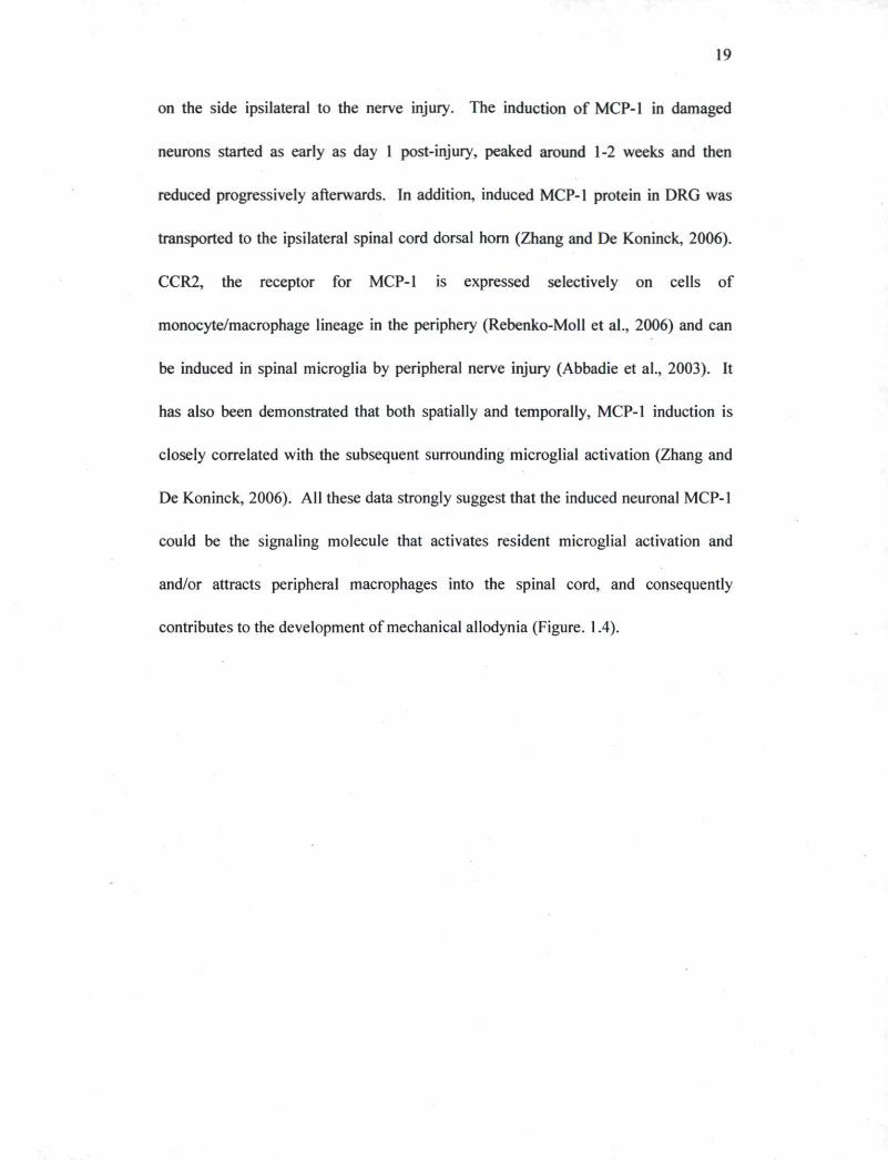
Figure 1.3 Two week after sciatic nerve injury, dense clusters of microglial cells occur in the ventral horn of the spinal cord, surrounding the cell bodies of motor neurons. Massive microglial activation is also found in the dorsal horn, in the projection territories of the central terminals of injured primary afferent fibers.
However, it remains unknown what is or are the trigger(s) that induce(s) spinal
glial activation following peripheral nerve injury.
18
1.4 MCP-1/CCR2
Chemokines are a family of small proteins (8-10 kDa), initially recognized as
chemoattractant cytokines controlling immune cell trafficking, a process important in
host immune surveillance. According to the number and spacing of the conserved N-
terminal cysteines, chemokines are classified into four classes (C, CC, CXC, CX3C)
and act through specific or shared receptors that all belong to the superfamily of G-
protein-coupled receptors (GPCR). Chemokines are extracellular signaling proteins
that produce endocrine, paracrine and autocrine effects. In contrast to circulating
endocrine hormones, they can exert their effects on nearby cells over short
extracellular distances at low concentration. It is well established that chemokines
play an important role in the pathogenesis of neuroinflammatory disease states, such
as multiple sclerosis (Bajetto et al., 2001).
Monocyte chemoattractant protein-1 (MCP-1) is a member ofthe CC chemokine
family that specifically attracts and activates monocytes to sites of inflammation
(Leonard et al., 1991). Recent studies have suggested a role for MCP-1 and its
receptor CÇR2 in nociception. Both intraplantar and intrathecal injections of this
chemokine have been shown to induce mechanical allodynia (Tanaka et al., 2004).
Mutant mice lacking the MCP-1 receptor CCR2 (CCR2"A) do not develop tactile
allodynia after nerve injury (Abbadie et al., 2003). Expressed normally by peripheral
immune cells, MCP-1 can be induced in neurons by nerve injury (Flugel et al., 2001;
Schreiber et al., 2001; Zhang and De Kononck, 2006). Following sciatic nerve injury,
MCP-1 mRNA and protein expression has been identified on DRG sensory neurons
19
on the side ipsilateral to the nerve injury. The induction of MCP-1 in damaged
neurons started as early as day 1 post-injury, peaked around 1-2 weeks and then
reduced progressively afterwards. In addition, induced MCP-1 protein in DRG was
transported to the ipsilateral spinal cord dorsal horn (Zhang and De Koninck, 2006).
CCR2, the receptor for MCP-1 is expressed selectively on cells of
monocyte/macrophage lineage in the periphery (Rebenko-Moll et al., 2006) and can
be induced in spinal microglia by peripheral nerve injury (Abbadie et al., 2003). It
has also been demonstrated that both spatially and temporally, MCP-1 induction is
closely correlated with the subsequent surrounding microglial activation (Zhang and
De Koninck, 2006). All these data strongly suggest that the induced neuronal MCP-1
could be the signaling molecule that activates resident microglial activation and
and/or attracts peripheral macrophages into the spinal cord, and consequently
contributes to the development of mechanical allodynia (Figure. 1.4).

20
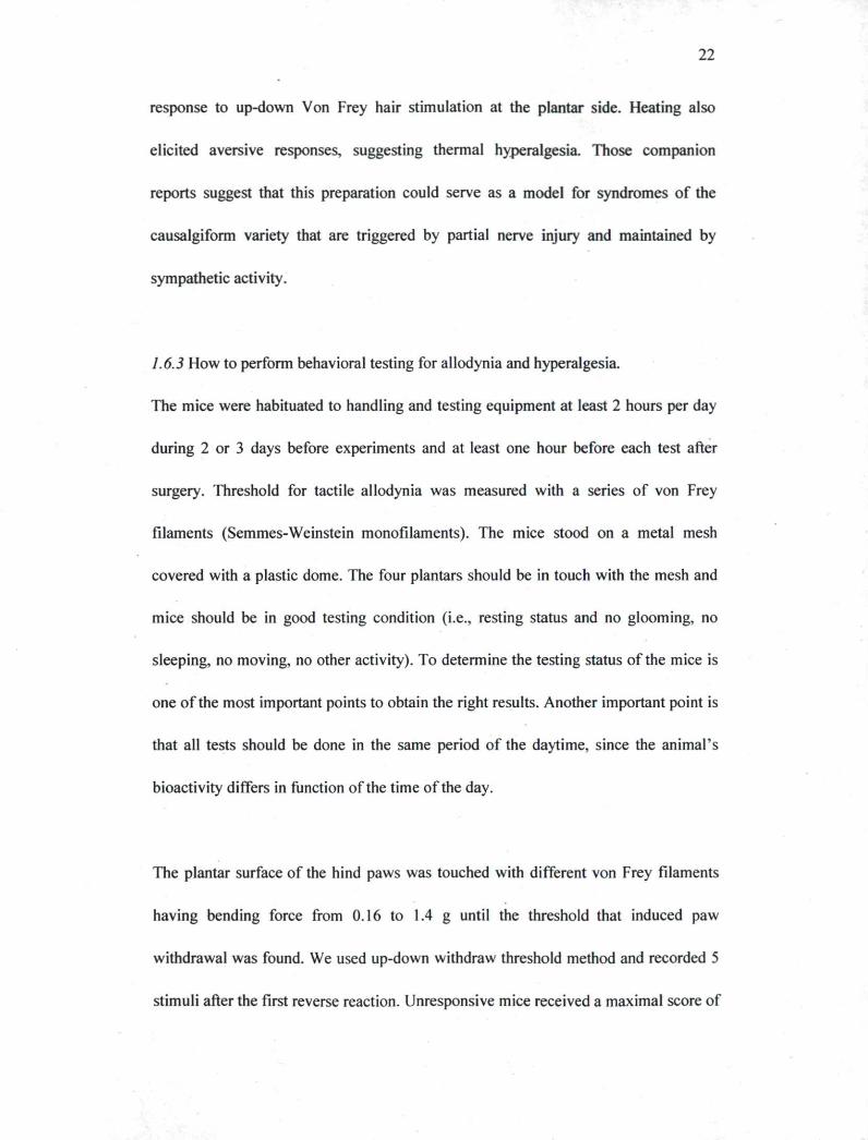
Microglial cell
Afferent terminal
Astrocyte
Transmission neuron
Figure 1.4 Microglial recruitment depends on signaling pathways involving chemokine CCL2 (MCP-1) and its receptor CCR2.
1.5 Research problem
The mechanisms underlying neuropathic pain after peripheral nerve injury are
still under investigation, especially the involvement of glial cells in the pathogenesis.
Neuropathic pain is generally chronic, severe, and resistant to treatment with counter
analgesics. The advanced research activities could unravel new targets for an
effective treatment. During my graduate studies, I tried to answer the following
questions:
/. What are the origins of these activated microglia?

21
Are they only CNS resident microglia or do they also include some newly
generated microglia? ■
Are they only CNS resident microglia or do some of them are derived from
infiltrating monocytes?
2. What triggers quiescent spinal cord glia to become activated in response to nerve
damage?
1.6 Animal model and behavioral testing
1.6.1 Why we chose the partial sciatic nerve ligation (PSNL) model
The PSNL model (Seltzer et al., 1990+) is widely used in the study of neuropathic
pain. Perhaps even more importantly, partial nerve injury is the main cause of
causalgiform pain disorders in humans. Thus, this model could similarly mimic
injuries observed in patients suffering from neuropathic pain.
1.6.2 How to make PSNL and the main characteristics ofthe model
In mice, like in rats, we unilaterally ligated about half of the sciatic nerve high in the
thigh. Within a few hours after the operation, and for several months thereafter, mice
developed guarding behavior of the ipsilateral hind paw and licked it often,
suggesting the possibility of spontaneous pain. The plantar surface of the foot was
evenly hyperesthetic to non-noxious and noxious stimuli. None of the mice
autotomized. There was a sharp decrease in the withdrawal thresholds ipsilaterally in
22
response to up-down Von Frey hair stimulation at the plantar side. Heating also
elicited aversive responses, suggesting thermal hyperalgesia. Those companion
reports suggest that this preparation could serve as a model for syndromes of the
causalgiform variety that are triggered by partial nerve injury and maintained by
sympathetic activity.
1.6.3 How to perform behavioral testing for allodynia and hyperalgesia.
The mice were habituated to handling and testing equipment at least 2 hours per day
during 2 or 3 days before experiments and at least one hour before each test after
surgery. Threshold for tactile allodynia was measured with a series of von Frey
filaments (Semmes-Weinstein monofilaments). The mice stood on a metal mesh
covered with a plastic dome. The four plantars should be in touch with the mesh and
mice should be in good testing condition (i.e., resting status and no glooming, no
sleeping, no moving, no other activity). To determine the testing status ofthe mice is
one ofthe most important points to obtain the right results. Another important point is
that all tests should be done in the same period ofthe daytime, since the animal's
bioactivity differs in function ofthe time ofthe day.
The plantar surface of the hind paws was touched with different von Frey filaments
having bending force from 0.16 to 1.4 g until the threshold that induced paw
withdrawal was found. We used up-down withdraw threshold method and recorded 5
stimuli after the first reverse reaction. Unresponsive mice received a maximal score of

23
2g. The variability in the withdrawal threshold was observed between the left and the
right hind paws after the PSNL. In this study, we focused on the Von Frey test to
analyze the development of neuropathic pain.
1.7 Presentation
The mode of presentation adopted for this master's thesis is the one ofthe series
articles. The research article included constitutes the second chapter and was
published in The Journal of Neuroscience. Nov 7. 2007. 27(45):12396-12406;
doi: 10.1523/ JNEUROSCI. 3016-07.2007. I note that I was responsible for the
majority ofthe experiments that were included in this manuscript. I conducted nerve
injury surgeries on mice with different genetic background and carried out all pain
behavioral testing before and after nerve injury. I performed immunohistochemical
studies, including double labeling, as well as qualitative and quantitative analyse of
images. I also participated in the preparation ofthe manuscript.

24
Chapter II
EXPRESSION OF CCR2 IN BOTH RESIDENT AND
BONE MARROW-DERIVED MICROGLIA PLAYS A
CRITICAL ROLE IN NEUROPATHIC PAIN
2.1 ABSTRACT
Neuropathic pain resulting from damage to or dysfunction of peripheral nerves is not
well understood and difficult to treat. Although CNS hyperexcitability is a critical
component, recent findings challenge the neuron-centric view of neuropathic pain
etiology and pathology. Indeed, glial cells were shown to play an active role in the
initiation and maintenance of pain hypersensitivity. However, the origins of these cells
and the triggers that induce their activation have yet to be elucidated. Here we show
that, after peripheral nerve injury induced by a partial ligation on the sciatic nerve, in
addition to activation of microglia resident to the CNS, hematogenous
macrophage/monocyte infiltrate the spinal cord, proliferate, and differentiate into
microglia. Signaling from chemokine monocyte chemoattractant protein-1 (MCP-1,
CCL2) to its receptor CCR2 is critical in the spinal microglial activation. Indeed,
intrathecal injection of MCP-1 caused activation of microglia in wild-type but not in
CCR2-deficient mice. Furthermore, treatment with an MCP-1 neutralizing antibody
prevented bone marrow-derived microglia (BMDM) infiltration into the spinal cord
after nerve injury. In addition, using selective knock-out of CCR2 in resident
microglia or BMDM, we found that, although total CCR2 knock-out mice did not

25
develop microglial activation or mechanical allodynia, CCR2 expression in either
resident microglia or BMDM is sufficient for the development of mechanical
allodynia. Thus, to effectively relieve neuropathic pain, both CNS resident microglia
and blood-borne macrophages need to be targeted. These findings also open the door
for a novel therapeutic strategy: to take advantage of the natural ability of bone
marrow-derived cells to infiltrate selectively affected CNS regions by using these cells
as vehicle for targeted drug delivery to inhibit hypersensitivity and chronic pain.
2.2 INTRODUCTION
The pathophysiological processes underlying the etiology of neuropathic pain involve
molecular and cellular changes in neuronal plasticity and anatomical reorganization at
various levels of the peripheral nervous system and CNS (Marx, 2004*; Baron,
2006*; Campbell and Meyer, 2006*). Recent findings have highlighted the active
involvement of glial cells in the pathogenesis of nerve injury-induced neuropathic
pain and uncover new targets for potential painkilling drugs (Marchand et al., 2005+;
Tsudaetal., 2005*).
Peripheral nerve injury induces activation of spinal microglial cells (Coyle, 1998+;
Colburn et al., 1999*; Fu et al., 1999*>; Zhang et al., 2003*). Activated microglia
contribute to neuropathic pain symptomology through the release of molecules that act
as direct modulators of neuronal excitability (Tsuda et al., 2003*; Coull et al., 2005*).

26
A major question remains unanswered: where do these activated microglial cells come
from and is there a specific population involved in pain? The normal CNS is
characterized by two major monocyte-related populations: highly ramified CNS
resident microglia and hematopoietic perivascular macrophages (Raivich and Banati,
2004*). The renewal of microglia in adulthood occurs not only through the
proliferation of preexisting cells but also through the recruitment of precursors that
derive from bone marrow (BM), because the perivascular macrophages replenished by
circulating monocyte could migrate through basal membrane into the CNS
parenchyma, a process enhanced in different forms of inflammatory neuropathology
(Streit et al., 1989*; Lawsonet al., 1992*; Priller etal., 2001 ♦; Sweitzer et al., 2002-»).
The relative contribution of resident and invading microglia to the pathogenesis may
vary depending on the setting and severity of the injury, as is evidenced by the
different dynamics of BM-derived cell accumulation (Furuya et al., 2003+; Priller et
al., 2006*; Solomon et al., 2006+; Denker et al., 2007*). An understanding of the
distinct contribution of cells of the monocytic lineage in injury-induced neuropathic
pain is important for directing the search for novel therapeutic targets.
Whenever neurons are injured, microglia become activated, both at the primary lesion
sites and remote from primary damage, at sites where the damaged neurons project
(Kreutzberg, 1996*). Thus, microglial activation is likely to be controlled by
endangered neurons. The identity of the molecules involved in neuron-microglia
signaling in different injury conditions remains an active subject of investigation.
Chemokines and their receptors constitute an elaborate signaling system that plays an

27
important role in cell-to-cell communication not only in the peripheral immune system
but also in the CNS (Ransohoff and Tani, 1998*; Ambrosini and Aloisi, 2004*; Moser
et al., 2004*; Rot and von Andrian, 2004*). Monocyte chemoattractant protein-1
(MCP-1), also named CCL2, is a member of the CC family chemokine that
specifically attracts and activates monocytes to the sites of inflammation (Leonard et
al., 1991*). Absent in normal CNS, MCP-1 was found to be induced in facial nucleus
neurons by facial nerve transection (Flugel et al., 2001*), in sympathetic ganglion
neurons after postganglionic axotomy (Schreiber et al., 2001*), and in DRG sensory
neurons and spinal cord motor neurons by chronic constriction of the sciatic nerve
(Tanaka et al., 2004*; Zhang and De Koninck, 2006*). CCR2, the receptor for MCP-
1, is expressed selectively on cells of monocyte/macrophage lineage in periphery
(Rebenko-Moll et al., 2006*) and can be induced in spinal microglia by peripheral
nerve injury (Abbadie et al., 2003*). We also demonstrated that, both spatially and
temporally, MCP-1 induction is closely correlated with the subsequent surrounding
microglial activation (Zhang and De Koninck, 2006*). We predicted that the induced
neuronal MCP-1 could be the signaling molecule that activates resident spinal
microglial and/or attracts peripheral macrophages into the spinal cord. Also, it could
contribute to peripheral sensitization by attracting macrophages to the injured nerve
and DRG. It has been demonstrated that mice lacking the CCR2 [CCR2 knock-out
(KO)] had impaired nociceptive response typically associated with neuropathy
(Abbadie et al., 2003*), but the exact contribution of CCR2 in resident and bone
marrow-derived microglia has yet to be clearly defined.

28
In the present study, we identified the origins of activated microglia by using chimeric
mice in which their bone marrow was replaced by one that expresses green fluorescent
protein (GFP). We show that, after peripheral nerve injury, in addition to activation of
microglia resident to the spinal cord, blood-borne macrophages have the ability to
infiltrate the spinal cord, proliferate, and differentiate into activated microglia. We
also showed that infiltration of peripheral macrophages into the spinal cord after nerve
injury involves direct MCP-1/CCR2 signaling from the CNS to the periphery. The fact
that both resident microglia and bone marrow-derived macrophages participate in the
modulation of central sensitization in neuropathic pain indicates that inhibition of
either resident microglia or of peripheral macrophages may not be an efficient
approach to relieve neuropathic pain. Both need to be targeted.
2.3 MATERIELS AND METHODS
Animals
Adult (7- to 12-week-old) male C57BL/6 mice were purchased from The Jackson
Laboratory (Bar Harbor, ME). Hemizygous transgenic mice expressing GFP under the
control of the chicken 6-actin promoter and cytomegalovirus enhancer and CCR2
knock-out mice were initially obtained from the same vendor. Local colonies of GFP
and CCR2KO mice were then established and maintained on a C57BL/6 background,
respectively. Mice were housed four per cage after weaning in a temperature- and
29
humidity-controlled vivarium, on a 14/10 h light/dark cycle (lights on at 6:00 A.M.
and off at 8:00 P.M.), with access to rodent chow and water ad libitum. Behavioral
experiments were conducted from 8:00 A.M. to 4:00 P.M. All protocols were
conducted according to the Canadian Council on Animal Care guidelines, as
administrated by the Laval University Animal Welfare Committee.
Generation of bone marrow-chimeric mice
Recipient mice were exposed to 10-gray total-body irradiation using a cobalt-60
source (Theratron-780 model; MDS Nordion, Ottawa, Ontario, Canada). A few hours
later, the animals were injected via tail vein with ~5 x IO6 bone marrow cells freshly
collected from donor mice. The cells were aseptically harvested by flushing femurs
with Dulbecco's PBS (DPBS) containing 2% fetal bovine serum. The samples were
combined, filtered through a 40 um nylon mesh, centrifuged, and passed through a 25
gauge needle. Recovered cells were resuspended in DPBS at a concentration of 5 x
IO6 vial nucleated cells per 200 pl. Irradiated mice transplanted with this suspension
were housed in autoclaved cages and treated with antibiotics (0.2 mg of trimethoprim
and 1 mg of sulfamethoxazole per milliliter of drinking water given for 7 d before and
2 weeks after irradiation). Animals were subjected to partial sciatic nerve ligation 3-5
months after transplantation.
GFP chimeric mice.
GFP-positive (GFP+) transgenic mice were used as BM donors. C57BL/6 mice were
irradiated and transplanted with GFP+ cells via the tail vein.

30
Central CCR2KO chimeric mice. CCR2KO mice were used as BM recipients. GFP+
transgenic BM cells were transplanted into irradiated CCR2KO mice.
Peripheral CCR2KO chimeric mice. GFP+ transgenic mice were used as BM
recipients. Bone marrow cells collected from CCR2KO mice were transplanted into
irradiated GFP+ transgenic mice.
The presence of GFP+ donor-derived cells in the peripheral circulation of transplant
recipients in each chimeric group was analyzed 8 weeks after transplantation by
fluorescence-activated cell sorting. The GFP chimeric mice and central CCR2KO
chimeric mice used in the protocol had all >80% (83.6 ± 5.03%; n = 50) of GFP+
peripheral blood leukocytes, and peripheral CCR2KO chimeric mice had only 1.53 ±
0.03% (n = 10) GFP+ cells in the blood.
Irradiation bone marrow chimeric mouse generation is currently widely used to
distinguish blood-derived and CNS resident microglia. To exclude the possibility that
the cell recruitment is an artifact of irradiation or bone marrow transplantation, some
additional approaches, such as intrasplenic injection of 6-carboxylfluorescein
diacétate, a long-lasting intracellular fluorescent tracer, and using unirradiated
parabionts with surgically anatomosed vasculature have been reported. As seen in
GFP bone marrow chimeras, monitoring invasion of blood-derived cells in the
absence of previous irradiation and bone marrow transplantation clearly revealed that
recruitment of leukocytes across the blood-brain barrier contributes to the
accumulation of ionizing calcium-binding adaptor molecule-positive (Iba-1+) cells

31
within the CNS parenchyma in different pathological conditions (Bechmann et al.,
2005*; Massengale et al., 2005*). More importantly, irradiation does not affect the
ability of resident cells to proliferate after spinal cord injury (Horky et al., 2006*).
Nerve injury model and behavioral studies
Partial sciatic nerve ligation was conducted according to the method of Seltzer et al.
(1990)* as adapted to mice (Malmbergand Basbaum, 1998*). Briefly, under
isoflurane anesthesia and aseptic conditions, the left sciatic nerve was exposed at
high-thigh level. The dorsum ofthe nerve was carefully freed from surrounding
connective tissues at a site near the trochanter. A 8-0 suture was inserted into the
nerve with a -Vncurved, reversed-cutting mini-needle (Tyco Health Care, Mississauga,
Ontario, Canada) and tightly ligated so that the dorsal one-third to one-half of the
nerve thickness was trapped in the ligature. The wound was then closed with two
muscle sutures (4-0) and two to three skin sutures (4-0). In sham-operated mice, the
nerve was exposed and left intact. The wound was closed as in injured mice.
All animals were assessed for mechanical sensitivity before surgery and from days 2-
3 after injury until they were killed for histological studies. The investigator was
totally blinded to the treatments the mice received. Paw-withdrawal threshold was
measured with calibrated von Frey fibers using the up-down method (Chaplan et al.,
1994*), as described previously (Mogil et al., 1999*). Mice were placed on a metal
mesh floor with small Plexiglas cubicles (9 x 5 x 5 cm high), and a set of eight
calibrated von Frey fibers (ranging from 0.008 to 1.40 g of force) were applied to the

32
plantar surface of the hindpaw until they bent. The threshold force required to elicit
withdrawal of the paw (median 50% paw withdrawal) was determined on two tests
separated by at least 1 h. All animals were habituated for at least 2 h to their
individual Plexiglas observation chamber before testing. Baseline data (day 0) was
obtained by averaging measurements made 1-2 d before surgery.
Intrathecal injections
In a subset of animals, recombinant mouse (rm) MCP-1 ( R & D Systems,
Minneapolis, MN) or neutralizing antibody against mouse MCP-1 (R&D Systems)
were injected by intrathecal punctions at the level of L5-L6 under isoflurane
anesthesia. The rmMCP-1 was delivered every 2 d (2 jig in 10 pl of saline per
injection) in adult naive CCR2KO and wild-type (C57BL/6) mice, and the animals
were killed at day 6 after the first injection and processed for immunohistochemistry
as described below. The MCP-1 neutralizing antibody was delivered in adult GFP
chimeric mice with nerve injury. Starting from the day of surgery, mice received an
injection ofthe antibody every 2d until day 13 after injury (4 pg in 10 |il of saline per
injection). Animals were then perfused for visualization of GFP cell infiltration at day
14 after injury. Mice in the control groups received intrathecal injections of an equal
volume of saline.
Immunohistochemistry
In wild-type (C57BL/6) and GFP chimeric mice, bromodeoxyuridine (BrdU) (50

33
mg/kg; Sigma, St. Louis, MO) was injected intraperitoneallyat day 3 after injury, and
animals were killed 2 h, 4 d, l id , and 27 d after injection. To collect the spinal cord
tissues of all animals used in the current study, mice were deeply anesthetized via an
intraperitoneal injection of a mixture of ketamine hydrochloride and xylazine and then
rapidly perfused transcardially with 0.9% saline, followed by 4% paraformaldehyde in
sodium phosphate buffer. Lumbar spinal cords were removed and postfixed overnight.
Lumbar spinal cord (L4-L5 segments) were cut into 30 pm sections, then collected in
a cold cryoprotectant solution (0.05 M sodium phosphate buffer, pH 7.3, 30%
ethylene glycol, and 20% glycerol), and stored at -20°C.
To allow the detection for BrdU-labeled cells, free-floating sections were pretreated
with 50% formamide in 2x SSC for 2 h at 65°C, followed by 15 min in 2x SSC at
room temperature, 30 min in 2N HCI at 37°C, 10 min in 0.1M borate buffer at room
temperature. Nonspecific labeling was blocked with TBS plus 0.25% Triton X-100,
1% BSA, and 3% normal goat serum for 1 h. A polyclonal goat anti-rat antibody
against BrdU (1:250; Accurate Chemicals, Westbury, NY) was incubated with tissue
sections for 48 h at 4°C. After primary antibody incubation, sections were rinsed in
TBS and incubated in Alexa 488-conjugated goat anti-rat IgG (in TBS containing
0.25% Triton X-100, 1% BSA, and 3% normal goat serum, 1:250; Invitrogen,
Carlsbad, CA) for 1 h. After rinses in TBS, sections were mounted onto slides and
coverslipped with Vectashield mounting medium (Vector Laboratories, Burlingame,
CA).

34
Regular immunofluorescent staining was performed to identify the phénotypes of
infiltrated BM cells and spinal microglia reaction to the peripheral nerve injury. Free-
floating sections were first treated in TBS containing 3% normal serum, 1% BSA, and
0.25 Triton X-100 for 1 h at room temperature and then 30 pm spinal cord sections
were incubated overnight at 4°C with antibodies listed below: mouse anti-neuron-
specific nuclear protein (NeuN) monoclonal antibody (for neurons, 1:1000;
Chemicon, Temecula, CA), rabbit anti-Iba-1 polyclonal antibody (for microglia and
macrophages, 1:1000; Wako Chemicals, Richmond, VA), rabbit anti-glial fibrillary
acid protein (GFAP) polyclonal antibody (for astrocytes, 1:1000; DakoCytomation,
Carpinteria, CA), rabbit anti-NG2 polyclonal antibody (chondroitin sulfate
proteoglycan, for oligodendrocyte progenitors, 1:250; Chemicon), and monoclonal rat
anti-CD31 (for endothelial cells, 1:1000; BD Biosciences PharMingen, San Diego,
CA), respectively, followed by a 60 min incubation at room temperature in
fluorochrome-conjugated goat secondary antibody. The sections were then mounted
onto SuperFrost slides (Fisher Scientific, Nepean, Ontario, Canada) and coverslipped
with Vectashield mounting medium (Vector Laboratories). In some cases, to better
identify the anatomical distribution of infiltrated cells, additional immunostaining was
performed using a polyclonal antibody against GFP (1:1000; Invitrogen), revealed by
a DAB-based enzymatic method; the tissue was then counterstained with thionin to
identify the parenchyma.
Image analysis

35
Images were acquired either using an Olympus Optical (Tokyo, Japan) microscope
(AX-70) equipped with a Spot Camera or a Zeiss (Oberkochen, Germany) LSM 510
confocal laser-scanning microscope. Colocalization was ensured with confocal Z
stacks at 1 pm intervals and visualization in three-dimensional orthogonal planes.
Quantitative analysis of the immunofluorescence intensity was performed on images
digitized using a constant set of parameters (exposure time, gain, and post-image
processing) with special care to avoid signal saturation. We measured the intensity of
Iba-1 immunofluorescence as the average pixel intensity within a rectangle (197 x 533
pixels) on the dorsal horn (DH) (lamina I—IV) and a rectangle (224 x 294 pixels) on
the ventral horn (VH) (lamina IX), on both sides relative to the side of injury
(MetaMorph, version 6.2r6; Universal Imaging, Downingtown, PA). GFP+ cells,
BrdU+ cells, and Iba-1+ microglial cells were counted by two independent
investigators in four different regions of interest [ipsilateral DH (DHi), contralateral
DH (DHc), VHi,and VHc]. Only ramified GFP+/Iba-1+ cells within parenchymal gray
matter were included.
Statistics
All data are presented as means ± SEM. Statistical analysis was based on the
following: (1) repeated-measures ANOVA followed by Dunnett's case-comparison
post hoc test for behavioral analyses; (2) paired / test for the difference in intensity of
Iba-1 signal between ipsilateral and contralateral side in the DH and VH, respectively;
(3) unpaired / test for the difference in intensity of Iba-1 signal between groups

36
(peripheral/central CCR2KO DHi vs GFP chimera DHi; peripheral/central CCR2KO
VHivs GFP chimera VHi).
2.4 RESULTS
Infiltration of bone marrow-derived cells into spinal cord after peripheral nerve
injury
To identify the origins of the activated microglia observed in the spinal cord after
peripheral nerve injury, we transplanted GFP-expressing bone marrow stem cells into
irradiated C57BL/6 mice (GFP chimeric mice). We found that, in naive animals, GFP+
cells were virtually absent in the spinal cord parenchyma, and the few GFP+ cells
found in the spinal cord had an elongated shape and were restricted to blood vessels
(Fig. IA). We then subjected the mice to either a sham surgery of the thigh or a
partial sciatic nerve ligation injury. In sham-operated mice, the number of GFP cells
was slightly higher in the spinal cord, but there was no significant difference between
the ipsilateral and contralateral sides (Fig. IA). In contrast, many ramified GFP-
expressing cells were present in the DHi and VHi ofthe L4-L5 spinal cord after nerve
injury (Fig. IA). The different morphologies of GFP+ cells are depicted in Figure
IB-D. The results were confirmed by immunolabeling with a polyclonal antibody
against GFP for infiltrated cells. Counterstaining with thionin helped better identify
the anatomical localization of GFP+ cells (Fig. IE-H).
37
Naive Sham Nerve-injured-d14 —
Y-* DHI
• *-. .
-- \_*K t - ■ ' • , JI VHi F G H
r/-* ' U
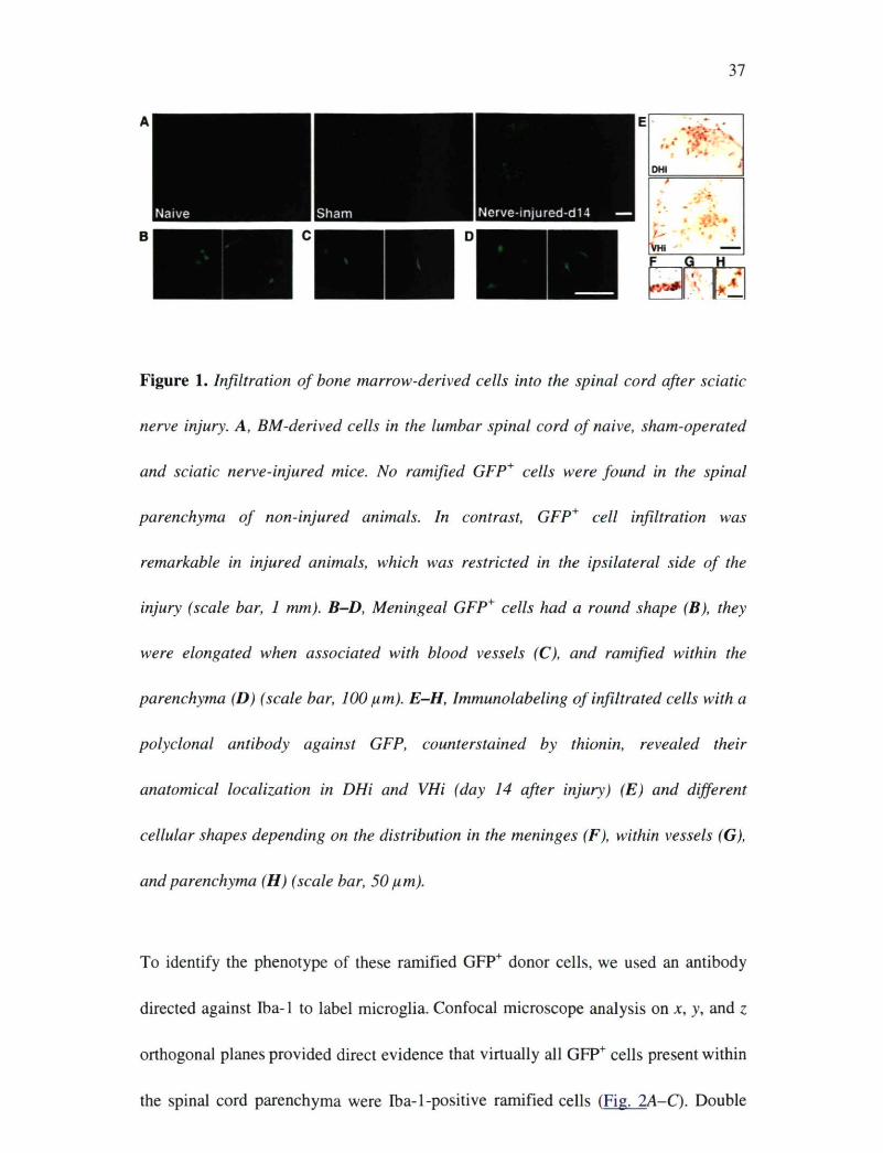
Figure 1. Infiltration of bone marrow-derived cells into the spinal cord after sciatic
nerve injury. A, BM-derived cells in the lumbar spinal cord of naive, sham-operated
and sciatic nerve-injured mice. No ramified GFP+ cells were found in the spinal
parenchyma of non-injured animals. In contrast, GFP+ cell infiltration was
remarkable in injured animals, which was restricted in the ipsilateral side of the
injury (scale bar, 1 mm). B-D, Meningeal GFP+ cells had a round shape (B), they
were elongated when associated with blood vessels (C), and ramified within the
parenchyma (D) (scale bar, 100 pm). E-H, Immunolabeling of infiltrated cells with a
polyclonal antibody against GFP, counter stained by thionin, revealed their
anatomical localization in DHi and VHi (day 14 after injury) (E) and different
cellular shapes depending on the distribution in the meninges (F), within vessels (G),
and parenchyma (H) (scale bar, 50 pm).
To identify the phénotype of these ramified GFP+ donor cells, we used an antibody
directed against Iba-1 to label microglia. Confocal microscope analysis on JC, y, and z
orthogonal planes provided direct evidence that virtually all GFP+ cells present within
the spinal cord parenchyma were Iba-1-positive ramified cells (Fig. 2A-Q. Double

38
immunolabeling of GFP with other cellular markers, NeuN for neurons, GFAP for
astrocytes, NG2 for oligodendrocyte progenitors, and CD31 for endothelial cells, were
also conducted. No evidence of GFP colocalization with the above markers has been
observed (Fig. 2D).
A DHL
Iba-1
GFP
Merge
DHc VHi VHc
Figure 2. Phénotype identification of infiltrated BM-derived cells in the lumbar spinal
cord. A, Intense Iba-1 labeling was found in activated microglia in the ipsilateral side
spinal cord (DH/VH), whereas Iba-1 immunofluorescence was weak (red) in the
contralateral DH/VH. Ramified GFP+ cells (green) overlapped with the Iba-1
immunoreactive signal in the ipsilateral DH/VH. Almost all ramified GFP+ cells
within the parenchyma were Iba-1+ (merge) (scale bar, 200 pm), which was
confirmed by confocal microscopic analysis in the x, y, and z orthogonal planes in
39
DH (B) and VH (C) (scale bar, 10 pm). D, No colocalization of GFP with other
cellular markers (NeuN, GFAP, NG2, and CD31) was observed in the lumbar spinal
cord dorsal horn 14 d after injury (scale bar, 100 pm).
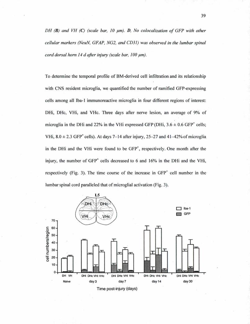
To determine the temporal profile of BM-derived cell infiltration and its relationship
with CNS resident microglia, we quantified the number of ramified GFP-expressing
cells among all Iba-1 immunoreactive microglia in four different regions of interest:
DHi, DHc, VHi, and VHc. Three days after nerve lesion, an average of 9% of
microglia in the DHi and 22% in the VHi expressed GFP (DHi, 3.6 ± 0.6 GFP+ cells;
VHi, 8.0 ± 2.3 GFP+cells). At days 7-14 after injury, 25-27 and 41^12% of microglia
in the DHi and the VHi were found to be GFP+, respectively. One month after the
injury, the number of GFP+ cells decreased to 6 and 16% in the DHi and the VHi,
respectively (Fig. 3). The time course of the increase in GFP+ cell number in the
lumbar spinal cord paralleled that of microglial activation (Fig. 3).
70-1
| 6 0
<D 5 0
2? © 40 _3 E -3 c 1 20 o
30-
10
| ~ | lba-1 I—I GFP
I
DH VH DHi DHc VHi VHc DHi DHc VHi VHc
Naive day 3 day?
Time post-injury (days)
- i — r DHi DHc VHi VHc DHi DHc VHi VHc
day 14 day 30

40
Figure 3. Temporal profile of BM-derived cell infiltration in the spinal cord
parenchyma. The numbers of Iba-1+ microglia (white bars) and GFP+/Iba-1+ BM-
derived microglia (gray bars) were determined in fours regions ofthe spinal cord (4—6
sections per mouse, 6 mice per group). Note the significant increase of BM-derived
microglia in the ipsilateral side DH/VH starting from day 3 and peaking at day 14.
Correspondingly, the number of microglia in the ipsilateral side, including BM-
derived microglia increased also from day 3 to day 14 (data are expressed as mean ±
SEM).
Proliferation and differentiation of bone marrow-derived cells within the spinal
cord parenchyma
We then assessed the plasticity of these infiltrating cells by determining their capacity
to proliferate and differentiate into microglia. Animals were injected with BrdU 3 d
-30).
Immunofluorescence staining of incorporated BrdU revealed that peripheral nerve
injury induced cell proliferation in the spinal cord, ipsilateral to the side of nerve
injury, from days 3 to 14 (Fig. 4A). Nerve injury increased the number of BrdU+ cells
in both irradiated GFP chimeric mice and non-irradiated control C57BL/6 mice
equally (data not shown). Double immunolabeling of BrdU with GFP demonstrated
after injury and perfused at different time points afterward (days 3-

41
that both resident cells (red arrow) and BM-derived hematopoietic cells (yellow
arrow) proliferated within the spinal cord parenchyma (Fig. 45), in which 19.7 and
22.3% of Brdlf cells derived from peripheral macrophages in the DHi and the VHi,
respectively (Fig. 4Q.

BrdU
42
ipsilateral contralateral on the meninges
DHi
VHi
80.3% 19.7%
i BrdU* BrdU+/GFP+
7 7 7 % 22.3%
43
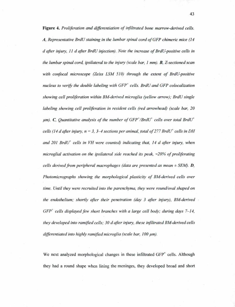
Figure 4. Proliferation and differentiation of infiltrated bone marrow-derived cells.
A, Representative BrdU staining in the lumbar spinal cord of GFP chimeric mice (14
d after injury, l i d after BrdU injection). Note the increase of BrdU-positive cells in
the lumbar spinal cord, ipsilateral to the injury (scale bar, 1 mm). B, Z-sectioned scan
with confocal microscope (Zeiss LSM 510) through the extent of BrdU-positive
nucleus to verify the double labeling with GFP+ cells. BrdU and GFP colocalization
showing cell proliferation within BM-derived microglia (yellow arrow); BrdU single
labeling showing cell proliferation in resident cells (red arrowhead) (scale bar, 20
pm). C, Quantitative analysis of the number of GFP+/Brdlf cells over total Brdlf
cells (14 d after injury, n = 3, 3-4 sections per animal, total of 277 Br d i f cells in DH
and 201 BrdU* cells in VH were counted) indicating that, 14 d after injury, when
microglial activation on the ipsilateral side reached its peak, ~20% of proliferating
cells derived from peripheral macrophages (data are presented as mean ± SEM). D,
Photomicrographs showing the morphological plasticity of BM-derived cells over
time. Until they were recruited into the parenchyma, they were round/oval shaped on
the endothelium; shortly after their penetration (day 3 after injury), BM-derived
GFP* cells displayed few short branches with a large cell body; during days 7-14,
they developed into ramified cells; 30 d after injury, these infiltrated BM-derived cells
differentiated into highly ramified microglia (scale bar, 100 pm).
We next analyzed morphological changes in these infiltrated GFP+ cells. Although
they had a round shape when lining the meninges, they developed broad and short

44
processes once they infiltrated the parenchyma (day 3). At later time points (days 7-
30), most GFP+ cells were highly ramified microglial cells with relatively small cell
bodies, resembling their resident counterparts (Fig. 4C). Thus, newly recruited
hematogenous macrophages invaded the spinal cord parenchyma proliferated and
differentiated gradually into highly ramified microglia.
Role of CCR2 in mediating microglial chemotaxis in the spinal cord
To test the hypothesis that CCR2 is critical in resident microglial activation and
peripheral macrophage infiltration, we first compared Iba-1 immunofluorescence in
sections of L4-L5 spinal cord taken from wild-type (C57BL/6) and CCR2-deficient
(CCR2KO) mice. Although nerve injury induced a striking increase of Iba-1
immunoreactivity in the DHi and VHi of wild-type (C57BL/6) mice at day 14 after
nerve injury, such an increase was almost completely abolished in CCR2KO mice
(Fig. 5A,B). This finding suggests that CCR2 expression is necessary for both
activation of resident microglia and chemotaxis of BM-derived cells after peripheral
nerve injury.
Chemotaxis of BM-derived cells may, however, occur secondarily to activation of
resident microglia expressing CCR2. To test for this possibility, we next generated
two other groups of chimeric mice by transplanting BM cells collected from GFP
transgenic mice into irradiated CCR2KO mice (central CCR2KO chimera) and by

45
transplanting CCR2K0 bone marrow cells into irradiated GFP transgenic mice
(peripheral CCR2KO chimera). The expression pattern of Iba-1 was similar in wild-
type non-irradiated mice and GFP chimeric mice, showing that irradiation and bone
marrow cell transplantation did not modify the ability of resident microglia and BM-
derived cells to respond to nerve injury (Fig. SA). We quantified the mean intensity of
Iba-1 immunoreactive signal in defined regions ofthe dorsal and ventral horns, in
which microglial activation was considered to be the most prominent. All groups of
chimeric mice exhibited significant differences in Iba-1 staining between ipsilateral
and contralateral sides after injury (Fig. 5_5). In addition, the Iba-1 signal was
significantly lower on the side ipsilateral to the injury in both groups of CCR2KO
chimeric mice when compared with the ipsilateral side in GFP chimeric mice (Fig.
5A,B).
46
Wild-type-naive Wild-type injured CCR2K0 injured
GFP chimera injured |Periph.CCR2K0 chi injured Cent.CCR2K0 chi injured
B
ilill !**
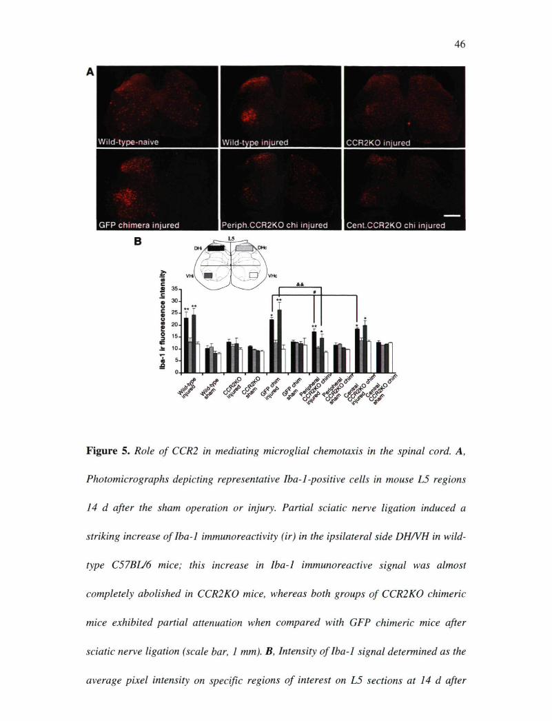
Figure 5. Role of CCR2 in mediating microglial chemotaxis in the spinal cord. A,
Photomicrographs depicting representative Iba-1-positive cells in mouse L5 regions
14 d after the sham operation or injury. Partial sciatic nerve ligation induced a
striking increase of Iba-1 immunoreactivity (ir) in the ipsilateral side DH/VH in wild-
type C57BL/6 mice; this increase in Iba-1 immunoreactive signal was almost
completely abolished in CCR2K0 mice, whereas both groups of CCR2K0 chimeric
mice exhibited partial attenuation when compared with GFP chimeric mice after
sciatic nerve ligation (scale bar, 1 mm). B, Intensity of Iba-1 signal determined as the
average pixel intensity on specific regions of interest on L5 sections at 14 d after

47
injury (4-6 sections per mouse, 4 mice per group; data are expressed as mean ±
SEM; *p < 0.05, **p < 0.01, ipsilateral vs contralateral; *p < 0.05,
peripheral/central CCR2KO DHi vs GFP DHi; &&p < 0.01, peripheral CCR2K0 VHi
vs GFP VHi).
The same result was obtained by counting Iba-1+ cells (Table 1). When CCR2 was
absent in the periphery, the decrease in Iba-1+ cell number (Table 1, A minus Q
corresponded to that of infiltrated cells in the GFP chimeric mice (Table 1, B).
Similarly, when CCR2 was absent in the CNS, the difference in Iba-1+ cell number
between central CCR2 KO and total CCR2KO (Table 1, D minus E) corresponded to
the number of infiltrated cells in the GFP chimeric mice (Table 1, B). The loss in Iba-
1 staining in each condition thus reflected the contribution of activated resident
microglia and bone marrow-derived infiltrated macrophages, respectively. The results
thus indicate that CCR2 expression is not only necessary for the activation of resident
microglia but is also directly responsible for recruitment of BM-derived cells to the
CNS.

48
Table 1. Quantification of Iba-1+ cell numbers in the lumber spinal cord of mice with different CCR2 genetic background 14 d after peripheral nerve injury
GFP chimeric mice Periph. CCR2KO mice
Cent. CCR2KO mice
CCR2KO mice
A. Iba-1 + cells
B. GFP+ cells
C. Iba-1 + cells
D. Iba-1 + cells
E. Iba-1 + cells
DHi 59.7±2.75 16±4.72 41.65±2.09** 42.50±3.63** 27.72±2.1** DHc 25.2±3.83 3±0.26 22.80±1.56 20.9O±3.53 23.78±1.33 VHi 56.55±6.03 21.3±8.1 33.05±4.37** 42.20±2.42* 26.02±3.56** VHc 28.65±2.95 4±1.67 26.80±0.43 25.40±2.92 26.94±0.98
The number of Iba-1* microglia was determined in four regions (DHi, DHc, VHi, and VHc) of the spinal cord (4-6 sections per mouse, 4 mice per group) in four groups (GFP chimeric, peripheral CCR2KO chimeric, central CCR2KO chimeric, and CCR2KO) of mice at 14 d after injury. Note that, on the ipsilateral side, the number of Iba-1* cells was significantly less in mice lacking peripherally, centrally, or totally CCR2 receptor than that in GFP chimeric mice (data are expressed as mean ± SEM; *p < 0.05, **p < 0.01, peripheral, central, and total CCR2KO DHi/VHi vs GFP DHi/VHi, respectively).
MCP-1 is the trigger for macrophage infiltration and activation of resident
microglia via its cognate receptor CCR2
To identify the ligand that activated the CCR2 receptor, we injected intrathecally
rmMCP-1 in wild-type and in CCR2KO mice. Exogenous MCP-1 induced an increase
in the size of microglial cell bodies as shown by Iba-1 immunostaining in intact wild-
type mice, and these changes were abolished in CCR2-deficient mice (Fig. 6A). In
addition, we injected intrathecally an antibody against mouse MCP-1 in GFP chimeric
mice having nerve injury to verify whether neutralization of MCP-1 could prevent
peripheral macrophage infiltration. In four of seven animals, GFP+ cell infiltration was
completely abolished. Overall, the number of ramified GFP+ cells was significantly
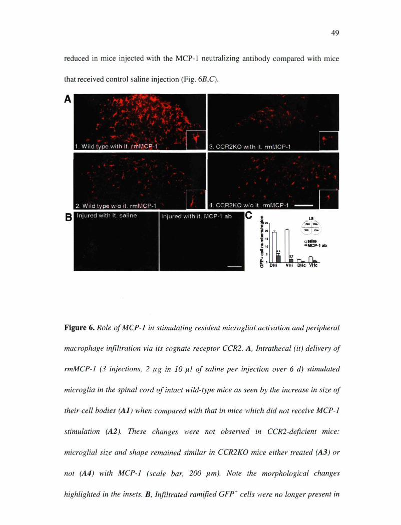
49
reduced in mice injected with the MCP-1 neutralizing antibody compared with mice
that received control saline injection (Fig. 6B,C).
■ _ J . 4 , _
■
1. Wild type with it. rmMCP-1 I 3. CCR2KO with it. rmMCP-
0 DHI VHi DHc VHc
Figure 6. Role of MCP-1 in stimulating resident microglial activation and peripheral
macrophage infiltration via its cognate receptor CCR2. A, Intrathecal (it) delivery of
rmMCP-1 (3 injections, 2 pg in 10 pl of saline per injection over 6 d) stimulated
microglia in the spinal cord of intact wild-type mice as seen by the increase in size of
their cell bodies (Al) when compared with that in mice which did not receive MCP-1
stimulation (A2). These changes were not observed in CCR2-deficient mice:
microglial size and shape remained similar in CCR2KO mice either treated (A3) or
not (A4) with MCP-1 (scale bar, 200 pm). Note the morphological changes
highlighted in the insets. B, Infiltrated ramified GFP* cells were no longer present in

50
the ipsilateral side DH and VH of injured mice treated with MCP-1 antibody (ab).
Only few elongated and scattered GFP* cells were found in blood vessels, although
the number of meningeal GFP* cells significantly increased (scale bar, 1 mm). C,
Quantitative analysis of GFP* cells in the spinal cord of GFP chimeric mice after
sciatic nerve injury and MCP-1 neutralization (data are shown as mean ± SEM; n = 7
mice/group; **p < 0.01, mice treated with MCP-1 antibody vs mice treated with
saline). MCP-1 antibody significantly reduced the number of GFP* cells within the
spinal cord parenchyma.
CCR2 in either CNS microglia or in bone marrow-derived macrophages is
sufficient for the development of mechanical allodynia
Development of mechanical hypersensitivity (allodynia) is a clinically relevant
characteristic of nerve injury. To address the relationship between mechanical
allodynia and the chemotaxis of resident and BM-derived microglia, we measured
paw-withdrawal threshold to mechanical stimuli in all animals before and after injury.
Before nerve injury, the withdrawal threshold was not affected by the CCR2 gene
deletion nor by the irradiation and bone marrow cell transplantation (Fig. 7). Wild-
type C57BL/6 mice showed a robust decrease in withdrawal threshold from 0.35 ±
0.01 g before surgery to 0.09 ±0.01 g (p < 0.01) at day 3 after surgery and maintained
this hypersensitivity to the end ofthe testing period (day 14) (Fig. IA). Mechanical
allodynia was significantly attenuated in CCR2KO mice (Fig. IB) and in GFP

51
chimeric mice treated with MCP-1 antibody (58 ± 3.8% reduction compared with
saline treated mice at day 12 after injury; n = 7 per group), indicating that MCP-
1/CCR2 signaling plays a critical role in the development ofthe hypersensitivity. In
contrast, however, neither selective peripheral CCR2KO nor selective central CCR2
KO mice had their allodynia significantly attenuated (Fig. 1D,E). This result indicates
that expression of CCR2 in either resident or BM-derived cells is sufficient for the
development of mechanical allodynia after peripheral nerve injury.
A. Wild-type mice B. CCR2KO mice
S o
■ injured-left ■ Injured-right - sham-left - sham-right
0 2 4 6 8 10 12 14 Time post-injury (days)
0 2 4 6 8 10 12 14 Time post-injury (days)
C. GFP chimera D. Periph.CCR2KO chimera E. Cent.CCR2KO chimera 0.5 -,
2 4 6 8 10 12 14 Time post-injury (days)
0 2 4 6 8 10 12 14 Time post-injury (days)
0 2 4 6 8 10 12 14 Time post-injury (days)
Figure 7. Mechanical allodynia in response to partial sciatic nerve ligation. Injured
paw-withdrawal thresholds decreased from baseline (~0.35 g) to below 0.1 g in all
groups, except for CCR2KO mice. Significant decrease in withdrawal threshold
occurred in all chimeric groups, indicating that CCR2 expression in either resident or
bone marrow-derived microglia is sufficient to cause mechanical allodynia. Data are
shown as mean ± SEM; *p < 0.05; **p < 0.01; n = 4-6 mice per group. Baseline
data (day 0) was obtained by an average of two measurements, 1-2 d before surgery.
52
2.5 DISCUSSION
Here we demonstrate that BM-derived macrophages have the ability to infiltrate the
spinal parenchyma after peripheral nerve injury. Interestingly, in contrast to spinal
cord injury, the blood-spinal cord barrier (BSCB) remains physically intact after
peripheral nerve injury, yet our results show that chemotaxis occurs across the BSCB.
These infiltrated macrophages proliferate and differentiate into microglia and,
together with their resident counterparts, contribute to CNS microgliosis in response
to peripheral nerve injury. We reported previously that MCP-1, the endogenous ligand
for CCR2 receptors, is produced by injured neurons (Zhang and De Koninck, 2006*).
In the current study, we demonstrated that exogenous MCP-1 could induce spinal
microglial activation and this activation is lost in CCR2KO mice. In addition,
neutralization of MCP-1 prevented peripheral macrophage infiltration after nerve
injury. Together, these findings imply a neuron-to-microglia and neuron-to-
macrophage signaling mechanism underlying the central component of neuropathic
pain pathogenesis. The fact that both resident and BM-derived microglia participate in
the development of the pathology has direct clinical importance. Inhibiting either
resident microglia or BM-derived macrophages may not be an effective approach to
relieve neuropathic pain.
Recruitment of circulating leukocytes into the CNS in normal physiological
conditions and in pathological states supports the essential functions of

53
immunosurveillance and host defense. Although the molecular signals and detailed
mechanisms responsible for the migration of specific inflammatory cells into the CNS
compartment are not completely identified, accumulating evidence suggests that
chemokines, in concert with adhesion molecules, are essential for the process (Charo
and Ransohoff, 2006*). MCP-1, identified originally as monocyte, memory T
lymphocytes andNK cell-specific chemoattractant (Valente et al., 1988*; Yoshimura
et al., 1989*) has been attributed a key role in regulating the infiltration of monocytes
during inflammation. MCP-1 knock-out mice exhibited deficient monocyte
recruitment in experimental autoimmune encephalomyelitis (Lu et al., 1998*; Huang
et al., 200 U). Entorhinodentate axotomy induces leukocyte infiltration in the
denervated hippocampus (Bechmann et al., 2005*; Ladeby et al., 2005*) in which
induced MCP-1 expression by glial cells has been considered as critical in directing
leukocytes to sites of axonal injury in the CNS (Babcock et al., 2003*). It is
interesting to note that, although numerous GFP cells were found in multiple regions
of normal brain (the current study; data not shown) (Vallieres and Sawchenko, 2003*;
Simard and Rivest, 2004*), such a process remains rare in the spinal cord of intact
mice. However, bone marrow-derived cells infiltrated massively the affected regions
ipsilateral to the peripheral nerve damage. This chemotaxis is dependent on MCP-1,
because MCP-1 antibody treatment successfully reduced the number of infiltrated
cells. This is also dependent on the MCP-1 receptor CCR2, because CCR2-deficient
mice no longer exhibited such an accumulation of microglial cells. MCP-1 has the
ability to alter expression of tight junction-associated proteins in endothelial cells of

54
the brain vascular system (Stamatovic et al., 2003*; Song and Pachter, 2004*), which
results in a local and temporary increase of BSCB permeability (Gordh et al., 2006*).
This may explain why MCP-1 and CCR2 play such a critical role in such a cell influx
in the affected spinal cord. We then took advantage of this model to determine the
respective contribution of BM-derived versus resident microglia in generating
chimeric mice and found that both types of cells participate in this process. MCP-1
production by damaged neurons after peripheral nerve injury (Zhang and De Koninck,
2006+) may then trigger chemotaxis through its cognate receptor CCR2 expressed in
resident and bone marrow-derived microglia.
In response to peripheral nerve injury, spinal glial cells, especially microglia,
proliferate (Echeverry et al., 2007*). The results from the current study showed that
both resident cells and blood-derived microglia retain their capacity to divide, in
which 20% of proliferating cells derived from peripheral macrophages. We also
observed that infiltrated blood-borne cells can differentiate into highly ramified Iba-1+
resident microglia but not in any other types of cells at all tested time points after
injury (days 3-30). The potential plasticity of hematopoietic stem cells raised the
questions on the trans-lineage differentiation. Several independent groups have
provided evidence that bone marrow-derived cells participate in adult neurogenesis
and angiogenesis by giving rise to neurons (Mezey et al., 2000*; Priller et al., 2001*),
endothelial cells (Bailey et al., 2006+), and astrocytes (Kopen et al., 1999*).
Consistent with some other studies (Simard and Rivest, 2004*; Massengale et al.,
2005*), our findings indicated that bone marrow-derived stem cells and their progeny

55
maintain lineage fidelity within the spinal cord parenchyma in the pathology of
neuropathic pain induced by peripheral nerve injury. The discrepancy may be
explained by technical problems (specificity of cellular markers and sensitivity of
histochemical methods) but most likely the difference may result from anatomical
distributions, because the BM-derived cells exhibiting the characteristic morphology
of cerebellar Purkinje neurons has been observed more frequently (Priller et al.,
2001+; Wright et al., 2001*) and also from different pathophysiological conditions.
The contribution of glia and glia-neuron communication in enhancing nociceptive
transmission has been well documented. Every animal model of nerve injury-induced
exaggerated pain is associated with the activation of glia within the pain-responsive
regions of the spinal cord (Tsuda et al., 2005*). Such exaggerated pain states are
mediated by glial activation, because they are blocked by drugs (e.g., fluorocitrate and
minocycline) that block glial activation (Milligan et al., 2003+; Raghavendra et al.,
2003*), by selective proinflammatory cytokine antagonists (Sweitzer et al., 2001+),
and by disrupting proinflammatory cytokine signaling pathway (Sweitzer et al.,
2004*). We revealed in this study that nerve injury induced microglial activation
comprises the activation of preexisting resident microglia, as well as the recruitment
of BM-derived peripheral macrophages. Of important impact is that both populations
are involved in the central component of sensitization to enhance spinal neuronal
excitability by dynamic glial modulators, such as ATP and BDNF (Tsuda et al.,
2003*; Coull et al., 2005*). Either resident microglia (central sensitization) or
peripheral macrophages (central sensitization by infiltration into the spinal cord and

56
peripheral sensitization by their activity at the injured site) is sufficient to cause the
neuropathic pain. The fact that Iba-1 staining was significantly reduced in both central
and peripheral CCR2KO chimeric mice (compared with GFP chimeric mice), while
neuropathic pain behavior remained identical in all three lines of mice, suggests that
there may be a floor effect on the behavior, i.e., that the amount of microglia
activation in the wild type is supramaximal, in other words, more than enough to
produce a full effect. Rutkowski et al. (2000)* demonstrated that mechanical allodynia
was not altered by either deactivation of macrophages with CNI-1493 or by depletion
of circulating macrophages using lisosome-encapsulated clodronate before peripheral
nerve injury. They concluded that macrophages have limited role in generation of
nerve injury-induced mechanical allodynia. It was also reported that macrophage
depletion by intravenous injection of liposome-encapsulated clodronate reduced the
number of macrophages in the injured nerve and alleviated slightly thermal
hyperalgesia (Liu et al., 2000*). The discrepancy with our results may stem from
differences in the behavioral outcomes measured (mechanical allodynia vs thermal
hyperalgesia). In light of our current findings, we suggested that peripheral
macrophages have significant contribution not only in peripheral but also in central
sensitization. However, blockade of only circulating macrophages is not enough to
attenuate hypersensitivity, because the involvement of spinal cord resident microglia
in the central component is not negligible.
Our results not only implicated MCP-1 as a necessary mediator for spinal microglial
activation, they point to the chemokine as being a major player for the development of

57
mechanical allodynia. These data are consistent with another study showing an
essential role of CCR2 in mediating neuropathic pain in mice (Abbadie et al., 2003*).
We further revealed the critical role of CCR2 in peripheral macrophages and resident
microglia through neuron-to-macrophages and neuron-to-microglia interaction in the
genesis of ongoing neuropathic pain. In addition, MCP-1 was reported to depolarize
sensory neurons after chronic compression of the dorsal root ganglion (White et al.,
2005*), which implicates a direct neuron-to-neuron interaction ofthe ligand with its
receptor. Some other mediators for neuron-to-glia communication that could lead to
glial activation and consequent enhancement of pain have been suggested. ATP
activates glia and the release of proinflammatory cytokines (Hide et al., 2000*;
Shigemoto-Mogami et al., 2001*). Mice lacking either purinergic P2X4 or P2X7
receptors show an impaired ability to develop neuropathic pain (Tsuda et al., 2003*;
Chessell et al., 2005*). The chemokine fractalkine has also been suggested to
contribute to the hypersensitivity evoked by nerve injury through microglial activation
(Verge et al., 2004*). Mice that bear a mutation in the gene encoding toll-like receptor
4 (TLR4) have a reduced microglial activation and do not develop thermal and
mechanical allodynia after peripheral nerve injury (Tanga et al., 2004*). Heat shock
protein 27 was proposed to be the potential endogenous ligand for TLR4 (Costigan et
al., 1998*). Although considerable redundancy may exist, microglial activation and
recruitment through MCP-1/CCR2 seem a critical component for the pathogenesis of
neuropathic pain.
58
The ability of macrophages to infiltrate and differentiate into fully functional
microglia in the CNS parenchyma opens the door for new therapeutic strategies to
treat neuropathic pain. Indeed, these cells are innately and specifically attracted to
injured and diseased regions and may thus be used as effective and selective vehicles
to deliver molecules that prevent neurodegeneration (Scholz et al., 2005*), eliminate
toxic proteins or pronociceptive signaling molecules (Shubayev and Myers, 2002*;
Coull et al., 2005*), or deliver anti-inflammatory agents (Ledeboer et al., 2007*). BM-
derived cells will be able to follow the chemoattracting gradient provided by the
distressed neurons (e.g., MCP-1) early after injury and specifically in the affected
region and deliver the necessary molecules to limit microgliosis and neuronal
hypersensitivity. A similar approach can be envisaged to treat neurodegenerative
diseases, such as Alzheimer's disease (Simard et al., 2006+), amyotrophic lateral
sclerosis (Solomon et al., 2006+), prion disease (Priller et al., 2006*), and multiple
sclerosis (Raivich and Banati, 2004*) in which BM-derived cells massively infiltrate
the affected regions.
59
2.6 Acknowledgements
We thank Martine Lessard for technical assistance for the generation of the chimeric
mice. This work was supported by Canadian Institutes of Health Research (CIHR)
grant (to J.Z.) and a team grant from Neuroscience Canada (Brain Repair program).
J.S.M. holds a Canada Research Chair in the Genetics of Pain. Y.D.K is a Senior
Scholar of the Fonds de la Recherche en Santé du Québec (FRSQ); S.R. holds a
Canada Research Chair in Neuroimmunology.
2.7 References
1. Campbell JN, Meyer RA (2006) Mechanisms of neuropathie pain. Neuron 52: 77-92.
2. Baron R (2006) Mechanisms of disease: neuropathie pain—a clinical perspective. Nat Clin Pract Neurol 2: 95-106.
3. Marx J (2004) Pain research. Prolonging the agony. Science 305: 326-329.
4. Marchand F, Perretti M, McMahon SB (2005) Role of the immune system in chronic pain. Nat Rev Neurosci 6: 521-532.
5. Tsuda M, Inoue K, Salter MW (2005) Neuropathic pain and spinal microglia: a big problem from molecules in "small" glia. Trends Neurosci 28: 101-107.
6. Colburn RW, Rickman AJ, DeLeo J A (1999) The effect of site and type of nerve injury on spinal glial activation and neuropathic pain behavior. Exp Neurol 157: 289-304.
7. Fu KY, Light AR, Matsushima GK, Maixner W (1999) Microglial reactions after subcutaneous formalin injection into the rat hind paw. Brain Res 825: 59-67.
8. Zhang J, Hoffert C, Vu HK, Groblewski T, Ahmad S, O'Donnell D (2003) Induction of CB2 receptor expression in the rat spinal cord of neuropathic but not inflammatory chronic pain models. Eur J Neurosci 17: 2750-2754.

60
9. Tsuda M, Shigemoto-Mogami Y, Koizumi S, Mizokoshi A, Kohsaka S, Salter MW, Inoue K (2003) P2X4 receptors induced in spinal microglia gate tactile allodynia after nerve injury. Nature 424: 778-783.
10. Coull JA, Beggs S, Boudreau D, Boivin D, Tsuda M, Inoue K, Gravel C, Salter MW, De Koninck Y (2005) BDNF from microglia causes the shift in neuronal anion gradient underlying neuropathic pain. Nature 438: 1017-1021.
11. Raivich G, Banati R (2004) Brain microglia and blood-derived macrophages: molecular profiles and functional roles in multiple sclerosis and animal models of autoimmune demyelinating disease. Brain Res Brain Res Rev 46: 261-281.
12. Priller J, Flugel A, Wehner T, Boentert M, Haas CA, Prinz M, Fernandez-Klett F, Prass K, Bechmann I, de Boer BA, Frotscher M, Kreutzberg GW, Persons DA, Dimagl U (2001) Targeting gene-modified hematopoietic cells to the central nervous system: use of green fluorescent protein uncovers microglial engraftment. Nat Med 7: 1356-1361.
13. Streit WJ, Graeber MB, Kreutzberg GW (1989) Expression of Ia antigen on perivascular and microglial cells after sublethal and lethal motor neuron injury. Exp Neurol 105: 115-126.
14. Lawson LJ, Perry VH, Gordon S (1992) Turnover of resident microglia in the normal adult mouse brain. Neuroscience 48: 405-415.
15. Furuya T, Tanaka R, Urabe T, Hayakawa J, Migita M, Shimada T, Mizuno Y, Mochizuki H (2003) Establishment of modified chimeric mice using GFP bone marrow as a model for neurological disorders. Neuroreport 14: 629-631.
16. Denker SP, Ji S, Dingman A, Lee SY, Derugin N, Wendland MF, Vexler ZS (2007) Macrophages are comprised of resident brain microglia not infiltrating peripheral monocytes acutely after neonatal stroke. J Neurochem 100: 893-904.
17. Solomon JN, Lewis CA, Ajami B, Corbel SY, Rossi FM, Krieger C (2006) Origin and distribution of bone marrow-derived cells in the central nervous system in a mouse model of amyotrophic lateral sclerosis. Glia 53: 744-753.
18. Priller J, Prinz M, Heikenwalder M, Zeller N, Schwarz P, Heppner FL, Aguzzi A (2006) Early and rapid engraftment of bone marrow-derived microglia in scrapie. J Neurosci 26: 11753-11762.
19. Kreutzberg GW (1996) Microglia: a sensor for pathological events in the CNS. Trends Neurosci 19: 312-318.
20. Ransohoff RM, Tani M (1998) Do chemokines mediate leukocyte recruitment in post-traumatic CNS inflammation? Trends Neurosci 21: 154-159.

61
21. Rot A, von Andrian UH (2004) Chemokines in innate and adaptive host defense: basic chemokinese grammar for immune cells. Annu Rev Immunol 22: 891-928.
22. Moser B, Wolf M, Walz A, Loetscher P (2004) Chemokines: multiple levels of leukocyte migration control. Trends Immunol 25: 75-84.
23. Ambrosini E, Aloisi F (2004) Chemokines and glial cells: a complex network in the central nervous system. Neurochem Res 29: 1017-1038.
24. Leonard EJ, Skeel A, Yoshimura T (1991) Biological aspects of monocyte chemoattractant protein-1 (MCP-1). Adv Exp Med Biol 305: 57-64.
25. Flugel A, Hager G, Horvat A, Spitzer C, Singer GM, Graeber MB, Kreutzberg GW, Schwaiger FW (2001) Neuronal MCP-1 expression in response to remote nerve injury. J Cereb Blood Flow Metab 21: 69-76.
26. Schreiber RC, Krivacic K, Kirby B, Vaccariello SA, Wei T, Ransohoff RM, Zigmond RE (2001) Monocyte chemoattractant protein (MCP)-l is rapidly expressed by sympathetic ganglion neurons following axonal injury. Neuroreport 12: 601-606.
27. Zhang J, De Koninck Y (2006) Spatial and temporal relationship between monocyte chemoattractant protein-1 expression and spinal glial activation following peripheral nerve injury. J Neurochem 97: 772-783.
28. Tanaka T, Minami M, Nakagawa T, Satoh M (2004) Enhanced production of monocyte chemoattractant protein-1 in the dorsal root ganglia in a rat model of neuropathic pain: possible involvement in the development of neuropathic pain. Neurosci Res 48: 463-469.
29. Rebenko-Moll NM, Liu L, Cardona A, Ransohoff RM (2006) Chemokines, mononuclear cells and the nervous system: heaven (or hell) is in the details. Curr Opin Immunol 18: 683-689.
30. Abbadie C, Lindia JA, Cumiskey AM, Peterson LB, Mudgett JS, Bayne EK, DeMartino JA, Maclntyre DE, Forrest MJ (2003) Impaired neuropathic pain responses in mice lacking the chemokine receptor CCR2. Proc Natl Acad Sci U S A 100:7947-7952.
31. Charo IF, Ransohoff RM (2006) The many roles of chemokines and chemokine receptors in inflammation. N Engl J Med 354: 610-621.
32. Valente AJ, Graves DT, Vial Ie-Valentin CE, Delgado R, Schwartz CJ (1988) Purification of a monocyte chemotactic factor secreted by nonhuman primate vascular cells in culture. Biochemistry 27: 4162-4168.

62
33. Yoshimura T, Robinson EA, Appella E, Matsushima K, Showalter SD, Skeel A, Leonard EJ (1989) Three forms of monocyte-derived neutrophil chemotactic factor (MDNCF) distinguished by different lengths of the amino-terminal sequence. Mol Immunol 26: 87-93.
34. Huang DR, Wang J, Kivisakk P, Rollins BJ, Ransohoff RM (2001) Absence of monocyte chemoattractant protein 1 in mice leads to decreased local macrophage recruitment and antigen-specific T helper cell type 1 immune response in experimental autoimmune encephalomyelitis. J Exp Med 193: 713-726.
35. Lu B, Rutledge BJ, Gu L, Fiorillo J, Lukacs NW, Kunkel SL, North R, Gerard C, Rollins BJ (1998) Abnormalities in monocyte recruitment and cytokine expression in monocyte chemoattractant protein 1-deficient mice. J Exp Med 187:601-608.
36. Vallieres L, Sawchenko PE (2003) Bone marrow-derived cells that populate the adult mouse brain preserve their hematopoietic identity. J Neurosci 23: 5197-5207.
37. Simard AR, Rivest S (2004) Bone marrow stem cells have the ability to populate the entire central nervous system into fully differentiated parenchymal microglia. FASEB J 18: 998-1000.
38. Stamatovic SM, Keep RF, Kunkel SL, Andjelkovic AV (2003) Potential role of MCP-1 in endothelial cell tight junction 'opening': signaling via Rho and Rho kinase. J Cell Sci 116: 4615-4628.
39. Song L, Pachter JS (2004) Monocyte chemoattractant protein-1 alters expression of tight junction-associated proteins in brain microvascular endothelial cells. Microvasc Res 67: 78-89.
40. Gordh T, Chu H, Sharma HS (2006) Spinal nerve lesion alters blood-spinal cord barrier function and activates astrocytes in the rat. Pain 124: 211-221.
41. Mezey E, Chandross KJ, Harta G, Maki RA, McKercher SR (2000) Turning blood into brain: cells bearing neuronal antigens generated in vivo from bone marrow. Science 290: 1779-1782.
42. Bailey AS, Willenbring H, Jiang S, Anderson DA, Schroeder DA, Wong MH, Grompe M, Fleming WH (2006) Myeloid lineage progenitors give rise to vascular endothelium. Proc Natl Acad Sci U S A 103: 13156-13161.
43. Kopen GC, Prockop DJ, Phinney DG (1999) Marrow stromal cells migrate throughout forebrain and cerebellum, and they differentiate into astrocytes after injection into neonatal mouse brains. Proc Natl Acad Sci U S A 96: 10711-10716.

63
44. Massengale M, Wagers AJ, Vogel H, Weissman IL (2005) Hematopoietic cells maintain hematopoietic fates upon entering the brain. J Exp Med 201: 1579-1589.
45. Wright DE, Wagers AJ, Gulati AP, Johnson FL, Weissman IL (2001) Physiological migration of hematopoietic stem and progenitor cells. Science 294: 1933-1936.
46. Milligan ED, Twining C, Chacur M, Biedenkapp J, O'Connor K, Poole S, Tracey K, Martin D, Maier SF, Watkins LR (2003) Spinal glia and proinflammatory cytokines mediate mirror-image neuropathic pain in rats. J Neurosci 23: 1026-1040.
47. Raghavendra V, Tanga F, DeLeo JA (2003) Inhibition of microglial activation attenuates the development but not existing hypersensitivity in a rat model of neuropathy. J Pharmacol Exp Ther 306: 624-630.
48. Sweitzer S, Martin D, DeLeo JA (2001) Intrathecal interleukin-1 receptor antagonist in combination with soluble tumor necrosis factor receptor exhibits an anti-allodynic action in a rat model of neuropathic pain. Neuroscience 103: 529-539.
49. Sweitzer SM, Medicherla S, Almirez R, Dugar S, Chakravarty S, Shumilla JA, Yeomans DC, Protter AA (2004) Antinociceptive action of a p38alpha MAPK inhibitor, SD-282, in a diabetic neuropathy model. Pain 109: 409-419.
50. Rutkowski MD, Pahl JL, Sweitzer S, van Rooijen N, Deleo JA (2000) Limited role of macrophages in generation of nerve injury-induced mechanical allodynia. Physiol Behav 71: 225-235.
51. Shigemoto-Mogami Y, Koizumi S, Tsuda M, Ohsawa K, Kohsaka S, Inoue K (2001) Mechanisms underlying extracellular ATP-evoked interleukin-6 release in mouse microglial cell line, MG-5. J Neurochem 78: 1339-1349.
52. Hide I, Tanaka M, Inoue A, Nakajima K, Kohsaka S, Inoue K, Nakata Y (2000) Extracellular ATP triggers tumor necrosis factor-alpha release from rat microglia. J Neurochem 75: 965-972.
53. Chessell IP, Hatcher JP, Bountra C, Michel AD, Hughes JP, Green P, Egerton J, Murfin M, Richardson J, Peck WL, Grahames CB, Casula MA, Yiangou Y, Birch R, Anand P, Buell GN (2005) Disruption ofthe P2X7 purinoceptor gene abolishes chronic inflammatory and neuropathic pain. Pain 114: 386-396.
54. Verge GM, Milligan ED, Maier SF, Watkins LR, Naeve GS, Foster AC (2004) Fractalkine (CX3CL1) and fractalkine receptor (CX3CR1) distribution in spinal cord and dorsal root ganglia under basal and neuropathic pain conditions. Eur J Neurosci 20: 1150-1160.

64
55. Tanga FY, Raghavendra V, DeLeo JA (2004) Quantitative real-time RT-PCR assessment of spinal microglial and astrocytic activation markers in a rat model of neuropathic pain. Neurochem Int 45: 397-407.
56. Costigan M, Mannion RJ, Kendall G, Lewis SE, Campagna JA, Coggeshall RE, Meridith-Middleton J, Tate S, Woolf CJ (1998) Heat shock protein 27: developmental regulation and expression after peripheral nerve injury. J Neurosci 18:5891-5900.
57. Scholz J, Broom DC, Youn DH, Mills CD, Kohno T, Suter MR, Moore KA, Decosterd I, Coggeshall RE, Woolf CJ (2005) Blocking caspase activity prevents transsynaptic neuronal apoptosis and the loss of inhibition in lamina II ofthe dorsal horn after peripheral nerve injury. J Neurosci 25: 7317-7323.
58. Shubayev VI, Myers RR (2002) Anterograde TNF alpha transport from rat dorsal root ganglion to spinal cord and injured sciatic nerve. Neurosci Lett 320: 99-101.
59. Ledeboer A, Jekich BM, Sloane EM, Mahoney JH, Langer SJ, Milligan ED, Martin D, Maier SF, Johnson KW, Leinwand LA, Chavez RA, Watkins LR (2006) Intrathecal interleukin-10 gene therapy attenuates paclitaxel-induced mechanical allodynia and proinflammatory cytokine expression in dorsal root ganglia in rats. Brain Behav Immun
60. Simard AR, Soulet D, Gowing G, Julien JP, Rivest S (2006) Bone marrow-derived microglia play a critical role in restricting senile plaque formation in Alzheimer's disease. Neuron 49: 489-502.
61. Seltzer Z, Dubner R, Shir Y (1990) A novel behavioral model of neuropathic pain disorders produced in rats by partial sciatic nerve injury. Pain 43: 205-218.
62. Malmberg AB, Basbaum AI (1998) Partial sciatic nerve injury in the mouse as a model of neuropathic pain: behavioral and neuroanatomical correlates. Pain 76: 215-222.
63. Chaplan SR, Bach FW, Pogrel JW, Chung JM, Yaksh TL (1994) Quantitative assessment of tactile allodynia in the rat paw. J Neurosci Methods 53: 55-63.
64. Mogil JS, Wilson SG, Bon K, Lee SE, Chung K, Raber P, Pieper JO, Hain HS, Belknap JK, Hubert L, Elmer GI, Chung JM, Devor M (1999) Heritability of nociception I: responses of 11 inbred mouse strains on 12 measures of nociception. Pain 80: 67-82.
65. Dissing-Olesen L, Ladeby L, Nielsen HH, Toft-Hansen H, Dalmau I, Finsen B (2007). "Axonal lesion-induced microglial proliferation and microglial cluster formation in the mouse". Neuroscience 149 (1): 112-122.

65
66. Kreutzberg GW (1995). "The First Line of Defense in Brain Pathologies". Drug-Research 45 (1): 357-360.
67. Gehrmann J, Matsumoto Y, Kreutzberg GW (1995). "Microglia: intrinsic immuneffector cell ofthe brain". Brain Research Reviews 20: 269-287.
66
Chapter III
CONCLUSION AND FUTURE DIRECTIONS
Our results revealed that, after peripheral nerve injury, the activated spinal
microglia is not only from the cells existed in the CNS, but also that ofthe population
of cells from the bone marrow like macrophages in periphery. Those two populations
of microglia (i.e., resident microglia and peripheral macrophages) participate in the
reaction of spinal microglia after partial ligation of the sciatic nerve. Bone marrow
derived macrophages have the ability to infiltrate into the spinal cord parenchyma,
proliferate and differentiate into fully functional microglia, like resident microglia.
The expression of CCR2 in resident microglia and peripheral macrophages is
necessary for the development ofthe neuropathic. The MCP-1/CCR2 system plays a
critic role acting as a trigger for microglial activation and MCP-1 not only activates
resident microglia, but also triggers the infiltration of circulating monocytes. To
efficiently treat the illness like neuropathic pain, the resident microglia in the CNS
and the macrophages derived from the bone marrow need to be targeted.
The blood-brain barrier (BBB) and the blood-spinal cord barrier (BSCB)
strongly contribute to the immune privileged status of the CNS. In normal
physiological conditions, only small, lipid-soluble molecules can freely diffuse
through the capillary endothelial membrane connected between themselves by the
tight junctions. Consequently, BBB/BSCB is an obstacle for CNS therapy and a
serious bottleneck in drug development for CNS disease. Our study demonstrated
that following peripheral nerve injury, not only resident microglia become activated,

67
under the guidance of the chemoattractant (e.g., MCP-1) gradient provided by the
injured neurons, bone marrow derived-macrophages have the ability to infiltrate into
the CNS parenchyma and differentiate into fully functional microglia, which could
reach up to 30-40% of total microglia in affected regions. In the future, we will
examine the possibility to use bone marrow-derived microglia/macrophages as
cellular vehicles to deliver analgesic agents into affected spinal cord dorsal horn and
around injured nerve to control neuropathic pain.
The expected results stem from the current study and some of our future
experiments by using bone marrow derived macrophages as cellular vehicles to
deliver anti-inflammatory agents directly into the spinal cord will provide a useful and
exciting new drug delivery technology and also a new concept by targeting glial cells
in the treatment of neuropathic pain. A similar approach can be envisaged to treat
other neurodegenerative diseases, such as Alzheimer's disease, amyotrophic lateral
sclerosis, prion disease, and multiple sclerosis in which BM-derived cells massively
infiltrate the affected regions.

68
REFERENCES FOR INTRODUCTION AND CONCLUSION
Dworkin, R.H. et al. Advances in neuropathie pain: diagnosis, mechanisms, and treatment recommendations. Arch. Neurol. 60, 1524-1534 (2003).
Streit, W.J. Microglia as neuroprotective, immunocompetent cells of the CNS. Glia 40, 133-139(2002).
Chan, W.Y., Kohsaka, S. & Rezaie, P. The origin and cell lineage of microglia: new concepts. Brain Res. Rev. 53, 344-354 (2007).
Hanani, M. Satellite glial cells in sensory ganglia: from form to function. Brain Res. Brain Res. Rev. 48, 457^176 (2005).
Perkins, N.M. & Tracey, D.J. Hyperalgesia due to nerve injury: role of neutrophils. Neuroscience 101, 745-757 (2000). Mueller, M. et al. Rapid response of identified resident endoneurial macrophages to nerve injury. Am. J. Pathol. 159, 2187-2197 (2001).
Perrin, F.E., Lacroix, S., Aviles-Trigueros, M. & David, S. Involvement of monocyte chemoattractant protein-1, macrophage inflammatory protein-1 and interleukin-1 in Wallerian degeneration. Brain 128, 854-866 (2005).
Shubayev, V.I. et al. TNF-induced MMP-9 promotes macrophage recruitment into injured peripheral nerve. Mol. Cell. Neurosci. 31, 407-^115 (2006).
White, F.A. et al. Excitatory monocyte chemoattractant protein-1 signaling is upregulated in sensory neurons after chronic compression of the dorsal root ganglion. Proc. Natl. Acad. Sci. USA 102, 14092-14097 (2005).
Zhang, J. & De Koninck, Y. Spatial and temporal relationship between monocyte chemoattractant protein-1 expression and spinal glial activation following peripheral nerve injury. J. Neurochem. 97, 772-783 (2006).
Hu, P. & McLachlan, E.M. Distinct functional types of macrophage in dorsal root ganglia and spinal nerves proximal to sciatic and spinal nerve transections in the rat. Exp. Neurol. 184, 590-605 (2003).
Tandrup, T., Woolf, CJ. & Coggeshall, R.E. Delayed loss of small dorsal root ganglion cells after transection ofthe rat sciatic nerve. J. Comp. Neurol. 422, 172-180 (2000).
69
Shi, T.J. et al. Effect of peripheral nerve injury on dorsal root ganglion neurons in the C57 BL/6J mouse: marked changes both in cell numbers and neuropeptide expression. Neuroscience 105, 249-263 (2001).
Costigan, M. et al. Replicate high-density rat genome oligonucleotide microarrays reveal hundreds of regulated genes in the dorsal root ganglion after peripheral nerve injury. BMC Neurosci. 3, 16 (2002).
Abbadie, C. et al. Impaired neuropathic pain responses in mice lacking the chemokine receptor CCR2. Proc. Natl. Acad. Sci. USA 100, 7947-7952 (2003).
Trinchieri, G. & Sher, A. Cooperation of Toll-like receptor signals in innate immune defence. Nat. Rev. Immunol. 7, 179-190 (2007). | Article | PubMed | ISI | ChemPort | Marshak-Rothstein, A. Toll-like receptors in systemic autoimmune disease. Nat. Rev. Immunol. 6, 823-835 (2006).
Scholz, J. et al. Blocking caspase activity prevents transsynaptic neuronal apoptosis and the loss of inhibition in lamina II ofthe dorsal horn after peripheral nerve injury. J. Neurosci. 25, 7317-7323 (2005).
Echeverry, S., Shi, X.Q. & Zhang, J. Characterization of cell proliferation in rat spinal cord following peripheral nerve injury and the relationship with neuropathic pain. Pain, published online 8, June 2007 (doi:doi: 10.1016/j.pain.2007.05.002). | Article | Coull, J.A. et al. BDNF from microglia causes the shift in neuronal anion gradient underlying neuropathic pain. Nature 438, 1017-1021 (2005).
TawfikV.L.,Nutile-McMenemyN.,Lacroix-FralishM.L.andDeLeo J.A. (2007) Efficacy of propentofylline, a glial modulating agent, on existing mechanical allodynia following peripheral nerve injury. Brain, Behavior and Immunity 21, 238-246.
Tsuda M., Inoue K. and Salter M.W. (2005) Neuropathic pain and spinal microglia: a big problem from molecules in "small" glia. Trends in Neurosciences 28, 101-107.
Tsuda M., Shigemoto-Mogami Y., Koizumi S., Mizokoshi A., Kohsaka S., Salter M.W. et al. (2003) P2X4 receptors induced in spinal microglia gate tactile allodynia after nerve injury. Nature 424, 778-783.
Watkins L.R. and Maier S.F. (2003) Glia: a novel drug discovery target for clinical pain. Nature Reviews Drug Discovery 2, 973-985.

70
Watkins L.R., Milligan E.D. and Maier S.F. (2001) Glial activation: a driving force for pathological pain. Trends in Neurosciences 24, 450-455.
Wen Y.R., Suter M.R., Kawasaki Y., Huang J., Pertin M., Kohno T. et al. (2007) Nerve conduction blockade in the sciatic nerve prevents but does not reverse the activation of p38 mitogen-activated protein kinase in spinal microglia in the rat spared nerve injury model. Anesthesiology 107, 312-321.
Bajetto A, Bonavia R, Barbero S, Florio T, Schettini G (2001) Chemokines and their receptors in the central nervous system. Front Neuroendocrinol 22:147-184.
Colburn RW, Rickman AJ, DeLeo J A (1999) The effect of site and type of nerve injury on spinal glial activation and neuropathic pain behavior. Exp Neurol 157:289-304.
Flugel A, Hager G, Horvat A, Spitzer C, Singer GM, Graeber MB, Kreutzberg GW, Schwaiger FW (2001) Neuronal MCP-1 expression in response to remote nerve injury. J Cereb Blood Flow Metab 21:69-76.
Fu KY, Light AR, Matsushima GK, Maixner W (1999) Microglial reactions after subcutaneous formalin injection into the rat hind paw. Brain Res 825:59-67.
Gehrmann J, Matsumoto Y, Kreutzberg GW (1995) Microglia: intrinsic immuneffector cell ofthe brain. Brain Res Brain Res Rev 20:269-287.
Hanisch UK, Kettenmann H (2007) Microglia: active sensor and versatile effector cells in the normal and pathologic brain. Nat Neurosci 10:1387-1394.
Hashizume H, DeLeo JA, Colburn RW, Weinstein JN (2000) Spinal glial activation and cytokine expression after lumbar root injury in the rat. Spine 25:1206-1217.

71
Leonard EJ, Skeel A, Yoshimura T (1991) Biological aspects of monocyte chemoattractant protein-1 (MCP-1). Adv Exp Med Biol 305:57-64.
Merskey H, Bogduk N (1994) IASP pain terminology. Seattle.
Milligan ED, Twining C, Chacur M, Biedenkapp J, O'Connor K, Poole S, Tracey K, Martin D, Maier SF, Watkins LR (2003) Spinal glia and proinflammatory cytokines mediate mirror-image neuropathic pain in rats. J Neurosci 23:1026-1040.
Raghavendra V, Tanga F, DeLeo JA (2003) Inhibition of microglial activation attenuates the development but not existing hypersensitivity in a rat model of neuropathy. J Pharmacol Exp Ther 306:624-630.
Raivich G, Bohatschek M, Kloss CU, Werner A, Jones LL, Kreutzberg GW (1999) Neuroglial activation repertoire in the injured brain: graded response, molecular mechanisms and cues to physiological function. Brain Res Brain Res Rev 30:77-105.
Rebenko-Moll NM, Liu L, Cardona A, Ransohoff RM (2006) Chemokines, mononuclear cells and the nervous system: heaven (or hell) is in the details. Curr Opin Immunol 18:683-689.
Scholz J, Woolf CJ (2007) The neuropathic pain triad: neurons, immune cells and glia. Nat Neurosci 10:1361-1368.
Schreiber RC, Krivacic K, Kirby B, Vaccariello SA, Wei T, Ransohoff RM, Zigmond RE (2001) Monocyte chemoattractant protein (MCP)-l is rapidly expressed by sympathetic ganglion neurons following axonal injury. Neuroreport 12:601-606.
Sindrup SH, Jensen TS (1999) Efficacy of pharmacological treatments of neuropathic pain: an update and effect related to mechanism of drug action. Pain 83:389-400.

72
Sweitzer S, Martin D, DeLeo JA (2001) Intrathecal interleukin-1 receptor antagonist in combination with soluble tumor necrosis factor receptor exhibits an anti-allodynic action in a rat model of neuropathic pain. Neuroscience 103:529-539.
Sweitzer SM, Medicherla S, Almirez R, Dugar S, Chakravarty S, Shumilla JA, Yeomans DC, Protter AA (2004) Antinociceptive action of a p38alpha MAPK inhibitor, SD-282, in a diabetic neuropathy model. Pain 109:409-419.
Tanaka T, Minami M, Nakagawa T, Satoh M (2004) Enhanced production of monocyte chemoattractant protein-1 in the dorsal root ganglia in a rat model of neuropathic pain: possible involvement in the development of neuropathic pain. Neurosci Res 48:463-469.
Watkins LR, Milligan ED, Maier SF (2003) Glial proinflammatory cytokines mediate exaggerated pain states: implications for clinical pain. Adv Exp Med Biol 521:1-21.
Watson CP (2000) The treatment of neuropathic pain: antidepressants and opioids. ClinJPainl6:S49-S55.
Wolf G, Gabay E, Tal M, Yirmiya R, Shavit Y (2006) Genetic impairment of interleukin-1 signaling attenuates neuropathic pain, autotomy, and spontaneous ectopic neuronal activity, following nerve injury in mice. Pain 120:315-324.
Zhang J, Hoffert C, Vu HK, Groblewski T, Ahmad S, O'Donnell D (2003) Induction of CB2 receptor expression in the rat spinal cord of neuropathic but not inflammatory chronic pain models. Eur J Neurosci 17:2750-2754.
Xie Y.F., Zhang S., Chiang C.Y., Hu J.W., Dostrovsky J.O. and Sessle B.J. (2007)Involvement of glia in central sensitization in trigeminal subnucleus caudalis(medullarydorsalhorn).BrainBehaviorandImmunity21,634-641.