Embed Size (px)
Citation preview

Journal of Surgical Oncology 61:223-229 (1996)
Expression of MRP and mdrl in Human Gastrointestinal Cancer Cell Lines:
A Correlation With Resistance Against Doxorubicin
MASAHARU MURASE, MD, YASUHIRO KODERA, MD, KEN KONDO, MD,
HlROYUKl SEKIGUCHI, MD, MICHITAKA FUJIWARA, MD, YASUSHI KASAI, MD,
SEIJI AKIYAMA, MD, KATSUKI ITO, MD, AND HROSHl TAKAGI, MD
From the Department of Surgery 11, Nagoya University School of Medicine, (M.M., K. K., H.S., M.F., YK., S.A., K.I., H.T;), Nagoya, Japan; and Department of Gastroenterological
Surgev, Aichi Cancer Center (Y K.), Nagoya, Japan ~ ~~~~
The mRNA expression of mdrl and MRP, each of which codes for a transport protein belonging to ATP-binding cassette superfamily and are reported to be responsible for multidrug resistance phenotype, were semi- quantified by RT-PCR in a panel of gastrointestinal cancer cell lines. Although the expression of MRP was predominant in esophageal cancer cell lines, expression of either or both of the genes was detected in all the cell lines tested. Expression of these two genes added together correlated significantly with chemosensitivity against doxorubicin, implicating that expression of both genes should be evaluated in the future analysis of multidrug resistance phenotype. The IDSo values for pirarubicin, although generally lower than the values for doxorubicin, correlated well with the latter, suggesting that the similar phenotype as that for doxorubicin might be responsible for drug resistance against this semisynthetic anthracycline glycoside. 0 1996 Wiley-Liss, Inc.
KEY WORDS: multidrug resistance, pirarubicin, doxorubicin, polymerase chain reaction
INTRODUCTION Gastrointestinal cancer is a disease that is still best
treated by surgery, and prognosis of unresectable disease is invariably poor even if treated by antineoplastic agents. One of the mechanisms that underlie inherent or acquired resistance of the cancer cells to anticancer drugs is multi- drug resistance. Multidrug resistance is a phenotype that confers resistance against anticancer drugs such as doxo- rubicin, etoposide, and vincristine and becomes a major impediment to the successful treatment of many human cancers.
The hallmark of multidrug resistance has long been recognized as the decreased intracellular drug accumula- tion that is related to the increased expression of the mdrl gene product, a 170 kD membrane glycoprotein termed P-glycoprotein [ 11. A study with a panel of xenograft lines resistant to various anticancer drugs has indeed revealed 0 1996 Wiley-Liss, Inc.
induction of detectable levels of mdr 1 only in a xenograft line resistant to doxorubicin [2]. However, constitutive levels of mdr 1 mRNA/P-glycoprotein expression may not necessarily result in the functional expression of the multidrug resistance phenotype in colon cancer [3] or small-cell lung cancer specimens [4]. We also have shown, through a study using surgically resected speci- mens of gastrointestinal cancer on the correlation between results of immunostaining with the monoclonal antibody to P-glycoprotein and chemosensitivity tests against doxorubicin, that 12 out of 16 P-glycoprotein negative specimens were nevertheless resistant to doxorubicin [5 ] .
Accepted for publication October 18, 1995. Address reprint requests to Dr. Yashuiro Kodera, Aichi Cancer Cen- ter, Department of Gastroenterological Surgery, 1-1, Kanokoden, Chikusa-ku, Nagoya 464, Japan.

224 Murase et al.
A number of multidrug-resistant cell lines have been described that do not overexpress P-glycoprotein but dis- play drug resistance patterns very similar to cell lines that do [6,7]. These results suggest that several additional drug resistance mechanisms other than mdrl/P-glycopro- tein might contribute to the overall level of intrinsic tumor drug resistance. Multidrug resistance-related protein (MRP), cloned originally from a multidrug-resistant small cell lung cancer cell line, is a 190kD N-glycosylated integral membrane phosphoprotein encode by a 6.5-kilo- base mRNA [8]. It has subsequently been found to be overeexpressed in a number of drug-selected cell lines derived from a variety of different tumor types [9-111. Moderate levels of resistance to anthracyclines, vincris- tine, and etoposide was exhibited in HeLa cells transfected with a fulllength MRP cDNA (12). MRP is also a novel member of the ATP-binding cassette super- family of transport proteins to which mdr-1 belongs. For these reasons, analyzing the expression of both members of the ATP-binding cassette superfamily might bring about more convincing data concerning the multidrug- resistance phenotype. A study with acute myeloid leuke- mia patients has already revealed that the functional multi- drug resistance phenotype correlated with the overexpres- sion of either one or both of these parameters in 94% of the samples studied [ 131. Studies to evaluate the expression of both mdrl and MRP now seem mandatory for cancer of several other organs.
In this study, mRNA expression of mdrl and MRP were semiquantified by reverse transcriptase polymerase chain reaction (RT-PCR) and correlation of these parame- ters with drug resistance against doxorubicin and pirarubi- cin, a semisynthetic anthracycline glycoside [ 141, has been studied, using gastrointestinal cancer cell lines.
MATERIALS AND METHODS Cell Lines
An epidermoid cancer cell line KB and its colchicine- resistant strain Chr24 are kind gifts from Dr. Kuwano (Oita Medical College, Oita, Japan). Gastric cancer cell lines MKN45 was obtained from Japanese Cancer Re- sources Bank (Tokyo, Japan). A gastric cancer cell line NUGC4 [ 151, a colon cancer cell line CC2NU, and esoph- ageal cancer cell lines ECINU and EC2NU were estab- lished at the Department of Surgery II (Nagoya Univer- sity). A colon cancer cell line col3-JCK was purchased from Tokyo Central Experimental Research Laboratory (Tokyo), and cisplatin (CDDP)-resistant strain co13/ CDDP was raised in vitro at the Department of Surgery I1 (Nagoya University) by exposure to sequential concen- trations of CDDP. A colon cancer cell line SW 1222 is a kind gift from Memorial Sloan Kettering Cancer Center (New York). A gastric cancer cell line H-111 was kindly donated by the Department of Surgery, Institute of Micro- biology (Osaka University, Osaka, Japan). An esophageal
cancer cell line WSSC was brought into a cell line from a xenograft, a kind gift from Dr. Hiroshi Watanabe (De- partment of Surgery, National Cancer Center Hospital, Tokyo). These cell lines were cultured in RPMI 1640 medium (Nissui Pharmaceutical, Tokyo) supplemented with 10% fetal calf serum.
Extraction of RNA, cDNA Synthesis, and RT-PCR Total RNA was extracted from the surgical specimens
by the method reported previously [16]. cDNA was syn- thesized essentially as described by Wang et al. [ 171 with 5 pg of total RNA and 0.5 pg of random hexadeoxy- nucleotide primer (Boehringer Mannheim, Mannheim, Germany) in 20 p1 of a solution containing 50 mM Tris- HCI (pH 8.3), 75 mM KCl, 3 mM MgCl?, 10 mM dithi- othreitol, 500 pM each dNTP, 28 units of RNAse inhibitor (Promega, Madison, WI), and 200 units of Moloney mu- rine leukemia virus reverse transcriptase (BRL, Gaithers- burg, MD). After 1 hour at 37°C cDNA was diluted at 1:lO with water and stored at -30°C until use.
PCR was carried out with cDNA derived from 50 ng of RNA, 0.5 unit of Taq DNA polymerase (Wako Jun- Yaku, Osaka, Japan), 0.2 pg of each primer, 200 FM of each dNTP, 50 m KCl, 10 mM Tris-HC1 (pH 8.8), 1.5 mM MgClz in a total volume of 20 p1. mdr-1-specific sequences were amplified by using the sense-strand primer (residues 2253-2262) and the antisense-strand primer (residues 2733-2752; antisense) designed by Noo- nan et al. [ 181, which yield a 167 base-pair product. The primers used for amplification of MRP specific sequences were residues 695-7 14 (sense strand) and residues 973- 992 (antisense strand) deduced from the nucleotide se- quence of MRP reported by Cole et al. [8] and yield a 298 base-pair product. The primers used for amplification of p-actin were residues 401421 (sense) and residues 1320-1 339 (antisense) deduced from the nucleotide se- quence of p-actin [19]. PCR was performed in a DNA thermal cycler (Perlun-Elmer/Cetus). Each cycle of PCR included 1 minute of denaturation at 94"C, 1 minute of primer annealing at 56°C and 3 minutes of extension at 70°C for a set of 26, 30, 34, and 40 cycles for mdrl, 22, 26, 30, and 40 cycles for MRP and 22 cycles for p-actin. All the necessary precautions against contamination of PCRs [20] were rigorously observed. The PCR products underwent electrophoresis on a 2% agarose gel, followed by ethidium bromide staining.
Semiquantitation of mdr-1 and MRP Genes The 167 bp band for mdrl was clearly detected after
16 cycles of PCR in cell lines Chr24 and SW1222 in which mRNA expression was detectable even with north- em blot hybridization [21]. The mdrl expression in these were designated as 4+, and mdrl levels detected only at 30, 34. and 40 cycles of PCR were designated as 3+. 2+, and +, respectively. Likewise, MRP gene expression

MRP, mdrl, and Gastrointestinal Cancer 225
TABLE I. Results of Reverse 'lkanscriptase Polymerase Chain Reaction (RT-PCR) and Chemosensitivity Test to Evaluate Correlation Between Expression of Multidrug Resistance Related Genes and Chemosensitivities Against Anthracyclines in Gastrointestinal Cancer Cell Lines
Gene expression' IDm (X 1 O-6M)
Cell lines mdrl MRP total ADM THP Epidermoid cancer
Chr24 4+ 3+ 7+ >3.5 1.03 KB I + 4+ 5+ 0.60 0.054
4+ 4+ 0.77 0.29 EC 1NU - wssc 1+ 4+ 5+ 0.52 0.75
4+ 4+ 0.93 0.041 EC2NU -
MKN45 1+ 3+ 4+ 0.43 0.073 NUGC4 3+ 2+ 5+ 0.28 0.057 H-111 3+ 3+ 0.3 1 0.040
c c 2 N u 3+ 3+ 0.45 0.16 sw1222 4+ 4+ 8+ 1.48 0.30 col3-JCK 2+ 4+ 6+ 0.62 0.072 col3KDDP I + 3+ 4+ 0.62 0.52
Esophageal cancer
Gastric cancer
- Colon cancer
-
Gene expression of mdrl and MRP mRNAs were semiquantified by RT-PCR and were expressed in terms of the FCR cycles required for the products to be detected. For mdrl, detection at 26 cycles was designated as 4+, and at 30, 34, and 40 cycles as 3+, 2+, and +. For MRP, detection at 22 cycles was designated as 4+, and at 26, 30, and 40 cycles as 3+, 2+, and +. Abbreviations: ADM; doxorubicin, THP, pirarubicin, mdr, multidmg resistance gene, MRP, multidrug resistance-related protein, IDso; the dose of an antineoplastic agent required to reduce the optical density value in the assay to 50% of the control.
of the cell lines whose 298 bp bands were detectable after 22 cycles of PCR was designated as 4+, and expression of those detectable only with 26, 30, and 40 cycles of PCR were designated as 3+, 2+, and +, respectively. Forty cycles of PCR was considered as the threshold for the detection of mdrl and MRP mRNA expression, since the yield of the mdrl or MRP specific products are found to reach plateau at -40 cycles in the current study. Noo- nan et al. [18] reported that 35 or more cycles may be used to discriminate between mdrl-positive and -negative samples in their PCR analysis, using the identical primers. No specific band at 40 cycles, therefore, was designated as - in this report.
Chemosensitivity Test Doxorubicin was kindly supplied by Kyowa Hakko
(Tokyo). Pirarubicin was kindly supplied by Meiji Seika (Tokyo). They were diluted into solutions of 0.01, 0.1, 1, and 10 p,g/ml with RPMI 1640 medium supplemented with 20% fetal calf serum, just before use in the chemo- sensitivity test. The MTT assay was performed as de- scribed [22,23]. In brief, cell suspensions were prepared and adjusted to 5 X l@ cells per ml of RPMI medium
supplemented with 20% fetal calf serum. A 100 FYwell of the suspensions are distributed to the 96-well microplates containing 100 p,1 of doxorubicin or pyrarubicin solutions at various concentrations. After incubating at 37"C, 5% C 0 2 for 72 hours, the cells were washed with phosphate- buffered saline, and 10 p,l of 0.5 M sodium succinate (Katayama Chemical, Osaka) and 10 (11 of 0.4% 3-(4, 5-dimethylthiazol-2-yl)2,5-d:phenylformazan bromide (MTT, Sigma Chemical, St. Louis, MO) were added to react for 3 hours at 37°C.
After completion of the reaction, 150 p,Ywell of di- methyl-sulfoxide (DMSO, Katayama Chemical) were added to dissolve formazan, a product of the reaction, and the optical density was measured with a sprectropho- tometer (Easy Reader EAR-340, SLT Lab Instruments, Salzburg, Austria) at a wavelength of 540 nm. The assay was performed in triplicate, and in each assay, a mean optical density value of six wells was worked out for each concentration of antineoplastic agent, and this value was divided by the mean optical density value of six control wells untreated by the anthracyclines to calculate the % viability of the cancer cells. The SDI test was considered valid only when the coefficient of variation from the optical density values of six control wells con-
226 Muraseetal.
mdr-1
B-actin
167 bp
167 bp
1 167bp 167 bp
428 bp
26 cycles
30 cycles
34 cycles
40 cycles
22 cycles
1 2 3 4 5 6 7 8 9101112
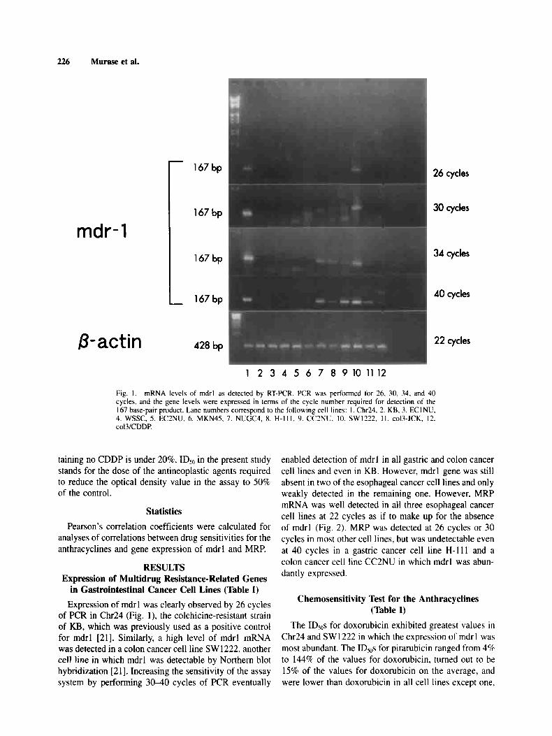
Fig. 1. mRNA levels of mdrl as detected by RT-PCR. PCR was performed for 26. 30, 34. and 40 cycles, and the gene levels were expressed in terms of the cycle number required for detection of the 167 base-pair product. Lane numbers correspond to the following cell lines: I . Chr24. 2. KB. 3. ECINU.
col3/CDDP. 4. WSSC, 5. EC2NU. 6. MKN45. 7. NUGC4, 8. H-Ill , 9. CC2NU, 10. SW1222. I I . col3-JCK. 12.
taining no CDDP is under 20%. ID50 in the present study stands for the dose of the antineoplastic agents required to reduce the optical density value in the assay to 50% of the control.
Statistics Pearson’s correlation coefficients were calculated for
analyses of correlations between drug sensitivities for the anthracyclines and gene expression of mdrl and MRP.
RESULTS Expression of Multidrug Resistance-Related Genes
in Gastrointestinal Cancer Cell Lines (Table I) Expression of mdrl was clearly observed by 26 cycles
of PCR in Chr24 (Fig. l) , the colchicine-resistant strain of KB, which was previously used as a positive control for mdrl [21]. Similarly, a high level of mdrl mRNA was detected in a colon cancer cell line SW 1222, another cell line in which mdrl was detectable by Northern blot hybridization [21]. Increasing the sensitivity of the assay system by performing 30-40 cycles of PCR eventually
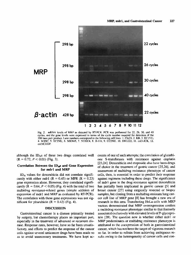
enabled detection of mdrl in all gastric and colon cancer cell lines and even in KB. However, mdrl gene was still absent in two of the esophageal cancer cell lines and only weakly detected in the remaining one. However, MRP mRNA was well detected in all three esophageal cancer cell lines at 22 cycles as if to make up for the absence of mdrl (Fig. 2). MRP was detected at 26 cycles or 30 cycles in most other cell lines, but was undetectable even at 40 cycles in a gastric cancer cell line H- 11 1 and a colon cancer cell line CC2NU in which mdrl was abun- dantly expressed.
Chemosensitivity Test for the Anthracyclines (Table I)
The IDSos for doxorubicin exhibited greatest values in Chr24 and SW1222 in which the expression of mdrl was most abundant. The IDSOS for pirarubicin ranged from 4% to 144% of the values for doxorubicin, turned out to be 15% of the values for doxorubicin on the average, and were lower than doxorubicin in all cell lines except one.
MRP, mdrl, and Gastrointestinal Cancer 227
- 298 bp 22 cycles
298 bp 26 cycles
298 bp
298 bp -
B-actin 428 bp
30 cycles
40 cycles
22 cycles
1 2 3 4 5 6 7 8 9 1 0 1 1 1 2
Fig. 2. mRNA levels of MRP as detected by RT-PCR. PCR was performed for 22, 26, 30, and 40 cycles, and the gene levels were expressed in terms of the cycle number required for detection of the 298 base-pair product. Lane numbers correspond to the following cell lines: 1. Chr24,2. KB, 3. EClNU,
col3/CDDP. 4. WSSC, 5 . EC2NU. 6. MKN45, 7. NUGC4, 8. H-111, 9. CC2NU, 10. SW1222, 11. CO~~-JCK, 12.
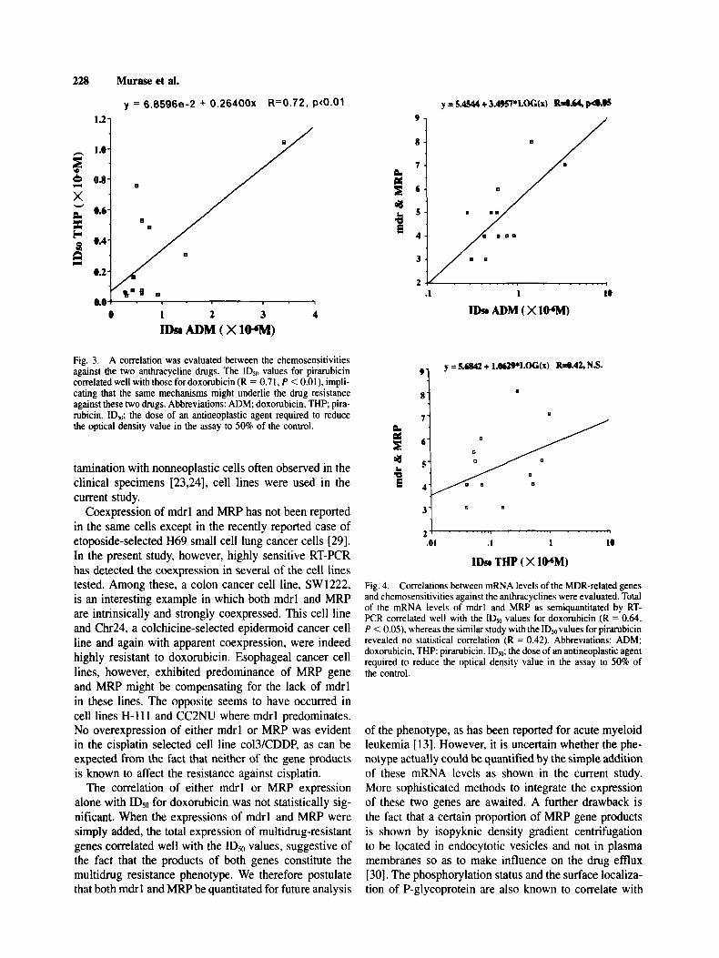
although the IDsos of these two drugs correlated well (R = 0.72, P < 0.01) (Fig. 3).
Correlation Between the ID* and Gene Expression for mdrl and MRP
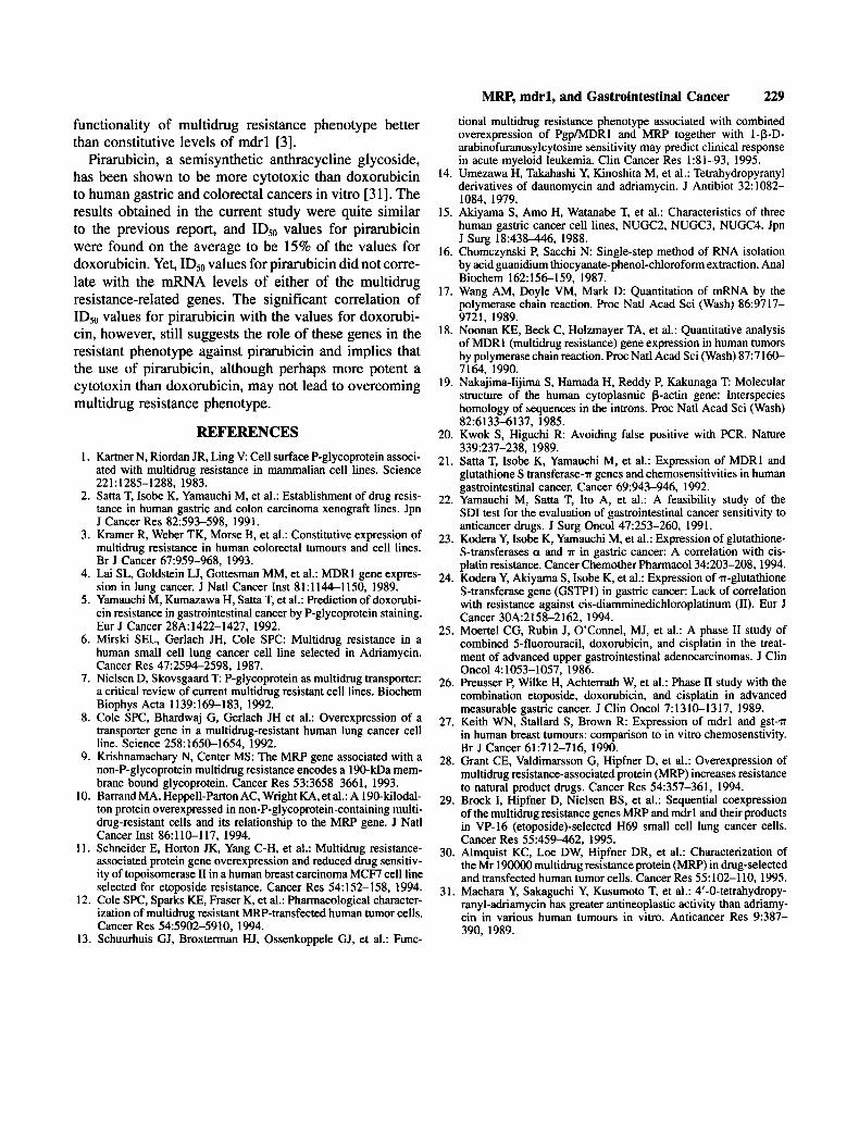
IDs0 values for doxorubicin did not correlate signifi- cantly with either mdrl (R = 0.45) or MPR (R = 0.23) gene expression alone. However, they correlated signifi- cantly (R = 0.64, P < 0.05) (Fig. 4) with the total of two multidrug resistance-related genes (simple addition of expression of mdrl and MRP as evaluated by RT-PCR). The correlation with these gene expressions was not sig- nificant for pirarubicin (R = 0.42) (Fig. 4).
DISCUSSION Gastrointestinal cancer is a disease primarily treated
by surgery, but chemotherapy places an important part, especially in the treatment of advanced or recurrent dis- ease. Response rates, however, have so far been unsatis- factory, and efforts to predict the response of the cancer cells against several anticancer drugs have been made so as to avoid unnecessary treatments. We have kept ac-
counts of one of such attempts; the correlation of glutathi- one S-transferases with resistance against cisplatin [23,24]. Doxorubicin and etoposide also have been drugs of choice in the treatment of gastric cancer [25,26], and assessment of multidrug resistance phenotype of cancer cells, then, is essential in order to predict their response against regimens including these drugs. The significance of mdrl gene in the drug resistance against doxorubicin has partially been implicated in gastric cancer [5] and breast cancer [27] using surgically resected or biopsy samples, but cloning from a multidrug-resistant lung can- cer cell line of MRP gene [8] has brought a new era of research in this area. Transfecting HeLa cells with MRP vectors demonstrated that MRP overexpression confers a multidrug resistance phenotype similar to that formerly associated exclusively with elevated levels of P-glycopro- tein [28]. The question now is whether either mdrl or MRP predominates or multidrug resistance phenotype is attributed to the coexpression of both in gastrointestinal cancer, which has not been the target of vigorous research so far. In order to refrain from achieving ambiguous re- sults owing to the heterogeneity of cancer cells and con-
228 Muraseetal.
y = 6.8596e-2 + 0.26400~ R=0.72, pt0.01
1.21
0 1 2 3 4
IDS, ADM ( X 1 W )
Fig. 3. A correlation was evaluated between the chemosensitivities against the two anthracycline drugs. The IDso values for pirarubicin correlated well with those fordoxorubicin (R = 0.71, P < 0.01), impli- cating that the same mechanisms might underlie the drug resistance against these two drugs. Abbreviations: ADM; doxorubicin, THP pira- rubicin. ID50; the dose of an antineoplastic agent required to reduce the optical density value in the assay to 50% of the control.
tamination with nonneoplastic cells often observed in the clinical specimens [23,24], cell lines were used in the current study.
Coexpression of mdrl and MRP has not been reported in the same cells except in the recently reported case of etoposide-selected H69 small cell lung cancer cells [29]. In the present study, however, highly sensitive RT-PCR has detected the coexpression in several of the cell lines tested. Among these, a colon cancer cell line, SW1222, is an interesting example in which both mdrl and MRP are intrinsically and strongly coexpressed. This cell line and Chr24, a colchicine-selected epidermoid cancer cell line and again with apparent coexpression, were indeed highly resistant to doxorubicin. Esophageal cancer cell lines, however, exhibited predominance of MRP gene and MRP might be compensating for the lack of mdrl in these lines. The opposite seems to have occurred in cell lines H-1 1 1 and CC2NU where mdrl predominates. No overexpression of either mdrl or MRP was evident in the cisplatin selected cell line col3/CDDP, as can be expected from the fact that neither of the gene products is known to affect the resistance against cisplatin.
The correlation of either mdrl or MRP expression alone with IDSo for doxorubicin was not statistically sig- nificant. When the expressions of mdrl and MRP were simply added, the total expression of multidrug-resistant genes correlated well with the IDSO values, suggestive of the fact that the products of both genes constitute the multidrug resistance phenotype. We therefore postulate that both mdrl and MRP be quantitated for future analysis
.1 1 I0
IDS, ADM ( X 1W)
0
3
2
0 0
* .01 .1 1 10
IDS THP ( X IWM)
Fig. 4. Correlations between mRNA levels of the MDR-related genes and chemosensitivities against the anthracyclines were evaluated. Total of the mRNA levels of mdrl and MRP as semiquantitated by RT- PCR correlated well with the ID, values for doxombicin (R = 0.64. P < 0.05), whereas the similar study with the IDS0 values for pirarubicin revealed no statistical correlation (R = 0.42). Abbreviations: ADM; doxorubicin, THP: pirarubicin. ID,; the dose of an antineoplastic agent required to reduce the optical density value in the assay to 508 of the control.
of the phenotype, as has been reported for acute myeloid leukemia [13]. However, it is uncertain whether the phe- notype actually could be quantified by the simple addition of these mRNA levels as shown in the current study. More sophisticated methods to integrate the expression of these two genes are awaited. A further drawback is the fact that a certain proportion of MRP gene products is shown by isopyknic density gradient centrifugation to be located in endocytotic vesicles and not in plasma membranes so as to make influence on the drug efflux [30]. The phosphorylation status and the surface localiza- tion of P-glycoprotein are also known to correlate with
MRP, mdrl, and Gastrointestinal Cancer 229
tional multidrug resistance phenotype associated with combined overexpression of PgpMDRl and MRP together with 1-p-D- arabinofuranosylcytosine sensitivity may predict clinical response in acute myeloid leukemia. Clin Cancer Res 1:81-93, 1995.
14. Umezawa H, Takahashi Y, Kinoshita M, et al.: Tetrahydropyranyl derivatives of daunomycin and adriamycin. J Antibiot 32: 1082- 1084, 1979.
15. Akiyama S, Amo H, Watanabe T, et al.: Characteristics of three human gastric cancer cell lines, NUGC2, NUGC3, NUGC4. Jpn J Surg 18:43846, 1988.
16. Chomczynski P, Sacchi N: Single-step method of RNA isolation by acid guanidium thiocyanate-phenol-chloroform extraction. Anal Biochem 162:15&159, 1987.
17. Wang AM, Doyle VM, Mark D: Quantitation of mRNA by the polymerase chain reaction. Proc Natl Acad Sci (Wash) 86:9717- 9721, 1989.
18. Noonan KE, Beck C, Holzmayer TA, et al.: Quantitative analysis of MDRl (multidrug resistance) gene expression in human tumors by polymerase chain reaction. Proc Natl Acad Sci (Wash) 87:716& 7164, 1990.
19. Nakajima-Iijima S, Hamada H, Reddy P, Kakunaga T Molecular structure of the human cytoplasmic p-actin gene: Interspecies homology of sequences in the introns. Proc Natl Acad Sci (Wash) 82:6 133-6 137, 1985.
20. Kwok S, Higuchi R: Avoiding false positive with PCR. Nature
21. Satta T, Isobe K, Yamauchi M, et al.: Expression of MDRl and glutathione S transferase-a genes and chemosensitivities in human gastrointestinal cancer. Cancer 69:943-946, 1992.
22. Yamauchi M, Satta T, Ito A, et al.: A feasibility study of the SDI test for the evaluation of gastrointestinal cancer sensitivity to anticancer drugs. J Surg Oncol47:253-260, 1991.
23. Kodera Y, Isobe K, Yamauchi M, et al.: Expression of glutathione- S-transferases a and w in gastric cancer: A correlation with cis- platin resistance. Cancer Chemother Pharmacol34:203-208,1994.
24. Kodera Y, Akiyama S, Isobe K, et al.: Expression of a-glutathione S-transferase gene (GSTPl) in gastric cancer: Lack of correlation with resistance against cis-diamminedichloroplatinum (11). Eur J Cancer 30A:2 158-2 162, 1994.
25. Moertel CG, Rubin J, O’Connel, MJ, et al.: A phase I1 study of combined 5-fluorouracil, doxorubicin, and cisplatin in the treat- ment of advanced upper gastrointestinal adenocarcinomas. J Clin
26. Preusser P, Wilke H, Achterrath W, et al.: Phase I1 study with the combination etoposide, doxorubicin, and cisplatin in advanced measurable gastric cancer. J Clin Oncol 7:1310-1317, 1989.
27. Keith WN, Stallard S, Brown R: Expression of mdrl and gst-w in human breast tumours: comparison to in vitro chemosenstivity. Br J Cancer 61:712-716, 1990.
28. Grant CE, Valdimarsson G, Hipfner D, et al.: Overexpression of multidrug resistance-associated protein (MRP) increases resistance to natural product drugs. Cancer Res 54:357-361, 1994.
29. Brock I, Hipfner D, Nielsen BS, et al.: Sequential coexpression of the multidrug resistance genes MRP and mdrl and their products in VP-16 (etoposide)-selected H69 small cell lung cancer cells. Cancer Res 55:459462, 1995.
30. Almquist KC, Loe DW, Hipfner DR, et al.: Characterization of the Mr 19oooO multidrug resistance protein (MRP) in drug-selected and transfected human tumor cells. Cancer Res 55: 102-1 10, 1995.
31. Maehara Y, Sakaguchi Y, Kusumoto T, et al.: 4’-0-tetrahydropy- ranyl-adriamycin has greater antineoplastic activity than adriamy- cin in various human tumours in vitro. Anticancer Res 9:387- 390, 1989.
339:237-238, 1989.
On~014:1053-1057, 1986.
functionality of multidrug resistance phenotype better than constitutive levels of mdrl [3].
Pirarubicin, a semisynthetic anthracycline glycoside, has been shown to be more cytotoxic than doxorubicin to human gastric and colorectal cancers in vitro [31]. The results obtained in the current study were quite similar to the previous report, and ID5,, values for pirarubicin were found on the average to be 15% of the values for doxorubicin. Yet, ID50 values for pirarubicin did not corre- late with the mRNA levels of either of the multidrug resistance-related genes. The significant correlation of ID50 values for pirarubicin with the values for doxorubi- cin, however, still suggests the role of these genes in the resistant phenotype against pirarubicin and implies that the use of pirarubicin, although perhaps more potent a cytotoxin than doxorubicin, may not lead to overcoming multidrug resistance phenotype.
REFERENCES 1. Kartner N, Riordan JR, Ling V Cell surface P-glycoprotein associ-
ated with multidrug resistance in mammalian cell lines. Science
2. Satta T, Isobe K, Yamauchi M, et al.: Establishment of drug resis- tance in human gastric and colon carcinoma xenograft lines. Jpn J Cancer Res 82593-598, 1991.
3. Kramer R, Weber TK, Morse B, et al.: Constitutive expression of multidrug resistance in human colorectal tumours and cell lines. Br J Cancer 67:959-968, 1993.
4. Lai SL, Goldstein W, Gottesman MM, et al.: MDRl gene expres- sion in lung cancer. J Natl Cancer Inst 81:1144-1150, 1989.
5 . Yamauchi M, Kumazawa H, Satta T, et al.: Prediction of doxorubi- cin resistance in gastrointestinal cancer by P-glycoprotein staining. Eur J Cancer 28A: 1422-1427, 1992.
6. Mirski SEL, Gerlach JH, Cole SPC: Multidrug resistance in a human small cell lung cancer cell line selected in Adriamycin. Cancer Res 47:2594-2598, 1987.
7. Nielsen D, Skovsgaard T P-glycoprotein as multidrug transporter: a critical review of current multidrug resistant cell lines. Biochem Biophys Acta 1139:169-183, 1992.
8. Cole SPC, Bhardwaj G, Gerlach JH et al.: Overexpression of a transporter gene in a multidrug-resistant human lung cancer cell line. Science 258: 1650-1654, 1992.
9. Krishnamachary N, Center MS: The MRP gene associated with a non-P-glycoprotein multidrug resistance encodes a 190-kDa mem- brane bound glycoprotein. Cancer Res 53:3658-3661, 1993.
10. Barrand MA, Heppell-Parton AC, Wright KA, et al.: A 190-kilodal- ton protein overexpressed in non-P-glycoprotein-containing multi- drug-resistant cells and its relationship to the MRP gene. J Natl Cancer Inst 86:110-117, 1994.
11. Schneider E, Horton JK, Yang C-H, et al.: Multidrug resistance- associated protein gene overexpression and reduced drug sensitiv- ity of topoisomerase I1 in a human breast carcinoma MCF7 cell line selected for etoposide resistance. Cancer Res 54: 152-158, 1994.
12. Cole SPC, Sparks KE, Fraser K, et al.: Pharmacological character- ization of multidrug resistant MRP-transfected human tumor cells. Cancer Res 545902-5910, 1994.
13. Schuurhuis GJ, Broxterman HJ, Ossenkoppele GJ, et al.: Func-
221~1285-1288, 1983.