Embed Size (px)
Citation preview

RESEARCH ARTICLE Open Access
Expression of taste receptors in SolitaryChemosensory Cells of rodent airwaysMarco Tizzano1,2*, Mirko Cristofoletti2, Andrea Sbarbati2, Thomas E Finger1
Abstract
Background: Chemical irritation of airway mucosa elicits a variety of reflex responses such as coughing, apnea, andlaryngeal closure. Inhaled irritants can activate either chemosensitive free nerve endings, laryngeal taste buds orsolitary chemosensory cells (SCCs). The SCC population lies in the nasal respiratory epithelium, vomeronasal organ,and larynx, as well as deeper in the airway. The objective of this study is to map the distribution of SCCs within theairways and to determine the elements of the chemosensory transduction cascade expressed in these SCCs.
Methods: We utilized a combination of immunohistochemistry and molecular techniques (rtPCR and in situhybridization) on rats and transgenic mice where the Tas1R3 or TRPM5 promoter drives expression of greenfluorescent protein (GFP).
Results: Epithelial SCCs specialized for chemoreception are distributed throughout much of the respiratory tree ofrodents. These cells express elements of the taste transduction cascade, including Tas1R and Tas2R receptormolecules, a-gustducin, PLCb2 and TrpM5. The Tas2R bitter taste receptors are present throughout the entirerespiratory tract. In contrast, the Tas1R sweet/umami taste receptors are expressed by numerous SCCs in the nasalcavity, but decrease in prevalence in the trachea, and are absent in the lower airways.
Conclusions: Elements of the taste transduction cascade including taste receptors are expressed by SCCsdistributed throughout the airways. In the nasal cavity, SCCs, expressing Tas1R and Tas2R taste receptors, mediatedetection of irritants and foreign substances which trigger trigeminally-mediated protective airway reflexes. Lowerin the respiratory tract, similar chemosensory cells are not related to the trigeminal nerve but may still trigger localepithelial responses to irritants. In total, SCCs should be considered chemoreceptor cells that help in preventingdamage to the respiratory tract caused by inhaled irritants and pathogens.
BackgroundChemical irritation of the respiratory and trachealmucosa causes various reflex responses such as cough-ing and apnea. Similarly, chemical stimulation of the lar-ynx results in a number of protective reflexes involvedin respiratory regulation, including startle, swallowing,apnea, laryngeal constriction, hypertension, and brady-cardia [1-7]. Such disturbance of respiration, if pro-longed, may cause profound hypoxemia and even death[8,9]. Despite obvious physiological and clinical impor-tance, not enough information is available regarding themeans by which chemical irritants are detected.
Until recently, the presumption has been that whiletaste buds may mediate chemical detection in the epi-glottis [2], free nerve endings are responsible for detec-tion of irritant chemicals lower in the respiratory tract[7]. Within the last decade, researchers have identified apopulation of specialized chemoreceptive epithelial cellsscattered along most of the respiratory tract from nasalcavity to bronchi [10-18]. These so-called solitary che-mosensory cells (SCCs) were first described in the gillapparatus and skin of aquatic vertebrates [19] and wereidentified by having a slender apical process and sub-stantial basolateral contacts with nerve fibers suggestingtheir role as sensory elements.The chemosensitive free nerve endings and SCCs of
the airways utilize different receptors and therefore areresponsive to different chemical irritants [20]. The nerveendings utilize various TRP channels (e.g. TrpV1 [21,22]
* Correspondence: [email protected] Mountain Taste and Smell Center, Department of Cell andDevelopmental Biology, University of Colorado at Denver & Health SciencesCenter, Aurora, USAFull list of author information is available at the end of the article
Tizzano et al. BMC Pulmonary Medicine 2011, 11:3http://www.biomedcentral.com/1471-2466/11/3
© 2011 Tizzano et al; licensee BioMed Central Ltd. This is an Open Access article distributed under the terms of the Creative CommonsAttribution License (http://creativecommons.org/licenses/by/2.0), which permits unrestricted use, distribution, and reproduction inany medium, provided the original work is properly cited.
or TrpA1 [23]), ASICs [24] and other chemosensitiveion channels. In contrast, the SCCs rely on taste recep-tors and their related downstream signalling cascade toactivate the system: the G-protein, a-gustducin; thephospholipase C beta2 (PLCb2); and the transient recep-tor potential channel M5 (TrpM5) [10-17].The population of SCCs within the airways has been
identified by expression of the TrpM5 channel[12,13,20,25]. Functional studies in TrpM5-knockoutmice show that activity of the TrpM5 channel is neces-sary for chemical transduction in both taste and SCCs[20,26,27]. The SCCs of the upper and lower airways arestructurally diverse and have different relationships tonerve fibers. In the nasal cavity, the SCCs have looseapical microvilli but are intimately associated with andsynapse onto sensory nerve terminals of the trigeminalnerve. In contrast, some of the SCCs of the lower air-ways have the key characteristics of brush cells [28]including the apical tuft of stiff microvilli. Despite thesedifferences in morphology, both the nasal SCCs and thetracheal SCC brush cells utilize the chemoreceptivetransduction cascade first described in taste buds. Like-wise, the SCCs in fish epidermis utilize some of thesame receptor proteins as in the taste system [29].The gustatory system uses different families of taste
receptors to detect nutritive or beneficial (sweet/umami)compounds on the one hand and potentially harmful(bitter) substances on the other. The appetitive qualities(sweet and umami) are detected via GPCRs of theTas1R family, namely Tas1R1, Tas1R2, and Tas1R3,characterized by a long extracellular NH2-terminal seg-ment [30-32]. Bitter substances are detected via GPCRsof the numerous members of the Tas2Rs family [33],characterized by a short extracellular NH2-domain[34-37]. The members of the Tas1R family of tastereceptors function as heterodimers [34]: the Tas1R2/Tas1R3 complex binds sweet-tasting stimuli, whereasTas1R1/Tas1R3 binds amino acids. Thus Tas1R3 is anobligatory subunit for both of the appetitive quality tastereceptors [31,38,39].Recently, it has been reported that most of the SCCs
in the nasal epithelium express Tas2Rs [10,40] with asmaller number expressing Tas1R3 (using a Tas1r3-WGA transgenic mice). Although the SCCs expresstaste receptor proteins, the sensations elicited by chemi-cal stimulation of the SCCs are not tastes, but ratherone of pain or irritation. This follows from the fact thatSCCs synapse onto polymodal nociceptors of the tri-geminal nerve rather than taste nerves [10,41] and it isthe nerves rather than the receptors that dictate thequality of a sensation [42]. Our recent studies indicatethat SCCs in the nasal cavity are activated by a varietyof substances [13,25], including bacteria quorum sensing
signalling molecules [20] and trigger protective airwayreflexes e.g. respiratory depression and apnea [10,20].Chemical stimulation of the larynx results in additionalprotective reflexes including startle, swallowing, laryn-geal constriction, hypertension, and bradycardia [1].Lower in the airways, chemical irritation largely triggerscough.To better understand the airway SCC system and its
receptors in two common laboratory rodents, we inves-tigated the distribution of SCCs throughout the lowerairways - from larynx to lung. The results show thatSCCs decrease in prevalence as one descends in therespiratory tract and that the two different classes oftaste receptors are differentially expressed in upper andlower airways.
MethodsAnimalsAdult transgenic mice in which either the Tas1R3 orTrpM5 promoter drives expression of GFP were used.Animals were a gift of Robert F. Margolskee (currently ofMonell Chem Senses Ctr., Philadelphia, PA) and SamiDamak (currently of Nestle, Lausanne, Switzerland). TheTas1R3-GFP construct contained 5’ to 3’: 13 kb of themouse Tas1R3 gene including the 5’ flanking region andthe entire 5’ untranslated region, and the codingsequence for eGFP [43]. The TrpM5-GFP construct con-tained 5’ to 3’: 11 kb of mouse TrpM5 5’ flankingsequence, TrpM5 Exon 1 (untranslated), Intron 1, andthe untranslated part of Exon 2, and eGFP [43]. AdultWistar rats were used for the in situ hybridization (ISH)and reverse trascriptase-polymerase chain reaction(RT-PCR) experiments.Experiments on mice were undertaken with the
approval of Univ. Colorado Denver Inst. Animal Careand Use Comm. Experiments on rats were conducted inaccordance with the guidelines for animal experimenta-tion according to Italian law.
Whole mount fluorescenceThe nose and trachea were dissected from Tas1R3- andTrpM5-GFP transgenic mice and stored in Tyrode’s buf-fer (145 mM NaCl, 5 mM KCl, 1 mM CaCl2, 1 mMMgCl2, 1 mM Na-pyruvate, 20 mM HEPES, 5 mM glu-cose, 7.2 pH with NaOH) to preserve the eGFP fluores-cence. Micrographs of whole-mounted tissues werecaptured with a RT Slider Spot Camera (DiagnosticInstruments) connected to a stereo microscope OlympusSZX12 (Olympus Corporation).
ImmunofluorescenceFor histological studies, tissue from Tas1R3-GFP andTrpM5-GFP transgenic mice was dissected after
Tizzano et al. BMC Pulmonary Medicine 2011, 11:3http://www.biomedcentral.com/1471-2466/11/3
Page 2 of 12
perfusion-fixation in 4% PFA/0.1M phosphate buffer(PB: 25 mM sodium phosphate dibasic anhydrous, 75mM sodium phosphate monobasic monohydrate; pH7.2) and postfixed in the same fixative for 30 minutesfollowed by cryoprotection in 20% sucrose in 0.1 M PBpH7.2 overnight at 4°C. After sectioning transversely ona cryostat, 16-μm sections were collected and dried ontoSuperfrost Plus slides (Fisher Scientific; USA). Afterthree times 10 min washes in 0.1 M phosphate-bufferedsaline (PBS: 150 mM sodium cloride, 25 mM sodiumphosphate dibasic anhydrous, 75 mM sodium phosphatemonobasic monohydrate; pH 7.2), slides were incubatedin blocking solution (2% normal goat serum, 1% bovineserum albumin, 0.3% Triton in PBS) for 1 hour at RT.Incubation with rabbit (rb) anti-a-gustducin antibody(1:500) (catalog # sc-395, Santa Cruz Biotechnology,USA), anti-Plcb2 (1:1000) (catalog # sc-206, Santa CruzBiotechnology, USA), anti PGP9.5 (1:500) (catalog #7863-0504, AbD Serotec, USA), anti-CGRP (1:1000)(catalog # T-4032, Peninsula Laboratories LLC, USA),anti-TrpM5 antibody (1:2,000) (Emily R. Liman, Univ.Southern California, USA) and rat anti-SubP (1:1000)(catalog # YMC1021, Accurate Chemical & ScientificCompany, NY, USA) all diluted in blocking solution wascarried out overnight. Three PBS washes were followedby 2 hours of incubation with Alexa568 goat anti-rb orgoat anti-rat (1:400; Molecular Probes, USA). The slidesthen were washed one time for 10 minutes in 0.1 M PBand two times for 10 minutes in 0.1 M PBS before cov-erslipping slides with Fluormount G (Southern Biotech-nology Associates, USA). Omission of the primaryantibody resulted in no apparent fluorescent signal.Similarly, in wild type mice no significant autofluores-cence is apparent (Additional file 1) at GFP wavelengths,thus indicating specificity of the fluorescence in the GFPtransgenic lines.All images were collected with an Olympus Fluoview
confocal laser scanning microscope (LSCM) FV300(Olympus Corporation). For each image, the channelswere collected sequentially with single wavelength exci-tation and then merged to produce the composite imageusing the Fluoview v5.0 software. This avoids the pro-blem resulting from side-band excitation of the fluoro-chromes. Brightness and contrast were adjusted inAdobe Photoshop.
Total RNA isolation and Reverse trascriptase - polymerasechain reaction (RT-PCR)Experimental tissues were dissected rapidly from 10 Wis-tar rats and frozen on dry ice. RNA was isolated fromtaste tissue (vallate papillae, foliate papillae, fungiformpapillae) and from other rat tissues (heart, trachea,bronchi, and lungs, nasal respiratory epithlium, larynx,spleen, liver, gut, stomach, testicle, brain) using TRIzol
Reagent (Invitrogen, life technologies). Samples of RNA(about 1 μg of total RNA) were digested with DNase I,Amp Grade (Invitrogen, life technologies), reverse tran-scribed and amplified with gene-specific primers usingthe SuperScript First-Strand Synthesis System forRT-PCR kit (Invitrogen, life technologies). Controlsomitting reverse transcriptase were done and were nega-tive (Additional file 2). Primer sequences used toamplify the target genes are shown in Table 1. Expres-sion of GAPDH (Glyceraldehyde-3 phosphate dehydro-genase) was used as the internal standard (Additionalfile 2). Amplification was performed with HotMasterTaq (Eppendorf) in an Eppendorf Gradient Martercyclerat 95°C × 30 sec, at 57-61°C × 30 sec, and at 72°C ×45-90 sec for 30 cycles. PCR products were visualizedwith ethidium bromide on a 1.5% agarose gel by electro-phoresis. Sequencing and nested PCR with 2 internalprimers of the PCR products were used to confirm tospecificity of the PCR and the primers used (data notshown). To check the quality of the cDNA templatesused for all the PCR experiments we amplified with pri-mers for GAPDH the cDNA treated with and without(Additional file 2) the reverse transcriptase (RT) enzyme.
In situ Hybridization (ISH)A longer cDNA insert (1.43 kb) for rat a-gustducin anda cDNA insert (1.85 kb) for rat Tas1R3 were generatedby RT-PCR, using cDNA from rat lingual tissue mRNAas template (primers in Table 1). This sequenceincluded the entire coding sequence (McLaughlin et al.1992) of a-gustducin and part of the extracellulardomain and the entire transmembrane sequences ofTas1R3. RT-PCR products were cloned using the TOPOTA Cloning kit (Invitrogen life technologies, USA) intothe pCRII-TOPO vector. Digoxigenin-labeled RNA(DIG-RNA) probes were transcribed in anti-sense andsense orientation using T7 and SP6 RNA polymerase.To estimate probe concentration, serial dilutions oflabeled probes and a standard labeled RNA were spottedon nylon membranes (HybondN+; Amersham, USA),immunodetected using anti-DIG-Fab-AP conjugatediluted 1:5000, and visualized with NBT and BCIPaccording to instructions from Roche. Probes werediluted to10 ng/μl and were stored in aliquots at -80°C.Tissues were dissected rapidly from adult Wistar rats
after perfusion with 0.1 M PB and fixation with 4% PFA/0.1M PB. The tissues were postfixed and cryoprotectedovernight at 4°C in 4% PFA/0.1M PB + 20% sucrose. Forpreparing cryosections, tissues were placed in chilledOCT embedding medium and snap-frozen in isopentaneprecooled in dry ice. Tissue blocks were stored at -80Cfor up to 4 weeks. Cryosections of 10-12 μm were cut at-20°C, collected on baked Superfrost Plus slides (FisherScientific; USA), and stored desiccated at -80°C.
Tizzano et al. BMC Pulmonary Medicine 2011, 11:3http://www.biomedcentral.com/1471-2466/11/3
Page 3 of 12
Cryosections were removed from -80°C storage and wereimmediately fixed in freshly prepared 4% PFA/0.1M PBSat 4°C for 20 minutes. Sections were rinsed twice in PBSfor 5 min each. Endogenous AP activity was quenchedwith 0.2 M HCl for 8 minutes, followed by two 5-minwashes in PBS. For tissue partial proteolysis, sectionswere permeabilized with 10 μg/ml proteinase K in 10mM Tris-HCl (pH 7.5) at room temperature (RT) for 10minutes (lingual tissue). After rinsing in 10 mM Tris-HCl, sections were equilibrated in 0.1 M triethanolamine(TEA) for 2 minutes and then were acetylated in freshlyprepared 0.25% acetic anhydride in 0.1 M TEA for 10minutes. After rinsing in 2× SSC for 10 minutes at RT,sections were air-dried for 5 minutes on a slide warmerat 60°C. Sections were encircled with rubber cement toenclose hybridization buffer and were used immediatelyin hybridization. DIG-RNA probes were freshly diluted in1 ml hybridization buffer (50% formamide, 2× SSC, 1×Denhardt’s, 10% dextran sulphate, 0,5 mg/ml yeast tRNA,0.5 mg/ml salmon sperm DNA), denatured at 95°C for5-10 minutes and cooled on ice for 2 minutes, all steps
in the dark (since digoxigenin is light-sensitive). Probe inhybridization buffer (400 μl/slide) was directly applied todry preheated sections. Slides were incubated overnightin a humid chamber containing paper towels moistenedwith 50% formamide and 2× SSC in the dark. The tem-perature for hybridization and post-hybridization highstringency washes were 59°C for a-gustducin and 57-60°Cfor Tas1R3. Sections were washed in 2× SSC at hybridiza-tion temperature for 10 minutes to remove excess probe.Sections were then subjected to two high stringencywashes in 50% formamide/1× SSC at the hybridizationtemperature for 20 minutes each time. Slides were rinsedin wash buffer (100 mM maleic acid, 150 mM NaCl, 0.3%Tween-20; pH 7.5) at RT. Non-specific binding wasblocked in freshly prepared blocking buffer (1% blockingreagent from Roche in wash buffer) for 30 minutes at37°C. Sections were incubated for 1 hr at RT with anti-DIG-Fab-AP conjugate (diluted 1:750) in blocking buffer.Sections were washed three times for 5 min each in washbuffer. Sections were first equilibrated for 10 minutes indetection buffer (100 mM NaCl, 100 mM Tris-HCl pH
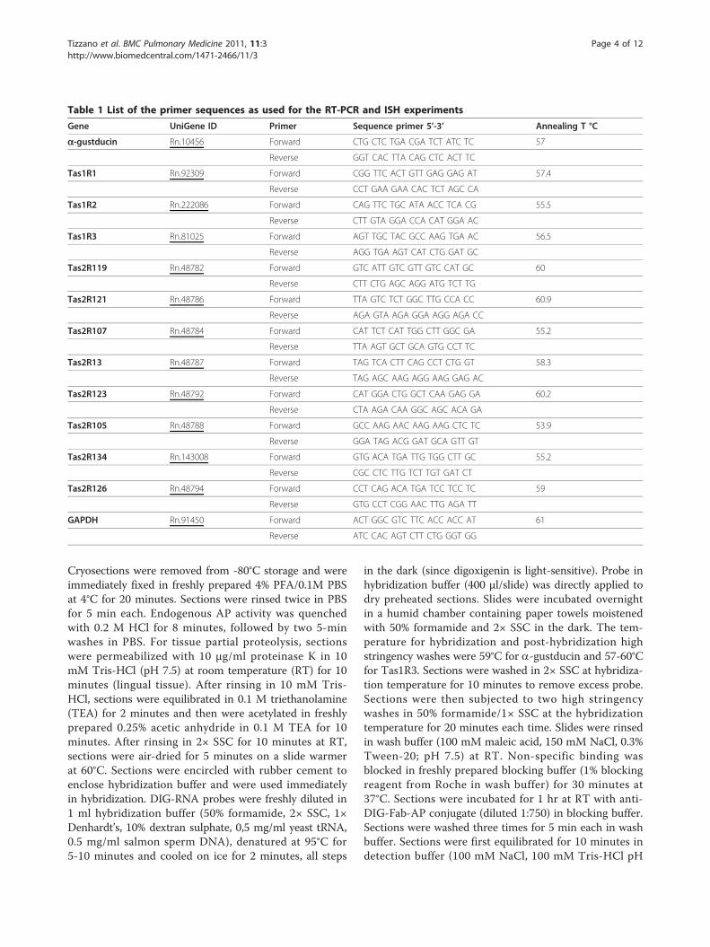
Table 1 List of the primer sequences as used for the RT-PCR and ISH experiments
Gene UniGene ID Primer Sequence primer 5’-3’ Annealing T °C
a-gustducin Rn.10456 Forward CTG CTC TGA CGA TCT ATC TC 57
Reverse GGT CAC TTA CAG CTC ACT TC
Tas1R1 Rn.92309 Forward CGG TTC ACT GTT GAG GAG AT 57.4
Reverse CCT GAA GAA CAC TCT AGC CA
Tas1R2 Rn.222086 Forward CAG TTC TGC ATA ACC TCA CG 55.5
Reverse CTT GTA GGA CCA CAT GGA AC
Tas1R3 Rn.81025 Forward AGT TGC TAC GCC AAG TGA AC 56.5
Reverse AGG TGA AGT CAT CTG GAT GC
Tas2R119 Rn.48782 Forward GTC ATT GTC GTT GTC CAT GC 60
Reverse CTT CTG AGC AGG ATG TCT TG
Tas2R121 Rn.48786 Forward TTA GTC TCT GGC TTG CCA CC 60.9
Reverse AGA GTA AGA GGA AGG AGA CC
Tas2R107 Rn.48784 Forward CAT TCT CAT TGG CTT GGC GA 55.2
Reverse TTA AGT GCT GCA GTG CCT TC
Tas2R13 Rn.48787 Forward TAG TCA CTT CAG CCT CTG GT 58.3
Reverse TAG AGC AAG AGG AAG GAG AC
Tas2R123 Rn.48792 Forward CAT GGA CTG GCT CAA GAG GA 60.2
Reverse CTA AGA CAA GGC AGC ACA GA
Tas2R105 Rn.48788 Forward GCC AAG AAC AAG AAG CTC TC 53.9
Reverse GGA TAG ACG GAT GCA GTT GT
Tas2R134 Rn.143008 Forward GTG ACA TGA TTG TGG CTT GC 55.2
Reverse CGC CTC TTG TCT TGT GAT CT
Tas2R126 Rn.48794 Forward CCT CAG ACA TGA TCC TCC TC 59
Reverse GTG CCT CGG AAC TTG AGA TT
GAPDH Rn.91450 Forward ACT GGC GTC TTC ACC ACC AT 61
Reverse ATC CAC AGT CTT CTG GGT GG
Tizzano et al. BMC Pulmonary Medicine 2011, 11:3http://www.biomedcentral.com/1471-2466/11/3
Page 4 of 12
9.5, 50 mM MgCl2) and incubated in substrate solution(337 μg/ml NBT, 175 μg/ml BCIP, 5 mM tetramisole; indetection buffer) in a humid chamber at RT in the dark.The reaction was continued for up to 96 hr and stoppedby rinsing slides in Tris-EDTA buffer (10 mM Tris and 1mM EDTA) followed by water. Sections were mounted inGelmount.
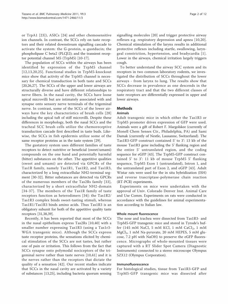
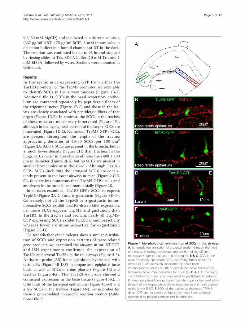
ResultsIn transgenic mice expressing GFP from either theTas1R3 promoter or the TrpM5 promoter, we were ableto identify SCCs in the airway mucosa (Figure 1B-F;Additional file 1). SCCs in the nasal respiratory epithe-lium are contacted repeatedly by peptidergic fibers ofthe trigeminal nerve (Figure 1B,C) and those in the lar-ynx are closely associated with peptidergic fibers of thatorgan (Figure 1D,E). In contrast, the SCCs in the tracheaof these mice are not densely innervated (Figure 1F),although in the hypoglossal portion of the larynx SCCs areinnervated (Figure 1D,E). Numerous TrpM5 GFP+ SCCsare present throughout the length of the tracheaapproaching densities of 40-50 SCCs per 100 μm2
(Figure 2A-B,D,F). SCCs are present in the bronchi, but ata much lower density (Figure 2H) than trachea. In thelungs, SCCs occur in bronchioles of more then 400 ± 100μm in diameter (Figure 2J-K) but no SCCs are present insmaller bronchioles or in the alveoli. Although Tas1R3GFP+ SCCs (including the laryngeal SCCs) are consis-tently present in the lower airways in mice (Figure 2 C,E,G), they are less numerous than TrpM5 GFP+ cells andare absent in the bronchi and more distally (Figure 2I).In all cases examined, Tas1R3 GFP+ SCCs co-express
TrpM5 (Figure 3A-C,) and a-gustducin (Figure 3D-F).Conversely, not all the TrpM5 or a-gustducin immu-noreactive SCCs exhibit Tas1R3-driven GFP expression,i.e. more SCCs express TrpM5 and gustducin thanTas1R3. In the trachea and bronchi, nearly all TrpM5-GFP expressing SCCs exhibit PLCb2 immunoreactivitywhereas fewer are immunoreactive for a-gustducin(Figure 3G-O).To test whether other rodents show a similar distribu-
tion of SCCs and expression patterns of taste-relatedgene products, we examined the airways in rat. RT-PCRand ISH experiments confirmed the expression ofTas1Rs and several Tas2Rs in the rat airways (Figure 4-5).Antisense probe (AS) for a-gustducin hybridized withtaste cells (Figure 4B-D,F) in tongue and epiglottis tastebuds, as well as SCCs in rhino pharynx (Figure 4E) andtrachea (Figure 4G). The Tas1R3 AS probe showed aconsistent expression in the taste tissue (Figure 4I-K), intaste buds of the laryngeal epithelium (Figure 4L-M) anda few SCCs in the trachea (Figure 4N). Sense probes forthese 2 genes yielded no specific reaction product (Addi-tional file 3).
Figure 1 Morphological relationships of SCCs in the airways.A. Schematic representation of a sagittal section through the headof a mouse showing the airways and position of the differentmicrographs (green, blue and red rectangles). B & C. SCCs in thenasal respiratory epithelium. SCCs expressing TrpM5 or Tas1R3(driven GFP) are intimately innervated by nerve fibersimmunoreactive for PGP9.5 (B) or peptidergic nerve fibers of thetrigeminal nerve immunoreactive for CGRP (C). D & E. In the larynx,Tas1R3-GFP+ SCCs are richly innervated by peptidergic (SubstanceP-immunoreactive) fibers probably from the superior laryngeal nerve(branch of the vagus) which shows responses to chemicals appliedto the larynx [5,50]. F. SCCs of the trachea as shown by TRPM5-driven GFP are not closely embraced by nerve fibers althoughoccasional en passant contacts can be observed.
Tizzano et al. BMC Pulmonary Medicine 2011, 11:3http://www.biomedcentral.com/1471-2466/11/3
Page 5 of 12
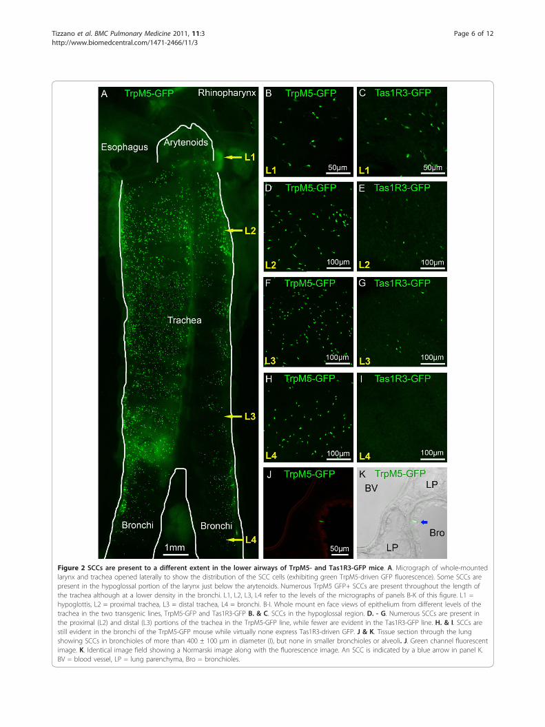
Figure 2 SCCs are present to a different extent in the lower airways of TrpM5- and Tas1R3-GFP mice. A. Micrograph of whole-mountedlarynx and trachea opened laterally to show the distribution of the SCC cells (exhibiting green TrpM5-driven GFP fluorescence). Some SCCs arepresent in the hypoglossal portion of the larynx just below the arytenoids. Numerous TrpM5 GFP+ SCCs are present throughout the length ofthe trachea although at a lower density in the bronchi. L1, L2, L3, L4 refer to the levels of the micrographs of panels B-K of this figure. L1 =hypoglottis, L2 = proximal trachea, L3 = distal trachea, L4 = bronchi. B-I. Whole mount en face views of epithelium from different levels of thetrachea in the two transgenic lines, TrpM5-GFP and Tas1R3-GFP B. & C. SCCs in the hypoglossal region. D. - G. Numerous SCCs are present inthe proximal (L2) and distal (L3) portions of the trachea in the TrpM5-GFP line, while fewer are evident in the Tas1R3-GFP line. H. & I. SCCs arestill evident in the bronchi of the TrpM5-GFP mouse while virtually none express Tas1R3-driven GFP. J & K. Tissue section through the lungshowing SCCs in bronchioles of more than 400 ± 100 μm in diameter (I), but none in smaller bronchioles or alveoli. J. Green channel fluorescentimage. K. Identical image field showing a Normarski image along with the fluorescence image. An SCC is indicated by a blue arrow in panel K.BV = blood vessel, LP = lung parenchyma, Bro = bronchioles.
Tizzano et al. BMC Pulmonary Medicine 2011, 11:3http://www.biomedcentral.com/1471-2466/11/3
Page 6 of 12
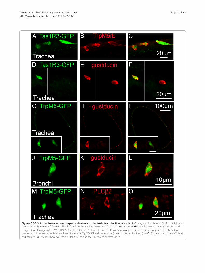
Figure 3 SCCs in the lower airways express elements of the taste transduction cascade. A-F. Single color channel (A & B; D & E) andmerged (C & F) images of Tas1R3 GFP+ SCC cells in the trachea co-express TrpM5 and a-gustducin. G-L. Single color channel (G&H; J&K) andmerged (I & L) images of TrpM5 GFP+ SCC cells in trachea (G-I) and bronchi (J-L) co-express a-gustducin. The insets of panels G-I show thata-gustducin is expressed only in a subset of the total TrpM5-GFP cell population (scale bar 10 μm for insets). M-O. Single color channel (M & N)and merged (O) images showing TrpM5 GFP+ SCC cells in the trachea co-express Plcb2.
Tizzano et al. BMC Pulmonary Medicine 2011, 11:3http://www.biomedcentral.com/1471-2466/11/3
Page 7 of 12
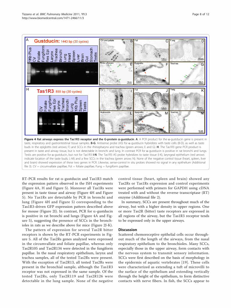
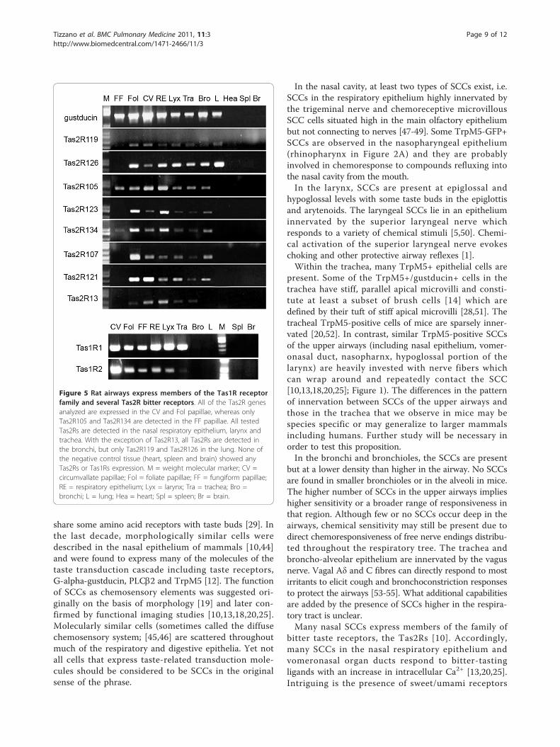
RT-PCR results for rat a-gustducin and Tas1R3 matchthe expression pattern observed in the ISH experiments(Figure 4A, H and Figure 5). Moreover all Tas1Rs werepresent in taste tissue and airway (Figure 4H and Figure5). No Tas1Rs are detectable by PCR in bronchi andlung (Figure 4H and Figure 5) corresponding to theTas1R3-driven GFP expression pattern described abovefor mouse (Figure 2I). In contrast, PCR for a-gustducinis positive in rat bronchi and lungs (Figure 4A and Fig-ure 5), suggesting the presence of SCCs in the bronch-ioles in rats as we describe above for mice (Figure 2J-K).The pattern of expression for several Tas2R bitter
receptors is shown by the RT-PCR experiments in Fig-ure 5. All of the Tas2Rs genes analyzed were expressedin the circumvallate and foliate papillae, whereas onlyTas2R105 and Tas2R134 were detected in the fungiformpapillae. In the nasal respiratory epithelium, larynx andtrachea samples, all of the tested Tas2Rs were present.With the exception of Tas2R13, all tested Tas2Rs werepresent in the bronchial sample, although the Tas1R3receptor was not expressed in the same sample. Of thetested Tas2Rs, only Tas2R119 and Tas2R126 weredetectable in the lung sample. None of the negative
control tissue (heart, spleen and brain) showed anyTas2Rs or Tas1Rs expression and control experimentswere performed with primers for GAPDH using cDNAtreated with and without the reverse transcriptase (RT)enzyme (Additional file 2).In summary, SCCs are present throughout much of the
airway, but with a higher density in upper regions. Oneor more Tas2R (bitter) taste receptors are expressed inall regions of the airway, but the Tas1R3 receptor tendsto be expressed only in the upper airways.
DiscussionScattered chemoreceptive epithelial cells occur through-out much of the length of the airways, from the nasalrespiratory epithelium to the bronchioles. Many SCCs,especially those in the upper airway, form contacts withthe nervous system to transmit sensory information.SCCs were first described on the basis of morphology inthe epidermis of aquatic vertebrates [19]. These cellswere characterized as extending a tuft of microvilli tothe surface of the epithelium and extending verticallythrough the height of the epithelium, to form distinctivecontacts with nerve fibers. In fish, the SCCs appear to
Figure 4 Rat airways express the Tas1R3 receptor and the G-protein a-gustducin. A. A PCR product for the a-gustducin gene is present intaste, respiratory and gastrointestinal tissue samples. B-G. Antisense probe (AS) for a-gustducin hybridizes with taste cells (B-D), as well as tastebuds in the epiglottis (red arrows; F) and SCCs in the rhinopharynx and trachea (green arrows; E and G). H. The Tas1R3 gene PCR product ispresent in taste and airway tissue, but is not detectable in bronchi and lung. In contrast PCR for a-gustducin is positive in rat bronchi and lungs.Testis are positive for a-gustducin, but not for Tas1R3 I-N. The Tas1R3 AS probe hybridizes to taste tissue (I-K), laryngeal epithelium (red arrowsindicate location of the taste buds; L-M) and a few SCCs in the trachea (green arrow; N). None of the negative control tissue (heart, spleen, liverand brain) showed expression of these two genes in PCR. Likewise, sense-control in situ probes showed no signal in any epithelium (Additionalfile 3). CV = circumvallate papillae; Fol = foliate papillae; Fung = fungiform papillae.
Tizzano et al. BMC Pulmonary Medicine 2011, 11:3http://www.biomedcentral.com/1471-2466/11/3
Page 8 of 12
share some amino acid receptors with taste buds [29]. Inthe last decade, morphologically similar cells weredescribed in the nasal epithelium of mammals [10,44]and were found to express many of the molecules of thetaste transduction cascade including taste receptors,G-alpha-gustducin, PLCb2 and TrpM5 [12]. The functionof SCCs as chemosensory elements was suggested ori-ginally on the basis of morphology [19] and later con-firmed by functional imaging studies [10,13,18,20,25].Molecularly similar cells (sometimes called the diffusechemosensory system; [45,46] are scattered throughoutmuch of the respiratory and digestive epithelia. Yet notall cells that express taste-related transduction mole-cules should be considered to be SCCs in the originalsense of the phrase.
In the nasal cavity, at least two types of SCCs exist, i.e.SCCs in the respiratory epithelium highly innervated bythe trigeminal nerve and chemoreceptive microvillousSCC cells situated high in the main olfactory epitheliumbut not connecting to nerves [47-49]. Some TrpM5-GFP+SCCs are observed in the nasopharyngeal epithelium(rhinopharynx in Figure 2A) and they are probablyinvolved in chemoresponse to compounds refluxing intothe nasal cavity from the mouth.In the larynx, SCCs are present at epiglossal and
hypoglossal levels with some taste buds in the epiglottisand arytenoids. The laryngeal SCCs lie in an epitheliuminnervated by the superior laryngeal nerve whichresponds to a variety of chemical stimuli [5,50]. Chemi-cal activation of the superior laryngeal nerve evokeschoking and other protective airway reflexes [1].Within the trachea, many TrpM5+ epithelial cells are
present. Some of the TrpM5+/gustducin+ cells in thetrachea have stiff, parallel apical microvilli and consti-tute at least a subset of brush cells [14] which aredefined by their tuft of stiff apical microvilli [28,51]. Thetracheal TrpM5-positive cells of mice are sparsely inner-vated [20,52]. In contrast, similar TrpM5-positive SCCsof the upper airways (including nasal epithelium, vomer-onasal duct, nasopharnx, hypoglossal portion of thelarynx) are heavily invested with nerve fibers whichcan wrap around and repeatedly contact the SCC[10,13,18,20,25]; Figure 1). The differences in the patternof innervation between SCCs of the upper airways andthose in the trachea that we observe in mice may bespecies specific or may generalize to larger mammalsincluding humans. Further study will be necessary inorder to test this proposition.In the bronchi and bronchioles, the SCCs are present
but at a lower density than higher in the airway. No SCCsare found in smaller bronchioles or in the alveoli in mice.The higher number of SCCs in the upper airways implieshigher sensitivity or a broader range of responsiveness inthat region. Although few or no SCCs occur deep in theairways, chemical sensitivity may still be present due todirect chemoresponsiveness of free nerve endings distribu-ted throughout the respiratory tree. The trachea andbroncho-alveolar epithelium are innervated by the vagusnerve. Vagal Aδ and C fibres can directly respond to mostirritants to elicit cough and bronchoconstriction responsesto protect the airways [53-55]. What additional capabilitiesare added by the presence of SCCs higher in the respira-tory tract is unclear.Many nasal SCCs express members of the family of
bitter taste receptors, the Tas2Rs [10]. Accordingly,many SCCs in the nasal respiratory epithelium andvomeronasal organ ducts respond to bitter-tastingligands with an increase in intracellular Ca2+ [13,20,25].Intriguing is the presence of sweet/umami receptors
Figure 5 Rat airways express members of the Tas1R receptorfamily and several Tas2R bitter receptors. All of the Tas2R genesanalyzed are expressed in the CV and Fol papillae, whereas onlyTas2R105 and Tas2R134 are detected in the FF papillae. All testedTas2Rs are detected in the nasal respiratory epithelium, larynx andtrachea. With the exception of Tas2R13, all Tas2Rs are detected inthe bronchi, but only Tas2R119 and Tas2R126 in the lung. None ofthe negative control tissue (heart, spleen and brain) showed anyTas2Rs or Tas1Rs expression. M = weight molecular marker; CV =circumvallate papillae; Fol = foliate papillae; FF = fungiform papillae;RE = respiratory epithelium; Lyx = larynx; Tra = trachea; Bro =bronchi; L = lung; Hea = heart; Spl = spleen; Br = brain.
Tizzano et al. BMC Pulmonary Medicine 2011, 11:3http://www.biomedcentral.com/1471-2466/11/3
Page 9 of 12
(Tas1R family members) in the airways. The presence ofthis class of receptors was first indicated by Tas1R3-driven expression of a transgene in the nasal epithelium[40]. We confirm with both PCR and Tas1R3 transgenicanimals that a subset of nasal SCCs expresses Tas1R3 asdo some in the trachea. Whereas Tas2Rs in the nosedetect bitter/toxic compounds as irritants [13,20,25],there are no reports of any chemosensitivity mediatedby Tas1R receptors in the airways or any experimentthat show the natural ligands for the Tas1R+ SCCs inthe respiratory tract.It is noteworthy that nasal SCCs co-express Tas1R3-
WGA with Tas2R5 and Tas2R8 [40]. In contrast, recep-tor cells in taste buds express either members of theTas1R or the Tas2R receptor families, but never bothtogether [32]. Further, individual SCCs co-expressTas1R3 and a-gustducin. In the taste buds of the pos-terior part of the tongue, taste receptor cells thatexpress Tas1R3 and Tas1R2 seldom express of a-gust-ducin [56]. The co-expression of Tas1R3 and gustducindoes, however occur in taste buds of fungiform papillaeand palate [57,58].Chemosensory cells expressing elements of the taste
transduction cascade are prevalent in both the gastroin-testinal tract [12,59] as well as the respiratory tree[10,12,13,16,17,25,60,61]. Despite this similarity in mole-cular expression, these diverse chemoresponsive cellsincluding SCCs and brush cells, are different in terms offunction and therefore should not be considered to be asingle cell type. Although the Tas2R/gustducin/TrpM5-expressing epithelial cells in the different tissues aresimilar in extending microvillous processes to the top ofthe epithelium and in utilizing similar receptor andtransduction cascades, the downstream effects of cellactivation are different. The SCCs in the nasal respira-tory epithelium form synapses to evoke a neuralresponse [20], whereas brush cells of the gut releasepeptides in a more paracrine fashion to modulate diges-tive activities [62].The function of the SCCs in the lower airway still
needs to be determined, but may involve local modula-tion of the airway epithelium (mucociliary clearance andsecretory functions; [60]) or induction of a localresponse of the innate immune system. For example,tracheal SCCs are likely to respond to bacterial signal-ling molecules or other irritants as do nasal SCCs, butinstead of communicating with nerve fibers, may releasecytokines and other modulators locally to evokeresponses in dendritic cells or macrophages. The SCCsof the nasal cavity, vomeronasal ducts and larynx holdcrucial positions at the entrance to different respiratoryorgans. Thus, SCCs in these situations may be especiallystrongly connected to nerves that trigger protectivereflexes of these respiratory organs.
Recently, three studies report Tas2R expression in theairways. The first study reports expression of Tas2Rfamily members and related downstream signalling com-ponents by tracheal brush cells (solitary chemosensorycells in our terminology) in mice [52]. These investiga-tors found that brush cells also express cholinergic traitsand lie close to, or contact, subepithelial nerve fibersthat express nicotinic acetylcholine receptors.The second study examines human airway epithelium
in vitro, reported that ciliated cells of respiratory epithe-lium express Tas2R bitter taste receptors and otherdownstream gustatory transduction components, e.g.gustducin. This study reported that the Tas2R moleculeslocalized to the motile cilia and that those ciliated cellsresponded to bitter-tasting compounds with an increasein intracellular Ca2+ and an increase in ciliary beat fre-quency [61]. Their results contrast with our observationin rodents that elements of the taste transduction cascadeare present only in SCCs and not in other cell types ofrespiratory tract, including ciliated cells. This may reflecta species difference, or the use of an in vitro system inthe Shah et al [61] study. The mere size difference in theairways between mice and humans is not the determiningfactor since the expression of gustducin in airway epithe-lium of another large mammal (i.e. cow), appearsrestricted to a distributed SCC population [11].The third report shows Tas2R expression in airways
describes the localization of taste transdcution compo-nents to cultured airway smooth muscle [63]. Theseinvestigators showed by PCR expression of Tas2Rs onhuman airway smooth muscle. We do not find evidencefor expression of bitter taste transduction elements inthe tracheal smooth muscle (as determined by immuno-cytochemistry or transgene expression in mice).Whether the presence of taste transduction componentsin smooth muscle of humans is again a function of thein vitro system or a species difference is unclear. TheDeshpande et al [63] study also reports that stimulationof cultured smooth muscle cells with bitter compoundsevoked increased intracellular calcium dependent on thebitter taste transduction cascade similar to the cellularactivation we have reported in nasal SCCs [13,20,25].Further, Deshpande et al report that, inhaled bittertastants decreased airway obstruction in a mouse modelof asthma. It is unclear from this report how the bitterligands are envisioned to reach the airway smooth mus-cle which is covered by respiratory epithelum. We sug-gest that the in vivo responsiveness may have beenattributable to activation of tracheal SCCs, which secon-darily activate the airway smooth muscle. Members ofthe Tas2R family are expressed in the SCCs of the nasaland tracheal [52] respiratory epithelia and these SCCsrespond to bitter compounds to evoke protectiverespiratory reflexes. Further study of both human tissue
Tizzano et al. BMC Pulmonary Medicine 2011, 11:3http://www.biomedcentral.com/1471-2466/11/3
Page 10 of 12

and animal models will be necessary to fully understandthis system.
ConclusionsIn summary, we find that epithelial cells specialized forchemoreception are distributed throughout much of therespiratory tree of rodents. The nasal cavity houses cyto-logically distinct SCCs that are intimately connected tosensory nerve fibers. In the lower airways, SCCs expres-sing similar transduction cascades include some brushcells with a distinctive tuft of stiff apical microvilli [52].Further, the molecular repertoire of chemoreceptor pro-teins differs somewhat between upper and lower airwayswith the sweet/umami receptor subunit Tas1R3 beingexpressed in many in nasal SCCs and being largelyabsent in the SCCs of the lower airway whereas theTas2R receptors are expressed throughout. The presenceof Tas1Rs in the airways is intriguing since hithertothese molecules were considered only to mediate posi-tive features of ingested foods, i.e. the presence of carbo-hydrates (sweet) or amino acids (umami). In the airways,these same receptors may be involved in detection ofchemicals in foodstuffs which would be appetitive in themouth or gut but which trigger protective reflexes inthe airways.
Additional material
Additional file 1: In wild type mice no significant autofluorescenceis apparent. A & C. SCCs in the nasal respiratory epithelium (A) andtrachea (B) immunoreactive for a-gustducin. C & D. The same epitheliashown in the green channel lack any autofluorescence, validating theGFP expression of the transgenic mice used to identify the SCCs.
Additional file 2: PCR experiments conducted with primers forGAPDH on cDNAs treated with and without the reversetranscriptase (RT) enzyme. The constitutive gene GAPDH is present inall the templates obtained by adding the RT enzyme during theproduction of the cDNA (+RT, upper line), whereas it is absent in alltemplates not treated with RT enzyme (-RT, lower line). PCR productlength is 273 bp. M = weight molecular marker; FF = fungiform papillae;Fol = foliate papillae; CV = circumvallate papillae; RE = respiratoryepithelium; Lyx = larynx; Tra = trachea; Bro = bronchi; L = lung; Hea =heart; Spl = spleen; Liv = liver; Br = brain; Sto = stomach; Tes = testis.
Additional file 3: In situ hybridization using sense-control probesshowed no signal in any epithelium. Both sense probes for a-gustducin (A) and Tas1R3 (B) show no staining in the epiglottis as well asin other tissue (not shown). The blue arrows indicate the location oflaryngeal taste buds circled by dotted lines.
List of abbreviationsAP: Alkaline phosphatase; BCIP: 5-bromo-4-chloro-3-indolyl-phosphate CGRP:Calcitonin gene related peptide; GFP: green fluorescent protein; GPCRs: Gprotein coupled receptors; ISH: in situ hybridization; LSCM: confocal laserscanning microscope; NBT: nitro blue tetrazolium; OCT: Tissue-Tekembedding medium; PB: phosphate buffer; PBS: phosphate-buffered saline;PFA: Paraformaldehyde; PGP9.5: Protein gene product 9.5 of the ubiquitincarboxy-terminal hydrolase; Plcྞ2: phospholipase C beta2; RT: roomtemperature; RT-PCR: reverse transcriptase-polymerase chain reaction; SCCs:solitary chemosensory cells; SSC: saline-sodium citrate; SubP: Substance P
peptide; Tas1R: taste receptor family 1; Tas2R: taste receptor family 2; TrpM5:transient receptor potential channel M5.
AcknowledgementsWe thank Dr. Robert F. Margolskee (Monell Chem Senses Ctr., Philadelphia,PA USA) and Dr. Sami Damak (Nestle, Lausanne, SWITZERLAND) for the GFPtransgenic mice and Dr. Emily R. Liman (University of Southern California,USA) for the anti-rabbit TrpM5 antibody.This study was supported by NIH grants NIDCD R01 DC006070 & P30DC04657 (to D.Restrepo. & T.E.F.) and DC009820 (to T.E.F. & S.C. Kinnamon).Thanks for useful discussion and reading of the manuscript: Anne Hansen.
Author details1Rocky Mountain Taste and Smell Center, Department of Cell andDevelopmental Biology, University of Colorado at Denver & Health SciencesCenter, Aurora, USA. 2Department of Morphological-Biomedical Sciences,University of Verona, Verona, Italy.
Authors’ contributionsMT, AS and TEF participated in design and coordination of the study. MTand MC carried out the RT-PCR assays. MT performed allimmunofluorescence and in situ hybridization experiments. Confocalmicroscopy was performed by MT. MT wrote the manuscript to which AS,MC and TEF added their contributions. All authors read and approved thefinal manuscript.
Competing interestsThe authors declare that they have no competing interests.
Received: 1 October 2010 Accepted: 13 January 2011Published: 13 January 2011
References1. Widdicombe JG: Chemoreceptor control of the airways. Respir Physiol
1992, 87(3):373-381.2. Bradley RM, Stedman HM, Mistretta CM: Superior laryngeal nerve response
patterns to chemical stimulation of sheep epiglottis. Brain Res 1983,276(1):81-93.
3. Stedman HM, Mistretta CM, Bradley RM: A quantitative study of catepiglottal taste buds during development. J Anat 1983, 136(Pt 4):821-827.
4. Altschuler SM: Laryngeal and respiratory protective reflexes. Am J Med2001, 111(Suppl 8A):90S-94S.
5. Smith DV, Hanamori T: Organization of gustatory sensitivities in hamstersuperior laryngeal nerve fibers. J Neurophysiol 1991, 65(5):1098-1114.
6. Dickman JD, Smith DV: Response properties of fibers in the hamstersuperior laryngeal nerve. Brain Res 1988, 450(1-2):25-38.
7. Mutoh T, Tsubone H: Hypersensitivity of laryngeal C-fibers induced byvolatile anesthetics in young guinea pigs. Am J Respir Crit Care Med 2003,167(4):557-562.
8. McIntosh HD, Estes EH, Warren JV: The mechanism of cough syncope. AmHeart J 1956, 52(1):70-82.
9. French JW, Morgan BC, Guntheroth WG: Infant monkeys–a model for cribdeath. Am J Dis Child 1972, 123(5):480-484.
10. Finger TE, Bottger B, Hansen A, Anderson KT, Alimohammadi H, Silver WL:Solitary chemoreceptor cells in the nasal cavity serve as sentinels ofrespiration. Proc Natl Acad Sci USA 2003, 100(15):8981-8986.
11. Tizzano M, Merigo F, Sbarbati A: Evidence of solitary chemosensory cellsin a large mammal: the diffuse chemosensory system in Bos taurusairways. J Anat 2006, 209(3):333-337.
12. Kaske S, Krasteva G, Konig P, Kummer W, Hofmann T, Gudermann T,Chubanov V: TRPM5, a taste-signaling transient receptor potential ion-channel, is a ubiquitous signaling component in chemosensory cells.BMC Neurosci 2007, 8:49.
13. Lin W, Ogura T, Margolskee RF, Finger TE, Restrepo D: TRPM5-expressingsolitary chemosensory cells respond to odorous irritants. J Neurophysiol2008, 99(3):1451-1460.
14. Sbarbati A, Merigo F, Benati D, Tizzano M, Bernardi P, Crescimanno C,Osculati F: Identification and characterization of a specific sensoryepithelium in the rat larynx. J Comp Neurol 2004, 475(2):188-201.
15. Sbarbati A, Merigo F, Benati D, Tizzano M, Bernardi P, Osculati F: Laryngealchemosensory clusters. Chem Senses 2004, 29(8):683-692.
Tizzano et al. BMC Pulmonary Medicine 2011, 11:3http://www.biomedcentral.com/1471-2466/11/3
Page 11 of 12
16. Merigo F, Benati D, Di Chio M, Osculati F, Sbarbati A: Secretory cells of theairway express molecules of the chemoreceptive cascade. Cell Tissue Res2007, 327(2):231-247.
17. Merigo F, Benati D, Tizzano M, Osculati F, Sbarbati A: alpha-Gustducinimmunoreactivity in the airways. Cell Tissue Res 2005, 319(2):211-219.
18. Ogura T, Krosnowski K, Zhang L, Bekkerman M, Lin W: Chemoreceptionregulates chemical access to mouse vomeronasal organ: role of solitarychemosensory cells. PLoS One 2010, 5(7):e11924.
19. Whitear M: Solitary Chemoreceptor Cells. In Chemoreception in Fishes. 2edition. Edited by: Hara TJ. London: Elsivier Press; 1992:103-125.
20. Tizzano M, Gulbransen BD, Vandenbeuch A, Clapp TR, Herman JP,Sibhatu HM, Churchill ME, Silver WL, Kinnamon SC, Finger TE: Nasalchemosensory cells use bitter taste signaling to detect irritants andbacterial signals. Proc Natl Acad Sci USA 2010, 107(7):3210-3215.
21. Geppetti P, Materazzi S, Nicoletti P: The transient receptor potentialvanilloid 1: role in airway inflammation and disease. Eur J Pharmacol2006, 533(1-3):207-214.
22. Caterina MJ, Schumacher MA, Tominaga M, Rosen TA, Levine JD, Julius D:The capsaicin receptor: a heat-activated ion channel in the painpathway. Nature 1997, 389(6653):816-824.
23. Jordt SE, Bautista DM, Chuang HH, McKemy DD, Zygmunt PM, Hogestatt ED,Meng ID, Julius D: Mustard oils and cannabinoids excite sensory nervefibres through the TRP channel ANKTM1. Nature 2004, 427(6971):260-265.
24. Olson TH, Riedl MS, Vulchanova L, Ortiz-Gonzalez XR, Elde R: An acidsensing ion channel (ASIC) localizes to small primary afferent neurons inrats. Neuroreport 1998, 9(6):1109-1113.
25. Gulbransen BD, Clapp TR, Finger TE, Kinnamon SC: Nasal solitarychemoreceptor cell responses to bitter and trigeminal stimulants invitro. J Neurophysiol 2008, 99(6):2929-2937.
26. Perez CA, Huang L, Rong M, Kozak JA, Preuss AK, Zhang H, Max M,Margolskee RF: A transient receptor potential channel expressed in tastereceptor cells. Nat Neurosci 2002, 5(11):1169-1176.
27. Zhang Y, Hoon MA, Chandrashekar J, Mueller KL, Cook B, Wu D, Zuker CS,Ryba NJ: Coding of sweet, bitter, and umami tastes: different receptorcells sharing similar signaling pathways. Cell 2003, 112(3):293-301.
28. Luciano L, Reale E, Ruska H: On a “chemoreceptive” sensory cell in thetachea of the rat. Z Zellforsch Mikrosk Anat 1968, 85(3):350-375.
29. Finger TE, Bryant BP, Kalinoski DL, Teeter JH, Bottger B, Grosvenor W,Cagan RH, Brand JG: Differential localization of putative amino acidreceptors in taste buds of the channel catfish, Ictalurus punctatus. JComp Neurol 1996, 373(1):129-138.
30. Li X, Staszewski L, Xu H, Durick K, Zoller M, Adler E: Human receptors forsweet and umami taste. Proc Natl Acad Sci USA 2002, 99(7):4692-4696.
31. Nelson G, Chandrashekar J, Hoon MA, Feng L, Zhao G, Ryba NJ, Zuker CS:An amino-acid taste receptor. Nature 2002, 416(6877):199-202.
32. Nelson G, Hoon MA, Chandrashekar J, Zhang Y, Ryba NJ, Zuker CS:Mammalian sweet taste receptors. Cell 2001, 106(3):381-390.
33. Meyerhof W, Batram C, Kuhn C, Brockhoff A, Chudoba E, Bufe B,Appendino G, Behrens M: The molecular receptive ranges of humanTAS2R bitter taste receptors. Chem Senses 2010, 35(2):157-170.
34. Adler E, Hoon MA, Mueller KL, Chandrashekar J, Ryba NJ, Zuker CS: A novelfamily of mammalian taste receptors. Cell 2000, 100(6):693-702.
35. Chandrashekar J, Mueller KL, Hoon MA, Adler E, Feng L, Guo W, Zuker CS,Ryba NJ: T2Rs function as bitter taste receptors. Cell 2000, 100(6):703-711.
36. Matsunami H, Montmayeur JP, Buck LB: A family of candidate tastereceptors in human and mouse. Nature 2000, 404(6778):601-604.
37. Shi P, Zhang J, Yang H, Zhang YP: Adaptive diversification of bitter tastereceptor genes in Mammalian evolution. Mol Biol Evol 2003, 20(5):805-814.
38. Damak S, Rong M, Yasumatsu K, Kokrashvili Z, Varadarajan V, Zou S, Jiang P,Ninomiya Y, Margolskee RF: Detection of sweet and umami taste in theabsence of taste receptor T1r3. Science 2003, 301(5634):850-853.
39. Zhao GQ, Zhang Y, Hoon MA, Chandrashekar J, Erlenbach I, Ryba NJ,Zuker CS: The receptors for mammalian sweet and umami taste. Cell2003, 115(3):255-266.
40. Ohmoto M, Matsumoto I, Yasuoka A, Yoshihara Y, Abe K: Genetic tracing ofthe gustatory and trigeminal neural pathways originating from T1R3-expressing taste receptor cells and solitary chemoreceptor cells. Mol CellNeurosci 2008, 38(4):505-517.
41. Gulbransen B, Silver W, Finger TE: Solitary chemoreceptor cell survival isindependent of intact trigeminal innervation. J Comp Neurol 2008,508(1):62-71.
42. Muller J: Handbuch der Physiologie des Menschen fuer Vorlesungen.Edited by: Clarke E, O’Malley CD. Coblenz: J. Hoelscher; , 2 1840.
43. Clapp TR, Medler KF, Damak S, Margolskee RF, Kinnamon SC: Mouse tastecells with G protein-coupled taste receptors lack voltage-gated calciumchannels and SNAP-25. BMC Biol 2006, 4:7.
44. Zancanaro C, Caretta CM, Merigo F, Cavaggioni A, Osculati F: alpha-Gustducin expression in the vomeronasal organ of the mouse. Eur JNeurosci 1999, 11(12):4473-4475.
45. Sbarbati A, Osculati F: The taste cell-related diffuse chemosensory system.Prog Neurobiol 2005, 75(4):295-307.
46. Sbarbati A, Bramanti P, Benati D, Merigo F: The diffuse chemosensorysystem: exploring the iceberg toward the definition of functional roles.Prog Neurobiol 2010, 91(1):77-89.
47. Elsaesser R, Montani G, Tirindelli R, Paysan J: Phosphatidyl-inositidesignalling proteins in a novel class of sensory cells in the mammalianolfactory epithelium. Eur J Neurosci 2005, 21(10):2692-2700.
48. Lin W, Ezekwe EA Jr, Zhao Z, Liman ER, Restrepo D: TRPM5-expressingmicrovillous cells in the main olfactory epithelium. BMC Neurosci 2008,9:114.
49. Hansen A, Finger TE: Is TrpM5 a reliable marker for chemosensory cells?Multiple types of microvillous cells in the main olfactory epithelium ofmice. BMC Neurosci 2008, 9:115.
50. Hanamori T, Smith DV: Central projections of the hamster superiorlaryngeal nerve. Brain Res Bull 1986, 16(2):271-279.
51. Rhodin J, Dalhamn T: Electron microscopy of the tracheal ciliated mucosain rat. Z Zellforsch Mikrosk Anat 1956, 44(4):345-412.
52. Krasteva G, Canning BJ, Veres T, Papadakis T, Hartmann P, Mühlfeld C,Schliecker K, Hans K, Tallini YN, Braun A, et al: Tracheal brush cells areneuronally connected cholinergic sensory cells [abstract]. Society forNeuroscience 2010, Online Program No 7737.
53. Fox AJ, Barnes PJ, Dray A: Stimulation of guinea-pig tracheal afferentfibres by non-isosmotic and low-chloride stimuli and the effect offrusemide. J Physiol 1995, 482(Pt 1):179-187.
54. Sant’Ambrogio G, Sant’Ambrogio FB, Davies A: Airway receptors in cough.Bull Eur Physiopathol Respir 1984, 20(1):43-47.
55. Kollarik M, Ru F, Brozmanova M: Vagal afferent nerves with the propertiesof nociceptors. Auton Neurosci 2010, 153(1-2):12-20.
56. Tizzano M, Dvoryanchikov G, Barrows JK, Kim S, Chaudhari N, Finger TE:Expression of Galpha14 in sweet-transducing taste cells of the posteriortongue. BMC Neurosci 2008, 9:110.
57. Stone LM, Barrows J, Finger TE, Kinnamon SC: Expression of T1Rs andgustducin in palatal taste buds of mice. Chem Senses 2007,32(3):255-262.
58. Kusakabe Y, Kim MR, Miura H, Shindo Y, Ninomiya Y, Hino A: Regionalexpression patterns of T1r family in the mouse tongue. Chem Senses2005, 30(Suppl 1):i23-24.
59. Wu SV, Rozengurt N, Yang M, Young SH, Sinnett-Smith J, Rozengurt E:Expression of bitter taste receptors of the T2R family in thegastrointestinal tract and enteroendocrine STC-1 cells. Proc Natl Acad SciUSA 2002, 99(4):2392-2397.
60. Sbarbati A, Tizzano M, Merigo F, Benati D, Nicolato E, Boschi F, Cecchini MP,Scambi I, Osculati F: Acyl homoserine lactones induce early response inthe airway. Anat Rec (Hoboken) 2009, 292(3):439-448.
61. Shah AS, Ben-Shahar Y, Moninger TO, Kline JN, Welsh MJ: Motile cilia ofhuman airway epithelia are chemosensory. Science 2009,325(5944):1131-1134.
62. Kokrashvili Z, Mosinger B, Margolskee RF: T1r3 and alpha-gustducin in gutregulate secretion of glucagon-like peptide-1. Ann N Y Acad Sci 2009,1170:91-94.
63. Deshpande DA, Wang WC, McIlmoyle EL, Robinett KS, Schillinger RM, An SS,Sham JS, Liggett SB: Bitter taste receptors on airway smooth musclebronchodilate by localized calcium signaling and reverse obstruction.Nat Med 2010, 16(11):1299-1304.
Pre-publication historyThe pre-publication history for this paper can be accessed here:http://www.biomedcentral.com/1471-2466/11/3/prepub
doi:10.1186/1471-2466-11-3Cite this article as: Tizzano et al.: Expression of taste receptors inSolitary Chemosensory Cells of rodent airways. BMC Pulmonary Medicine2011 11:3.
Tizzano et al. BMC Pulmonary Medicine 2011, 11:3http://www.biomedcentral.com/1471-2466/11/3
Page 12 of 12