Embed Size (px)
Citation preview
METHODS AND PROTOCOLS
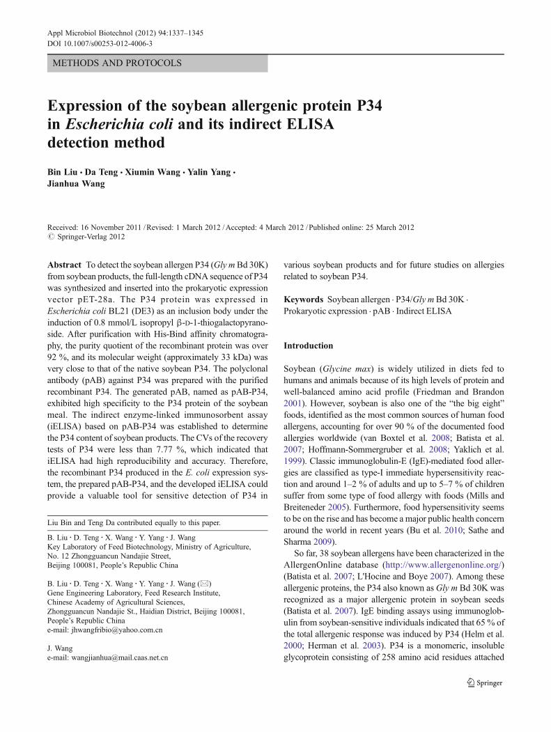
Expression of the soybean allergenic protein P34in Escherichia coli and its indirect ELISAdetection method
Bin Liu & Da Teng & Xiumin Wang & Yalin Yang &
Jianhua Wang
Received: 16 November 2011 /Revised: 1 March 2012 /Accepted: 4 March 2012 /Published online: 25 March 2012# Springer-Verlag 2012
Abstract To detect the soybean allergen P34 (Gly mBd 30K)from soybean products, the full-length cDNA sequence of P34was synthesized and inserted into the prokaryotic expressionvector pET-28a. The P34 protein was expressed inEscherichia coli BL21 (DE3) as an inclusion body under theinduction of 0.8 mmol/L isopropyl β-D-1-thiogalactopyrano-side. After purification with His-Bind affinity chromatogra-phy, the purity quotient of the recombinant protein was over92 %, and its molecular weight (approximately 33 kDa) wasvery close to that of the native soybean P34. The polyclonalantibody (pAB) against P34 was prepared with the purifiedrecombinant P34. The generated pAB, named as pAB-P34,exhibited high specificity to the P34 protein of the soybeanmeal. The indirect enzyme-linked immunosorbent assay(iELISA) based on pAB-P34 was established to determinethe P34 content of soybean products. The CVs of the recoverytests of P34 were less than 7.77 %, which indicated thatiELISA had high reproducibility and accuracy. Therefore,the recombinant P34 produced in the E. coli expression sys-tem, the prepared pAB-P34, and the developed iELISA couldprovide a valuable tool for sensitive detection of P34 in
various soybean products and for future studies on allergiesrelated to soybean P34.
Keywords Soybean allergen . P34/Gly mBd 30K .
Prokaryotic expression . pAB . Indirect ELISA
Introduction
Soybean (Glycine max) is widely utilized in diets fed tohumans and animals because of its high levels of protein andwell-balanced amino acid profile (Friedman and Brandon2001). However, soybean is also one of the “the big eight”foods, identified as the most common sources of human foodallergens, accounting for over 90 % of the documented foodallergies worldwide (van Boxtel et al. 2008; Batista et al.2007; Hoffmann-Sommergruber et al. 2008; Yaklich et al.1999). Classic immunoglobulin-E (IgE)-mediated food aller-gies are classified as type-I immediate hypersensitivity reac-tion and around 1–2 % of adults and up to 5–7 % of childrensuffer from some type of food allergy with foods (Mills andBreiteneder 2005). Furthermore, food hypersensitivity seemsto be on the rise and has become a major public health concernaround the world in recent years (Bu et al. 2010; Sathe andSharma 2009).
So far, 38 soybean allergens have been characterized in theAllergenOnline database (http://www.allergenonline.org/)(Batista et al. 2007; L'Hocine and Boye 2007). Among theseallergenic proteins, the P34 also known asGly m Bd 30K wasrecognized as a major allergenic protein in soybean seeds(Batista et al. 2007). IgE binding assays using immunoglob-ulin from soybean-sensitive individuals indicated that 65 % ofthe total allergenic response was induced by P34 (Helm et al.2000; Herman et al. 2003). P34 is a monomeric, insolubleglycoprotein consisting of 258 amino acid residues attached
Liu Bin and Teng Da contributed equally to this paper.
B. Liu :D. Teng :X. Wang :Y. Yang : J. WangKey Laboratory of Feed Biotechnology, Ministry of Agriculture,No. 12 Zhongguancun Nandajie Street,Beijing 100081, People’s Republic China
B. Liu :D. Teng :X. Wang :Y. Yang : J. Wang (*)Gene Engineering Laboratory, Feed Research Institute,Chinese Academy of Agricultural Sciences,Zhongguancun Nandajie St., Haidian District, Beijing 100081,People’s Republic Chinae-mail: [email protected]
J. Wange-mail: [email protected]
Appl Microbiol Biotechnol (2012) 94:1337–1345DOI 10.1007/s00253-012-4006-3
by disulfide linkages in the 7S globulin fraction and may playa role in protein folding (Helm et al. 2000). As a thiol protease,P34 is a member of the papain superfamily (Sewekow et al.2008) and has a tendency to bind to lipid and oil, which islikely significant in its role as an allergen (Mills et al. 2004).P34 exhibits post-translational production derived from a 46-to 47-kDa precursor protein by the partial removal of 122 N-terminal amino acid residues, and its tertiary conformationwas similar to the papain family (Ogawa et al. 2000).
To date, the studies on P34 have focused on their structuraland functional properties including their HPLC assay andimmunological detection by means of monoclonal antibodybased on epitope predication, and in the meanwhile, thechange and diversity of P34 structure frequently occurring inprocessing was seldomly considered and tracked when theseassays were tried to develop (You et al. 2008; Wilson et al.2008; Amnuaycheewa and de Mejia 2010; Egounlety andAworh 2003; Hotz and Gibson 2007; van Boekel et al.2010). The detection of allergens in food products is verydifficult as allergens are often present in trace amounts andcan be masked by the food matrix (Poms et al. 2004).Although the quantification of soybean allergens can be car-ried out by high-performance liquid chromatography andimmunohistochemical techniques, these methods requiretime-consuming cleanup steps and expensive equipment(Mujoo et al. 2003; Pedersen et al. 2008; L'Hocine and Boye2007). Immunoassay methods have been widely applied inmany fields including detection for food allergen because theyinvolve the use of relatively inexpensive equipment and aresuitable for high-throughput screening (Asensio et al. 2008).The indirect enzyme-linked immunosorbent assay (iELISA)had been developed to quantify P34 in various commercialsoy ingredients and products based on the existing monoclo-nal α-P34 antibody (Tsuji et al. 1995; Wilson et al. 2008). Butsome problems exist in the above typical immunoassay ap-proach based on the monoclonal antibody: on the one hand,the preparation of pure P34 from raw soybean and highlysensitive monoclonal antibody against P34 from animal im-munization are critical and crucial; in fact, besides clinicaldiagnosis in hospital, the detection assay of soy allergensincluding P34 with merits such as more simple, quick, effi-cient, or high-throughput screening is more urgently requiredin many fields such as food and feed processing and someregions where people are often insensitive to soy allergenssuch as Asia. Moreover, on the other hand, the immunoassayassay based on monoclonal antibody is limited on a singleepitope and shows probably no effect when this epitope wasdestroyed during the process. On the contrary, the assay basedon polyclonal antibody could cover multiple epitopes andshowed a lower detection limit (Pedersen et al. 2008). Sofar, there is no iELISA method to detect P34 using polyclonalantibody pAB against recombinant P34 fusion protein. In thisstudy, the P34 protein was expressed in E. coli, pAB was
prepared, and then an iELISA for the detection of P34 usingpAB was established and evaluated.
Materials and methods
Preparation of the substances
Soybean seed (SBS), the soybean meal (SBM), and fermen-tation soybean meal (FSBM) were ground into powder withan FW100 Series of High-Speed Universal Grinder (BeijingZhongxingweiye Instrument Co., Ltd.) and passed through a60-mesh sieve. Each gram of powder above was added to15 mL of 0.01 mol/L PBS (8.0 g NaCl, 0.2 g KH2PO4, 2.13 gNa2HPO4, and 0.2 g KCl per liter, pH 7.4) and kept overnightat 4 °C. After centrifugation at 13,500×g for 15 min at 4 °C,the supernatants were stored at −20 °C until analysis.
Construction of the pET28a-30K plasmid
The 771-bp cDNA sequence of P34 mature peptide(GenBank: EU883600.1) was optimized for expression inE. coli and synthesized by Sangon Biotech Co., Ltd.(Shanghai, China). The full length of the P34 cDNA wasamplified by PCR with forward primer 30K-F, 5′-GGAATTCCATATG AAGAAAATGAAGAAGGAA-3′(Nde I site underlined) and reverse primer 30K-R, 5′-CGGAATTCAAGAGGAGAGTGATCAAC-3′ (EcoR I siteunderlined). The PCR products were digested with Nde Iand EcoR I and inserted into similarly digested pET-28a togenerate plasmid pET28a-30K.
Expression of P34 in E. coli
The plasmid pET28a-30K was transformed into E. coliBL21(DE3) and was identified by restriction enzymes NdeI and EcoR I. The transformant cells were cultured in 5 mLof Luria–Bertani medium containing 50 μg/mL kanamycin(Kan), with shaking at 37 °C overnight. The preculture(5 mL) was then transferred into 500 mL fresh TerrificBroth medium containing 50 μg/mL Kan and grown withshaking at 37 °C until OD600 was 0.5–0.8, at which timeprotein expression was induced by adding 0.8 mM isopropylβ-D-1-thiogalactopyranoside (IPTG).
Purification of recombinant P34 protein
After 6 h of induction, the cultured cells were harvested bycentrifugation at 5,000 rpm for 30 min at 4 °C and resus-pended in 50 mL of lysis buffer (0.5 mol/L NaCl, 20 mmol/L Tris–HCl, 8 mol/L urea, 1 mmol/L PMSF, pH 7.9). Thesuspended cells were sonicated for 20 min with anUltrasonic Crasher Noise Isolating Chamber (SCIENTZ,
1338 Appl Microbiol Biotechnol (2012) 94:1337–1345
Ningbo Science Biotechnology Co. Ltd, China) on ice. Thesuspension was centrifuged at 12,000 rpm for 10 min toseparate the unlysed cells from the lysate, and the superna-tant fraction was analyzed by 12 % sodium dodecyl sulfate-polyacrylamide gel electrophoresis (SDS-PAGE) andstained with Coomassie Brilliant Blue R250.
The supernatant of the lysate was loaded onto a His-BindResin column (Novagen, USA), pre-equilibrated with thelysis buffer. The loaded column was extensively washedwith washing buffer (50 mmol/L Na2HPO4, pH 8.0,300 mmol/L NaCl, and 6 mol/L urea) containing 10, 20,40, 60, 80, 120, and 200 mmol/L imidazole, respectively.The washing buffers were tested by 12 % SDS-PAGE. Thepurified recombinant P34 proteins were lyophilized at afreeze dryer (ALPHA 1-2 LD plus, Christ, Germany).
Preparation of the polyclonal antibody
The recombinant P34 lyophilized powder was dissolved in0.01 mol/L PBS (pH 7.4) to 1 mg/mL. Two New Zealandwhite rabbits (named A and B), weighing 2 kg (BeijingCoWin Biotech Co., Ltd.), were immunized with the recom-binant P34 solution emulsified with complete or incompleteFreund's adjuvant of the same volume by three subcutane-ous injections at multiple subcutaneous sites. The first in-jection consisted of 600 μg (0.6 mL) of recombinant P34emulsified with complete Freund's adjuvant (0.6 mL)(PIERCE Biotechnology, USA). Subsequent two injectionsconsisted of 400 μg (0.4 mL) of recombinant P34 emulsi-fied with incomplete Freund's adjuvant (0.4 mL; PIERCEBiotechnology, USA) were given every 2 weeks. Therabbits were then bled by cardiac puncture 10 days afterthe final injection, and the blood was clotted and retractedon ice overnight. Then, serum was isolated by centrifugationat 3,000 rpm for 30 min using refrigerated centrifuge (HC-3018R , ANHUI USTC ZONKIA SCIENTIF ICINSTRUMENTS CO., LTD, China). The serum antibodytiters were determined using iELISA and the recombinantP34 protein. A higher titer serum was named pAB-P34.
SDS-PAGE and Western blotting analysis
In order to assess the specificity of the pAB-P34 to P34, theWestern blot technique, as described below, was performed.The recombinant P34 and total proteins of SBM were dis-solved in 0.01 M PBS, respectively; the protein concentra-tion of the samples was measured using Bradford ProteinAssay kit (TIANGEN BIOTECH Co., Ltd, Beijing, China)and bovine serum albumin (BSA) as the standard. Then,samples (15 μg/well) were subjected to SDS-PAGE using aminigel apparatus (BioRad Laboratories, Hertfordshire, UK)with 12 % acrylamide separating gel and 5 % acrylamidestacking gel according to the method of You et al. (2008).
The separated protein was transferred electrophoreticallyfrom gel to a 0.45-μm polyvinylidene fluoride microporousmembrane (Millipore Corporation, USA) in a semi-dry elec-trophoretic transfer cell (BioRad, USA). Semi-dry blottingwas performed at 12 V for 40 min. The membrane wasblocked in Tris-buffered saline (TBS-T) (10 mmol/L Tris,150 mmol/L NaCl, pH 7.5) containing Tween-20 (0.1 %)and 5 % BSA overnight at 4 °C. The membrane was rinsedagain four times for 10 min each in TBS-T and incubatedwith pAB-P34 diluted 8,000-fold with TBS-T and 1 % BSAfor 2 h at room temperature (RT). After washing with TBS-T, the membrane was probed with the secondary antibody(goat anti-rabbit IgG (H+L)-AP, Beijing CoWin BiotechCo., Ltd.) diluted 5,000-fold for 30 min at RT. After wash-ing with TBS-T four times, bound antibodies were detectedwith the BCIP/NBT Chromogenic Substrate Kit (TIANGENBIOTECH Co., Ltd.).
iELISA protocol
All of the assay parameters were optimized and standard-ized. The coating antigen was serially diluted from 5 to80 ng/mL, and the pAB-P34 was diluted threefold from 1/1,000 to 729,000.
One hundred microliters of recombinant P34 or extracts ofsoybean samples in 0.05 M carbonate buffer (pH 9.6) wereincubated overnight at 4 °C in 96-well microtiter plates(Nunc-Immuno Plate MaxiSorp, Denmark). After washingthree times with 300 μL/well of 0.01 M PBST (8.0 g NaCl,0.2 g KH2PO4, 2.13 g Na2HPO4, 0.2 g KCl/L, 0.05 % Tween-20, pH 7.4), the plate was blocked for 2 h at 37 °C by adding200 μL per well of 0.01 M PBST containing 2 % BSA. Theplate was washed three times again. Next, 100 μL of pAB ofsoybean P34 (diluted with 1 % BSA in 0.01 M PBST) wasadded to the wells and incubated for 1.5 h at 37 °C. Afterwashing the plate three times, 100 μL goat anti-rabbit-HRPIgG (H+L)-HRP was diluted 1:2,000 in 0.01 M PBSTcontaining 1 % BSA and added to each well. The plateswere incubated for 1.5 h at 37°C and then washed threetimes. Finally, 100 μL of TMB diluted in substratebuffer was added to each well and incubated in thedark at RT for 20 min. The color reaction was stoppedby adding 50 μL of 2 M H2SO4 to each well. Theabsorbance of each well at 450 nm was determined byan automatic ELISA plate reader. Positive and negativeserum controls were included in each plate (BeijingPuLang New Technology Ltd., Beijing, China).
Establishment of iELISA standard curve
A standard curve is constructed by plotting the absorbanceobtained from each reference standard against its concentra-tion in nanograms per milliliter.
Appl Microbiol Biotechnol (2012) 94:1337–1345 1339

Validation and repeatability of the assay
The repeatability of the experiment was assessed by assay-ing eight replicates of the P34 samples at 20, 40, 60, and100 ng/mL, respectively. The recovery and coefficient ofvariation (CV) were calculated by measuring eight repli-cates each of four P34 samples. Eight replicates were deter-mined for each sample.
Detection of the contents of P34 in soybean products
Protein solutions from SS, SM, and FSM were diluted inassay buffer to their proper concentrations before assess-ment. The P34 content from these soybean products wasanalyzed by the developed iELISA based on recombinantP34 and pAB-P34.
Results
Construction of the recombinant plasmid pET28a-30K
As described in the “Materials and methods,” the recombi-nant plasmid pET28a-P34 was successfully constructed andidentified by PCR method and restriction enzyme digestion(Fig. 1). Nucleotide sequencing confirmed that the constructwas in the correct reading frame.
Expression and purification of P34
The expression of recombinant P34 protein in E. coli BL21(DE3) was induced for 6 h by adding 0.8 mM IPTG at 37 °C.In order to analyze the expression and the solubility of therecombinant proteins, lysate and soluble and insoluble frac-tions were examined by SDS-PAGE. As shown in Fig. 2, mostof the expressed P34 was present in the insoluble bodies (lane4) leaving only a minute fraction in the soluble form (lane 3).The inclusion bodies could be soluble in 6 M urea. The
molecular weight of the recombinant P34 was approximately33 kDa, which is the expected size of the fusion protein.
The purification of this recombinant protein P34 wasachieved by a His-Bind Resin column (lane 1; Fig. 3a).The purified P34 was of more than 92 % without otherbands and met the requirement for the next preparation ofpolyclonal antibody pAB-P34.
Preparation of pAB-P34 and Western blotting analysis
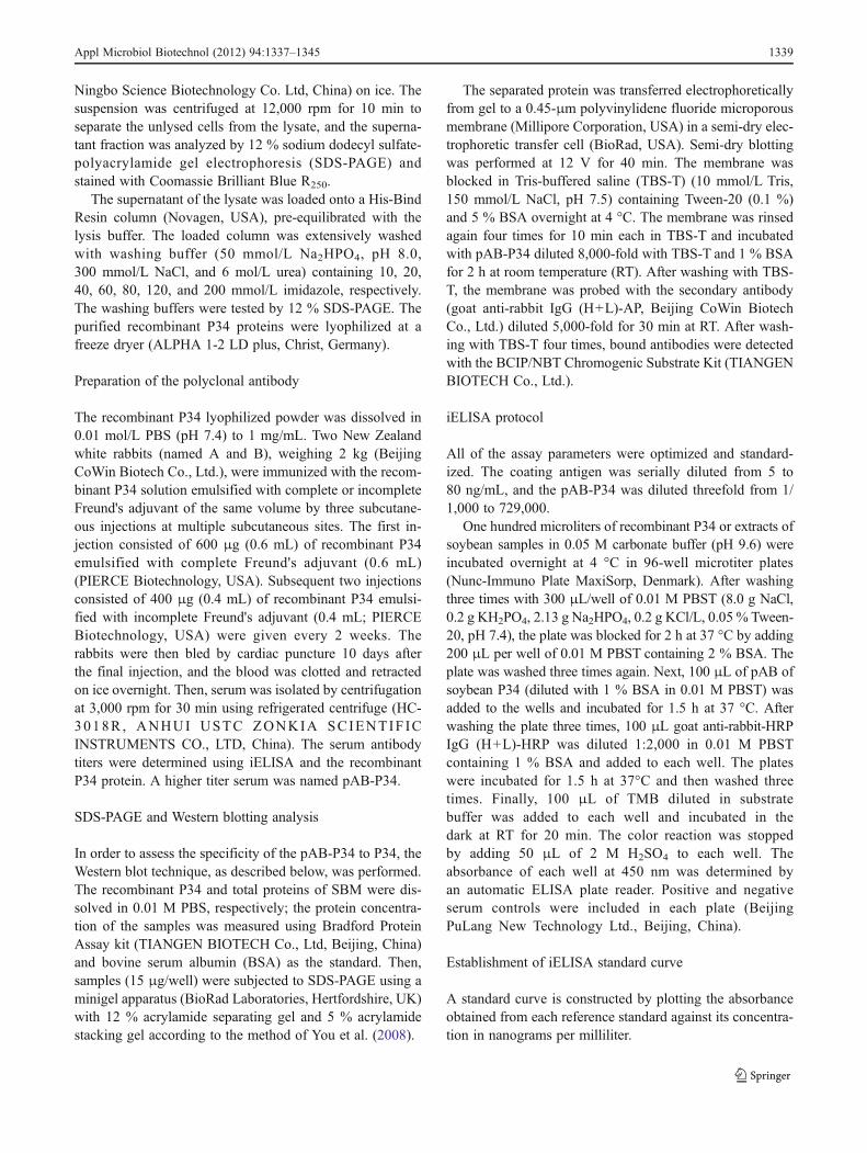
The sera titers of two rabbits were detected by iELISA usingP34 as a coating antigen. When the sera titer of rabbit Awas
A BM 1 2 3bp
5000
1000
750
Fig. 1 Schematic diagram ofthe pET28a-P34 plasmid anddigestion of the restrictionenzymes. a Digestion of therestriction enzymes. b Schematicdiagram of the pET28a-P34plasmid. 1 pET-28a plasmid;2 pET28a-P34 plasmid/Nde I,EcoR I; 3 P34 gene; M DNAmarker
M 1 2 3 4kDa
35.0
26.0
Fig. 2 SDS-PAGE analysis of the expression pattern of the recombi-nant P34 protein. M standard protein marker; 1 the lysates of pET28a-P34/BL21(DE3) (non-induced); 2 the lysates of pET28a-P34/BL21(DE3) (induced); 3 the soluble fractions of pET28a-P34/BL21(DE3)(induced); 4 the insoluble fractions of pET28a-P34/BL21(DE3)(induced)
1340 Appl Microbiol Biotechnol (2012) 94:1337–1345
lower than 81,000, its ratio of P/N was higher than 2.1 andthe P value was larger than 0.1. When the sera titer of rabbitB was lower than 729,000, its ratio of P/N was higher than2.1 and the P value was larger than 0.1 (Fig. 4). Thus, thesera of rabbit B was chosen and named as pAB–P34.
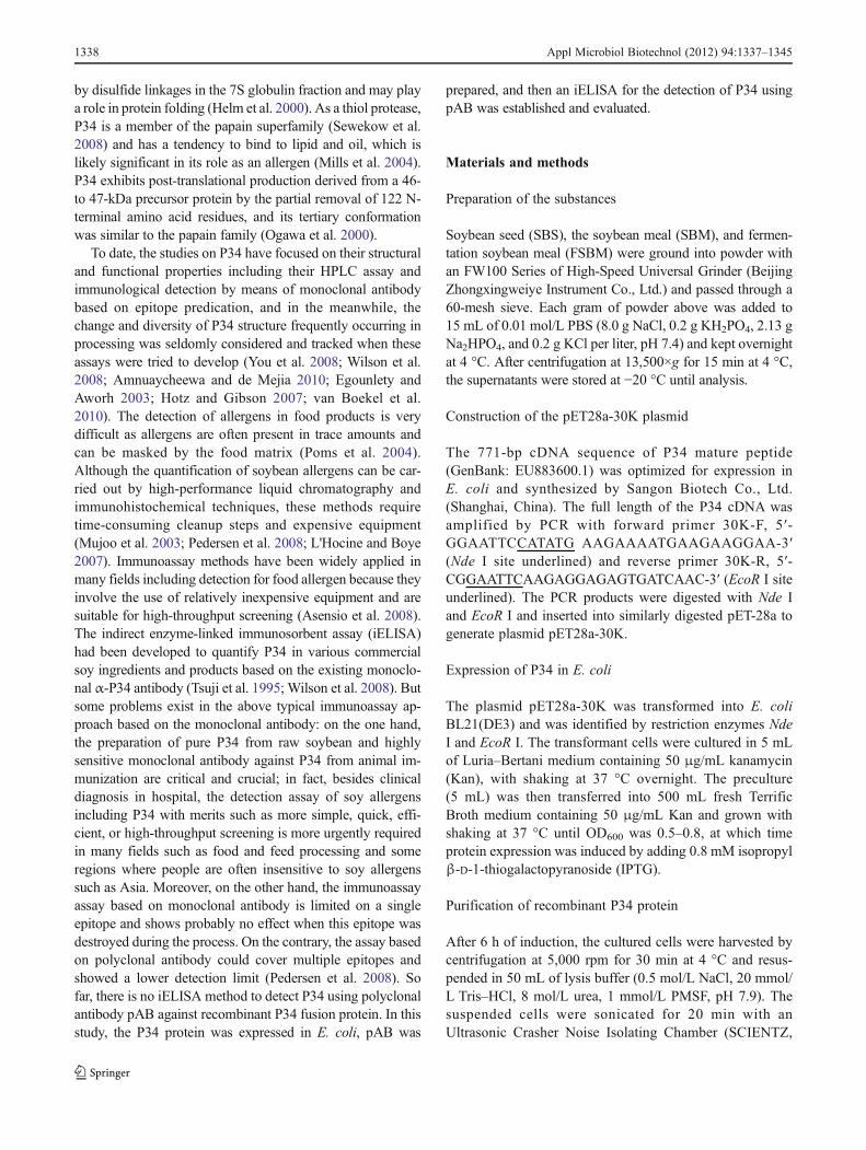
Western blot analysis indicated that pAB-P34 could spe-cifically recognize the purified recombinant P34 andextracted proteins from SBM with a specific signal at33 kDa (Fig. 3) but could not recognize any other SBMproteins, indicating that the recombinant P34 protein in-duced a specific immunological response, and thus, theprepared pAB-P34 antibody had a high level of specificity.
iELISA
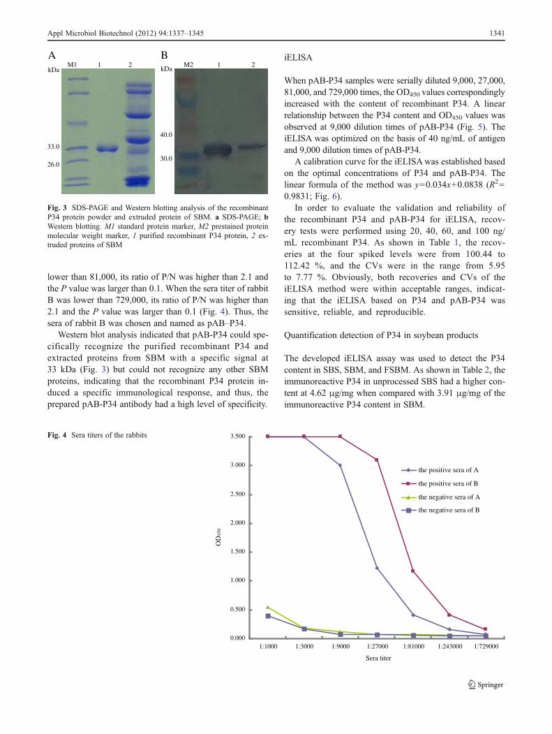
When pAB-P34 samples were serially diluted 9,000, 27,000,81,000, and 729,000 times, the OD450 values correspondinglyincreased with the content of recombinant P34. A linearrelationship between the P34 content and OD450 values wasobserved at 9,000 dilution times of pAB-P34 (Fig. 5). TheiELISA was optimized on the basis of 40 ng/mL of antigenand 9,000 dilution times of pAB-P34.
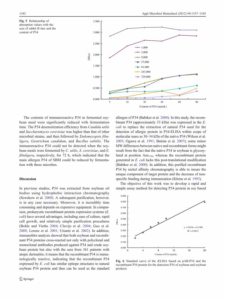
A calibration curve for the iELISAwas established basedon the optimal concentrations of P34 and pAB-P34. Thelinear formula of the method was y00.034x+0.0838 (R20
0.9831; Fig. 6).In order to evaluate the validation and reliability of
the recombinant P34 and pAB-P34 for iELISA, recov-ery tests were performed using 20, 40, 60, and 100 ng/mL recombinant P34. As shown in Table 1, the recov-eries at the four spiked levels were from 100.44 to112.42 %, and the CVs were in the range from 5.95to 7.77 %. Obviously, both recoveries and CVs of theiELISA method were within acceptable ranges, indicat-ing that the iELISA based on P34 and pAB-P34 wassensitive, reliable, and reproducible.
Quantification detection of P34 in soybean products
The developed iELISA assay was used to detect the P34content in SBS, SBM, and FSBM. As shown in Table 2, theimmunoreactive P34 in unprocessed SBS had a higher con-tent at 4.62 μg/mg when compared with 3.91 μg/mg of theimmunoreactive P34 content in SBM.
kDa
A BM1 1 2 M2 1 2
kDa
33.0
26.0
40.0
30.0
Fig. 3 SDS-PAGE and Western blotting analysis of the recombinantP34 protein powder and extruded protein of SBM. a SDS-PAGE; bWestern blotting. M1 standard protein marker, M2 prestained proteinmolecular weight marker, 1 purified recombinant P34 protein, 2 ex-truded proteins of SBM
0.000
0.500
1.000
1.500
2.000
2.500
3.000
3.500
1:1000 1:3000 1:9000 1:27000 1:81000 1:243000 1:729000
Sera titer
OD
450
the positive sera of A
the positive sera of B
the negative sera of A
the negative sera of B
Fig. 4 Sera titers of the rabbits
Appl Microbiol Biotechnol (2012) 94:1337–1345 1341
The contents of immunoreactive P34 in fermented soy-bean meal were significantly reduced with fermentationtime. The P34 desensitization efficiency from Candida utilisand Saccharomyces cerevisiae was higher than that of othermicrobial strains, and then followed by Endomycopsis fibu-ligera, Geotrichum candidum, and Bacillus subtilis. Theimmunoreactive P34 could not be detected when the soy-bean meals were fermented by C. utilis, S. cerevisiae, and E.fibuligera, respectively, for 72 h, which indicated that themain allergen P34 of SBM could be reduced by fermenta-tion with these microbes.
Discussion
In previous studies, P34 was extracted from soybean oilbodies using hydrophobic interaction chromatography(Sewekow et al. 2008). A subsequent purification, however,is in any case necessary. Moreover, it is incredibly timeconsuming and depends on expensive equipment. In compar-ison, prokaryotic recombinant protein expression systems (E.coli) have several advantages, including ease of culture, rapidcell growth, and relatively simple purification procedures(Bohle and Vieths 2004; Clavijo et al. 2004; Guo et al.2008; Lorenz et al. 2001; Utsumi et al. 2002). In addition,immunoblot analysis showed that both soybean and recombi-nant P34 proteins cross-reacted not only with polyclonal andmonoclonal antibodies produced against P34 and crude soy-bean protein but also with the sera from 361 patients withatopic dermatitis; it means that the recombinant P34 is immu-nologically reactive, indicating that the recombinant P34expressed by E. coli has similar epitope structures to naturalsoybean P34 protein and thus can be used as the standard
allergen of P34 (Babiker et al. 2000). In this study, the recom-binant P34 (approximately 33 kDa) was expressed in the E.coli to replace the extraction of natural P34 used for thedetection of allergic protein in P34-ELISA within scope ofmolecular mass as 30–34 kDa of the native P34 (Wilson et al.2005; Ogawa et al. 1991; Batista et al. 2007); some minorMWdifferences between native and recombinant forms mightresult from the fact that the native P34 in soybean is glycosy-lated at position Asn170, whereas the recombinant proteingenerated in E. coli lacks this post-translational modification(Babiker et al. 2000). In addition, this purified recombinantP34 by nickel affinity chromatography is able to insure theunique component of target protein and the decrease of non-specific binding during immunization (Kleine et al. 1993).
The objective of this work was to develop a rapid andsimple assay method for detecting P34 protein in soy based
0.000
0.500
1.000
1.500
2.000
2.500
3.000
3.500
5 10 20 40 60 80
Content of P34 (ng/mL)
OD
450
1,000
3,000
9,000
27,000
81,000
243,000
729,000
Fig. 5 Relationship ofabsorption values with thesera of rabbit B titer and thecontent of P34
y = 0.034x + 0.1368
R2 = 0.9831
0.000
0.100
0.200
0.300
0.400
0.500
0.600
0.700
0.800
0.900
0 4 8 12 16 20
Content of P34 (ng/mL)
OD
450
Fig. 6 Standard curve of the iELISA based on pAB-P34 and therecombinant P34 protein for the detection P34 of soybean and soybeanproducts
1342 Appl Microbiol Biotechnol (2012) 94:1337–1345
on pAB. In previous studies, the sandwich ELISA andcELISA methods aimed at native soy proteins (such asP34, Gly m Bd 28K, β-conglycinin, and glycinin) had beendeveloped (Tsuji et al. 1993; Bando et al. 1998; You et al.2008; Ma et al. 2010). However, these methods based onmonoclonal antibodies need a longer time for the prepara-tion of monoclonal antibody and the higher manufacturecost compared with those assays depending on polyclonalantibody (Pedersen et al. 2008). By comparison, a rabbitpAB against the recombinant P34 protein was easily pro-duced in terms of time and cost special for immunoassays ofP34. Western blot analysis revealed that the prepared pAB-P34 antibody reacted strongly with the purified recombinantP34 protein corresponding to the size of the P34 protein insoy (Fig. 3). The result indicated that pAB-P34 was able tospecifically identify P34 protein similar to a previous report(Kleine et al. 1993) and thus could be used to developimmunoassay methods for detecting P34.
In the previous studies, using the sandwich ELISA basedon two different kinds of monoclonal antibodies, the P34protein content in SBS was determined as 7.29 μg/mg (Tsujiet al. 1995). By ELISA, Wilson et al. (2008) determined theP34 protein content in 138 soybean cultivars from a diversegermplasm collection from ten different countries, and theresults indicated that P34 protein was present in all strainstested, ranging from 2.3 to 12 μg/mg. Our result showedthat the P34 protein content in SBS was 4.62 μg/mgdetected by the developed iELISA based on the pAB-P34(Table 2), which was compatible with the previous value,and the advantage over pAB-P34 is low cost and shortertime consumed by comparison with the assay based onmonoclonal antibody. In previous reports, the contents ofP34 in total soybean proteins, which account for 40.0 % ofthe total weight of the seed (You et al. 2008), were near 1 %(Herman et al. 2003; Wang et al. 2003). These data indicatethat the P34 content of the total SBS was about 4.0 μg/mg.Obviously, the assay for P34 detection showed high sensi-tivity and low cost of equipment from our results, as can beseen from Table 2; the content of the P34 protein in SBS(4.62 μg/mg) was higher than that of SBM (3.91 μg/mg).
As can be seen from Table 2, the immunoreactive P34contents in soybean products especially in FSBM (i.e.,soybean meal and fermented soybean meal by B. subtilis,G. candidum, E. fibuligera, C. utilis, and S. cerevisiae) were
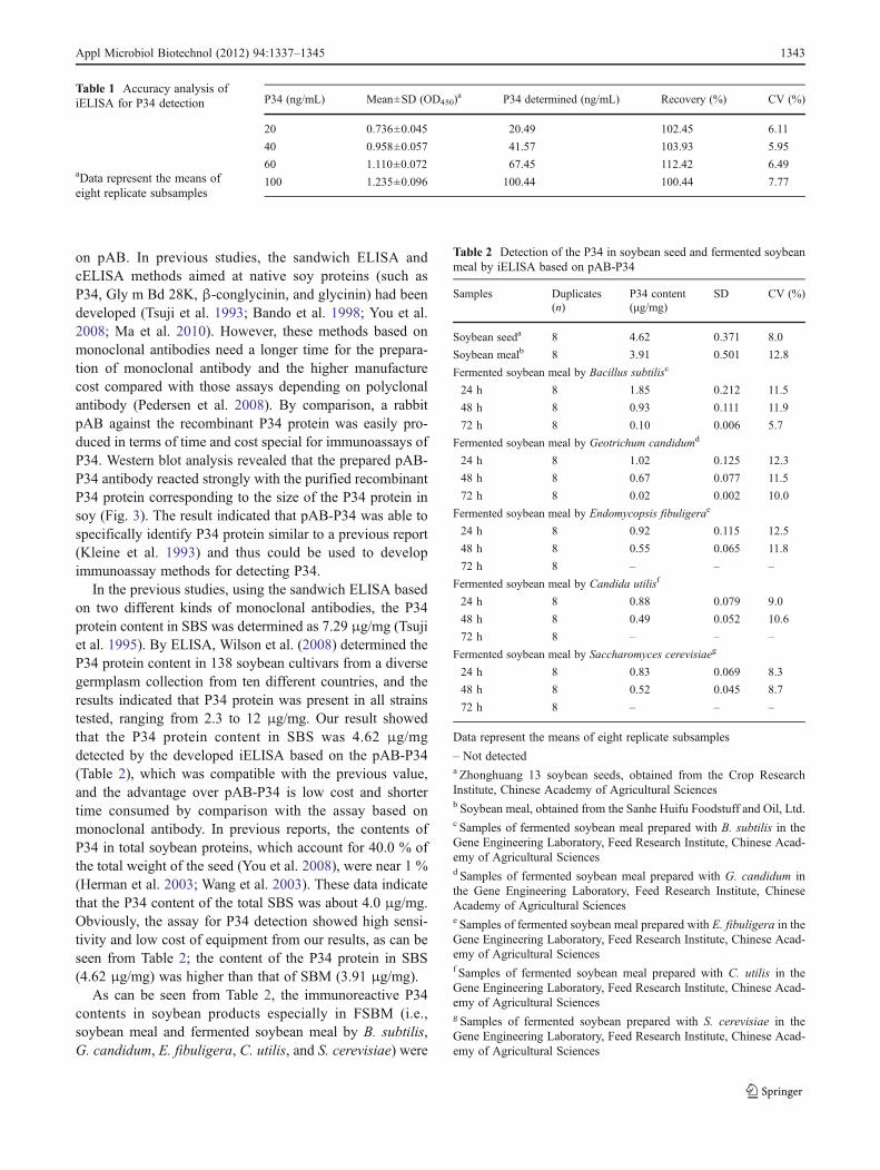
Table 1 Accuracy analysis ofiELISA for P34 detection
aData represent the means ofeight replicate subsamples
P34 (ng/mL) Mean±SD (OD450)a P34 determined (ng/mL) Recovery (%) CV (%)
20 0.736±0.045 20.49 102.45 6.11
40 0.958±0.057 41.57 103.93 5.95
60 1.110±0.072 67.45 112.42 6.49
100 1.235±0.096 100.44 100.44 7.77
Table 2 Detection of the P34 in soybean seed and fermented soybeanmeal by iELISA based on pAB-P34
Samples Duplicates(n)
P34 content(μg/mg)
SD CV (%)
Soybean seeda 8 4.62 0.371 8.0
Soybean mealb 8 3.91 0.501 12.8
Fermented soybean meal by Bacillus subtilisc
24 h 8 1.85 0.212 11.5
48 h 8 0.93 0.111 11.9
72 h 8 0.10 0.006 5.7
Fermented soybean meal by Geotrichum candidumd
24 h 8 1.02 0.125 12.3
48 h 8 0.67 0.077 11.5
72 h 8 0.02 0.002 10.0
Fermented soybean meal by Endomycopsis fibuligerae
24 h 8 0.92 0.115 12.5
48 h 8 0.55 0.065 11.8
72 h 8 – – –
Fermented soybean meal by Candida utilisf
24 h 8 0.88 0.079 9.0
48 h 8 0.49 0.052 10.6
72 h 8 – – –
Fermented soybean meal by Saccharomyces cerevisiaeg
24 h 8 0.83 0.069 8.3
48 h 8 0.52 0.045 8.7
72 h 8 – – –
Data represent the means of eight replicate subsamples
– Not detecteda Zhonghuang 13 soybean seeds, obtained from the Crop ResearchInstitute, Chinese Academy of Agricultural Sciencesb Soybean meal, obtained from the Sanhe Huifu Foodstuff and Oil, Ltd.c Samples of fermented soybean meal prepared with B. subtilis in theGene Engineering Laboratory, Feed Research Institute, Chinese Acad-emy of Agricultural Sciencesd Samples of fermented soybean meal prepared with G. candidum inthe Gene Engineering Laboratory, Feed Research Institute, ChineseAcademy of Agricultural Sciencese Samples of fermented soybean meal prepared with E. fibuligera in theGene Engineering Laboratory, Feed Research Institute, Chinese Acad-emy of Agricultural Sciencesf Samples of fermented soybean meal prepared with C. utilis in theGene Engineering Laboratory, Feed Research Institute, Chinese Acad-emy of Agricultural Sciencesg Samples of fermented soybean prepared with S. cerevisiae in theGene Engineering Laboratory, Feed Research Institute, Chinese Acad-emy of Agricultural Sciences
Appl Microbiol Biotechnol (2012) 94:1337–1345 1343
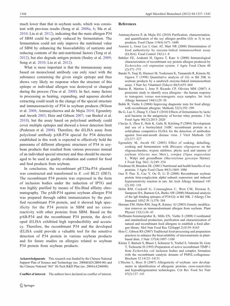
much lower than that in soybean seeds, which was consis-tent with previous results (Song et al. 2008a, b; Ma et al.2010; Liu et al. 2012), indicating that the main allergen P34of SBM could be greatly reduced by fermentation. Thefermentation could not only improve the nutritional valueof SBM by enhancing the bioavailability of nutrients andreducing contents of the anti-nutritional factors (Teng et al.2012), but also degrade antigen protein (Sealey et al. 2009;Song et al. 2010; Liu et al. 2012).
What is more important is that the immunoassay assaybased on monoclonal antibody can only react with thesubstance containing the given single epitope and thusshows very likely no response when the structure of thisepitope or individual allergen was destroyed or changedduring the process (You et al. 2008). In fact, many factorsin processing as heating, expanding, and aqueous alcoholextracting could result in the change of the special structureand immunoreactivity of P34 in soybean products (Wilsonet al. 2008; Amnuaycheewa and de Mejia 2010; Egounletyand Aworh 2003; Hotz and Gibson 2007; van Boekel et al.2010), but the assay based on polyclonal antibody couldcover multiple epitopes and showed a lower detection limit(Pedersen et al. 2008). Therefore, the iELISA assay frompolyclonal antibody pAB-P34 special for P34 detectionestablished in this work is expected to effectively hold thepanorama of different allergenic structures of P34 in soy-bean products that resulted from various processes insteadof an individual special structure and thus should be encour-aged to be used in quality evaluation and control for foodand feed products from soybean.
In conclusion, the recombinant pET28a-P34 plasmidwas constructed and transformed to E. coli BL21 (DE3).The recombinant P34 protein was expressed in the formof inclusion bodies under the induction of IPTG andwas highly purified by means of His-Bind affinity chro-matography. The pAB-P34 against soybean allergen P34was prepared through rabbit immunization by the puri-fied recombinant P34 protein, and it showed high spec-ificity for the P34 protein in SBM and no cross-reactivity with other proteins from SBM. Based on thepAB-P34 and the recombinant P34 protein, the devel-oped iELISA exhibited high reproducibility and accura-cy. Therefore, the recombinant P34 and the developediELISA could provide a valuable tool for the sensitivedetection of P34 protein in various soybean productsand for future studies on allergies related to soybeanP34 protein from soybean products.
Acknowledgments This research was funded by the Chinese NationalSupport Plan of Science and Technology (no. 2006BAD12B04-06) andthe Chinese National ‘863’ Hi-Tech R&D Plan (no. 2004AA246040).
Conflict of interest The authors have declared no conflict of interest.
References
Amnuaycheewa P, de Mejia EG (2010) Purification, characterisation,and quantification of the soy allergen profilin (Gly m 3) in soyproducts. Food Chem 119(4):1671–1680
Asensio L, Gonz Lez I, Garc AT, Mart NR (2008) Determination offood authenticity by enzyme-linked immunosorbent assay(ELISA). Food Control 19(1):1–8
Babiker EE, Azakami H, Ogawa T, Kato A (2000) Immunologicalcharacterization of recombinant soy protein allergen produced byEscherichia coli expression system. J Agric Food Chem 48(2):571–575
Bando N, Tsuji H, Hiemori M, Yoshizumi K, Yamanishi R, Kimoto M,Ogawa T (1998) Quantitative analysis of Gly m Bd 28K insoybean products by a sandwich enzyme-linked immunosorbentassay. J Nutr Sci Vitaminol (Tokyo) 44(5):655–664
Batista R, Martins I, Jeno P, Ricardo CP, Oliveira MM (2007) Aproteomic study to identify soya allergens—the human responseto transgenic versus non-transgenic soya samples. Int ArchAllergy Immunol 144(1):29–38
Bohle B, Vieths S (2004) Improving diagnostic tests for food allergywith recombinant allergens. Methods 32(3):292–299
Bu G, Luo Y, Zhang Y, Chen F (2010) Effects of fermentation by lacticacid bacteria on the antigenicity of bovine whey proteins. J SciFood Agric 90(12):2015–2020
Clavijo A, Zhou E, Hole K, Galic B, Kitching P (2004) Developmentand use of a biotinylated 3ABC recombinant protein in asolid-phase competitive ELISA for the detection of antibodiesagainst foot-and-mouth disease virus. J Virol Methods 120(2):217–227
Egounlety M, Aworh OC (2003) Effect of soaking, dehulling,cooking and fermentation with Rhizopus oligosporus on theoligosaccharides, trypsin inhibitor, phytic acid and tannins ofsoybean (Glycine max Merr.), cowpea (Vigna unguiculataL. Walp) and groundbean (Macrotyloma geocarpa Harms).J Food Eng 56(2–3):249–254
Friedman M, Brandon DL (2001) Nutritional and health benefits of soyproteins. J Agric Food Chem 49(3):1069–1086
Guo P, Piao X, Cao Y, Ou D, Li D (2008) Recombinant soybeanprotein beta-conglycinin alpha′-subunit expression and inducedhypersensitivity reaction in rats. Int Arch Allergy Immunol 145(2):102–110
Helm RM, Cockrell G, Connaughton C, West CM, Herman E,Sampson HA, Bannon GA, Burks AW (2000) Mutational analysisof the IgE-binding epitopes of P34/Gly m Bd 30K. J Allergy ClinImmunol 105(2 Pt 1):378–384
Herman EM, Helm RM, Jung R, Kinney AJ (2003) Genetic modifica-tion removes an immunodominant allergen from soybean. PlantPhysiol 132(1):36–43
Hoffmann-Sommergruber K, Mills EN, Vieths S (2008) Coordinatedand standardized production, purification and characterization ofnatural and recombinant food allergens to establish a food aller-gen library. Mol Nutr Food Res 52(Suppl 2):S159–S165
Hotz C, Gibson RS (2007) Traditional food-processing and preparationpractices to enhance the bioavailability of micronutrients in plant-based diets. J Nutr 137(4):1097–1100
Kleine T, Bartsch S, Blaser J, Schnierer S, Triebel S, Valentin M, GoteT, Tschesche H (1993) Preparation of active recombinant TIMP-1from Escherichia coli inclusion bodies and complex formationwith the recombinant catalytic domain of PMNL-collagenase.Biochem 32:14125–14131
L'Hocine L, Boye JI (2007) Allergenicity of soybean: new develop-ments in identification of allergenic proteins, cross-reactivitiesand hypoallergenization technologies. Crit Rev Food Sci Nutr47(2):127–143
1344 Appl Microbiol Biotechnol (2012) 94:1337–1345
Liu B, Teng D, Yang L, Wang X, Wang J (2012) Development of acompetitive ELISA for the detection of soybean α subunit of β-conglycinin. Process Biochem 47(2):280–287
Lorenz A, Scheurer S, Haustein D, Vieths S (2001) Recombinant foodallergens. J Chromatogr B Biomed Sci Appl 756(1–2):255–279
Ma X, Sun P, He P, Han P, Wang J, Qiao S, Li D (2010) Development ofmonoclonal antibodies and a competitive ELISA detection methodfor glycinin, an allergen in soybean. Food Chem 121(2):546–551
Mills ENC, Breiteneder H (2005) Food allergy and its relevance toindustrial food proteins. Biotechnol Adv 23(6):409–414
Mills EN, Jenkins JA, Alcocer MJ, Shewry PR (2004) Structural,biological, and evolutionary relationships of plant food allergenssensitizing via the gastrointestinal tract. Crit Rev Food Sci Nutr44(5):379–407
Mujoo R, Trinh DT, Ng PKW (2003) Characterization of storageproteins in different soybean varieties and their relationship totofu yield and texture. Food Chem 82(2):265–273
Ogawa T, BandoN, Tsuji H, Okajima H,Nishikawa K, Sasaoka K (1991)Investigation of the IgE-binding proteins in soybeans by immuno-blotting with the sera of the soybean-sensitive patients with atopicdermatitis. J Nutr Sci Vitaminol (Tokyo) 37(6):555–565
Ogawa A, Samoto M, Takahashi K (2000) Soybean allergens andhypoallergenic soybean products. J Nutr Sci Vitaminol (Tokyo)46(6):271–279
Pedersen MH, Holzhauser T, Bisson C, Conti A, Jensen LB, Skov PS,Bindslev-Jensen C, Brinch DS, Poulsen LK (2008) Soybeanallergen detection methods—a comparison study. Mol NutrFood Res 52(12):1486–1496
Poms RE, Klein CL, Anklam E (2004) Methods for allergen analysis infood: a review. Food Addit Contam 21(1):1–31
Sathe SK, Sharma GM (2009) Effects of food processing on foodallergens. Mol Nutr Food Res 53(8):970–978
Sealey WM, Barrows FT, Smith CE, Overturf K, LaPatra SE (2009)Soybean meal level and probiotics in first feeding fry diets alterthe ability of rainbow trout Oncorhynchus mykiss to utilize highlevels of soybean meal during grow-out. Aquaculture 293(3–4):195–203
Sewekow E, Kessler LC, Seidel-Morgenstern A, Rothkotter HJ (2008)Isolation of soybean protein P34 from oil bodies using hydropho-bic interaction chromatography. BMC Biotechnol 8:27
Song YS, Martinez-Villaluenga C, De Meijia EG (2008a) Quantificationof human IgE immunoreactive soybean proteins in commercial soyingredients and products. J Food Sci 73(6):T90–T99
Song YS, Frias J, Martinez-Villaluenga C, Vidal-Valdeverde C,Gonzalez de Mejia E (2008b) Immunoreactivity reduction ofsoybean meal by fermentation effect on amino acid composition
and antigenicity of commercial soy products. Food Chem108:571–581
Song YS, Perez VG, Pettigrew JE, Martinez-Villaluenga C, de MejiaEG (2010) Fermentation of soybean meal and its inclusion indiets for newly weaned pigs reduced diarrhea and measuresof immunoreactivity in the plasma. Anim Feed Sci Tech 159(1–2):41–49
Teng D, Gao M, Yang Y, Liu B, Tian Z, Wang J (2012) Bio-modificationof soybean meal with Bacillus subtilis or Aspergillus oryzae.Biocatalysis Agric Biotechnol 1(1):32–38
Tsuji H, Bando N, Kimoto M, Okada N, Ogawa T (1993) Preparationand application of monoclonal antibodies for a sandwich enzyme-linked immunosorbent assay of the major soybean allergen, Gly mBd 30K. J Nutr Sci Vitaminol 39(4):389–397
Tsuji H, Okada N, Yamanishi R, Bando N, Kimoto M, Ogawa T (1995)Measurement of Gly m Bd 30K, a major soybean allergen, insoybean products by a sandwich enzyme-linked immunosorbentassay. Biosci Biotechnol Biochem 59(1):150–151
Utsumi S, Maruyama N, Satoh R, Adachi M (2002) Structure–functionrelationships of soybean proteins revealed by using recombinantsystems. Enzyme Microb Tech 30(3):284–288
van Boekel M, Fogliano V, Pellegrini N, Stanton C, Scholz G, LalljieS, Somoza V, Knorr D, Jasti PR, Eisenbrand G (2010) A reviewon the beneficial aspects of food processing. Mol Nutr Food Res54(9):1215–1247
van Boxtel EL, van den Broek LA, Koppelman SJ, Gruppen H (2008)Legumin allergens from peanuts and soybeans: effects of dena-turation and aggregation on allergenicity. Mol Nutr Food Res 52(6):674–682
Wang TL, Domoney C, Hedley CL, Casey R, Grusak MA (2003) Canwe improve the nutritional quality of legume seeds? Plant Physiol131(3):886–891
Wilson S, Blaschek K, de Mejia E (2005) Allergenic proteins insoybean: processing and reduction of P34 allergenicity. NutrRev 63(2):47–58
Wilson S, Martinez-Villaluenga C, De Mejia EG (2008) Purification,thermal stability, and antigenicity of the immunodominant soy-bean allergen P34 in soy cultivars, ingredients, and products. JFood Sci 73(6):T106–T114
Yaklich R, Helm R, Herman E (1999) Analysis of the distribution ofthe major soybean allergen in a core collection of Glycine maxaccessions. Crop Sci 39:1444–1447
You J, Li D, Qiao S, Wang Z, He P, Ou D, Dong B (2008)Development of a monoclonal antibody-based competitiveELISA for detection of [beta]-conglycinin, an allergen from soy-bean. Food Chem 106(1):352–360
Appl Microbiol Biotechnol (2012) 94:1337–1345 1345
![Food Allergenic Information Gyu-Kaku Grand …Food Allergenic Information Gyu-Kaku Grand_Menu [Last updated on 2020/5/27] ① ※【 】:Contain allergen 【-】:Without allergen](https://img.pdfslide.net/doc/110x75/5f2400760af06f7aec55da5d/food-allergenic-information-gyu-kaku-grand-food-allergenic-information-gyu-kaku.jpg)