Embed Size (px)
Citation preview

대한구강해부학회지 제 39권 제1호
Korean J Oral Anatomy Vol. 39, no. 1 (2018) pp.1~8
1
Expression patterns of Zfhx1a and Zfhx1b during mouse craniofacial development
Jeong-Oh Shin1,*, Jong-Min Lee4,*, Jinwoong Bok1,2,3, Han-Sung Jung4,†
1Department of Anatomy, 2BK21 PLUS project for Medical Science, 3Department of Otorhinolaryngology, Yonsei University College of Medicine, Seoul, Republic of Korea. 4Division in Anatomy and
Developmental Biology, Department of Oral Biology, Oral Science Research Center, BK21 PLUS Project, Yonsei University College of Dentistry, Seoul, Korea.
접수: 2018년 9월 27일/ 수정접수: 2018년 10월 18일/ 게재 승인: 2018년 10월 18일/ 출간: 2018년 12월 31일
Recent studies have demonstrated that Zfhx1a and Zfhx1b are transcription factors involved in many im-
portant signaling pathways. They are known to be essential for neural development, and for the development
of other neural-crest-derived tissues. However, much remains to be learned about their expression patterns and
functions in the developing tissues of the craniofacial region. We determined the unique expression patterns
of Zfhx1a and Zfhx1b during mouse craniofacial development from embryonic day (E) 13.5 to E16.5. In the
epithelium of the circumvallate papilla facing the oral cavity, Zfhx1a and Zfhx1b were strongly and weakly
expressed, respectively. The epithelial component of the submandibular gland expressed Zfhx1a and Zfhx1b. In
the developing eye, Zfhx1a and Zfhx1b were expressed strongly in the retina, and in the anterior region of the
lens at E13.5 and E14.5. At E16.5, transcripts of Zfhx1a and Zfhx1b were detected in the developing eyelids.
These findings demonstrate the spatial and temporal expression patterns of Zfhx1a and Zfhx1b during mouse
craniofacial development.
Keywords: Zfhx1a, Zfhx1b, circumvallate papilla, submandibular gland, eye.
* These authors contributed equally to this work† Correspondence to: Han-Sung Jung, Ph.D.Division in Anatomy and Developmental Biology, Department of Oral Biology, College of Dentistry, Yonsei University, 03722, Seoul, Korea. Tel: +82-2-2228-3065; Fax: +82-2-312-8012; E-mail: [email protected]

Expression patterns of Zfhx1a and Zfhx1b during mouse craniofacial development
Jeong-Oh Shin, Jong-Min Lee, Jinwoong Bok, Han-Sung Jung
2
IntroductionZfhx1a and Zfhx1b , the two z inc- f inger
E-box-binding homeobox factors, are two transcrip-
tion regulators of the vertebrate that are closely as-
sociated with Zfh-1 of Drosophila 1,2,3,4,5,6). Zfhx1a is
diversely known as Zeb15, 7, 8), δEF13), and Zfhep9,10,11);
Zfhx1b is also known as Zeb2 and Sip15,8,12). Zfhx1a
and Zfhx1b may function as mediators of other sig-
naling pathways13). Zfhx1a is involved in transform-
ing growth factor beta (Tgf-β) signaling in vascular
smooth muscle cell differentiation14), and in sonic
hedgehog (Shh) signaling during mouse limb devel-
opment15). Zfhx1a is strongly expressed in the neural
tube, brain, mesoderm, and neural-crest-derived
tissues such as the limb buds, somites, and branchial
arches10,16). In addition, the Zfhx1a-knockout mouse
exhibits cleft palate, suggesting its role as a regulator
of cell proliferation during secondary palate develop-
ment16). Zfhx1b is a transcription repressor of the Zfh-
1 family that acts as a downstream mediator of Tgf-β
and bone morphogenetic protein signaling (BMP)8,12).
Zfhx1b is widely expressed in humans and mice,
most prominently in the heart and the neural tis-
sues17,18). Mutations causing Zfhx1b haploinsufficien-
cy during embryogenesis is related to Mowat-Wilson
syndrome, which is characterized by mental retarda-
tion, dysmorphic facial features, microcephaly, sei-
zures19,20,21). Knockout of Zfhx1b in mice is embryonic
lethal at embryonic day (E) 9.5–E10.5, with the mice
exhibiting developmental defects in neural crest for-
mation22,23,24) that are caused by ectopic expression of
E-cadherin. Zfhx1b is expressed in numerous tissues
during embryonic development, including the neural
crest, neuroepithelium, and limb buds23). However,
expression of Zfhx1a and Zfhx1b in the internal organ
of craniofacial region was not determined. Here, we
determined the unique and overlapping expression
patterns of Zfhx1a and Zfhx1b in the developing
mouse craniofacial region by in situ hybridization in
the circumvallate papilla of the tongue, submandib-
ular gland, and the developing eye at E13.5, E14.5,
and E16.5.
Materials and MethodsAll experiments were performed according to
the guidelines of the Intramural Animal Use and Care
Committee of the College of Dentistry, Yonsei Uni-
versity.
Animals
Adult ICR mice were housed in a tempera-
ture-controlled room (22°C) under artificial illumina-
tion (lights on from 05:00 to 17:00) and 55% relative
humidity, with access to food and water ad libitum.
The embryos were obtained from time-mated preg-
nant mice. E0 was designated as the day on which
the presence of a vaginal plug was confirmed. Em-
bryos at each developmental stage (E13.5, E14.5, and
E16.5) were used in this study.
In situ hybridization
In situ hybridization on whole mouse embryos
was performed as previously described25) in paraffin
wax sections by using standard protocols. Briefly,
embryos were fixed in 4% PFA, embedded in paraffin
wax and sectioned at 7 μm. Sections were incubated
at 60°C, dewaxed in xylene, re-hydrated through a
graded series of alcohol washes and post-fixed in
4% PFA. Sections were prehybridized in a humid
대한구강해부학회지 제 39권 제1호
Korean J Oral Anatomy Vol. 39, no. 1 (2018) pp.1~8
3
chamber containing 50% formamide in 2× saline so-
dium citrate buffer at 58°C for 30 min. Digoxigenin
(DIG)-labelled RNA probes were prewarmed to 85°C
and hybridized to sections overnight at 58°C. Mouse
DNA Zfhx1a and Zfhx1b plasmids were used as tem-
plates for the synthesis of DIG-labeled RNA probes.
Results and Discussion
Expression patterns of Zfhx1a and Zfhx1b in the
developing circumvallate papilla of the tongue and
the submandibular gland
The expression patterns of Zfhx1a and Zfhx1b
were examined on sections of developing mouse
circumvallate papilla region of the tongue (Fig. 1).
At E13.5, Zfhx1a was expressed strongly in the epi-
thelium of the circumvallate-papilla-forming region,
including the arch-like structure (i.e., the epithelium
of the circumvallate papilla facing the oral cavity;
Fig. 1A and B). Zfhx1a was weakly expressed in the
mesenchyme underlying the epithelium of the cir-
cumvallate papilla (Fig. 1A and B). Zfhx1b was ex-
pressed weakly in the mesenchyme of the underlying
circumvallate papilla (Fig. 1D and E). Interestingly,
Zfhx1b was strongly expressed in the epithelium
where the trench of the circumvallate papilla was
developing, but weakly in the arch-like structure
of the circumvallate papilla (Fig. 1D and E). At
E14.5, Zfhx1a was expressed strongly in the overall
epithelium, including the arch-like structure of the
circumvallate papilla, but it was not observed in the
mesenchymal cells underlying the epithelium of the
circumvallate papilla (Fig. 1G and H). Zfhx1b tran-
scripts were detected in the epithelium of the circum-
vallate papilla, except the arch-like structure, but it
was localized in the overall mesenchyme underlying
the circumvallate papilla (Fig. 1J and K).
Figure 1. Localization of Zfhx1a and Zfhx1b in the developing circumvallate papilla and submandibular gland at E13.5, E14.5, and E16.5.All samples are frontal sections. (A, D, G, J, M, P) Low magnification images of the posterior mandible at the level of the developing circumvallate papilla of the tongue and the submandibular gland. Red and blue boxes are higher magnification images of panel A, D, G, J, M, and P. (B, H, N) Expression of Zfhx1a in the circumvallate papilla of the posterior tongue. (E, K, Q) Expression of Zfhx1b in the circumvallate papilla of the posterior tongue. (C, I, O) Expression of Zfhx1a in the submandibular gland. (F, L, R) Expression of Zfhx1b in the submandibular gland. yellow dotted line, epithelium outline in circumvallate papilla. Blue arrowhead, arch-like structure in circumval-late papilla; black arrowhead, strong expression region in apex region and trench wall in circumvallate papilla. CVP, circumvallate papilla; SG, submandibular gland. Scale bars – A, D, G, J: 200 μm; B, C, E, F, H, K, O, R: 50 μm;
I, L, N, Q: 100 μm; M, P: 400 μm
At E16.5, the developing circumvallate papilla
underwent prominent morphological changes, result-
ing in a more bulbous shape, deeper location of the
floor epithelium of the trench, and uplifting of the
arch-like region (apex) of the trench-wall epithelium
(Fig. 1N and Q). At this stage, Zfhx1a was expressed
throughout the epithelium of the circumvallate papil-
la (Fig. 1M and N). It was strongly expressed in the
arch-like region of the trench-wall epithelium and

Expression patterns of Zfhx1a and Zfhx1b during mouse craniofacial development
Jeong-Oh Shin, Jong-Min Lee, Jinwoong Bok, Han-Sung Jung
4
the floor epithelium of the trench of the circumval-
late papilla (Fig. 1N). Zfhx1b was expressed in the
epithelium and mesenchyme of the circumvallate
papilla region, but not in the arch-like structure of the
circumvallate epithelium at E16.5 (Fig. 1Q).
The expression pattern of Zfhx1a in the devel-
oping circumvallate papilla region (Fig. 1H and N)
was similar to that of Patched, while that of Zfhx1b
(Fig. 1K and Q) was similar to that of Shh26). The
relationship between Zfhx1a and Shh signaling has
been investigated during mouse limb development15).
Patched is the molecular target of Shh27), and it is
suggested that the Shh signaling pathway plays an
important role in the developing circumvallate papil-
la28). Therefore, we suggest that Zfhx1a and Zfhx1b,
in association with the Shh signaling pathway, are
involved in the morphogenesis and pattern formation
of the circumvallate papilla. Both Zfhx1a and Zfhx1b
were also expressed in the muscle fibers of the tongue
below the developing circumvallate papilla region at
E13.5, E14.5, and E16.5 (Fig. 1B, E, H, K, N and Q).
These results are in agreement with the expression
patterns of Zfhx1a and Zfhx1b found in the muscle
cells of the developing mouse embryo16), and suggest
that Zfhx1a and Zfhx1b are involved in the devel-
opment of the muscle fibers in the circumvallate
papilla region. In addition, Zfhx1a and Zfhx1b were
expressed in the mylohyoid muscle and the digastric
muscle at E13.5 and E14.5, but their levels were di-
minished at E16.5 (Fig. 1A, D, G, J, M and P).
The development of the mouse submandibular
gland is initiated between E11.5 and E12.5. By
E13.5, the epithelial bud begins to cleft and branch.
Branching morphogenesis occurs continuously in
the immature submandibular gland, resulting in the
formation of multiple cords by E14.5. Finally, at
E17, differentiation and lumenization occur in the
ducts and terminal buds29). The expression patterns
of Zfhx1a and Zfhx1b on sections of developing sub-
mandibular gland are presented in Fig. 2. At E13.5,
Zfhx1a and Zfhx1b were expressed strongly in the na-
scent epithelial bud of the developing submandibular
gland, but weakly in the surrounding mesenchyme
(Fig. 1A, C, D and F). At E14.5, Zfhx1a and Zfhx1b
were strongly expressed in the proliferating and cleft-
ing epithelial bud of the embryonic submandibular
gland. However, they were weakly expressed in the
mesenchyme of the submandibular gland (Fig. 1C
and F). At E16.5, Zfhx1a and Zfhx1b were strongly
expressed in the epithelial buds that will form the
submandibular acini (Fig. 1M, O, P and R). Weaker
expressions were found in multiple epithelial cords
that will form the submandibular ducts (Fig. 1M, O,
P and R).
Expression patterns of Zfhx1a and Zfhx1b in the
developing eye
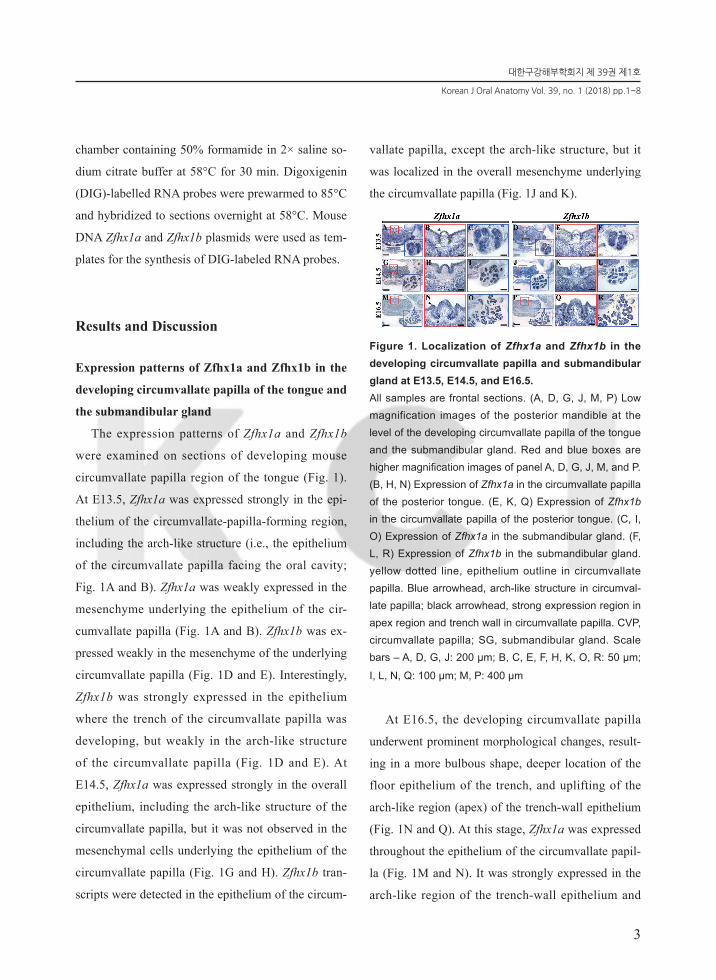
The expression patterns of Zfhx1a and Zfhx1b on
sections of the developing mouse eye are presented
in Fig. 2. In the lens, Zfhx1a was expressed in the
anterior half of the lens fibers at E13.5, and gradu-
ally decreased at E14.5 (Fig. 2A, B, E and F). On
the other hand, Zfhx1b was expressed strongly in the
lens epithelium at E13.5 and E14.5 (Fig. 2C, D, G
and H). Despite the expression patterns of Zfhx1a
and Zfhx1b differing at E13.5 and E14.5, these genes
were both expressed in the same region at E16.5, the
region of cell elongation (Fig. 2I, J, K and L). Tran-
scripts of Zfhx1a and Zfhx1b were also observed in
the mesenchyme in the edges of the upper and lower

대한구강해부학회지 제 39권 제1호
Korean J Oral Anatomy Vol. 39, no. 1 (2018) pp.1~8
5
developing eyelids at E16.5 (Fig. 2I and K). Zfhx1a
and Zfhx1b are known to be crucial factors in neural
development10,11,18,24), and we found that Zfhx1a and
Zfhx1b were also strongly expressed in the nervous
tunic layer, including the retina, at E13.5, E14.5, and
E16.5 (Fig. 2A, C, E, G, I and K).
Figure 2. Localization of Zfhx1a and Zfhx1b in the de-veloping eye at E13.5, E14.5, and E16.5.All samples are frontal sections. Expression of Zfhx1a in the developing eye. (A, C, E, G, I, K) Low magnification images of the developing eye. (B, D, F, H, J, L ) Red boxes are higher magnification images of panel. Ls, lens; C, cornea; Re, retina; LF, lens fiber; LE, lens epithelium; ILO, inner layer of optic cup. Scale bars – A, C, E, G: 200 μm; B, D, F, H, J, L: 50 μm ; I, K: 400 μm
In summary, this study has demonstrated the
unique expression patterns of Zfhx1a and Zfhx1b
in the craniofacial region from E13.5 to E16.5.
Zfhx1a and Zfhx1b are known to be important in
ectoderm-derived organ and the development of
neural-crest-derived tissues10,11,18,24). Zfhx1a (but not
Zfhx1b) was expressed in the arch-like epithelial
layer of the circumvallate papilla facing the oral
cavity. The epithelial component of the submandib-
ular gland, but not the mesenchymal cells, expressed
Zfhx1a and Zfhx1b (Fig. 1). In the developing eye,
strong expression of Zfhx1a and Zfhx1b were found
in the retina and the anterior region of the lens
(Fig. 2). These findings improve the spatial and tem-
poral understanding of the expressions of Zfhx1a and
Zfhx1b during mouse craniofacial development.
AcknowledgementsThis work was supported by the Nation-
al Research Foundation of Korea (NRF) Grant
funded by the Korea Government (MSIP) (NRF-
2016R1A5A2008630).
References1. Cabanillas AM, Darling DS. Alternative splicing gives
rise to two isoforms of Zfhep, a zinc finger/home-odomain protein that binds T3-response elements. DNA Cell Biol 15:643-651. 1996. DOI: 10.1089/dna.1996.15.643.
2. Fortini ME, Lai ZC, Rubin GM. The Drosophila zfh-1 and zfh-2 genes encode novel proteins containing both zinc-finger and homeodomain motifs. Mech Dev 34:113-122. 1991. DOI: 10.1016/0925-4773(91)90048-B.
3. Funahashi J, Sekido R, Murai K, Kamachi Y, Kondoh H. Delta-Crystallin Enhancer-Binding Protein Del-ta-Ef1 Is a Zinc Finger-Homeodomain Protein Impli-cated in Postgastrulation Embryogenesis. Development 119:433-446. 1993. PMID: 7904558.
4. Lai ZC, Fortini ME, Rubin GM. The embryonic expres-sion patterns of zfh-1 and zfh-2, two Drosophila genes encoding novel zinc-finger homeodomain proteins. Mech Dev 34:123-134. 1991. PMID: 1680377.
5. Postigo AA, Dean DC. ZEB, a vertebrate homolog of Drosophila Zfh-1, is a negative regulator of muscle differentiation. EMBO J 16:3935-3943. 1997. DOI: 10.1093/emboj/16.13.3935.
6. Sekido R, Takagi T, Okanami M, Moribe H, Yamamura M, Higashi Y, Kondoh H. Organization of the gene en-

Expression patterns of Zfhx1a and Zfhx1b during mouse craniofacial development
Jeong-Oh Shin, Jong-Min Lee, Jinwoong Bok, Han-Sung Jung
6
coding transcriptional repressor deltaEF1 and cross-spe-cies conservation of its domains. Gene 173:227-232. 1996. DOI: 10.1016/0378-1119(96)00185-0.
7. Genetta T, Ruezinsky D, Kadesch T. Displacement of an E-Box-Binding Repressor by Basic Helix-Loop-He-lix Proteins - Implications for B-Cell Specificity of the Immunoglobulin Heavy-Chain Enhancer. Molec-ular and Cellular Biology 14:6153-6163. 1994. DOI: 10.1128/MCB.14.9.6153.
8. Postigo AA, Depp JL, Taylor JJ, Kroll KL. Regulation of Smad signaling through a differential recruitment of coactivators and corepressors by ZEB proteins. EMBO J 22:2453-2462. 2003. DOI: 10.1093/emboj/cdg226.
9. Darling DS, Gaur NK, Zhu B. A zinc finger home-odomain transcription factor binds specific thyroid hor-mone response elements. Mol Cell Endocrinol 139:25-35. 1998. DOI: 10.1016/S0303-7207(98)00076-8.
10. Darling DS, Stearman RP, Qi Y, Qiu MS, Feller JP. Expression of Zfhep/deltaEF1 protein in palate, neu-ral progenitors, and differentiated neurons. Gene Expr Patterns 3:709-717. 2003. DOI: 10.1016/S1567-133X(03)00147-9.
11. Yen G, Croci A, Dowling A, Zhang S, Zoeller RT, Dar-ling DS. Developmental and functional evidence of a role for Zfhep in neural cell development. Brain Res Mol Brain Res 96:59-67. 2001. DOI: 10.1016/S0169-328X(01)00267-4.
12. Verschueren K, Remacle JE, Collart C, Kraft H, Baker BS, Tylzanowski P, Nelles L, Wuytens G, Su MT, Bod-mer R, Smith JC, Huylebroeck D. SIP1, a novel zinc finger/homeodomain repressor, interacts with Smad proteins and binds to 5'-CACCT sequences in candidate target genes. J Biol Chem 274:20489-20498. 1999. doi: 10.1074/jbc.274.29.20489.
13. Postigo AA. Opposing functions of ZEB proteins in the regulation of the TGFbeta/BMP signaling pathway. EMBO J 22:2443-2452. 2003. DOI: 10.1093/emboj/cdg225.
14. Nishimura G, Manabe I, Tsushima K, Fujiu K, Oishi Y, Imai Y, Maemura K, Miyagishi M, Higashi Y, Kondoh H, Nagai R. DeltaEF1 mediates TGF-beta signaling in vascular smooth muscle cell differentiation. Dev Cell
11:93-104. 2006. DOI: 10.1016/j.devcel.2006.05.011.
15. Moribe H, Takagi T, Kondoh H, Higashi Y. Suppres-sion of polydactyly of the Gli3 mutant (extra toes) by deltaEF1 homozygous mutation. Dev Growth Differ 42:367-376. 2000. DOI: 10.1046/j.1440-169x.2000.00523.x.
16. Takagi T, Moribe H, Kondoh H, Higashi Y. DeltaEF1, a zinc finger and homeodomain transcription factor, is required for skeleton patterning in multiple lineages. Development 125:21-31. 1998. PMID: 9389660.
17. Bassez G, Camand OJ, Cacheux V, Kobetz A, Das-tot-Le Moal F, Marchant D, Catala M, Abitbol M, Goossens M. Pleiotropic and diverse expression of ZF-HX1B gene transcripts during mouse and human devel-opment supports the various clinical manifestations of the "Mowat-Wilson" syndrome. Neurobiol Dis 15:240-250. 2004. DOI: 10.1016/j.nbd.2003.10.004.
18. Yamada K, Yamada Y, Nomura N, Miura K, Wakako R, Hayakawa C, Matsumoto A, Kumagai T, Yoshimura I, Miyazaki S, Kato K, Sonta S, Ono H, Yamanaka T, Nagaya M, Wakamatsu N. Nonsense and frameshift mutations in ZFHX1B, encoding Smad-interacting pro-tein 1, cause a complex developmental disorder with a great variety of clinical features. Am J Hum Genet 69:1178-1185. 2001. DOI: 10.1086/324343.
19. Cacheux V, Dastot-Le Moal F, Kaariainen H, Bondu-rand N, Rintala R, Boissier B, Wilson M, Mowat D, Goossens M. Loss-of-function mutations in SIP1 Smad interacting protein 1 result in a syndromic Hirschsprung disease. Hum Mol Genet 10:1503-1510. 2001. DOI: 10.1093/hmg/10.14.1503.
20. Dastot-Le Moal F, Wilson M, Mowat D, Collot N, Niel F, Goossens M. ZFHX1B mutations in patients with Mowat-Wilson syndrome. Hum Mutat 28:313-321. 2007. DOI: 10.1002/humu.20452.
21. Wakamatsu N, Yamada Y, Yamada K, Ono T, Nomu-ra N, Taniguchi H, Kitoh H, Mutoh N, Yamanaka T, Mushiake K, Kato K, Sonta S, Nagaya M. Mutations in SIP1, encoding Smad interacting protein-1, cause a form of Hirschsprung disease. Nat Genet 27:369-370. 2001. DOI: 10.1038/86860.
22. Higashi Y, Maruhashi M, Nelles L, Van de Putte T,

대한구강해부학회지 제 39권 제1호
Korean J Oral Anatomy Vol. 39, no. 1 (2018) pp.1~8
7
Verschueren K, Miyoshi T, Yoshimoto A, Kondoh H, Huylebroeck D. Generation of the floxed allele of the SIP1 (Smad-interacting protein 1) gene for Cre-mediat-ed conditional knockout in the mouse. Genesis 32:82-84. 2002. DOI: 10.1002/gene.10048.
23. Van de Putte T, Maruhashi M, Francis A, Nelles L, Kondoh H, Huylebroeck D, Higashi Y. Mice lacking ZFHX1B, the gene that codes for Smad-interacting protein-1, reveal a role for multiple neural crest cell defects in the etiology of Hirschsprung disease-mental retardation syndrome. Am J Hum Genet 72:465-470. 2003. DOI: 10.1086/346092.
24. Vandewalle C, Van Roy F, Berx G. The role of the ZEB family of transcription factors in development and disease. Cell Mol Life Sci 66:773-787. 2009. DOI: 10.1007/s00018-008-8465-8.
25. Eblaghie MC, Song SJ, Kim JY, Akita K, Tickle C, Jung HS. Interactions between FGF and Wnt signals and Tbx3 gene expression in mammary gland initia-tion in mouse embryos. J Anat 205:1-13. 2004. DOI: 10.1111/j.0021-8782.2004.00309.x.
26. Lee MJ, Kim JY, Lee SI, Sasaki H, Lunny DP, Lane EB, Jung HS. Association of Shh and Ptc with keratin localization in the initiation of the formation of circum-vallate papilla and von Ebner's gland. Cell Tissue Res 325:253-261. 2006. DOI: 10.1007/s00441-006-0160-1.
27. Torroja C, Gorfinkiel N, Guerrero I. Patched controls the Hedgehog gradient by endocytosis in a dynamin-de-pendent manner, but this internalization does not play a major role in signal transduction. Development 131:2395-2408. 2004. DOI: 10.1242/dev.01102.
28. Kim JY, Lee MJ, Cho KW, Lee JM, Kim YJ, Kim JY, Jung HI, Cho JY, Cho SW, Jung HS. Shh and ROCK1 modulate the dynamic epithelial morphogenesis in circumvallate papilla development. Dev Biol 325:273-280. 2009. DOI: 10.1016/j.ydbio.2008.10.034.
29. Jaskoll T, Chen H, Min Zhou Y, Wu D, Melnick M. De-velopmental expression of survivin during embryonic submandibular salivary gland development. BMC Dev Biol 1:5. 2001. DOI: 10.1186/1471-213X-1-5.

Expression patterns of Zfhx1a and Zfhx1b during mouse craniofacial development
Jeong-Oh Shin, Jong-Min Lee, Jinwoong Bok, Han-Sung Jung
8
한글초록
생쥐 두개 안면 성장 동안 Zfhx1a와 Zfhx1b의 발현 양상
신정오1, 이종민2, 복진웅1, 정한성2
1연세대학교 의과대학 해부학교실, BK21 플러스 의생명과학단, 2연세대학교 치과대학 구강생물학교실, BK21 플러스 통합구강생명과학단
최근의 연구에 따르면 Zfhx1a와 Zfhx1b는 많은 중요한 신호 전달 경로에 관여하는 전사 인자이다. 이 유
전자들은 신경 발달 및 신경능선세포로부터 유래되는 다양한 조직의 발생에 필수적인 것으로 알려져 있다.
그러나, 두개 안면 발생 시 Zfhx1a와 Zfhx1b의 발현 양상과 기능에 대한 연구는 입천장과 치아 발생을 제외
하고는 미흡한 편이다. 본 연구에서는 배아 발생 13.5일에서 16.5일까지 생쥐 두개 안면 성장 동안 Zfhx1a
와 Zfhx1b의 발현 양상을 혀의 성곽유두와 턱밑 샘, 눈에서 확인하였다. 발생 중인 혀 성곽유두의 상피에서,
Zfhx1a와 Zfhx1b 의 발현 양상을 시기별로 비교하였다. 또한 턱밑샘 발생 중 이 유전자들의 발현 양상 또
한 비교 분석하였다. 발생 중인 눈에서의 Zfhx1a와 Zfhx1b 시공간적 발현 양상도 확인하였다. 이러한 시공
간적 발현의 차이는 두개안면 발생 동안 Zfhx1a 및 Zfhx1b가 중요한 역할을 하고 있음을 시사한다고 할 수
있다.
주제어: Zfhx1a , Zfhx1b , 성곽유두, 턱밑샘, 눈