Embed Size (px)
Citation preview
EXPRESSION, PURIFICATION AND
CHARACTERIZATION OF
PEPTIDOGLYCAN RECOGNITION
PROTEIN 1 FROM
MANDUCA SEXTA (L.)
By
NIRANJI SUMATHIPALA
Bachelor of Science
University of Colombo
Colombo, Sri Lanka
2005
Submitted to the Faculty of the Graduate College of the
Oklahoma State University in partial fulfillment of the requirements for
the Degree of MASTER OF SCIENCE
May, 2009
ii
EXPRESSION, PURIFICATION AND
CHARACTERIZATION OF
PEPTIDOGLYCAN RECOGNITION
PROTEIN 1 FROM
MANDUCA SEXTA (L.)
Thesis Approved:
Dr. Haobo Jiang
Thesis Adviser
Dr. Jack Dillwith
Dr. Deborah Jawroski
Dr. A. Gordon Emslie
Dean of the Graduate College
iii
ACKNOWLEDGMENTS I would like to express my gratitude and sincere thanks to my major advisor Dr.
Haobo Jiang for his guidance, support, advice, motivation and understanding my
capabilities during my graduate studies at Oklahoma State University.
My sincere thanks to my committee members Dr. Jack Dillwith and Dr. Deborah
Jaworski for their acceptance to serve on my committee, for their advice and guidance.
A very special thank you to Yang Wang the senior laboratory technician for
teaching me the research techniques and for her continuous support during my stay in the
laboratory.
I would like to thank my present and past lab members Picheng, Fan, Dr. Siwei,
Manfei, Ray, Hong, Dr. Zhiqiang, Rudan and Dr. Zhen Zou for helping me in my work,
for helpful suggestions and being very good friends.
My sincere thanks go to all the members of the Department of Entomology and
Plant Pathology for their help and support during my program of study.
I would like to thank my husband Udana for being understanding, very supportive
especially during stressful times and his valuable help in completing the thesis. My
sincere gratitude to my parents and my brother for their support and blessings to achieve
success in my life. Also my appreciation is extended to all my friends, especially all the
Sri Lankan friends at OSU.
iv
TABLE OF CONTENTS
Chapter Page I. INTRODUCTION ......................................................................................................1 II. REVIEW OF LITERATURE Insect immunity .......................................................................................................5 Physiochemical barriers ...........................................................................................5 Recognition ..............................................................................................................6 Peptidoglycan structure ............................................................................................6 Peptidoglycan recognition proteins........................................................................10 Cellular responses ..................................................................................................12 Humoral responses .................................................................................................12 Initiation of proPO cascade in Manduca sexta ......................................................12 proPO cascade ........................................................................................................14 Roles of PGRP in proPO cascade ..........................................................................15 Signaling pathways and transcriptional activation ................................................16 Insect antimicrobial peptides .................................................................................17 Function of Drosophila PGRPs .............................................................................17 Function of PGRPs in other insects .......................................................................21 Mammalian PGRPs ................................................................................................22 III. MATERIAL AND METHODS Insect rearing and hemolymph collection .............................................................24 Detection of PGRP1 in hemolymph .....................................................................24 RT-PCR .................................................................................................................25 Expression and purification of M. sexta PGRP1 from insect cells .......................25 Isolation of peptidoglycan .....................................................................................27 Plate assay of PGRP1 binding to soluble peptidoglycan ......................................28 Binding of PGRP1 to insoluble peptidoglycan .....................................................29 Binding of PGRP1 to bacterial cells .....................................................................29 proPO activation and PO activity assays ..............................................................30 Antibacterial activity assay ...................................................................................30
v
Chapter Page
IV. RESULTS AND DISCUSSION Sequence analysis of PGRP1 .................................................................................32 Detection of PGRP1 in hemolymph .....................................................................33 Inducibility of PGRP1............................................................................................33 Expression of PGRP1 in different tissues .............................................................35 Expression and purification of PGRP1 from insect cells ......................................35 Binding study analysis ..........................................................................................36 proPO activation analysis .....................................................................................40 Antibacterial activity .............................................................................................44 V. CONCLUSIONS .....................................................................................................45 REFERENCES ............................................................................................................70
vi
LIST OF TABLES
Table Page 1. Binding of M. sexta PGRP1 to peptidoglycan and bacterial cells ........................47 2. Relationship between binding and PPO activation of PGRP1..............................48 3. Relationship between binding of PGRP1 to s. peptidoglycan & PPO activation 49 4. Antibacterial activity results of PGRP1 ................................................................50
vii
LIST OF FIGURES
Figure Page 1. Structure of peptidoglycan monomer......................................................................7 2. Structure of Lys-type and DAP-type peptidoglycan. ..............................................8 3. Nucleotide and deduced amino acid sequences of M. sexta PGRP1 cDNA. ........51 4. Multiple sequence alignment of PGRP sequences from various insects. .............52 5. Detection of M. sexta PGRP1 in hemolymph. ......................................................53 6. Inducibility of M. sexta PGRP1. ...........................................................................54 7. Expression of M. sexta PGRP1 in different tissues. .............................................55 8. Isolation of M. sexta PGRP1 from the baculovirus-infected insect cells. ............56 9. Binding of PGRP1 to soluble peptidoglycan . .....................................................57 10. Binding of M. sexta PGRP1 to M. luteus cells and peptidoglycan ......................58 11. Binding of M. sexta PGRP1 to S. aureus cells and peptidoglycan. .....................59 12. Binding of M. sexta PGRP1 to B. megabacterium cells and peptidoglycan ........60 13. Binding of M. sexta PGRP1 to B. subtilis cells and peptidoglycan. ....................61 14. Increase of PO activity in the control hemolymph by M. sexta PGRP1.. ............62 15. PPO activation by PGRP1 with M.luteus cells and peptidoglycan ......................63 16. PPO activation by PGRP1 with S.aureus cells and peptidoglycan. .....................64 17. PPO activation by PGRP1 with S.aureus soluble peptidoglycan ........................65 18. PPO activation by PGRP1 with B. megabacterium cells and peptidoglycan. .....66 19. PPO activation by PGRP1 with B. subtilis cells and peptidoglycan. ..................67 20. PPO activation by PGRP1 with E.coli cells and peptidoglycan.. ........................68 21. PPO activation by PGRP1 with curdlan.. ............................................................69
1
CHAPTER I
INTRODUCTION
Insects are the most diverse group of animals on earth. They are found in nearly
all environments on the planet. For successful colonization in various environments,
insects rely on an innate immune system to fight against invading pathogens and
parasites. Insects have a well developed defense system which closely resembles the
vertebrate innate immune system (Gillespie and Kanost, 1997; Lavine and Strand, 2002).
The innate immune system functions by encoding factors for recognition and killing of/or
invading microorganisms (Fearon, 1997). The insect immunity includes phagocytosis,
nodulation, encapsulation, synthesis of antimicrobial peptides (AMPs), activation of
proteolytic cascades that lead to melanization, blood coagulation, and release of stress
responsive proteins and molecules which function in opsonization and iron sequestration
(Jiravanichpaisal et al, 2006).
Insect immune responses are stimulated by recognizing conserved pathogen-
associated molecular patterns (PAMPs) which are unique components of almost all
microorganisms (Janeway, 1989). Peptidoglycans, lipopolysaccharides, β-1,3-glucans
and β-1,3-mannans act as PAMPs in insects (Gillespie and Kanost, 1997). Lectins,
hemolin, lipopolysaccharide-binding protein, Gram-negative bacteria-binding protein,
2
peptidoglycan recognition proteins (PGRPs), β-1,3-glucan recognition proteins (βGRPs)
recognize different PAMPs (Ochai and Ashida, 1999).
Peptidoglycan is a structural component of bacterial cell wall. It is a polymer that
contains unbranched glycan strands connected through short peptides. The glycan strands
are composed of alternating β-1,4-linked N-acetyl glucosamine and N-acetyl muramic
acid residues. A short peptide chain is attached to the muramic acid residue. The cross-
linking occurs between the peptide connected to the glycan strand. Lys- and DAP-type
peptidoglycans are the two most common types of peptidoglycans in nature. Lys-type
peptidoglycan is mainly found in Gram-positive bacterial cell wall while DAP-type
peptidoglycan is mainly found in Gram-negative bacterial cell wall. The difference
between these two types of peptidoglycan lies on the third amino acid in the peptide
chain connected to the glycan strand. In Lys-type peptidoglycan the third amino acid is a
L-lysine residue and in DAP-type peptidoglycan meso-diaminopimelic acid is found at the
third position (Schleifer and Kandler, 1972; Meroueh et al., 2006; Volmer et al., 2008).
Peptidoglycan recognition proteins (PGRPs) are immunity-related proteins
involved in recognition of peptidoglycan in bacterial cell wall. PGRPs are conserved
from insects to humans. The first PGRP was characterized in the silkworm Bombyx mori
(Yoshida et al., 1996). The conserved carboxy-terminal PGRP domain is approximately
165 amino acid residues long and homologous to lysozyme of bacteriophage T7 (Yoshida
et al., 1996; Werner et al., 2000; Liu et al., 2001; Ochiai and Ashida, 1999; Kang et al.,
1998). PGRPs have been identified in several insects. Thirteen PGRP genes have been
identified in Drosophila (Aggrawal and Silverman, 2007). In Anopheles gambiae seven
PGRP genes have been identified (Christophides et al., 2002). Also PGRPs have been
3
identified in the lepidopteran insects (Yoshida et al., 1996; Onoe et al., 2007; Hashimotoa
et al., 2007).
Recognition of pathogens (by pathogen recognition molecules) activates cellular
and humoral defense responses. Hemocytes function in cell-mediated responses, which
include phagocytosis of microorganisms, trapping microorganisms by nodulation and
encapsulation.
Humoral defense responses include antimicrobial peptide (AMP) synthesis and
melanization (Jiravanichpaisal et al., 2006). The synthesis of AMPs is regulated by Toll
and IMD pathways in insects, which leads to the translocation of NF-κB proteins that
transcriptionally activates the expression of immunity-related genes (Brennan and
Anderson, 2004). The Toll pathway is mainly activated during fungal and Gram-positive
bacterial infections, whereas the IMD pathway is activated during Gram-negative
bacterial infection (Hetru et al., 2003).
Insects PGRPs function in cell activating, phagocytosis and hydrolysis of
peptidoglycan (Werner et al., 2000). Cell-activating PGRPs activate either Toll
(Drosophila PGRP-SA, PGRP-SD and PGRP-SC1) or IMD (Drosophila PGRP-LC)
pathways (Li et al., 2007). The Toll pathway is preferentially triggered by Lys-type
peptidoglycan and the IMD pathway by DAP-type peptidoglycan (Leulier et al., 2003).
Some PGRPs activate the prophenoloxidase (proPO) system (Yoshida et al., 1996; Park
et al., 2006). Catalytic PGRPs are known N-acetylmuramoyl-L-alanine amidases which
hydrolyze the lactyl-amide bond between N-acetyl muramic acid and L-alanine in the
peptide stem. Drosophila PGRP-LB, PGRP-SC1B and PGRP-SB1 are N-
4
aetylmuramoyl-L-alanine amidases (Kim et al., 2003; Mellroth et al., 2003; Mellroth and
Stiener, 2006).
In lepidopteran insects the role of PGRPs in innate immune system has been
studied. Bombyx mori PGRP binds to M. luteus peptidoglycan and activate proPO system
(Yoshida et al., 1996). PGRP-A from wild silkworm Samia cynthia ricini binds to both
Lys- and DAP-type peptidoglycans in vitro (Onoe et al., 2007).
Our laboratory works on the humoral immune responses of the lepidopteran
insect Manduca sexta which is a model organism for insect immune research. In M. sexta
cDNA of PGRP1 has been isolated from induced fat body by substractive hybridization
(Zhu et al., 2003). The expression of PGRP1 is constitutive and induced after a bacterial
challenge (Kanost et al., 2004; Yu et al., 2002).
Specific objectives of my research include:, 1) study the inducibility and
expression of PGRP1 from different tissues, 2) expression and purification of PGRP1
from the baculovirus expression system, and 3) functional analysis of PGRP1 (i.e.,
binding of PGRP1 to peptidoglycan and bacterial cells, role of PGRP1 in proPO cascade
in M. sexta hemolymph, and antibacterial activity of PGRP1).
5
CHAPTER II
REVIEW OF LITERATURE
Insect immunity
Insects as all other multicellular organisms possess an efficient immune system
against pathogens and parasites (Tzou et al., 2002; Lemaitre and Hoffmann, 2007;
Pinheiro and Ellar, 2006; Royet, 2004). Though insects lack an acquired immune system
they have a well-developed innate immune system that closely resembles vertebrate
innate immune system. Also the integument and gut act as physical barriers for insects.
When the foreign entities pass these physical barriers, heomocyte responses are activated
and synthesis of antimicrobial peptides by fat body is induced (Gillespie and Kanost,
1997; Lavine et al., 2002).
Physiochemical barriers
Insect cuticle acts as the first physical barrier against invading microorganisms (Brey
et al., 1993). The peritrophic membrane (chitinous lining) in the gut and trachea also act
as a secondary physical barrier. The low pH in the gut maintained by lysozymes also
prevents colonization of microbes (Tzou et al., 2002). In lepidopteran insects an extreme
high pH is maintained in the gut (Appel and Maines, 1995).
6
Recognition
Insects have the ability to distinguish foreign molecules from self molecules, and
have evolved a system for recognizing characteristic molecular patterns of microbial
polysaccharides (Janeway, 1994). Peptidoglycan unique to bacterial cell walls,
lipopolysaccharide from the outer membrane of Gram-negative bacteria, β-1,3-glucans,
and β-1,3-mannans from fungal cell walls can be recognized by the insect immune
system (Gillespie and Kanost, 1997; Yu et al., 2002; Kanost et al., 2004).
After Manduca sexta and Bombyx mori larvae are injected with peptidoglycan, the
synthesis of hemolymph proteins by the fat body is stimulated as observed after injection
of whole bacteria to the larvae (Kanost et al., 1988; Ladendorff and Kanost, 1990;
Morishima et al., 1995).
The microbial polysaccharides are recognized by both cell surface receptors and
pattern recognition proteins in the plasma. Insects carry several proteins that can serve as
pattern recognition proteins. These proteins include lectins, hemolin, lipopolysaccharide-
binding protein, Gram-negative bacteria-binding protein, peptidoglycan recognition
protein (PGRP), β-1,3-glucan recognition protein (βGRP) (Ochai and Ashida, 1999;
Kanost et al., 2004; Jiang, 2008). Binding of foreign molecules by PGRP and βGRP
triggers the activation of prophenoloxidase cascade which results in melanization (Ashida
and Brey, 1997).
Pepidoglycan
Peptidoglycan (PGN) is a polymer present in the bacterial cell wall. It is the only
cell wall polymer common to both Gram-positive and Gram-negative bacteria.
7
Peptidoglycan polymer contains unbranched glycan strands connected through short
peptides. The glycan strands are composed of alternating β-1,4-linked N-acetyl
glucosamine and N-acetyl muramic acid residues (Schleifer and Kandler, 1972). The
glycan strands are normally 5 to 10 disaccharides units long in the Gram negative
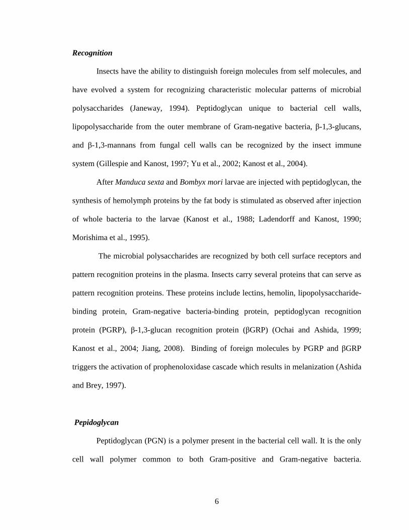
bacteria E. coli (Hartz et al., 1990). In the general peptidoglycan structure, the short
peptide is composed of L-alanine bound to muramic acid, followed by D-glutamic acid,
the γ-carboxyl group of D-glutamic acid is linked to L-diamino acid. And the final alanine
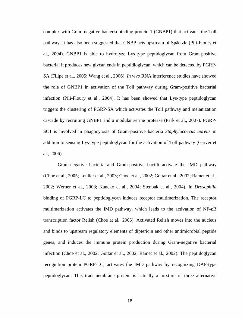
residue is attached to the diamino acid (Fig. 1). A peptide unit forms cross-link from ω-
amino group of the diamino acid of one of the peptide subunit to the D-Ala carboxyl
group of another peptide subunit (Schleifer and Kandler, 1972).
O
HO O
CH3CONH
O
CH2OH
CH2OH
O
O
CH3CONH
CH2
CH2
CO NH CH CO NH CH COOH
CH2
CH2
H2N
CH2
CH COOH
CH3
CH
CO NH CH CO NH CH COOH
CH3
CH3
Figure 1. Structure of peptidoglycan monomer of DAP-type peptidoglycan of E. coli. The monomer contains the N-acetyl glucosamine and N-acetyl muramic acid disaccharide and the tetrapeptide subunit connected to the N-acetyl muramic acid residue (Vollmer et al., 2008). N-acetylmuramoyl-L-alanine amidase cleavage site is marked with an arrow (Royet and Dziarski, 2007).
8
A B
O
HO O
CH3CONH
O
CH2OH
CH2OH
CH3CONHCH
CO
CH3
L-Ala
D- Glu
D-Ala
O
O
n
m DAP D-Ala
D Glu
m DAP
L-Ala
GlcNAc β 1,4 MurNAcn
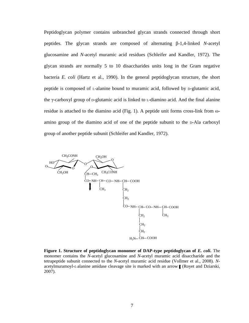
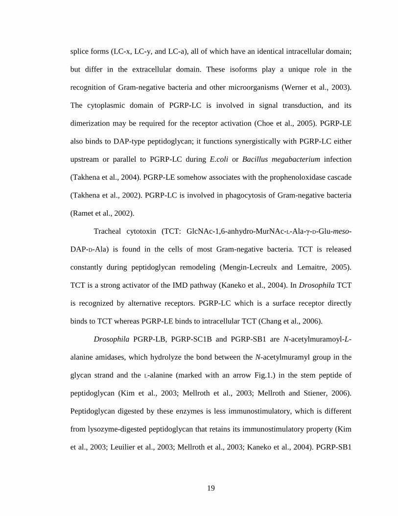
Figure 2. Structures of DAP-type (A) and Lys-type peptidoglycan (B).
The glycan structure in peptidoglycan has a uniform composition in both Gram-
positive and Gram-negative bacteria peptidoglycan (Fig.2). The peptide subunit attached
to the muramic acid residue shows variation due to different amino acid composition. The
muramic acid linking amino acid is usually L-Ala, but in some cases it can be replaced by
Gly or L-Ser. The highest variation in the cross-linking subunit occurs at position three,
where usually diamino acid is present. Most common diamino acid is meso-
diaminopimelic acid (m-Dpm) which is present in probably all Gram-negative bacteria
and Gram-positive bacterial species belonging to Bacillaceae, Lactobacillaceae,
Corynebacteriaceae, and Propionibacteriaceae (Schleifer and Kandler, 1972). L-lysine,
the second most common amino acid is at the third position of the cross-linking peptide
subunit from most other Gram-positive bacteria. Lys-type peptidoglycan is more
heterogeneous, due to the variability of inter-peptide. This bridge can be made up of a
single amino acid residue or of homo-oligopeptides of up to six residues. In
O
HO O
CH3CONH
O
CH2OH
CH2OH
CH3CONHCH
CO
CH3
L-Ala
D- iGln
L- Lys Gly 5
D-Ala
O
O
D-Ala
L-Lys
D- iGln
L-Ala
GlcNAc β 1,4 MurNAcn
n
9
Staphylococcus aureus the interpeptide bridge contains five glycine residues (Schleifer
and Kandler, 1972; Meroueh et al., 2006; Volmer et al., 2008).
In addition to the most common types, DAP- and Lys-type peptidoglycan, there
are other types of peptidoglycan which varies in the third position of the peptide subunit.
These include L-Orn, L, L-Dpm, meso-2, 6-diamino-3-hydroxy-β-pimelic acid (m-hyDpm)
and hydroxy-lysine. In some bacterial species the third position diamino acid is not
involved in cross-linking. In these peptides cross-linking occurs in D-Glu at position 2.
Peptidoglycans can be further modified by amidation of free carboxyl group of D-Glu or
meso-Dpm and less common O-acetylation of N-acetyl muramic acid residue. The
complete resistance to lysozyme by Staphylococcus aureus is due to O-acetylation of
peptidoglycan (Bera et al., 2005; Schleifer and Kandler, 1972).
The compositions of Gram-positive and Gram-negative bacterial cell walls are
different. Major components of Gram-negative bacterial cell wall include
lipopolysaccharide and lipo-protein. The peptidoglycan composition is less than 10% of
the total cell wall contents. In Gram-positive bacterial cell walls the major component is
peptidoglycan, which is about 30-70% of the total cell wall contents. There is high
variation in peptidoglycan composition and structure among Gram-positive bacteria. The
structure of peptidoglycan is constant among Gram-negative bacteria (Schleifer and
Kandler, 1972).
The structure of peptidoglycan brings unique characteristics to bacteria. N-acetyl
muramic acid in the glycan strand of peptidoglycan is a hexose, only present in bacteria.
The presence of D-amino acids is rare in eukaryotic organisms. The alternating D- and L-
amino acids in the peptide segment of peptidoglycan are a unique feature to bacteria.
10
These unique features in bacteria can be utilized by the pattern recognition molecules to
recognize bacteria (Mellroth, 2005).
Peptidoglycan recognition proteins (PGRPs)
Peptidoglycan recognition proteins (PGRPs) are a class of immunity related
proteins involved in microbe recognition. The first PGRP was characterized in the silk-
worm Bombyx mori (Yoshida et al., 1996). PGRPs have been identified in insects,
mollusks, echinoderms, and vertebrates including mammals. PGRPs are absent in plants
and nematodes (Dziarski and Gupta, 2006). PGRPs are expressed in secretory, cytosolic
and transmembrane forms, and all these forms contain at least one carboxyl-terminal
PGRP domain of approximately 165 amino acid residues which is homologous to
lysozyme of bacteriophage T7 (Yoshida et al., 1996; Werner et al., 2000; Liu et al., 2001;
Ochiai and Ashida, 1999; Kang et al., 1998).
Insect PGRPs have been classified to short-form or long-form according to its
length. In short PGRPs a signal peptide is followed by the PGRP domain. The long
PGRPs contain an N-terminal transmembrane with the C-terminal PGRP domain. In
Drosophila PGRP-SA and PGRP-SD are short secretory PGRPs and PGRP-LC is a long
form with a transmembrane domain (Kaneko et al., 2006).
In insects the short PGRPs are secreted into the hemolymph and they are also
present in the cuticle, gut, epidermal cells, and fat-body cells. The short PGRPs are either
constitutively expressed or induced after an immune challenge. The long membrane-
bound PGRPs are mainly expressed in hemocytes. The Drosophila PGRP-LE is a long
PGRP present in the hemolymph. Long PGRP production is induced following a bacterial
11
exposure. They can also be induced by purified peptidoglycan (Kang et al., 1998; Werner
et al., 2000; Ochiai and Ashida, 1999; Dimopoulos et al., 2002; Christophides et al.,
2002).
Two closely spaced cysteine residues in the middle of the PGRP domain which
forms a disulfide bond is important for the function of PGRPs (Dziarski and Gupta,
2006). A mutation in one of the two residues (Cys80Tyr) in Drosphila PGRP-SA
abolishes its ability for activation of the Toll pathway upon binding to Gram-positive
peptidoglycan (Michel et al., 2001). Mutation in one of the cysteine residues in human
PGLYPR-2 (Cys419Ala) leads to complete loss of its amidase activity (Wang et al.,
2003).
Crystal structures of Drosophila PGRP-LB, SA and SD, human PGRP-1α and S
have been reported (Kim et al., 2003; Reise et al., 2004; Guan et al., 2004a; Guan et al.,
2005; Leone et al., 2008). All structures reveal a general fold consisting of several central
β-strands and three peripheral α-helices. These proteins have a conserved peptidoglycan
binding cleft but differ in N-terminal. PGRPs with catalytic activity have an active site
cleft with a Zn2+-binding site. The zinc binding site consists of two histidines, one
tyrosine, and one cysteine residue. In catalytic PGRPs Lys128 in T7 lysozyme was
replaced with a conserved threonine, which is important in Zn2+ binding (Kim et al.,
2003). In PGRPs which does not have the amidase activity, the Cys residue important for
Zn2+ binding is replaced by a serine residue (Mellroth et al., 2003; Wang et al., 2003).
12
Cellular responses
Insects produce several types of hemocytes that protect the insect from invading
microorganisms (Lavine and Strand, 2002). Hemocytes function in recognition,
phagocytosis, melanotic encapsulation and cytotoxicity (Cerenius and Söderhäll, 2004;
Tzou et al., 2002). Hemocytes also respond to external wounds by participating in clot
formation (Lavine and Strand, 2002).
Humoral responses
In insects, the humoral reactions comprise of activation of proteolytic pathways
and induced synthesis of immunity related peptides (Hultmark, 1993; Hoffmann, 1995;
Meister et al., 1997; Gillespie and Kanost, 1997 and Lehrer and Ganz, 1999). Most
commonly produced ones are antibacterial or antifungal peptides that are synthesized
mainly in the fat body (Zhu et al., 2003).
Initiation of proPO activation cascade in Manduca sexta
Upon recognition of foreign molecules, prophenoloxidases (proPOs) are activated
through a regulated serine protease cascade pathway. Recognition of pathogen-associated
molecular patterns (PAMPs) by pattern recognition receptors (PRRs) initiates the
protease cascade that leads to proPO activation and other immune responses (Ashida and
Brey, 1997; Yu et al., 2002). In Manduca sexta several PRRs have been identified which
bind to PAMPs to activate the proPO cascade. These proteins include immulectin-1
(IML1), IML2, β-1,3-glucan recognition protein-1 (βGRP1), βGRP2, and hemolymph
protease 14 (HP14) (Kanost et al., 2004; Ji et al., 2004; Eleftherianos et al., 2006a).
13
Immunolectins are C-type lectins containing two carbohydrate binding domains.
M. sexta IML2 stimulates the proPO cascade by binding to lipopolysaccharide from
Gram-negative bacteria (Yu et al., 2000; Yu et al., 2006). IML1, less specific than IML2,
binds to both Gram-positive and Gram-negative bacteria (Yu et al., 1999). Knockdown of
IML2 by RNAi caused great reduction in host resistance against pathogenic bacteria
Photorhabdus asymbiotica (Eleftherianos et al., 2006a).
β-1,3-glucan recognition protein 1 (βGRP1) and βGRP2 both contain a glucanase-
like domain but lack the enzyme activity. Both proteins bind and agglutinate yeasts and
bacteria. The proPO system is activated by βGRP1 and βGRP2 through binding to
laminarin (Ma and Kanost, 2000; Jiang et al., 2004).
In M. sexta two PGRP cDNA clones have been identified by subtractive
hybridization, which have identical sequences except to a few differences in the signal
peptide (Zhu et al., 2003). The PGRP1 expression is constitutive in naïve larvae and is
induced after a bacterial challenge (Kanost et al., 2004; Yu et al., 2002). Supplementation
of recombinant PGRP1 to larval plasma did not enhance proPO activation after exposure
to Micrococcus luteus (Kanost et al., 2004). Knockdown of PGRP1 by RNAi did not
have any effect on the cellular immune function (Eleftherianos et al, 2007). However,
knocking down PGRP1 expression increased the susceptibility of larvae to P.
asymbiotica (Eleftherianos et al, 2002a and 2002b).
Hemolymph protease-14 (HP14) became active either by binding to a complex of
β-1, 3 glucan and βGRP2 (Wang and Jiang, 2006) or binding to peptidoglycan directly (Ji
et al., 2004). Autoactivation of HP14 initiates the proPO cascade via several proteolytic
steps (Jiang, 2008).
14
ProPO cascade
Activation of proPO cascade in insects is a highly regulated process. Recognition
of PAMPs, such as peptidoglycans and lipopolysaccharides in bacterial cell wall and β-1,
3-glucan in fungal cell wall, by specific recognition proteins triggers the serine protease
pathway (Cerenius et al., 2007). A terminal protease, PAP (proPO activating protease)
cleaves proPO to form active phenoloxidase (PO), and PO catalyzes the melanin
formation (Cerenius and Söderhäll, 2004).
In M. sexta recognition of pathogens leads to the activation of a hemolymph
protease cascade. HP14 precursor gets activated by complexing with β-1,3-glucan bound
to a βGRP2. Active HP14 activates downstream proHP21. HP21 cleaves PAP2 and PAP3
precursors at a specific peptide bond (Jiang, 2008; Gorman et al., 2007). Two serine
protease homologs (SPH1 and SPH2), each containing a protease-like domain at the
carboxyl-terminus lacking an active site serine residue, function as a cofactor for proPO
activation by a PAP (Yu et al., 2003). In M. sexta proPO is activated in the presence of
proPO, PAP and SPHs simultaneously (Gupta et al., 2005).
Melanization is a tightly regulated process because excessive melanin formation
can also be harmful to host tissues and cells. Serine protease inhibitors of the serpin
superfamily play a vital role in regulating melanization, as several critical steps of the
proPO cascade including proteolytic cleavage of proPO are controlled by multiple serpins
(Cerenius et al., 2007). In M. sexta serpin-1J, a variant of serpin-1 gene product inhibits
all three PAPs (Jiang et al., 2003a; Gupta et al., 2005). Serpin-4 and serpin-5 regulate
hemolymph proteases upstream to the PAPs (Tong and Kanost, 2005; Tong et al., 2005).
Serpin-6 inhibits PAP3 in a concentration dependent manner (Wang and Jiang, 2004; Zou
15
and Jiang, 2005) and it also controls HP8 by forming a covalent complex with the
proteases in vivo.
Role of PGRPs in proPO cascade
The role of PGRPs in insect proPO cascade has been studied in Bombyx mori,
Drosophila melanogaster and Tenebrio molitor (Yoshida et al, 1996; Takhena et al.,
2002; Park et al., 2006; Park et al., 2007). The first PGRP was identified in the silkworm,
which acts as an entry point for the proPO activation system. Recognition of DAP-type
peptidoglycan by Drosophila PGRP-LE increases melanization and antimicrobial peptide
expression (Takhena et al., 2002). A soluble form of Lys-type peptidoglycan, which
contains a long glycan chain with a short peptide stem, binds to PGRP-SA and functions
as a potent activator of the T. molitor Toll pathway and proPO cascade. T. molitor PGRP-
SA binds Lys-type peptidoglycan to form clusters, and the clustering is required for
activating proPO cascade. Partial digestion of peptidoglycan by lysozyme appears to
enhance the clustering of TmPGRP-SA around peptidoglycan and recruitment of Gram-
negative bacteria-binding protein (GNBP) and a modular serine protease orthologous to
M. sexta HP14 (Park et al., 2007). Activation of proPO cascade has been further
characterized using a Lys-type peptiodoglycan fragment (T-4P2), which competitively
inhibits melanization stimulated by the natural peptidoglycan. The T-4P2-coupled column
has been used to purify TmPGRP-SA from the hemolymph without activating the proPO
cascade. The purified TmPGRP-SA recognizes both Lys-type and DAP-type
peptidoglycans but the former is a stronger elicitor than the latter in stimulating
melanization (Park et al., 2006).
16
Signaling pathways and transcriptional activation
Activation of Imd and Toll pathways results in the translocation of NF-κB like
factors to the fat body nucleus which induces the transcription of defense related genes
(Brennan et al., 2004).
Toll pathway is mainly activated during fungal and Gram-positive bacterial
infection in Drosophila. Pathway is initiated by proteolytically cleaved form of Späetzle.
Activated Späetzle interacts with the extracellular domain of Toll receptor. The receptor-
Späetzle complex signals to the ankyrin domain protein Cactus, to dissociate from the
NF-κB/Rel protein Dif. Dissociation from its partner causes the exposure of nuclear
localization signal (NLS) on Dif which then translocates into the nucleus to initiate
transcription of antmicrobial genes (Hetru et al., 2003).
IMD pathway is mainly activated during Gram-negative bacterial infection in
Drosophila, which regulates the synthesis of antimicrobial peptides including diptericin,
drosocin, cecropins, and attacins. The IMD pathways stimulate the synthesis of
antimicrobial peptides through relish an NF-κB/Rel protein. Inactive Relish contains
ankyrin repeats that block the NLS (Hoffmann, 2003). After proteolytic cleavage by a
caspase the N-terminal fragment of Drosophila Relish which contain a Rel homology
domain, transloslocates into the nucleus and initiate transcription of immunity related
genes. The C-terminal portion of Relish with ankyrin repeats remains in the cytoplasm
after caspase cleavage (Stoven et al., 2000).
17
Insect Antimicrobial peptides
Synthesis of antimicrobial peptides (AMPs) is an important humoral response in
insects. AMPs are small polypeptides less than 150-200 amino acids. Most AMPs are
cationic at physiological pH due to high percentage of arginine and lysine residues (Bulet
et al., 2004).
In Drosophila seven structurally diverse AMP have been identified (Tzou et al.,
2000). Drosomycins and metchnikowin act as antifungal peptides. Defensin kill Gram-
positive bacteria. Attacins, cecropins, drosocin and diptericins are active against Gram-
negative bacteria. These AMPs function together to inhibit the growth of invading
microorganisms in the hemolymph (Hoffmann, 2003).
Functions of Drosophila PGRPs
Insects PGRPs play important roles in the innate immune system, such as
recognition, signaling and sometimes effectors (Dziarski and Gupta, 2006). In
Drosophila 13 PGRP genes encode approximately 17 PGRP proteins through alternative
splicing (Aggrawal and Silverman, 2007). Drosophila PGRP-SA, PGRP-SD and PGRP-
SC1 activate the Toll pathway by Gram-positive bacteria (Michel et al., 2001; Bischoff et
al., 2004; Garver et al., 2006). Sensing Gram-positive bacteria by these PGRPs leads to
the proteolytic cleavage of Späetzle. The interaction of activated Späetzle with Toll
receptor signals the protein Cactus to dissociate from the NF-κB transcription factor Dif.
Dissociation from its partner and moving into the nucleus leads to the transcription of
drosomycin and the other antimicrobial peptides (Hetru et al., 2003). During Gram-
positive bacterial infection Drosophila PGRP-SA circulating in the hemolymph forms a
18
complex with Gram negative bacteria binding protein 1 (GNBP1) that activates the Toll
pathway. It has also been suggested that GNBP acts upstream of Späetzle (Pili-Floury et
al., 2004). GNBP1 is able to hydrolyze Lys-type peptidoglycan from Gram-positive
bacteria; it produces new glycan ends in peptidioglycan, which can be detected by PGRP-
SA (Filipe et al., 2005; Wang et al., 2006). In vivo RNA interference studies have showed
the role of GNBP1 in activation of the Toll pathway during Gram-positive bacterial
infection (Pili-Floury et al., 2004). It has been showed that Lys-type peptidoglycan
triggers the clustering of PGRP-SA which activates the Toll pathway and melanization
cascade by recruiting GNBP1 and a modular serine protease (Park et al., 2007). PGRP-
SC1 is involved in phagocytosis of Gram-positive bacteria Staphylococcus aureus in
addition to sensing Lys-type peptidoglycan for the activation of Toll pathway (Garver et
al., 2006).
Gram-negative bacteria and Gram-positive bacilli activate the IMD pathway
(Choe et al., 2005; Leulier et al., 2003; Choe et al., 2002; Gottar et al., 2002; Ramet et al.,
2002; Werner et al., 2003; Kaneko et al., 2004; Stenbak et al., 2004). In Drosophila
binding of PGRP-LC to peptidoglycan induces receptor multimerization. The receptor
multimerization activates the IMD pathway, which leads to the activation of NF-κB
transcription factor Relish (Choe at al., 2005). Activated Relish moves into the nucleus
and binds to upstream regulatory elements of diptericin and other antimicrobial peptide
genes, and induces the immune protein production during Gram-negative bacterial
infection (Choe et al., 2002; Gottar et al., 2002; Ramet et al., 2002). The peptidoglycan
recognition protein PGRP-LC, activates the IMD pathway by recognizing DAP-type
peptidoglycan. This transmembrane protein is actually a mixture of three alternative
19
splice forms (LC-x, LC-y, and LC-a), all of which have an identical intracellular domain;
but differ in the extracellular domain. These isoforms play a unique role in the
recognition of Gram-negative bacteria and other microorganisms (Werner et al., 2003).
The cytoplasmic domain of PGRP-LC is involved in signal transduction, and its
dimerization may be required for the receptor activation (Choe et al., 2005). PGRP-LE
also binds to DAP-type peptidoglycan; it functions synergistically with PGRP-LC either
upstream or parallel to PGRP-LC during E.coli or Bacillus megabacterium infection
(Takhena et al., 2004). PGRP-LE somehow associates with the prophenoloxidase cascade
(Takhena et al., 2002). PGRP-LC is involved in phagocytosis of Gram-negative bacteria
(Ramet et al., 2002).
Tracheal cytotoxin (TCT: GlcNAc-1,6-anhydro-MurNAc-L-Ala-γ-D-Glu-meso-
DAP-D-Ala) is found in the cells of most Gram-negative bacteria. TCT is released
constantly during peptidoglycan remodeling (Mengin-Lecreulx and Lemaitre, 2005).
TCT is a strong activator of the IMD pathway (Kaneko et al., 2004). In Drosophila TCT
is recognized by alternative receptors. PGRP-LC which is a surface receptor directly
binds to TCT whereas PGRP-LE binds to intracellular TCT (Chang et al., 2006).
Drosophila PGRP-LB, PGRP-SC1B and PGRP-SB1 are N-acetylmuramoyl-L-
alanine amidases, which hydrolyze the bond between the N-acetylmuramyl group in the
glycan strand and the L-alanine (marked with an arrow Fig.1.) in the stem peptide of
peptidoglycan (Kim et al., 2003; Mellroth et al., 2003; Mellroth and Stiener, 2006).
Peptidoglycan digested by these enzymes is less immunostimulatory, which is different
from lysozyme-digested peptidoglycan that retains its immunostimulatory property (Kim
et al., 2003; Leuilier et al., 2003; Mellroth et al., 2003; Kaneko et al., 2004). PGRP-SB1
20
is an amidase with antibacterial activity prefers DAP-type peptidoglycan, and shows
antibacterial activity against Bacillus megabacterium (Mellroth and Steiner, 2006). The
exact function of catalytic PGRPs in insects is not known. It has been proposed that they
may modulate immune responses by scavenging peptidoglycan or act directly as
antibacterial factors (Mellroth et al., 2003). Drosophila PGRP-LB modulates the IMD
pathway during Gram-negative bacterial infection (Zaidman-Remy et al., 2006).
Drosophila PGRP-SC1 prevents over-activation of the IMD pathway in the gut which is
important to prevent larval death and bacteria-induced developmental defects (Bischoff et
al., 2006).
PGRP-SA has an L, D-carboxypeptidase activity only against DAP-type
peptidoglycans. The Ser158 and His42 residues in the docking groove of PGRP-SA may
be involved in the hydrolytic activity. The carboxyl group of the DAP type peptidoglycan
interact with the docking groove residues. Ser158 in the docking groove is important for
peptidoglycan binding and activation of the Toll pathway (Chang et al., 2004).
PGRPs discriminate Gram-positive and Gram-negative peptidoglycans. The
structural difference between the two peptidoglycan types lies in the presence of lysine
residue or DAP residue at the third position of the peptide stem. These two residues differ
by the presence of a carboxyl-group on their side chain (Fig.2) The carboxy group in
DAP-type peptidoglycan is recognized by a conserved arginine residue in the DAP
recognizing PGRPs. This Arg residue is conserved among PGRPs recognizing DAP-type
peptidoglycan. In PGRP-LE the guanidine group of Arg254 charge balances with the
carboxyl group of DAP-type peptidoglycan (Lim et al., 2006). For PGRPs recognizing
Lys-type peptidoglycan, lysine residue in the third position of the stem peptide is unlikely
21
to serve as the determinant for discrimination. This may be due to the fact: 1) charge
repulsion 2) diverse cross linking patterns are observed in Gram-positive bacterial
peptidoglycan (Lim et al., 2006; Schlefifer and Kandler, 1972). It is speculated that the
inter-peptide bridges of Lys-type peptidoglycan may be the determinant for PGRPs
recognizing Lys-type peptidoglycan (Lim et al., 2006).
Drosophila PGRP-LF is a membrane-bound PGRP which has two extracellular
PGRP domains with different affinity for peptidoglycan. The z-domain shows affinity to
peptidoglycan of Lys, DAP and ornithine-types but the w-domain only binds to DAP-
type peptidoglycan from E.coli. PGRP-LF also plays a regulatory role in the immune
responses (Persson et al., 2007).
PGRPs in other insects
The first insect PGRP was characterized in the silk-worm Bombyx mori (Yoshida
et al., 1996). In the mosquito Anopheles gambiae seven PGRP genes, four long and three
short ones have been identified. Many of the PGRP isoforms are similar in structure to
Drosophia isoforms. For an example, the exon-intron organization of A. gambiae PGRP-
LC gene is identical to that of Drosophila PGRP-LC and produces three different
spliceforms (Christophides et al., 2002).
In the silk-worm (Bombyx mori) and mealworm (Tenebrio molitor) PGRPs are
present in the hemolymph (Yoshida et al., 1996; Park et al., 2006). The silk-worm PGRP
activates the prophenoloxidase cascade in the presence of Lys-type peptidoglycan
(Yoshida et al., 1996). Tenebrio molitor PGRP binds to both Lys-type and DAP-type
peptidoglycan to activate the prophenoloxidase cascade (Park et al., 2006).
22
In the beetle Holotrichia diomphalia PGRP binds to peptidoglycan and the fungal
cell wall component β-1,3 glucan. Interestingly, it was shown that PGRP binds to β-1,3
glucan for the activation of prophenoloxidase cascade and binding of PGRP to
peptidoglycan did not trigger the activation of prophenoloxidase cascade (Lee et al.,
2004).
Mammalian PGRPs
In mammals PGRPs have two main functions: antibacterial activity and amidase
activity (Dziarski and Gupta, 2006). Mammalian PGLYPR-2 is produced by the liver
constitutively and secreted to the blood (Zhang et al., 2005). This is similar to synthesis
of insect PGRPs in the fat body and secretion to the hemolymph (Ochiai and Ashida,
1999; Hashimoto et al., 2007; Werner et al., 2000; Dziarski, 2003). In insects, fat body is
functionally analogous to mammalian liver (Vierstraete et al., 2003; Gutierrez et al.,
2007). Mammalian PGLYPR-2 is also expressed in the intestinal epithelial cells
(Dziarski, 2003), similar to expression of insect PGRPs in the gut (Dziarski, 2003; Ochiai
and Ashida, 1999; Werner et al., 2000).
Different isoforms in Drosophila PGRP-LC are produced by alternative splicing
(Werner et al., 2003). Similarly, in some mammals multiple splice forms of PGLYPR-2
are produced. In pigs two isoforms of PGLYPR-2 show differential expression and
regulation patterns, but both isoforms have N-acetylmuramoyl-L-alanine amidases
activity (Sang et al., 2005).
The crystallographic structures of mammalian PGLYPR-1 and the carboxy
terminal domain of PGLYPR-3 have revealed that these proteins have a ligand binding
23
groove that binds specifically to peptidoglycan (Guan et al., 2004a; Guan et al., 2005).
Insect PGRPs also have ligand binding groove for peptidoglycan binding (Kim et al.,
2003: Reiser et al., 2004; Guan et al., 2004b; Chang et al., 2005; Lim et al., 2006; Chang
et al., 2006).
Some mammalian PGRPs (carboxy terminal of human PGLYPR-3) have a
preference for the binding to Lys-type peptidoglycan over DAP-type peptidoglycan.
However human PGLYPR-1 has a high affinity to DAP-type peptidoglycan over Lys-
type peptidoglycan (Kumar et al., 2005; Swaminathan et al., 2006). Similarly Drosophila
PGRP-LC and PGRP-LE have a preference to DAP-type peptidoglycan over Lys-type
peptidoglycan (Werner et al., 2003).
Most of the mammalian PGRPs function as bactericidal proteins, in difference to
insect PGRPs which very rarely show bactericidal activity. Drosophila PGRP-SB1 shows
antibacterial activity against Bacillus megabacterium (Mellroth and Steiner, 2006).
Mammalian PGLYPR-2 is a Zn2+-dependent N-acetylmuramoyl-L-alanine amidase
similar to Drosophila PGRP-LB, SC1B and SB-1 (Gelius et al., 2003; Wang et al., 2003;
Kim et al., 2003; Mellroth et al., 2003; Mellroth and Stiener, 2006).
24
CHAPTER III
MATERIALS AND METHODS
Insect rearing, bacterial challenge, and hemolymph collection
M. sexta eggs were purchased from Carolina Biological Supply and larvae were
reared on an artificial diet (Dunn and Drake, 1983). Control hemolymph was collected
from cut prolegs of day 2, fifth instar larvae. Day 2, fifth instar larvae were injected with
formaldehyde-killed E. coli (2 x 10⁸ cells/larvae). Hemolymph was collected in the same
way from the larvae 6 h, 12 h, and 24 h after the immune challenge. Hemolymph samples
from the naïve and induced insects were aliquoted and stored at -80˚C.
Detection of PGRP1 in hemolymph
Control and induced hemolymph samples (6, 12, and 24 h after E. coli injection)
were analyzed by mixing 2 µl of hemolymph with 6 µl of 20 mM Tris-Cl, pH 8.0, and 4
µl of 5xSDS sample buffer. After incubation at 95˚C for 5 min, 7 µl of the mixture was
separated by 15% SDS-polyacrylamide gel electrophoresis (SDS-PAGE), transferred to a
nitrocellulose membrane and reacted with 1:2000 diluted PGRP1 polyclonal antiserum
(obtained from Dr. Kanost at Kansas State University). Antibody-antigen complexes
were detected using alkaline phosphatase conjugated to goat-anti-rabbit IgG (Biorad) as
the secondary antibody.
25
RT-PCR analysis
Hemocyte and fat body total RNA samples were prepared from the naïve and
induced, day 3, fifth instar larvae. Total RNA samples were prepared from muscle,
nervous tissue, cuticle, salivary gland, malpighian tubule, and trachea from day 3, fifth
instar naïve larvae. The total RNA was extracted using Micro-to-Midi Total RNA
Purification System (Invitrogen). The RNA sample (2-4 µg), oligo(dT) (0.5 µg), and
dNTPs (1 µl, 10 mM each) were mixed with diethylpyrocarbonate-treated H2O in a final
volume of 12 µl, denatured at 65˚C for 5 min, and quickly chilled on ice for 3 min. M-
MLV reverse transcriptase (1 µl, 200 U/µl, Invitrogen), 5xbuffer (4 µl), 0.1 M
dithiothreitol (2 µl), and RNase OUT (1 µl, 40U/µl, Invitrogen) were added to the
denatured RNA sample (12 µl) for first strand cDNA synthesis at 37˚C for 50 min. The
M. sexta ribosomal protein S3 (rpS3) mRNA was used as an internal control to normalize
the cDNA samples using specific primers j501 (5´-GCCGTTCTTGCCCTGTT-3´) and
j504 (5´CGCGAGTTGACTTCGGT-3´). Primers j297 and j298 (5´-GAACGAAGATCC
GATGTCCAGTC-3´) were used to amplify M. sexta PGRP1 cDNA under conditions
empirically chosen to avoid saturation: 30 cycles of 94˚C, 30 s; 50˚C, 30s; 72˚C, 30s in a
multiplex PCR reaction. The relative levels of PGRP1 mRNA in the normalized samples
were determined by 1.5% agarose gel electrophoresis.
Expression and purification of M. sexta PGRP1 in insect cells
M. sexta PGRP1 cDNA (obtained from Dr. Kanost at Kansas State University)
was amplified using PCR primer j285 (5´-GGAATTCACTGCAACGTCGTC-3´) and
j288 (5´-CTCGAGGTCTTTATATTCGGACAC-3´). The PCR product was T/A cloned
26
into pGEM-T (Promega) and completely verified by DNA sequencing. From the resulting
plasmid a 528 bp EcoRI-XhoI fragment was retrieved by restriction digestion and
directionally inserted to the same sites of pMFH6, a plasmid vector modified from
pFastBac1 (Lu and Jiang, 2007), to generate the recombinant plasmid (PGRP1/pMFH6).
In vivo transposition of the expression cassette, selection of bacterial colonies carrying
the recombinant bacmid, and isolation of the bacmid DNA were performed according to
manufacturer’s protocols (Invitrogen Life Technologies). The initial viral stock (V0) was
obtained by transfecting Spodoptera frugiperda Sf 9 cells with a bacmid-cellFECTIN
mixture, and its titer was improved through serial infections. The V6 viral stock,
containing the highest level of baculovirus, was stored at -70˚C for further experiments.
Sf 9 cells (at 2.0x10⁶ cells/ml) in 100 ml of insect serum-free medium (Invitrogen Life
Technologies) were separately infected with the baculovirus stocks at a multiplicity of
infection of 10 and grown at 27˚C for 96 h with gentle agitation (100 rpm). The cells
were removed by centrifugation at 5,000g for 10 min. Protein purification was carried out
in batches of 100 ml of the conditioned medium. The culture supernatant was mixed with
an equal volume of distilled water at 4˚C for 20 min. After centrifugation at 22,100g for
20 min, the cleared supernatant (~200 ml) was applied to a dextran sulfate (DS)-
Sepharose column (5 ml) (Nakamura et al., 1985) equilibrated with buffer A (10 mM
potassium phosphate, pH 6.4, 1 mM benzamidine). Following a washing step with 25 ml
of buffer A, bound proteins were eluted with a linear gradient of 0-1.0 M NaCl in buffer
A (30 ml). Fractions containing M. sexta PGRP1 were pooled and applied onto a Ni2+
column (1 ml), equilibrated with buffer B (50 mM potassium phosphate, pH 8.0, 300 mM
NaCl, 10 mM imidazole, 0.005% Tween-20). After washing with 5 ml of buffer B, bound
27
proteins were eluted from the column with a linear gradient of 10-100 mM imidazole in
buffer B (20 ml). Finally, tightly bound proteins were eluted with 5 ml of buffer B
containing 250 mM imidazole. All the purification steps were carried out at 4˚C. After
electrophoretic analysis, PGRP1 fractions were combined and concentrated using
Amicon ultracentrifugal 5K MWCO filter device (Millipore). Concentrated protein was
buffer exchanged with 20 mM Tris-Cl, pH 7.5, 50 mM NaCl on the same device and
stored at -80˚C in aliquots.
Purification of insoluble peptidoglycan from Gram-positive bacteria
Bacterial cells were grown in LB medium (2000 ml) overnight at 37˚C with
shaking. The cells, separated from the medium by centrifugation at 2000g, 4˚C for 20
min, were resuspended in 100 ml saline (0.85% NaCl) and heated at 100˚C for 20 min.
The cells were washed twice with saline, once with water, three times with acetone. Each
time 50 ml solution was used for resuspension and removed after centrifugation. The
bacterial cells were dried at 37˚C for 8 h (Rosenthal and Dziarski, 1994). Peptidoglyan
was extracted from the cells by following a modified protocol described by (Tsuchia et al
1996). Dried cells (10 g) were stirred in 300 ml of 10% trichloroacetic acid (TCA) in a
boiling water bath for 20 min. After centrifugation at 10,000g for 30 min, the pellet was
washed with 250 ml water for three times and once with 250 ml of buffer A (100 mM
Tris-HCl, pH 7.5, 20 mM MgCl2 and 1 mM CaCl2). The resuspension in 100 ml buffer A
was incubated with 30 mg of bovine trypsin at 37˚C for 18-24 h with gentle agitation.
After trypsin was inactivated with 1 mM PMSF at 37˚C for 30 min, the treated cells were
centrifuged at 10,000g for 30 min and washed with 200 ml of H2O for ten times. Each
28
time the precipitate was completely resuspended by sonication and, after the final wash,
lyophilized and stored at -20˚C.
Plate assay of PGRP1 binding to soluble peptidoglycan
Soluble peptidoglycans from E. coli or S. aureus (InvivoGen) were used to study
specific binding. The ligand (2 µg) was applied to each well in a 96-well microplate, air
dried overnight at room temperature, and fixed to the well at 60˚C for 30 min. After
blocking with 200 µl of 1 mg/ml BSA in TBS at 37˚C for 2 h and washing with 200 µl
TBS four times (5 min each), PGRP1 (300 ng) in 50 µl TBS containing 0.1 mg/ ml BSA
was added to the wells and incubated at room temperature for 3 h. For competition assay
PGRP1 (300 ng) was first incubated with 200 µg of the ligand at room temperature
before adding to the well for incubation. Following a washing step with TBS, 100 µl of
1:1000 His-5 monoclonal antibody (Bio-Rad) diluted in TBS containing 0.1 mg/ml BSA
was added to the wells and incubated at 37˚C for 2 h. After washing with 200 µl of TBS
for four times, 100 µl of 1:1500 goat- anti-mouse IgG conjugated to alkaline phosphatase
(Bio-Rad) diluted in TBS containing 0.1 mg/ ml BSA was added to the wells and
incubated at 37˚C for 2 h. After washing with TBS four times and 0.5 MgCl2, 10 mM
diethanolamine once, aliquots of 50 µl of p-nitrophernyl phosphate (1.0 mg/ml in 0.5 M
MgCl2, 10 mM diethanolamine) were added to the wells and absorbance at 405 nm was
monitored in the kinetic mode on a VersaMax microplate reader (Molecular Devices).
BSA was used as a negative control.
29
Binding of PGRP1 to insoluble peptidoglycan
One mg of insoluble peptidoglycan was mixed with 10 µl (0.3 µg/µl) of PGRP1
and 40 µl of buffer C (20 mM Tris-HCl, pH 8.0, 20 mM NaCl). After incubation for 2 h
at 4˚C with mixing, the mixture was centrifuged at 16,000g for 15 min. The supernatant
was treated with 5x SDS sample buffer and analyzed as unbound fraction. The pellet was
washed 3 times with 200 µl of buffer C and boiled with 20 µl of 2xSDS for 5 min to
obtain the bound fraction. The unbound (7 µl) and bound (7 µl) samples were separated
by 15% SDS-PAGE followed by immunoblot analysis using 1:2000 diluted His-5
antibody as the first antibody and goat-anti-mouse IgG-conjugated to alkaline
phosphatase as the second antibody.
Binding of PGRP1 to bacterial cells
A single bacterial colony was grown overnight at 37˚C. Overnight bacterial
cultures were subcultured into 4 ml of Luria-Bertani (LB) broth until the OD600 was close
to 0.8. After centrifugation at 1000g and washing with buffer C twice, cells from 4 ml
subculture were resuspended in 40 µl of the same buffer. PGRP1 (10 µl, 0.3 µg/µl) was
added to the cell suspension and incubated for 2 h at 4˚C with mixing. After
centrifugation at 4,000g for 15 min, the supernatant was treated with 5xSDS sample
buffer and analyzed as unbound fraction. The cell pellet was washed 3 times with 200 µl
of buffer C, suspended with 20 µl of 2xSDS buffer, and heated at 95˚C for 5 min to
obtain the bound fraction. The unbound (7 µl) and bound (7 µl) protein fractions were
analyzed as described above.
30
Role of PGRP1 in proPO activation in hemolymph from naïve M. sexta larvae
Hemolymph from day 3, fifth instar naïve larvae was centrifuged at 500g for 5
min at 4˚C to remove hemocytes. The plasma was diluted ten times with buffer D (20
mM Tris-HCl pH 8.0, 1 mM CaCl2, 0.001% Tween-20). Five µl of diluted sample was
mixed with 19 µl of buffer D and incubated at 25˚C for 10 min. PO activity was
determined using dopamine as a substrate on a microplate reader (Jiang et al., 2003a).
Control hemolymph with low PO activity was selected for the study. Hemolymph was
stored in 10 µl aliquots at -80 ˚C.
Five µl of diluted plasma was incubated with purified recombinant PGRP1 (1 µl,
0.2 µg/µl) and different elicitors separately to find out which ones trigger PGRP1-
enhanced proPO activation. Micrococcus luteus, Staphylococcus auerus, Bacilus
megabacterium, and Bacillus subtilis insoluble peptidoglycans (2 µl, 1 mg/ml), S. auerus
and E. coli soluble peptidoglycans (1 µl, 1 mg/ml), curdlan (1 µl, 10 mg/ml), M. luteus, S.
auerus, B. megabacterium, B. subtilis and E. coli cells (1 µl, 2 x 10⁵ cells) were tested.
The controls were mixtures of diluted hemolymph with buffer, elicitor or PGRP1. The
total volume of the test and control mixtures was adjusted to 24 µl with buffer D and
incubated at 25˚C for 1 h. PO activity was determined using dopamine as a substrate on a
microplate reader (Jiang et al., 2003b).
Antibacterial activity assay
Recombinant PGRP1 was tested against Gram-negative bacteria S. typhimurium,
K. pneumonia, P. aeruginosa and E. coli and Gram-positive bacteria M. luteus, S. aureus
and B. megabacterium. Single bacterial colonies were grown overnight and subcultured
31
in 4 ml of Trypticase Soy Broth (TSB) for 3-5 h until the bacteria reached mid-log phase.
After centrifugation at 1000g at 4˚C and washing with 10 mM Tris-HCl, pH 8.0, the cells
were suspended in 5% TSB to 5x10⁵ cfu/ml. Aliquots of the diluted cultures (90 µl) were
mixed with 10 µl of recombinant PGRP1 (1.2 µg) or 10 mM Tris-HCl, pH 8.0, and
cultured at 37˚C for 8 h in a 96-well cell culture plate. Optical density at 600 nm was
recorded after 8 h incubation for comparison between the treatment and control groups.
32
CHAPTER IV
RESULTS AND DISCUSSION
Sequence analysis of PGRP1
M. sexta PGRP1 was initially identified as a pattern recognition receptor in
plasma (Yu et al., 2002; Zhu et al., 2003). The open reading frame encodes a 192-residue
protein sequence including a 21-residue signal peptide (Fig. 3). No potential N- or O-
linked glycosylation sites are present in the sequence. A BLAST search of mature PGRP1
indicated it is 71%, 69%, 64% and 58% identical to PGRPs from the lepidopteran insects
Antheraea mylitta (Gandhe et al., 2006), Samia cynthia ricini (Onoe at al., 2007),
Trichoplusia ni (Kang et al., 1998), and Bombyx mori (Ochiai and Ashida, 1999)
respectively. In PGRP1 the conserved PGRP domain is located between residues 22-164.
Sequence alignment of M. sexta PGRP1 with other insect PGRPs revealed that M.
sexta PGRP1 has a serine residue in the position equivalent to Cys130 in T7 lysozyme,
which is a common feature of all receptor-type PGRPs. (Fig. 4). This strongly suggests
that PGRP1 is not an amidase (Mellroth et al., 2003). (In M. sexta PGRP2 and PGRP3,
however, Cys is conserved in the position corresponding to Cys130 in T7 lysozyme,
suggesting that these two proteins are amidases). In addition, Ser100 in M. sexta PGRP1 is
equivalent in position to Arg254 of Drosophila PGRP-LE, which interacts with the
33
carboxyl group of DAP-type peptidoglycans (Lim et al., 2006). This Arg residue is
conserved in all known Drosophila and human PGRPs that recognize DAP-type
peptidoglycans, but not always found in PGRPs that bind Lys-type peptidoglycans (Onoe
et al., 2007). Arg254 of Drosophila PGRP-LE corresponds to Arg110 and Arg115 of M.
sexta PGRP2 and PGRP3, respectively.
Detection of PGRP1 in M. sexta larval hemolymph
M. sexta PGRP1 was detected in plasma by immunoblot analysis using PGRP1
antibodies. Present at a low level in the naïve plasma, it significantly increased in a time-
dependent manner after an immune challenge with E. coli (Fig. 5). This is consistent
with the data on PGRP1 concentration change after a bacterial challenge (Yu et al.,
2002). PGRPs have been detected in plasma of the silkworm (Yoshida et al., 1996), the
beetle Holotrichia diomphalia (Lee et al., 2004), and the mealworm Tenebrio molitor
(Park et al., 2006). In Drosophila PGRP-SA, PGRP-LE and PGRP-LB are secreted into
plasma (Werner et al., 2000; Takhena et al., 2004; Zaidman-Remy et al., 2006).
Inducibility of PGRP1
Semi-quantitative RT-PCR was performed to examine the inducibility of PGRP1
in fat body and hemocytes. M. sexta ribosomal protein S3 (rpS3) transcripts were used as
an internal control to normalize the cDNA templates. Relative band intensities indicated
that PGRP1 was constitutively expressed in the fat body; its level became greatly
abundant after an immune challenge (Fig. 6). PGRP1 gene was weakly expressed in
34
hemocytes from naïve larvae, and its mRNA level also largely increased after the
immune challenge.
These results are consistent with the previous report that M. sexta PGRP1 gene is
constitutively expressed at a low level in fat body of naïve insects and its mRNA level
increased after an immune challenge with E. coli, M. luteus, Photorhabdus luminescens
or P. asymbiotica (Yu et al., 2002; Eleftherianosa et al, 2006b; Zhu et al., 2003;
Eleftherianosa et al., 2006a). Other pattern recognition proteins (e.g. M. sexta βGRP1,
IML2, and IML4) showed similar expression patterns (Ma and Kanost, 2000; Yu et al.,
2000 and 2006). M. sexta IML1, hemolin and βGRP2 have a different expression pattern;
they are produced in the fat body only after an immune challenge (Yu et al., 1999; Wang
et al., 2005; Jiang et al., 2004). Hemolin is also expressed in hemocytes and its
transcription increased after E. coli injection (Eleftherianos et al., 2007).
Other lepidopteran insects show similar expression patterns for PGRPs. In
Bombyx mori PGRP1 is constitutively expressed in hemocytes and fat body. Its mRNA
became more abundant after a bacterial challenge (Ochiai and Ashida, 1999). The
expression of PGRP-A in the wild silkworm Samia cynthia ricini exhibits the same
pattern in fat body and hemocytes (Onoe et al., 2007). S. cynthia PGRP-C and PGRP-D
transcripts were absent in the naïve larvae, but became much higher in fat body after an
immune challenge (Hashimoto et al., 2007).
The inducibility and expression pattern of Drosophila PGRPs have been studied.
PGRP-SA mRNA is present in the fat body of naïve larvae, but the level did not increase
after a bacterial challenge. Drosophila PGRP-SB1 and PGRP-SD are mainly expressed in
35
fat body of induced larvae, whereas PGRP-LA, LC and LD mRNA are present in
hemocytes of naïve larvae (Werner et al., 2000).
Expression of PGRP1 in different tissues
Expression of M. sexta PGRP1 was studied in the following tissues: malpighian
tubule, cuticle, nerve tissue, salivary gland, trachea, muscle, midgut, fat body and
hemocytes from naïve larvae by semi-quantitative RT-PCR. PGRP1 was expressed in all
tissues tested and transcript levels in fat body and cuticle were higher than the other
tissues (Fig. 7). Tissue-specific expression of PGRP has been studied in other insects. B.
mori PGRP1 is expressed in fat body, hemocytes, and epidermal cells, but not in
malpighian tubules, silk gland, or midgut of naïve larvae (Ochiai and Ashida, 1999). S.
cynthia ricini PGRP-B is constitutively expressed in midgut at a high level in naïve
larvae (Hashimoto et al., 2007). In Drosophila, PGRP-SC1 and -SC2 are constitutively
transcribed in gut; PGRP-LE is also a constitutively expressed but only weakly in gut,
hemocytes, and carcass which include all the epidermal layers. PGRP-SA is expressed in
fat body and epidermis of naïve larvae (Werner et al., 2000).
Expression and purification of PGRP1 from insect cells
The protein was expressed using the baculovirus expression system. PGRP1 cDNA was
cloned to pMHF6 which contains honeybee melittin signal peptide-coding region. The
honeybee melittin signal peptide increases the secretion of recombinant proteins (Jarvis et
al., 1993). The carboxy-terminal hexahistidine tag facilitates the purification of
recombinant protein by affinity chromatography.
36
The recombinant protein was soluble and secreted into the cell culture medium.
After removing the cells from the culture the protein was captured by ion exchange
chromatography and eluted from the Dextan Sulfate (DS) column in a small volume. The
PGRP1 fractions were affinity purified by Ni-NTA agarose column. The protein was
eluted with a linear gradient of imidazole. Fig. 8.A illustrates the purification procedure
with comassie blue staning. The immunoblots (Fig. 8.B and 8.C) illustrates the
purification procedure using anti-His-5 anibody and PGRP1 antibody respectively.
Coomassie blue staining analysis following SDS-polyacrylamide gel electrophoresis
indicated that the affinity purified protein was essentially pure (Fig. 8.A). Recombinant
PGRP1 run as a 19 kDa band under reducing conditions (Fig. 8.A, 8.B and 8.C).
Binding of PGRP1 to peptidoglycan In vitro binding assays of M. sexta PGRP1 showed, it binds to purified Lys-type
peptidoglycan from M. luteus, DAP-type soluble peptidoglycan from E. coli and
amidated DAP-type peptidoglycan from Bacillus subtilis and Bacillus megabacterium.
Recombinant PGRP1 did not show any binding to insoluble and soluble Lys-type
peptidoglycan from S. aureus. The specific binding of recombinant PGRP1 to E. coli
(DAP-type) soluble peptidoglycan was confirmed by using a competition assay with
ELISA (Fig.9.A). PGRP1 did not show any specific binding to soluble S. aureus (Lys-
type) soluble peptidoglycan (Fig.9.B). Complete binding of PGRP1 was observed to
insoluble peptidoglycan from the bacterial strains M. luteus (Fig.10.A), B.
megabacterium (Fig.12.A) and B. subtilis (Fig.13.A). Consistent with ELISA results
37
recombinant PGRP1 did not show any binding to insoluble peptidoglycan from S. aureus
(Fig.11.A).
Both mammalian and insect PGRPs have conserved residues for peptidoglycan
binding. Sixteen residues have been identified as ligand contacting residues. These
residues are invariant in both insect and mammalian PGRPs (Guan et al., 2004b). Five
residues which are highly conserved in the ligand binding groove have been identified,
these residues in human PGRP-1αC are His-208, His-231, Tyr-242, His-264, and Asn-
269. In M. sexta PGRP1 His-52, His-75, Ser-86, Pro-108 and Asn-113 are present in the
corresponding positions. These conserved residues form a nearly contiguous patch on the
floor of binding groove (Guan et al., 2004b). Highly variable residues in the
peptidoglycan-binding groove are important in discriminating between Lys- and DAP-
type peptidoglycan. The crystal structure of human PGRP-1αC has revealed the Asn-236
and Phe-237 are involved in forming large number of van der Waals bonds with the side
chain of lysine (Guan et al., 2004b). Variability of sequences at these two positions may
be important for the capability of some PGRPs to discriminate between Lys- and DAP-
type peptidoglycan (Michel et al., 2001; Choe et al., 2002; Gottar et al., 2002; Leulier et
al., 2003; Werner et al., 2003; Kaneko et al., 2004). In Drosophila PGRP-SA which
recognizes Lys-type peptidoglycan Asp-96-Phe-97 are the corresponding residues to
Asn-236 and Phe-237 of human PGRP-1αC. In contrast to PGRPs recognizing Lys-type
peptidoglycan in Drosophila, PGRP-LCx and LE which recognize DAP-type
peptidoglycan, Gly-Trp are present at the corresponding position (Hoffmann et al., 2003;
Kaneko et al., 2004).
38
In M. sexta PGRP1 Asn-80-Tyr-81 are in the corresponding position. In the
lepidopteran Trichoplusia ni the same residues are found at the corresponding positions.
PGRPs from both species recognize both Lys- and DAP-type peptidoglycan (Kang et al.,
1998). It has been suggested Arg254 in Drosophila PGRP-LE interacts with the carboxyl
group of DAP-type peptidoglycan (Lim et al., 2006). This arginine residue is conserved
in Drosophila and human PGRPs which recognize DAP-type peptidoglycan, but not
always present in PGRPs that recognize Lys-type peptidoglycan (Onoe et al., 2007). M.
sexta PGRP1 has a serine residue at the corresponding position of Arg254 in Drosophila
PGRP-LE (marked by a filled arrowhead in Fig. 4). Similar to M. sexta PGRP1 in the
lepidopteran Samia cynthia ricini a serine residue is found at the corresponding position.
PGRPs from both species recognize both Lys and DAP-type peptidoglycan (Onoe et al.,
2007).
Binding of PGRP to peptidoglycan in vitro has been studied in lepidopteran
insects. Bombyx mori PGRP purified from hemolymph binds to M. luteus peptidoglycan
(Yoshida et al., 1996). PGRP-A from Samia cynthia ricini binds to M. luteus (Lys-type)
and B. licheniformis (DAP-type) peptidoglycans (Onoe et al., 2007). Several Drosophila
PGRPs bind to both Lys- and DAP-type peptidoglycans include PGRP-LB (Kim et al.,
2003), PGRP-SC1B (Mellroth et al., 2003), PGRP-SA (Chang et al., 2004), PGRP-LCx
(Mellroth et al., 2005) and PGRP-LF (Persson et al., 2007).
It has been suggested that in lepidopteran insects the DAP or Lysine residue at the
third position of the stem peptide may be not critical in recognition of peptidoglycan, but
the structure of cross-linking peptide is important for recognition of peptidoglycan (Onoe
et al., 2007). This is different to selective recognition of Lys- and DAP-type
39
peptidoglycans by Drosophila and human PGRPs (Leuilier et al., 2003; Swaminathan et
al., 2006; Kumar et al., 2005).
Recombinant PGRP1 did not show any binding to either soluble or insoluble S.
aureus peptidoglycan (Fig.9.B and 11.A). This result is supported by the observation that
M. sexta PGRP1 in induced hemolymph does not bind to S. aureus cells (Ragan, 2008).
When recombinant PGRP1 was incubated with S. aureus cells a partial binding was
observed (Fig.11.B). The partial binding of PGRP1 to S. aureus bacterial cells may be
due to the homogenous environment in the medium during the binding study, compared
to the induced hemolymph which is a complex system with a heterogeneous environment
in which no binding of PGRP to S. aureus cells has been observed. The binding
properties coincide with the proPO activation results of recombinant PGRP1 not
triggering proPO activation in control hemolymph by binding to S. aureus live cells,
insoluble peptidoglycan and soluble peptidoglycan (Fig.16 and 17). Some insect PGRPs
Holotrichia diomphalia (Lee et al., 2004) Drosophila PGRPs SC1B, LB and LF
(Mellroth et al., 2003; Kim et al., 2003; Persson et al., 2007) have been shown binding to
S. aureus peptidoglycan.
When recombinant PGRP1 was incubated with M. luteus (Fig. 10.B), B.
megabacterium (Fig.12.B) and B. subtilis (Fig.13.B) cells, only partial binding was
observed although a complete binding was observed when purified insoluble
peptidoglycan was used from these strains. Recombinant PGRP1 showing higher affinity
to peptidoglycan may be due to the less complexity and high accessibility of
peptidoglycan compared to cells. This hypothesis is supported by the information from
Samia synthia ricini PGRP-SA which binds to uncross-linked Lys-type peptidoglycan
40
from M. luteus with a high affinity compared to cross-linked peptidoglycan (Onoe et al.,
2007).
ProPO activation
Role of PGRP1 in proPO activation cascade was studied using M. sexta control
hemolymph. Control hemolymph was incubated with different elicitors and recombinant
PGRP1 separately to find which binding triggers proPO activation. When recombinant
PGRP1 was added to the control hemolymph without a bacterial elicitor there was an
increase in proPO activation (Fig.14). High PGRP1 concentration produced high proPO
activity. A sigmoidal curve which is typical for biological processes was obtained when
proPO activity was plotted against the PGRP1 concentration. There was a greater
increase of proPO activity when PGRP1 was added to the control hemolymph, and with
the increase of PGRP1 concentration, proPO activity was increased at a lesser rate and it
slowly reaches the asymptote. An increase in melanization has been observed in the
absence of microorganisms when Drosophila PGRP-LE was expressed in a higher level.
PGRP-LE is constitutively present in the hemolymph and is probably involved in the first
line of self-defense by recognizing pathogens and transmission of the signals to
downstream effectors involved in defense reactions (Takhena et al., 2002). Similar to
Drosophila PGRP-LE M. sexta PGRP1 is a constitutive protein in the hemolymph and it
is probably involved in first line of self-defense against invading pathogens. M. sexta
IML2 which function as pattern recognition molecule enhances melanization (Yu and
Kanost, 2004; Ling and Yu, 2006).
41
When bacterial cells were used as elicitors there was no increase in proPO
activation by binding PGRP1 to bacterial cells (Fig.15.B, 16.B, 18.B, 19.B, 20.B) .This
was consistent with the reported data that there is no increase in proPO activaton when
recombinant PGRP1 was incubated with M. luteus cells (Kanost et al., 2004). This may
be due to complex structure of cell wall, compared to purified peptidoglycan. Activation
of proPO cascade by peptidoglycan but not by bacterial cells coincides with the results of
binding studies. The binding was complete with insoluble peptidoglycan but was partial
with bacterial cells. There was increase in proPO activation when PGRP1 was incubated
with sonicated peptidoglycan from M. luteus and B. megabacterium and soluble
peptidoglycan from E. coli (Fig.15.A, 18.A, 20.A). PGRP purified from the hemolymph
of Bombyx mori activates proPO cascade by binding M. luteus peptidoglycan (Yoshida et
al., 1996). Drosophila PGRP-LC triggers the IMD pathway in the presence of lightly
cross-linked (25%) peptidoglycan, but PGRP-LC does not activate the IMD pathway with
heavily cross-linked peptidoglycan (75%). This result supports the fact that PGRPs sense
degree of cross-linking in peptidoglycan (Kaneko et al., 2005).
It has been reported melanization in Drosophila and Galleria has increased in the
presence of peptidoglycan (Bidla et al., 2008). When M. sexta control hemolymph was
incubated with sonicated peptidoglycan there was no increase in proPO activation, but it
was increased when sonicated peptidoglycan was incubated with recombinant PGRP1.
Sonication of peptidoglycan might increase accessibility to peptidoglycan. This
hypothesis is supported by the fact that Samia cynthia ricini PGRP-A binds to less
complex uncross-linked M. luteus peptidoglycan with high affinity than to cross-linked
peptidoglycan (Onoe et al., 2007). It has been reported when sonicated peptidoglycan
42
was added to the silk worm plasma there was an increase in proPO activation (Tsuchiya
et al., 1996) but this was not observed in M. sexta control hemolymph incubated with
peptidoglycan.
PGRP1 increased proPO activation by binding to both Lys-type peptidoglycan
from M. luteus and DAP-type peptidoglycan from B. megabacterium and E. coli.
Similarly PGRP from army meal worm Tenebrio molitor activates proPO cascade by
binding to both Lys- and DAP-type peptidoglycan (Park et al., 2006). PGRP-LE activates
proPO cascade in Drosophila larvae by binding DAP-type peptidoglycan (Takhena et al.,
2002; Takhena et al., 2004).
Incubation of recombinant M. sexta PGRP1 with S. aureus cells, insoluble and
soluble peptidoglycan did not increase the proPO activation (Fig.16 and 18). There was
lowering in PO activity when S. aureus cells and insoluble peptidoglycan were incubated
with PGRP1 compared to controls with PGRP1 without S. aureus cells or insoluble
peptidoglycan. Binding experiments showed that recombinant PGRP1 does not bind to S.
aureus peptidoglycan. The binding characteristic coincides with the proPO activation of
PGRP protein with peptidoglycan. The lowering of proPO activation by S. aureus cells
and peptidoglycan may be due to the complex peptidoglycan structure down regulating
the proPO cascade. When soluble S. aureus peptidoglycan was incubated with PGRP1 no
lowering effect was observed (Fig.17). This is due to the fact peptidoglycan is not
complexly cross-linked in the soluble form of peptidoglycan. The cell wall of S. aureus is
very complex with high degree of cross-linking and it is resistant to lysozyme (Dmitriev
et al., 2004; Bera et al., 2005). S. aureus cells might be using this complex cell wall
structure as a resistance mechanism to host immune responses. Drosophila IMD pathway
43
is activated by M. luteus peptidoglycan, but not S. aureus (Kaneko et al., 2005). S. aureus
peptidoglycan is highly cross-linked with 5-glycine cross-bridge, whereas M. luteus uses
a branched pentapeptide (identical to its stem-peptide) for the cross-bridge (Shokman et
al., 1983).
When Drosophila and silk worm are infected with live S. aureus bacterial cells
the insects die within a few days (Needham et al., 2004; Kaito et al., 2002). In silk worm
larvae infected with S. aureus, it shows a systemic infection of S. aureus bacterial cells
proliferating in blood, tissues and epithelial surface of the mid gut (Kaito et al., 2002).
These results suggest that the insect immune system does not show strong immune
responses against S. aureus infection. In Anophels gambiae it has been shown
peptidoglycan from S. aureus down regulate PGRPS3 (Christophides et al., 2002). In
Bombyx mori PGRP genes are weakly induced in the fat body after injection with S.
aureus compared to E. coli and B. subtilis infection and some PGRP genes are not
induced with S. aureus peptidoglycan (Tanaka et al., 2008). But it has been shown that
soluble S. aureus peptidoglycan is able to stimulate the proPO cascade in silk worm and
Tenebrio molitor (Kaneko et al., 2004).
PGRP1 did not increase proPO activation by binding to B. subtilis peptidoglycan
(Fig.19.A) although the protein binds strongly to peptidoglycan from B. subtilis. Both B.
subtilis cells and peptidoglycan function as weak elicitors in M. sexta (observed results).
This may be due to the fact that B. subtilis is a non pathogenic microorganism to insects
(Needham et al., 2004).
There was an increase in proPO activation by binding recombinant PGRP1 to
DAP-type soluble peptidoglycan from E. coli (Fig.20.A). This is consistent with the
44
specific binding results by ELISA. There was no increase in proPO activation by PGRP1
by binding to E. coli cells (Fig.20.B). This may due to the fact that soluble peptidoglycan
is more accessible to PGRP1 compared to bacterial cells, and also the fact that in E. coli
the peptidoglycan layer in the cell wall is not directly exposed to the surface.
In the beetle Holotrichia diomphalia PGRP binds to β-1,3-glucan for the
activation of prophenoloxidase cascade (Lee et al., 2004). The ability of M. sexta PGRP1
to activate proPO cascade by binding to curdlan (soluble β-1,3 glucan) was tested, but
there was no increase in proPO activation by binding PGRP1 to curdlan was observed
(Fig.21).
Antibacterial activity of PGRP1
Recombinant PGRP1 did not show any antibacterial activity against any of the
Gram-positive and Gram-negative bacterial strains tested (Table.4). Only insect PGRP
which has shown bactericidal activity is PGRP-SB1 from Drosophila. PGRP-SB1 shows
antibacterial activity against Bacillus megabacterium (Mellroth and Steiner, 2006). In
contrast to insect PGRPs, most mammalian PGRPs are bactericidal (Liu et al., 2000; Lu
et al., 2006).
45
CHAPTER V
CONCLUSION
The in vitro biochemical analysis of M. sexta recombinant PGRP1 showed that it
functions as a pattern recognition protein which triggers the humoral cascade of proteases
leading to the activation of PPO pathway upon binding to bacterial peptidoglycan. M.
sexta PGRP1 functions as a common recognition molecule for Lys- and DAP-type
peptidoglycan (Table.1).
There was a strong relationship between binding and PGRP1 mediated PPO
activation (Table.2 and 3). PGRP1 did not show a complete binding to bacterial cells and
when bacterial cells were incubated with recombinant PGRP1 in the control hemolymph
there was no PGRP1 mediated PPO activation (Table.2). PGRP1 showed complete
binding with bacterial peptidoglycan (except S. aureus peptidoglycan) and consistent
with this binding, an increase in PPO activation was seen when recombinant PGRP1 was
incubated with bacterial peptidoglycan (Table. 2 and 3).
These results illustrate that PGRP1 recognize bacterial peptidoglycan more
effectively than bacterial cells. When bacterial cells are inside the insect hemolymph they
can be processed by the lysozymes, catalytic PGRPs which have amidase activity and by
phagocytosis. Hultmark and Borge-Renberg have proposed a mechanism for processing
of bacteria for the recognition by recognition protein. It has suggested the bacteria can be
46
directly identified by hemocytes which are involved in phagocytosis. Hemocytes have an
efficient mechanism for the export of digestion products, such as peptidoglycan
fragments, from phagocytosed microorganisms (Hultmark and Borge-Renberg, 2007).
This processed peptidoglycan can be recognized by PGRPs to activate the PPO activation
and the signaling pathways which regulate the synthesis of antimicrobial peptides.
The inducibility, constitutive expression and role of PGRP1 in the PPO activation
suggest that this recognition protein plays an important role in the M. sexta immune
system.
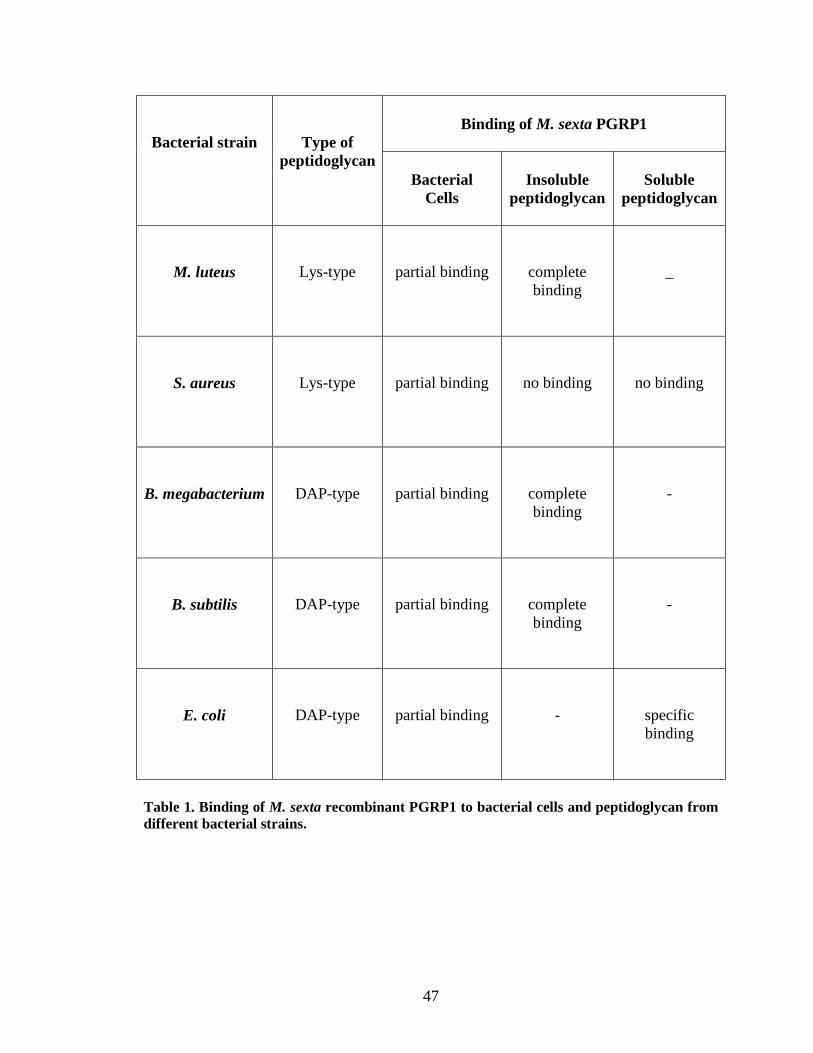
47
Bacterial strain
Type of peptidoglycan
Binding of M. sexta PGRP1
Bacterial Cells
Insoluble
peptidoglycan
Soluble
peptidoglycan
M. luteus
Lys-type
partial binding
complete binding
_
S. aureus
Lys-type
partial binding
no binding
no binding
B. megabacterium
DAP-type
partial binding
complete binding
-
B. subtilis
DAP-type
partial binding
complete binding
-
E. coli
DAP-type
partial binding
-
specific binding
Table 1. Binding of M. sexta recombinant PGRP1 to bacterial cells and peptidoglycan from different bacterial strains.
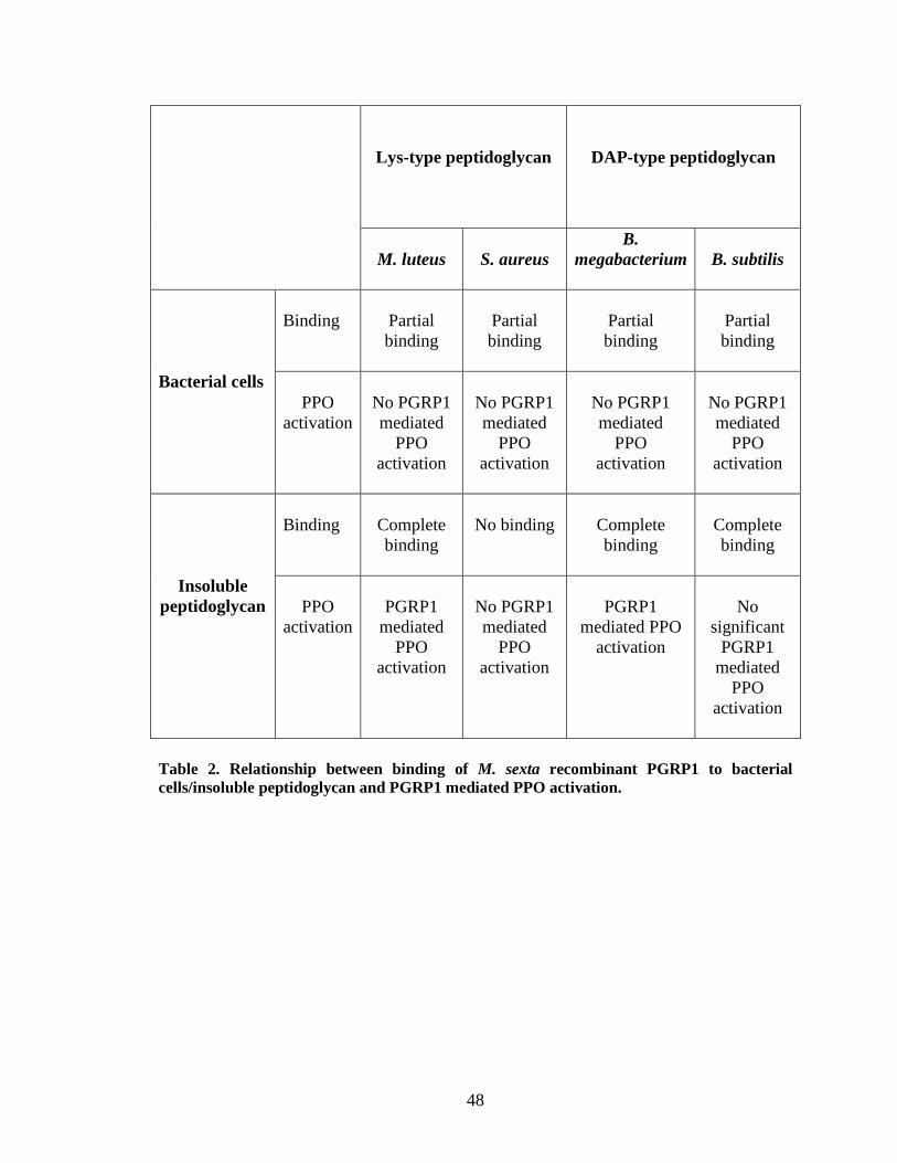
48
Lys-type peptidoglycan
DAP-type peptidoglycan
M. luteus
S. aureus
B. megabacterium
B. subtilis
Bacterial cells
Binding
Partial binding
Partial binding
Partial binding
Partial binding
PPO
activation
No PGRP1 mediated
PPO activation
No PGRP1 mediated
PPO activation
No PGRP1 mediated
PPO activation
No PGRP1 mediated
PPO activation
Insoluble peptidoglycan
Binding
Complete binding
No binding
Complete binding
Complete binding
PPO
activation
PGRP1
mediated PPO
activation
No PGRP1 mediated
PPO activation
PGRP1
mediated PPO activation
No
significant PGRP1
mediated PPO
activation
Table 2. Relationship between binding of M. sexta recombinant PGRP1 to bacterial cells/insoluble peptidoglycan and PGRP1 mediated PPO activation.
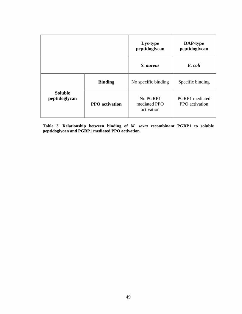
49
Lys-type peptidoglycan
DAP-type
peptidoglycan
S. aureus
E. coli
Soluble peptidoglycan
Binding
No specific binding
Specific binding
PPO activation
No PGRP1
mediated PPO activation
PGRP1 mediated PPO activation
Table 3. Relationship between binding of M. sexta recombinant PGRP1 to soluble peptidoglycan and PGRP1 mediated PPO activation.
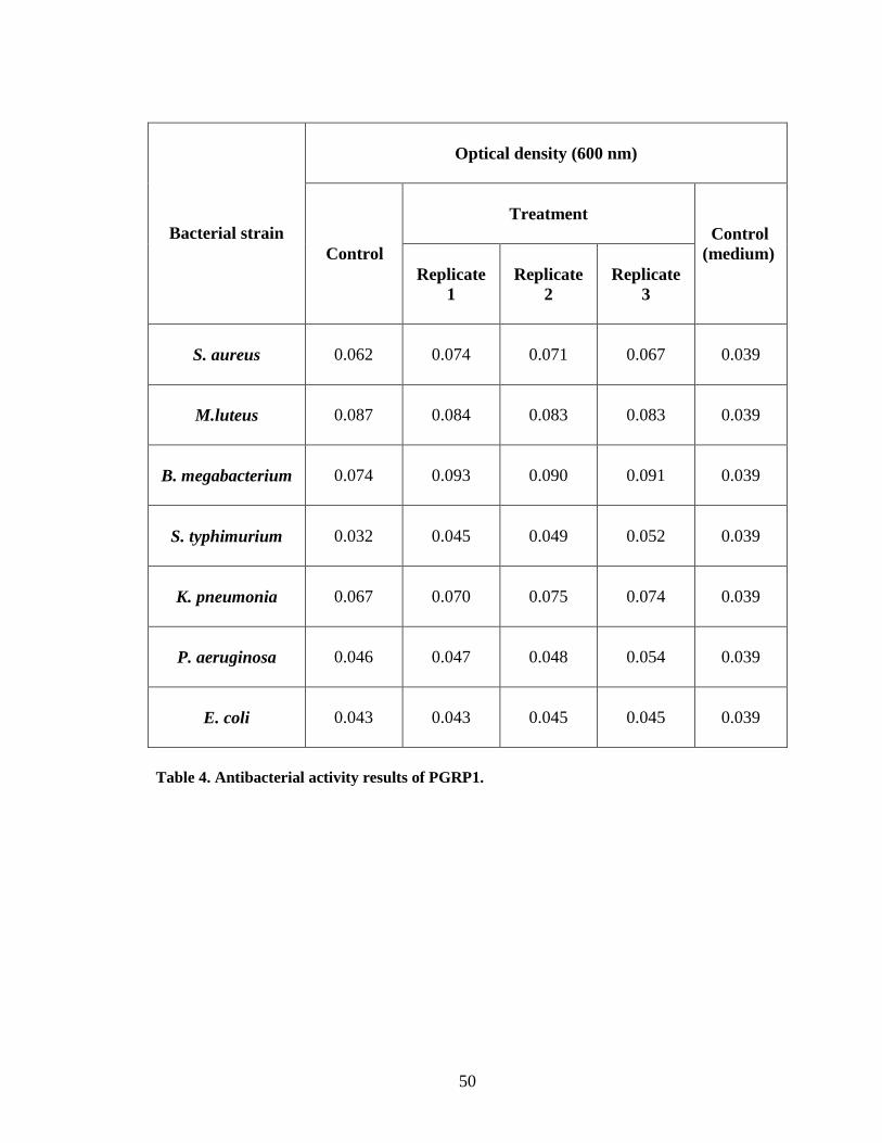
50
Bacterial strain
Optical density (600 nm)
Control
Treatment
Control (medium)
Replicate 1
Replicate
2
Replicate
3
S. aureus
0.062
0.074
0.071
0.067
0.039
M.luteus
0.087
0.084
0.083
0.083
0.039
B. megabacterium
0.074
0.093
0.090
0.091
0.039
S. typhimurium
0.032
0.045
0.049
0.052
0.039
K. pneumonia
0.067
0.070
0.075
0.074
0.039
P. aeruginosa
0.046
0.047
0.048
0.054
0.039
E. coli
0.043
0.043
0.045
0.045
0.039
Table 4. Antibacterial activity results of PGRP1.
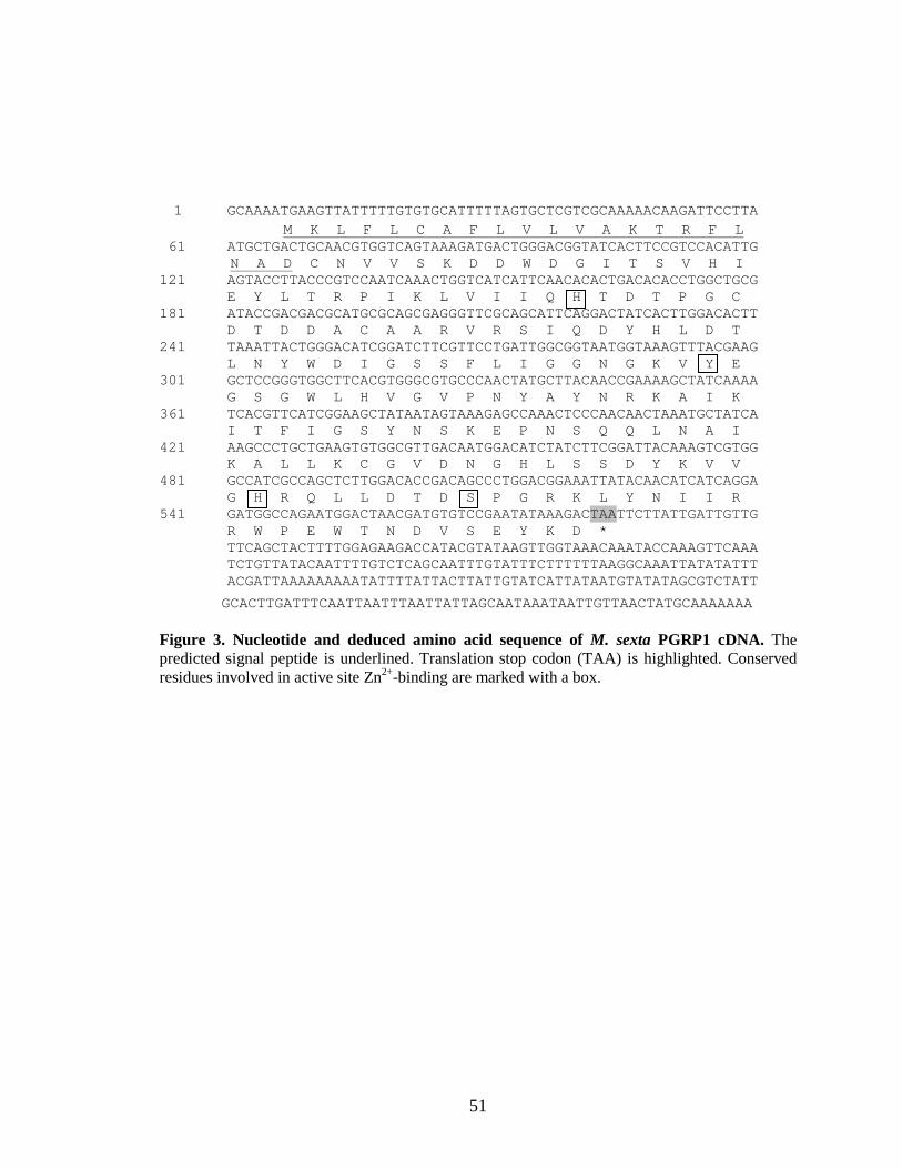
51
1 GCAAAATGAAGTTATTTTTGTGTGCATTTTTAGTGCTCGTCGCAAAAACAAGATTCCTTA
M K L F L C A F L V L V A K T R F L 61 ATGCTGACTGCAACGTGGTCAGTAAAGATGACTGGGACGGTATCACTTCCGTCCACATTG N A D C N V V S K D D W D G I T S V H I 121 AGTACCTTACCCGTCCAATCAAACTGGTCATCATTCAACACACTGACACACCTGGCTGCG E Y L T R P I K L V I I Q H T D T P G C 181 ATACCGACGACGCATGCGCAGCGAGGGTTCGCAGCATTCAGGACTATCACTTGGACACTT D T D D A C A A R V R S I Q D Y H L D T 241 TAAATTACTGGGACATCGGATCTTCGTTCCTGATTGGCGGTAATGGTAAAGTTTACGAAG L N Y W D I G S S F L I G G N G K V Y E 301 GCTCCGGGTGGCTTCACGTGGGCGTGCCCAACTATGCTTACAACCGAAAAGCTATCAAAA G S G W L H V G V P N Y A Y N R K A I K 361 TCACGTTCATCGGAAGCTATAATAGTAAAGAGCCAAACTCCCAACAACTAAATGCTATCA I T F I G S Y N S K E P N S Q Q L N A I 421 AAGCCCTGCTGAAGTGTGGCGTTGACAATGGACATCTATCTTCGGATTACAAAGTCGTGG K A L L K C G V D N G H L S S D Y K V V 481 GCCATCGCCAGCTCTTGGACACCGACAGCCCTGGACGGAAATTATACAACATCATCAGGA G H R Q L L D T D S P G R K L Y N I I R 541 GATGGCCAGAATGGACTAACGATGTGTCCGAATATAAAGACTAATTCTTATTGATTGTTG R W P E W T N D V S E Y K D * TTCAGCTACTTTTGGAGAAGACCATACGTATAAGTTGGTAAACAAATACCAAAGTTCAAA TCTGTTATACAATTTTGTCTCAGCAATTTGTATTTCTTTTTTAAGGCAAATTATATATTT ACGATTAAAAAAAAATATTTTATTACTTATTGTATCATTATAATGTATATAGCGTCTATT
GCACTTGATTTCAATTAATTTAATTATTAGCAATAAATAATTGTTAACTATGCAAAAAAA Figure 3. Nucleotide and deduced amino acid sequence of M. sexta PGRP1 cDNA. The predicted signal peptide is underlined. Translation stop codon (TAA) is highlighted. Conserved residues involved in active site Zn2+-binding are marked with a box.
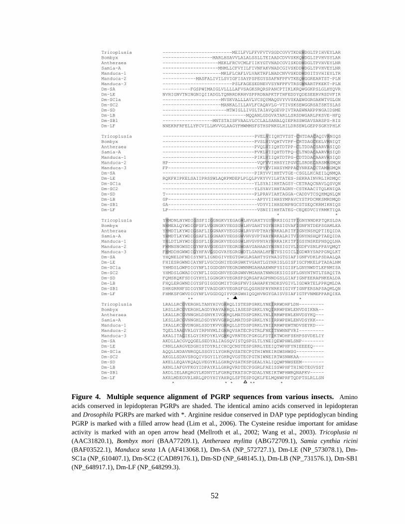
52
Tricoplusia -------------------------MEILFVLFFVFVTVSGDCGVVTKDEWDGLTPIHVEYLAR Bombyx ------------------MARLHSAVVLALALSSLLTEIAADCDVVSKKQWDGLIPVHVSYLAR Antheraea --------------------MEKLFRCVCMLFIIKYGTVNADCGVISKDDWDGLTPVHVEYLNR Samia-A --------------------MNMLLCFVYILFIVNFAKVNADCGIVSKDDWDGLTPVHVEYLNR Manduca-1 ---------------------MKLFLCAFLVLVAKTRFLNADCNVVSKDDWDGITSVHIEYLTR Manduca-2 ------------MASFALIVILSVIGFISAYPSPEGYSSAFNFPFVTKEQWGGREARTST-PLN Manduca-3 -------------------------PSLFAGESEDNEVVSYNFPFVTRSGWNARTPKEKT-PLN Dm-SA ----------FGSPWIMAIGLVLLLLAFVSAGKSRQRSPANCPTIKLKRQWGGKPSLGLHYQVR Dm-LE NVHIGNVTNINGNIQIIADGLTQNRRDRRHVSPPRDNAPKTPTHFEDDYQDESEERVRSDVFIR Dm-SC1a ---------------------MVSKVALLLAVLVCSQYMAQGVYVVSKAEWGGRGAKWTVGLGN Dm-SC2 ---------------------MANKALILLAVLFCAQAVLG-VTIVSKSEWGGRSATSKTSLAS Dm-SD -----------------------MTWIGLLIVGLTAIAVQGEVPIVTRAEWNAKPPNGAIDSME Dm-LB ------------------------------MQQANLGDGVATARLLSRSDWGARLPKSVE-HFQ Dm-SB1 ------------------MNTSTAISFVAALVLCCLALSANALQIEPRSSWGAVSARSPS-RIS Dm-LF NNEKRFRFELLYFCVILLMVVGLAAGYFMWMMSFSTHSPNKGLHILDRSEWLGEPPSGKYPHLK Tricoplusia ---------------------------------PVELVIIQHTVTST-CNTDAACAQIVRNIQS Bombyx ---------------------------------PVSLVIVQHTVTPF-CRTDAGCEELVRNIQT Antheraea ---------------------------------PVQLVIIQHTDTPP-CLTDDACSARVRSIQD Samia-A ---------------------------------PVKLVIIQHTDTPQ-CLTNDACAARVRSIQD Manduca-1 ---------------------------------PIKLVIIQHTDTPG-CDTDDACAARVRSIQD Manduca-2 HP--------------------------------VQFVVIHHSYIPGVCLSRDECARSMRSMQN Manduca-3 FP--------------------------------VPYVVIHHSYMPPACYNREACCTAMRGMQN Dm-SA ---------------------------------PIRYVVIHHTVTGE-CSGLLKCAEILQNMQA Dm-LE RQKFKIPKELSAIIPRSSWLAQKPMDEPLPLQLPVKYVVILHTATES-SEKRAINVRLIRDMQC Dm-SC1a ---------------------------------YLSYAIIHHTAGSY-CETRAQCNAVLQSVQN Dm-SC2 ---------------------------------YLSYAVIHHTAGNY-CSTKAACITQLKNIQA Dm-SD T--------------------------------PLPRAVIAHTAGGA-CADDVTCSQHMQNLQN Dm-LB GP--------------------------------APYVIIHHSYMPAVCYSTPDCMKSMRDMQD Dm-SB1 GA--------------------------------VDYVIIHHSDNPNGCSTSEQCKRMIKNIQS Dm-LF LP--------------------------------VSNIIIHHTATEG-CEQEDVCIYRMKTIQA * * Tricoplusia YHMDNLNYWDIGSSFIIGGNGKVYEGAGWLHVGAHTYGYNRKSIGITFIGNYNNDKPTQKSLDA Bombyx NHMEALQYWDIGPSFLVGGNGKVYEGSGWLHVGAHTYGYNSRSIGVAFIGNFNTDEPSGAMLEA Antheraea YHMDTLKYWDIGSAFLIGGNAKVYEGSGWLRVSVPTHAYNRKALRITFIGNYNSHQPTIEQIDA Samia-A YHMDTLKYWDIGSAFLIGGNAKVYEGSGWVHVSVPTHAYNRKALRITVIGNYNSHQPTAEQIDA Manduca-1 YHLDTLNYWDIGSSFLIGGNGKVYEGSGWLHVGVPNYAYNRKAIKITFIGSYNSKEPNSQQLNA Manduca-2 FHMNSNGWSDIGYNFAVGGEGSVYEGRGWDAVGAHAAGYNSNSIGIVLIGDFVSNLPPAVQMQT Manduca-3 FHMDDHGWWDIGYHFAVGSDGVAYEGRGWDTLGAHALHFNTVSIGICLIGDWRYSAPPGNQLKT Dm-SA YHQNELDFNDISYNFLIGNDGIVYEGTGWGLRGAHTYGYNAIGTGIAFIGNFVDKLPSDAALQA Dm-LE FHIESRGWNDIAYNFLVGCDGNIYEGRGWKTVGAHTLGYNRISLGISFIGCFMKELPTADALNM Dm-SC1a YHMDSLGWPDIGYNFLIGGDGNVYEGRGWNNMGAHAAEWNPYSIGISFLGNYNWDTLEPNMISA Dm-SC2 YHMDSLGWADIGYNFLIGGDGNVYEGRGWNVMGAHATNWNSKSIGISFLGNYNTNTLTSAQITA Dm-SD FQMSKQKFSDIGYHYLIGGNGKVYEGRSPSQRGAFAGPNNDGSLGIAFIGNFEERAPNKEALDA Dm-LB FHQLERGWNDIGYSFGIGGDGMIYTGRGFNVIGAHAPKYNDKSVGIVLIGDWRTELPPKQMLDA Dm-SB1 DHKGRRNFSDIGYNFIVAGDGKVYEGRGFGLQGSHSPNYNRKSIGIVFIGNFERSAPSAQMLQN Dm-LF FHMKSFGWVDIGYNFLVGGDGQIYVGRGWHIQGQHVNGYGAISVSIAFIGTFVNMEPPARQIEA ** * * * Tricoplusia LRALLRCGVERGHLTANYHIVGHRQLISTESPGRKLYNEIRRWDHFLDN--------- Bombyx LRSLLRCGVERGHLAGDYRAVAHRQLIASESPGRKLYNQIRRWPEWLENVDSIKNA-- Antheraea LKALLRCGVNNGHLDSNYKIVGHRQLMATDSPGRKLYNLIRRWPEWLENVDSYKQ--- Samia-A LKSLLRCGVNNGHLDSDYNVVGHRQLMATDSPGRKLYNIIRRWPEWLENVDSYKK--- Manduca-1 IKALLKCGVDNGHLSSDYKVVGHRQLLDTDSPGRKLYNIIRRWPEWTNDVSEYKD--- Manduca-2 TQELIAAGVRLGYIRPNYMLIGHRQVSATECPGTRLFNEITNWNNFVRI--------- Manduca-3 AKALITAGIELGYIKPDYKLVGHKQVRNTECPGKGLFDTIKTWDHFSEHPSSVDELIY Dm-SA AKDLLACGVQQGELSEDYALIAGSQVISTQSPGLTLYNEIQEWPHWLSNP-------- Dm-LE CRNLLARGVEDGHISTDYRLICHCQCNSTESPGRRLYEEIQTWPHFYNIEEEEQ---- Dm-SC1a AQQLLNDAVNRGQLSSGYILYGHRQVSATECPGTHIWNEIRGWSHWSG---------- Dm-SC2 AKGLLSDAVSRGQIVSGYILYGHRQVGSTECPGTNIWNEIRTWSNWKAA--------- Dm-SD AKELLEQAVKQAQLVEGYKLLGHRQVSATKSPGEALYALIQQWPNWSEEM-------- Dm-LB AKNLIAFGVFKGYIDPAYKLLGHRQVRDTECPGGRLFAEISSWPHFTHINDTEGVSST Dm-SB1 AKDLIELAKQRGYLKDNYTLFGHRQTKATSCPGDALYNEIKTWPHWRQNAPKV----- Dm-LF AKRLMDEGVRLHRLQPDYHIYAHRQLSPTESPGQKLFELMQNWPRFTQDPTSLRLLSN
* * * ** *
Figure 4. Multiple sequence alignment of PGRP sequences from various insects. Amino acids conserved in lepidopteran PGRPs are shaded. The identical amino acids conserved in lepidopteran and Drosophila PGRPs are marked with *. Arginine residue conserved in DAP type peptidoglycan binding PGRP is marked with a filled arrow head (Lim et al., 2006). The Cysteine residue important for amidase activity is marked with an open arrow head (Mellroth et al., 2002; Wang et al., 2003). Tricoplusia ni (AAC31820.1), Bombyx mori (BAA77209.1), Antheraea mylitta (ABG72709.1), Samia cynthia ricini (BAF03522.1), Manduca sexta 1A (AF413068.1), Dm-SA (NP_572727.1), Dm-LE (NP_573078.1), Dm-SC1a (NP_610407.1), Dm-SC2 (CAD89176.1), Dm-SD (NP_648145.1), Dm-LB (NP_731576.1), Dm-SB1 (NP_648917.1), Dm-LF (NP_648299.3).
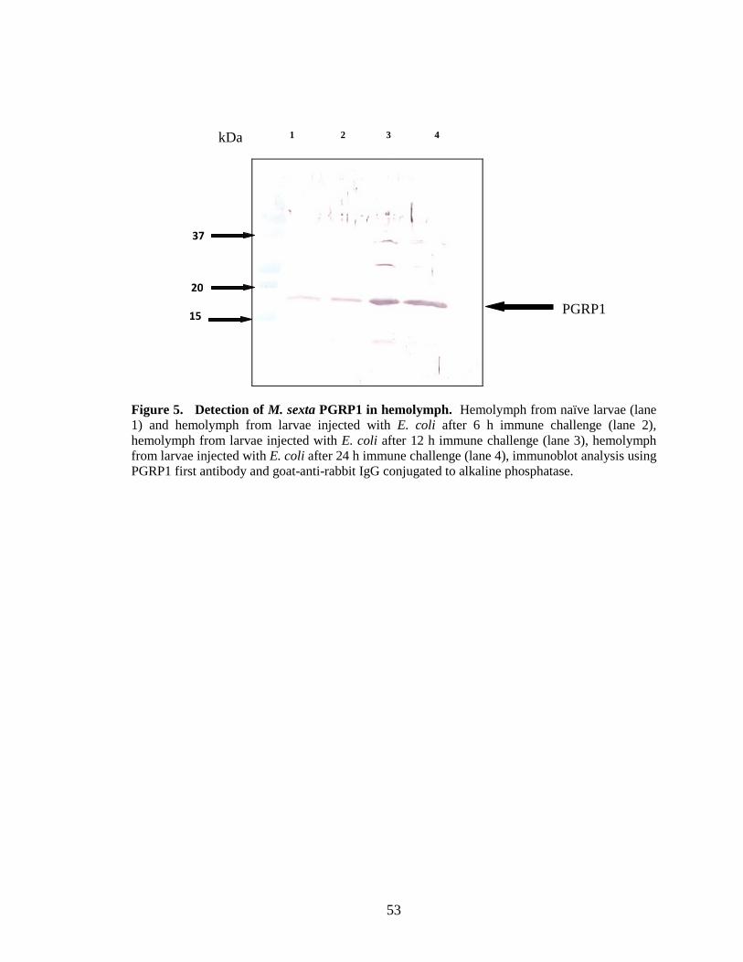
53
Figure 5. Detection of M. sexta PGRP1 in hemolymph. Hemolymph from naïve larvae (lane 1) and hemolymph from larvae injected with E. coli after 6 h immune challenge (lane 2), hemolymph from larvae injected with E. coli after 12 h immune challenge (lane 3), hemolymph from larvae injected with E. coli after 24 h immune challenge (lane 4), immunoblot analysis using PGRP1 first antibody and goat-anti-rabbit IgG conjugated to alkaline phosphatase.
1 2 3 4
20
15
37
kDa
PGRP1
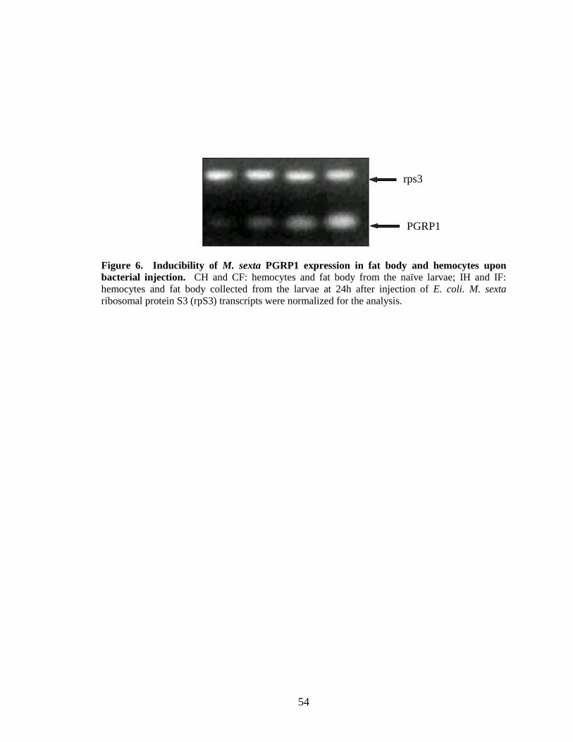
54
Figure 6. Inducibility of M. sexta PGRP1 expression in fat body and hemocytes upon bacterial injection. CH and CF: hemocytes and fat body from the naïve larvae; IH and IF: hemocytes and fat body collected from the larvae at 24h after injection of E. coli. M. sexta ribosomal protein S3 (rpS3) transcripts were normalized for the analysis.
rps3
PGRP1
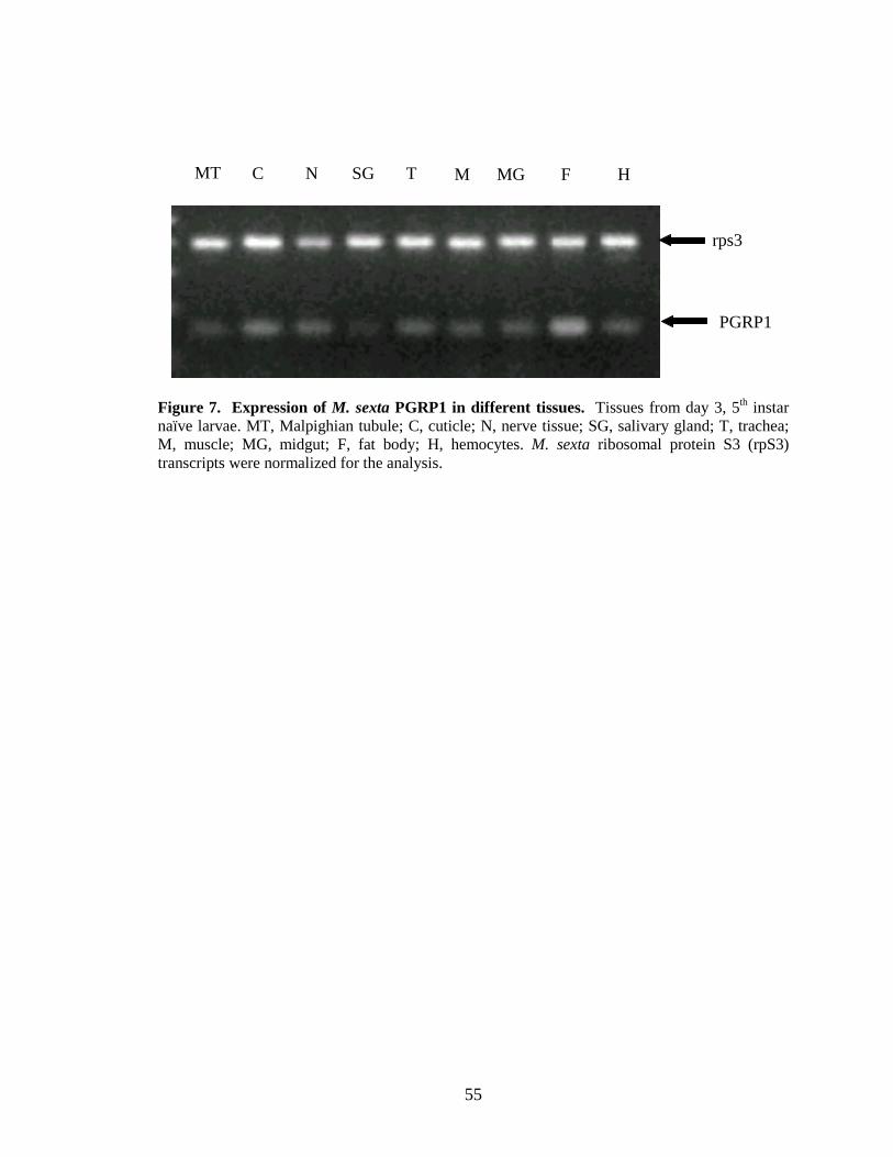
55
Figure 7. Expression of M. sexta PGRP1 in different tissues. Tissues from day 3, 5th instar naïve larvae. MT, Malpighian tubule; C, cuticle; N, nerve tissue; SG, salivary gland; T, trachea; M, muscle; MG, midgut; F, fat body; H, hemocytes. M. sexta ribosomal protein S3 (rpS3) transcripts were normalized for the analysis.
MTTT
C N SG T M MG F H
rps3
PGRP1
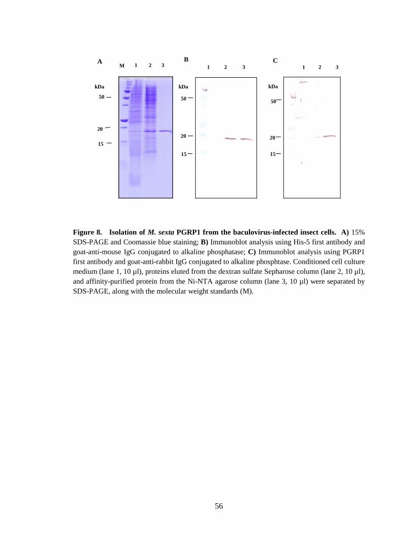
56
Figure 8. Isolation of M. sexta PGRP1 from the baculovirus-infected insect cells. A) 15% SDS-PAGE and Coomassie blue staining; B) Immunoblot analysis using His-5 first antibody and goat-anti-mouse IgG conjugated to alkaline phosphatase; C) Immunoblot analysis using PGRP1 first antibody and goat-anti-rabbit IgG conjugated to alkaline phosphtase. Conditioned cell culture medium (lane 1, 10 µl), proteins eluted from the dextran sulfate Sepharose column (lane 2, 10 µl), and affinity-purified protein from the Ni-NTA agarose column (lane 3, 10 µl) were separated by SDS-PAGE, along with the molecular weight standards (M).
A B M 1 2 3 1 2 3 1 2 3
20
15
kDa
50
15
20
50
kDa
20
15
50
kDa
C
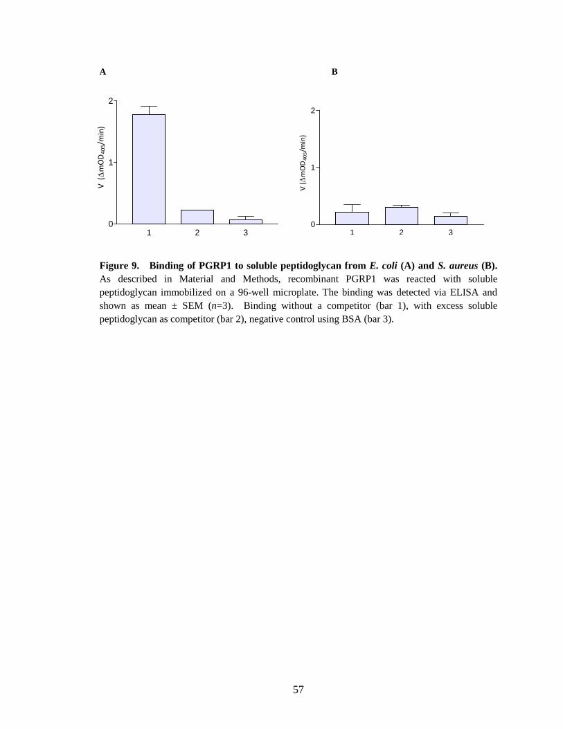
57
A B
Figure 9. Binding of PGRP1 to soluble peptidoglycan from E. coli (A) and S. aureus (B). As described in Material and Methods, recombinant PGRP1 was reacted with soluble peptidoglycan immobilized on a 96-well microplate. The binding was detected via ELISA and shown as mean ± SEM (n=3). Binding without a competitor (bar 1), with excess soluble peptidoglycan as competitor (bar 2), negative control using BSA (bar 3).
1 2 30
1
2V
(∆
mO
D4
05/m
in)
1 2 30
1
2
V (∆
mO
D4
05/m
in)

58
A B
Figure 10. Binding of M. sexta PGRP1 to M. luteus cells (B) and insoluble peptidoglycan (A). Binding was tested by analyzing the unbound and bound fractions by immunoblotting using His-5 first antibody and goat-anti-mouse IgG conjugated to alkaline phosphatase. Control total protein (lane 1), unbound fraction (lane 2), bound fraction (lane 3).
15-
1 2 3
37 -
20-
37-
20-
15-
1 2 3 kDa kDa
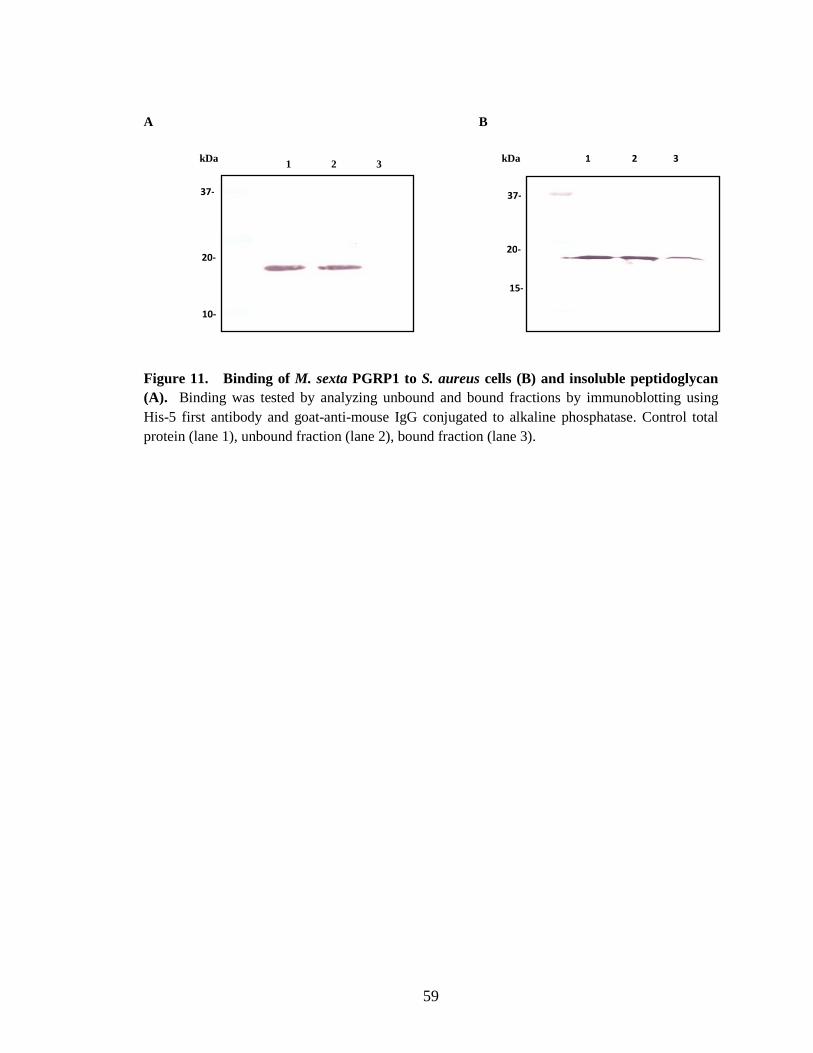
59
A B Figure 11. Binding of M. sexta PGRP1 to S. aureus cells (B) and insoluble peptidoglycan (A). Binding was tested by analyzing unbound and bound fractions by immunoblotting using His-5 first antibody and goat-anti-mouse IgG conjugated to alkaline phosphatase. Control total protein (lane 1), unbound fraction (lane 2), bound fraction (lane 3).
1 2 3 1 2 3
37-
20-
10-
37-
20-
15-
kDa kDa
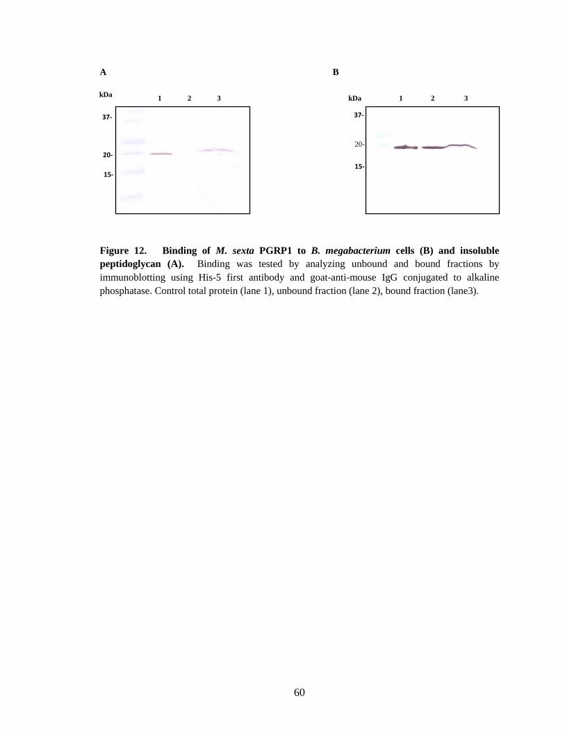
60
A B
Figure 12. Binding of M. sexta PGRP1 to B. megabacterium cells (B) and insoluble peptidoglycan (A). Binding was tested by analyzing unbound and bound fractions by immunoblotting using His-5 first antibody and goat-anti-mouse IgG conjugated to alkaline phosphatase. Control total protein (lane 1), unbound fraction (lane 2), bound fraction (lane3).
1 2 3
37-
20-
15-
37-
20-
15-
1 2 3 kDa kDa
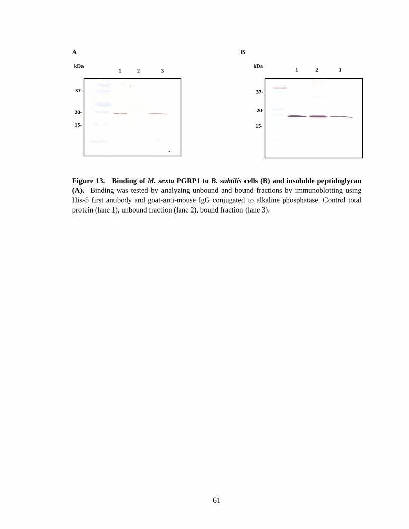
61
A B Figure 13. Binding of M. sexta PGRP1 to B. subtilis cells (B) and insoluble peptidoglycan (A). Binding was tested by analyzing unbound and bound fractions by immunoblotting using His-5 first antibody and goat-anti-mouse IgG conjugated to alkaline phosphatase. Control total protein (lane 1), unbound fraction (lane 2), bound fraction (lane 3).
1 2 3 1 2 3
37-
20-
15-
37-
20-
15-
kDa kDa
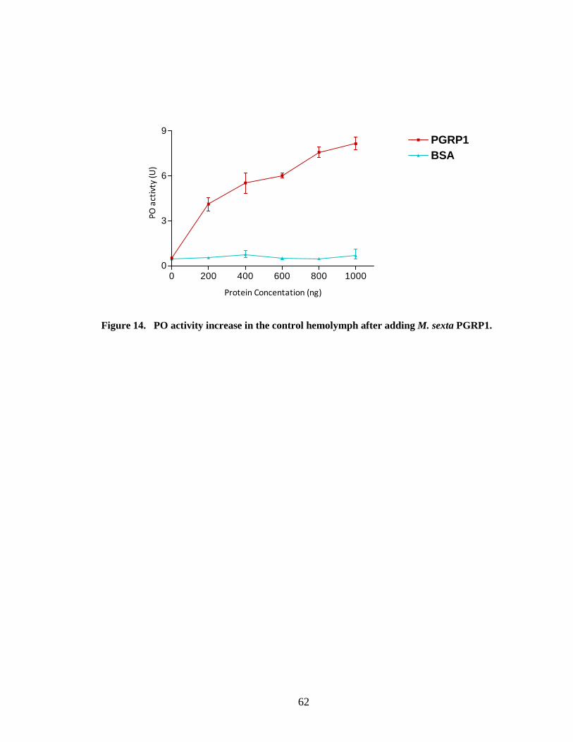
62
Figure 14. PO activity increase in the control hemolymph after adding M. sexta PGRP1.
0 200 400 600 800 10000
3
6
9PGRP1BSA
Protein Concentation (ng)
PO
act
ivty
(U
)
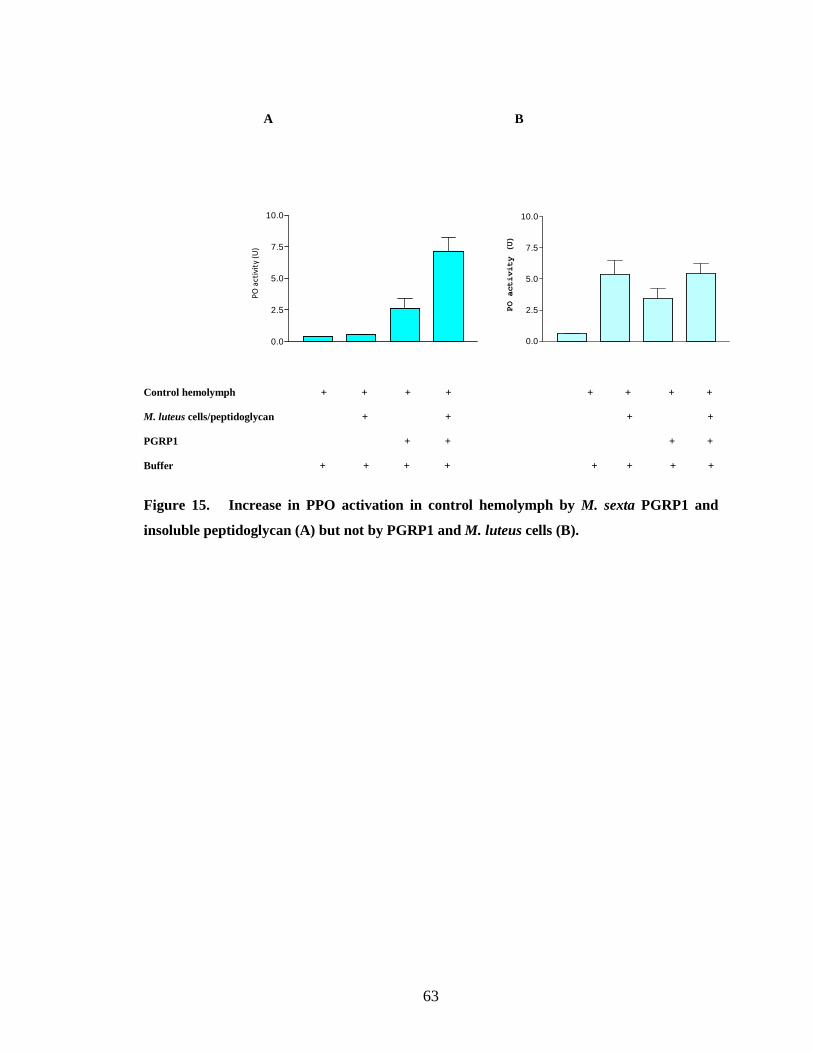
63
A B
Control hemolymph + + + + + + + + M. luteus cells/peptidoglycan + + + + PGRP1 + + + + Buffer + + + + + + + +
Figure 15. Increase in PPO activation in control hemolymph by M. sexta PGRP1 and
insoluble peptidoglycan (A) but not by PGRP1 and M. luteus cells (B).
0.0
2.5
5.0
7.5
10.0
PO activity (U)
0.0
2.5
5.0
7.5
10.0
PO
act
ivit
y (U
)
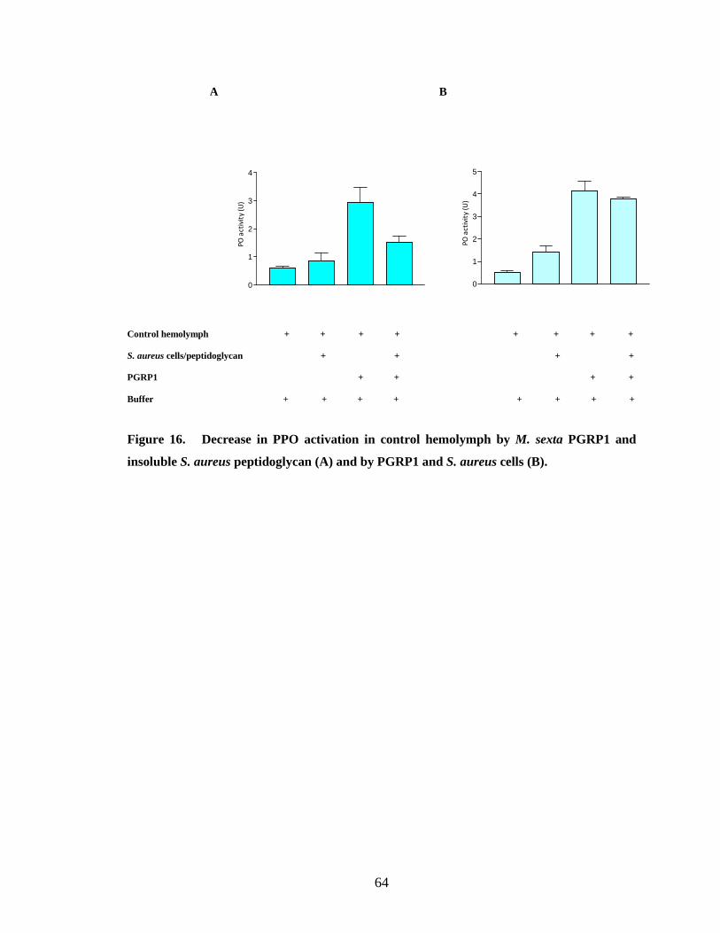
64
A B
Control hemolymph + + + + + + + + S. aureus cells/peptidoglycan + + + + PGRP1 + + + + Buffer + + + + + + + +
Figure 16. Decrease in PPO activation in control hemolymph by M. sexta PGRP1 and
insoluble S. aureus peptidoglycan (A) and by PGRP1 and S. aureus cells (B).
0
1
2
3
4
5
PO
act
ivit
y (U
)
0
1
2
3
4
PO
act
ivit
y (U
)
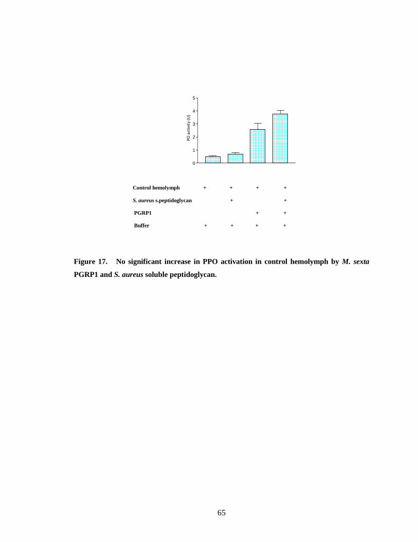
65
Control hemolymph + + + +
S. aureus s.peptidoglycan + +
PGRP1 + +
Buffer + + + +
Figure 17. No significant increase in PPO activation in control hemolymph by M. sexta
PGRP1 and S. aureus soluble peptidoglycan.
0
1
2
3
4
5
PO
act
ivit
y (U
)
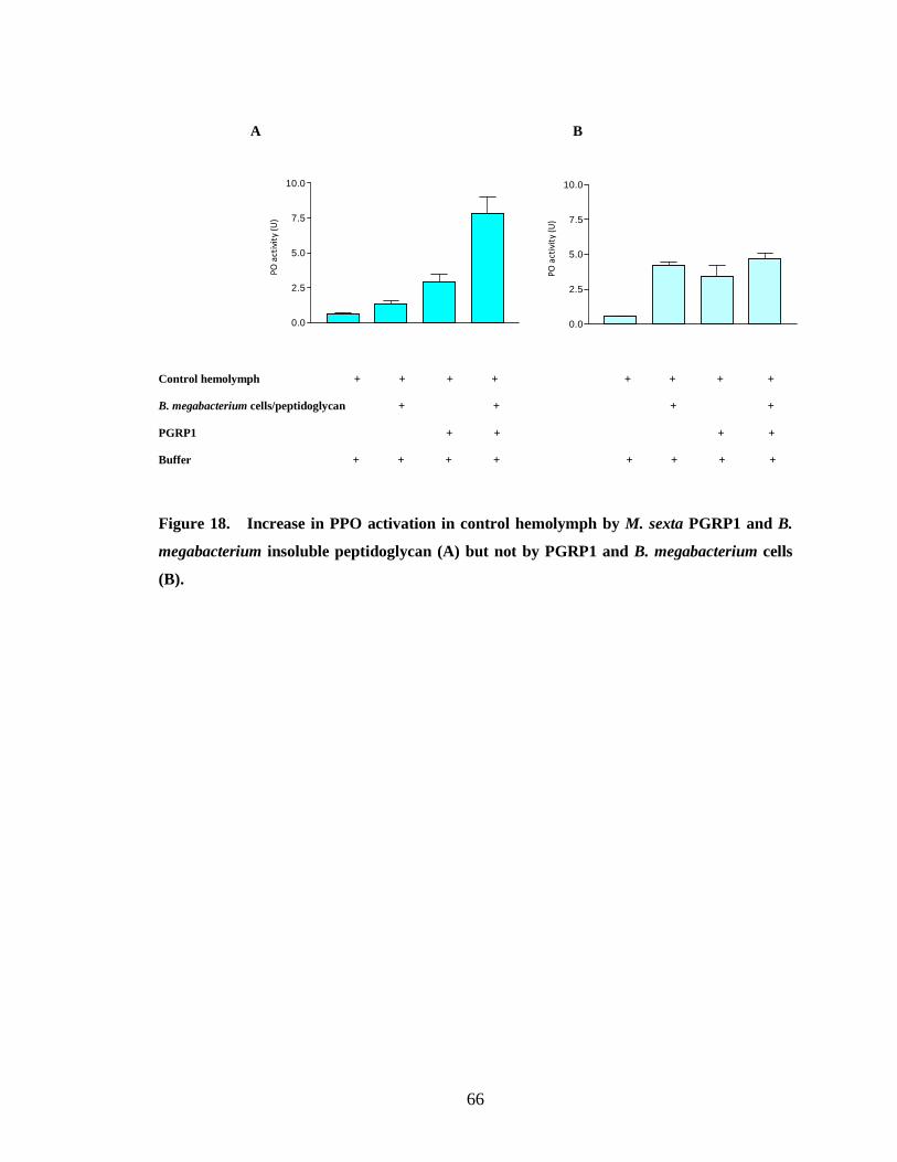
66
A B
Control hemolymph + + + + + + + + B. megabacterium cells/peptidoglycan + + + + PGRP1 + + + + Buffer + + + + + + + +
Figure 18. Increase in PPO activation in control hemolymph by M. sexta PGRP1 and B.
megabacterium insoluble peptidoglycan (A) but not by PGRP1 and B. megabacterium cells
(B).
0.0
2.5
5.0
7.5
10.0
PO
act
ivit
y (U
)
0.0
2.5
5.0
7.5
10.0
PO
act
ivit
y (U
)
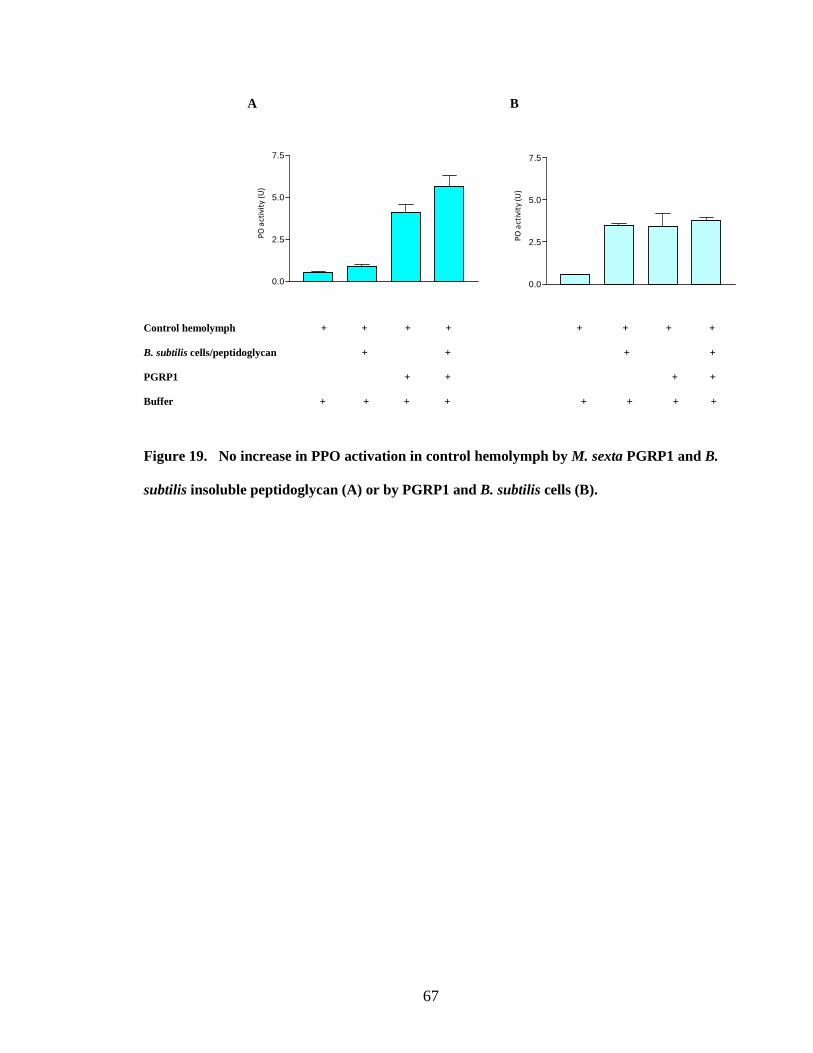
67
A B
Control hemolymph + + + + + + + + B. subtilis cells/peptidoglycan + + + + PGRP1 + + + + Buffer + + + + + + + +
Figure 19. No increase in PPO activation in control hemolymph by M. sexta PGRP1 and B.
subtilis insoluble peptidoglycan (A) or by PGRP1 and B. subtilis cells (B).
0.0
2.5
5.0
7.5
PO
act
ivit
y (U
)
0.0
2.5
5.0
7.5
PO
act
ivit
y (U
)
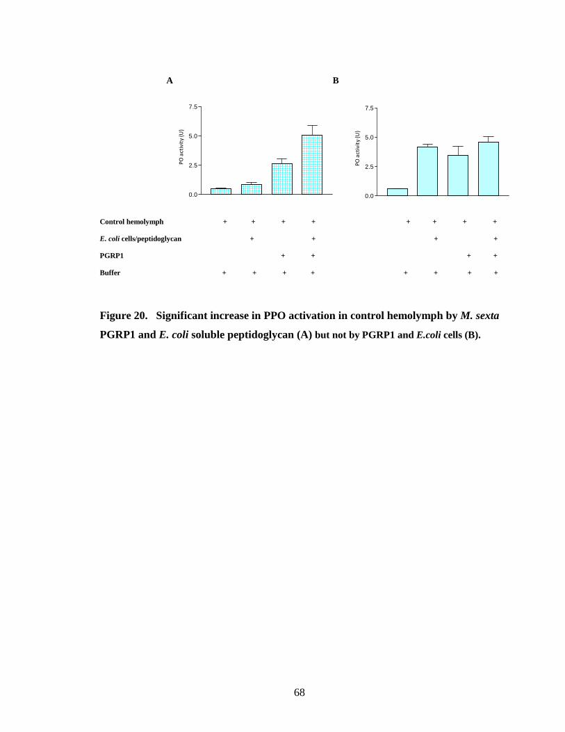
68
A B Control hemolymph + + + + + + + + E. coli cells/peptidoglycan + + + + PGRP1 + + + + Buffer + + + + + + + +
Figure 20. Significant increase in PPO activation in control hemolymph by M. sexta
PGRP1 and E. coli soluble peptidoglycan (A) but not by PGRP1 and E.coli cells (B).
0.0
2.5
5.0
7.5
PO
act
ivit
y (U
)
0.0
2.5
5.0
7.5
PO
act
ivit
y (U
)
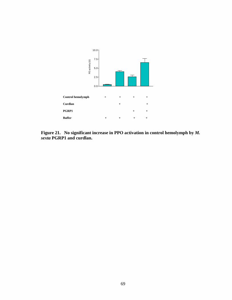
69
Control hemolymph + + + + Curdlan + + PGRP1 + + Buffer + + + +
Figure 21. No significant increase in PPO activation in control hemolymph by M. sexta PGRP1 and curdlan.
0.0
2.5
5.0
7.5
10.0
PO
act
ivit
y (U
)
70
REFERENCES
Aggrawal, K. and Silverman, N. (2007) Peptidoglycan recognition in Drosophila.
Biochem. Soc. Trans. 35, 1496-1499.
Appel, H.M. and Maines, L.W. (1995) The influence of host-plant on gut conditions of
gypsy moth (Lymantria dispar) caterpillars. J. Insect. Physiol. 41, 241-246.
Ashida, M. and Brey, P.T. (1997) Recent advances on the research of the insect
prophenoloxidase cascade. In: Brey, P.T. and Hultmark, D. (Eds.), Molecular
Mechanisms of Immune Responses in Insects. Chapman and Hall, London, pp.
135-172.
Bera, A., Herbert, S., Jakob, A., Vollmer, W. and Gotz, F. (2005) Why are pathogenic
staphylococci so lysozyme resistant? The peptidoglycan O-acetyltransferase OatA
is the major determinant for lysozyme resistance of Staphylococcus aureus. Mol.
Microb. 55, 778-787.
Bidla, G., Hauling, T., Dushay, M.S. and Theopold, U. (2008) Activation of Insect
Phenoloxidase after Injury: Endogenous versus Foreign Elicitors. J. Innate.
Immune. DOI: 10.1159/000168009.
Bischoff, V., Vignal, C., Boneca, I.G., Michel, T., Hoffmann, J.A. and Royet, J. (2004)
Function of the Drosophila pattern-recognition receptor PGRP-SD in the
detection of Gram-positive bacteria. Nat. Immun. 4, 1175-1180.
71
Bischoff, V., Vignal, C., Duvic, B., Boneca, I.G., Hoffmann, J. and Royet, J. (2006)
Downregulation of the Drosophila immune response by peptidoglycan-
recognition proteins SC1 and SC2. PLoS. Biol. 2, 0139-0147.
Brennan, C.A. and Anderson, K.V. (2004) DROSOPHILA: The Genetics of Innate
Immune Recognition and Response. Ann. Rev. of. Immunol. 22, 457-483.
Brey, P.T., Lee, W.J., Yamakawa, M., Koizumi, Y., Perrot, S., François, M. and Ashida,
M. (1993) Role of the integument in insect immunity: epicuticular abrasion and
induction of cecropin synthesis in cuticular epithelial cells. Proc. Natl. Acad. Sci.
USA. 90, 6275-6279.
Bulet, P., Stocklin, R., and Menin, L. (2004) Anti-microbial peptides: from invertebrates
to vertebrates. Immuno Rev. 198, 169-184.
Cerenius, L., Lee, B.L. and Söderhäll, K. (2007) The proPO-system: pros and cons for
its role in invertebrate immunity. Trends. Immunol. 29, 263-271.
Cerenius, L. and Soderhall, K. (2004) The prophenoloxidase-activating system in
invertebrates. Immunol. Rev. 198, 116-126.
Chang, C-I., Pili-Floury, S., Herve, M., Parquet, C., Chelliah, Y., Lemaitre, B., Mengin-
Lecreulx, D. and Deisenhofer, J. A. (2004) Drosophila pattern recognition
receptor contains a peptidoglycan docking groove and unusual L,D-
carboxypeptidase activity. PLoS. Biol. 2, e277.
Chang, C.I., Ihara, K., Chelliah, Y., Mengin-Lecreulx, D., Wakatsuki, S. and
Deisenhofer, J. (2005) Structure of the ectodomain of Drosophila peptidoglycan-
recognition protein LCa suggests a molecular mechanism for pattern recognition.
Proc. Natl. Acad. Sci. USA. 102, 10279-10284.
72
Chang, C.I., Chelliah, Y., Borek, D., Mengin-Lecreulx, D. and Deisenhofer. J. (2006)
Structure of Tracheal Cytotoxin in Complex with a Heterodimeric Pattern-
Recognition Receptor. Science. 311, 1761-1764.
Choe, K-M., Werner, T., Stoven, S., Hultmark, D. and Anderson, K.V. (2002)
Requirement for a peptidoglycan recognition protein (PGRP) in Relish activation
and antibacterial immune responses in Drosophila. Science. 296, 259-262.
Choe, K-M., Lee, H. and Anderson, K.V. (2005) Drosophila peptidoglycan recognition
protein LC (PGRP-LC) acts as a signal-transducing innate immune receptor. Proc.
Natl. Sci. USA. 102, 1122-1126.
Christophides, G.K., Zdobnov, E., Barillas-Mury, C., Birney, E., Stephanie, B., Blass, C.,
Brey, P.T., Collins, F.H., Danielli, A., Dimopoulos, G. et al., (2002) Immunity-
Related Genes and Gene Families in Anopheles gambiae. Science. 198, 159-165.
Dimopoulos, G., Christophides, G.K., Meister, S., Schultz, J., White, K.P., Barillas-
Mury, C. and Kafatos, F.C. (2002) Genome expression analysis of Anopheles
gambiae: Responses to injury, bacterial challenge, and malaria infection. Proc.
Natl. Acad. Sci. USA. 99, 8814-8819.
Dmitriev, B.A., Toukach, F.V., Holst, O., Rietschel, E.T. and Ehlers, S. (2004) Tertiary
Structure of Staphylococcus aureus Cell Wall Murein. J. Bacteriol. 186, 7141-
7148.
Dunn, P. and Drake, D. (1983) Fate of bacteria injected into naïve and immunized larvae
of the tobacco hornworm, Manduca sexta. J. Invert. Pathol. 41, 77-85.
Dziarski, R. (2003) Peptidoglycan recognition proteins (PGRPs). Mol. Immuno. 40, 877-
886.
73
Dziarski, R. and Gupta, D. (2006) The peptidoglycan recognition proteins (PGRPs).
Genome. Biol. 7, 232.
Eleftherianosa, I., Millichapa, P.J., Ffrench-Constanta, R.H. and Reynolds, S.E. (2006a)
RNAi suppression of recognition protein mediated immune responses in the
tobacco hornworm Manduca sexta causes increased susceptibility to the insect
pathogen Photorhabdu. Dev. Comp. Immunol. 30, 1099-1107.
Eleftherianosa, I., Marokhazi, J., Millichap, P.J., Hodgkinson, A.J., Sriboonlert, A.,
Ffrench-Constant, R.H. and Reynolds, S.E. (2006b) Prior infection of Manduca
sexta with non-pathogenic Escheichia coli elicits immunity to pathogenic
Photorhabdus luminescens: Roles of immune-related proteins shown by RNA
interference. Insect Biochem. Mol. Biol. 36, 517-525.
Eleftherianosa, I., Gokcen, F., Felfold, G., Millichap, P.J., Trenczek, T.E., ffrench-
Constant, H. and Reynolds, S.E. (2007) The immunoglobulin family protein
Hemolin mediates cellular immune responses to bacteria in the insect Manduca
sexta. Cell. Microbiol. 9, 1137-1147.
Fearon, D.T. (1997) Seeking wisdom in innate immunity. Nature. 388, 323-324.
Filipe, S.R, Tomasz, A. and Ligoxygakis, P. (2005) Requirements of peptidoglycan
structure that allow detection by the Drosophila Toll pathway. EMBO reports. 6,
327-333.
Gandhe, A.S., Arunkumar, K.P., John, S.H. and Nagaraju, J. (2006) Analysis of bacteria-
challenged wild silkmoth, Antheraea mylitta (lepidoptera) transcriptome reveals
potential immune genes. BMC Genomics. 7,184.
74
Garver, L.S., Wu, J. and Wu, L.P. (2006) The peptidoglycan recognition protein PGRP-
SC1a is essential for Toll signaling and phagocytosis of Staphylococcus aureus in
Drosophila. Proc. Natl. Acad. Sci. USA. 103, 660-665.
Gelius, E., Persson, C., Karlsson, J. and Steiner, H. (2003) A mammalian peptidoglycan
recognition protein with N-acetylmuramoyl-L-alanine amidase activity. Biochem
Biophys Res Comm. 306, 988-994.
Gillespie, J.P. and Kanost, M.R. (1997) Biological Mediators Of Insect Immunity. Ann.
Rev. of. Immunol. 42, 611-643.
Gorman, M.J., Wang, Y., Jiang, H. and Kanost, M.R. (2007) Manduca sexta
Hemolymph Proteinse 21 Activates Prophenoloxidase-activating proteinase 3 in
an Insect Innate Immune Proteinase Cascade. J. Biol. Chem. 282, 11742-11749.
Gottar, M., Gobert, V., Michel, T., Belvin, M., Duyk, G. and Hoffmann, J.A. (2002) The
Drosophila immune response against Gram-negative bacteria is mediated by a
peptidoglycan recognition protein. Nature. 416, 640-644.
Guan, R., Malchiodi, M.L., Wang, Q., Schuck, P. and Mariuzza, R.A. (2004a) Crystal
structure of the C-terminal peptidoglycan-binding domain of human
peptidoglycan recognition protein Iα. J. Biol. Chem. 279, 31873-31882.
Guan, R., Roychowdhury, A., Ember, B., Kumar, S., Boons, G-J. and Mariuzza, R.A.
(2004b) Structural basis for peptidoglycan binding by peptidoglycan recognition
proteins. Proc. Natl. Acad. Sci. USA. 101, 17168-17173.
Guan, R., Wang, Q., Sundberg, E.J. and Mariuzza, R.A. (2005) Crystal structure of
human peptidoglycan recognition protein S (PGRP-S) at 1.70 A˚ resolution. J.
Mol. Biol. 347, 683-691.
75
Gupta, S., Wang, Y. and Jiang, H. (2005) Manduca sexta prophenoloxidase (proPO)
activation requires proPO-activating proteinase (PAP) and serine proteinase
homologs (SPHs) simultaneously. Insect. Biochem. Mol. Biol. 35, 241-248.
Gutierrez, E., Wiggins, D., Fielding, B. and Gould, A.P. (2007) Specialized hepatocyte-
like cells regulate Drosophila lipid metabolism. Nature. 445, 275-280.
Harz, H., Burgdorf, K. and Holtje, J-V. (1990) Isolation and Seperation of the Glycan
Strands from Murein of Escherichia coli by Reversed-Phase High-Performance
Liquid Chromatography. Analytic. BioChem. 190, 120-128.
Hashimotoa, K., Megaa, K., Matsumotoa, Y., Bao, Y., Yamano, Y. and Morishima, I.
(2007) Three peptidoglycan recognition protein (PGRP) genes encoding potential
amidase from eri-silkworm, Samia cynthia ricini. Comp. Biochem. Physio. 148,
322-328.
Hetru, C., Troxler, L., and Hoffmann, J.A. (2003) Drosophila melanogaster
Antimicrobial Defense. J. Infectious. Diseases. 187, s327-s324.
Hoffmann, J.A. (1995) Innate immunity of insects. Curr. Opin. Immunol. 7, 4-10.
Hoffmann, J.A. (2003) The immune response of Drosophila. Nature. 6, 33-38.
Hultmark, D. (1993) Immune reactions in Drosophila and other insects: a model for
innate immunity. Trends. Genet. 9, 178-183.
Hultmark, D, and Borge-Renberga, K. (2007) Drosophila Immunity: Is Antigen
Processing the First Step? Curr Biol. 17, R22-24.
Janeway, C.A. Jr (1994) Phylogenetic Perspectives in Immunity: The Insect Host
Defense Boca Raton. CRC Press. 115-122.
76
Jarvis, D.L., Summers, M.D., Garcia, A. Jr and Bohlmeyer, D.A. Influence of different
signal peptides and prosequences on expression and secretion of human tissue
plasminogen activator in the baculovirus system. J. Biol. Chem. 268, 16754-
16762.
Ji, C., Wang, Y., Guo, X., Hartson, S. and Jiang, H. (2004) A Pattern Recognition Serine
Proteinase Triggers the Prophenoloxidase Activation Cascade in the Tobacco
Hornworm, Manduca sexta. J. Biol. Chem. 279, 34101-34106.
Jiang, H., Wang, Y., Yu, X-Q., Zhu, Y. and Kanost, M.R. (2003a) Prophenoloxidase-
activating proteinase-3 (PAP-3) from Manduca sexta hemolymph: a clip domain
serine proteinase regulated by serpin-1J and serine proteinase homologs. Insect.
Biochem. Mol. Biol. 33, 1049-1060.
Jiang, H., Wang, Y., Yu, X-Q. and Kanost, M.R. (2003b) Prophenoloxidase-activating
Proteinase-2 from Hemolymph of Manduca sexta A BACTERIA-INDUCIBLE
SERINE PROTEINASE CONTAINING TWO CLIP DOMAINS. J. Biol. Chem.
278, 3552-3561.
Jiang, H., Ma, C., Lu, Z-Q. and Kanost, M.R. (2004) β-1, 3-Glucan recognition protein-2
(βGRP2) from Manduca sexta: an accute-phase protein that binds to β-1,3-glucan
and lipoteichoic acid to aggregate fungi and bacteria and stimulate
prophenoloxidase activation. Insect. Biochem. Mol. Biol. 34, 89-100.
Jiang, H. (2008) The biochemical basis of antimicrobial responses in Manduca sexta. J.
Insect Sci. 15, 53-66.
77
Jiravanichpaisala, P., Lee, B.L., and Söderhäll, K. (2006) Cell-mediated immunity in
arthropods: Hematopoiesis, coagulation, melanization and opsonization.
Immunobiology. 211, 213-236.
Kaito, C., Akimitsu, N., Watanabe, H. and Sekimizu, K. (2002) Silkworm larvae as an
animal model of bacterial infection pathogenic to humans. Microb Pathog. 32,
183-190.
Kaneko, T., Goldman, W.E., Mellroth, P., Steiner, H., Fukase, K., Kusumoto, S., Harley,
W., Fox, A., Golenbock, D. and Silverman, N. (2004) Monomeric and polymeric
gram-negative peptidoglycan but not purified LPS stimulate the Drosophila IMD
pathway. Immunity. 20, 637-649.
Kaneko, T., Golenbock, D. and Silverman, N. (2005) Peptidoglycan recognition by the
Drosophila Imd pathway. J Endotox Research. 11, 383-389.
Kaneko, T., Yano, T., Aggarwal, K., Lim, J., Ueda, K., Oshima, Y., Peach, C., Erturk-
Hasdemir, D., Goldman, W.E., Oh, B., Kurata, S. and Silverman, N. (2006)
PGRP-LC and PGRP-LE have essential yet distinct functions in the Drosophila
immune response to monomeric DAP-type peptidoglycan. Nat. Immuno. 7, 715-
723.
Kang, D., Liu, G., Lundstrom, A., Gelius, E. and Steiner, H. (1998) A peptidoglycan
recognition protein in innate immunity conserved from insects to humans. Proc.
Natl. Acad. Sci. USA. 95, 10078-10082.
Kanost, M.R., Dai, W. and Dunn, P.E. (1988) Peptidoglycan fragments elicit antibacterial
protein synthesis in larvae of Manduca sexta. Arch. Insect Biochem Physio. 8, 1
47-164.
78
Kanost, M.R., Jiang, H. and Yu, X.Q. (2004) Innate immune responses of a lepidopteran
insect, Manduca sexta. Immunol. Rev. 198, 97-105.
Kim, M-S., Byun, M. and Oh, B-H. (2003) Crystal structure of peptidoglycan
recognition protein LB from Drosophila melanogaster. Nat. Immunol. 4, 787-
793.
Kumar, S., Roychowdhury, A., Ember, B., Wang, Q., Guan, R., Mariuzza, R.A. and
Boons, G-J. (2005) Selective recognition of synthetic lysine and meso-
diaminopimelic acid-type peptidoglycan fragments by human peptidoglycan
recognition proteins Iα and S. J. Biol. Chem. 280, 37005-37012.
Ladendorff, N.E. and Kanost, M.R. (1990) Isolation and characterization of bacteria-
induced protein P4 from hemolymph of Manduca sexta. Arch Insect Biochem
Physiol. 15, 33-41.
Lavine, M.D. and Strand, M.R. (2002) Insect hemocytes and their role in immunity.
Insect Biochem. Mol. Biol. 32, 1295-1309.
Lee, M.H., Osaki, T., Lee, J.Y., Baek, M.J., Zhang, R., Park, J.W., Kawabata, S.,
Soderhall, K. and Lee, B.L. (2004) Peptidoglycan Recognition Proteins Involved
in 1, 3-β-D-Glucan-dependent Prophenoloxidase Activation System of Insect. J.
Biol. Chem. 279, 3218-3227.
Lehrer, R. and Ganz, T. (1999) Antimicrobial peptides in mammalian and insect host
defense. Curr Opin Immunol. 11, 23-27.
Lemaitre, B. and Hoffmann, J. (2007) The host defense of Drosophila melanogaster.
Annu. Rev. Immunol. 25, 697-743.
79
Leone, P., Bischoff, V., Kellenberger, C., Hetru, C., Royet, J. and Roussel, A. (2008)
Crystal structure of Drosophila PGRP-SD suggests binding to DAP-type but not
lysine-type peptidoglycan. Mol. Immuno. 45, 2521-2530.
Leulier, F., Parquet, C., Pili-Floury, S., Ryu, J.H., Caroff, M., Lee, W.J., Mengin-
Lecreulx, D. and Lemaitre, B. (2003) The Drosophila immune system detects
bacteria through specific peptidoglycan recognition. Nat. Immunol. 4, 478-484.
Li, X., Wang, S., Qi, J., Echtenkamp, S.F., Chatterjee, R., Wang, M., Boons, G-J.,
Dziarski, R. and Gupta, D. (2007) Zebrafish Peptidoglycan Recognition Proteins
Are Bactericidal Amidases Essential for Defense against Bacterial Infections.
Immunity. 27, 518-529.
Lim, J-H., Kim, M-S., Kim, H-E., Yano, T., Oshima, Y., Aggarwal, K., Goldman, W.E.,
Silverman, N., Kurata, S. and Oh, B-H. (2006) Structural basis for preferential
recognition of diaminopimelic acid-type peptidoglycan by a subset of
peptidoglycan-recognition proteins. J. Biol. Chem. 281, 8286-8295.
Ling, E. and Yu, X-Q. (2006) Cellular encapsulation and melanization are enhanced by
immulectins, pattern recognition receptors from the tobacco hornworm Manduca
sexta. Dev Comp Immuno. 30, 289-299.
Liu, C., Gelius, E., Liu, G., Steiner, H. and Dziarski, R. (2000) Mammalian
Peptidoglycan Recognition Protein Binds Peptidoglycan with High Affinity, Is
Expressed in Neutrophils, and Inhibits Bacterial Growth. J. Biol. Chem. 275,
24490-24499.
80
Liu, C., Xu, Z., Gupta, D. and Dziarski, R. (2001) Peptidoglycan recognition proteins: a
novel family of four human innate immunity pattern recognition molecules. J.
Biol. Chem. 276, 34686-34694.
Lu, X., Wang, M., Qi, J., Wang, H., Li, X., Gupta, D. and Dziarski, R. (2006)
Peptidoglycan Recognition Proteins Are a New Class of Human Bactericidal
Proteins. J. Biol. Chem. 281, 5895-5907.
Lu, Z. and Jiang, H. (2007) Expression of Manduca sexta serine proteinase homolog
precursors in insect cells and their proteolytic activation. Insect Biochem. Mol.
Biol. 38, 89-98.
Ma, C. and Kanost, M.R. (2000) A β1,3-Glucan Recognition Protein from an insect,
Manduca sexta, Agglutinates Microorganisms and Activates the Phenoloxidase
Cascade. J. Biol. Chem. 275, 7505-7514.
Meister, M., Lemaitre, B., and Hoffmann, J.A. (1997) Antimicrobial peptide defense in
Drosophila. BioEssays. 19, 1019-1026.
Mellroth, P., Karlsson, J. and Steiner, H. (2003) A scavenger function for a Drosophila
peptidoglycan recognition protein. J. Biol. Chem. 278, 7059-7064.
Mellroth, P. (2005) Peptidoglycan Recognition Proteins: Major Regulators of
Drosophila Immunity. PhD thesis.
Mellroth, P., Karlsson, J., Hakansson, J., Schultz, N., Goldman, W.E. and Steiner, H.
(2005) Ligand-induced dimerization of Drosophila peptidoglycan recognition
proteins in vitro. Proc. Natl. Acad. Sci. USA. 12, 6455-6460.
81
Mellroth, P. and Steiner, H. (2006) PGRP-SB1: An N-acetylmuramoyl L-alanine
amidase with antibacterial activity. Biochem Biophys Res Commun. 350, 994-
999.
Mengin-Lecreulx, D. and Lemaitre, B. (2005) Structure and metabolism of
peptidoglycan and molecular requirements allowing its detection by the
Drosophila innate immune system. J Endotoxin Res. 11, 105-111.
Meroueh, S.O., Bencze, K.Z., Hesek, D., Lee, M., Fisher, J.F., Stemmler, T.L. and
Mobashery, S. (2006) Three-dimensional structure of the bacterial cell wall
peptidoglycan. Proc. Natl. Acad. Sci. USA. 103, 4404-4409.
Michel, T., Reichhart, J.M., Hoffmann, J.A and Royet, J. (2001) Drosophila Toll is
activated by Gram-positive bacteria through a circulating peptidoglycan
recognition protein. Nature. 414, 756-759.
Morishma, I., Horiba, T., Iketani, M., Nishioka, E., Horiba, T., Iketani, M., Nishioka, E.
and Yamano, Y. (1995) Parallel induction of cecropin and lysozyme in larvae of
the silkworm Bombyx mori. Dev Comp Immunol. 19, 357-363.
Nakamura, M., Inomata, M., Hayashi, M., Imahori, K. and Kawashima, S. (1985)
Purification and characterization of 210,000-dalton inhibitor of calcium-activated
neutral protease from rabbit skeletal muscle and its relation to 50,000-dalton
inhibitor. J. Biol. Chem. 98, 757-765.
Needham, A.J., Kibart, M., Crossley, H., Ingham, P.W. and Foster, S.J. (2004)
Drosophila melanogaster as a model host for Staphylococcus aureus infection.
Microbiology. 150, 2347-2355.
82
Ochiai, M. and Ashida, M. (1999) A pattern recognition protein for peptidoglycan:
cloning the cDNA and the gene of the silk worm, Bombyx mori. J. Biol. Chem.
274, 11854-11858.
Onoe, H., Matsumoto, A., Hashimoto, K., Yamano, Y. and Morishima, I. (2007)
Peptidoglycan recognition protein (PGRP) from eri-silkworm, Samia cynthia
ricini; protein purification and induction of the gene expression. Comp Biochem
Physiol. 147, 512-519.
Park, J.W., Je, B.R., Piao, S., Inamura, S., Fujimoto, Y., Fukase, K., Kusumoto, S.,
Söderhäll, K., Ha, N.C. and Lee, B.L. (2006) A synthetic peptidoglycan fragment
as a competitive inhibitor of the melanization cascade. J. Biol. Chem. 281, 7747-
7755.
Park, J., Kim, C., Kim, J., Je, B., Roh, K., Kim, S., Lee, H., Ryu, J., Lim, J., Oh, B., Lee,
W., Ha, N. and Lee, B. (2007) Clustering of peptidoglycan recognition protein-
SA is required for sensing lysine-type peptidoglycan in insects. Proc. Natl. Acad.
Sci. USA. 104, 6602-6607.
Persson, C., Oldenvi, S. and Steiner, H. (2007) Peptodoglycan recognition protein LF: A
negative regulator of Drosophila immunity. Insect Biochem. Mol. Biol. 37, 1309-
1316.
Pili-Floury, S., Franc, O., Takahashi, K., Saigo, K., Samain, E., Ueda, R. and Lemaitre,
B. (2004) In Vivo RNA Interference Analysis Reveals an Unexpected Role for
GNBP1 in the Defense against Gram-positive Bacterial Infection in Drosophila
Adults. J. Biol. Chem. 279, 12848-12853.
83
Pinheiro, V.B. and Ellar, D.J. (2006) How to kill a mocking bug? Cell Microbiol. 8, 545-
557.
Ragan, E. J. (2008) Immune-related protein complexes and serpin-1 isoforms in
Manduca sexta plasma. PhD thesis.
Ramet, M., Manfruelli, P., Pearson, A., Mathey-Prevot, B. and Ezekowitz, R.A.B.
(2002) Functional genomic analysis of phagocytosis and identification of a
Drosophila receptor for E. coli. Nature. 416, 644-648.
Reiser, J-B., Teyton, L. and Wilson, I.A. (2004) Crystal structure of the Drosophila
peptidoglycan recognition protein (PGRP)-SA at 1.56 A˚ resolution. J Mol Biol.
340, 909-917.
Rosenthal, R.S. and Dziarski, R. (1994) Isolation of peptidoglycan and soluble
peptidoglycan fragments. Methods Enzymol. 235, 253-285.
Royet, J. (2004) Infectious non-self recognition in invertebrates: lessons from
Drosophila and other insect models. Mol Immunol. 41, 1063-1075.
Royet, J. and Dziarski, R. (2007) Peptidoglycan recognition proteins: pleiotropic sensors
and effectors of antimicrobial defences. Nature. 5, 264-277.
Sang, Y., Ramanathan, B., Ross, C.R. and Blecha, F. (2005) Gene Silencing and
Overexpression of Porcine Peptidoglycan Recognition Protein Long Isoforms:
Involvement in ß-Defensin-1 Expression. Infection and Immunity. 73, 7133-7141.
Schilefifer, K.H. and Kandler, O. (1972) Peptidoglycan Types of Bacterial Cell Walls
and their Taxonomic Implications. Bacterio Rev. 36, 407-477.
Shockman, G.D. and Barrett, J.F. (1983) Structure, function, and assembly of cell walls
of Gram-positive bacteria. Annu Rev Microbiol. 37, 501-527.
84
Stenbak, C.R., Ryu, J.H., Leulier, F., Pili-Floury, S., Parquet, C., Herve, M., Chaput, C.,
Boneca, I.G., Lee, W.J., Lemaitre, B. et al. (2004) Peptidoglycan molecular
requirements allowing detection by the Drosophila immune deficiency pathway.
J. Immuno. 173, 7339-7348.
Stoven, S., Ando, I., Kadalayil, L., Engstrom, Y., Hultmark, H. (2000) Activation of the
Drosophila NF-κB factor Relish by rapid endoproteolytic cleavage. EMBP J. 16,
347-352.
Swaminathan, C.P., Brown, P.H., Roychowdhury, A., Wang, Q., Guan, R., Silverman,
N., Goldman, W.E., Boons, G.J. and Mariuzza, R.A. (2006) Proc. Natl. Acad. Sci.
USA. 103, 684-699.
Takehana, A., Katsuyama, T., Yano, T., Oshima, Y., Takada, H., Aigaki, T. and Kurta,S.
(2002) Overexpression of a pattern-recognition receptor, peptidoglycan-
recognition protein-LE, activates imd/relish-mediated antibacterial defense and
the prophenoloxidase cascade in Drosophila larvae. Proc. Natl. Acad. Sci. USA.
99, 13705-13710.
Takehana, A., Yano, T., Mita, S., Kotani, A., Oshima, Y. and Kurata, S. (2004)
Peptidoglycan recognition protein (PGRP)-LE and PGRP-LC act synergistically
in Drosophila immunity. EMBO J. 23, 4690-4700.
Tanakaa, H., Ishibashia, J., Fujitaa, K. et al., (2008) A genome-wide analysis of genes
and gene families involved in innate immunity of Bombyx mori. Insect Biochem.
Mol. Biol. 38, 1087-1110.
85
Tong, Y., Jiang, H. and Kanost, M.R. (2005) Identification of Plasma Proteases Inhibited
by Manduca sexta Serpin-4 and Serpin-5 and Their Association with Components
of the Prophenol Oxidase Activation Pathway. J. Biol. Chem. 280, 14932-14942.
Tong, Y. and Kanost, M.R. (2005) Manduca sexta serpin-4 and serpin-5 inhibit the
prophenol oxidase activation pathway: cDNA cloning, protein expression, and
characterization. J. Biol. Chem. 280, 14923-14931.
Tsuchiya, M., Asahi, N., Suzuoki, F., Ashida, M. and Matsuura, S. (1996) Detection of
peptidoglycan and β-glucan with silkworm larvae plasma test. FEMS Immunol
Med Microbiol. 15, 129-134.
Tzou, P., De Gregorio, E. and Lemaitre, B. (2002) How Drosophila combats microbial
infection: a model to study innate immunity and host–pathogen interactions. Curr
opini Microbiol. 5, 102-110.
Vierstraete, E., Verleyen, P., Baggerman, G., D'Hertog, W., Van den Bergh, G., Arckens,
L., De Loof, A. and Schoofs, L. (2003) A proteomic approach for the analysis of
instantly released wound and immune proteins in Drosophila melanogaster
hemolymph. Proc. Natl. Acad. Sci. USA. 101, 470-475.
Vollmer, W., Blanot, D. and de Pedro, M.A. (2008) Peptidoglycan structure and
architecture. FEMS Micrbiol Rev. 32, 149-167.
Wang, L., Weber, A.N.R., Atilano, M.L., Filipe, S.R., Gay, N.J. and Ligoxygakis, P.
(2006) Sensing of Gram-positive bacteria in Drosophila: GNBP1 is needed to
process and present peptidoglycan to PGRP-SA. EMBO J. 25, 5005-5014.
86
Wang, Y., Willott, E. and Kanost, M.R. (2005) Organization and expression of the
hemolin gene, a member of the immunoglobulin superfamily in an insect,
Manduca sexta. Insect Biochem. Mol. Biol. 4, 113-123.
Wang, Y. and Jiang, H. (2004) Purification and characterization of Manduca sexta
serpin-6: a serine proteinase inhibitor that selectively inhibits prophenoloxidase-
activating proteinase-3. Insect Biochem. Mol. Biol. 34, 387-395.
Wang, Y. and Jiang, H. (2006) Interaction of β- 1,3-Glucan with Its Recognition Protein
Activates Hemolymph Proteinase 14, an Initiation Enzyme of Prophenoloxidase
Activation System in Manduca sexta. J. Biol. Chem. 81, 9271-9278.
Wang, Z.M., Li, X., Cocklin, R.R., Wang, M., Wang, M., Fukase, K., Inamura, S.,
Kusumoto, S., Gupta, D. and Dziarski, R. (2003) Human peptidoglycan
recognition protein-L is an N-acetylmuramoyl-L-alanine amidase. J. Biol. Chem.
278, 49044-49052.
Werner, T., Liu, G., Kang, D., Ekengren, S., Steiner, H. and Hultmark, D. (2000) A
family of peptidoglycan recognition proteins in the fruit fly Drosophila
melangaster. Proc. Natl. Acad. Sci. USA. 97, 13772-13777.
Werner, T., Borge-Renberg, K. Mellroth, P. Steiner, H. and Hultmar, K.D. (2003)
Functional diversity of the Drosophila PGRP-LC gene cluster in the response to
lipopolysaccharide and peptidoglycan. J. Biol. Chem. 278, 26319-26322.
Yoshida, H., Kinoshita, K and Ashida, M. (1996) Purification of a Peptidoglycan
Recognition Protein from Hemolymph of the Silkworm, Bombyx mori. J. Biol.
Chem. 271, 13854-13860.
87
Yu, X-Q., Gan, H. and Kanost, M.R. (1999) Immulectin, an inducible C-type lectin from
an insect, Manduca sexta, stimulates activation of plasma prophenol oxidase.
Insect Biochem. Mol. Biol. 29, 585-597.
Yu, X-Q., Jiang, H., Wang, Y. and Kanost, M.R. (2003) Nonproteolytic serine proteinase
homologs are involved in prophenoloxidase activation in the tobacco hornworm,
Manduca sexta. Insect Biochem. Mol. Biol. 33, 197-208.
Yu, X.Q., Zhu, Y.F., Ma, C., Fabrik, J.A. and Kanost, M.R. (2002) Pattern recognition
proteins in Manduca sexta plasma. Insect Biochem. Mol. Biol. 32, 1287-1293.
Yu, X.Q., Ling, E. Tracy, M.E. and Zhu, Y. (2006) Immunolectin-4 from the tobacco
hornworm Manduca sexta binds to lipopolysaccharide and lipoteichoic acid.
Insect Biochem. Mol. Biol. 15, 119-128.
Yu, X-Q. and Kanost, M.R. (2000) Immunolectin-2, a Lipopolysaccharide-specific
Lectin from an Insect, Manduca sexta, Is Induced in Response to Gram-negative
Bacteria. J. Biol. Chem. 275, 37373-37381.
Yu, X-Q. and Kanost, M.R. (2004) Immulectin-2, a pattern recognition receptor that
stimulates hemocyte encapsulation and melanization in the tobacco hornworm,
Manduca sexta. Dev Comp Immuno. 28, 891-900.
Zaidman-Remy, A., Herve, M., Poidevin, M., Pili-Floury, S., Kim, M.S., Blanot, D., Oh,
B.H., Ueda, R., Mengin-Lecreulx, D. and Lemaitre, B. (2006) The Drosophila
amidase PGRP-LB modulates the immune response to bacterial infection.
Immunity. 24, 463-473.
88
Zhang, Y., Fits, L.V.D., Voerman, J.S., Melief, M-J., Laman, J.D., Wang, M., Wang, H.,
Wang, M., Lid, X., Wallsa, C.D., Gupta. D. and Dziarski, R. (2005) Identification
of serum N-acetylmuramoyl-L-alanine amidase as liver peptidoglycan recognition
protein 2. Biochemica et Biophysica acta, 1752, 34-46.
Zhu, Y., Johnson, T.J., Myers, A.A. and Kanost, M.R. (2003) Identification by
subtractive suppression hybridization of bacteria-induced genes expressed in
Manduca sexta fat body. Insect Mol Biol. 33, 541-559.
Zou, Z. and Jiang, H. (2005) Manduca sexta serpin-6 regulates immune serine
proteinases PAP-3 and HP8: cDNA cloning, protein expression, inhibition
kinetics, and functional elucidations. J. Biol. Chem. 280, 14341-14348.
VITA
Niranji Sumathipala
Candidate for the Degree of
Master of Science Thesis: EXPRESSION, PURIFICATION AND CHARACTERIZATION OF
PEPTIDOGLYCAN RECOGNITION PROTEIN-1 FROM MANDUCA SEXTA (L.)
Major Field: Entomology Biographical:
Personal Data: Born on Jan.21, 1982, Colombo, Sri Lanka Education: Completed the requirements for the Master of Science in Entomology at Oklahoma State University, Stillwater, Oklahoma in May, 2009. Completed the requirements for the Bachelors of Science in Biochemistry and Molecular Biology, University of Colombo, Sri Lanka in September, 2005. Experience: Professional Memberships: Entomological Society of America
ADVISER’S APPROVAL: Haobo Jiang
Name: Niranji Sumathipala Date of Degree: May, 2009 Institution: Oklahoma State University Location: Stillwater, Oklahoma Title of Study: EXPRESSION, PURIFICATION AND CHARACTERIZATION OF
PEPTIDOGLYCAN RECOGNITION PROTEIN-1 FROM MANDUCA SEXTA (L.) Pages in Study: 88 Candidate for the Degree of Master of Science
Major Field: Entomology Scope and Method of Study: Findings and Conclusions: Insects have an effective defense system against invading microbes. Peptidoglycan recognition proteins (PGRPs) in hemolymph detect peptidoglycans (a structural component of bacterial cell walls) and trigger a serine proteinase network that mediates and coordinates various host immune responses. To elucidate its biochemical functions, we have expressed Manduca sexta PGRP1 in the baculovirus expression system and purified the protein from the conditioned culture medium. Purified PGRP1 has a molecular mass of 19 kDa. The recombinant PGRP1 specifically binds to Lys- and DAP-type peptidoglycans from Gram-positive and Gram–negative bacteria, respectively and leads to the proteolytic activation of prophenoloxidase. There was no increase in prophenoloxidase activity by binding recombinant PGRP1 to bacterial cells. Recombinant PGRP1 showed high binding affinity for all polymeric peptidoglycans tested, except for the one from Staphylococcus aureus. The recombinant protein showed specific binding to E. coli soluble peptidoglycan. Recombinant PGRP1 did not show any antibacterial activity against any of the Gram positive and Gram negative bacterial strains tested. M. sexta PGRP1 is produced at a low, constitutive level by hemocytes and fat body, and its transcripts become highly abundant in both tissues after larvae are challenged by bacterial injection. PGRP1 is expressed in the all the tissues tested. PGRP1 protein is constitutively present in the hemolymph of naïve larvae, and its level significantly increases after a bacterial injection.