Embed Size (px)
Citation preview
RESEARCH ARTICLE
Extended antibiotic treatment in salmon
farms select multiresistant gut bacteria with a
high prevalence of antibiotic resistance genes
Sebastian Higuera-Llanten1, Felipe Vasquez-Ponce1, Beatriz Barrientos-Espinoza1,
Fernando O. Mardones2, Sergio H. Marshall1, Jorge Olivares-Pacheco1,3*
1 Laboratorio de Genetica e Inmunologıa Molecular, Instituto de Biologıa, Facultad de Ciencias, Pontificia
Universidad Catolica de Valparaıso, Campus Curauma, Valparaıso, CP, Chile, 2 Escuela de Medicina
Veterinaria, Facultad de Ecologıa y Recursos Naturales, Universidad Andres Bello, Republica 252, CP,
Santiago, Chile, 3 Millenium Nucleus on Interdisciplinary approach to Antimicrobial Resistance, Lo
Barnechea, Santiago, CP, Chile
Abstract
The high use of antibiotics for the treatment of bacterial diseases is one of the main prob-
lems in the mass production of animal protein. Salmon farming in Chile is a clear example of
the above statement, where more than 5,500 tonnes of antibiotics have been used over the
last 10 years. This has caused a great impact both at the production level and on the envi-
ronment; however, there are still few works in relation to it. In order to demonstrate the
impact of the high use of antibiotics on fish gut microbiota, we have selected four salmon
farms presenting a similar amount of fish of the Atlantic salmon species (Salmo salar), rang-
ing from 4,500 to 6,000 tonnes. All of these farms used treatments with high doses of antibi-
otics. Thus, 15 healthy fish were selected and euthanised in order to isolate the bacteria
resistant to the antibiotics oxytetracycline and florfenicol from the gut microbiota. In total, 47
bacterial isolates resistant to florfenicol and 44 resistant to oxytetracycline were isolated,
among which isolates with Minimum Inhibitory Concentrations (MIC) exceeding 2048 μg/mL
for florfenicol and 1024 μg/mL for oxytetracycline were found. In addition, another six differ-
ent antibiotics were tested in order to demonstrate the multiresistance phenomenon. In this
regard, six isolates of 91 showed elevated resistance values for the eight tested antibiotics,
including florfenicol and oxytetracycline, were found. These bacteria were called “super-
resistant” bacteria. This phenotypic resistance was verified at a genotypic level since most
isolates showed antibiotic resistance genes (ARGs) to florfenicol and oxytetracycline. Spe-
cifically, 77% of antibiotic resistant bacteria showed at least one gene resistant to florfenicol
and 89% showed at least one gene resistant to oxytetracycline. In the present study, it was
demonstrated that the high use of the antibiotics florfenicol and oxytetracycline has, as a
consequence, the selection of multiresistant bacteria in the gut microbiota of farmed fish of
the Salmo salar species at the seawater stage. Also, the phenotypic resistance of these bac-
teria can be correlated with the presence of antibiotic resistance genes.
PLOS ONE | https://doi.org/10.1371/journal.pone.0203641 September 11, 2018 1 / 22
a1111111111
a1111111111
a1111111111
a1111111111
a1111111111
OPENACCESS
Citation: Higuera-Llanten S, Vasquez-Ponce F,
Barrientos-Espinoza B, Mardones FO, Marshall SH,
Olivares-Pacheco J (2018) Extended antibiotic
treatment in salmon farms select multiresistant gut
bacteria with a high prevalence of antibiotic
resistance genes. PLoS ONE 13(9): e0203641.
https://doi.org/10.1371/journal.pone.0203641
Editor: Yi Luo, Nankai University, CHINA
Received: April 26, 2018
Accepted: August 26, 2018
Published: September 11, 2018
Copyright: © 2018 Higuera-Llanten et al. This is an
open access article distributed under the terms of
the Creative Commons Attribution License, which
permits unrestricted use, distribution, and
reproduction in any medium, provided the original
author and source are credited.
Data Availability Statement: All relevant data are
within the paper and its Supporting Information
files.
Funding: This work was supported by Proyecto de
la Comision nacional de ciencia y Tecnologıa
(CONICYT) FONDECYT 11150858 (http://www.
conicyt.cl/fondecyt/) and the Proyecto de la
Direccion de investigacion de la Pontificia
Universidad Catolica de Valparaıso, Chile.
ENVIROTRACKER: 039.461/2017 (http://www.
pucv.cl/uuaa/site/edic/base/port/vriea.html). The
Introduction
The phenomenon of antibiotic resistance is, according to the General Assembly of the United
Nations, a priority topic for human development, being on par with global warming [1]. The
number of bacteria resistant to all known antibiotics increases each day, and some have pre-
dicted that by 2050, humanity may return to an era without antibiotics [2]. Antibiotics are not
limited to medical applications in humans; in fact, notable quantities are used in farm animals
and agricultural crops [3,4]. The aims of such usage are to prevent or cure infectious diseases,
as well as to promote livestock/crop growth [5]. The issue of antibiotic resistance is further
aggravated by climate change, which has accelerated the global food crisis. On this point, esti-
mates indicate that if food production does not improve within the next 40 years, the world
will witness a serious global food shortage [6]. Aquaculture is one of the most promising alter-
natives for efficiently and sustainably increasing the production of animal proteins [7]. Never-
theless, farmed fish are not exempt from antibiotics use. By contrast, antibiotics are routinely
administered in aquaculture farms to treat a range of diseases [8]. Antibiotic usage in this
industry is largely uncontrolled, and measures must be enacted to prevent consequent harm to
human health and the environment [9].
Considering the current and projected situation, antibiotic resistance must be addressed
not only from perspectives of human health, but also with considerations to veterinary health
and the impact that the use and liberation of antibiotic substances may have on the environ-
ment [10]. To this end, the “one-health” concept was developed [9,11] to promote multidisci-
plinary and multiarea research on antibiotics resistance. One of the principal concerns that
arose out of this new investigative paradigm was that of commercial producers of animal pro-
teins [12]. Chile, as the second largest worldwide producer of farmed salmon after Norway
[13], has evidenced particular interest in addressing antibiotic resistance in salmon farms.
The Chilean salmon industry is constantly affected by bacterial, parasitic, fungal, and viral
infections that cause a series of diseases, many of which can result in the death of millions of
fish and, consequently, significant production losses [14]. Without doubt, the pathogen that
has mostly plagued the salmon industry over the last 30 years is the facultative intracellular
bacterium Piscirickettsia salmonis, which is the causative agent of salmonid rickettsial syn-
drome (SRS) [15]. This bacterium is responsible for more than 80% of fish deaths occuring
due to infectious diseases in the three principal fish species farmed in Chile, i.e., Atlantic
salmon (Salmo salar), coho salmon (Oncorhynchus kisutch), and rainbow trout (O. mykiss)[16]. Although this pathogen is present in other salmonid-producing countries, such as Nor-
way [17], Canada [17,18], Scotland, Ireland [17], and Australia [19], this bacterium is much
more aggressive in Chile due to certain genetic traits [15,20]. Although P. salmonis is the pri-
mary pathogen in the Chilean salmon farming, none of the 40 commercially available vaccines
are sufficiently effective at protecting against disease [21–24]. Consequently, antibiotics are the
primary tool employed in controlling this bacterium, meaning that, over the past 40 years,
large quantities of antibiotics have been used by fish farms.
According to a report by the National Fisheries Service (Sernapesca), the salmon industry
used more than 5,500 tons of antibiotics between 2007 and 2017, with each ton of produced
salmon receiving, on average, 500 g of antibiotics [25]. Antibiotics are mostly used during the
fattening stage in marine sites. The two most administered antibiotics in this industry are flor-
fenicol and oxytetracycline, both broad spectrum antibiotcs. In just 2017, 393,9 tons of antibi-
otics were used, 92,2% of which was florfenicol, and 6,7% of which was oxytetracycline. The
remaining 1% corresponded to antibiotics such as erythromycin and amoxycillin [25].
Antibiotics in the aquaculture industry are administered through medicated feed, immer-
sion baths, or, in extreme circumstances, through an intramuscular or intraperitoneal
Isolation and characterization of multiresistant bacteria from farmed salmon gut
PLOS ONE | https://doi.org/10.1371/journal.pone.0203641 September 11, 2018 2 / 22
funders had no role in study design, data collection
and analysis, decision to publish, or preparation of
the manuscript.
Competing interests: The authors have declared
that no competing interests exist.
injection [26,27]. Medicated feed is not fully digested by fish and, in many cases, is in fact
poorly digested and metabolized, with the consequence being a constant liberation of antibi-
otic substances into the environment [28]. Without doubt, medicated feed also fundamentally
affects the intestinal microbiota of fish [29]. Constant exposure to antibiotic substances leads
to the selection of resistant bacteria and an increase in the horizontal transfer of antibiotic-
resistance genes (ARGs) [30]. This situation ultimately means that the feces of medicated-feed
fish are rich in ARGs.
The primary aim of this study was to characterize the antibiotic-resistant bacteria present in
the intestinal microbiota of farmed Atlantic salmon treated with high antibiotic doses. Four
salmon farms were sampled, and different antibiotic-resistant bacteria were recorded at each.
Specific genetic elements involved in resistance to florfenicol and oxytetracycline were identi-
fied, as were integron elements. This paper establishes a clear relationship between the use of
antibiotics, the presence of resistant bacteria and the high prevalence of antibiotic resistance
genes in the intestinal-microbiota system from the Atlantic Salmon. Additionally, which could
be an important source for the dispersion and liberation of ARGs into the environment, with
potential impacts for human health.
Materials and methods
Sample collection
Four salmon farms located in the Aysen Region, in the Cupquelan Fjord (Northern Patagonia,
Chile) were assessed. The farms were chosen for four fundamental characteristics: (i) all the
farms contain fish of the Salmo salar species in a similar period of the productive cycle; (ii)
similar mass of fish (between 4500 and 6000 tons); (iii) all the farms had more than one out-
break of SRS in the productive cycle; and (iv) the four farms had more than one treatment
with medicated food at the time of sampling. (Table 1).
Isolation of bacteria from intestinal fish microbiota
In order to obtain the largest number of bacterial isolates, two sources were used: (i) fecal mat-
ter; and (ii) the intestines. For this, 15 apparently healthy (i.e., no clinical signs of SRS) Atlantic
salmon were randomly selected from each salmon farm. Fecal matter was obtained by applying
perianal stimulation to the fish. Prior to fecal extraction procedure fish were anesthetized with
benzocaine (25 μg/mL). The collected fecal samples from three individuals were pooled, stored
in a 5 mL saline solution (0.85% NaCl), and kept on ice until laboratory analyses 24 h later. To
collect the intestine samples, the animals were euthanized by immersion in a solution of 50
mg/L of benzocaine. The intestines of three fish were also pooled. This study was carried out
in accordance with law 20,380 regarding animal welfare, as set out by the Chilean Health Min-
istry in the use of wild or protected animal species in biomedical research and approved by the
National Fisheries Service (SERNAPESCA) and the Pontificia Universidad Catolica de Valpa-
raıso Bioethical Committee.
Table 1. Number of florfenicol (FLO) and oxytetracycline (OTC) oral treatments, sum of the amount of medi-
cated feed with antibiotics and total fish weight in each Atlantic salmon farm at the time that sampling.
Farm No. treatments and types Amount of medicated feed
with antibiotics (in kg)
Total weight of fish (in tons)
I 3 FCL + 1 OTC 725 5,904
II 4 FCL 807 4,512
III 1 FCL 55 5,262
IV 3 FCL 283 5,578
https://doi.org/10.1371/journal.pone.0203641.t001
Isolation and characterization of multiresistant bacteria from farmed salmon gut
PLOS ONE | https://doi.org/10.1371/journal.pone.0203641 September 11, 2018 3 / 22

To obtain the bacterial isolates, homogenates were obtained from the pooled fecal matter
and intestines. A dilution series was applied, where 10−4 and 10−5 dilutions (100 μL) were
seeded in sextuplet on tryptic soy agar plates (TSA) (Difco, USA). Plates were incubated at
25˚C, 30˚C, and 37˚C for 48 h and at 15˚C for 72 h, the aim of which being to assess the widest
temperature range possible. TSA medium and incubation temperature have been commonly
used to isolate bacteria from fish microbiota [31–33]. To prevent fungus and yeast growth, a
50 μg/mL concentration of cycloheximide was added as an antifungal agent.
Construction of a bank of bacterial isolates
Colonies grown in TSA medium were classified according to standard patterns: shape, color,
texture and shape of the colony-border. Each of the differentiated colonies was seeded in
96-well plates in TSB medium and grown at the temperature at which it was isolated, regard-
less of whether it came from intestine or fecal material. In order to ensure the purity of the iso-
lates, each of the colonies grown in TSB medium were Gram-characterized and the mixed
cultures or cultures contaminated with yeasts were discarded.
Determinations of minimum inhibitory concentration (MIC)
The MICs for all the bacterial isolates were determined following the agar double-dilution pro-
tocol established by the Clinical and Laboratory Standards Institute[34,35]. Briefly, each MIC
was estimated by inoculating square plates (120 mm2) with the Muller-Hinton medium
(Sigma-Aldrich, USA) and increasing concentrations of florfenicol and oxytetracycline (0–
2,048 μg/mL), using a 96 pin replicator (Boekel 140500). As a control was used the strain E.
coli K12. Once the MIC of each isolate was calculated, those isolated showing a MIC� 128 μg/
mL for florfenicol and� 64 μg/mL for oxytetracycline were considered to be resistant bacteria,
according to EUCAST clinical standard [36]. Finally, two banks of resistant bacteria were cre-
ated–a florfenicol-resistant bank (FB) and an oxytetracycline-resistant bank (OB)
Determination of the multiresistant phenotype
The cross-resistance potentials of isolates from both banks (i.e., FB and OB) were measured by
estimating MICs to treatment with chloramphenicol (CHL), tetracycline (TET), ciprofloxacin
(CYP), erythromycin (ERY), ampicillin (AMP), and kanamycin (KAN). The previously
described methodology for establishing MICs was applied, using increasing concentrations of
each antibiotic (0–2,048 μg/mL). Resistance levels was defined by the EUCAST values for any
antibiotic in E. coli K12: AMP� 64 μg/mL, CHL� 32 μg/mL, CIP� 4 μg/mL, ERY� 16 μg/
mL, KAN 16 μg/mL and TET 32 μg/mL [36]
Molecular identification of resistant bacteria
To identify the resistant isolates, 16S rRNA gene sequencing analysis was used for taxonomic
classification. Genomic DNA extracts were obtained with Chelex-100 (Bio-rad, USA) accord-
ing to manufacturer instructions. PCR amplification of the 16S gene was carried out using the
universal primers 27F (5'-AGAGTTTGATCMTGGCTCAG-3’) and 1492R (5’-GGTTACCTTGTTACGACTT-3’). The amplification conditions were as follows: initial denaturalization at
95˚C for 3 min, followed by 35 cycles at 95˚C for 45 s, 55˚C for 30 s, and 72˚C for 1 min, with
final extension at 72˚C for 5 min. The obtained sequences were analyzed using the BLAST tool
[37], with comparisons conducted against sequences available in GenBank (NCBI). Five
rounds of amplification and sequenciation were used to verify the sequences. The obtained
Isolation and characterization of multiresistant bacteria from farmed salmon gut
PLOS ONE | https://doi.org/10.1371/journal.pone.0203641 September 11, 2018 4 / 22
16S rRNA sequences for each resistant bacterial isolate were submitted to the GenBank data-
base (S1 Table).
Characterizing genetic determinants of resistance
The incidences of genetic determinants implicated in resistance to florfenicol and oxytetracy-
cline were evaluated through PCR and sequencing of the most relevant genes described in rela-
tion to antibiotic resistance. The partial sequences of the genes (three or four for fish farm)
were submitted to the GenBanK database (S2 Table). For florfenicol, these genes included floR[38,39], fexA [40,41], and cfr [42]. For oxytetracycline, these genes included tetA, tetB, tetE,
tetL, tetH, tetM, tet34, and tet35 [43]. The primers used for each gene are given in Table 2. All
primers were validated with the NCBI primer-Blast tool before their use.
Statistical data analysis
Statistical analyses were performed using SPSS 22.0 for Windows (Chicago, IL). The preva-
lence of antibiotic resistance bacteria and antibiotic resistance genes was compared among
Table 2. Primers, amplicon size (base pairs [bp]), annealing temperatures (˚ C), and references for amplified PCR products.
Primer Sequence Annealing Gene descriptionPF Source Length
Temperature
27 F 5’-AGAGTTTGATCMTGGCTCAG-3’ 58˚C 16S rDNA Universal 1460 bp
1492 R 5’-GGTTACCTTGTTACGACTT-3’
floR F 5'-CCGTCATTCCTCACCTTCAT-3' 58˚C floR MFS efflux pump This study 408 bp
floR R 5'-GACAAGGGAAATGAGCGGTA-3'
fexA F 5'-TTTCGCTGTTCTTGTGTTCG-3' 56˚C fexA MFS efflux pump This study 358 bp
fexA R 5'-ACCTTGGAAAATCCCCATTC-3'
cfr F 5'-TGAAGTATAAAGCAGGTTGGGAGTCA-3' 62˚C cfr RNAr metiltransferase This study 746 bp
cfr R 5'-ACCATATAATTGACCACAAGCAGC-3'
tetL F 5'-TTATCGTTAGCGTGCTGTCATTCC-3' 60˚C tetL MFS efflux pump Miranda et al., (2003) 450 bp
tetL R 5'-TTAAGCAAACTCATTCCAGC-3'
tetH F 5'-ATACTGCTGATCACCG-3' 56˚C tetH MFS efflux pump Miranda et al., (2003) 135 bp
tetH R 5'-TCCCAATAAGCGACGC-3'
tet34 F 5'-ATGAAAACGAACGCTAATTAACCA-3' 60˚C tet34 tetracycline resistance gene Miranda et al., (2003) 270 bp
tet34 R 5'-ACATAGAGATCGATGCTAGTACTA-3'
tet35 F 5'-ATGCGCAAGACCGTCCTAC-3' 60˚C tet35 MFS efflux pump Miranda et al., (2003) 700 bp
tet35 R 5'-CACACACTAGTAACGGTCGAA-3'
tetA F 5'-GCGCGATCTGGTTCACTCG -3' 60˚C tetA MFS efflux pump This study 164 bp
tetA R 5'- AGTCGACAGYRGCGCCGGC-3'
tetB F 5'-TACGTGAATTTATTGCTTCGG-3' 58˚C tetB MFS efflux pump This study 206 bp
tetB R 5'-ATACAGCATCCAAAGCGCAC-3'
tetE F 5'-GTTATTACGGGAGTTTGTTGG-3' 58˚C tetE MFS efflux pump This study 213 bp
tetE R 5'-AATACAACACCCACACTACGC-3'
tetM F 5'-GTGGACAAAGGTACAACGAG-3' 58˚C tetM tetracycline resistance gene This study 406 bp
tetM R 5'-CGGTAAAGTTCGTCACACAC-3'
int1 F 5'-CAGTGGACATAAGCCTGTTC-3' 59˚C Class 1 integrase This study 160 bp
int1 R 5'-CCCGAGGCATAGACTGTA-3'
PF: Partial fragment of the gene
https://doi.org/10.1371/journal.pone.0203641.t002
Isolation and characterization of multiresistant bacteria from farmed salmon gut
PLOS ONE | https://doi.org/10.1371/journal.pone.0203641 September 11, 2018 5 / 22
isolates from different origins or taxonomic groups using the chi-square test at a significance
level of 0.05.
Results
Characterization of the bacterial isolates bank
More than 12,000 colonies were obtained in the TSA plates without antibiotics from the four
farms sampled. Of these colonies, 28% was obtained at 15˚C, while 63% of the isolates came
from the growth at 25˚C. Finally only 9% of the colonies came from the growth at 30˚C. In
none of the fish farms were bacteria found that could grow at 37˚C. After the classification of
the colonies according their pattern of size, shape, color, texture and the shape of the border,
and after discarding the mixed cultures or those contaminated with yeast, the number of colo-
nies was considerably reduced: 2,628 coming from the intestine and 2,390 colonies from the
feces, showing no differences between this two different sources. The individual analysis per
farm showed that 1,248 colonies were obtained from the farm I; while 1,035 are from farm II;
1,472 from farm III; and 1263 from farm IV. The amount and diversity of the patters of size,
shape, texture, color and shape of the border from the colonies were similar for the four farms.
Determinations of MICs for all isolates of the bacterial bank
The MICs to florfenicol or oxytetracycline were determined using microdilutions in Muller-
Hinton agar, adding increasing concentrations (0–2,048 μg/mL) of each antibiotic for all iso-
lates of the bacterial bank. Once MICs were determined, those isolates evidencing resistance to
�64 μg/mL for oxytetracycline or�128 μg/mL for florfenicol were classified as resistant. A
total of 47 (23 from feces and 24 from intestine) florfenicol-resistant and 44 (21 from feces and
23 from intestine) oxytetracycline-resistant isolates were classified (Figs 1 and 2).
Of the bacteria resistant to florfenicol, 93.6% presented a MIC >256 μg/mL, and 38.2% of
these isolates had a MIC > 1,024 μg/mL. Of the bacteria resistant to oxytetracycline, 56.8%
presented an MIC >256 μg/mL, and 34.1% of this group had an MIC of 1,024 μg/mL. These
findings suggest a high degree of resistance in bacteria from the gut microbiota of fish fed with
medicated feed containing these antibiotics. Even at the three farms where medicated food did
not contain oxytetracycline, high MICs against this antibiotic were found.
With this data the Antibiotic Resistance Rate (ARR) (resistant bacteria/total culturable bac-
teria) was calculated, and for the florfenicol resistance samples this rate was estimated in
9.4x10⁻³ and 8.7x10⁻⁴ for the oxytetracycline resistance samples. In other words, less than 1%
of the total isolates were classified as resistant bacteria. If this value is analyzed by farm and by
resistant bank, it can be seen that farm I shows a value of 8x10⁻³, while farm II shows a value of
1.2x10⁻², and farms III and IV show values of 7.4x10³ and 7.9x10⁻³ respectively flor florfenicol.
In this case the farm II shows an antibiotic resistance rate almost two times higher compared
with the others three farms. This results could be explained by the amount of florfenicol treat-
ment used in the farm II. In the case of the OB the values are the following: Farm I: 1.4 x10⁻²;
farm II 9.6x10⁻³; farm III 4,7x10⁻³; and farm IV 7.1x10⁻³. In this case it is the farm II that has a
value almost two times higher compared to the rest of the farms, which could be explained as
it is the only farm where oxytetracycline is used as a treatment in seawater.
Multiresistant bacterial isolates to florfenicol and oxytetracycline
To estimate the multiresistance capacity against other antibiotics in strains classified as resis-
tant to florfenicol and/or oxytetracycline (i.e., MIC�64 μg/mL for oxytetracycline or
�128 μg/mL for florfenicol), MIC values were determined against the following antibiotics:
Isolation and characterization of multiresistant bacteria from farmed salmon gut
PLOS ONE | https://doi.org/10.1371/journal.pone.0203641 September 11, 2018 6 / 22

Fig 1. Minimum inhibitory concentrations (MIC) in bacteria resistant to florfenicol. (μg/mL).
https://doi.org/10.1371/journal.pone.0203641.g001
Fig 2. Minimum inhibitory concentrations (MIC) in bacteria resistant to oxytetracycline. (μg/mL).
https://doi.org/10.1371/journal.pone.0203641.g002
Isolation and characterization of multiresistant bacteria from farmed salmon gut
PLOS ONE | https://doi.org/10.1371/journal.pone.0203641 September 11, 2018 7 / 22

chloramphenicol, tetracycline, erythromycin, ampicillin, ciprofloxacin, and kanamycin.
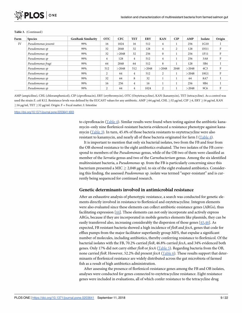
Among the FB resistant isolates, 4.3% showed resistance phenotype to the eight assessed anti-
biotics, and 57.3% showed resistance to at least four antibiotics (Table 3). Different results
were found for OB resistant bacteria, with a 8.5% of the bacterial isolates showed resistance to
the eight antibiotics. Nevertheless, 100% of OB-classified isolates were resistant to at least four
of the tested antibiotics (Table 4). These findings evidence a clear selection tendency for multi-
resistant bacteria when florfenicol and oxytetracycline are used as treatments.
Particularly interesting results were found for resistance tests against ciprofloxacin and
kanamycin, because only two of the florfenicol-resistant isolates showed resistance for cipro-
floxacin (Table 3). But in the case of the OB a 54.5% of the bacterial isolates showed resistance
Table 3. Minimum inhibitory concentrations of florfenicol-resistant bacteria to other antibiotic families (μg/mL).
Farm Species GenBank Similarity OTC CFC TET ERY KAN CIP AMP Isolate Origin
I Pseudomonas fragi 99% 512 1024 4 128 4 2 128 13H4 F
Pseudomonas sp 99% 512 2048 256 128 8 2 256 13G1 F
Pseudomonas fragi 99% 128 2048 64 1024 8 1 512 7C5 F
Pseudomonas fragi 99% 64 2048 64 128 2 1 128 5B1 I
Pseudomonas fluorescens 99% 512 2048 64 512 8 1 512 7A3 I
Pseudomonas sp 99% 256 2048 256 64 2 1 128 4B2 F
Serratia sp 99% 512 2048 512 512 8 1 >2048 4D1 I
Pseudomonas sp 99% 64 2048 32 128 2 1 128 9A1 I
Pseudomonas azotoformans 99% 512 2048 128 256 2 1 >2048 8C2 F
Serratia sp 99% 512 64 512 256 2 1 512 9A2 I
II Serratia sp 99% 128 512 128 512 4 1 256 1B4 F
Pseudomonas fragi 99% 256 >2048 64 512 4 1 256 1E2 F
Pseudomonas fluorescens 99% 256 >2048 32 512 8 1 256 2B4 F
Pseudomonas sp 99% 32 2048 32 64 4 1 128 1D2 I
Pseudomonas fragi 99% 512 128 32 64 4 1 128 6A4 I
Pseudomonas sp 99% 128 2048 64 512 8 1 128 6C5 F
Pseudomonas sp 99% 8 256 8 1024 32 1 >2048 5B8 I
Pseudomonas fluorescens 99% 64 2048 64 512 4 1 128 6H4 I
Pseudomonas sp 100% 2 256 8 512 32 1 >2048 5C8 I
Pseudomonas fluorescens 99% 128 2048 64 1024 8 1 128 7B8 I
Pseudomonas fluorescens 99% 512 16 256 1 2 1 16 6D3 F
Pseudomonas psychrophila 99% 64 16 32 1 2 1 16 6A5 I
Pseudomonas psychrophila 99% 512 >2048 512 >2048 2048 1024 >2048 4G7 I
Pseudomonas sp 99% 2 2048 16 64 4 1 128 6H3 F
Pseudomonas sp 100% 64 256 128 512 128 1 128 4D7 F
III Pseudomonas sp 98% 4 512 4 1024 32 1 256 1A8 I
Pseudomonas sp 100% 4 512 4 1024 32 1 256 1C7 F
Pseudomonas migulae 99% 4 128 4 512 2 1 256 3C4 F
Pseudomonas fluorescens 99% 512 512 128 512 8 1 >2048 4A11 F
Pseudomonas migulae 99% 2 64 256 1024 8 1 >2048 7F11 I
Hafnia sp 99% 256 128 256 2048 8 1 128 6B1 F
Pseudomonas fragi 99% 64 2048 32 128 2 1 64 8B12 I
Pseudomonas fluorescens 99% 64 64 32 128 2 1 128 8F12 F
Pseudomonas fluorescens 99% 2 256 4 512 32 1 >2048 11H8 F
Pseudomonas sp 99% 2 64 4 1024 32 1 >2048 9F10 I
Aeromonas molluscorum 99% 2 64 4 64 8 1 >2048 9E11 I
(Continued)
Isolation and characterization of multiresistant bacteria from farmed salmon gut
PLOS ONE | https://doi.org/10.1371/journal.pone.0203641 September 11, 2018 8 / 22
to ciprofloxacin (Table 4). Similar results were found when testing against the antibiotic kana-
mycin–only nine florfenicol-resistant bacteria evidenced a resistance phenotype against kana-
mycin (Table 3). In turn, 45.4% of those bacteria resistants to oxytetracycline were also
resistant to kanamycin, and nearly all of these bacteria originated for farm I (Table 4).
It is important to mention that only six bacterial isolates, two from the FB and four from
the OB showed resistance to the eight antibiotics evaluated. The two isolates of the FB corre-
spond to members of the Pseudomonas genus, while of the OB two of them were classified as
member of the Serratia genus and two of the Carnobacterium genus. Among the six identified
multiresistant bacteria, a Pseudomonas sp. from the FB is particularly concerning since this
bacterium presented a MIC� 2,048 μg/mL to six of the eight evaluated antibiotics. Consider-
ing this finding, the assessed Psudomonas sp. isolate was termed “super-resistant” and is cur-
rently being sequenced for continued research.
Genetic determinants involved in antimicrobial resistance
After an exhaustive analysis of phenotypic resistance, a search was conducted for genetic ele-
ments directly involved in resistance to florfenicol and oxytetracycline. Integron elements
were also evaluated since these elements can collect antibiotic-resistance genes (ARGs), thus
facilitating expression [44]. These elements can not only incorporate and actively express
ARGs, because if they are incorporated in mobile genetics elements like plasmids, they can be
easily transferred also, increasing considerably the dispersion of those genes [45,46]. As
expected, FB resistant bacteria showed a high incidence of floR and fexA, genes that code for
efflux pumps from the major facilitator superfamily group MFS, that expulse a significant
number of molecules, including antibiotics, thereby conferring resistance to florfenicol. Of the
bacterial isolates with the FB, 70.2% carried floR, 46.8% carried fexA, and 34% evidenced both
genes. Only 17% did not carry either floR or fexA (Table 5). Regarding bacteria from the OB,
none carried floR. However, 52.2% did present fexA (Table 6). These results support that deter-
minants of florfenicol resistance are widely distributed across the gut microbiota of farmed
fish as a result of high antibiotics administration.
After assessing the presence of florfenicol-resistance genes among the FB and OB isolates,
analyses were conducted for genes connected to oxytetracycline resistance. Eight resistance
genes were included in evaluations, all of which confer resistance to the tetracycline drug
Table 3. (Continued)
Farm Species GenBank Similarity OTC CFC TET ERY KAN CIP AMP Isolate Origin
IV Pseudomonas jessenii 99% 16 1024 16 512 4 1 256 1G10 I
Pseudomonas sp 99% 32 2048 32 128 4 2 128 1H11 F
Pseudomonas sp 99% 32 >2048 32 256 0 1 256 1F11 F
Pseudomonas sp 99% 4 128 4 512 4 1 256 3A8 F
Pseudomonas fragi 99% 64 2048 64 512 8 1 128 5B4 I
Pseudomonas sp 99% 512 >2048 512 >2048 >2048 2048 >2048 4C3 F
Pseudomonas sp 99% 2 64 4 512 2 1 >2048 10G1 F
Pseudomonas sp 99% 32 64 8 32 1 1 64 8A7 I
Pseudomonas sp 99% 16 256 4 16 1 1 256 9B4 I
Pseudomonas sp 99% 2 64 4 1024 2 1 >2048 9C6 F
AMP (ampiciline), CHL (chloramphenicol), CIP (ciprofloxacin), ERY (erythromycin), OTC (Oxytetracycline), KAN (kanamycin), TET (tetracycline). As a control was
used the strain E. coli K12. Resistance levels was defined by the EUCAST values for any antibiotic. AMP�64 μg/ml, CHL�32 μg/ml, CIP�4, ERY�16 μg/ml, KAN
�16 μg/ml, TET�32 μg/ml. Origin: F = Fecal matter; I: Intestine
https://doi.org/10.1371/journal.pone.0203641.t003
Isolation and characterization of multiresistant bacteria from farmed salmon gut
PLOS ONE | https://doi.org/10.1371/journal.pone.0203641 September 11, 2018 9 / 22
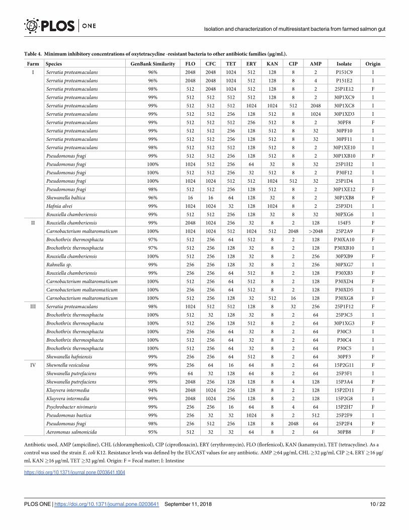
Table 4. Minimum inhibitory concentrations of oxytetracycline -resistant bacteria to other antibiotic families (μg/mL).
Farm Species GenBank Similarity FLO CFC TET ERY KAN CIP AMP Isolate Origin
I Serratia proteamaculans 96% 2048 2048 1024 512 128 8 2 P151C9 I
Serratia proteamaculans 96% 2048 2048 1024 512 128 8 4 P151E2 I
Serratia proteamaculans 98% 512 2048 1024 512 128 8 2 25P1E12 F
Serratia proteamaculans 99% 512 512 512 512 128 8 2 30P1XC9 I
Serratia proteamaculans 99% 512 512 512 1024 1024 512 2048 30P1XC8 I
Serratia proteamaculans 99% 512 512 256 128 512 8 1024 30P1XD3 I
Serratia proteamaculans 99% 512 512 512 256 512 8 2 30PF8 F
Serratia proteamaculans 99% 512 512 256 128 512 8 32 30PF10 I
Serratia proteamaculans 99% 512 512 256 128 512 8 32 30PF11 I
Serratia proteamaculans 98% 512 512 512 128 512 8 2 30P1XE10 I
Pseudomonas fragi 99% 512 512 256 128 512 8 2 30P1XB10 F
Pseudomonas fragi 100% 1024 512 256 64 32 8 32 25P1H2 I
Pseudomonas fragi 100% 512 512 256 32 512 8 2 P30F12 I
Pseudomonas fragi 100% 1024 1024 512 512 1024 512 32 25P1D4 I
Pseudomonas fragi 98% 512 512 256 128 512 8 2 30P1XE12 F
Shewanella baltica 96% 16 16 64 128 32 8 2 30P1XB8 F
Hafnia alvei 99% 1024 1024 32 128 1024 8 2 25P3D1 I
Rouxiella chamberiensis 99% 512 512 256 128 32 8 32 30PXG6 I
II Rouxiella chamberiensis 99% 2048 1024 256 32 8 2 128 154F5 F
Carnobacterium maltaromaticum 100% 1024 1024 512 1024 512 2048 >2048 25P2A9 F
Brochothrix thermosphacta 97% 512 256 64 512 8 2 128 P30XA10 F
Brochothrix thermosphacta 97% 512 256 128 32 8 2 128 P30XB10 I
Rouxiella chamberiensis 100% 512 256 128 32 8 2 256 30PXB9 F
Rahnella sp. 99% 256 256 128 32 8 2 256 30PXG7 I
Rouxiella chamberiensis 99% 256 256 64 512 8 2 128 P30XB3 F
Carnobacterium maltaromaticum 100% 512 256 64 512 8 2 128 P30XD4 F
Carnobacterium maltaromaticum 100% 256 256 64 512 8 2 128 P30XD5 I
Carnobacterium maltaromaticum 100% 512 256 128 32 512 16 128 P30XG8 F
III Serratia proteamaculans 98% 1024 512 512 128 8 32 256 25P1F12 F
Brochothrix thermosphacta 100% 512 32 128 32 8 2 64 25P3C5 I
Brochothrix thermosphacta 100% 512 256 128 512 8 2 64 30P1XG3 F
Brochothrix thermosphacta 100% 256 256 64 32 8 2 64 P30C3 I
Brochothrix thermosphacta 100% 512 256 64 32 8 2 64 P30C4 I
Brochothrix thermosphacta 100% 512 256 64 32 8 2 64 P30C5 I
Shewanella hafniensis 99% 256 256 64 512 8 2 64 30PF3 F
IV Shewnella vesiculosa 99% 256 64 16 64 8 2 64 15P2G11 F
Shewanella putrefaciens 99% 64 32 128 64 8 2 64 25P3F1 I
Shewanella putrefaciens 99% 2048 256 128 128 8 4 128 15P3A4 F
Kluyvera intermedia 94% 2048 1024 256 128 8 2 128 15P2D11 F
Kluyvera intermedia 99% 2048 1024 256 128 8 2 128 15P2G8 I
Psychrobacter nivimaris 99% 256 256 16 64 8 4 64 15P2H7 F
Pseudomonas baetica 99% 256 32 32 1024 8 2 512 25P2F9 I
Pseudomonas fragi 98% 256 512 256 128 8 2048 64 25P2F4 F
Aeromonas salmonicida 95% 512 32 32 64 8 2 64 30PB8 F
Antibiotic used, AMP (ampiciline), CHL (chloramphenicol), CIP (ciprofloxacin), ERY (erythromycin), FLO (florfenicol), KAN (kanamycin), TET (tetracycline). As a
control was used the strain E. coli K12. Resistance levels was defined by the EUCAST values for any antibiotic. AMP�64 μg/ml, CHL�32 μg/ml, CIP�4, ERY�16 μg/
ml, KAN�16 μg/ml, TET�32 μg/ml. Origin: F = Fecal matter; I: Intestine
https://doi.org/10.1371/journal.pone.0203641.t004
Isolation and characterization of multiresistant bacteria from farmed salmon gut
PLOS ONE | https://doi.org/10.1371/journal.pone.0203641 September 11, 2018 10 / 22
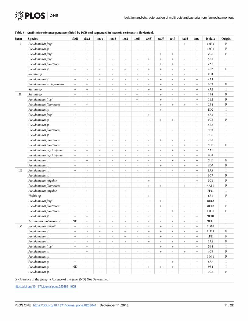
Table 5. Antibiotic resistance genes amplified by PCR and sequenced in bacteria resistant to florfenicol.
Farm Species floR fexA tet34 tet35 tetA tetB tetE tetH tetL tetM int1 Isolate Origin
I Pseudomonas fragi - + - - - - - - - + + 13H4 F
Pseudomonas sp - + - - + - - - - - + 13G1 F
Pseudomonas fragi + + - - - - - + + - + 7C5 F
Pseudomonas fragi + + - - - - + + + - + 5B1 I
Pseudomonas fluorescens + + - - - - - + + - + 7A3 I
Pseudomonas sp + - - - + - + + - - - 4B2 F
Serratia sp + + - - + - - - - - + 4D1 I
Pseudomonas sp + - - - - - - + - - + 9A1 I
Pseudomonas azotoformans + + - - - - - + - - + 8C2 F
Serratia sp + + - - - - + + - - + 9A2 I
II Serratia sp + - - - - + - + - - + 1B4 F
Pseudomonas fragi - - - - - + - + - - + 1E2 F
Pseudomonas fluorescens + + - - - - - + + + + 2B4 F
Pseudomonas sp + - - - - - - + - - + 1D2 I
Pseudomonas fragi + - - - - - + - - - + 6A4 I
Pseudomonas sp + + - - - - - + + - + 6C5 F
Pseudomonas sp + - - - - - - - - - + 5B8 I
Pseudomonas fluorescens + + - - - - - - - - + 6H4 I
Pseudomonas sp - - - - - - - - - - + 5C8 I
Pseudomonas fluorescens + + - - - - - + - - + 7B8 I
Pseudomonas fluorescens + - - - - - - - - + + 6D3 F
Pseudomonas psychrophila + + - - - - - - + - + 6A5 I
Pseudomonas psychrophila + - - - - - - - - - + 4G7 I
Pseudomonas sp - + - - - - - - + - + 6H3 F
Pseudomonas sp + - - - - - - + - + + 4D7 F
III Pseudomonas sp + - - - - - - - - - + 1A8 I
Pseudomonas sp - - - - - - - - - - + 1C7 F
Pseudomonas migulae - - - - - - + - - - + 3C4 F
Pseudomonas fluorescens + + - - - - + + - + + 4A11 F
Pseudomonas migulae + + - - + - - - - - + 7F11 I
Hafnia sp + - - - + - + - - - - 6B1 F
Pseudomonas fragi - - - - - - - + - - + 8B12 I
Pseudomonas fluorescens + + - - - - - + - - + 8F12 F
Pseudomonas fluorescens - - - - - - - - + - + 11H8 F
Pseudomonas sp + + - - - - - - - - + 9F10 I
Aeromonas molluscorum ND + - - - - - - - - + 9E11 I
IV Pseudomonas jessenii + - - - - - - + - - + 1G10 I
Pseudomonas sp + - - - + - + + - - + 1H11 F
Pseudomonas sp + - - - + - - + - - + 1F11 F
Pseudomonas sp - - - - - - + - - - + 3A8 F
Pseudomonas fragi + + - - - - - + + - + 5B4 I
Pseudomonas sp - + - - - - - + - + + 4C3 F
Pseudomonas sp - - - - - - - - - - + 10G1 F
Pseudomonas sp + - - - - - - - + - + 8A7 I
Pseudomonas sp ND - - - + - + + + - - 9B4 I
Pseudomonas sp + + - - - - - - - - + 9C6 F
(+) Presence of the gene; (-) Absence of the gene; (ND) Not Determined.
https://doi.org/10.1371/journal.pone.0203641.t005
Isolation and characterization of multiresistant bacteria from farmed salmon gut
PLOS ONE | https://doi.org/10.1371/journal.pone.0203641 September 11, 2018 11 / 22

Table 6. Antibiotic resistance genes amplified by PCR and sequenced in bacteria resistant to oxytetracycline.
Farm Species floR fexA tet34 tet35 tetA tetB tetE tetH tetL tetM Int1 Isolate Origin
I Serratia proteamaculans - - - + + + - - + + + P151C9 I
Serratia proteamaculans - - - - - + - - + + + P151E2 I
Serratia proteamaculans - - - + - + + - - + + 25P1E12 F
Serratia proteamaculans - - - - - + + - + + + 30P1XC9 I
Serratia proteamaculans - - - - - + + - + - + 30P1XC8 I
Serratia proteamaculans - + - - + + + - + - + 30P1XD3 I
Serratia proteamaculans - + - - - + + - + + + 30PF8 F
Serratia proteamaculans - + - - - + + - + + + 30PF10 I
Serratia proteamaculans - + - - - + + - + - + 30PF11 I
Serratia proteamaculans - - - - - + + - + - + 30P1XE10 I
Pseudomonas fragi - + - + + + - + - + + 30P1XB10 F
Pseudomonas fragi - - - + + - - + + + + 25P1H2 I
Pseudomonas fragi - + + - + - - - - + + P30F12 I
Pseudomonas fragi - - - + + - - + - - + 25P1D4 I
Pseudomonas fragi - + - - + - - - - + + 30P1XE12 F
Shewanella baltica - + - - - + - - + + + 30P1XB8 F
Hafnia alvei - - - - - - - - - + + 25P3D1 I
Rouxiella chamberiensis - + - - - + - + + + + 30PXG6 I
II Rouxiella chamberiensis - - - - + - + - - + + 154F5 F
Carnobacterium maltaromaticum - - - - - - - - + + + 25P2A9 F
Brochothrix thermosphacta - + - - - - + - + + + P30XA10 F
Brochothrix thermosphacta - + - - - - - - + + + P30XB10 I
Rouxiella chamberiensis - + - - - - - + + + + 30PXB9 F
Rahnella sp. - + + - - - - + + + + 30PXF7 I
Rouxiella chamberiensis - + - - - - - + + + + P30XB3 F
Carnobacterium maltaromaticum - + - - + - + - + + + P30XD4 F
Carnobacterium maltaromaticum - + - - - + - - + + + P30XD5 I
Carnobacterium maltaromaticum - + - - - - + - + - + P30XG8 F
III Serratia proteamaculans - - - - + + + - + + + 25P1F12 F
Brochothrix thermosphacta - + - - + - - - + + + 25P3C5 I
Brochothrix thermosphacta - ND - - + - - - + + + 30P1XG3 F
Brochothrix thermosphacta - + - - - - - - + + + P30C3 I
Brochothrix thermosphacta - + - - - - - + + + + P30C4 I
Brochothrix thermosphacta - ND - - - - + - + + + P30C5 I
Shewanella hafniensis - + - + - + + + + + + 30PF3 F
IV Shewnella vesiculosa - + - - - + - - - - + 15P2G11 F
Shewanella putrefaciens - + - - - + + - - - + 25P3F1 I
Shewanella putrefaciens - - - + - + - - - + + 15P3A4 F
Kluyvera intermedia - + - - - - + - - - + 15P2D11 F
Kluyvera intermedia - - - + - - + - - + + 15P2G8 I
Psychrobacter nivimaris - ND - - + - - - + - + 15P2H7 F
Pseudomonas baetica - - - + + + + + - + + 25P2F9 I
Pseudomonas fragi - - - - - - + - - - + 25P2F4 F
Aeromonas salmonicida - ND - - + + + - + - + 30PB8 F
(+) Presence of the gene; (-) Absence of the gene; (ND) Not Determined.
https://doi.org/10.1371/journal.pone.0203641.t006
Isolation and characterization of multiresistant bacteria from farmed salmon gut
PLOS ONE | https://doi.org/10.1371/journal.pone.0203641 September 11, 2018 12 / 22
family. These apply named tet genes included tetA, tetB, tetE, tetH, tetL, tet34, and tet 35, all of
which were previously described in sediments associated with salmon farms [43], but not with
intestinal microbiota of the farmed fish. The presence of tetM was also included considering
prior reports of being a widely dispersed and prevalent resistance mechanism against tetracy-
clines [47,48]. For OB resistance bacteria, 100% of isoaltes presented at least one of the evalu-
ated tet genes, the more prevalent of which was, not surprisingly, tetM (72.7%). Following in
prevalence was tetL (68.1%). Among this group of bacterial isolates, 4.5% of the bacterial iso-
lates carried six tet genes, 9,1% five and 15.9% four. In other words, 29.5% of oxytetracycline-
resistant bacteria carried at least four different resistance genes to this antibiotic (Table 6).
Regarding those bacteria included in the FB, 78.7% of isolates had at least one of the evaluate
tet genes, 48.9% of which were carries of tetH (i.e., the most abundant in florfenicol-resistant
bacteria; Table 5). Likewise, an estimated 12.8% of the FB isolates carried at least three of the
evaluated tet genes. The presence of specific ARGs against florfenicol and oxytetracycline cor-
roborate the phenotypic resistances observed for each of the resistant isolates. Further research
is needed regarding those resistant isolates for which no ARGs were detected. To understand
these results, it is necessary to take into account the limitations of detection using the PCR
technique, since only eight out of 28 tet genes were evaluated [49], the most frequently
described in aquaculture [50]. The same happens with the genes of resistance to phenicoles
since only those that are detected more frequently at environmental level were evaluated
[38,51]. Therefore a metagenomic analysis would be very useful to detect all the possible genes
involved in the resistance against these antibiotics.
Once resistance elements to the most widely used antibiotics were determined, a search for
integron-like elements was conducted. The presence of type I integrase was established
through PCR analyses and posterior sequencing. In the case of OB isolates, 100% were positive
for type I integrase, while 96% of FB isolates presented type I integrase (Tables 5 and 6). These
results suggest that this type of element might be widely distributed among resistant bacteria
residing within the gut microbiota of fish. These are highly dangerous elements that play a fun-
damental role in the distribution and phenotypic expression of ARGs. A dedicated study on
the detected integrons is currently underway in our lab.
Taxonomic differences among bacteria resistant to florfenicol and
oxytetracycline
In contrast to previously published reports, the present study simultaneously provides informa-
tion related to the phenomena of resistance to florfenicol and oxytetracycline, with the different
tested isolates having been directly obtained from the intestinal microbiota of farmed salmon
exposed to high concentrations of antibiotics. Of note, the conducted analyses detected differences
in species richness for resistant bacteria. More specifically, the FB isolates evidenced a high pre-
dominance of Pseudomonas species, a phenomenon that was found across the sampled farming
centers and among all the evaluated temperatures. To a lesser degree, members of the Gammapro-
tebacteria class were also isolated, including Aeromonas, Serratia, and Hafnia species. Regarding
OB isolates, a high incidence of Gammaprotebacteria were also observed, as represented by the
genera Serratia, Hafnia, Rouxiella, Rahnella, Kluyvera, Shewanella, psychrobacter, Aeromonas, and
Pseudomonas. Furthermore, Gram-positive bacteria were represented by the Firmicutes phylum,
notable among which were Carnobacterium and Brochothrix, among others.
Discussion
Antibiotic use in the production of animal proteins is a practice that should be controlled on a
global scale, specifically since the application of these medicines for veterinary ends many
Isolation and characterization of multiresistant bacteria from farmed salmon gut
PLOS ONE | https://doi.org/10.1371/journal.pone.0203641 September 11, 2018 13 / 22

times lacks the degree of strict control imposed on human medicines. One of the primary
methods for administering antibiotics to industrially farmed animals is medicated feed [52–
54]. This delivery method is the most popular among farmed fish due to the notable technical
difficulties associated with administering antibiotics through other means, such as injection
[55,56]. The ingestion of medicated feed implicates that the intestinal microbiota of these ani-
mals is strongly affected. Antibiotics substantially change the bacterial diversity of gut micro-
biota in farmed animals [53], which, often, can result in functional and growth problems [57].
Another effect caused by the high use of the medicated food on the intestinal microbiota is
that the feces could contain resistant bacteria, which could be easily dispersed in the environ-
ment [58,59]. In this work it was possible to demonstrate that the feces contain multiresistant
bacteria, which are constantly being released into the environment. Additionally, it was
observed that the diversity of resistant isolates is similar to that found in the intestine, reaffirm-
ing in this way that eventually any resistant bacteria selected in the intestine carrying ARGs
could be disseminated through the feces.
Various studies have extensively characterized the gut-microbiota composition of Atlantic
salmon, reporting total dominance by the Proteobacteria phylum. More specific still, Pseudo-monas species of the Gammaproteobacteria class are the most represented [29,60]. These prior
descriptions were supported by the present study, with most of the resistant isolates being of
the Pseudomonas genus. Prior research has similarly characterized the effect that oxytetracy-
cline use has on the microbiota composition of Atlantic salmon. For example, Navarrete et al.(2008) [29] exposed Atlantic salmon to high oxytetracycline concentrations under experimen-
tal conditions (i.e., not in the field) and found a notable enrichment of Aeromonas salmonicida,
a bacterial pathogen. In the present study, only one Aeromonas isolate was found in the OB
group, thus corroborating the possible selection of this bacterium under high oxytetracycline
concentrations.
A fundamental finding of the present study is that oxytetracycline-resistant isolates were
found to be more diverse than those resistant to florfenicol. This could have direct implications
on treatments and the strategies applied to the use of these antibiotics. Since the early report of
P. salmonis infection by the end of the 80s, oxytetracycline and quinolones have been used to
tackle the infection. Also, oxytetracycline is the most widely used antibiotic during freshwater
rearing, with florfenicol use nearly null during this stage [25]. Consequently, the large majority
of farmed fish with a freshwater stage have been subjected to oxytetracycline treatments [25].
This could, in turn, mean that the gut microbiota of these fish is already rich in bacteria resis-
tant to oxytetracycline. Little research has reported on substantial changes to the microbiota of
freshwater- versus seawater-stage salmon. However, some authors speculate that a large part of
the microbiota would be conserved between systems [61]. In contrast to freshwater farming,
florfenicol is the most widely used antibiotic in seawater farming. Supporting this claim, all of
the currently assessed fish farms had applied three to four oral applications of florfenicol, and
only farm I had used an additional treatment of oxytetracycline at the time of sampling. This
might explain the lesser diversity of bacteria resistant to florfenicol; i.e., the constant florfenicol
treatments may have selected and maintained only those bacteria with the most efficient mech-
anisms against this antibiotic. This is the perfect scenario for Pseudomonas members, the
genomes of which are equipped with multiple resistance elements that make these bacteria
competitive in environments saturated with antibiotics [62,63].
In addition to the well-known antibiotic resistance capacity of Pseudomonas, the present
study detected most of the resistance genes included in assessments, and the majority of these
genes, excepting tetM, were drug efflux pumps. These findings corroborate that these genetic
elements are a principal trait of Pseudomonas bacteria [64]. Considering the phenotypic versa-
tility of Pseudomonas, it was unsurprising that the majority of the obtained florfenicol-resistant
Isolation and characterization of multiresistant bacteria from farmed salmon gut
PLOS ONE | https://doi.org/10.1371/journal.pone.0203641 September 11, 2018 14 / 22
isolates were of this genus. Regarding oxytetracycline-resistant isolates, the detected genera, as
with Pseudomonas, are highly ubiquitous in different systems. For example, Hafnia alvei has
been isolated from the intestinal microbiota of various organisms, including pigs, cows, and
fish [65,66]. This bacterium is an important contaminant of packaged food [67], meaning that
H. alvei is an ideal resistant-isolate candidate in any system. These traits can also be found in
species of Shewanella [68,69], Serratia [70,71], Carnobacterium [72], Brochotrhix [73,74], and
Kluyvera [75,76]. All of these bacteria can colonize multiple environments, and the ability to
acquire resistance in any given environment represents a significant risk for human health.
Only Aeromonas molluscorum [77,78] and Psychrobacter nivimaris [79] are exclusive to the
aquatic environment, which translates into a highly probable exchange of ARGs between bac-
teria directly related to the human system and environmental bacteria from the gut microbiota
of fish.
Tetracyclines are commonly used in veterinary medicine around the world, and, specifi-
cally, oxytetracycline is the most widely used antibiotic during the freshwater stage of salmon
growth in Chile [25]. Origin records for the Atlantic salmon sampled in the present study were
not obtained, but it is very likely that the sampled fish were exposed to high doses of oxytetra-
cycline during the freshwater stage. This would explain the high instance of oxytetracycline-
resistant bacteria, even when oxytetracycline was not applied during the seawater stage. Simi-
larly, the high prevalence of tet genes found for the FB and OB could be explained by the high
frequency at which these genes localize in mobile genetic elements, such as plasmids or trans-
posons [49]. The genes tetA, tetB, tetE, tetL, tetH, tet34, and tet35 have been recorded in the
sediments and water column of Chilean salmon farms [43,80]. An interisting case is the gene
tetM, this globally distributed gene presents a clinically confirmed resistance mechanism to tet-
racyclines [81,82]. One trait of this gene is that it is frequently associated with transposons and
plasmids in human pathogenic bacteria [83,84]; this could be a clear sign of the impacts this
gene is having from the clinical to the environmental spheres. Consequently, antibiotics used
in industrially farmed animals could substantially contribute to the maintenance and disper-
sion of ARGs in natural environments.
As with the tet genes, the presence of floR, a drug efflux pump of the major facilitator super-
family (MFS), has previously been reported in bacteria collected from marine sediments asso-
ciated with salmon farming in Chile [39]. This gene has even been detected in a plasmid that
also transported tet and qnr genes, the latter of which are implicated in resistance to quino-
lones [85]. However, the present study is the first to report on the existence of floR in the gut
microbiota of salmon. Furthermore, this investigation is the first to find and report on fexA, an
efflux pump of the major facilitator superfamily also, in association with Chilean salmon farm-
ing, with no previously published study having detected this gene in relation to the Chilean
aquaculture industry. This fenicol-resistance gene was first described in the bacterium Staphy-lococcus lentus [40], and has since almost been exclusively described in members of the Staphy-lococcus genus [41,42,51,86–88], both for humans and animals, but never in the marine
environment. Apart from the Staphylococcus genus, fexA has been described in members of
the Enterococcus genus, specifically in association with industrially farmed terrestrial animals
[89,90]. No Staphylococcus or Enterococcus bacteria were found among any of the obtained
resistant isolates, but the presented findings do support the ability of fexA to disperse across a
wide variety of both Gram-positive and–negative bacteria.
One curious outcome of the conducted study was that none of the oxytetracycline-resistant
bacteria presented floR, but, by contrast, 52,2% did present fexA. As mentioned, the most
widely used antibiotic used in Chilean salmon farming is oxytetracycline, and there is a nearly
null use of florfenicol in the freshwater stage. Prior reports have found fexA in plasmids con-
taining a great number of resistance genes to other antibiotics [87]. Therefore, this gene has
Isolation and characterization of multiresistant bacteria from farmed salmon gut
PLOS ONE | https://doi.org/10.1371/journal.pone.0203641 September 11, 2018 15 / 22
probably been selected during the freshwater stage in plasmids that also carry tetracylcin-resis-
tance genes. This co-selection is likely since the water used in the freshwater rearing stage is
collected from the lakes and rivers most exposed to human activities, including livestock
farms, and, as stated, fexA has almost exclusively been isolated from industrially farmed terres-
trial animals. This is another compelling case for ARGs primarily originating from human
activities and not directly from salmon farming.
In contrast to findings from sediments related to intense salmon farming in Chile [91], the
present study found a high incidence of class I integrons in resistant isolates from both the FB
and OB databases. In fact, 100% of OB isolates and 96% of FB isolates were positive for type I
integrase. These genetic elements are the greatest contributors to the evolution and dispersion
of ARGs [92]. Integrons are true genetic platforms that acquire exogenous genes through
mobile genetic cassettes, having the ability to collect up to hundreds of genes in the same unit
[93]. Nevertheless, these elements did not originate in bacterial pathogens, nor is their primary
function to collect antibiotic-resistance genes. Integrons frequently appear in Betaproteobac-teria native to soils, freshwater, and saltwater. Genetic cassettes are incorporated alongside ele-
ments that are highly varied and much more diverse than ARGs [94]. Integrons are very
successful and ubiquitous in natural environments, and it is therefore unsurprising that the
majority of the isolated OB and FB bacteria would present these elements. Concern arises
when these elements have a clinical origin and carry ARGs for most of the known antibiotics
[95]. Integrons carrying ARGs against quinolones (qnrA and qnrB), trimethoprims (drfA12),
and aminoglycosides (aad2) have recently been described in the Chilean farming system,
which is in addition to some clinical Escherichia coli also carrying these plasmid-transferred
integrons [96]. This appears to be credible proof that there is an interaction between the clini-
cal environment and that of salmon farming. However, a number of questions remain–What
is the origin of these integrons? Are they from aquaculture-associated bacteria? Or have contri-
butions from the clinical environment contaminated bacteria from salmon farming? These
questions need to be answered. No integron from either the OB or FB bacteria presented the
traits described by Tomova et al. (2018) [90].
Another important aspect to consider in this work is the Antibiotic Resistance Rate (ARR).
Overall, it was found that less than 1% of the total bacterial isolates were resistant to oxytetra-
cycline or florfenicol (0.94% for florfenicol and 0.87% for oxytetracycline). Although there are
few works in which this rate is addressed at an environmental level [97,98], values close to 1%
can be considered high [97–100]. In these works it can be seen that when the concentration of
antibiotics in the medium increases, the value of this range increases too. This phenomenon is
corroborated in the present work since the ARR in the FB for farm II exceeds 1%, almost twice
as much as that presented by the other farms and it is precisely the farm II that the major num-
ber of treatments with florfenicol presents, four. The same effect is observed in the OB in farm
I where ARR again exceeds 1% and the farm I is the only one that uses oxytetracycline as a
treatment in its seawater stage.
The information collected in the present study clearly indicate that using large amounts of
antibiotics to treat industrially farmed animals has the consequence of selecting for multiresis-
tant bacteria in the intestinal microbiota, which then have undeniable advantages when liber-
ated into the environment through the feces. While the obtained results are somewhat biased
towards “culturable” bacteria, metagenomics data are currently being analyzed, the results of
which will reveal all of the bacteria that did not grow in a TSA medium. This information will
provide a complete picture for the behaviors of the bacterial populations interacting in this
complex system. Meanwhile, the currently reported data do show that the intestinal micro-
biota, as a system “semi-isolated” from the environment yet in direct contact with applied anti-
biotics, is the perfect place for interactions to occur between bacteria from different
Isolation and characterization of multiresistant bacteria from farmed salmon gut
PLOS ONE | https://doi.org/10.1371/journal.pone.0203641 September 11, 2018 16 / 22
environments, including from the human clinical context. Therefore, the gut microbiota of
farmed salmon could also serve as the perfect reservoir for ARGs, the dispersion of which
would be fundamentally related to feces. The release of feces with resistant bacteria and ARGs
is creating an environment constantly rich in resistance elements, creating an ideal situation
for genetic exchanges to occur between different bacterial populations, whether in the water
column or in marine sediments. These populations are precisely where fish pathogens can be
found, thus notably increasing the risk that these bacteria will acquire resistance elements. If
this occurs, no effective treatments will exist for the control of these pathogens. Fortunately, a
number of aquaculture producers are beginning to understand the gravity of the situation.
Measures aimed at decreasing antibiotic usage are increasingly being adopted, with expecta-
tions that by 2020, there will be at least a 50% decrease in the use of these substances.
Conclusion
In this study, bacterial isolates from fish gut microbiota of the Salmo salar species, showing
high resistance levels to antibiotics oxytetracycline and florfenicol, were characterised. The
feces, represent important vehicles of dispersion of resistant bacteria and ARGs. The analyses
showed that the high use of these antibiotics selects bacteria that are multiresistant to a wide
range of antibiotics. In turn, these bacteria show a high prevalence of antibiotic resistance
genes, thus, verifying the phenotypic resistance with the mechanism present in the bacteria to
cope with these substances. Furthermore, almost 100% of these (antibiotic) resistant isolates
showed class 1 integrons, dangerous elements involved in the resistance phenomenon due to
their capacity of dispersion.
Supporting information
S1 Table. Genbank access numbers for 16S rRNA genes amplified from different bacterial
isolates.
(DOCX)
S2 Table. Genbank access numbers for the partial sequence of the antibiotic resistance
genes (ARGs) amplified by PCR.
(DOCX)
Acknowledgments
Thanks to Dr. Lina Jimena Cortes Salinas for figures advising and text correction.
Author Contributions
Conceptualization: Sebastian Higuera-Llanten, Jorge Olivares-Pacheco.
Data curation: Sebastian Higuera-Llanten, Felipe Vasquez-Ponce, Beatriz Barrientos-
Espinoza.
Formal analysis: Sebastian Higuera-Llanten, Felipe Vasquez-Ponce, Fernando O. Mardones,
Sergio H. Marshall, Jorge Olivares-Pacheco.
Funding acquisition: Jorge Olivares-Pacheco.
Methodology: Sebastian Higuera-Llanten, Felipe Vasquez-Ponce.
Writing – original draft: Sebastian Higuera-Llanten, Jorge Olivares-Pacheco.
Isolation and characterization of multiresistant bacteria from farmed salmon gut
PLOS ONE | https://doi.org/10.1371/journal.pone.0203641 September 11, 2018 17 / 22
Writing – review & editing: Fernando O. Mardones, Sergio H. Marshall, Jorge Olivares-
Pacheco.
References1. Berrian AM, Smith MH, van Rooyen J, Martınez-Lopez B, Plank MN, Smith WA, et al. A community-
based One Health education program for disease risk mitigation at the human-animal interface. One
Heal. 2018; 5: 9–20. https://doi.org/10.1016/j.onehlt.2017.11.002
2. Durão P, Balbontın R, Gordo I. Evolutionary mechanisms shaping the maintenance of antibiotic resis-
tance. Trends Microbiol. 2018; 26(8): 677–691. https://doi.org/10.1016/j.tim.2018.01.005 PMID:
29439838
3. Baquero F, Alvarez-Ortega C, Martinez JL. Ecology and evolution of antibiotic resistance. Env Micro-
biol Reports. 2009; 1: 469–476. https://doi.org/10.1111/j.1758-2229.2009.00053.x
4. Martinez JL, Olivares J. Environmental pollution by antibiotic resistance genes. In: Keen P, Montforts
M, editors. Antimicrobial Resistance in the Environment. New Jersey: Willey-Blackwell; 2012. pp.
151–172.
5. Martinez JL, Fajardo A, Garmendia L, Hernandez A, Linares JF, Martinez-Solano L, et al. A global
view of antibiotic resistance. FEMS Microbiol Rev. 2009; 33: 44–65. https://doi.org/10.1111/j.1574-
6976.2008.00142.x PMID: 19054120
6. Hadley C, Crooks DL. Coping and the biosocial consequences of food insecurity in the 21st century.
Am J Phys Anthropol. 2012; 149: 72–94. https://doi.org/10.1002/ajpa.22161 PMID: 23109261
7. Liao IC, Chao N-H. Aquaculture and food crisis: opportunities and constraints. Asia Pac J Clin Nutr.
2009; 18: 564–9. http://www.ncbi.nlm.nih.gov/pubmed/19965349 PMID: 19965349
8. Done HY, Venkatesan AK, Halden RU. Does the recent growth of aquaculture create antibiotic resis-
tance threats different from those associated with land animal production in agriculture? AAPS J.
2015; 17: 513–524. https://doi.org/10.1208/s12248-015-9722-z PMID: 25700799
9. Lubroth J. FAO and the One Health approach. Current topics in microbiology and immunology.
2012. pp. 65–72. https://doi.org/10.1007/82_2011_137
10. Sleeman JM, DeLiberto T, Nguyen N. Optimization of human, animal, and environmental health by
using the One Health approach. J Vet Sci. 2017; 18: 263. https://doi.org/10.4142/jvs.2017.18.S1.263
PMID: 28859266
11. Lebov J, Grieger K, Womack D, Zaccaro D, Whitehead N, Kowalcyk B, et al. A framework for One
Health research. One Heal. 2017; 3: 44–50. http://doi.10.1016/j.onehlt.2017.03.004
12. Baum SE, Machalaba C, Daszak P, Salerno RH, Karesh WB. Evaluating one health: Are we demon-
strating effectiveness? One Heal. 2017; 3: 5–10. http://doi.10.1016/j.onehlt.2016.10.004
13. FAO. The State of World Fisheries and Aquaculture 2016. http://doi.92-5-105177-1
14. Asche F, Hansen H, Tveteras R, Tveterås S. The salmon disease crisis in Chile. Mar Resour Econ.
MRE Foundation, Inc. 2009; 24(4): 405–411. http://doi.10.5950/0738-1360-24.4.405
15. Rozas M, Enrıquez R. Piscirickettsiosis and Piscirickettsia salmonis in fish: a review. J Fish Dis. 2014;
37: 163–188. https://doi.org/10.1111/jfd.12211 PMID: 24279295
16. Makrinos DL, Bowden TJ. Growth characteristics of the intracellular pathogen, Piscirickettsia salmo-
nis, in tissue culture and cell-free media. J Fish Dis. 2017; 40: 1115–1127. https://doi.org/10.1111/jfd.
12578 PMID: 28026007
17. House M, Bartholomew J, Winton J, Fryer J. Relative virulence of three isolates of Piscirickettsia sal-
monis for coho salmon Oncorhynchus kisutch. Dis Aquat Organ. 1999; 35: 107–113. https://doi.org/
10.3354/dao035107 PMID: 10092973
18. Arkush K, McBride A, Mendonca H, Okihiro M, Andree K, Marshall S, et al. Genetic characterization
and experimental pathogenesis of Piscirickettsia salmonis isolated from white seabass Atractoscion
nobilis. Dis Aquat Organ. 2005; 63: 139–149. https://doi.org/10.3354/dao063139 PMID: 15819429
19. Corbeil S, Hyatt AD, Crane MS. Characterisation of an emerging rickettsia-like organism in Tasmanian
farmed Atlantic salmon Salmo salar. Dis Aquat Organ. 2005; 64: 37–44. https://doi.org/10.3354/
dao064037 PMID: 15900686
20. Marshall SH, Flores-Herrera P, Henrıquez FA, Gomez FA. Identification and characterization of two
variants of the Hfq-sRNA-chaperone in the fish pathogen Piscirickettsia salmonis. J Fish Dis. 2018;
41(3): 501–509. https://doi.org/10.1111/jfd.12752 PMID: 29159897
21. EvensenØ. Immunization Strategies against Piscirickettsia salmonis Infections: Review of vaccination
approaches and modalities and their associated immune response profiles. Front Immunol. 2016; 7:
482. https://doi.org/10.3389/fimmu.2016.00482 PMID: 27917172
Isolation and characterization of multiresistant bacteria from farmed salmon gut
PLOS ONE | https://doi.org/10.1371/journal.pone.0203641 September 11, 2018 18 / 22
22. Fuentealba P, Aros C, Latorre Y, Martınez I, Marshall S, Ferrer P, et al. Genome-scale metabolic
reconstruction for the insidious bacterium in aquaculture Piscirickettsia salmonis. Bioresour Technol.
2017; 223: 105–114. https://doi.org/10.1016/j.biortech.2016.10.024 PMID: 27788423
23. Maisey K, Montero R, Christodoulides M. Vaccines for piscirickettsiosis (salmonid rickettsial septicae-
mia, SRS): the Chile perspective. Expert Rev Vaccines. 2017; 16: 215–228. https://doi.org/10.1080/
14760584.2017.1244483 PMID: 27690686
24. Tandberg J, Oliver C, Lagos L, Gaarder M, Yañez AJ, Ropstad E, et al. Membrane vesicles from Pis-
cirickettsia salmonis induce protective immunity and reduce development of salmonid rickettsial septi-
cemia in an adult zebrafish model. Fish Shellfish Immunol. 2017; 67: 189–198. https://doi.org/10.
1016/j.fsi.2017.06.015 PMID: 28600194
25. Sernapesca. Informe sobre uso de antimicrobianos por la salmonicultura nacional: Año 2017. Servicio
Nacional de Pesca y Acuicultura; 2018. http://www.sernapesca.cl/presentaciones/Comunicaciones/
Informe_Sobre_Uso_de_Antimicrobianos-2017.pdf
26. Sørum H. Antimicrobial drug resistance in fish pathogens. ASM Pub2Web; 2006. pp. 213–238. http://
doi.10.1128/9781555817534.CH13
27. Smith PR, Le Breton A, Horsberg TE, Corsin F. Guidelines for antimicrobial use in aquaculture. Guide
to antimicrobial use in animals. Oxford, UK: Blackwell Publishing; 2009. pp. 207–218. http://doi.10.
1002/9781444302639.ch12
28. Tomova A, Ivanova L, Buschmann AH, Rioseco ML, Kalsi RK, Godfrey HP, et al. Antimicrobial resistance
genes in marine bacteria and human uropathogenic Escherichia coli from a region of intensive aquacul-
ture. Environ Microbiol Rep. 2015; 7: 803–9. https://doi.org/10.1111/1758-2229.12327 PMID: 26259681
29. Navarrete P, Mardones P, Opazo R, Espejo R, Romero J. Oxytetracycline treatment reduces bacterial
diversity of intestinal microbiota of Atlantic salmon. J Aquat Anim Health. 2008; 20: 177–183. https://
doi.org/10.1577/H07-043.1 PMID: 18942594
30. Martınez JL. Ecology and evolution of chromosomal gene transfer between environmental microorgan-
isms and pathogens. Microbiol Spectr. 2018; 6(1). http://doi.10.1128/microbiolspec.MTBP-0006-2016
31. Salas Leiva J, Opazo R, Remond C, Uribe E, Velez A, Romero J. Characterization of the intestinal
microbiota of wild caught and farmed fine flounder (Paralichthys adspersus). Lat Am J Aquat Res.
2017; 45: 370–378. http://doi.10.3856/vol45-issue2-fulltext-12
32. Navarrete P, Magne F, Mardones P, Riveros M, Opazo R, Suau A, et al. Molecular analysis of intesti-
nal microbiota of rainbow trout (Oncorhynchus mykiss). FEMS Microbiol Ecol. 2010; 71: 148–156.
https://doi.org/10.1111/j.1574-6941.2009.00769.x PMID: 19780831
33. Cantas L, Sørby JRT, Alestrom P, Sørum H. Culturable gut microbiota diversity in zebrafish. Zebrafish.
2012; 9: 26–37. https://doi.org/10.1089/zeb.2011.0712 PMID: 22428747
34. Alvarez-Ortega C, Wiegand I, Olivares J, Hancock RE, Martinez JL. Genetic determinants involved in
the susceptibility of Pseudomonas aeruginosa to beta-lactam antibiotics. Antimicrob Agents Che-
mother. 2010; 54: 4159–4167. https://doi.org/10.1128/AAC.00257-10 PMID: 20679510
35. Fernandez L, Alvarez-Ortega C, Wiegand I, Olivares J, Kocincova D, Lam JS, et al. Characterization
of the polymyxin B resistome of Pseudomonas aeruginosa. Antimicrob Agents Chemother. 2013; 57:
110–119. https://doi.org/10.1128/AAC.01583-12 PMID: 23070157
36. EUCAST. (2017) Breakpoint tables for interpretation of MICs and zone diameters Version 7.1, 3–7.
37. Altschul SF, Gish W, Miller W, Myers EW, Lipman DJ. Basic local alignment search tool. J Mol Biol.
1990; 215: 403–10. https://doi.org/10.1016/S0022-2836(05)80360-2 PMID: 2231712
38. Miranda CD, Rojas R. Occurrence of florfenicol resistance in bacteria associated with two Chilean
salmon farms with different history of antibacterial usage. Aquaculture. 2007; 266: 39–46. http://doi.
10.1016/j.aquaculture.2007.02.007
39. Fernandez-Alarcon C, Miranda CD, Singer RS, Lopez Y, Rojas R, Bello H, et al. Detection of the floR
gene in a diversity of florfenicol resistant gram-negative bacilli from freshwater salmon farms in Chile.
Zoonoses Public Health. 2010; 57: 181–188. https://doi.org/10.1111/j.1863-2378.2009.01243.x
PMID: 19538451
40. Kehrenberg C, Schwarz S. fexA, a novel Staphylococcus lentus gene encoding resistance to florfeni-
col and chloramphenicol. Antimicrob Agents Chemother. 2004; 48: 615–8. https://doi.org/10.1128/
AAC.48.2.615-618.2004 PMID: 14742219
41. Couto N, Belas A, Rodrigues C, Schwarz S, Pomba C. Acquisition of the fexA and cfr genes in Staphy-
lococcus pseudintermedius during florfenicol treatment of canine pyoderma. J Glob Antimicrob Resist.
2016; 7: 126–127. https://doi.org/10.1016/j.jgar.2016.08.008 PMID: 27750159
42. Kehrenberg C, Schwarz S. Distribution of florfenicol resistance genes fexA and cfr among chloram-
phenicol-resistant Staphylococcus isolates. Antimicrob Agents Chemother. 2006; 50: 1156–1163.
https://doi.org/10.1128/AAC.50.4.1156-1163.2006 PMID: 16569824
Isolation and characterization of multiresistant bacteria from farmed salmon gut
PLOS ONE | https://doi.org/10.1371/journal.pone.0203641 September 11, 2018 19 / 22
43. Miranda CD, Kehrenberg C, Ulep C, Schwarz S, Roberts MC. Diversity of tetracycline resistance
genes in bacteria from Chilean salmon farms. Antimicrob Agents Chemother. 2003; 47: 883–888.
https://doi.org/10.1128/AAC.47.3.883-888.2003 PMID: 12604516
44. Mazel D. Integrons: agents of bacterial evolution. Nat Rev Microbiol. 2006; 4: 608–620. https://doi.
org/10.1038/nrmicro1462 PMID: 16845431
45. Cambray G, Guerout A-M, Mazel D. Integrons. Annu Rev Genet. 2010; 44: 141–166. https://doi.org/
10.1146/annurev-genet-102209-163504 PMID: 20707672
46. Antunes P, Machado J, Sousa JC, Peixe L. Dissemination amongst humans and food products of ani-
mal origin of a Salmonella typhimurium clone expressing an integron-borne OXA-30 -lactamase. J
Antimicrob Chemother. 2004; 54: 429–434. https://doi.org/10.1093/jac/dkh333 PMID: 15243023
47. Connell SR, Tracz DM, Nierhaus KH, Taylor DE. Ribosomal protection proteins and their mechanism
of tetracycline resistance. Antimicrob Agents Chemother. 2003; 47: 3675–81. https://doi.org/10.1128/
AAC.47.12.3675-3681.2003 PMID: 14638464
48. Donhofer A, Franckenberg S, Wickles S, Berninghausen O, Beckmann R, Wilson DN. Structural basis
for TetM-mediated tetracycline resistance. Proc Natl Acad Sci. 2012; 109: 16900–16905. https://doi.
org/10.1073/pnas.1208037109 PMID: 23027944
49. Grossman TH. Tetracycline Antibiotics and Resistance. Cold Spring Harb Perspect Med. 2016; 6:
a025387. https://doi.org/10.1101/cshperspect.a025387 PMID: 26989065
50. Han Y, Wang J, Zhao Z, Chen J, Lu H, Liu G. Combined impact of fishmeal and tetracycline on resis-
tomes in mariculture sediment. Environ Pollut. 2018; 1–9 http://doi.10.1016/j.envpol.2018.07.101
51. Wang Y, Zhang W, Wang J, Wu C, Shen Z, Fu X, et al. Distribution of the Multidrug Resistance Gene
cfr in Staphylococcus Species Isolates from Swine Farms in China. Antimicrob Agents Chemother.
2012; 56: 1485–1490. https://doi.org/10.1128/AAC.05827-11 PMID: 22183168
52. Pourabedin M, Zhao X. Prebiotics and gut microbiota in chickens. FEMS Microbiol Lett. 2015; 362
(15): fnv122. https://doi.org/10.1093/femsle/fnv122 PMID: 26208530
53. Woolhouse M, Ward M, van Bunnik B, Farrar J. Antimicrobial resistance in humans, livestock and the
wider environment. Philos Trans R Soc B Biol Sci. 2015; 370: 20140083–20140083. http://doi.10.
1098/rstb.2014.0083
54. Angelakis E. Weight gain by gut microbiota manipulation in productive animals. Microb Pathog. 2017;
106: 162–170. https://doi.org/10.1016/j.micpath.2016.11.002 PMID: 27836763
55. HasinaKarki AM, ArlisLaMaster SD. Antibiotic resistant bacteria in the gut of hatchery-reared Tilapia
and Coho Salmon. Univers J Microbiol Res. 2013; 1: 43–46. http://doi.10.13189/UJMR.2013.010303
56. Ringø E, Zhou Z, Vecino JLG, Wadsworth S, Romero J, Krogdahl Å, et al. Effect of dietary compo-
nents on the gut microbiota of aquatic animals. A never-ending story? Aquac Nutr. 2016; 22: 219–
282. http://doi.10.1111/anu.12346
57. Banerjee S, Sar A, Misra A, Pal S, Chakraborty A, Dam B. Increased productivity in poultry birds by
sub-lethal dose of antibiotics is arbitrated by selective enrichment of gut microbiota, particularly short-
chain fatty acid producers. Microbiology. 2018; 164: 142–153. https://doi.org/10.1099/mic.0.000597
PMID: 29393019
58. Wu J, Huang Y, Rao D, Zhang Y, Yang K. Evidence for environmental dissemination of antibiotic resis-
tance mediated by wild birds. Front Microbiol. 2018; 9: 745. https://doi.org/10.3389/fmicb.2018.00745
PMID: 29731740
59. Wang M, Liu P, Zhou Q, Tao W, Sun Y, Zeng Z. Estimating the contribution of bacteriophage to the
dissemination of antibiotic resistance genes in pig feces. Environ Pollut. 2018; 238: 291–298. https://
doi.org/10.1016/j.envpol.2018.03.024 PMID: 29573711
60. Gajardo K, Rodiles A, Kortner TM, Krogdahl Å, Bakke AM, Merrifield DL, et al. A high-resolution map
of the gut microbiota in Atlantic salmon (Salmo salar): A basis for comparative gut microbial research.
2016; 6: 30893. https://doi.org/10.1038/srep30893 PMID: 27485205
61. Dehler CE, Secombes CJ, Martin SAM. Seawater transfer alters the intestinal microbiota profiles of
Atlantic salmon (Salmo salar L.). Sci Rep. 2017; 7: 13877. https://doi.org/10.1038/s41598-017-
13249-8 PMID: 29066818
62. Palleroni NJ. The Pseudomonas story. Environ Microbiol. 2010; 12: 1377–1383. https://doi.org/10.
1111/j.1462-2920.2009.02041.x PMID: 20553550
63. Olivares J, Bernardini A, Garcia-Leon G, Corona F, M BS, Martinez JL. The intrinsic resistome of bac-
terial pathogens. Front Microbiol. 2013; 4: 103. https://doi.org/10.3389/fmicb.2013.00103 PMID:
23641241
64. Alvarez-Ortega C, Olivares J, Martinez JL. RND multidrug efflux pumps: what are they good for? Front
Microbiol. 2013; 4: 7. https://doi.org/10.3389/fmicb.2013.00007 PMID: 23386844
Isolation and characterization of multiresistant bacteria from farmed salmon gut
PLOS ONE | https://doi.org/10.1371/journal.pone.0203641 September 11, 2018 20 / 22
65. Savini V, Santarelli A, Polilli E, Astolfi D, Pompilio A, Di Bonaventura G, et al. Hafnia alvei from the
farm to the delivery room. Vet Microbiol. 2013; 163: 202–203. https://doi.org/10.1016/j.vetmic.2012.
12.002 PMID: 23317805
66. Orozova P, Sirakov I, Chikova V, Popova R, Al-Harbi AH, Crumlish M, et al. Recovery of Hafnia alvei
from diseased brown trout, Salmo trutta L., and healthy noble crayfish, Astacus astacus (L.), in Bul-
garia. J Fish Dis. 2014; 37: 891–898. https://doi.org/10.1111/jfd.12212 PMID: 24422558
67. Tan J-Y, Yin W-F, Chan K-G. Quorum Sensing Activity of Hafnia alvei Isolated from Packed Food.
Sensors. 2014; 14: 6788–6796. https://doi.org/10.3390/s140406788 PMID: 24736131
68. Hambright WS, Deng J, Tiedje JM, Brettar I, Rodrigues JLM. Shewanella baltica Ecotypes have wide
transcriptional variation under the same growth conditions. mSphere. 2016; 1: e00158–16. https://doi.
org/10.1128/mSphere.00158-16 PMID: 27777983
69. Leyva-Dıaz JC, Poyatos JM, Barghini P, Gorrasi S, Fenice M. Kinetic modeling of Shewanella baltica
KB30 growth on different substrates through respirometry. Microb Cell Fact. 2017; 16: 189. https://
doi.org/10.1186/s12934-017-0805-7 PMID: 29100519
70. Fougy L, Coeuret G, Champomier-Vergès M-C, Chaillou S. Draft genome sequence of Serratia pro-
teamaculans MFPA44A14-05, a model organism for the study of meat and seafood spoilage. Genome
Announc. 2017; 5: e00491–17. https://doi.org/10.1128/genomeA.00491-17 PMID: 28596408
71. Nwaiwu O. Data on evolutionary relationships of Aeromonas hydrophila and Serratia proteamaculans
that attach to water tanks. Data Br. 2018; 16: 10–14. http://doi.10.1016/j.dib.2017.10.073
72. Loch TP, Kumar R, Xu W, Faisal M. Carnobacterium maltaromaticum infections in feral Oncorhynchus
spp. (Family Salmonidae) in Michigan. J Microbiol. 2011; 49: 703–713. https://doi.org/10.1007/
s12275-011-0527-1 PMID: 22068485
73. Leroi F, Fall PA, Pilet MF, Chevalier F, Baron R. Influence of temperature, pH and NaCl concentration
on the maximal growth rate of Brochothrix thermosphacta and a bioprotective bacteria Lactococcus
piscium CNCM I-4031. Food Microbiol. 2012; 31: 222–228. https://doi.org/10.1016/j.fm.2012.02.014
PMID: 22608227
74. Papadopoulou OS, Doulgeraki AI, Botta C, Cocolin L, Nychas G-JE. Genotypic characterization of
Brochothrix thermosphacta isolated during storage of minced pork under aerobic or modified atmo-
sphere packaging conditions. Meat Sci. 2012; 92: 735–738. https://doi.org/10.1016/j.meatsci.2012.
06.030 PMID: 22789459
75. Boechat CL, Giovanella P, Amorim MB, de Sa ELS, de Oliveira Camargo FA. Metal-resistant rhizobacteria
isolates improve Mucuna deeringiana phytoextraction capacity in multi-metal contaminated soils from a
gold mining area. Environ Sci Pollut Res. 2017; 24: 3063–3073. http://doi.10.1007/s11356-016-8103-1
76. Thele R, Gumpert H, Christensen LB, Worning P, Schønning K, Westh H, et al. Draft genome
sequence of a Kluyvera intermedia isolate from a patient with a pancreatic abscess. J Glob Antimicrob
Resist. 2017; 10: 1–2. https://doi.org/10.1016/j.jgar.2017.05.007 PMID: 28576740
77. Minana-Galbis D, Farfan M, Fuste MC, Loren JG. Aeromonas molluscorum sp. nov., isolated from
bivalve molluscs. Int J Syst Evol Microbiol. 2004; 54: 2073–2078. https://doi.org/10.1099/ijs.0.63202-
0 PMID: 15545437
78. Spataro N, Farfan M, Albarral V, Sanglas A, Loren JG, Fuste MC, et al. Draft Genome Sequence of
Aeromonas molluscorum Strain 848TT, Isolated from Bivalve Molluscs. Genome Announc. 2013; 1:
e00382-13-e00382-13. http://doi.10.1128/genomeA.00382-13
79. Kudo T, Kidera A, Kida M, Kawauchi A, Shimizu R, Nakahara T, et al. Draft Genome Sequences of
Psychrobacter Strains JCM 18900, JCM 18901, JCM 18902, and JCM 18903, Isolated Preferentially
from Frozen Aquatic Organisms. 2014; 2: e00280-14-e00280-14. https://doi.org/10.1128/genomeA.
00280-14 PMID: 24699965
80. Miranda CD, Tello A, Keen PL. Mechanisms of antimicrobial resistance in finfish aquaculture environ-
ments. Front Microbiol. 2013; 4. http://doi.10.3389/fmicb.2013.00233
81. Roberts MC. Update on acquired tetracycline resistance genes. FEMS Microbiol Lett. 2005; 245:
195–203. https://doi.org/10.1016/j.femsle.2005.02.034 PMID: 15837373
82. Feng J, Li B, Jiang X, Yang Y, Wells GF, Zhang T, et al. Antibiotic resistome in a large-scale healthy
human gut microbiota deciphered by metagenomic and network analyses. Environ Microbiol. 2018;
20: 355–368. https://doi.org/10.1111/1462-2920.14009 PMID: 29194931
83. Thong K, Hanifah Y, Lim K, Yusof M. ermA, ermC, tetM and tetK are essential for erythromycin and tet-
racycline resistance among methicillin-resistant Staphylococcus aureus strains isolated from a tertiary
hospital in Malaysia. Indian J Med Microbiol. 2012; 30: 203. https://doi.org/10.4103/0255-0857.96693
PMID: 22664438
84. Haubert L, Kroning IS, Iglesias MA, da Silva WP. First report of the Staphylococcus aureus isolate
from subclinical bovine mastitis in the South of Brazil harboring resistance gene dfrG and transposon
Isolation and characterization of multiresistant bacteria from farmed salmon gut
PLOS ONE | https://doi.org/10.1371/journal.pone.0203641 September 11, 2018 21 / 22
family Tn 916–1545. Microb Pathog. 2017; 113: 242–247. https://doi.org/10.1016/j.micpath.2017.10.
022 PMID: 29051059
85. Buschmann AH, Tomova A, Lopez A, Maldonado MA, Henrıquez LA, Ivanova L, et al. Salmon aqua-
culture and antimicrobial resistance in the marine environment. PLoS One. 2012; 7: e42724. https://
doi.org/10.1371/journal.pone.0042724 PMID: 22905164
86. Gomez-Sanz E, Kadlec K, Feßler AT, Zarazaga M, Torres C, Schwarz S. A Novel FexA Variant from a
Canine Staphylococcus pseudintermedius Isolate That Does Not Confer Florfenicol Resistance. Anti-
microb Agents Chemother. 2013; 57: 5763–5766. https://doi.org/10.1128/AAC.00948-13 PMID:
23979755
87. Li D, Wang Y, Schwarz S, Cai J, Fan R, Li J, et al. Co-location of the oxazolidinone resistance genes
optrA and cfr on a multiresistance plasmid from Staphylococcus sciuri. J Antimicrob Chemother. 2016;
71: 1474–1478. https://doi.org/10.1093/jac/dkw040 PMID: 26953332
88. Li J, Jiang N, Ke Y, Feßler AT, Wang Y, Schwarz S, et al. Characterization of pig-associated methicil-
lin-resistant Staphylococcus aureus. Vet Microbiol. 2017; 201: 183–187. https://doi.org/10.1016/j.
vetmic.2017.01.017 PMID: 28284608
89. Tamang MD, Moon DC, Kim S-R, Kang HY, Lee K, Nam H-M, et al. Detection of novel oxazolidinone
and phenicol resistance gene optrA in enterococcal isolates from food animals and animal carcasses.
Vet Microbiol. 2017; 201: 252–256. https://doi.org/10.1016/j.vetmic.2017.01.035 PMID: 28284617
90. Wang Y, Lv Y, Cai J, Schwarz S, Cui L, Hu Z, et al. A novel gene, optrA, that confers transferable resis-
tance to oxazolidinones and phenicols and its presence in Enterococcus faecalis and Enterococcus
faecium of human and animal origin. J Antimicrob Chemother. 2015; 70: 2182–2190. https://doi.org/
10.1093/jac/dkv116 PMID: 25977397
91. Shah SQA, Cabello FC, L’Abee-Lund TM, Tomova A, Godfrey HP, Buschmann AH, et al. Antimicro-
bial resistance and antimicrobial resistance genes in marine bacteria from salmon aquaculture and
non-aquaculture sites. Environ Microbiol. 2014; 16: 1310–1320. https://doi.org/10.1111/1462-2920.
12421 PMID: 24612265
92. Gillings M, Boucher Y, Labbate M, Holmes A, Krishnan S, Holley M, et al. The evolution of class 1 inte-
grons and the rise of antibiotic resistance. J Bacteriol. 2008; 190: 5095–100. https://doi.org/10.1128/
JB.00152-08 PMID: 18487337
93. Gillings MR. Class 1 integrons as invasive species. Curr Opin Microbiol. 2017; 38: 10–15. https://doi.
org/10.1016/j.mib.2017.03.002 PMID: 28414952
94. Gillings MR. Integrons: Past, Present, and Future. Microbiol Mol Biol Rev. 2014; 78: 257–277. https://
doi.org/10.1128/MMBR.00056-13 PMID: 24847022
95. Carattoli A. Importance of integrons in the diffusion of resistance. Vet Res. 2001; 32: 243–259. https://
doi.org/10.1051/vetres:2001122 PMID: 11432416
96. Tomova A, Ivanova L, Buschmann AH, Godfrey HP, Cabello FC. Plasmid-Mediated quinolone resis-
tance (PMQR) genes and class 1 integrons in quinolone-resistant marine bacteria and clinical isolates
of Escherichia coli from an aquacultural area. Microb Ecol. 2018; 75: 104–112. https://doi.org/10.
1007/s00248-017-1016-9 PMID: 28642992
97. Bai X, Ma X, Xu F, Li J, Zhang H, Xiao X. The drinking water treatment process as a potential source
of affecting the bacterial antibiotic resistance. Sci Total Environ. 2015; 533: 24–31. https://doi.org/10.
1016/j.scitotenv.2015.06.082 PMID: 26150304
98. Papandreou S, Pagonopoulou O, Vantarakis A, Papapetropoulou M. Multiantibiotic resistance of
gram-negative bacteria isolated from drinking water samples in southwest Greece. J Chemother. Tay-
lor & Francis; 2000; 12: 267–273. https://doi.org/10.1179/joc.2000.12.4.267 PMID: 10949974
99. Ten Hove R-J, Tesfaye M, Ten Hove WF, Nigussie M. Profiling of antibiotic resistance of bacterial spe-
cies recovered from routine clinical isolates in Ethiopia. Ann Clin Microbiol Antimicrob. 2017; 16: 46.
https://doi.org/10.1186/s12941-017-0221-1 PMID: 28651533
100. Parajuli NP, Maharjan P, Parajuli H, Joshi G, Paudel D, Sayami S, et al. High rates of multidrug resis-
tance among uropathogenic Escherichia coli in children and analyses of ESBL producers from Nepal.
Antimicrob Resist Infect Control. 2017; 6: 9. https://doi.org/10.1186/s13756-016-0168-6 PMID:
28096977
Isolation and characterization of multiresistant bacteria from farmed salmon gut
PLOS ONE | https://doi.org/10.1371/journal.pone.0203641 September 11, 2018 22 / 22