Embed Size (px)
Citation preview

EXTENDED REPORT
Comparison of the American-European ConsensusGroup Sjögren’s syndrome classification criteriato newly proposed American College ofRheumatology criteria in a large, carefullycharacterised sicca cohortAstrid Rasmussen,1 John A Ice,1 He Li,1,2 Kiely Grundahl,1 Jennifer A Kelly,1
Lida Radfar,3 Donald U Stone,4 Kimberly S Hefner,5 Juan-Manuel Anaya,6
Michael Rohrer,7 Rajaram Gopalakrishnan,8 Glen D Houston,2 David M Lewis,2
James Chodosh,9 John B Harley,10,11 Pamela Hughes,12 Jacen S Maier-Moore,1
Courtney G Montgomery,1 Nelson L Rhodus,13 A Darise Farris,1 Barbara M Segal,14
Roland Jonsson,15,16 Christopher J Lessard,1,2 R Hal Scofield,1,17,18
Kathy L Moser Sivils1
Handling editor Tore K Kvien
▸ Additional material ispublished online only. To viewplease visit the journal online(http://dx.doi.org/10.1136/annrheumdis-2013-203845).
For numbered affiliations seeend of article.
Correspondence toDr Kathy L Moser Sivils,Arthritis and ClinicalImmunology Research Program,Oklahoma Medical ResearchFoundation, 825 N.E. 13thStreet, MS57, Oklahoma City,OK 73104, USA;[email protected]
Received 26 April 2013Revised 24 July 2013Accepted 26 July 2013Published Online First22 August 2013
▸ http://dx.doi.org/10.1136/annrheumdis-2013-203953
To cite: Rasmussen A,Ice JA, Li H, et al. AnnRheum Dis 2014;73:31–38.
ABSTRACTObjective To compare the performance of theAmerican–European Consensus Group (AECG) and thenewly proposed American College of Rheumatology (ACR)classification criteria for Sjögren’s Syndrome (SS) in awell-characterised sicca cohort, given ongoing efforts toresolve discrepancies and weaknesses in the systems.Methods In a multidisciplinary clinic for the evaluationof sicca, we assessed features of salivary and lacrimalgland dysfunction and autoimmunity as defined by testsof both AECG and ACR criteria in 646 participants.Global gene expression profiles were compared in asubset of 180 participants.Results Application of the AECG and ACR criteriaresulted in classification of 279 and 268 participants withSS, respectively. Both criteria were met by 244participants (81%). In 26 of the 35 AECG+/ACRparticipants, the minor salivary gland biopsy focal scorewas ≥1 (74%), while nine had positive anti-Ro/La (26%).There were 24 AECG−/ACR+ who met ACR criteriamainly due to differences in the scoring of cornealstaining. All patients with SS, regardless of classification,had similar gene expression profiles, which were distinctfrom the healthy controls.Conclusions The two sets of classification criteria yieldconcordant results in the majority of cases and geneexpression profiling suggests that patients meeting eitherset of criteria are more similar to other SS participantsthan to healthy controls. Thus, there is no clear evidencefor increased value of the new ACR criteria over the oldAECG criteria from the clinical or biological perspective. Itis our contention, supported by this report, thatimprovements in diagnostic acumen will require a morefundamental understanding of the pathogenicmechanisms than is at present available.
INTRODUCTIONSjögren’s Syndrome (SS) is a chronic, systemic diseasethat may be second only to rheumatoid arthritis in
prevalence among the rheumatic autoimmune dis-eases.1 2 The principal manifestations of the diseaseare dry eyes and dry mouth resulting from immune-mediated damage and dysfunction of the lacrimal andsalivary glands3 4 which develop a characteristiclymphocytic infiltrate that can be objectively measuredwith a focus score.5 Approximately 67% of patientswith SS have circulating autoantibodies to anti-Ro(SSA) and/or anti-La (SSB).6 Extraglandular manifesta-tions, which include vasculitis, peripheral neuropathy,renal tubular acidosis, pulmonary involvement, lym-phoproliferative disease and/or immunological abnor-malities, are present in a subset of patients and foundmost commonly among those with high levels ofanti-Ro and anti-La autoantibodies.7 8
The diagnosis of SS commonly requires a multidis-ciplinary approach and may be difficult to establish.sicca symptoms are common, non-specific, and there isno gold standard diagnostic test. For research purposes,11 sets of classification criteria have been proposedsince the mid-1960s.9–19 The last of these, the 2002revised American–European Consensus Group(AECG) Classification Criteria, have had widespreadacceptance and adoption in clinical and research studiesof SS, having been cited >1500 times.20 They consistof six criteria, two subjective and four objective (table1).19 In 2012, the American College of Rheumatology(ACR) endorsed a new set of preliminary criteria pro-posed by the Sjögren’s International CollaborativeClinical Alliance (sicca).21 22 These criteria are centredaround three objective features (table 1).We undertook this study to compare the new
ACR criteria to the revised AECG criteria in acohort of participants with sicca symptoms thathave been carefully evaluated for SS.
METHODSParticipant recruitmentThe participating individuals were evaluated in theSjögren’s Research Clinic at Oklahoma Medical
Editor’s choiceScan to access more
free content
Rasmussen A, et al. Ann Rheum Dis 2014;73:31–38. doi:10.1136/annrheumdis-2013-203845 31
Clinical and epidemiological research
on Novem
ber 17, 2021 by guest. Protected by copyright.
http://ard.bmj.com
/A
nn Rheum
Dis: first published as 10.1136/annrheum
dis-2013-203845 on 22 August 2013. D
ownloaded from
Research Foundation or at a similar clinic at the University ofMinnesota. Participants were self-referred or physician-referred.Each potential clinic participant was interviewed via phone bytrained personnel who assessed the presence of ocular and oralsymptoms by asking the six standardised and validated17 questionsin the subjective criteria of the revised AECG Classification Criteria
(table 1).19 In order to be eligible for an appointment at the clinic,at least one ocular and one oral question had to be answeredaffirmatively. The exclusion criteria for evaluation at the clinic werealso based on the recommendations of the AECG (table 1).19
Additionally, we excluded individuals who presented with knowncurrent pregnancy or inability to provide informed consent.
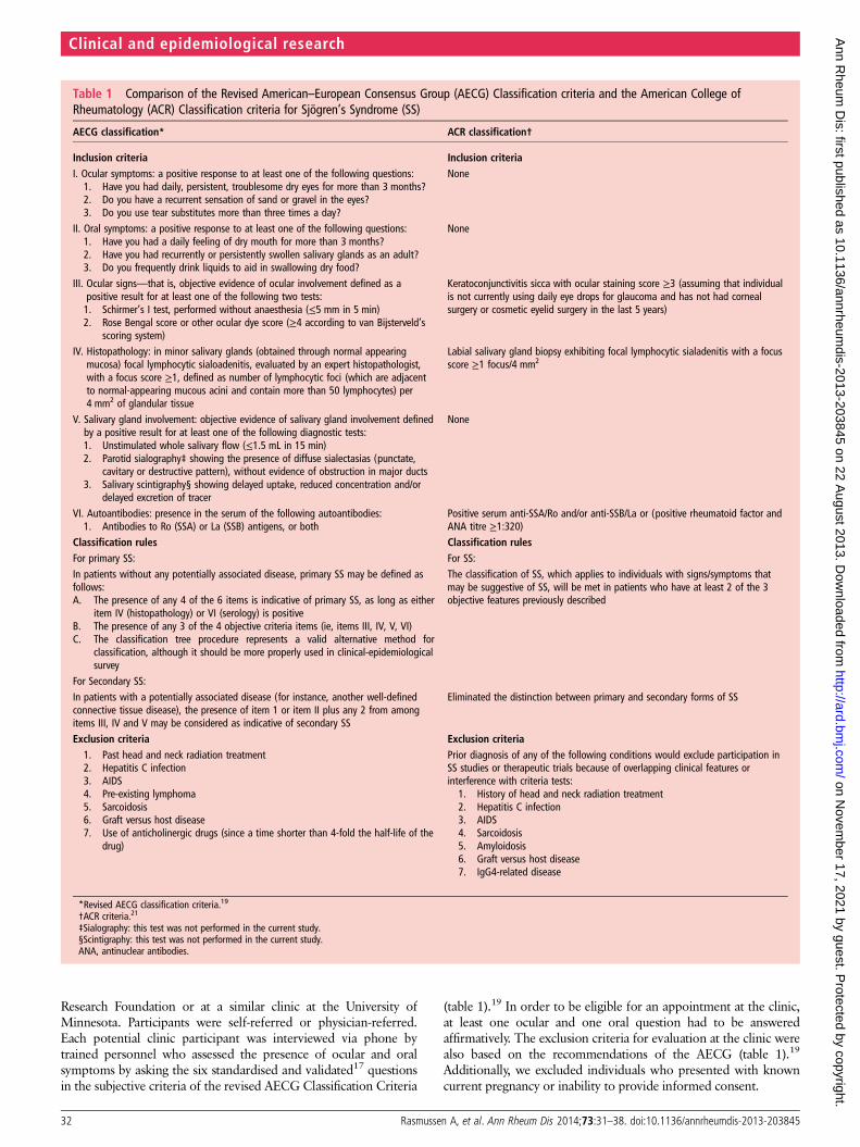
Table 1 Comparison of the Revised American–European Consensus Group (AECG) Classification criteria and the American College ofRheumatology (ACR) Classification criteria for Sjögren’s Syndrome (SS)
AECG classification* ACR classification†
Inclusion criteria Inclusion criteriaI. Ocular symptoms: a positive response to at least one of the following questions:1. Have you had daily, persistent, troublesome dry eyes for more than 3 months?2. Do you have a recurrent sensation of sand or gravel in the eyes?3. Do you use tear substitutes more than three times a day?
None
II. Oral symptoms: a positive response to at least one of the following questions:1. Have you had a daily feeling of dry mouth for more than 3 months?2. Have you had recurrently or persistently swollen salivary glands as an adult?3. Do you frequently drink liquids to aid in swallowing dry food?
None
III. Ocular signs—that is, objective evidence of ocular involvement defined as apositive result for at least one of the following two tests:1. Schirmer’s I test, performed without anaesthesia (≤5 mm in 5 min)2. Rose Bengal score or other ocular dye score (≥4 according to van Bijsterveld’s
scoring system)
Keratoconjunctivitis sicca with ocular staining score ≥3 (assuming that individualis not currently using daily eye drops for glaucoma and has not had cornealsurgery or cosmetic eyelid surgery in the last 5 years)
IV. Histopathology: in minor salivary glands (obtained through normal appearingmucosa) focal lymphocytic sialoadenitis, evaluated by an expert histopathologist,with a focus score ≥1, defined as number of lymphocytic foci (which are adjacentto normal-appearing mucous acini and contain more than 50 lymphocytes) per4 mm2 of glandular tissue
Labial salivary gland biopsy exhibiting focal lymphocytic sialadenitis with a focusscore ≥1 focus/4 mm2
V. Salivary gland involvement: objective evidence of salivary gland involvement definedby a positive result for at least one of the following diagnostic tests:1. Unstimulated whole salivary flow (≤1.5 mL in 15 min)2. Parotid sialography‡ showing the presence of diffuse sialectasias (punctate,
cavitary or destructive pattern), without evidence of obstruction in major ducts3. Salivary scintigraphy§ showing delayed uptake, reduced concentration and/or
delayed excretion of tracer
None
VI. Autoantibodies: presence in the serum of the following autoantibodies:1. Antibodies to Ro (SSA) or La (SSB) antigens, or both
Positive serum anti-SSA/Ro and/or anti-SSB/La or (positive rheumatoid factor andANA titre ≥1:320)
Classification rules Classification rulesFor primary SS: For SS:In patients without any potentially associated disease, primary SS may be defined asfollows:A. The presence of any 4 of the 6 items is indicative of primary SS, as long as either
item IV (histopathology) or VI (serology) is positiveB. The presence of any 3 of the 4 objective criteria items (ie, items III, IV, V, VI)C. The classification tree procedure represents a valid alternative method for
classification, although it should be more properly used in clinical-epidemiologicalsurvey
The classification of SS, which applies to individuals with signs/symptoms thatmay be suggestive of SS, will be met in patients who have at least 2 of the 3objective features previously described
For Secondary SS:In patients with a potentially associated disease (for instance, another well-definedconnective tissue disease), the presence of item 1 or item II plus any 2 from amongitems III, IV and V may be considered as indicative of secondary SS
Eliminated the distinction between primary and secondary forms of SS
Exclusion criteria Exclusion criteria1. Past head and neck radiation treatment2. Hepatitis C infection3. AIDS4. Pre-existing lymphoma5. Sarcoidosis6. Graft versus host disease7. Use of anticholinergic drugs (since a time shorter than 4-fold the half-life of the
drug)
Prior diagnosis of any of the following conditions would exclude participation inSS studies or therapeutic trials because of overlapping clinical features orinterference with criteria tests:1. History of head and neck radiation treatment2. Hepatitis C infection3. AIDS4. Sarcoidosis5. Amyloidosis6. Graft versus host disease7. IgG4-related disease
*Revised AECG classification criteria.19
†ACR criteria.21
‡Sialography: this test was not performed in the current study.§Scintigraphy: this test was not performed in the current study.ANA, antinuclear antibodies.
32 Rasmussen A, et al. Ann Rheum Dis 2014;73:31–38. doi:10.1136/annrheumdis-2013-203845
Clinical and epidemiological research
on Novem
ber 17, 2021 by guest. Protected by copyright.
http://ard.bmj.com
/A
nn Rheum
Dis: first published as 10.1136/annrheum
dis-2013-203845 on 22 August 2013. D
ownloaded from
With very few exceptions, participants were evaluated in asingle morning clinic visit using standardised protocols. Patientsunderwent an oral exam consisting of measurement of stimu-lated and timed whole unstimulated salivary flow (WUSF), a lipbiopsy and collection and storage of saliva. Participant evalu-ation did not include sialography or scintigraphy. The ocularspecialist performed ocular surface staining with lissamine greenand fluorescein, an unanaesthetised Schirmer’s I test, and collec-tion and storage of tears. The ocular vital dye score was deter-mined using the quantitative dot-counting method23 rather thanby descriptive features,24 and the score for each section wasrecorded independently before generating a final score for eacheye. Blood samples were collected for general laboratorytests and extraction of DNA, RNA and serum. A physician com-pleted a detailed history and physical examination, includinggeneral medical, rheumatological and neurological evaluations.If patients gave a history of a past diagnosis of rheumatoid arth-ritis, mixed connective tissue disease, systemic sclerosis, myo-sitis, primary biliary cirrhosis, multiple sclerosis, or systemiclupus erythematosus, classification criteria for these illnesseswere specifically ascertained by history, medical record reviewand testing for the corresponding autoantibodies.
All procedures were approved by the Oklahoma MedicalResearch Foundation and University of Minnesota InstitutionalReview Boards. Each participant provided written informedconsent prior to entering the study.
BiopsyThe dentist performed lip biopsies to obtain minor salivaryglands in all patients, unless slides from a previous biopsy wereavailable and contained sufficient tissue for re-examination byour pathologists. A portion of each specimen was formalin-fixedand paraffin-embedded, sections were cut and stained withhematoxylin-eosin, while other fragments were cryologicallypreserved. Two dental pathologists reviewed the specimens inde-pendently; the results were compared and a consensus readingwas generated. The lymphocytic infiltration of the glands wasgraded by focus score.5
Clinical laboratory and serologyAnti-Ro/SSA and anti-La/SSB autoantibodies were determinedby multiple methods. Additionally, all patients were tested forrheumatoid factor (RF), antinuclear antibodies (ANA), precipi-tins for autoantibodies associated with other connective tissuedisorders, hepatitis C serology, complete blood count (CBC)with differential, immunoglobulin profile and urinalysis (seeonline supplementary text).
ClassificationEach study participant was classified according to both therevised AECG,19 and to the newly proposed ACR criteria.21 Weeliminated from analysis the participants that did not haveresults for all the features of both classification systems with theexception of sialography and scintigraphy (table 1).
Peripheral blood mRNA transcript measurementsGlobal gene expression profiles comprising transcript levels for>15 000 loci were compared in a subset of 180 participants(see online supplementary text).
Statistical analysisPerformance of the tests was assessed via sensitivity, specificity,positive predictive value and negative predictive value estimatedby considering the AECG criteria as the ‘gold standard’, andsummarising the results with exact binomial 95% CI.McNemar’s Test of paired samples was used to assess whetherthe two sets of criteria were significantly different with respectto dichotomous variables. The κ statistic was used to quantifythe degree of agreement between the new classification criteriaand the AECG criteria. Details of the statistical analyses for thegene expression data are available in the online supplementarytext.
RESULTSThe initial cohort of participants evaluated at either theSjögren’s Research Clinic at Oklahoma Medical ResearchFoundation or the Sjögren’s Clinic in the University ofMinnesota comprised 837 individuals. Of these, 646 had alldata points of both AECG and ACR classification criteria and,thus, constitute the study cohort. The demographic characteris-tics of both cohorts are comparable in makeup with respect toage, sex, race and ethnicity (see online supplementary table S1).
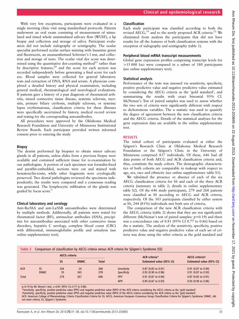
We tabulated the presence or absence of each of the sixAECG classification criteria for SS and each of the three ACRcriteria (summary in table 2; details in online supplementarytable S2). Of the 646 study participants, 279 and 268 patientswere classified as SS according to AECG and ACR criteria,respectively. Of the 303 participants classified by either systemas SS, 244 (81%) individuals met both sets of criteria.
The comparison of the new ACR classification criteria withthe AECG criteria (table 2) shows that they are not significantlydifferent (McNemar’s test of paired samples: p=0.19) and therewas a concordance rate of 0.81 (95% CI 0.77 to 0.86) based onthe κ statistic. The analysis of the sensitivity, specificity, positivepredictive value and negative predictive value of each set of cri-teria was done using the other criteria as the gold standard and
Table 2 Comparison of classification by AECG criteria versus ACR criteria for Sjögren’s Syndrome (SS)
AECG criteria ACR criteria* AECG criteria†SS DNMC Total Estimated value (95% CI) Estimated value (95% CI)
ACR SS 244 24 268 Sensitivity 0.87 (0.83 to 0.91) 0.91 (0.87 to 0.94)DNMC 35 343 378 Specificity 0.93 (0.90 to 0.96) 0.91 (0.87 to 0.93)
Total 279 367 646 PPV 0.91 (0.87 to 0.94) 0.87 (0.83 to 0.91)NPV 0.90 (0.87 to 0.93) 0.93 (0.90 to 0.96)
p=0.19 by Mc Nemar’s test; κ=0.81 (95% CIs 0.77 to 0.86).*Sensitivity, specificity, positive predictive value (PPV) and negative predictive value (NPV) of the ACR criteria considering the AECG criteria as the ‘gold standard’.†Sensitivity, specificity, positive predictive value (PPV) and negative predictive value (NPV) of the AECG criteria considering the ACR criteria as the ‘gold standard’.ACR, American College of Rheumatology Criteria Classification Criteria for SS; AECG, American European Consensus Group Classification Criteria for Sjögren’s Syndrome; DNMC, didnot meet criteria; SS, Sjögren’s Syndrome.
Rasmussen A, et al. Ann Rheum Dis 2014;73:31–38. doi:10.1136/annrheumdis-2013-203845 33
Clinical and epidemiological research
on Novem
ber 17, 2021 by guest. Protected by copyright.
http://ard.bmj.com
/A
nn Rheum
Dis: first published as 10.1136/annrheum
dis-2013-203845 on 22 August 2013. D
ownloaded from
was similar for both classification systems. The sensitivity of theACR criteria was 87.5 (95% CI 82.9 to 90.9) with a specificityof 93.4 (95% CI 90.3 to 95.7); the positive predictive value was91.0 (95% CI 86.8 to 94.0) and the negative predictive valuewas 90.7 (95% CI 87.2 to 93.4). Thus, 12.5% (35 of 279) ofparticipants classified as SS under the AECG criteria were notconsidered SS when evaluated by the ACR criteria; conversely,8.9% (24 of 268) met only the ACR criteria.
The differences between how the two systems classified thesicca participants revolved around which objective measures ofocular and oral involvement were included in addition to thehistology and Ro/La serology. Namely, the van Bijsterveld (vBS)grading system of ocular staining, Schirmer’s I test, and WUSFvolume for the AECG criteria, and a different version of thefirst of these (the sicca ocular staining score or OSS) for theACR criteria plus positive ANA (≥1:320) and positive RF as analternative measure of serological activity.
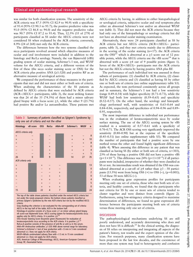
We compared the performance of these measures in the parti-cipants that met and did not meet either or both sets of criteria.When analysing the characteristics of the SS patients asdefined by AECG criteria that were excluded by ACR criteria(ACR−/AECG+ participants; table 3), the most striking featurewas that 26 of the 35 (74.3%) of them had a minor salivarygland biopsy with a focus score ≥1, while the other 9 (25.7%)had positive Ro and/or La autoantibodies. These patients met
AECG criteria by having, in addition to either histopathologicalor serological criteria, subjective ocular and oral symptoms pluseither an abnormal Schirmer’s test and/or an abnormal WUSFtest (table 3). They did not meet the ACR criteria because theyhad only one of the histopathology or serology criteria but didnot have an abnormal ocular staining examination.
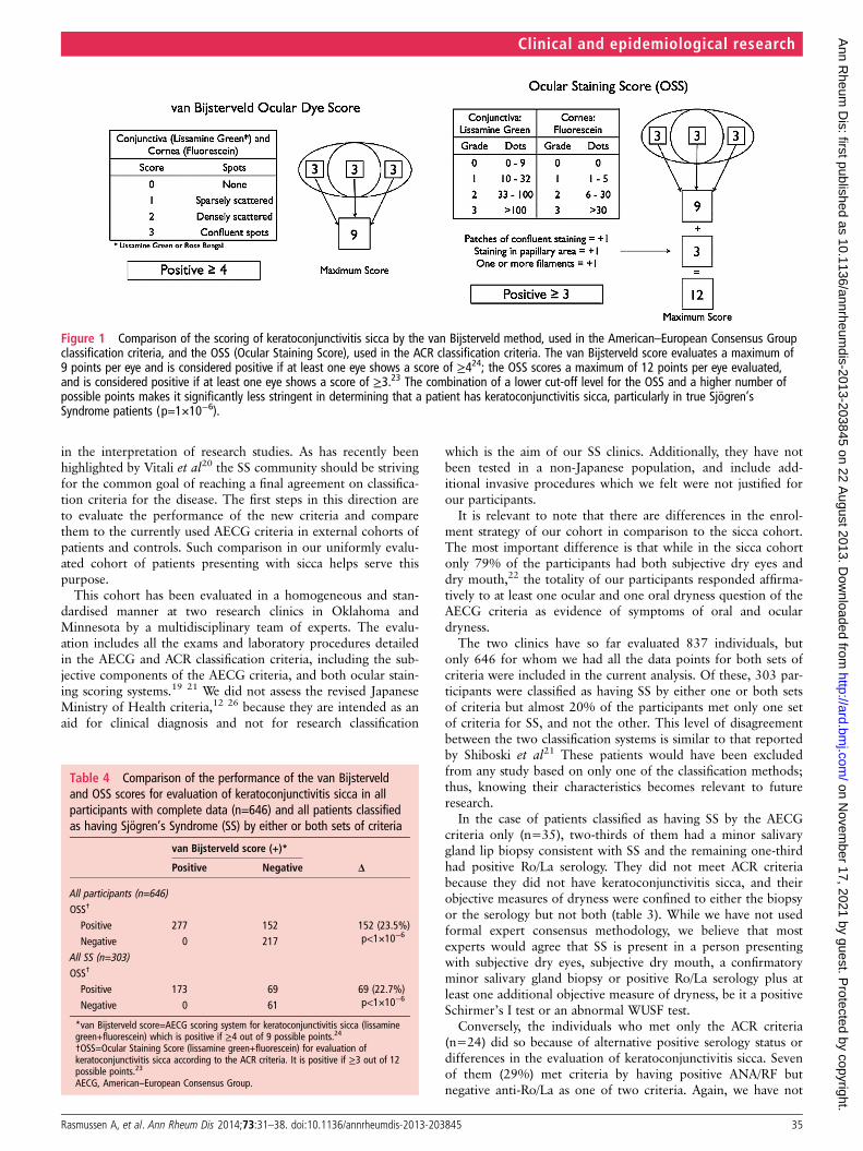
Alternatively, there were 24 participants classified as SS byACR criteria but not AECG criteria (ACR+/AECG− partici-pants; table 3), and they met criteria mainly due to differencesin the scoring of the ocular staining (n=17): the ACR criteriause the OSS23 which is abnormal at a ≥3 score out of 12 pos-sible points, while the AECG criteria use the vBS score24 that isabnormal with a score ≥4 out of 9 possible points (figure 1).Seven of the ACR+/AECG– participants met the ACR criteriabut not the AECG criteria by having positive ANA plus RF.
The performance of each individual test was assessed in threesubsets of participants: (1) classified by ACR criteria, (2) classi-fied by AECG criteria and (3) classified as having SS by eitherone or both sets of criteria (see online supplementary table S3).As expected, the tests performed consistently across all groupsand in summary, the Schirmer’s I test had a low sensitivity(range 0.49–0.54) with higher specificity (0.71–0.73), while theWUSF had both low sensitivity (0.59–0.65) and specificity(0.52–0.57). On the other hand, the serology and histopath-ology performed well, with sensitivities of 0.63–0.64 and0.84–0.86, respectively, and specificities of 0.94–0.96 and 0.89–0.95, respectively.
The most important difference in individual test performancewas in the evaluation of keratoconjunctivitis sicca by ocularsurface staining. The use of the AECG scoring system by vBSresulted in a sensitivity of 0.57–0.61 with a specificity of0.70–0.71. The ACR OSS scoring very significantly improved thesensitivity (0.80–0.90) but at the expense of the specificity(0.45–0.51) (see online supplementary table S3). We comparedthe number of participants that had a positive score by onemethod versus the other and found highly significant differences(table 4). When assessing this difference in any patient that wasclassified as having SS (by either or both sets of criteria, n=303),23% of those having a positive OSS did not have a positive vBS(p<1×10⁻⁶). This difference was 24% (p<1×10⁻⁶) if all partici-pants were included, irrespective of whether they were classified asSS or not. An intermediate result was obtained if the OSS was con-sidered abnormal at a cut-off of ≥4 rather than ≥3 : 58 partici-pants (13.5%) went from being OSS (+) to OSS (−), (p=0.001);53 of these 58 were AECG (−).
When evaluating gene expression profiles for participantsmeeting only one set of criteria, those who met both sets of cri-teria, and healthy controls, we found that the participants whomet criteria for SS by one or more sets of criteria tended tocluster together and were distinct from controls (figure 2).Furthermore, using low-stringency criteria designed to maximisedetermination of differences, we found no gene expression dif-ference between the participants meeting both sets of criteriaversus those meeting only one set of criteria.
DISCUSSIONThe pathophysiological mechanisms underlying SS are stillpoorly understood, and accurately determining who does anddoes not have SS is difficult.25 In the clinical setting, the diagno-sis of SS relies on interpreting and integrating all aspects of thepatient’s history, test results and the expert opinion of the clin-ician. For research purposes, many classification systems havebeen proposed in the last few decades, and the coexistence ofmore than one system may lead to heterogeneity and confusion
Table 3 Summary of patients classified as Sjögren’s Syndrome byonly one set of criteria and not the other
Histology vB score OSS Ro/La ANA+RF Schirmer’s WUSF n
AECG criteria only+ − − + − 8+ − − + + 10+ − − − + 8− − + + − 0− − + + + 4− − + − + 526 0 9 14 27 35ACR criteria only
+ + − − 13+ − + − 0+ − − + 2+ − + + 0− + + − 4− + − + 5− + + + 015 22 4 7 24
The top of the table shows patientss classified under the revised AECG criteria butnot under the newly proposed criteria; the bottom shows patients classified asprimary Sjögren’s Syndrome by the new ACR criteria but not by the modified AECGcriteria.Gray shading=the criterion is not evaluated for the corresponding set of criteria:AECG in the top half of the table; ACR in the bottom half.Histology=A positive consists of a focus score ≥1 on minor salivary gland biopsy.vB score=van Bijsterveld Score. AECG scoring system for keratoconjunctivitis sicca;applies only for AECG criteria. It is positive ≥4.24OSS=Ocular Staining Score (lissamine green+fluorescein) for evaluation ofkeratoconjunctivitis sicca according to the ACR criteria. It is positive ≥3.23Ro/La=A positive consists of the presence of anti-Ro or anti-La in the serum.ANA+RF=Antinuclear antibodies ≥1:320 AND above normal range for laboratory.Schirmer’s=Schirmer’s I test of tear production with <5 mm in 5 min consideredabnormal (+). Does not apply for ACR criteria.WUSF=Whole unstimulated salivary flow with <1.5 mL in 15 min consideredabnormal (+). Does not apply for ACR criteria.ACR, American College of Rheumatology; AECG, American–European ConsensusGroup; RF, rheumatoid factor.
34 Rasmussen A, et al. Ann Rheum Dis 2014;73:31–38. doi:10.1136/annrheumdis-2013-203845
Clinical and epidemiological research
on Novem
ber 17, 2021 by guest. Protected by copyright.
http://ard.bmj.com
/A
nn Rheum
Dis: first published as 10.1136/annrheum
dis-2013-203845 on 22 August 2013. D
ownloaded from
in the interpretation of research studies. As has recently beenhighlighted by Vitali et al20 the SS community should be strivingfor the common goal of reaching a final agreement on classifica-tion criteria for the disease. The first steps in this direction areto evaluate the performance of the new criteria and comparethem to the currently used AECG criteria in external cohorts ofpatients and controls. Such comparison in our uniformly evalu-ated cohort of patients presenting with sicca helps serve thispurpose.
This cohort has been evaluated in a homogeneous and stan-dardised manner at two research clinics in Oklahoma andMinnesota by a multidisciplinary team of experts. The evalu-ation includes all the exams and laboratory procedures detailedin the AECG and ACR classification criteria, including the sub-jective components of the AECG criteria, and both ocular stain-ing scoring systems.19 21 We did not assess the revised JapaneseMinistry of Health criteria,12 26 because they are intended as anaid for clinical diagnosis and not for research classification
which is the aim of our SS clinics. Additionally, they have notbeen tested in a non-Japanese population, and include add-itional invasive procedures which we felt were not justified forour participants.
It is relevant to note that there are differences in the enrol-ment strategy of our cohort in comparison to the sicca cohort.The most important difference is that while in the sicca cohortonly 79% of the participants had both subjective dry eyes anddry mouth,22 the totality of our participants responded affirma-tively to at least one ocular and one oral dryness question of theAECG criteria as evidence of symptoms of oral and oculardryness.
The two clinics have so far evaluated 837 individuals, butonly 646 for whom we had all the data points for both sets ofcriteria were included in the current analysis. Of these, 303 par-ticipants were classified as having SS by either one or both setsof criteria but almost 20% of the participants met only one setof criteria for SS, and not the other. This level of disagreementbetween the two classification systems is similar to that reportedby Shiboski et al21 These patients would have been excludedfrom any study based on only one of the classification methods;thus, knowing their characteristics becomes relevant to futureresearch.
In the case of patients classified as having SS by the AECGcriteria only (n=35), two-thirds of them had a minor salivarygland lip biopsy consistent with SS and the remaining one-thirdhad positive Ro/La serology. They did not meet ACR criteriabecause they did not have keratoconjunctivitis sicca, and theirobjective measures of dryness were confined to either the biopsyor the serology but not both (table 3). While we have not usedformal expert consensus methodology, we believe that mostexperts would agree that SS is present in a person presentingwith subjective dry eyes, subjective dry mouth, a confirmatoryminor salivary gland biopsy or positive Ro/La serology plus atleast one additional objective measure of dryness, be it a positiveSchirmer’s I test or an abnormal WUSF test.
Conversely, the individuals who met only the ACR criteria(n=24) did so because of alternative positive serology status ordifferences in the evaluation of keratoconjunctivitis sicca. Sevenof them (29%) met criteria by having positive ANA/RF butnegative anti-Ro/La as one of two criteria. Again, we have not
Figure 1 Comparison of the scoring of keratoconjunctivitis sicca by the van Bijsterveld method, used in the American–European Consensus Groupclassification criteria, and the OSS (Ocular Staining Score), used in the ACR classification criteria. The van Bijsterveld score evaluates a maximum of9 points per eye and is considered positive if at least one eye shows a score of ≥424; the OSS scores a maximum of 12 points per eye evaluated,and is considered positive if at least one eye shows a score of ≥3.23 The combination of a lower cut-off level for the OSS and a higher number ofpossible points makes it significantly less stringent in determining that a patient has keratoconjunctivitis sicca, particularly in true Sjögren’sSyndrome patients (p=1×10⁻⁶).
Table 4 Comparison of the performance of the van Bijsterveldand OSS scores for evaluation of keratoconjunctivitis sicca in allparticipants with complete data (n=646) and all patients classifiedas having Sjögren’s Syndrome (SS) by either or both sets of criteria
van Bijsterveld score (+)*
ΔPositive Negative
All participants (n=646)OSS†
Positive 277 152 152 (23.5%)p<1×10⁻⁶Negative 0 217
All SS (n=303)OSS†
Positive 173 69 69 (22.7%)p<1×10⁻⁶Negative 0 61
*van Bijsterveld score=AECG scoring system for keratoconjunctivitis sicca (lissaminegreen+fluorescein) which is positive if ≥4 out of 9 possible points.24
†OSS=Ocular Staining Score (lissamine green+fluorescein) for evaluation ofkeratoconjunctivitis sicca according to the ACR criteria. It is positive if ≥3 out of 12possible points.23
AECG, American–European Consensus Group.
Rasmussen A, et al. Ann Rheum Dis 2014;73:31–38. doi:10.1136/annrheumdis-2013-203845 35
Clinical and epidemiological research
on Novem
ber 17, 2021 by guest. Protected by copyright.
http://ard.bmj.com
/A
nn Rheum
Dis: first published as 10.1136/annrheum
dis-2013-203845 on 22 August 2013. D
ownloaded from

Figure 2 Assessment of the American–European Consensus Group (AECG) and ACR criteria using whole-blood gene expression profiling inSjögren’s Syndrome (SS). Heat maps are displayed using fold changes for differentially expressed (DE) transcripts; overexpressed transcripts (FC>0)are bright yellow, while underexpressed transcripts (FC<0) are light blue. Data is displayed in rows and columns, with rows defined by transcriptsand columns defined by individual samples. Colours above each column header denote healthy controls (blue; n=73) and cases (red, yellow, orgreen). For cases, the colour represents the criteria used to define SS: red denotes cases meeting only ACR criteria (n=4); yellow denotes casesmeeting only AECG criteria (n=25); and green denotes cases meeting both ACR and AECG criteria (n=127). A Sjögren’s-specific set of DE transcriptswas defined by comparing all SS cases regardless of classification criteria to healthy controls. Hierarchical clustering was performed with respect totranscripts and samples, and dendrograms generated to visualise sample clustering. In panel A, all samples are displayed, with cases and controlsgenerally segregating. In panel B, hierarchical clustering was performed using the DE transcripts defined in panel A, but removing cases meetingboth AECG and ACR criteria. Panel C further limits the clustering to those individuals meeting only ACR or AECG. Distinct clustering of patientsmeeting only ACR or AECG criteria was not observed, suggesting molecular similarity between cases defined by either ACR or AECG criteria.
36 Rasmussen A, et al. Ann Rheum Dis 2014;73:31–38. doi:10.1136/annrheumdis-2013-203845
Clinical and epidemiological research
on Novem
ber 17, 2021 by guest. Protected by copyright.
http://ard.bmj.com
/A
nn Rheum
Dis: first published as 10.1136/annrheum
dis-2013-203845 on 22 August 2013. D
ownloaded from
done formal expert testing, but it is unlikely that these indivi-duals would be considered to have SS based on expert opinion,especially without information about sicca symptoms. Theremaining 71% met criteria for keratoconjunctivitis sicca byACR but not by AECG criteria.
It has been proposed that it would be useful to know if theOSS developed for the ACR criteria can be substituted by theAECG vBS.21 Few already established cohorts that we are awareof, if any, are currently able to directly compare the perform-ance of the vBS with the OSS. In cohorts that were evaluatedbefore the publication of the OSS in 2010, determining the OSSwould require access to the breakdown of the scoring of eacheye: individual scores for medial and lateral bulbar conjunctivaand cornea plus the description of patches of confluent staining,staining in the papillary area and presence of filaments(figure 1). The vBS does not take into consideration these lastthree features,24 which add three possible points to the score ofeach eye in the case of the OSS. The vBS is considered abnormalwith a score of ≥4 out of 9 possible points24 while the OSS ispositive at ≥3 out of 12 points.23
We are in the unique position of having recorded separatelyeach of the 12 possible scoring points for each eye in all ourcohort participants. Thus, we were able to determine both theirvBS and OSS scores and compare the performance of eachsystem. To reduce interobserver and intraobserver variabilityinherent to the vBS scoring system,24 the vital dye score foreach section of the ocular surface was determined using thesicca dot counting method. While there are no studies validatingthe conversion of this scoring method with the traditional vBStechnique, the two are similar; we felt that an objective scoringmethod would be more meaningful and reproducible in thecontext of multiple observers. As expected, participants weremore likely to have an abnormal OSS score than vBS, resultingin ∼25% of patients having a positive OSS but negative vBS.This difference was highly significant in all cohort participantsand in patients who were classified as having SS by one or bothsets of criteria (p<1×10⁻⁶). The OSS is superior in includingtrue positive cases but has a poor performance ruling out thosewho do not have SS (ie, it is very sensitive but has poor specifi-city); the opposite is the case for the vBS. It is pertinent to notethat only a minor proportion of cases of keratoconjunctivitissicca are due to SS.27 It remains to be seen how other prospect-ive cohorts evaluate these two scoring systems vis à vis, in orderto determine what the optimal threshold should be. It is note-worthy that one of the main goals of the development of newclassification criteria by the sicca consortium was to come upwith a system that has high specificity to avoid exposingunaffected individuals to the potentially serious adverse effectsof novel investigational therapies.21
The two tests that performed the best across all comparisongroups were the minor salivary gland biopsy and anti-Ro/La ser-ology, which performed similarly to reports in previousstudies.21 28 The Schirmer’s and WUSF tests while less useful indistinguishing true SS patients from participants withnon-Sjögren’s sicca syndrome, are easy to perform and are non-invasive. It has recently been suggested that more emphasisshould be given to tests that in addition to identifying true casesand excluding unaffected individuals, can be done at earlystages, multiple times, and with minimal distress to the partici-pant.29 In the future, we may see salivary gland ultrasonographyplaying this role in SS.30 But with the current sets of criteria,there is an interesting difference in terms of accessibility; whilethe ACR criteria require evaluation by a practitioner specialisingin eyes and by a practitioner who can perform a lip biopsy, both
the Schirmer’s and the WUSF tests can be performed in a stand-ard medical office without the need for sophisticated equipmentor medical specialists. Thus, patients can be assessed for subject-ive dry eyes and dry mouth, the presence of autoantibodiesalong with Schirmer’s and WUSF testing by a rheumatologist. IfAECG criteria are not met with such an assessment, then biopsyand eye examination can be pursued. In some clinical care set-tings or research situations that do not include exposing theselected participants to the risk of significant adverse events(such as some therapeutic trials), a stepwise approach such asthis may be useful and cost effective.
The comparison of the AECG criteria with the proposedACR classification demonstrates that neither system is clearlysuperior to the other when classifying a patient with SS;a finding already reported in the initial publication of the ACRcriteria.21 The lack of highly sensitive, specific and reproduciblecriteria may, in part, be due to our current limited understand-ing of SS physiopathology; such knowledge would provide themost rational basis for disease classification. In the currentsetting, the ACR criteria may be best suited for stricter studiesfocused on high specificity to reduce the risk of drug-relatedtoxicity, while the AECG criteria may be applicable to broaderuse, particularly in less risky medical research, or in non-treatment clinical or translational research settings. Moreoverour findings of similar gene expression profiles across all pos-sible patients affected by SS, which is different from what isobserved in healthy controls, supports our notion that modify-ing classification using only clinical criteria is not likely to leadto consequential improvements in our ability to identify patientswith SS. We believe that such improvements in diagnosticacumen will require a more fundamental understanding of thepathogenic mechanisms than is at present available.
Author affiliations1Arthritis and Clinical Immunology Research Program, Oklahoma Medical ResearchFoundation, Oklahoma City, Oklahoma, USA2Department of Pathology, University of Oklahoma Health Sciences Center,Oklahoma City, Oklahoma, USA3Department of Oral Diagnosis and Radiology, University of Oklahoma College ofDentistry, Oklahoma City, Oklahoma, USA4Department of Ophthalmology, University of Oklahoma Health Sciences Center,Oklahoma City, Oklahoma, USA5Hefner Eye Care and Optical Center, Oklahoma City, Oklahoma, USA6Center for Autoimmune Diseases Research (CREA), Universidad del Rosario,Bogotá, Colombia7Hard Tissue Research Laboratory, University of Minnesota School of Dentistry,Minneapolis, Minnesota, USA8Division of Oral Pathology, Department of Developmental and Surgical Science,University of Minnesota School of Dentistry, Minneapolis, Minnesota, USA9Department of Ophthalmology, Harvard Medical School, Boston, Massachusetts,USA10Division of Rheumatology, Cincinnati Children’s Hospital Medical Center,Cincinnati, Ohio, USA11US Department of Veterans Affairs Medical Center, Cincinnati, Ohio, USA12Division of Oral and Maxillofacial Surgery, Department of Developmental andSurgical Science, University of Minnesota School of Dentistry, Minneapolis,Minnesota, USA13Department of Oral Surgery, University of Minnesota School of Dentistry,Minneapolis, Minnesota, USA14Hennepin County Medical Center, Minneapolis, Minnesota, USA15Broegelmann Research Laboratory, Department of Clinical Science, University ofBergen, Bergen, Norway16Department of Rheumatology, Haukeland University Hospital, Bergen, Norway17Department of Medicine, University of Oklahoma Health Sciences Center,Oklahoma City, Oklahoma, USA18Department of Veterans Affairs Medical Center, Oklahoma City, Oklahoma, USA
Correction notice This article has been corrected since it was published OnlineFirst. Occurrences of ‘SICCA’ have been changed to lower case (‘sicca’).
Rasmussen A, et al. Ann Rheum Dis 2014;73:31–38. doi:10.1136/annrheumdis-2013-203845 37
Clinical and epidemiological research
on Novem
ber 17, 2021 by guest. Protected by copyright.
http://ard.bmj.com
/A
nn Rheum
Dis: first published as 10.1136/annrheum
dis-2013-203845 on 22 August 2013. D
ownloaded from
Acknowledgements We are grateful to all the individuals with SS and those servingas healthy controls who participated in this study. We would like to thank the followingindividuals for their help in the collection and ascertainment of the samples used in thisstudy: Erin Rothrock, Judy Harris, Sharon Johnson, Sarah Cioli, Nicole Weber, DominiqueWilliams, Wes Daniels, Cherilyn Pritchett-Frazee, Kylia Crouch, Laura Battiest, JustinRodgers, James Robertson, Thuan Nguyen, Amanda Crosbie, Ellen James,Carolyn Meyer, Amber McElroy, Eshrat Emamian, Julie Ermer, Kristine Rohlf, JoanliseLeon, Anita Petersen, Danielle Hartle, Jill Novizke, Ward Ortman, Carl Espy, Beth Cobb,Gudlaug Kristjansdottir and Marianne Eidsheim. We would also like to thank StuartGlenn and Jared Ning for their ongoing assistance in developing and maintaining thecomputational infrastructure used to perform this study.
Contributors All authors of the manuscript contributed to: conception and design,or analysis and interpretation of data; drafting the article or revising it critically forimportant intellectual content and final approval of the version to be published.
Funding This publication was made possible by grants 5R01 AR50782 (KLS), P50AR0608040 (KLS, CJL, RHS, and ADF), 5U19 AI 082714 (KLS, CJL), 5R01DE018209 (KLS, JBH), 5R37AI024717-22S1 ( JBH, AR). The contents are the soleresponsibility of the authors and do not necessarily represent the official views ofthe NIH. Additional funding was obtained from the Phileona Foundation (KLS) andthe Oklahoma Medical Research Foundation (CJL and KLS). DUS received fundingfrom an unrestricted grant from Research to Prevent Blindness to the University ofOklahoma Department of Ophthalmology.
Competing interests None.
Ethics approval Oklahoma Medical Research Foundation Internal Review Boardand University of Minnesota Internal Review Board.
Provenance and peer review Not commissioned; externally peer reviewed.
Data sharing statement The authors would consider requests for the datagenerated in this project through collaborative arrangements.
REFERENCES1 Helmick C G, Felson D T, Lawrence R C, et al. National Arthritis Data Workgroup.
Estimates of the prevalence of arthritis and other rheumatic conditions in the UnitedStates. Part I. Arthritis Rheum 2008;58:15–25.
2 Gøransson LG, Haldorsen K, Brun JG, et al. The point prevalence of clinicallyrelevant primary Sjögren’s syndrome in two Norwegian counties. Scand J Rheumatol2011;40:221–4.
3 Fox RI. Sjögren’s syndrome. Lancet 2005;366:321–31.4 Amador-Patarroyo MJ, Arbelaez JG, Mantilla RD, et al. Sjögren’s syndrome at the
crossroad of polyautoimmunity. J Autoimmun 2012;39:199–205.5 Daniels TE. Labial salivary gland biopsy in Sjögren’s syndrome. Assessment as a
diagnostic criterion in 362 suspected cases. Arthritis Rheum 1984;27:147–56.6 Reichlin M, Scofield RH. Ro (SS-A) antibodies. In: Shoenfeld Y, Gershwin ME, Meroni PL.
eds. Textbook of Autoantibodies. 2nd edn. Amsterdam: Elsevier, 2006:783–8.7 Ramos-Casals M, Solans R, Rosas J, et al. Primary Sjögren syndrome in Spain:
clinical and immunologic expression in 1010 patients. Medicine 2008;87:210–19.8 Anaya JM, Delgado-Vega AM, Castiblanco J. Genetic basis of Sjogren’s syndrome.
How strong is the evidence?. Clin Dev Immunol 2006;13:209–22.9 Bloch KJ, Buchanan WW, Wohl MJ, et al. Sjögren’s Syndrome. A Clinical,
Pathological, and Serological Study of Sixty-Two Cases. Medicine (Baltimore)1965;44:187–231.
10 Shearn MA. Sjögren’s syndrome. In: Smith LH.ed. Major Problems in InternalMedicine. vol. II. Philadelphia: WB Saunders Co., 1971:12–14.
11 Daniels TE, Silverman S Jr, Michalski JP, et al. The oral component of Sjögren’ssyndrome. Oral Surg Oral Med Oral Pathol 1975;39:875–85.
12 Ohfuji T. Review on research reports. Annual report of the ministry of Health andWelfare: Sjögren’s disease Research Committee, Japan, 1977.
13 Homma M, Tojo T, Akizuki M, et al. Criteria for Sjögren’s syndrome in Japan. ScandJ Rheumatol 1986;61(Suppl):P26–7.
14 Manthorpe R, Frost-Larsen K, Isager H, et al. Sjögren’s syndrome. A review withemphasis on immunological features. Allergy 1981;36:139–53.
15 Skopouli FN, Drosos AA, Papaioannou T, et al. Preliminary diagnostic criteria forSjögren’s syndrome. Scand J Rheumatol 1986;61(Suppl):22–5.
16 Fox RI, Robinson CA, Curd JG, et al. Sjögren’s syndrome. Proposed criteria forclassification. Arthritis Rheum 1986;29:577–85.
17 Vitali C, Bombardieri S, Moutsopoulos HM, et al. Preliminary criteria for theclassification of Sjögren’s syndrome. Results of a prospective concerted actionsupported by the European Community. Arthritis Rheum 1993;36:340–7.
18 Fujibayashi T. Revised diagnostic criteria for Sjögren’s syndrome. Rheumatology(Oxford) 2000;24:421–8.
19 Vitali C, Bombardieri S, Jonsson R, et al. Classification criteria for Sjögren’ssyndrome: a revised version of the European criteria proposed by theAmerican-European Consensus Group. Ann Rheum Dis 2002;61:554–8.
20 Vitali C, Bootsma H, Bowman SJ, et al. Classification criteria for Sjögren’ssyndrome: we actually need to definitively resolve the long debate on the issue.Ann Rheum Dis 2013;72:476–8.
21 Shiboski SC, Shiboski CH, Criswell L, et al. New classification criteria for Sjögren’sSyndrome: a data-driven expert-clinician consensus approach within the SICCACohort. Arthritis Care Res 2012;64:475–87.
22 Daniels TE, Criswell LA, Shiboski C, et al. An early view of the internationalSjogren’s syndrome registry. Arthritis Rheum 2009;61:711–14.
23 Van Bijsterveld OP. Diagnostic tests in the SICCA syndrome. Arch Ophtal1969;82:10–14.
24 Jonsson R, Vogelsang P, Volchenkov R, et al. The complexity of Sjögren’s syndrome:novel aspects on pathogenesis. Immunol Lett 2011;141:1–9.
25 Whitcher JP, Shiboski CH, Shiboski SC, et al. A simplified quantitative method forassessing keratoconjunctivitis SICCA from the Sjögren’s Syndrome InternationalRegistry. Am J Ophtalmol 2010;149:405–15.
26 Tsuboi H, Hagiwara S, Asashima H, et al. Validation of different sets of criteria forthe diagnosis of Sjögren’s syndrome in Japanese patients. Mod Rheumatol2013;23:219–25.
27 Yazdani C, McLaughlin T, Smeeding JE, et al. Prevalence of treated dry eye diseasein a managed care population. Clin Ther 2001;23:1672–82.
28 Vitali C, Moutsoupoulos HM, Bombardieri S, et al. The European Community StudyGroup on Diagnostic Criteria for Sjögren’s Syndrome. Sensitivity and specificity oftests for ocular and oral involvement in Sjögren’s syndrome. Ann Rheum Dis1994;53:637–47.
29 Bootsma H, Spijkervet FKL, Kroese FGM, et al. Toward New Classification Criteriafor Sjögren’s Syndrome? Arthritis Rheum 2013;65:21–3.
30 Milic V, Petrovic R, Boricic I, et al. Ultrasonography of major salivary glands couldbe an alternative tool to sialoscintigraphy in the American-Europeanclassification criteria for primary Sjögren’s syndrome. Rheumatology (Oxford)2012;51:1081–5.
38 Rasmussen A, et al. Ann Rheum Dis 2014;73:31–38. doi:10.1136/annrheumdis-2013-203845
Clinical and epidemiological research
on Novem
ber 17, 2021 by guest. Protected by copyright.
http://ard.bmj.com
/A
nn Rheum
Dis: first published as 10.1136/annrheum
dis-2013-203845 on 22 August 2013. D
ownloaded from