Embed Size (px)
Citation preview

ORIGINAL PAPER
Extract from Ribes nigrum leaves in vitro activates nitric oxidesynthase (eNOS) and increases CD39 expression in humanendothelial cells
Boguslawa Luzak &Magdalena Boncler & Joanna Rywaniak &Dominika Dudzinska &
Marek Rozalski & Urszula Krajewska & Ewa Balcerczak & Anna Podsedek &
Malgorzata Redzynia & Cezary Watala
Received: 3 December 2013 /Accepted: 10 November 2014 /Published online: 19 November 2014# University of Navarra 2014
Abstract The aim of the present study was to evaluatewhether blackcurrant leaf extract (BLE) modulates en-dothelium antithrombotic function, namely increasesthe expression/activity of ADPase (CD39) and aug-ments the production of nitric oxide in human umbilicalvein endothelial cells (HUVEC). It was found that BLEwith proanthocyanidins (60 % of the total polyphenolcontent) increased the CD39-positive endothelial cellfraction (up to 10 % for 2.5 μg/ml, and up to 33 % for15 μg/ml, p<0.05 or less) in a concentration-dependentmanner, and enhanced endothelial nitric oxide synthase(eNOS) activation (T495 phosphorylation decreased by31±6% for 2.5 μg/ml and 48±6 % for 15 μg/ml; S1177phosphorylation increased by 13±3% for 2.5μg/ml and18±7 % for 15 μg/ml, compared to untreated cells,p<0.05 or less). Additionally, incubation for 24 or48 h with BLE at a lower range of polyphenol
concentrations, significantly increased cell viability witha maximal effect at 2.5 μg/ml (viability increased by24.8±1.0 % for 24 h and by 32.5±2.7 % for 48-h timeincubation, p<0.0001). The increased CD39 expressionand the increased eNOS activation in HUVEC can beregarded as the beneficial markers of the improvementof antiplatelet action of endothelial cells. Unexpectedly,these assumptions were not confirmed in the experimen-tal model of platelet-endothelial cell interactions. Theseobservations lead to the conclusion that BLE may im-prove endothelial cell viability at low physiologicalconcentrations without affecting the antiplatelet actionof endothelium.
Keywords Blackcurrant leaf extract . eNOS activation .
CD39 . ecto-ADPase . Endothelial cells . Cardiovasculardisease
Introduction
The endothelium mediates a number of responses (re-laxation or contraction) of arteries and veins from ani-mals and humans, but the special role of endothelialcells is the regulation of blood platelet functions. Plate-lets, in general, appear to be designed to adhere to thevessel wall only when endothelial integrity is disruptedand several mechanisms, such as the secretion of nitricoxide (NO) and prostacyclin, evolved to prevent theiradhesion to the intact endothelium. Both mediators arepotent inhibitors of platelet adhesion and activation. The
J Physiol Biochem (2014) 70:1007–1019DOI 10.1007/s13105-014-0370-z
B. Luzak (*) :M. Boncler : J. Rywaniak :D. Dudzinska :C. WatalaDepartment of Haemostasis and Haemostatic Disorders,Medical University of Lodz,6/8 Mazowiecka Street, 92-215 Lodz, Polande-mail: [email protected]
M. Rozalski :U. Krajewska : E. BalcerczakDepartment of Pharmaceutical Biochemistry, MolecularBiology Laboratory, Medical University of Lodz,Lodz, Poland
A. Podsedek :M. RedzyniaDepartment of Biotechnology and Food Sciences, Institute ofTechnical Biochemistry, Lodz University of Technology,Lodz, Poland
endothelial cells also produce an ecto-ADPase (CD39),which degrades ADP released from erythrocytes andaggregating platelets. The function of endothelial cellsis dependent on many variables including lipid levels,blood pressure and also the normal ageing process,which influence the turnover and regeneration of endo-thelial cells, which may, in turn, result in abnormalfunction: the loss of ability to release a relaxing factor[8]. Under conditions of disrupted homeostasis such asthose found in inflammatory states, platelets can evenbind to the intact endothelium. This is partly becauseendothelium-dependent mechanisms of platelet inhibi-tion are impaired, and partly because new adhesionmolecules are expressed on the surface of activatedendothelial cells. Several agents have been proposed aspotential modulators of endothelial function. Most ofthe available approaches include pharmaceutical agents(statins, angiotensin converting enzyme inhibitors), di-etary supplements (f.i. folic acid), functional foods andnutraceuticals [21]. Numerous epidemiological studieshave indicated that diets rich in fruits, vegetables andbeverages, such as red wine and tea, are associated withthe reduced risk of cardiovascular diseases. Polyphenolsmay protect the cardiovascular system by preventingoxidation of low-density lipoprotein, platelet aggrega-tion and adhesion and smooth muscle cell migration andviability. Alternatively, vascular protection may also bedue to the direct action of polyphenols on the endothe-lial function [24].
Blackcurrant leaves (Ribes nigrum L.) are used inEuropean traditional medicine for treating disorders ofinflammatory nature, as for instance, rheumatic disease[27]. Several studies have focused on the therapeuticpotential of blackcurrant with regard to hypertensionand other cardiovascular-associated illnesses, neoplas-tic, neurodegenerative and ocular diseases and diabeticneuropathy [7]. It has been shown that blackcurrant seedoil acts as a platelet inhibitor and potentiates an antico-agulant effect by inhibiting fibrin formation [25]. Otherreports have demonstrated the influence ofblackcurrants on serum lipid profile: increased HDL-cholesterol level and decreased triglyceride or total cho-lesterol [33], as well as reduced serum inflammatorymarkers [30].
The aim of the present study was to investigate theantithrombotic function of human umbilical vein endo-thelial cells (HUVEC) in the presence of blackcurrant leafextract. The first section elaborates on of the procedurefor obtaining a leaf extract rich in proanthocyanidins, the
second provides an estimate of endothelial cell viabilityafter incubation with an extract and selects the optimalconcentration of extract for further analyses. The finalsection details the monitoring of selected parameters ofendothelial cell function which might be related to themodulation of blood platelet reactivity. We hypothesisethat the blackcurrant leaf extract may improve the anti-platelet action of endothelium by increasing CD39 ex-pression and nitric oxide production, monitored as endo-thelial nitric oxide synthase (eNOS) activation and eNOSgene expression. The results demonstrated thatblackcurrant extract rich in proanthocyanidins (up to60 % of the total content of polyphenols), used at aconcentration of 2.5 to 15 μg/ml, increased the CD39-positive cell fraction and enhanced eNOS activation byincreasing S1177 phosphorylation and decreasing T495phosphorylation in the eNOS molecule. Unexpectedly,these assumptions were not confirmed in the experimen-tal model of platelet-endothelial cell interactions, becausewe did not observe the significant changes in plateletreactivity after their incubation with the extract-treatedHUVEC. In conclusion, our study provides evidence fora potential beneficial role of blackcurrant leaf extract inthe regulation of endothelial function, but further studiesare needed to resolve still existing ambiguities.
Materials and methods
Reagents
Chlorogenic acid; gallic acid; ellagic acid; myricetin;kaempferol; methyl gallate; quercetin; vanillin; (+)-cat-echin; 6-hydroxy-2,5,7,8-tetramethychroman-2-carbox-ylic acid (Trolox); 2,4,6-Tri(2-pyridyl)-s-triazine(TPTZ); 1,1-diphenyl-2-picrylhydrazyl (DPPH); 2,2 -azinobis (3-ethylbenzothiazo-line-6-sulfonic acid)(ABTS); potassium persulphate, 5 -adenosine diphos-phate (ADP); protease inhibitor cocktails; bovine de-oxyribonuclease I (DNase I); BCA Kit, 3-(4,5-dimeth-ylthiazol-2-yl)-2,5-diphenyltetrazolium bromide(MTT); enhanced avian HS RT-PCR kit; SYBR®GreenJumpStart™ Taq ReadyMix™; apyrase (EC 3.6.1.5)and adenosine 5′-triphosphate disodium salt (ATP) werepurchased from Sigma-Aldrich (St. Louis, MO, USA).HPLC grade acetonitrile and acetic acid were purchasedfrom J.T. Baker (Avantor Performance Materials, Inc.Center Valley, PA, USA). All other chemicals werereagent grade products purchased from POCH (Gliwice,
1008 B. Luzak et al.
Poland). Reagents for flow cytometry studies (BD™Cytometric Bead Array (CBA) Cell Signalling MasterBuffer Kit, BD™ Cytometric Bead Array (CBA)Phospho eNOS (T495) Flex Set, BD™ CytometricBead Array (CBA), Phospho eNOS (S1177) Flex Set,anti-human mAb CD61/PerCp, CD62/PE, PAC-1/FITC, CellFix and VacutainerTM containing 0.105-Mbuffered sodium citrate were from Becton Dickinson(San Diego, CA, USA). Anti-human mAb CD39/R-PE, mouse IgG1 (R-PE) isotype control were obtainedfromAncell (Bayport, MN, USA). TheMalachite GreenPhosphate Assay Kit was from BioAssay Systems(Hayward, CA, USA).
Characteristics of plant material
Blackcurrant leaves (Ribes nigrum L.) were harvested incentral Poland by hand on August 2009. The leaveswere air-dried in a laboratory dryer at 40 °C for 8 h,ground to a fine powder, and stored in closed vessels inthe refrigerator.
Dried blackcurrant leaves were extracted three timeswith 70 % (v/v) aqueous acetone while stirring at roomtemperature for 30, 15 and 15 min, followed by centri-fugation (4000 rpm) for 15 min. The solid to liquid ratiowas 1:10 (w/v) every time. After removal of acetonewith a vacuum rotary evaporator (Rotavapor RII, Büchi,Switzerland) at <40 °C, the extract was submitted to aliquid-liquid partition using chloroform (six times, 1:1v/v). The water fraction was concentrated in a vacuum,and lyophilized (Alpha 1-2 LD plus, Christ) giving apercentage yield of 20.6 %. For future analysis, 50 mgof dry extract was dissolved in 10ml 10% (v/v) aqueousdimethyl sulfoxide (DMSO). The total content of poly-phenols of the extract was determined using Folin-Ciocalteu reagent, according to the method describedby Bordonaba at el. [3]. The gallic acid was used as areference standard. The results are expressed asmilligramme gallic acid equivalent (GAE) per grammeof dry weight of extract (mg GAE/g). Determination oftotal content of flavan-3-ols in the extracts was per-formed using the vanillin assay according to the Swainand Hillis protocol [26]. The flavanol content was cal-culated from a calibration curve, using (+) catechin as astandard. The results are expressed as milligramme (+)catechin equivalents per gramme of dry weight of ex-tract (mg CE/g). The proanthocyanidins were deter-mined after acid depolymerisation to the correspondinganthocyanidins as described by Rösch et al. [23]. The
content of proanthocyanidins (mg cyanidingequivalents/g of extract) was calculated by the molare x t i n c t i o n c o e f f i c i e n t o f c y a n i d i n ( ε =17,360 L mol−1 cm−1 and molar mass 287 g mol−1).The content of hydrolysable tannins in the obtainedextracts was estimated by high-performance liquid chro-matography (HPLC) after the acidic hydrolysis ofgallotannins into methyl gallate and the acidic hy-drolysis of ellagitannins into ellagic acid. Themethyl gallate and ellagic acid were analysed di-rectly by HPLC as described below, and quantifiedas milligrammes of methyl gallate or ellagic acidequivalents per gramme of extract, respectively [4].Free flavonols were identified and quantified in theextract after acid hydrolysis of flavonol conjugates.An aliquot (3.75 mg) of extract was dissolved in0.75 ml of water, and added to 0.25 ml of 0.2 Methylenediaminetetraacetic acid (antioxidant),followed by 0.25 ml of concentrated hydrochloricacid and 1.25 ml of methanol. The mixture wasrefluxed at 85 °C for 0.5 h. After cooling, theflavonol aglycones were analysed directly by HPLCat 360 nm.
HPLC-PDA analysis of phenolic compounds wasperformed using an analytical reversed-phase HPLCsystem (Waters) with 2707 autosampler and 1525binary HPLC pump coupled to a 996 photodiodearray detector (2998), controlled by Waters Breeze2 software (Waters, Milford, MA). Separation wasperformed on a SYMMETRY C18 (250 mm×4.6 mm, 5 μm) column (Waters). The binary mo-bile phase, according to Dyrby et al. [4], consistedof water and formic acid at a ratio of 90:10 (v/v),respectively (solvent A), water, acetonitrile andformic acid in the ratio of 49:50:10 (v/v/v), respec-tively (solvent B). The separation of phenolics wereperformed using the following gradient programmewith a flow rate of 1 ml/min: 0 min, 88 % A +12 % B; 26 min, 70 % A +30 % B; 40–43 min,0 % A +100 % B; 43–50 min, 88 % A +12 % B.The detector was set at 320 nm for hydroxycinnamicacid derivatives and 360 nm for flavonols, and thesecompounds were identified on the basis of absorptionspectra (200–500 nm). The amount of hydroxycinnamicacids was expressed as chlorogenic acid equivalents andflavonols as quercetin equivalents. The flavonolaglycons were identified by comparing the reten-tion times and UV spectra of the analytes withthose of the reference compounds.
Extract from R. nigrum leaves modulates endothelial cell function 1009

Determination of radical scavenging activity(ABTS●+ assay), DPPH• scavenging activityand antioxidant reducing power (FRAP assay)of the extracts
The scavenging effects on stable ABTS•+ radicalswas determined according to the procedure de-scribed by Re et al. [22]. The ferric reducing anti-oxidant power (FRAP assay) was evaluated accord-ing to the procedure developed by Benzie andStrain [2]. DPPH (2,2-diphenyl-1-picrylhydrazyl)scavenging activity was determined using the meth-od of Kim et al. [11]. The results for all threemethods are expressed as micromoles of troloxequivalent per gramme of extract (trolox equivalentantioxidant capacity (TEAC)).
Maintaining of HUVEC culture
Human umbilical vein endothelial cells (HUVEC) andall reagents needed for cell culture were purchased fromCascade Biologics (Portland, OR, USA). The HUVECwere cultured according to the manufacturer’s instruc-tions and the cells underwent 2–6 passages.
Determination of the effect polyphenol extractfrom blackcurrant leaf on HUVEC viability
HUVEC were seeded onto 96-well plate (10,000cells/well) and these cells were further grown in ahumidified atmosphere of 5 % CO2 in air at 37 °Cfor 24 h. Cells were then exposed to polyphenolextract (final concentrations 0.05, 0.1, 0.25, 0.5, 1,2.5, 5, 10, 25, 30, 50 and 100 μg GAE/mlblackcurrant extract) for three different periods oftime: 1, 24 and 48 h. After incubation, the culturemedium containing polyphenolic extract was re-placed with fresh medium and cell viability wasdetermined by the MTT assay [9]. The cells weretreated with the MTT reagent for 2 h. MTT-formazan crystals were dissolved in 20 % SDSand 50 % DMF at pH 4.7 and absorbance was readat 570 nm on multifunctional ELISA-plate readerVictor3 (PerkinElmer, Turku, Finland). The effect ofthe extract on HUVEC was analysed as the relativecell viability in comparison to the untreated cells,and expressed as a percent of control sample.
Analysis of CD39/NTPDase expression in blackcurrantextract-treated HUVEC
HUVEC were cultured at a density of 5×10⁴ cells/wellfor 24 h in 6-well plate to confluence and then treatedwith the blackcurrant leaf extract (2.5 and 15 μg GAE/ml). Following 24-h incubation, the culture mediumcontaining polyphenolic extract was replaced with freshmedium and another 24 h later, the cells were detachedfrom the plate (0.01 % EDTA, 0.1 % bovine serumalbumin (BSA)), centrifuged (6 min, 100×g), re-suspended in a staining solution (phosphate-bufferedsaline (PBS), 1 % BSA, 0.01 % sodium azide) andstained for 30 min with the monoclonal antibodies spe-cific to human CD39 antigen. Mouse IgG1 (R-PE)isotype control was used as a negative control. Afterstaining, the cells were washed twice, suspended in astaining buffer and analysed using an LSR II FlowCytometer (Becton Dickinson, San Diego, CA, USA).
Analysis of the NTPDase activity
The apyrase (NTPDase) activity in the presence ofblackcurrant leaf extract was determined by malachitegreen assay used to the measuring inorganic phosphaterelease from ATP according to manufacturer’s protocol.The solution preparation, enzyme reactions and standardcurve were performed in disposable sterile phosphate-free plastic labware. Apyrase activity was determined inphosphate-free 50 mM Tris buffer, pH 7.5 containing1 mM CaCl2. Before the addition of a substrate of theenzymatic reaction (ATP at final concentration of3 mM), apyrase (0.1 U/ml) was incubated with theextract in the concentrations: 0.25, 0.5, 1, 2.5, 10 and15 μg GAE/ml for 15 min at 37 °C. The enzymaticreaction was provided by 15 min at 37 °C, and next itwas stopped after by the dilution of the reaction mix-tures with buffer (if it was necessary) and/or by thetransfer of 80 μl of reaction buffer to a 96-well plateand the subsequent addition of 20μl ofMalachite GreenReagent. Calibration curve and Malachite Green Re-agent were prepared according to Malachite Green Kitproducer instruction. Absorbance was read at 655 nm.Simultaneously, the interference of the absorbance forblackcurrant extract alone with the absorbance for thereaction Pi-malachite green product was estimated and itwas excluded that extract might influence on thespectophotomeric measurements in this assay.
1010 B. Luzak et al.
Monitoring of 1177-Ser and 495-Thr phosphorylationin eNOS protein
HUVEC (1×10⁶) were placed in a 75 cm2 culture bot-tles and after 24 h, the cells were incubated with 2.5 or15 μg GAE/ml blackcurrant leaf extract for further 24 h.After the incubation, the cells were harvested bytrypsinization and centrifuged (100×g, 6 min, RT). Theobtained cell pellets were subsequently denatured withdenaturation buffer previously diluted in deionised wa-ter and supplemented with protease inhibitor cocktailand phosphatase inhibitor cocktail 2 (5 min, 100 °C).The presence of DNA in denaturated samples wereremedied by adding DNase I (15 min, 37 °C) whichwas further inactivated by sample heating at 75 °C for10 min. The obtained cells lysates were stored at −70 °Cuntil determination of protein concentration by BCAassay or eNOS phosphorylation. Before further analysis,the samples were centrifuged to pellet the debris(10,000×g, 3 min). The measurements of endothelialnitric oxide synthase (eNOS) phosphorylation at Ser-1177 and Thr-495 were performed with the use of thecommercial BD™ Flex Set Kits, for Phospho eNOSS1177 and Phospho eNOS T495 according to the man-ufacturer’s instructions. The samples were measured ona Becton Dickinson FACSCanto II flow cytometer andthe results were analysed with Becton Dickinson FCAPArray software.
Determination of eNOS gene expression
To estimate the eNOS gene expression, HUVEC (1×10⁶) were located in 75-cm2 culture bottles (NUNC,Roskilde, Denmark) and further grown for 48 h. Theculture medium was then replaced with blackcurrantleaf extract containing medium for 24 h (the finalconcentrations of 0, 0.25, 2.5 and 15 μg of GAE/ml). The HUVEC were than rinsed four times withPBS, scraped and spun down (250×g, 6 min, 4 °C).Finally, 1 ml of fenozole was added to the cell pelletfor RNA isolation. RNA was isolated by Total RNAPrep Plus Minicolumn Kit (A&A Biotechnology,Poland). The isolated RNA had an A260/280 ratio of1.6–1.8. The reverse transcriptase (RT) reaction wasperformed using Enhanced Avian HS RT-PCR Kit,according to the two-step manufacturer’s protocol.Reaction mixture for Reverse Transcriptioncontained at the first step 0.01 μg/μl of total RNA,1 μl anchored oligo (dT)23 (the final concentration
of 3.5 μM), 1 μl of deoxynucleotide (dNTP) mix(the final concentration of 500 μM each dNTP) anddeionised, nuclease-free water. The mixture wascentrifuged and incubated at 70 °C for 10 min.Then, the mixture was chilled on ice and the follow-ing components for the second step were added: 2 μlof ×10 buffer for AMV-RT, 1 μl of RNase inhibitor,1 U/μl and 1 μl of enhanced avian RT 1 U/μl. Themixture was then mixed, centrifuged and incubatedat 42 °C for 50 min. Total volume was 20 μl. Thecomplementary DNA (cDNA) was used immediate-ly or stored at −20 °C. Before the quantitative anal-ysis of gene expression by real-time PCR reaction,the parameters were checked using qualitative PCR.The PCR reaction mixture for PCR amplificationconsisted of a cDNA template, 0.5 μM of eachprimer, ×10 AccuTaq Buffer, 0.5 U of AccuTaqLA DNA Polymerase Mix, 0.2 mM each dNTP,and water to a final volume of 20 μl. A negativecontrol was included in each experiment (the samplewithout a cDNA template). The primer sequencesfor both genes, target eNOS and reference GAPDH(glyceraldehyde phosphate dehydrogenase), weredesigned by using Primer3 software: The WWWprimer tool (http://biotools.umassmed.edu/bioapps/primer3_www.cgi). Real-time PCR was performedon the corresponding cDNA synthesised from eachsample using an MX3005P™ System. The eNOSgene and a reference GAPDH were amplified inparallel for each sample in separate wells, duringthe same PCR run. GAPDH was utilised as aninternal positive control and as a normaliser for thecorrection of gene expression. For each PCR run, areaction mixture was prepared consisting of 12.5 μlSYBR® Green JumpStart™ Taq ReadyMix™, 0.5 μl forward primer (final concentration 0.2 μM),0.5 μl reverse primer, 9 μl nuclease-free water, and2.5 μl template cDNA. The thermal cycling condi-tions comprised an initial denaturation step at 95 °Cfor 2 min, 35 cycles at 94 °C for 30 s, 59 °C for 30 sand 72 °C for 30 s and a final extension step at72 °C for 3 min. After reaction, a melting curve wasperformed to confirm reaction specificity. Experi-ments for all samples were performed in triplicate.The quantification of eNOS messenger RNA(mRNA) level was carried out by the ΔΔCT method,which was described previously by Livak andSchmittgen [16], using GAPDH as a referencehousekeeping gene.
Extract from R. nigrum leaves modulates endothelial cell function 1011

Measurement of platelet reactivity in the presenceof HUVEC
Blood collection and platelet isolation
Blood was collected from 12 healthy donors (3 men and9 women, with a mean age of 26.1±4.4 years) into avacuum tube containing 0.105-M buffered sodium cit-rate. None of the donors had taken aspirin or other drugsaffecting platelet function for at least 14 days prior toblood collection. The study was performed under theguidelines of the Helsinki Declaration for human re-search and approved by the committee on the Ethics ofResearch in Human Experimentation at the MedicalUniversity of Lodz.
The blood was centrifuged for 12 min at 190×g toobtain platelet-rich plasma (PRP). Then 1 ml of PRPwas layered onto the top of BSA-Sepharose 2B gelcolumn and platelets were eluted from the column withTyrode’s buffer (134 mM NaCl, 12 mM NaHCO3,2.9 mM KCl, 0.34 mM Na2HPO4, 1 mM MgCl2,10 mM HEPES, 5 mM glucose, 0.3 % BSA, pH 7.4).The final platelet count in the suspensions of isolatedcells estimated by photometric method was about 2×10⁸/ml.
Measurement of platelets activation markers
Flow cytometric measurements of activated GPIIb/IIIa(PAC-1) and P-selectin (CD62P) expression were per-formed in suspensions of isolated platelets, which wereincubated (10 min at 37 °C) with HUVEC (8×10⁴ cells/well in 12-well plate) pretreated with 2.5 or 15 μg GAE/ml the blackcurrant leaf extract for 24 h. Optionally,10 μM ADP was added to the platelet suspensions inthe wells with the HUVEC. After incubation, plateletswere aspirated from the wells, mixed and labelled withanti-CD62 or PAC-1 antibodies and immediately fixedwith CellFix (2 h at RTor overnight at 4 °C). Followingthis, the expression of platelet surface CD62P and theactivated form of GPIIb/IIIa (monitored as PAC-1 bind-ing) was analysed for 5000 platelets using a BDFACSCanto System (Becton Dickinson, San Diego,CA, USA).
Statistical analysis
Mean±SE or median and interquartile ranges are givenfor all parameters. The Shapiro-Wilk test was used to
verify whether the data was normally distributed.Levene’s test was used to verify the homogeneity ofvariances. The significance of differences between sam-ples and controls (untreated cells) was determined withANOVA for repeated measures, followed by multiplecomparisons paired tests (either the paired Student t testor Wilcoxon signed-rank test with Bonferroni’s correc-tion for multiple comparisons). For unpaired compari-sons (analysis of the extract antioxidant activity deter-mined by different methods; analysis of time dependent-differences between EC50 and EC80 values, analysis ofthe extract effects on S1177 or T495 phosphorylation,analysis of the extract influence on apyrase activity),either the parametric one-way ANOVA or Kruskal-Wallis non-parametric test was used, depending on datadistribution and variance homogeneity, followed byStudent t test for independent samples or Mann-Whitney U test. For the analysis of the related(indirect) values from the cell viability (presented as apercent of control - untreated cells) a single sample t testwas used. The EC50 and EC80 values were determinedby nonlinear regression analysis, and the comparisons ofcurve parameters (EC50 and Hill’s slope) were based onStudent t test statistics (GraphPad Prism v. 5; GraphPadSoftware, San Diego, CA, USA).
Results
Phenolic composition and antioxidant activityof blackcurrant leaf extract
The phenolic composition, expressed as milligrammesper gramme of blackcurrant leaf extract, is summarisedin Table 1. The results showed that flavanols were themost common phenolic compounds, representing 68 %of the total polyphenols present in extract and thatproanthocyanidins accounted for almost 90 % of totalflavanol content. In addition to monomers, flavanols arepresented as oligomers and as polymers called con-densed tannins or proanthocyanidins. The content ofhydroxycinnamic acids and flavonols was above sixtimes lower than that of total flavanols. The main com-ponent of flavonol was quercetin (quantified after acidhydrolysis), whichmade up almost 60% of flavonol andamounted to near 69 % of the sum of estimatedaglycones.
The extract from blackcurrant leaves was seen topossess a strong antioxidant capacity. However, the
1012 B. Luzak et al.

antioxidant activity measured by ABTS was significant-ly higher than that determined by the FRAP method(2835±48 vs. 2198±5 μM Trolox/g of extract, respec-tively, p<0.01, n=5). The antioxidant activity ofblackcurrant extract measured by DPPH assay (2597±81 μM Trolox/g, n=6) was not different from thoseobtained from ABTS and FRAP methods.
The influence of R. nigrum leaf extract on HUVECviability
The influence of blackcurrant leaf extract on viability ofendothelial cells was evaluated after 1-, 24- or 48-hincubation of HUVEC with plant extract. It was foundthat 24- and 48-h incubation with low concentrations ofextract significantly increased cell viability, with a max-imum at 2.5 μg GAE/ml (Fig. 1a). This effect wasespecially evident for 48-h incubation where the signif-icant enhancement in cell viability was at 0.1 μg GAE/ml, p<0.001 (Fig. 1a). At higher concentrations (above30 μg GAE/ml for 24 and 48 h), blackcurrant leafextract significantly decreased HUVEC viability com-pared to untreated cells (Fig. 1b). The dose-dependent
inhibition of viability was also analysed by calculatingthe concentrations of extract that elicited a response of80 % (EC80) or 50 % (EC50) of maximal HUVECviability for the cells incubated for 24 or 48 h. TheEC80 was 15.6±0.4 μg GAE/ml for 24 h and wassignificantly higher than EC80 for 48 h (9.2±1.8 μg GAE/ml), p<0.01. The 24 and 48 h EC50 valueswere comparable (43.8±1.8 and 39.9±5.0 μg GAE/ml,respectively). As blackcurrant extract was observed tohave a weak effect on HUVEC viability after 1-h expo-sure, these results were excluded from further analyses.
To further analyse HUVEC function, we selected theextract concentration based on the viability results. Thelowest used concentration was 2.5 μg GAE/ml.HUVEC incubated with the extract at 2.5 μg GAE/mldemonstrated the maximal cell viability: HUVEC (via-bility became increased by 25% for 24-h incubation andby 32 % for 48-h incubation). Furthermore, we chose15 μg/ml (EC80) as the maximal concentration of theextract, at which the extract did not significantly de-crease HUVEC viability compared to the extract-untreated cells, although the maximal HUVEC viability(observed at 2.5 μg GAE/ml) became reduced by 20 %.We assumed that the extract concentration of15 μg GAE/ml (relevant to the EC80 value) mightinfluence HUVEC functionwithout the significant mod-ification of cell viability.
CD39 expression on HUVEC incubated with the extractfrom blackcurrant leaves and determination of apyraseactivity
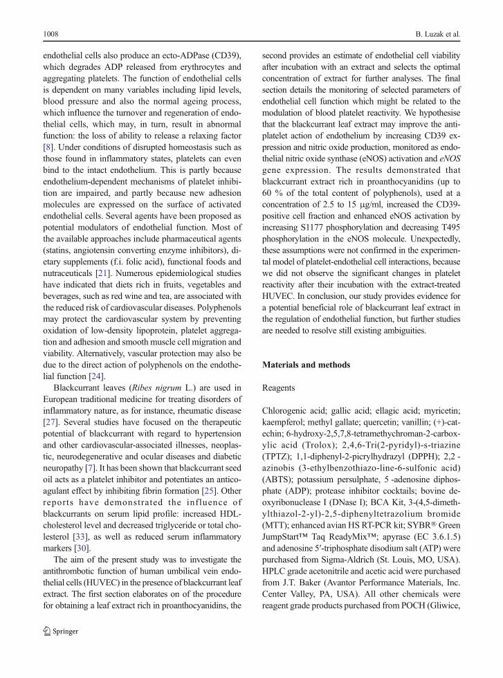
After 24-h incubation of HUVEC with blackcurrant leafextract, the CD39 expression significantly increased in adose-dependent manner (up to 10% for 2.5 μg GAE/ml,up to 25 % for 10 μg GAE/ml and up to 33 % for15 μg GAE/ml, Fig. 2). The NTPDase (apyrase) activitywas determined in the in vitro experimental model with-out cells, where the amount of Pi released from ATP byapyrase enzymatic reaction was measured. We observedthat after the incubation of apyrase with the extract at thehighest concentrations (10 and 15 μg GAE/ml) theenzyme activity was significantly reduced (Table 2).Simultaneously, the lower concentrations (0.25–2.5 μg GAE/ml) of extract did not change apyraseactivity in the significant way. Also, the absorbancespectrum of blackcurrant leaf extract did not interferewith the absorbance of the product of malachite greenassay measured at 655 nm.
Table 1 Polyphenolic composition of blackcurrant leaf extract
Content [mg/g weight of extract]
Total polyphenolsa 249.2±2.5
Total flavanolsb 169.4±3.2
Total proanthocyanidinsc 152.2±2.5
Hydroxycinnamic acidsd 27.0±0.3
Flavonol derivativese 23.8±0.1
Flavonol aglyconsf
Quercetin 13.6±0.2
Kaempferol 5.4±0.1
Myricetin 0.8±0.0
Gallotanninsg 1.2±0.1
Data shown as a mean±SE, n=10aDetermined by Folin-Ciocalteu reagent as gallic acid equivalentsb Determined by vanillin reagent as (+) catechin equivalentsc Determined after acid depolymerization as cyanidin equivalentsd Determined by HPLC method at 320 nm as chlorogenic acidequivalentse Determined by HPLC at 360 nm as quercetin equivalentsf Determined byHPLC at 360 nm after acid hydrolysis as referencecompoundsgDetermined by HPLC at 280 nm after acid hydrolysis as methylgallate equivalents
Extract from R. nigrum leaves modulates endothelial cell function 1013

The effect of blackcurrant leaf extract on S1177and T495 phosphorylation in eNOS protein
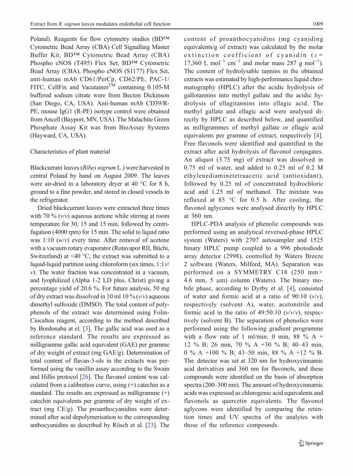
In endothelial cells incubated for 24 h with 2.5 and15 μg GAE/ml blackcurrant leaf extract, the activity ofeNOS was analysed by monitoring protein phosphory-lation at serine 1177 (S1177) and threonine 495 (T495)using flow cytometry. Blackcurrant leaf extractinduced significant eNOS phosphorylation atS1177 (p<0.05 or less) and significantly decreasedeNOS phosphorylation at T495 (p<0.01) (Fig. 3).The effects of dephosphorylation were significantlyhigher (p<0.05) for both concentrations (the de-crease in T495 phosphorylation was 31±6 % for2.5 μg GAE/ml and 48±6 % for 15 μg GAE/ml
vs. the increase in S1177 phosphorylation, 13±3 %for 2.5 μg GAE/ml and 18±7 % for 15 μg GAE/ml,compared to untreated cells).
The effect of blackcurrant leaf extract on eNOS geneexpression
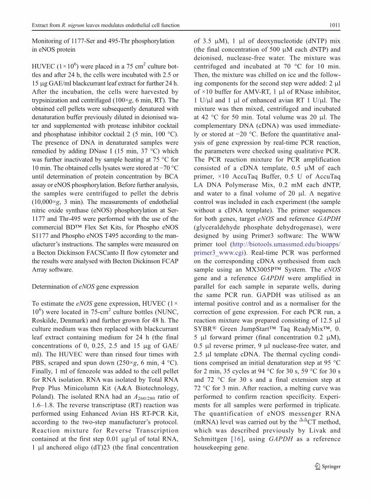
The basal relative mRNA level for eNOS was 8.7±1.4in untreated cells and was unchanged after incubationwith the tested extract (Fig. 4). Nevertheless, the incu-bation with the polyphenolic extract led to the increasedrelative mRNA level, mostly at the lower concentration(0.25 μg GAE/ml); this difference, however, remainedbeyond statistical significance.
Fig. 1 The effect of blackcurrantleaf extract on HUVEC viability.Data shown as a percent ofcontrol (untreated cells). Errorbars refer to the standard errorsobtained for eight experiments(each in four repeats). a Theextract in low concentrationsignificantly increased cellviability after 24 h (white boxes)and 48 h (grey boxes), *p<0.001or less. b The extract significantlyreduced cell viability for bothincubation times, *p<0.001(significance of differences wasestimated by the single sampletest with Bonferroni’s correctionin comparison to control:untreated cells)
1014 B. Luzak et al.
The effect of blackcurrant leaf extracton platelet-endothelium interactions
This study encompasses a simple model of monitoringblood platelet reactivity by analysing the expressions ofselectin P (CD62P antigen) and the active form ofGPIIb/IIIa (PAC-1) in blood platelets incubated withHUVEC pretreated with 2.5 and 15 μg GAE/mlblackcurrant extract. A significant reduction of ADP-induced platelet reactivity was seen after incubation
with HUVEC. The effect was especially more pro-nounced for the fraction of PAC-1-positive platelets(43.7±2.9 % for platelets unicubated with HUVEC vs.26.0±2.1 % for platelets incubated with extract-untreated HUVEC, p<0.001). Nevertheless, platelet
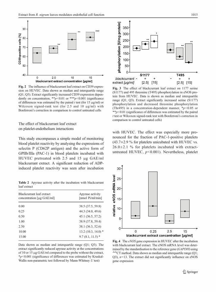
Fig. 2 The influence of blackcurrant leaf extract on CD39 expres-sion on HUVEC. Data shown as median and interquartile range(Q1; Q3). Extract significantly increased CD39 expression depen-dently on concentration, **p<0.01 or ***p<0.001 (significanceof differences was estimated by the paired t test (for 15 μg/ml) orWilcoxon signed-rank test (for 2.5 and 10 μg/ml) withBonferroni’s correction in comparison to control untreated cells
Table 2 Apyrase activity after the incubation with blackcurrantleaf extract
Blackcurrant leaf extractconcentration [μg GAE/ml]
Apyrase activity[nmol Pi/ml/min]
0.00 38.5 (37.5, 59.8)
0.25 44.3 (34.0, 49.6)
0.50 45.1 (36.5, 57.2)
1.00 38.9 (37.8, 59.4)
2.50 38.1 (36.3, 52.6)
10.00 13.2 (10.3, 14.0) *
15.00 9.7 (8.1, 11.5) *
Data shown as median and interquartile range (Q1; Q3). Theextract significantly reduced apyrase activity at the concentrationsof 10 or 15 μg GAE/ml compared to the probe without the extract,*p<0.001 (significance of differences was estimated by Kruskal-Wallis non-parametric test followed by Mann-Whitney U test)
Fig. 3 The effect of blackcurrant leaf extract on 1177 serine(S1177) and 495 threonine (T495) phosphorylation in eNOS pro-tein from HUVEC. Data is shown as median and interquartilerange (Q1; Q3). Extract significantly increased serine (S1177)phosphorylation and decreased threonine phosphorylation(Thr495) in a concentration-dependent manner, *p<0.05 or**p<0.01 (significance of differences was estimated by the pairedt test or Wilcoxon signed-rank test with Bonferroni’s correction incomparison to control untreated cells)
Fig. 4 The eNOS gene expression in HUVEC after the incubationwith blackcurrant leaf extract. The eNOS mRNA level was deter-mined by the standardisation to the reference gene (GAPDH) usingΔΔCTmethod. Data shown as median and interquartile range (Q1;Q3), n=13. The extract did not significantly influence on eNOSgene expression
Extract from R. nigrum leaves modulates endothelial cell function 1015

reactivity did not change after exposure to extract-treatedHUVEC compared to controls: platelets incubated withuntreated HUVEC.
Discussion
Plant materials may contain varying quantities of differ-ent subclasses of phenolics, ranging from simple phe-nolic acids to polymerised tannins. Therefore, it is verydifficult to develop the procedure suitable for the extrac-tion of a whole spectrum of plant phenolics. For theextraction of phenolics from blackcurrant leaves, theacetone mixture was found to be more effective thanother solvents and it enables the extraction of severalclasses of phenolic compounds with a good yield forflavonols, flavan-3-ols and anthocyanins [28, 29]. Theresults from the present study confirmed these observa-tions: blackcurrant leaf acetone extract, additionallywashed with chloroform to remove fat-soluble sub-stances (especially chlorophylls), is rich in phenoliccompounds (Table 1). The blackcurrant leaf extract isknown to contain 25 % phenolic compounds, most ofwhich are f l avanol s , 90 % of them beingproanthocyanidins. The content of hydroxycinnamicacids and flavonols was less than one sixth that of totalflavanols. Quercetin content, as quantified after acidhydrolysis, was 2.5 times higher than kaempferol andalmost 17 times higher than myricetin content.Oszmiański et al. [20] and Liu et al. [15] described adetailed composition of blackcurrant leaf ethanol extractand indicated that the most important groups of phenoliccompounds are flavonols (flavonol glycosides such asquercetin or kaempferol), proanthocyanidins and phe-nolic acids (hydroxycinnaminic acid conjugates). Also,Vagiri et al. reported the phenolic composition andindicated an optimised method for analysis of phenoliccompounds in buds, leaves and fruits of blackcurrantwhich are known as very good sources of active pheno-lic compounds [31].Many of the compounds detected inthe leaves have been reported to have antiviral, antimi-crobial and antidiabetic activities. Proantocyanidins, themain phenolic compounds in blackcurrant leaf extract,and naturally occurring in various plants, have beenknown for its anti-inflammatory and antiarthritic activ-ities. They are reported to prevent heart diseases, andgrape seed proanthocyanidins should be a potentcardioprotective compound.
Berries are known as a rich source of phenolics [7],but the recent studies underline a significant role of thepolyphenols extracted from raw materials other thanberry tissue, such as leaves [20]. It was reported thatthe leaves from apple, thornless black raspberry, redraspberry and strawberry had high antioxidant capacitiesand total phenolic content compared to their fruit tissues[32]. Tabart et al. demonstrated that blackcurrant leaveshad a higher phenolic and antioxidant content than fullyripened berries, where the total phenolic level was cor-related with antioxidant activity [28]. In addition,blackcurrant leaf extract possessed the strongest antiox-idant activity compared to berries or buds as demon-strated by in vitro classical assays, but also in cellularmodels, such as endothelial cells and polymorphonucle-ar neutrophils. This study emphasises that the intracel-lular and extracellular scavenging properties ofblackcurrant extract have no toxic effects [27]. Ourstudy confirms that blackcurrant leaf extract is a goodsource of antioxidants with scavenging potential againstfree synthetic radicals ABTS●+ and DPPH●, and withhigh reducing power towards Fe3+ ions. Although theseresults may indicate a potential mechanism of thein vivo action of blackcurrant leaf extract, only clinicalstudies could finally confirm this assumption. Polyphe-nols, as the constituents of food, are poorly absorbedand extensively metabolised. In addition, they can bindto plasma proteins and form relatively stable complexesthat are less active.
The aim of the present study was to investigate theantithrombotic function of human umbilical vein endo-thelial cells (HUVEC) in the presence of blackcurrantleaf extract.We targeted this objective by the monitoringof selected parameters of endothelial cell function thatmight be related to the modulation of blood plateletreactivity. We further hypothesised that the blackcurrantleaf extract might improve the antiplatelet action ofendothelium by the increasing of CD39 expression andnitric oxide production, monitored as endothelial nitricoxide synthase (eNOS) activation and eNOS geneexpression.
Typically, endothelial cells release NO in response tofluid shear stress and autacoids that mobilise intracellu-lar calcium, such as thrombin or ADP. The eNOS en-zyme is regulated post-translationally, but can also beinfluenced on the transcriptional level. Plant extractsrich in polyphenolic compounds were shown to be ableto up-regulate eNOS and increase the activity of eNOSin cultured endothelial cells [5]. The consumption of
1016 B. Luzak et al.

flavanol-rich cocoa and purified anthocyanin supple-mentation improved endothelial function and vascularreactivity in hypercholesterolemic patients [33]. More-over, in healthy subjects, an improved endothelial func-tion was observed after administration of (-)-epicatehin(a major flavanol of cocoa), or after red wine or purplegrape juice [24]. Our findings show that the extract fromblackcurrant leaves significantly increased eNOS activ-ity by modulating the phosphorylation rate of this pro-tein; increased S1177 phosporylation (an activator site),and decreased T495 phosporylation (an inhibitor site)were observed in the extract-treated cells (Fig. 3). Thesefindings are corroborated by other studies performedwith other phenolic compounds. The phenolic-rich frac-tion of black tea was found to sharply enhance NOformation subsequent to the phosphorylation of eNOSat S1177 and the dephosphorylation of eNOS at T495 inporcine aortic endothelial cells. This stimulatory effectwas calcium dependent, involving both intracellular andextracellular calcium and involved the p38 MAPK up-stream of PI3-kinase/Akt pathway [1]. Theendothelium-dependent relaxation was observed in re-sponse to exposure to some berry extracts, includingblackcurrant extract, and the biological activity waspredominantly associated with the fraction enriched inellagitannins or anthocyanins [24]. Edirisinghe et al.report that anthocyanins from blackcurrant juice (1 μl/ml) significantly increased the phosphorylation of Akt atS473 and eNOS at S1177 in human endothelial cellsunder in vitro conditions [5]. Also, it has been shownthat red wine polyphenols induce the redox-sensitiveactivation of the PI3-kinase/Akt pathway in endothelialcells which, in turn, causes phosphorylation of eNOS,resulting in an increased formation of NO [19]. Themechanism underlying the activation of endothelialNO synthase (eNOS) remains unclear, but it has beensuggested that polyphenols induce intracellular forma-tion of reactive oxygen species such as superoxide an-ions and trigger the activation of eNOS by the PI3-kinase/Akt pathway or they may act in a calcium-dependent manner [24].
It was reported that blackcurrant leaf extract pos-sesses anti-inflammatory properties [6, 27]. The presentstudy demonstrates for the first time that blackcurrantleaf extract may modulate endothelial cell function suchas an increase in CD39 (ecto-ADPase) expression andeNOS activation. The ecto-ADPase expression in endo-thelial cells is one of the endogenousmechanisms whichregulate blood platelet reactivity. The preserved function
of CD39/ATPase is critical in the inhibition of plateletand neutrophil activation by keeping adenosine nucleo-tide levels at a low level [18]. Kaneider et al. report thatthrombin downregulates CD39/ATPase activity butquercetin and resveratrol restores the decreasedCD39/ATPase activity in human umbilical vein endo-thelial cells in response to thrombin, as demonstrated bythe increased adenosine monophosphate (AMP) andadenosine levels seen in endothelial culture superna-tants. Both polyphenols were found to inhibitthrombin-induced MAPK, JNK and focal adhesion ki-nase activities in endothelial cells: clear evidence thatthe levels of CD39 expression correlate with ATPaseactivity in human endothelial cells, platelets and select-ed monocytes, NK cells and megakaryocyte cell lines[10]. ATPase expressed by the investigated cells mightinfluence signal transduction initiated by purinergic re-ceptors by hydrolysis of their agonists. Koziak assumesthat activity and expression of CD39/ATPase in thevasculature and other cells may have profound conse-quences for modulation of signals triggered bypurinergic receptors, including apoptosis, anti-inflammatory and prothrombotic responses [13].
The increased CD39 expression and the increasedeNOS activation in HUVEC can be regarded as thebeneficial markers of the improvement of antiplateletaction of endothelial cells. Unexpectedly, these assump-tions were not confirmed in the experimental model ofplatelet-endothelial cell interactions. We did not observethe significant changes in platelet reactivity after theirincubation with the extract-treated HUVEC. The expla-nation of this observation may relate to the analysis ofapyrase activity after the incubation with blackcurrantleaf extract. The increased CD39 expression appears nota sufficient indicator of antiplatelet action of HUVEC,because blackcurrant extract at the higher concentra-tions (10 or 15 μg GAE/ml) significantly reduced apy-rase activity or did not affect apyrase activity at lowerconcentrations (up to 2.5 μg GAE/ml). It is possible thatsome compounds of extract may interact with apyrasefollowed by the enzyme inhibition. Also, the increasedeNOS activation without the accompanying increase ofthe eNOS mRNA in HUVEC treated with the extractwas not associated with the reduced blood plateletreactivity.
In this study, the biological antithrombotic effects ofblackcurrant leaf extract were investigated at a few con-centrations selected in the cell viability assay.We noticedthat blackcurrant leaf extract may modulate the viability
Extract from R. nigrum leaves modulates endothelial cell function 1017
of endothelial cells in a time- and concentration-dependent manner. Regardless of exposure time, at lowerconcentrations, blackcurrant leaf extract was found toimprove HUVEC viability, whereas at higher concentra-tions (above 30 μg GAE/ml), it significantly decreasedHUVEC viability as compared to the untreated cells.However, EC80 values estimated for 24 and 48-h incu-bation of HUVEC with blackcurrant extract were signif-icantly different, which means that longer incubation ofHUVEC with blackcurrant leaf extract intensifies itsanti-proliferative effect. Such an increased cell viabilityeffect of blackcurrant leaf extract for lower concentra-tions, which are nutritionally relevant doses for phenoliccompounds, may play a crucial role in physiologicalangiogenic processes, such as wound repair or tissueregeneration. Additionally, we observed that in the rangeof concentrations known to improve cell viability, theextract also increased eNOS activation or enhancedCD39 expression. Per analogiam, some authors havesuggested a significant role of phenolic compounds iso-lated from Panax ginseng in angiogenesis. The extractsignificantly enhanced HUVEC viability and this effectwas accompanied by phosphorylation of eNOS, as wellas the increased NO production [12]. In the presence ofSKE-treated cells (the extract from Stewartia koreanaleaves, 50 μg/ml, incubation by 24 h) cell proliferationincreased twofold, and almost no cytotoxic effects orinducedmorphological changes were noted in the treatedcells, thus indicating that the enhanced proliferation ofendothelial cells occurred at non-cytotoxic concentra-tions. Based on these outcomes, Stewartia koreana ex-tracts have been proposed as potential agents useful inaccelerating vascular wound healing or promoting thegrowth of collateral blood vessels in ischaemic tissues[14].
We are aware that the most relevant limitation of ourstudy is the choice of blackcurrant leaf extract concen-trations to monitor the endothelial cell functions. Wehave known that the selected concentrations (2.5; 10 or15 μg/ml) were higher compared to those used in thepolyphenol in vivo studies (where the maximal plasmaconcentration of polyphenols and its metabolites usuallydid not exceed 10 μM (1.8 μg/ml) and depended uponthe kind of phenolic compound) [17]. Epidemiologicalstudies have shown that the significant biological effectsof polyphenols can be observed after a few days or a fewweeks of the diets rich in polyphenolic compounds. Inour experimental model of human endothelial cells intissue cultures, we monitored the cell function after the
short 24-h incubation instead of a few days’ incubationand we used the higher concentrations to demonstratethe extract effects. Simultaneously, we have been con-scious that the increasing of extract concentrations couldresult in experimental artefacts. However, in many ofthe published in vitro studies, the health benefits havebeen demonstrated using the concentrations muchhigher (1–100 μM) than those achievable in a body.
In conclusion, blackcurrant leaf extract, which is richin proanthocyanidins, may modulate endothelial cellfunction in a concentration-dependent manner. The ex-tract has been found to damage endothelial cells atconcentrations above 30 μg GAE/ml. However, at thelower concentrations, its effects were beneficial. Theextract increased endothelial cell viability and increasedCD39 expression, but collaterally reduced apyrase ac-tivity; it also up-regulated eNOS activation by influenc-ing enzyme phosphorylation rate, however, with noconcomitant affecting eNOS gene expression. The-se observations lead to the conclusion that theblackcurrant leaf extract may improve endothelialcell viability at low physiological concentrations,however, without affecting antiplatelet action ofendothelium. Importantly, the increasing of theextract concentrations apparently reverses thesebeneficial effects. Overall, we have to sum up withthe stating that the in vitro findings on biologicalactivity of extracts cannot be easily translated toin vivo physiological conditions.
Acknowledgments This work was supported by the grant “Pro-duction of polyphenol extracts of plant origin with antiplatelet andcardioprotective properties – FLAWOPIRYNA” UDA-POIG.01.03.01-10-129/08-00, cofinanced by the European Unionfrom the European Regional Development Fund within the frame-work of the Innovative Economy Operational Programme.
References
1. Anter E, Thomas SR, Schulz E, Shapira OM, Vita JA, KeaneyJF Jr (2004) Activation of endothelial nitric-oxide synthase bythe p38 MAPK in response to black tea polyphenols. J BiolChem 279:46637–46643
2. Benzie IF, Strain JJ (1996) The ferric reducing ability ofplasma (FRAP) as a measure of "antioxidant power": theFRAP assay. Anal Biochem 239:70–76
3. Bordonaba JG, Terry LA (2008) Biochemical profiling andchemometric analysis of seventeen UK-grown black currantcultivars. J Agric Food Chem 56:7422–7430
1018 B. Luzak et al.

4. Dyrby M, Westergaard N, Stapelfedt H (2001) Light and heatsensitivity of red cabbage extract in soft drink model systems.Food Chem 72:431–437
5. Edirisinghe I, Banaszewski K, Cappozzo J, McCarthy D,Burton-Freeman BM (2011) Effect of black currant anthocy-anins on the activation of endothelial nitric oxide synthase(eNOS) in vitro in human endothelial cells. J Agric FoodChem 59:8616–8624
6. Garbacki N, Tits M, Angenot L, Damas J (2004) Inhibitoryeffects of proanthocyanidins from Ribes nigrum leaves oncarrageenin acute inflammatory reactions induced in rats.BMC Pharmacol 4:25
7. Gopalan A, Reuben SC, Ahmed S, Darvesh AS, Hohmann J,Bishayee A (2012) The health benefits of blackcurrants. FoodFunct 3:795–809
8. Goubareva I, Gkaliagkousi E, ShahA,Queen L, Ritter J, FerroA (2007) Age decreases nitric oxide synthesis and responsive-ness in human platelets and increases formation of monocyte-platelet aggregates. Cardiovasc Res 75:793–802
9. Hansen MB, Nielsen SE, Berg K (1989) Re-examination andfurther development of a precise and rapid dye method formeasuring cell growth/cell kill. J Immunol Methods119:203–210
10. Kaneider NC, Mosheimer B, Reinisch N, Patsch JR,Wiedermann CJ (2004) Inhibition of thrombin-induced sig-naling by resveratrol and quercetin: effects on adenosinenucleotide metabolism in endothelial cells and platelet-neutrophil interactions. Thromb Res 114:185–194
11. Kim DO, Lee KW, Lee HJ, Lee CY (2002) Vitamin C equiv-alent antioxidant capacity (VCEAC) of phenolic phytochem-icals. J Agric Food Chem 50:3713–3717
12. Kim YM, Namkoong S, Yun YG, Hong HD, Lee YC, Ha KS,Lee H,KwonHJ, KwonYG,KimYM (2007)Water extract ofKorean red ginseng stimulates angiogenesis by activating thePI3K/Akt-dependent ERK1/2 and eNOS pathways in humanumbilical vein endothelial cells. Biol Pharm Bull 30:1674–1679
13. Koziak K, Sevigny J, Robson SC, Siegel JB, Kaczmarek E(1999) Analysis of CD39/ATP diphosphohydrolase(ATPDase) expression in endothelial cells, platelets and leu-kocytes. Thromb Haemost 82:1538–1544
14. Lee TH, Lee GW, Kim CW, Bang MH, Baek NI, KimSH, Chung DK, Kim J (2010) Stewartia koreana extractstimulates proliferation and migration of human endo-thelial cells and induces neovasculization in vivo.Phytother Res 24:20–25
15. Liu P, Kallio H, Yang B (2014) Flavonol glycosides and otherphenolic compounds in buds and leaves of different varietiesof black currant (Ribes nigrum L.) and changes during grow-ing season. Food Chem 160:180–189
16. Livak KJ, Schmittgen TD (2001) Analysis of relative geneexpression data using real-time quantitative PCR and the 2(-delta delta C(T)) method. Methods 25:402–408
17. Manach C, Williamson G, Morand C, Scalbert A, Remesy C(2005) Bioavailability and bioefficacy of polyphenols inhumans. I. Review of 97 bioavailability studies. Am J ClinNutr 81:230S–242S
18. Marcus AJ, BroekmanMJ, Drosopoulos JH, Olson KE, IslamN, Pinsky DJ, Levi R (2005) Role of CD39 (NTPDase-1) inthromboregulation, cerebroprotection, and cardioprotection.Semin Thromb Hemost 31:234–246
19. Ndiaye M, Chataigneau M, Lobysheva I, Chataigneau T,Schini-Kerth VB (2005) Red wine polyphenol-induced,endothelium-dependent NO-mediated relaxation is due to theredox-sensitive PI3-kinase/Akt-dependent phosphorylation ofendothelial NO-synthase in the isolated porcine coronary ar-tery. FASEB J 19:455–457
20. Oszmianski J, Wojdylo A, Gorzelany J, Kapusta I (2011)Identification and characterization of low molecular weightpolyphenols in berry leaf extracts by HPLC-DAD and LC-ESI/MS. J Agric Food Chem 59:12830–12835
21. Ramli J, CalderonArtero P, Block RC, Mousa SA (2011)Novel therapeutic targets for preserving a healthy endotheli-um: strategies for reducing the risk of vascular and cardiovas-cular disease. Cardiol J 18:352–363
22. Re R, Pellegrini N, Proteggente A, Pannala A, Yang M, Rice-Evans C (1999) Antioxidant activity applying an improvedABTS radical cation decolorization assay. Free Radic BiolMed 26:1231–1237
23. Rosch D, Bergmann M, Knorr D, Kroh LW (2003) Structure-antioxidant efficiency relationships of phenolic compoundsand their contribution to the antioxidant activity of sea buck-thorn juice. J Agric Food Chem 51:4233–4239
24. Schini-Kerth VB, Auger C, Kim JH, Etienne-Selloum N,Chataigneau T (2010) Nutritional improvement of the endo-thelial control of vascular tone by polyphenols: role of NO andEDHF. Pflugers Arch 459:853–862
25. Stone DA, Hawke MW, LaMonte M, Kittner SJ, Acosta J,Corretti M, Sample C, Price TR, Plotnick GD (1995)Ulcerated atherosclerotic plaques in the thoracic aorta areassociated with cryptogenic stroke: a multiplane transesopha-geal echocardiographic study. Am Heart J 130:105–108
26. Swain T, Hillis WE (1959) The phenolics constituents ofPrunus domestica III. J Sci Food Agric 10:63–68
27. Tabart J, Franck T, Kevers C, Pincemail J, Serteyn D,Defraigne JO, Dommes J (2012) Antioxidant and anti-inflammatory activities of Ribes nigrum extracts. FoodChem 131:1116–1122
28. Tabart J, Kevers C, Evers D, Dommes J (2011) Ascorbic acid,phenolic acid, flavonoid, and carotenoid profiles of selectedextracts from Ribes nigrum. J Agric Food Chem 59:4763–4770
29. Tabart J, Kevers C, Pincemail J, Defraigne JO, Dommes J(2006) Antioxidant capacity of black currant varies with or-gan, season, and cultivar. J Agric Food Chem 54:6271–6276
30. Tahvonen RL, Schwab US, Linderborg KM, Mykkanen HM,Kallio HP (2005) Black currant seed oil and fish oil supple-ments differ in their effects on fatty acid profiles of plasmalipids, and concentrations of serum total and lipoprotein lipids,plasma glucose and insulin. J Nutr Biochem 16:353–359
31. Vagiri M, EkholmA, Andersson SC, Johansson E, RumpunenK (2012) An optimized method for analysis of phenolic com-pounds in buds, leaves, and fruits of black currant (Ribesnigrum L.). J Agric Food Chem 60:10501–10510
32. Wang SY, Lin HS (2000) Antioxidant activity in fruits andleaves of blackberry, raspberry, and strawberry varies withcultivar and developmental stage. J Agric Food Chem 48:140–146
33. Zhu Y, Xia M, Yang Y, Liu F, Li Z, Hao Y, Mi M, Jin T, LingW (2011) Purified anthocyanin supplementation improvesendothelial function via NO-cGMP activation in hypercholes-terolemic individuals. Clin Chem 57:1524–1533
Extract from R. nigrum leaves modulates endothelial cell function 1019