Embed Size (px)
Citation preview
El
JLa
b
a
ARRAA
KGNCALL
1
lWcncir2tVeo
ttwfMb
0h
Carbohydrate Polymers 91 (2013) 229– 235
Contents lists available at SciVerse ScienceDirect
Carbohydrate Polymers
jo u rn al hom epa ge: www.elsev ier .com/ locate /carbpol
xtraction and characterization of nanocellulose structures from raw cottoninter
oão Paulo Saraiva Moraisa,∗, Morsyleide de Freitas Rosab, Men de sá Moreira de Souza Filhob,idyane Dias Nascimentoa, Diego Magalhães do Nascimentob, Ana Ribeiro Cassalesb
Embrapa Algodão, Rua Oswaldo Cruz, 1143, CEP 58428-095 Campina Grande, PB, BrazilEmbrapa Agroindústria Tropical, Rua Dra. Sara Mesquita, 2270, CEP 60511-110 Fortaleza, CE, Brazil
r t i c l e i n f o
rticle history:eceived 11 May 2012eceived in revised form 20 July 2012ccepted 3 August 2012vailable online 11 August 2012
a b s t r a c t
This study aimed to characterize nanocellulose extracted from cotton (Gossypium hirsutum) linters.The nanocellulose was subjected to electronic microscopy, thermal analysis, X-ray diffractometry, lightscattering, and contact angle. The properties of the nanocellulose are considerably different from thelinter. The acidic hydrolyses applied to extract the nanocrystals increased the crystallinity index and thehydrophilicity and decreased the thermal stability. On average, the nanocrystals were 177 nm long and12 nm wide, with an aspect ratio of 19 when measured by microscopy. The light scattering results were
eywords:ossypium hirsutumanowhiskero-productgroindustrial wasteight scattering diffraction
coherent with the crystal dimensions. Cotton linter is a potential source of nanocellulose crystals, par-ticularly to be used in the production of hydrophilic nanocomposites. Extraction of nanocellulose fromraw cotton linter does not require pulping.
© 2012 Elsevier Ltd. Open access under the Elsevier OA license.
ignocellulosic characterization
. Introduction
Linter is an important by product of the textile industry. Cottoninter is the short fiber that cannot be used in the textile process.
hen the regular cotton fibers are extracted in the ginning pro-ess, the linter remains attached to the seed coat. The fuzzy seedeeds to be subjected to an additional process that will mechani-ally remove the linter. The amount of linter produced worldwides around 2.5 million metric tons, considering the 42 million met-ic tons of cotton lint produced in 2010 (FAOSTAT, 2012; Sczostak,009). Traditional products made from linter are: absorbent cot-on, special papers, cellulose nitrate, and acetate (Sczostak, 2009;ieira, Beltrão, Lima, & Leão, 2008). In some cases, the linter is notxtracted, but kept with the seed (when it is used for oil extraction)r chemically dissolved (for planting the seed).
Producing cellulose nanocrystals is an interesting use for lin-er. Nanocrystals of cellulose, with diameters ranging from 2 nmo 20 nm and length ranging from 100 nm to 2.1 �m are calledhiskers, nanowhiskers, or nanofibrils, and they can be obtained
rom many natural fibers (Capadona et al., 2009; Pandey, Ahn, Lee,ohanty, & Misra, 2010; Rosa et al., 2010). Natural fibers are used
ecause they are cheap, abundant, renewable, and biodegradable
∗ Corresponding author. Tel.: +55 83 3182 4300; fax: +55 83 3182 4367.E-mail address: [email protected] (J.P.S. Morais).
144-8617 © 2012 Elsevier Ltd. ttp://dx.doi.org/10.1016/j.carbpol.2012.08.010
Open access under the Elsevier OA license.
(Eichhorn et al., 2010; Siqueira, Bras, & Dufresne, 2009; Teixeiraet al., 2010). Nanocrystals can be used as fillers in composites(Capadona et al., 2009; Eichhorn et al., 2010; Stelte & Sanadi, 2009;Teixeira et al., 2010) because they have interesting mechanicalproperties such as low gas permeability (Stelte & Sanadi, 2009)and stiffness enhancing capacity (Pääkkö et al., 2008). They canalso be used as reinforcements for adhesives, components of elec-tronic devices, biomaterials, foams, aerogels, and textiles (Eichhornet al., 2010; Pääkkö et al., 2008; Pandey et al., 2010; Ummartyotin,Juntaro, Sain, & Manuspiya, 2012).
Crystalline and amorphous regions are found in cellulose fibersin proportions that vary among plant species. For that reason,the characteristics (particularly the dimensions) of nanocellulosicmaterials depend largely on the raw material. Even though all cel-lulose nanocrystals are made of the same biopolymer, differentraw materials can be used to obtain nanowhiskers tailored to spe-cific needs (Beck-Candanedo, Roman, & Gray, 2005; Eichhorn et al.,2010; Pandey et al., 2010; Rosa et al., 2010).
Cotton fiber is a traditional source of cellulose nanostructures(Rånby, 1949), but its chemical composition can be influenced bymany factors including the genotype and the environment whereit was produced. However, in the literature on cotton nanocrys-
tals there is scarce information on how and where the cotton wasproduced (Ibrahim, El-Zawawy, & Nassar, 2010; Lin, Chen, Huang,Dufresne, & Chang, 2009). The use of regular cotton fiber can alsoresult in a product different from those made of linter (Ass, Ciacco, &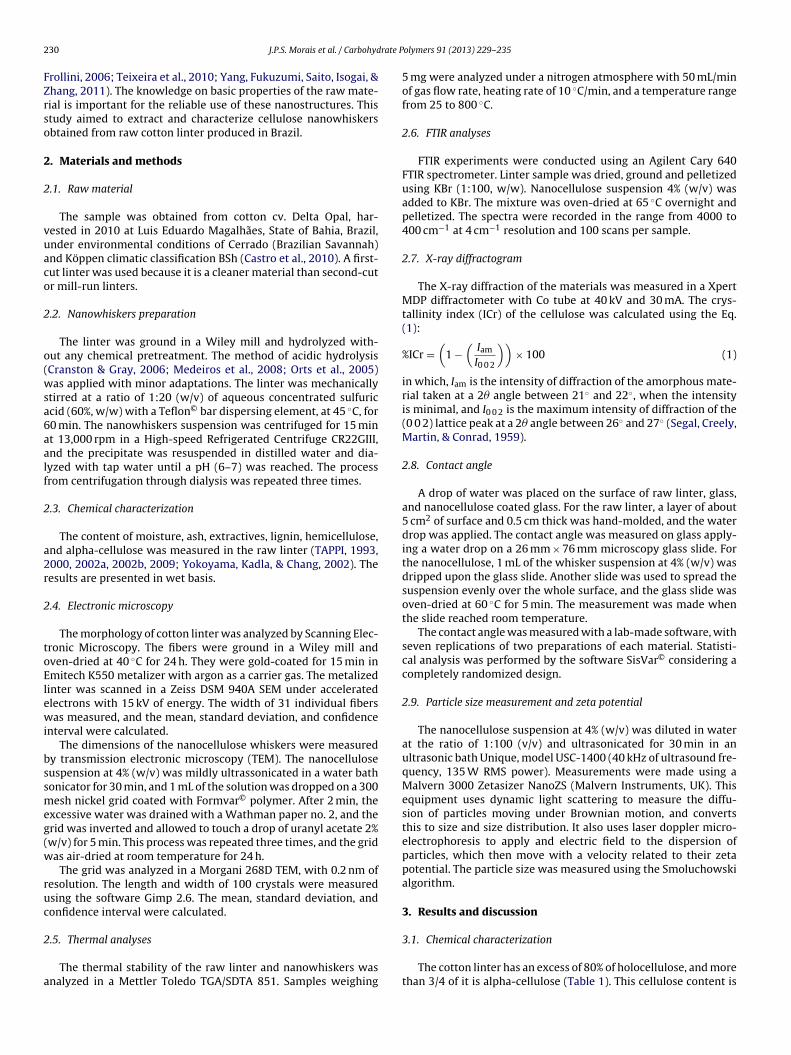
2 rate P
FZrso
2
2
vuaco
2
o(wsa6aalf
2
a2r
2
toElewi
bssmeg(w
ruc
2
a
30 J.P.S. Morais et al. / Carbohyd
rollini, 2006; Teixeira et al., 2010; Yang, Fukuzumi, Saito, Isogai, &hang, 2011). The knowledge on basic properties of the raw mate-ial is important for the reliable use of these nanostructures. Thistudy aimed to extract and characterize cellulose nanowhiskersbtained from raw cotton linter produced in Brazil.
. Materials and methods
.1. Raw material
The sample was obtained from cotton cv. Delta Opal, har-ested in 2010 at Luis Eduardo Magalhães, State of Bahia, Brazil,nder environmental conditions of Cerrado (Brazilian Savannah)nd Köppen climatic classification BSh (Castro et al., 2010). A first-ut linter was used because it is a cleaner material than second-cutr mill-run linters.
.2. Nanowhiskers preparation
The linter was ground in a Wiley mill and hydrolyzed with-ut any chemical pretreatment. The method of acidic hydrolysisCranston & Gray, 2006; Medeiros et al., 2008; Orts et al., 2005)as applied with minor adaptations. The linter was mechanically
tirred at a ratio of 1:20 (w/v) of aqueous concentrated sulfuriccid (60%, w/w) with a Teflon© bar dispersing element, at 45 ◦C, for0 min. The nanowhiskers suspension was centrifuged for 15 mint 13,000 rpm in a High-speed Refrigerated Centrifuge CR22GIII,nd the precipitate was resuspended in distilled water and dia-yzed with tap water until a pH (6–7) was reached. The processrom centrifugation through dialysis was repeated three times.
.3. Chemical characterization
The content of moisture, ash, extractives, lignin, hemicellulose,nd alpha-cellulose was measured in the raw linter (TAPPI, 1993,000, 2002a, 2002b, 2009; Yokoyama, Kadla, & Chang, 2002). Theesults are presented in wet basis.
.4. Electronic microscopy
The morphology of cotton linter was analyzed by Scanning Elec-ronic Microscopy. The fibers were ground in a Wiley mill andven-dried at 40 ◦C for 24 h. They were gold-coated for 15 min inmitech K550 metalizer with argon as a carrier gas. The metalizedinter was scanned in a Zeiss DSM 940A SEM under acceleratedlectrons with 15 kV of energy. The width of 31 individual fibersas measured, and the mean, standard deviation, and confidence
nterval were calculated.The dimensions of the nanocellulose whiskers were measured
y transmission electronic microscopy (TEM). The nanocelluloseuspension at 4% (w/v) was mildly ultrassonicated in a water bathonicator for 30 min, and 1 mL of the solution was dropped on a 300esh nickel grid coated with Formvar© polymer. After 2 min, the
xcessive water was drained with a Wathman paper no. 2, and therid was inverted and allowed to touch a drop of uranyl acetate 2%w/v) for 5 min. This process was repeated three times, and the gridas air-dried at room temperature for 24 h.
The grid was analyzed in a Morgani 268D TEM, with 0.2 nm ofesolution. The length and width of 100 crystals were measuredsing the software Gimp 2.6. The mean, standard deviation, andonfidence interval were calculated.
.5. Thermal analyses
The thermal stability of the raw linter and nanowhiskers wasnalyzed in a Mettler Toledo TGA/SDTA 851. Samples weighing
olymers 91 (2013) 229– 235
5 mg were analyzed under a nitrogen atmosphere with 50 mL/minof gas flow rate, heating rate of 10 ◦C/min, and a temperature rangefrom 25 to 800 ◦C.
2.6. FTIR analyses
FTIR experiments were conducted using an Agilent Cary 640FTIR spectrometer. Linter sample was dried, ground and pelletizedusing KBr (1:100, w/w). Nanocellulose suspension 4% (w/v) wasadded to KBr. The mixture was oven-dried at 65 ◦C overnight andpelletized. The spectra were recorded in the range from 4000 to400 cm−1 at 4 cm−1 resolution and 100 scans per sample.
2.7. X-ray diffractogram
The X-ray diffraction of the materials was measured in a XpertMDP diffractometer with Co tube at 40 kV and 30 mA. The crys-tallinity index (ICr) of the cellulose was calculated using the Eq.(1):
%ICr =(
1 −(
Iam
I0 0 2
))× 100 (1)
in which, Iam is the intensity of diffraction of the amorphous mate-rial taken at a 2� angle between 21◦ and 22◦, when the intensityis minimal, and I0 0 2 is the maximum intensity of diffraction of the(0 0 2) lattice peak at a 2� angle between 26◦ and 27◦ (Segal, Creely,Martin, & Conrad, 1959).
2.8. Contact angle
A drop of water was placed on the surface of raw linter, glass,and nanocellulose coated glass. For the raw linter, a layer of about5 cm2 of surface and 0.5 cm thick was hand-molded, and the waterdrop was applied. The contact angle was measured on glass apply-ing a water drop on a 26 mm × 76 mm microscopy glass slide. Forthe nanocellulose, 1 mL of the whisker suspension at 4% (w/v) wasdripped upon the glass slide. Another slide was used to spread thesuspension evenly over the whole surface, and the glass slide wasoven-dried at 60 ◦C for 5 min. The measurement was made whenthe slide reached room temperature.
The contact angle was measured with a lab-made software, withseven replications of two preparations of each material. Statisti-cal analysis was performed by the software SisVar© considering acompletely randomized design.
2.9. Particle size measurement and zeta potential
The nanocellulose suspension at 4% (w/v) was diluted in waterat the ratio of 1:100 (v/v) and ultrasonicated for 30 min in anultrasonic bath Unique, model USC-1400 (40 kHz of ultrasound fre-quency, 135 W RMS power). Measurements were made using aMalvern 3000 Zetasizer NanoZS (Malvern Instruments, UK). Thisequipment uses dynamic light scattering to measure the diffu-sion of particles moving under Brownian motion, and convertsthis to size and size distribution. It also uses laser doppler micro-electrophoresis to apply and electric field to the dispersion ofparticles, which then move with a velocity related to their zetapotential. The particle size was measured using the Smoluchowskialgorithm.
3. Results and discussion
3.1. Chemical characterization
The cotton linter has an excess of 80% of holocellulose, and morethan 3/4 of it is alpha-cellulose (Table 1). This cellulose content is
J.P.S. Morais et al. / Carbohydrate P
Table 1Lignocellulosical composition of cotton linter cv. Delta Opal.
Component Content (%, w/w)a
Moisture 6.33 ± 0.06Ashes 2.32 ± 0.04Extractives 5.59 ± 1.91Insoluble lignin 0.68 ± 0.35Holocellulose 81.51 ± 4.12Hemicellulose 4.60 ± 0.60
wiwic
haN(rM(
161 to 193), 12 nm wide (ranging from 10 to 13), and have an aspect
Alpha-cellulose 76.91 ± 7.19
a Mean ± standard error.
ithin the normal range for cotton linter (Sczostak, 2009), and its compared to the cellulose content of naturally colored cotton,
hich ranges from 74.0% to 80.3% (Teixeira et al., 2010). However, its lower than the 97.7% of cellulose found in hydrophilic (medicinal)otton.
The linter is an attractive source of nanowhiskers because itas more cellulose than other natural fibers commonly used suchs: sisal (Agave sisalana) (67–78%) (Oksman, Mathew, Långström,yström, & Joseph, 2009), banana (Musa spp.) (54–64.4%)
Cherian et al., 2008; Oksman et al., 2009), sugarcane (Saccha-
um officinarum) bagasse (44.9–45%) (Cerqueira, Rodrigues Filho, &eireles, 2010; Zhao, Wang, & Liu, 2008), bamboo (Bambusa spp.)41.8–54.0%) (Ardanuy, Claramunt, García-Horta, & Barra, 2011;
Fig. 1. SEM pictures of cotton linter cv. Delta Opal.
olymers 91 (2013) 229– 235 231
Chen, Yu, Liu, Hai, & Zhang, 2011), and coconut (Cocos nucifera)husk (32.5–45.9%) (Brígida, Calado, Gonc alves, & Coelho, 2010; Rosaet al., 2010). Cotton linter is also available in large amounts becauseit is a by-product of the textile industry. Upscaling of linter for com-mercial production of cellulosic nanowhiskers requires a supplywith little variation in the cellulose content and low impurities con-tent such as seed coat, soil, plant residues, and other contaminants.
3.2. Electronic microscopy
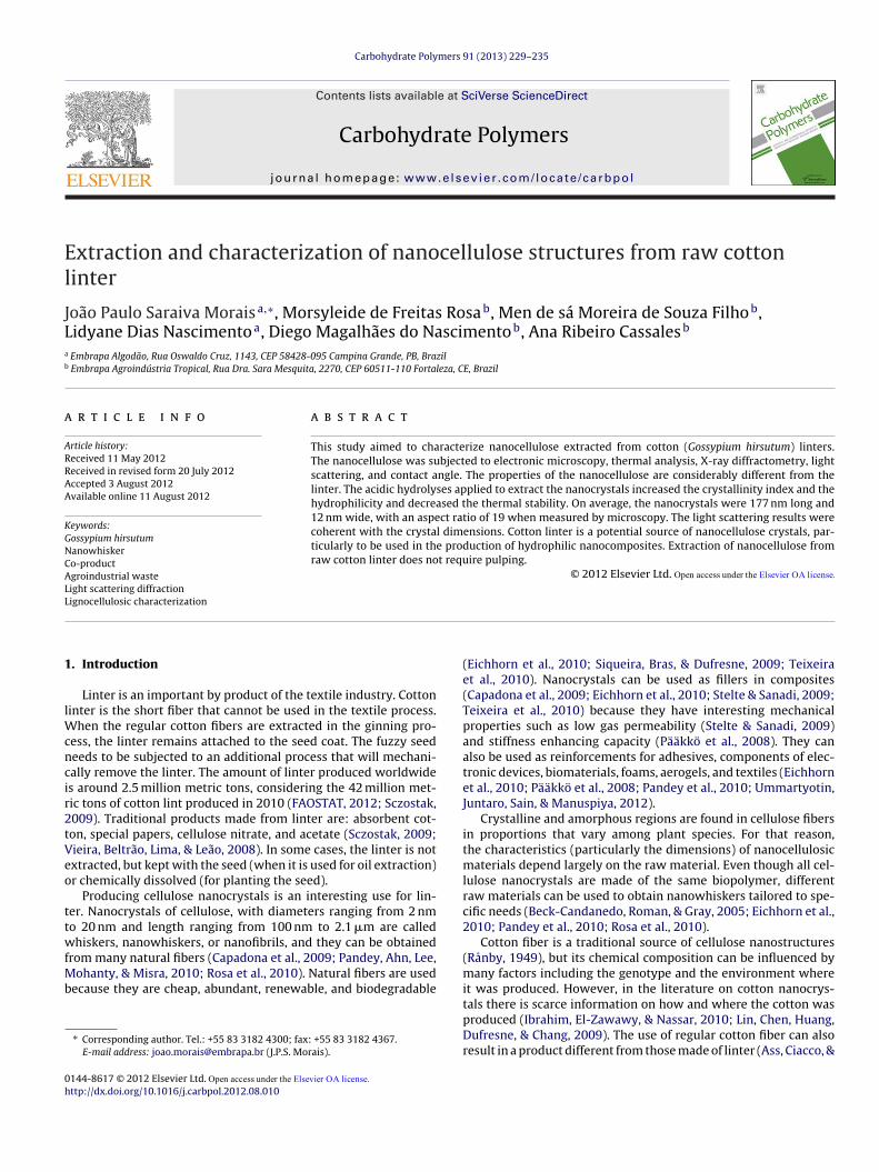
A curled and soft-flat shape was observed in the SEM pictures ofthe linter (Fig. 1a). The surface was rough with some pits. The aver-age width was 23.04 �m, with a confidence interval of 1.01 �m(Fig. 1b), which is in accordance with reports in the literature(Sczostak, 2009). This curled shape increases the surface area andmakes the fiber more reactive than typical cotton fibers. The flatshape of this fiber increases its specific area and favors chemicalreactions such as acidic hydrolysis.
The nanocellulose suspension had a white gel appearance(Fig. 2a). Bundles of crystals are depicted in the TEM pictures(Fig. 2b). On average, the whiskers are 177 nm long (ranging from
ratio (L/D) of 19 (ranging from 20 to 24). The TEM pictures (Fig. 2b)also depict agglomeration of nanocellulose bundles, points withdispersed crystallites, and individual crystals.
Fig. 2. Nanocellulose suspension (a) and TEM picture of cotton linter nanowhiskers(b).

232 J.P.S. Morais et al. / Carbohydrate Polymers 91 (2013) 229– 235
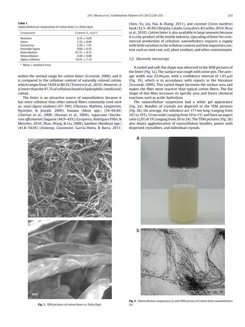
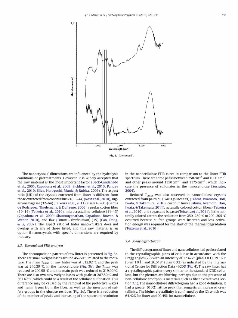
Fig. 3. Thermal decomposition profile of raw linter (a) and linter nanowhiskers (b), and FTIR spectra (c) of raw linter (bottom) and linter nanowhiskers (top).
J.P.S. Morais et al. / Carbohydrate Polymers 91 (2013) 229– 235 233
Fig. 3. (Continued ).
cteertad((W&ooi
3
TtwrT3dafo
The nanocrystals’ dimensions are influenced by the hydrolysisonditions or pretreatments. However, it is widely accepted thathe raw material is the most important factor (Beck-Candanedot al., 2005; Capadona et al., 2009; Eichhorn et al., 2010; Pandeyt al., 2010; Silva, Haraguchi, Muniz, & Rubira, 2009). The aspectatio (L/D) of the crystals extracted from linter is different fromhose extracted from coconut husks (35–44) (Rosa et al., 2010), sug-rcane bagasse (32–64) (Teixeira et al., 2011), sisal (43–60) (Garciae Rodriguez, Thielemans, & Dufresne, 2006), regular cotton fiber10–14) (Teixeira et al., 2010), microcrystalline cellulose (11–13)Capadona et al., 2009; Shanmuganathan, Capadona, Rowan, &
eder, 2010), and flax (Linum usitatissimum) (15) (Cao, Dong, Li, 2007). The aspect ratio of linter nanowhiskers does notverlap with any of those listed, and this raw material is anption if nanocrystals with specific dimensions are required byndustry.
.3. Thermal and FTIR analyses
The decomposition pattern of raw linter is presented in Fig. 3a.here are small weight losses around 45–50 ◦C related to the mois-ure. The main Tonset of raw linter was at 312.92 ◦C and the peakas at 340.29 ◦C. In the nanocellulose (Fig. 3b), the Tonset was
educed to 200.95 ◦C and the main peak was reduced to 219.00 ◦C.here are also two new weight losses with peaks at 287.50 ◦C and67.67 ◦C, which could be a result of the cellulose sulfonation. This
ifference may be caused by the removal of the protective waxesnd lignin layers from the fiber, as well as the insertion of sul-ate groups in the glucose residues (Fig. 3c). There is a reductionf the number of peaks and increasing of the spectrum resolutionin the nanocellulose FTIR curve in comparison to the linter FTIRspectrum. There are some peaks between 750 cm−1 and 1000 cm−1
and other peaks around 1350 cm−1 and 1175 cm−1, which indi-cate the presence of sulfonates in the nanocellulose (Socrates,2004).
Reduced Tonset was also observed in nanocellulose crystalsextracted from palm oil (Elaeis guineensis) (Fahma, Iwamoto, Hori,Iwata, & Takemura, 2010), coconut husk (Fahma, Iwamoto, Hori,Iwata, & Takemura, 2011), naturally colored cotton fibers (Teixeiraet al., 2010), and sugarcane bagasse (Teixeira et al., 2011). In the nat-urally colored cotton, the reduction from 250–280 ◦C to 200–205 ◦Coccurred because sulfate groups were inserted and less activa-tion energy was required for the start of the thermal degradation(Teixeira et al., 2010).
3.4. X-ray diffractogram
The diffractograms of linter and nanocellulose had peaks relatedto the crystallographic plans of cellulose in accordance with theBragg angles (2�) with an intensity of 17.422◦ (plan 1 0 1), 19.169◦
(plan 1 0 1), and 26.518◦ (plan 0 0 2) as indicated by the Interna-tional Centre for Diffraction Data – ICDD (Fig. 4). The raw linter hasa crystallographic pattern very similar to the standard ICDD cellu-lose, but the pictures are blurring, perhaps due to the presence ofnon-cellulosic amorphous materials such as fiber extractives (Sec-
tion 3.1). The nanocellulose diffractogram had a good definition. Ithad a greater (0 0 2) lattice peak that suggests an increased crys-tallinity. The higher crystallinity is confirmed by the ICr which was64.42% for linter and 90.45% for nanocellulose.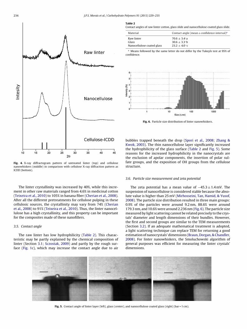
234 J.P.S. Morais et al. / Carbohydrate Polymers 91 (2013) 229– 235
Fig. 4. X-ray diffractogram pattern of untreated linter (top) and cellulosenanowhiskers (middle) in comparison with cellulose X-ray diffraction pattern asI
m(Acelf
3
tlf
Table 2Contact angles of raw linter cotton, glass slide and nanocellulose coated glass slide.
Material Contact angle (mean ± confidence interval)a
Raw linter 70.6 ± 3.4◦aGlass 30.6 ± 3.3◦bNanocellulose coated glass 23.2 ± 4.0 ◦c
a Means followed by the same letter do not differ by the Tukeyıs test at 95% ofconfidence.
Fig. 6. Particle size distribution of linter nanowhiskers.
CDD (bottom).
The linter crystallinity was increased by 40%, while this incre-ent in other raw materials ranged from 4.6% in medicinal cotton
Teixeira et al., 2010) to 105% in banana fiber (Cherian et al., 2008).fter all the different pretreatments for cellulose pulping in theseellulosic sources, the crystallinity may vary from 74% (Cheriant al., 2008) to 91% (Teixeira et al., 2010). Thus, the linter nanocel-ulose has a high crystallinity, and this property can be importantor the composites made of these nanofillers.
.5. Contact angle
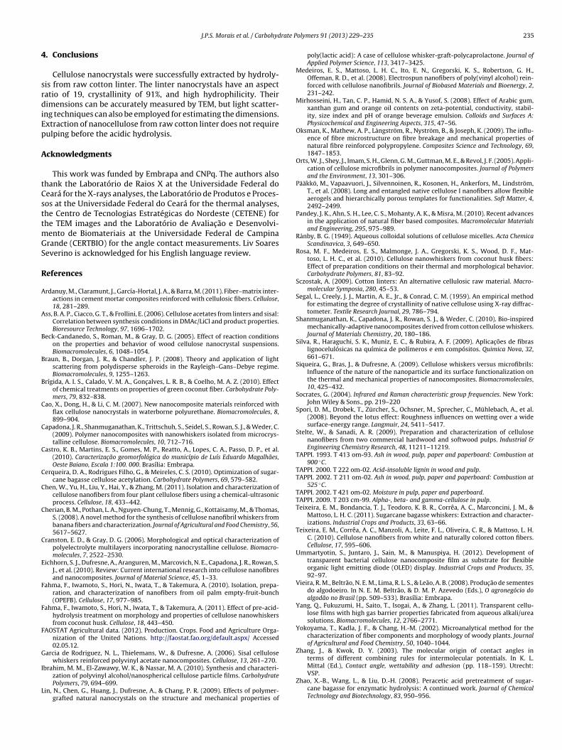
The raw linter has low hydrophilicity (Table 2). This charac-eristic may be partly explained by the chemical composition ofinter (Section 3.1; Sczostak, 2009) and partly by the rough sur-ace (Fig. 1c), which may increase the contact angle due to air
Fig. 5. Contact angle of linter layer (left), glass (center), a
bubbles trapped beneath the drop (Spori et al., 2008; Zhang &Kwok, 2003). The thin nanocellulose layer significantly increasedthe hydrophilicity of the glass surface (Table 2 and Fig. 5). Somereasons for the increased hydrophilicity in the nanocrystals arethe exclusion of apolar components, the insertion of polar sul-fate groups, and the exposition of OH groups from the cellulosestructure.
3.6. Particle size measurement and zeta potential
The zeta potential has a mean value of −45.3 ± 1.4 mV. Thesuspension of nanocellulose is considered stable because the abso-lute value is higher than 25 mV (Mirhosseini, Tan, Hamid, & Yusof,2008). The particle size distribution resulted in three main groups:0.9% of the particles were around 9.2 nm, 88.6% were around179.3 nm, and 10.6% were around 2.236 nm (Fig. 6). The particle sizemeasured by light scattering cannot be related precisely to the crys-tals’ diameter and length dimensions of their bundles. However,the first and second groups are similar to the TEM measurements(Section 3.2). If an adequate mathematical treatment is adopted,a light scattering technique can replace TEM for returning a goodestimation of nanocrystals’ dimensions (Braun, Dorgan, & Chandler,
2008). For linter nanowhiskers, the Smoluchowski algorithm ofgeneral purposes was efficient for measuring the linter crystals’dimensions.nd nanocellulose coated glass (right) (bar = 1 cm).
rate P
4
srdiEp
A
tCsttmGS
R
A
A
B
B
B
C
C
C
C
C
C
C
E
F
F
F
G
I
L
J.P.S. Morais et al. / Carbohyd
. Conclusions
Cellulose nanocrystals were successfully extracted by hydroly-is from raw cotton linter. The linter nanocrystals have an aspectatio of 19, crystallinity of 91%, and high hydrophilicity. Theirimensions can be accurately measured by TEM, but light scatter-
ng techniques can also be employed for estimating the dimensions.xtraction of nanocellulose from raw cotton linter does not requireulping before the acidic hydrolysis.
cknowledgments
This work was funded by Embrapa and CNPq. The authors alsohank the Laboratório de Raios X at the Universidade Federal doeará for the X-rays analyses, the Laboratório de Produtos e Proces-os at the Universidade Federal do Ceará for the thermal analyses,he Centro de Tecnologias Estratégicas do Nordeste (CETENE) forhe TEM images and the Laboratório de Avaliac ão e Desenvolvi-
ento de Biomateriais at the Universidade Federal de Campinarande (CERTBIO) for the angle contact measurements. Liv Soareseverino is acknowledged for his English language review.
eferences
rdanuy, M., Claramunt, J., García-Hortal, J. A., & Barra, M. (2011). Fiber–matrix inter-actions in cement mortar composites reinforced with cellulosic fibers. Cellulose,18, 281–289.
ss, B. A. P., Ciacco, G. T., & Frollini, E. (2006). Cellulose acetates from linters and sisal:Correlation between synthesis conditions in DMAc/LiCl and product properties.Bioresource Technology, 97, 1696–1702.
eck-Candanedo, S., Roman, M., & Gray, D. G. (2005). Effect of reaction conditionson the properties and behavior of wood cellulose nanocrystal suspensions.Biomacromolecules, 6, 1048–1054.
raun, B., Dorgan, J. R., & Chandler, J. P. (2008). Theory and application of lightscattering from polydisperse spheroids in the Rayleigh–Gans–Debye regime.Biomacromolecules, 9, 1255–1263.
rígida, A. I. S., Calado, V. M. A., Gonc alves, L. R. B., & Coelho, M. A. Z. (2010). Effectof chemical treatments on properties of green coconut fiber. Carbohydrate Poly-mers, 79, 832–838.
ao, X., Dong, H., & Li, C. M. (2007). New nanocomposite materials reinforced withflax cellulose nanocrystals in waterborne polyurethane. Biomacromolecules, 8,899–904.
apadona, J. R., Shanmuganathan, K., Trittschuh, S., Seidel, S., Rowan, S. J., & Weder, C.(2009). Polymer nanocomposites with nanowhiskers isolated from microcrys-talline cellulose. Biomacromolecules, 10, 712–716.
astro, K. B., Martins, E. S., Gomes, M. P., Reatto, A., Lopes, C. A., Passo, D. P., et al.(2010). Caracterizac ão geomorfológica do município de Luís Eduardo Magalhães,Oeste Baiano, Escala 1:100. 000. Brasília: Embrapa.
erqueira, D. A., Rodrigues Filho, G., & Meireles, C. S. (2010). Optimization of sugar-cane bagasse cellulose acetylation. Carbohydrate Polymers, 69, 579–582.
hen, W., Yu, H., Liu, Y., Hai, Y., & Zhang, M. (2011). Isolation and characterization ofcellulose nanofibers from four plant cellulose fibers using a chemical-ultrasonicprocess. Cellulose, 18, 433–442.
herian, B. M., Pothan, L. A., Nguyen-Chung, T., Mennig, G., Kottaisamy, M., & Thomas,S. (2008). A novel method for the synthesis of cellulose nanofibril whiskers frombanana fibers and characterization. Journal of Agricultural and Food Chemistry, 56,5617–5627.
ranston, E. D., & Gray, D. G. (2006). Morphological and optical characterization ofpolyelectrolyte multilayers incorporating nanocrystalline cellulose. Biomacro-molecules, 7, 2522–2530.
ichhorn, S. J., Dufresne, A., Aranguren, M., Marcovich, N. E., Capadona, J. R., Rowan, S.J., et al. (2010). Review: Current international research into cellulose nanofibresand nanocomposites. Journal of Material Science, 45, 1–33.
ahma, F., Iwamoto, S., Hori, N., Iwata, T., & Takemura, A. (2010). Isolation, prepa-ration, and characterization of nanofibers from oil palm empty-fruit-bunch(OPEFB). Cellulose, 17, 977–985.
ahma, F., Iwamoto, S., Hori, N., Iwata, T., & Takemura, A. (2011). Effect of pre-acid-hydrolysis treatment on morphology and properties of cellulose nanowhiskersfrom coconut husk. Cellulose, 18, 443–450.
AOSTAT Agricultural data. (2012). Production. Crops. Food and Agriculture Orga-nization of the United Nations. http://faostat.fao.org/default.aspx/ Accessed02.05.12.
arcia de Rodriguez, N. L., Thielemans, W., & Dufresne, A. (2006). Sisal cellulosewhiskers reinforced polyvinyl acetate nanocomposites. Cellulose, 13, 261–270.
brahim, M. M., El-Zawawy, W. K., & Nassar, M. A. (2010). Synthesis and characteri-zation of polyvinyl alcohol/nanospherical cellulose particle films. CarbohydratePolymers, 79, 694–699.
in, N., Chen, G., Huang, J., Dufresne, A., & Chang, P. R. (2009). Effects of polymer-grafted natural nanocrystals on the structure and mechanical properties of
olymers 91 (2013) 229– 235 235
poly(lactic acid): A case of cellulose whisker-graft-polycaprolactone. Journal ofApplied Polymer Science, 113, 3417–3425.
Medeiros, E. S., Mattoso, L. H. C., Ito, E. N., Gregorski, K. S., Robertson, G. H.,Offeman, R. D., et al. (2008). Electrospun nanofibers of poly(vinyl alcohol) rein-forced with cellulose nanofibrils. Journal of Biobased Materials and Bioenergy, 2,231–242.
Mirhosseini, H., Tan, C. P., Hamid, N. S. A., & Yusof, S. (2008). Effect of Arabic gum,xanthan gum and orange oil contents on zeta-potential, conductivity, stabil-ity, size index and pH of orange beverage emulsion. Colloids and Surfaces A:Physicochemical and Engineering Aspects, 315, 47–56.
Oksman, K., Mathew, A. P., Långström, R., Nyström, B., & Joseph, K. (2009). The influ-ence of fibre microstructure on fibre breakage and mechanical properties ofnatural fibre reinforced polypropylene. Composites Science and Technology, 69,1847–1853.
Orts, W. J., Shey, J., Imam, S. H., Glenn, G. M., Guttman, M. E., & Revol, J. F. (2005). Appli-cation of cellulose microfibrils in polymer nanocomposites. Journal of Polymersand the Environment, 13, 301–306.
Pääkkö, M., Vapaavuori, J., Silvennoinen, R., Kosonen, H., Ankerfors, M., Lindström,T., et al. (2008). Long and entangled native cellulose I nanofibers allow flexibleaerogels and hierarchically porous templates for functionalities. Soft Matter, 4,2492–2499.
Pandey, J. K., Ahn, S. H., Lee, C. S., Mohanty, A. K., & Misra, M. (2010). Recent advancesin the application of natural fiber based composites. Macromolecular Materialsand Engineering, 295, 975–989.
Rånby, B. G. (1949). Aqueous colloidal solutions of cellulose micelles. Acta ChemicaScandinavica, 3, 649–650.
Rosa, M. F., Medeiros, E. S., Malmonge, J. A., Gregorski, K. S., Wood, D. F., Mat-toso, L. H. C., et al. (2010). Cellulose nanowhiskers from coconut husk fibers:Effect of preparation conditions on their thermal and morphological behavior.Carbohydrate Polymers, 81, 83–92.
Sczostak, A. (2009). Cotton linters: An alternative cellulosic raw material. Macro-molecular Symposia, 280, 45–53.
Segal, L., Creely, J. J., Martin, A. E., Jr., & Conrad, C. M. (1959). An empirical methodfor estimating the degree of crystallinity of native cellulose using X-ray diffrac-tometer. Textile Research Journal, 29, 786–794.
Shanmuganathan, K., Capadona, J. R., Rowan, S. J., & Weder, C. (2010). Bio-inspiredmechanically-adaptive nanocomposites derived from cotton cellulose whiskers.Journal of Materials Chemistry, 20, 180–186.
Silva, R., Haraguchi, S. K., Muniz, E. C., & Rubira, A. F. (2009). Aplicac ões de fibraslignocelulósicas na química de polímeros e em compósitos. Quimica Nova, 32,661–671.
Siqueira, G., Bras, J., & Dufresne, A. (2009). Cellulose whiskers versus microfibrils:Influence of the nature of the nanoparticle and its surface functionalization onthe thermal and mechanical properties of nanocomposites. Biomacromolecules,10, 425–432.
Socrates, G. (2004). Infrared and Raman characteristic group frequencies. New York:John Wiley & Sons., pp. 219–220
Spori, D. M., Drobek, T., Zürcher, S., Ochsner, M., Sprecher, C., Mühlebach, A., et al.(2008). Beyond the lotus effect: Roughness influences on wetting over a widesurface-energy range. Langmuir, 24, 5411–5417.
Stelte, W., & Sanadi, A. R. (2009). Preparation and characterization of cellulosenanofibers from two commercial hardwood and softwood pulps. Industrial &Engineering Chemistry Research, 48, 11211–11219.
TAPPI. 1993. T 413 om-93. Ash in wood, pulp, paper and paperboard: Combustion at900 ◦C.
TAPPI. 2000. T 222 om-02. Acid-insoluble lignin in wood and pulp.TAPPI. 2002. T 211 om-02. Ash in wood, pulp, paper and paperboard: Combustion at
525 ◦C.TAPPI. 2002. T 421 om-02. Moisture in pulp, paper and paperboard.TAPPI. 2009. T 203 cm-99. Alpha-, beta- and gamma-cellulose in pulp.Teixeira, E. M., Bondancia, T. J., Teodoro, K. B. R., Corrêa, A. C., Marconcini, J. M., &
Mattoso, L. H. C. (2011). Sugarcane bagasse whiskers: Extraction and character-izations. Industrial Crops and Products, 33, 63–66.
Teixeira, E. M., Corrêa, A. C., Manzoli, A., Leite, F. L., Oliveira, C. R., & Mattoso, L. H.C. (2010). Cellulose nanofibers from white and naturally colored cotton fibers.Cellulose, 17, 595–606.
Ummartyotin, S., Juntaro, J., Sain, M., & Manuspiya, H. (2012). Development oftransparent bacterial cellulose nanocomposite film as substrate for flexibleorganic light emitting diode (OLED) display. Industrial Crops and Products, 35,92–97.
Vieira, R. M., Beltrão, N. E. M., Lima, R. L. S., & Leão, A. B. (2008). Produc ão de sementesdo algodoeiro. In N. E. M. Beltrão, & D. M. P. Azevedo (Eds.), O agronegócio doalgodão no Brasil (pp. 509–533). Brasília: Embrapa.
Yang, Q., Fukuzumi, H., Saito, T., Isogai, A., & Zhang, L. (2011). Transparent cellu-lose films with high gas barrier properties fabricated from aqueous alkali/ureasolutions. Biomacromolecules, 12, 2766–2771.
Yokoyama, T., Kadla, J. F., & Chang, H.-M. (2002). Microanalytical method for thecharacterization of fiber components and morphology of woody plants. Journalof Agricultural and Food Chemistry, 50, 1040–1044.
Zhang, J., & Kwok, D. Y. (2003). The molecular origin of contact angles interms of different combining rules for intermolecular potentials. In K. L.
Mittal (Ed.), Contact angle, wettability and adhesion (pp. 118–159). Utrecht:VSP.Zhao, X.-B., Wang, L., & Liu, D.-H. (2008). Peracetic acid pretreatment of sugar-cane bagasse for enzymatic hydrolysis: A continued work. Journal of ChemicalTechnology and Biotechnology, 83, 950–956.