Embed Size (px)
Citation preview

RADIOLOGY PAGE
639
Extrarenal Angiomyolipoma: differential diagnosis of retroperitoneal masses_______________________________________________Marcelo Wroclawski 1, Willy Baccaglini 2, Cristiano Linck Pazeto 2, Cristina Carbajo 2, Chaline Matushita 2, Arie Carneiro 2, Alexandre Pompeo 2, Sidney Glina 2, Antonio Carlos Lima Pompeo 2, Lívia Barreira Cavalcante 3
1 Hospital Israelita Albert Einstein, São Paulo, SP, Brasil; 2 Departamento de Urologia, Faculdade de Medicina do ABC, Santo André, SP, Brasil; 3 Centro de Imuno-Histoquímica, Citopatologia e Anatomia Patológica (CICAP) São Paulo, SP, Brasil
_______________________________________________________________________________________
INTRODUcTION
Angiomiolipomas (AML) are benign me-senchymal tumors of unknown origin, that con-sist of mature adipose tissue, muscle fibers and blood vessel with thickened wall (1).
Renal AMLs represent 1% of renal tumors. However, extra-renal AMLs are extremely rare, and 60 cases have been described. Most extra--renal AMLs were observed at liver (18 patients) and at retroperitoneum (16 cases) (2). We report a retroperitoneum extra-renal AML.
cASE REpORT
A fifty-one years old man with right lum-bar pain for one day was submitted to computer tomography that showed an incidental left retro-
peritoneal nodule, in close contact to ipsilateral adrenal gland.
Magnetic resonance confirmed the presen-ce of a heterogeneous nodule, close to left adrenal, hypervascularized, with sparse focus with loss of signal in sequences with fat saturation, with appro-ximately 2.4x2.1cm (Figures 1 and 2). Blood analy-sis excluded a functional adrenal tumor.
The lesion was excised by transperitoneal laparoscopy without complications, and it was diagnosed a mesenchymal lesion compatible to AML, confirmed by immune-histochemical assay (Table-1 and Figure-3).
DIScUSSION
AMLs are mainly asymptomatic inciden-talomas. However, it was reported spontaneous
Vol. 44 (3): 639-641, May - June, 2018
doi: 10.1590/S1677-5538.IBJU.2016.0670
figure 1 - Magnetic ressonance with axial T1wI image in phases before contrast (A), arterial (B), portal (c) and Equilibrium (D), showing a heterogenous nodule, hypervascularized, with peripheral progressive highlight due to contrast at the retroperitoneum, measuring 2.4x2.1cm.
A B c D

ibju | RADIOLOGY PAGE
640
bleeding (particularly in tumors with >4cm dia-meter), thromboembolic events and compres-sion of adjacent structures (3-6). Also, the risk of malignization of such tumors is unknown.
Most reported retroperitoneal AMLs in-clude symptomatic and big tumors (2); the pre-sent tumor was a small incidental lesion.
Image exams with presence of macros-copic fat are not conclusive, since liposarcoma
figure 2 - Magnetic resonance images (EcO Gradient sequences GRE) T1w1 inside and outside of phase (A and B) and in T2 with and without fat suppression (c and D); they show sparse areas of signal loss corresponding to lipomatous foci.
A B c D
Table 1 - Immuno-histochemical results that confirm angiomiolipoma.
ANTIGEN RESULT
HMB45 Reactivity in rare cells
Melan A Negative
CD34 Positive
Smooth muscle actine Positive
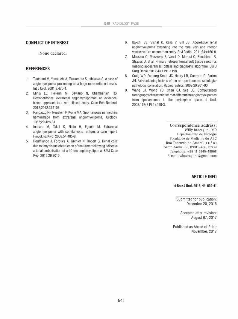
figure 3 – A) triphasic tumor, including mature adipose tissue, blood vessels with thickened wall and smooth muscle cells; B) component of elongated smooth muscle cells irradiating from gibbous vessels walls, permeating adipose tissue; c) angiomiolipoma, interface between mature fat cells and smooth muscle cells without atypia.
A B c
represent most of retroperitoneal sarcomas ad-jacent to adrenal gland that can be confused to myelolipoma, particularly in well-defined lesions such as the one here described (7). Other possible diagnosis include lipomas, lymphoma, adenocarcinoma metastasis and germ cell tu-mors, extra-gonadal dermoid cyst, hibernomas and lipoblastomas, among others (8).
Percutaneous biopsy may be inconclusi-ve; therefore, treatment of choice must be exci-
sion, preferably by minimally invasive techni-que. In the present case, lesion excision allowed histologic confirmation, preclude follow-up with image exams and had low morbidity to patient.
It is not uncommon the need of immu-ne-histochemical exams for diagnosis, due to histologic similarities with other tumors, such as liposarcoma, leiomyoma and lipoma (9).
ibju | RADIOLOGY PAGE
641
cONfLIcT Of INTEREST
None declared.
REfERENcES
1. Tsutsumi M, Yamauchi A, Tsukamoto S, Ishikawa S. A case of angiomyolipoma presenting as a huge retroperitoneal mass. Int J Urol. 2001;8:470-1.
2. Minja EJ, Pellerin M, Saviano N, Chamberlain RS. Retroperitoneal extrarenal angiomyolipomas: an evidence-based approach to a rare clinical entity. Case Rep Nephrol. 2012;2012:374107.
3. Randazzo RF, Neustein P, Koyle MA. Spontaneous perinephric hemorrhage from extrarenal angiomyolipoma. Urology. 1987;29:428-31.
4. Inahara M, Takei K, Naito H, Eguchi M. Extrarenal angiomyolipoma with spontaneous rupture: a case report. Hinyokika Kiyo. 2008;54:485-8.
5. Rouffilange J, Forgues A, Grenier N, Robert G. Renal colic due to fatty tissue obstruction of the ureter following selective arterial embolisation of a 10 cm angiomyolipoma. BMJ Case Rep. 2015;29:2015.
6. Bakshi SS, Vishal K, Kalia V, Gill JS. Aggressive renal angiomyolipoma extending into the renal vein and inferior vena cava - an uncommon entity. Br J Radiol. 2011;84:e166-8.
7. Messiou C, Moskovic E, Vanel D, Morosi C, Benchimol R, Strauss D, et al. Primary retroperitoneal soft tissue sarcoma: Imaging appearances, pitfalls and diagnostic algorithm. Eur J Surg Oncol. 2017;43:1191-1198.
8. Craig WD, Fanburg-Smith JC, Henry LR, Guerrero R, Barton JH. Fat-containing lesions of the retroperitoneum: radiologic-pathologic correlation. Radiographics. 2009;29:261-90.
9. Wang LJ, Wong YC, Chen CJ, See LC. Computerized tomography characteristics that differentiate angiomyolipomas from liposarcomas in the perinephric space. J Urol. 2002;167(2 Pt 1):490-3.
_______________________
Correspondence address:Willy Baccaglini, MD
Departamento de UrologiaFaculdade de Medicina do ABC
Rua Tancredo do Amaral, 131/ 83Santo André, SP, 09015-430, Brasil
Telephone: +55 11 9545-48968E-mail: [email protected]
ARTIcLE INfO
Int Braz J Urol. 2018; 44: 639-41
_____________________Submitted for publication:
December 20, 2016_____________________
Accepted after revision:August 07, 2017
_____________________Published as Ahead of Print:
November, 2017