Embed Size (px)
Citation preview

F334 – The Thread of Life:
The structure of DNA:
DNA is a polymer which is formed from many nucleotides. A DNA nucleotide is made from 2 sugar phosphate backbones, in between which are
nitrogen bases which can hydrogen bond to each other. DNA is double stranded. The sugar phosphate backbone is made from a deoxyribose sugar attached to a phosphate
group; the two molecules bond together in a condensation reaction. There are four possible bases in between the two sugar phosphate backbones. The four possible bases are adenine and guanine, cytosine and thymine. Adenine is the complimentary base to thymine and forms two hydrogen bonds with it.
Cytosine is the complimentary base to guanine and forms three hydrogen bonds with it.
Base pairing allows a DNA molecule to copy itself before cell division – a process called replication.
The two strands of DNA become paired off with each other to form a double helix (proposed by Francis Crick and James Watson in 1953).
NOTE: a nucleotide contains one sugar, one phosphate and one base joined together. Crucial to the double helix arrangement are the fact that the complimentary nitrogen bases
can hydrogen bond with each other:
The interactions between other groups hold the DNA molecule into a double helix.
Protein synthesis:
It is the nitrogen bases on the DNA molecule that code for proteins. The code in DNA is a triplet code; three bases code for each amino acid (three bases make
up each gene). Because there are three pieces of code that are needed for each amino acid, and there are
four bases that each of the three pieces of code can take, there are 64 combinations of code, yet there are only 20 amino acids.
Thus, some of the amino acids have more than one DNA code.

To synthesis proteins, the information in the DNA has to get into the cytoplasm of the cell, and then to the ribosomes.
DNA is too large to leave the nuclear envelope. A copy of the information in the gene is made in the form of a smaller molecule; this smaller molecule is Messenger RNA, or mRNA. mRNA is small enough to leave the nuclear envelope.
Once the mRNA strand has been synthesised from the DNA, it moves out of the nucleus to a ribosome.
Transfer RNA (tRNA) is now used to carry amino acids to the correct place, according to complimentary base pairing rules.
At the base of the molecule is a group of three unpaired bases called the anticodon. This code determines which amino acid is carried and makes the tRNA molecule specific to one type of Amino Acid.
On the tRNA molecule is an OH group which, with the aid of enzymes, forms an ester bond with the COOH group on a specific amino acid.
The anticodon on the tRNA molecule forms temporary hydrogen bonds with complimentary codons on the mRNA molecule. The amino acids on two adjacent tRNA molecules react together forming a peptide bond.
This process continues down the length of the mRNA molecule forming a polypeptide.
As previously mentioned, the tRNA molecules that bond to the mRNA molecules have an
anticodon that is complimentary to the mRNA codon. Complimentary base pairing happens because the correct pairs of bases have complimentary shapes, with groups in just the right places to allow hydrogen bonding to occur:
The DNA code is present in every cell in the body (those which contain a nucleus, i.e. not red
blood cells); however, certain genes are only switched on in certain cells.

In addition to just the genes, DNA molecules also contain codes which start or stop RNA production, and other regions which appear to have no function (exons).
The differences between RNA and DNA are:
DNA is made from the deoxyribose sugar, whereas RNA is made from the ribose sugar. The base thymine (DNA) is replaced by Uracil in RNA. DNA is double stranded (two sugar phosphate backbones), whereas RNA is single stranded. Each DNA molecule contains the code for all the proteins needed in the body, each mRNA
molecule codes for only one protein.
DNA Finger Printing and Ethical Issues:
The sequence of DNA base sequencing in an individual is unique. This sequencing is the same in every cell of the individual’s body.
This means that a person can be identified from a sample of a cell from their blood, skin, hair, or semen (male).
The DNA is first extracted from the cell. It is then cut into fragments using a restriction enzyme The resulting solution is applied to a gel and is subjected to an electric field in gel
electrophoresis (DNA negatively charged due to phosphate group). Different sized fragments move at different speeds towards the positive electrode. Each fragment is marked with a probe and then exposed on an x-ray film, producing a
pattern of black bars. Ethical ownership issues:
Who should have access to personal genetic information?Who owns and controls genetic information?Who owns genes and other piecies of DNA?
DNA Testing issues:Should parents have the right to have their children tested?Should tests be performed for genetic diseases?Should an individual always be given their genetic information?
Rates of Chemical Reactions:
All chemical reactions have different rates of reactions. The rate of a chemical reaction can be increased by: Increasing the concentration of a solution. Raising the pressure of a gas. Increasing the temperature of the reaction. Increasing the surface area of a solid. Adding a catalyst.
The effect of changing the concentration:
An example often used when demonstrating chemical reactions is the reaction between hydrochloric acid and calcium carbonate:

CaCO3(s) + 2HCl(aq) CaCl2(s) + H2O(l) + CO2(g)
The rate of reaction can be altered by changing the size of the calcium carbonate chips or the concentration of the acid.
If the chips are large and in excess, then only a small fraction of the solid reacts and the chips don’t change shape. This means that the calcium carbonate does not affect the rate of reaction, only the concentration of the hydrochloric acid does.
The rate will decrease over time as the hydrochloric acid is used up (its concentration decreases).
The rate of reaction could be worked out either by measuring the pH of the solution, the decrease in the mass of calcium carbonate, or the increase in the volume of carbon dioxide.
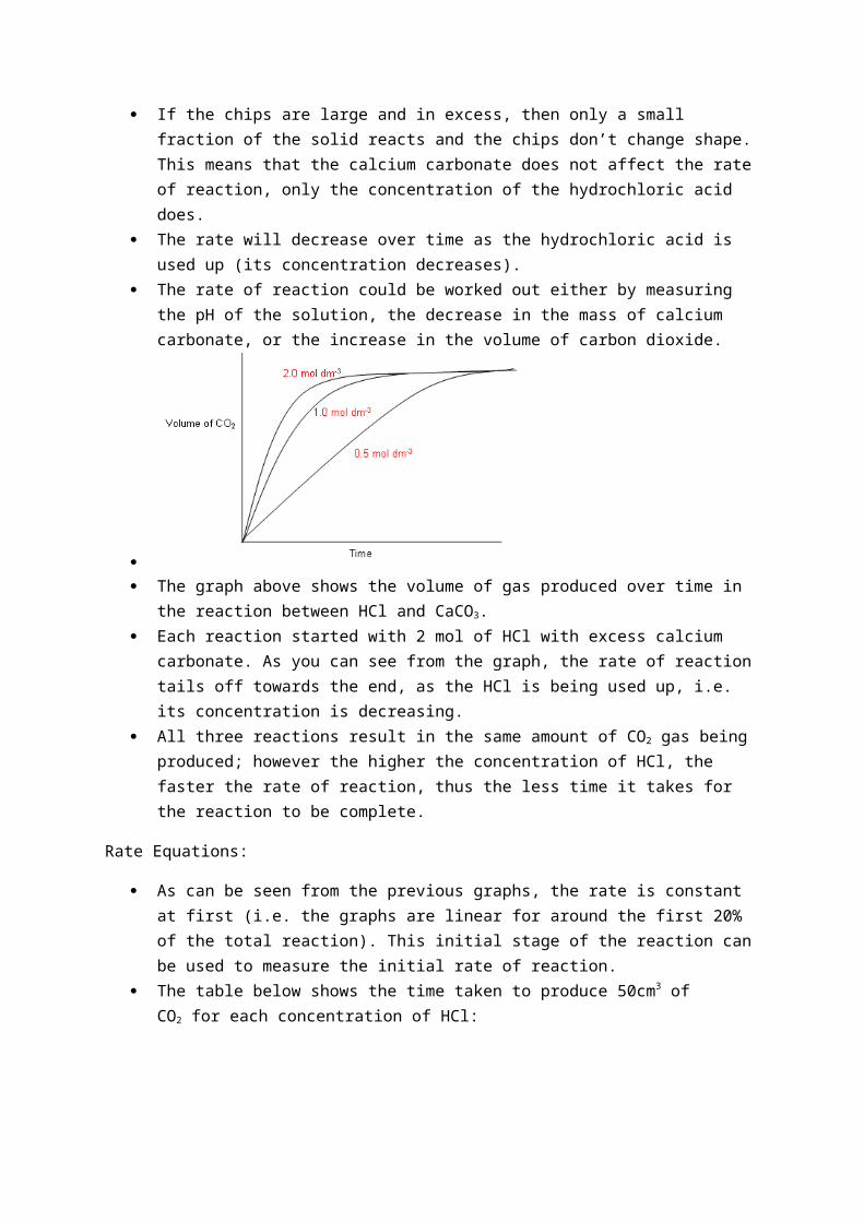
The graph above shows the volume of gas produced over time in the reaction between HCl
and CaCO3. Each reaction started with 2 mol of HCl with excess calcium carbonate. As you can see from
the graph, the rate of reaction tails off towards the end, as the HCl is being used up, i.e. its concentration is decreasing.
All three reactions result in the same amount of CO2 gas being produced; however the higher the concentration of HCl, the faster the rate of reaction, thus the less time it takes for the reaction to be complete.
Rate Equations:
As can be seen from the previous graphs, the rate is constant at first (i.e. the graphs are linear for around the first 20% of the total reaction). This initial stage of the reaction can be used to measure the initial rate of reaction.
The table below shows the time taken to produce 50cm3 of CO2 for each concentration of HCl:
The units of the rate above are measured in cm3 of CO2 per second; this is because it is how
the rate of reaction has been measured. If the rate was measured using the decrease in concentration of HCl instead, the units of rate would be mol dm-3 s-1, or if it was measured by decrease in mass it would be g s-1.
From the results in the table above, it can be worked out that the rate of reaction is directionally proportional to the concentration of hydrochloric acid, so if you halve the concentration of HCl the rate will be halved, if you quarter the concentration, the rate will be quartered.
This relationship can be converted into a rate equation by replacing the proportionality sign
with an equals sign and inserting a constant: Rate = k[HCl]1
k is the rate constant. If we are measuring the rate of the reaction using the change in concentration, then the
units will be:
The rate is measured in s-1 in the case above, but in other rate equations it may be different;
it all depends on the units being used to measure the rate and the number of reactants and products.
Orders of Reactions:
The reaction of calcium carbonate with hydrochloric acid is said to be first order with respect to hydrochloric acid.
This is because the rate depends upon the concentration of hydrochloric acid to the power one.
Take the rate equation below: Rate = k[A]n[B]m
n is the order of the reaction with respect to A and m is the order of reaction with respect to B.
The values of n and m are usually 0, 1 or 2 corresponding to zero, first and second orders; however some reactions can have fractional or negative orders.
Orders of reactions must be worked out experimentally; they are totally unrelated to the chemical equation.
Working out a rate equation: An experiment was conducted to find out how the concentration of X and Y affect the rate of
formation of the product Z. the results were as follows:
Firstly to work out the order of the reaction. Starting with X, we need to look in the table for
two different values of [X], when the concentration of Y is kept constant. Experiments 2 and 3 can be used to work out the order of X.

When [X] is doubled the rate quadruples, this means that the order of the reaction with
respect to [X] is 2. Now to work out the order of the reaction with respect to X, in this case we need to use
reaction 1 and 2:
The order of this reaction with respect to Y is one, as the relationship between [Y] and rate is
directionally proportional. The rate equation for the reaction is: Rate = k[X]2[Y] The overall order of the reaction is three (the two orders added together). Now to work out the constant.
This can be worked out by using values from the table:
Note that this k value is only applicable for one temperature. The overall reaction is
Rate = 80[X]2[Y] Now for the units:
Cancelling down gives mol-2 dm6 s-1.
Half-Lives:
When analysing a rate graph, it is possible to look at the initial rate of the reaction to work out the order (as done previously); however it is also possible to look at the half-life for a first order reaction.
The half-life is the time taken for the concentration of a substance to fall to half its starting value. It is often given the symbol t/2.
The reaction of calcium carbonate with hydrochloric acid (looked at previously) can be used to analyse half-lives:
CaCO3(s) + 2HCl(aq) CaCl2(s) + H2O(l) + CO2(g)

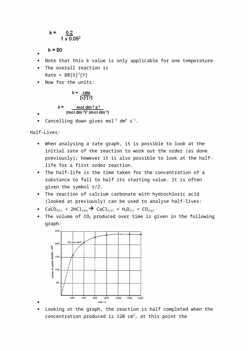
The volume of CO2 produced over time is given in the following graph:
Looking at the graph, the reaction is half completed when the concentration produced is 120
cm3, at this point the concentration of HCl is 1 mol dm-3. The time taken to reach this point is the half-life of the reaction with a starting concentration of 2 mol dm-3. The half-life for this is 141 seconds.
The time taken for the volume of CO2 to change from 120 cm3 to 180 cm3 corresponds to a change in concentration of HCl from 1 mol dm-3 to 0.5 mol dm-3, i.e. it is the t/2 value for the reaction with a starting concentration of 1 mol dm-3. The half-life for this is 144 seconds.
The next half-life can be found by calculating the time it takes for the volume of CO2 to increase from 180cm3 to 210cm3. This corresponds to [HCl] falling from 0.5 mol dm-3 to 0.25 mol dm-3. The half-life for this is 147 seconds.
The half-lives for the three different parts of the equation are roughly the same; they are independent of the starting concentration of HCl. Therefore the reaction is first order with respect to [HCl].
Second and zero order processes do not have constant half-lives. First order processes are never over, the mass is constantly halving every t years. A reaction
can be classed as over when the change can no longer be measured.
Rate Determining Steps:
As previously mentioned, the rate of a chemical reaction cannot be worked out by looking at the chemical equation.
This is because most reactions do not take place in one step, but in many. The chemical equation only shows the overall effect of these many small steps.
For example, the reaction below shows the nucleophilic substitution reaction of 2-bromo-2-methylpropane with OH-:
The reaction is first order with respect to C(CH3)3Br and zero order with respect to OH-: Rate = k[C(CH3Br]
The [OH-] is not involved in the rate equation, i.e. altering it does not have an effect upon the rate of the reaction.
The reason for this is that the reaction does not occur in one step, but it occurs in two steps: The C-Br bond breaks heterolytically:
This only involves C(CH3)3Br, thus its rate is only dependent on [C(CH3)3Br]. The carbocation reacts with OH-:
The reaction for step 2 is much faster than the reaction in step 1. The rate of reaction is
always determined by the slowest reaction, therefore it is reaction 1 that is the rate determining step and its equation becomes the rate equation for the whole reaction.
Graphs to determine order of reaction:
Ways of measuring rate of reaction:
You need to measure a property that changes during a reaction and that is proportional to the concentration of a particular reactant or product.
This is information comes from experiment data. If a gas is produced you can collect it in a gas syringe, the volume produced can be used to
follow the rate of a reaction. You could also measure by recording the loss in mass. You can follow the pH of a reaction if the acid/alkali concentration changes throughout. A colorimeter can be used to measure the change in colour of a reaction. All these techniques do not interfere with the progress of the reaction. Chemical analysis involves taking samples of the reaction mixture at regular intervals, and
stopping the reaction in the sample by a process known as quenching. If the reaction is acid catalysed you can neutralise it with sodium hydrogencarbonate. Anything unreacted can be analysed via titration. You can also quench with ice cold water to decrease the concentration, diluting the reaction
mixture.
Summary:
Step 1: decide on a property of a reactant or product which you can measure. Step 2: measure the change in property in a certain time
Step 3: find the rate in terms of - change∈ property
timetaken
Optical Isomerism:
When four single bonds are formed around a central carbon atom(chiral centre), the molecule formed is tetrahedral in shape.
A chiral centre is an atom that four different atoms or groups of atoms attached to it. If the four groups are all different, the molecule can exist in two isomeric forms. This can be
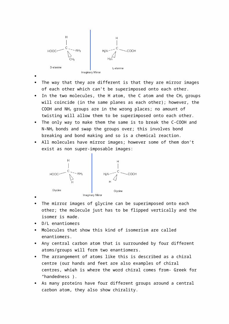
seen with the amino acid, alanine:
The way that they are different is that they are mirror images of each other which can’t be
superimposed onto each other. In the two molecules, the H atom, the C atom and the CH3 groups will coincide (in the same
planes as each other); however, the COOH and NH2 groups are in the wrong places; no amount of twisting will allow them to be superimposed onto each other.
The only way to make them the same is to break the C-COOH and N-NH2 bonds and swap the groups over; this involves bond breaking and bond making and so is a chemical reaction.
All molecules have mirror images; however some of them don’t exist as non super-imposable images:

The mirror images of glycine can be superimposed onto each other; the molecule just has to
be flipped vertically and the isomer is made. D/L enantiomers Molecules that show this kind of isomerism are called enantiomers. Any central carbon atom that is surrounded by four different atoms/groups will form two
enantiomers. The arrangement of atoms like this is described as a chiral centre (our hands and feet are
also examples of chiral centres, which is where the word chiral comes from- Greek for “handedness”).
As many proteins have four different groups around a central carbon atom, they also show chirality.
All the proteins within our body are built up from L-enantiomers (they have the same geometric arrangement around the central carbon atom).
L-enantiomers can be identified using the CORN rule: with the H atom pointing upwards, looking down from the H atom to the carbon and moving clockwise, the L-amino acid has the order COOH, R, NH2, i.e.
Enantiomers behave identically in all chemical reactions and most of their physical
properties are the same. The only difference comes when they are in the presence of other chiral molecules. D and L enantiomers will react differently with our enzymes (like trying to put a left hand
glove onto your right hand). Examples of different effects include: Enantiomers react differently with chiral taste-buds (D amino acids are sweet and L-amino
acids are bitter/tasteless). Enantiomers can smell differently. Many enantiomers are beneficial medicines; however their isomers can have drastic effects
(e.g. thalidomide). D-amino acids do exist in nature, for example penicillin works by breaking down the peptide
links in D-alanine; these occur in the cell walls of bacteria but not in humans. Thus penicillin only affects bacterial cells.

Test for enantiomers:
A plane of polarised light is rotated in opposite directions by each enantiomer. If a solution containing equal amounts of each enantiomer is placed in a polarimeter, the
solution will be optically inactive.
Amino Acids:
Alpha-Amino acids contain both the NH2 (amino) group and the COOH (carboxylic acid) group attached to the same carbon atom.
Proteins are made from amino acids and so they are very important in living organisms.
Amino acids have two functional groups; they are therefore examples of bifunctional
compounds. Bifunctional compounds often share the same properties of the two functional groups. However, this is not the case with amino acids, as both of their functional groups interact. The COOH group acts as an acid (donating protons) and the NH2 group acts as a base
(accepting protons); therefore these two groups can react with each other forming zwitterions(compounds containing both anionic and cationic groups):
An aqueous solution of an amino acid is formed mainly of zwitterions (solution is neutral,
unless there is an extra COOH or NH2 on the R group); this gives the solution certain characteristics that are common of ionic compounds:
They are soluble in water. They have a higher melting and boiling point than expected. When they dissolve in water, the NH2 group accepts a proton and the COOH group donates
one to another water molecule.
H2O + H2N—CH(CH3)—COOH + H2O HO- + +H3N—CH(CH2)—COO- + H3O-
The OH- and H3O+ ions then go on to react with each other, to restore the equilibrium within water:
OH-(aq) + H3O+
(aq) 2H2O(l)
This maintains that the overall solution is neutral. Zwitterions act as good buffers; addition of small quantities of acid or alkali to the solution
has little affect upon the pH of the solution:
OH-(aq) + +H3N—CH(R)—COO-
(aq) H2O(l)+ H2N—CH(R)-COO-(aq)
H3O+(aq) + +H3N—CH(R)—COO-
(aq) H2O(l) + +H3N—CH(R)-COOH(aq)
Proteins:
There are only twenty -amino acids in the world. These twenty amino acids are joined together in different sequences to form every protein present in living organisms.
The order in which amino acids are joined to one another is called the primary structure. The twenty -amino acids have the same general structure (above); however they have
different side-chains (R-group). For example,
What makes the proteins different from each other are the order in which the amino acids
are joined; the primary structure of the protein.
Synthesising Peptides:
Amino acids join together to form polypeptides, which make proteins. The reaction is an example of a condensation reaction, as one amino acid joins onto the next
forming a larger molecule with the loss of a smaller molecule. The amino acids join together when the carboxylic acid group on one amino acid joins on to
the amino group on the next (a molecule of water is lost each time). The –CONH– (red) is called a peptide group; this may look familiar, in designer polymers you
came across this group in nylon; however it is referred to as a secondary amide group. In the molecule below, the two peptides have joined together to form a dipeptide; many
peptides join together to form a polypeptide. Chemists cannot make amino acids react directly together. Instead, they make the COOH
group more reactive by converting it into an acyl chloride. Chemists also have to look at another property of amino acids when synthesising proteins. Amino acids are not flat molecules; they have a three-dimensional shape, based upon a
tetrahedron. This means that they exist in two isometric forms (except glycine) known as D or L isomers.
Naming an Amino Acid Sequence:

Amino acid sequences are named in the direction with the free NH2 group on the left; for example:
Secondary and Tertiary Structures of Proteins:
When a polypeptide chain forms an alpha helix or a beta sheet this is called it’s secondary structure. Both helix and sheet arrangements occur as a result of hydrogen bonding.
The helix arrangement is when the chain is tightly coiled where the C=O group of one peptide link forms a hydrogen bond to an N-H group four peptide links along the chain.
The other arrangement is when it is stretched out into regions of extended chain, which lie alongside one another and hydrogen bonded to form a sheet.
The ‘global’ folding of a polypeptide chain to give it a unique shape is called its tertiary structure.
There are four types of interaction responsible for maintaining tertiary structure: Instantaneous dipole – Induced dipole bonds between non polar side chains. Hydrogen bonds between polar side chains. Ionic bonds between ionisable side chains. Covalent bonds (sulphur bridges)
Hydrolysing Polypeptides:
As polypeptides are held together by peptide linkages, which are the same as amide linkages, they can be hydrolysed in the same way as polyamides.
In the body, enzymes would be used to help hydrolyse the peptide bond; however the peptide bonds can be broken in a reflux reaction with a moderately concentrated acid or alkali.
The C-N bond is broken in the peptide link to give its constituents. Under acid hydrolysis conditions the –NH2 groups are protonated to give –NH3
+
Whereas under alkaline conditions the –COOH groups are deprotonated to give –COO-
Chromatography works on the basis that some compounds are more soluble than others, and so when left to move with a solvent up chromatography paper, the compounds will leave the solvent at different points (i.e. the more soluble the compounds, the further they will travel up the chromatography paper).
Technique:
Draw a pencil line approximately 1cm from the bottom of the paper. Mark a cross and produce a concentrated spot of the compounds to be used by repeated application followed by drying.
Mark a reference spot. Mark a spot of the product from the hydrolysis. Place in chromatography tank; ensure that the spots are above the solvent

Cover with lid. Allow time for the solvent to run up the chromatography plate, so it reaches just below the
end of the paper. Use locating agent to find spots (ninhydrin), and then heat using a hair dryer. Compare the products from the hydrolysis of aspartame with the reference compounds.
Enzymes:
Enzymes are biological catalysts that speed up chemical reactions. Enzymes speed up chemical reactions in the body by a factor of between 106 to 1012 times, allowing vital chemical reactions to take place at normal temperatures
There are around 40,000 enzymes in the human body. Enzymes are complex structures of three-dimensional proteins (quaternary protein
structure). They are many times larger than the molecules involved in the reactions they catalyse.
The reaction takes place in a small area of the enzyme called the Active Site, the rest of the
protein acts as scaffolding holding the protein together. The arrangement of the amino acids in the active site makes it specific for only one type of
substrate. The enzyme is complementary in shape, charge and hydrophobic/hydrophilic areas.

Even when other substrates are present (2 and 3) only the substrate with the specific shape will bind to the enzyme (1). The enzyme and substrate are like jigsaw pieces that fit together.
When the substrate and enzyme “collide”, hydrogen bonds or interactions between ionic side groups hold the substrate and enzyme together. After the new products are formed, they are released and the enzyme’s active site is free for more substrates to bind to.
If a molecule of similar shape to the substrate enters and binds strongly to the active site it might not react but still occupy that active site, preventing the entry of the correct substrate. This is called competitive inhibition.
Changing the pH:
Enzymes are very sensitive to any changes in pH. This is because the change in concentration of H+ ions affects the bonding holding the tertiary structure together. Disrupts ionic interactions leaving the enzyme inactive as it will have been denatured.
Changing the temperature:
Enzymes are denatured at higher temperatures. This is because high temperatures break the hydrogen bonding holding the tertiary structure together.
Enzymes as catalysts:
A catalyst is a substance that alters the rate of a reaction without being used up itself in the process.
Most catalysts speed up the rates of reactions. They do this by providing an alternate reaction pathway that has a lower activation energy. Enzymes are very useful as catalysts; without them reactions that occur within our body
would need to take place at extremely high temperatures, rather than just 37oC. The energy profile diagram below shows the effect of enzymes on the activation energy of a reaction:
Enzymes are usually only present in small traces. At low concentrations of substrate the rate equation is:
rate = k[E][S] There are plenty of active sites for the substrate to bind to, so doubling the substrate
concentration doubles the rate. The reaction is first order with respect to the substrate. At high concentrations of substrate the rate equation is:
rate = k[E]

All the active sites on the enzyme molecules have become saturated. The reaction becomes zero order with respect to the substrate.
Enzymes in industry:
Enzymes are used increasingly as catalysts in industry because: They are specific – they can ‘select’ a particular substrate from a feedstock containing a
mixture of reactants. They work effectively at low temperatures – this helps reduce energy costs of an industrial
process. They work well in an aqueous environment – this reduces the need for organic solvents,
which can be flammable and harmful to the environment. They can often convert reactant to product in a one-step reaction – this increases the
percentage atom economy of the process.