Embed Size (px)
Citation preview

FABRICATION OF A CARBON-BASED 1MMUNO-
BIOSENSOR FOR HEPATITIS B SURFACE ANTIGEN DETECTION
WILLY WONG XItJ FA
Thesis submitted in partial fulfilment of the requirements for the award of the degree of
Bachelor of Chemical Engineering (Biotechnology)
Faculty of Chemical & Natural Resources Engineering UNIVERSITI MALAYSIA PAHANG
JULY 2014
©WILLY WONG XIU FA (2014)
III

ABSTRACT
Since the early diagnosis of HBV infection is crucial for the successful antiviral
treatment, sensitive methods are urgently needed for measuring bio-diagnosis markers
present at ultra-low levels during early stages of the infection. In this proposal, a
graphene based biosensor is presented for performing highly sensitive pathogenic virus
detection, particularly toward the detection of Hepatitis B virus surface antigen
(HBsAg). A free-standing conductive graphene will be prepared using a modified
Hummers method. Graphite will be oxidized to graphene oxide which will then be
reduced to graphene by using hydrazine as the reducing agent. The product before and
alter hydrazine reduction was tested with UV-Vis spectrometer to obtain the peak at
certain wavelength. Before reduction, the peak was shown around 230-240 nm while
after reduction, the peak was around 270 rim. The peak shift from 230 nm to 270 nm
shows that the synthesis of graphene was successful. The final product of the reduction
was a black color powder. The immobilization of the antibody will be done using this
graphene as substratum. The graphene is used to grow the Nafion-graphene nanosheet,
and was then soak in Thionine solution. Following that, a solution of primary antibodies
against hepatitis B surface antigen (anti-HBsAg IgG) was incubated on the biosensor.
After the immobilization, the antigen was dropped onto the biosensor, follow by
incubation of secondary antibodies conjugated to Horseradish Peroxidase (HRP). The
color change to blue upon addition of Tetramethylbenzidine (TMB). Sulfuric acid stop
solution was then added and colour changes from blue to yellow was observed. This
colour change indicate that the immobilization of anti-HB sAg on the graphene surface
was successful.
VIII

TABLE OF CONTENTS
DECLARATIONSUPERVISOR'S ........................................................................ IV
DECLARATION V STUDENT'S ..............................................................................
VI Dedication...........................................................................................................VII ACKNOWLEDGEMENT....................................................................................
VIII ABSTRACT.......................................................................................................
CONTENTS TABLEOF .......................................................................................IX
LISTOF FIGURES .............................................................................................Xl XII LIST OF TABLES ...............................................................................................
LISTOF ABBREVIATIONS .............................................................................. XIII
1 INTRODUCTION............................................................................................I
1 .1 Background of Study ...............................................................................3
1.2 Statement of Problem .............................................................................3
1.3 Objective of Research.............................................................................
1.4 Scope of Proposed Research ................................................................. 4 4
1.5 Expected Outcomes................................................................................4
1.6 Significance of Study ...............................................................................4
1.7 Summary of Chapter ...............................................................................5
2 LITERATURE REVIEW .................................................................................. 5 2.1 Overview .................................................................................................
5 2.2 Immuno-BiosenSOrs (ImmunosenSOrs) ...................................................
6 2.3 Hepatitis B Virus (HBV)...........................................................................
6 2.3.1 Virus Morphology................................................................................
6 2.3.2 Virus Detection Methods......................................................................
2.4 Graphene Oxide (GO).............................................................................
2.4.1 Morphology ........................................................................................8
2.4.2 Electrochemical Properties ....................................................................9
2.5 Graphene ................................................................................................
2.5.1 Morphology ........................................................................................ 10
2.5.2 Properties ..........................................................................................11
2.6 Summary of Chapter.............................................................................12
3 MATERIALS AND METHODS .....................................................................12 3.1 Overview .............................. ..................................................................12 3.2 Materials .......... ........................................ ..............................................12 3.3 Instruments ...........................................................................................13 3.4 Research Process/Procedures .............................................................13 3.4.1 Synthesis of Graphite Oxide................................................................
3.4.2 Reduction of Graphene Oxide (GO) to Graphene.................................. 14 15 3.4.3 Fabrication of Biosensor.....................................................................
3.4.4 Immobilization of Hepatitis B Surface Antigen Antibody (Anti-HB sAg). 15
3.4.5 Identification of Graphene Oxide and Graphene.................................... 15 15 3.5 Summary of Chapter.............................................................................16 4 RESULT AND DISCUSSION.........................................................................16 4 .1 Overview ...............................................................................................16 4.2 Synthesis of Graphene..........................................................................
L,1

4.3 Coating of Graphene onto Microtiter Plate Surface ..............................18 4.4 Detection of Hepatitis B Virus Surface Antigen (HBsAg) ......................21
5 CONCLUSION AND RECOMMENDATIONS ................................................23
5 .1 Conclusion ............................................................................................23
5 .2 Recommendations ................................................................................23
REFRENCES ....................................................................................................... 24 APPENDIXES ....................................................................................................28
VA
LIST OF FIGURES
Figure 2.1: Advantages of Immuno-Biosensor.......................................................6
Figure 2.2: Model of Human Hepatitis B Virus (HBV) (Henderson, 2004).............7
Figure 2.3: Scheme of structural model of graphene and graphene oxide (GO), showing that graphene consists of only trigonally bonded sp2 carbon atoms while GO consists of a partially broken sp2-carbon network with phenol, hydroxyl, and epoxide groups on the basal plane and carboxylic acid groups at the edges (Chen et aL,2010) ........................................... . ................................................................. 10
Figure 3.1: Process Flow Diagram of the Fabrication of Carbon-Based Immune-Biosensor for Hepatitis B Surface Antigen (fiBsAg) Detection . ........................... 13
Figure 3.2: Process Flow Diagrams for the Synthesis of Graphene from Graphite ..............................................................................................14
Figure 4.1: Setup for vaccum filtration . .............................................................. 17
Figure 4.2: Graphite oxide cake after vacuum filtration ...................................... 17
Figure 4.3: Peak shift before and after hydrazine reduction using UV-Vis Spectrometry.................................................................. .................................... 18
Figure 4.4: The relationship between the time taken and the amount of graphene-Naflon dropped onto the microtiter plate surface ................................................ 19
Figure 4.5: Well dispersed graphene-nafion solution before drying ....................20
Figure 4.6: Attachment of Graphene-Naflon on the plate surface after drying .... 21
Figure 4.7: Detection of fiBsAg. Addition of TMB substrate yields blue colour solution. With the addition of sulfuric acid, the colour changes to yellow . ........... 22

LIST OF TABLES
Table 4.1: The relationship between the amounts of graphene-Nafion, time taken to dried and distribution of the graphene layer . .................................................. 19

LIST OF ABBREVIATIONS
Anti-HBsAg Antibodies against Hepatitis B virus surface antigen FTIR Fourier Transform Infrared Spectroscopy GO Graphene Oxide rGO reduced Graphene Oxide (graphene) HIBsAg Hepatitis B virus surface antigen HBV Hepatitis B virus SEM Scanning Electron Microsope TMB Tetramethylbenzidine UV-Vis Ultraviolet-visible light

1 INTRODUCTION
1.1 Background of Study
Micro-organisms, such as bacteria and viruses, are found widely in the environment, in
food, marine and estuarine waters, soil, and also in the body fluids of humans and
animals. Many of these organisms have an essential function in nature, but certain
potentially harmful micro-organisms can have profound negative effects on both
animals and humans (Paul et al., 2002). One of the examples would be Hepatitis B virus
that causes Hepatitis B.
Hepatitis B is a disease caused by HBV which infect the liver of hominoidae. As
reported by Ganem and Varmus in 1987, chronic Hepatits B infections can cause a
spectrum of different diseases ranging from inactive carrier state to the development of
cirrhosis-related complication and hepatocellular carcinoma (liver cancel). Besides,
Hepatits B virus is blood-borne, transfusion-transmitted human pathogen that has a
major impact on blood safety and public health worldwide (Hsia et at., 2007). Hepatitis
B virus (HBV) infection is a major global health problem and the tenth leading cause of
death worldwide (Lavanchy, 2004). About a third of the world's population, an
estimated 2 billion people have been infected with Hepatitis B virus and more than 360
million have chronic Hepatitis B. Additionally, at least 600,000 people die annually
from acute or chronic consequences of Hepatitis B virus infections (Verma et at., 2011).
Verma et al.(201 1)'s research also shows that HBV is 50 to 100 times more infectious
than Human Immunodeficiency Virus (HIV) that causes Acquired Immunodeficiency
Syndrome (AIDS).
According to Sung et al. (2009)'s research, a high viral load in patients is the main
cause of Hepatitis B progression, thus the ultimate goal for the treatment of Hepatitis B
is to eliminate the virus before irreversible liver damage occurs. In other words, early
diagnosis of HBV infections is crucial in making clinical decision for successful
antiviral treatment. Therefore, sensitive methods are urgently needed for measuring the
bio-diagnosis markers present at ultra-low levels during early stages of the infection.
Sensitive and early detection of HBV may not only help monitor the viral dynamics associated with treatment but could also improve therapeutic decision making (Geng et

al., 2005). Conventional methods for detection of HBV have shown a negative outcome
of being time consuming and expensive. As a matter of fact, the development of novel
biosensor for highly sensitive, selective and rapid pathogen detection is of paramount
importance for medical diagnostics, food safety screening and environment pollution
monitoring.
As reported by Vashist et al. (2011), the electrochemical immunoassays and immuno-
sensors are drawing more attention in a wide range of uses, due to their merits such as
low cost, small size, short response time and the possibility of using in vivo. The
immunoassay techniques, based on highly specific molecular recognition of antigens by
antibodies, have become the main analytical tools in clinical and biochemical analyses
and in other areas such as environmental control, food quality control, etc (Liu et a!,
2006). The electrochemical methods in inimuno-sensing have become very popular
recently. Impedance technique, a type of electrochemical biosensors have been proven
to be a promising method for pathogenic detection due to its portability, rapidity,
sensitivity, and more importantly it could be used for on-the-spot detection.
Generally, the impedance detection techniques can be classified into two types
depending on the presence or absence of specific bio-recognition elements. The first
type works by measuring the impedance change caused by binding of targets to bio-
receptors (antibodies and nucleic acids) immobilized onto the electrode surface, while
the detection principle of the second type is based on metabolites produced by bacterial
cells as a result of growth (Wang et al., 2012).
Owing to the emerging of nanotechnology, a variety of nanomaterial such as
semiconductor quantum dots, metallic or semiconductor nanowires, carbon nanotubes
(CNTs), and nanostructured conductive polymers has been incorporated into the optical
and electrical transducers of biosensors. As Mabmoud and Luong (2003)'s stated, these
incorporations lead to a significant improvement in the sensitivity and selectivity of the
sensors. Among them, the two-dimensional carbon nanostructure, known as graphene,
has stimulated research interest due to its remarkable electrical, mechanical and thermal
properties (Haque et al., 2012). The additional findings of biocompatibility, facile
surface modification with biomolecules, good water dispersibility, high surface-area-to-
volume ratio, and unique optical properties endow graphene with high potential for
bioelecironics and biosensing application (Liu et al., 2011). In conclusion, the
2

integration of nanomaterial (graphene) and biosensors has a promising future in
detecting HBV.
1.2 Statement of Problem
A high viral load in patients is the main cause of Hepatitis B progression, thus the
ultimate goal for the treatment of Hepatitis B is to eliminate the virus before irreversible
liver damage occurs. In order to do so, diagnosis of HBV infections in the early stages
is crucial for successful antiviral treatment. However, accurate detection of HBV using
the conventional methods requires high cost. Moreover, most of the conventional
methods for accurate detection are time consuming. Therefore, cheap, rapid and
sensitive methods are needed to measure the bio-diagnosis markers present at ultra-low
level during early stages of infections. As a result, the electrochemical immunoassays
and immunosensors are drawing more attention in a wide range of uses such as in the
analysis of trace substances in environmental science, pharmaceutical and food
industries because of its low cost, small-sized, short response time and the possibility of
using in vivo. Moreover, merging nanomaterial such as graphene oxide and graphene
into biosensor enhance the performances of the detection for HBV. Both graphene oxide
and graphene can be processed into a wide variety of novel materials with distinctly
different morphological features, where the carbonaceous nanosheets can serve as either
the sole component, as in papers and thin films, or as fillers in polymer and/or inorganic
nanocomposites. Graphene is better than graphene oxide due to its biocompatibility,
high surface area, facile surface modification with biomoleciiles, good water
dispersibility, high conductivity and capacitance. Subsequently, incorporation of
graphene with biosensor is believed to augment the detection of HBV in term of
accuracy and time.
1.3 Objective of Research
The study aims to:
i) To reduce graphene oxide to graphene
ii) To immobilize Hepatitis B antibody (anti-HBsAg) onto graphene.

1.4 Scope of Proposed Research Graphene, Hepatits B Virus and Hepatitis B antibodies (anti-HB sAg) were used in this
research. Graphene oxide (GO) was synthesized from natural graphite based on the
modified Hummer's methods. This GO was then being reduced to graphene. UV-Vis
spectroscopy was used to confirm the peak shift. A peak shift from 230 nm to 270 nm
indicates that the reduction was successful. The amount of graphene-Nafion solution to
be dropped onto the 96-well microtiter plate was studied as well. The fabricated
biosensor was tested with Hepatitis B virus surface antigen (HBsAg).
1.5 Expected Outcomes This study would claim to produce a biosensor with high selectivity for rapid and
simple detection of Hepatitis B virus surface antigen (HBsAg) in human blood serum,
and it also studies the potential of the biosensor as a diagnostic tool for rapid and direct
detection of viral antigens in clinical samples for preliminary pathogenic screenings.
1.6 Significance of Study The biggest beneficiary will be the medical industry. This biosensor aims to detect the
HBV in human blood serum. It appears to be a diagnostic tool for rapid and direct
detection of HBV. Early diagnosis of Hepatitis B using this biosensor reduced the time
consuming and cost for the detection of HBV. Thus improve the therapeutic decision
making for the antiviral treatment.
1.7 Summary of Chapter
This chapter has explained on the background information of the research itself in term
of Hepatitis B, type of detection for HBV and the promising benefits of graphene-based
biosensor. Problem statement, research objectives and significance of research are
discussed to explain the purpose and needs of this research. Lastly, scope of the
research and expected outcomes are stated to ensure that the research objectives could
be achieved.
4

2 LITERATURE REVIEW
2.1 Overview
This chapter will discuss in detail about the immuno-biosensors, characteristic of
Hepatitis B virus, properties of graphene oxide and graphene.
2.2 Immuno-Biosensors (Imtnunosensors)
Biosensors are analytical devices which combine a biologically sensitive element with a
physical or chemical transducer to selectively and quantitatively detect the presence of
specific compounds in a given external environment (Nicolini et al., 1992). Immuno-
biosensors or immunosensors will be an example of an easy-handling biosensor
(Selvakumar & Thakur, 2012). As stated by Fu et al. in 2009, this type of biosensor
have been extensively used for clinical diagnostics, environmental monitoring, and also
for food safety. In fact, immunobiosensors are of great attention due to its potential
utility as specific and direct detection tools and their simplicity compared to standard
immunological test, which include Enzyme-Linked Immunosorbent Assays (ELISAs)
(Sibbald, 1986). ELISAs are time consuming (Marquette, Coulet & Blum, 1999).
In addition, immunosensors, like other types of biosensors, uses a molecular recognition
element that consists of a transduction system coupled to a receptor (Buch & Recbnitz,
1989; Thompson & Krull, 1991). The common recognition element in this
immunosensors is achived by sensing the specific antigen-antibody binding reaction at
the receptor (DeSelva et al., 1995). For instances, Tang et al. (2006) and Wang et al.
(2005)'s research demonstrate an electrochemical immunobiosensor by immobilizing
the antigens onto the surface of electrodes. When the antibodies bind to the immobilized
antigens, electrical signals are generated. Next, the transduction system identifies and
responds to changes in an optical, spectroscopic, chemical, electrochemical, radiochemical or electrical parameter of the receptor environment caused by the specific antigen-antibody binding (Tang et al., 2004). The study also highlights that the high
selectivity and affinity of antibodies molecule to their corresponding antigens is the
reason why this recognition element has become the most common element for fluflunobiosensor.
5

'Lke ) is1t
(ci . ) (
T11-ZIP tw-
Figure 2.1: Advantages of Immuno-Biosensor
2.3 Hepatitis B Virus (HBV)
2.3.1 Virus Morphology
The HBV is an envelope virus belonging to the Hepadnaviridae family. It contains a
3.2-kb, partially double stranded open circular genome enclosed by a nucleocapsid core
(HBcAg) and a viral encoded DNA polymerase (Aliyu et al., 2003). The HBV capsid is
surrounded by a lipid bilayer envelope comprising three related surface glycoproteins
known as L (large), M (middle), and S (short) surface antigen (HBsAg)(Ganem,1991).
The viral capsid with an icosahedral structure is made up of 180 or 240 subunits of core
antigen (HBcAg) (Crowther et al., 1994). Each HBcAg subunit consists of 183 or 185
amino acid residues (depending on virus subtypes) with a carboxy (C)-terminal region
about 40 residues which are highly rich in positively charged residues (Tan et al., 2003).
2.3.2 Virus Detection Methods
Presently, enzyme-linked immunosorbent assays (ELISA) is the main method to
clinically diagnose HBV (Moriya et al., 2002). However, information obtained from this
method is indirect and the method itself has a low sensitivity. Moreover, ELISA is time
consuming (Marquette & Blum, 1999).

The endogenous DNA amplification and dot blot methods can only detect 0.1pg HBV
DNA, which corresponds to 3 xl 04 virosomes (Liu et al., 1999). But, the sensitivity of
clinical diagnosis has been enormously improved by the Polymerase Chain Reaction
(PCR) method, which can sense 10-5pg HBV DNA (Desmet et al., 1994). In addition
to the above methods, methods based on the use of fluorescence dye markers (Park et
al., 2000; Stefanini et al., 1983), radioactive isotopes (Jilbert, 2000; Barlet et al., 1994),
or chemiluminescence labels (Young et al., 2002) have also been developed for the
detection of viral hepatitis. But, the radioactive isotope is difficult to handle and it is
hazardous; while the fluorescence markers are prone to bleaching and the
chemiluminescence labels may yield irreproducible results in some cases (Moriya et al.,
2002). In conclusion, methods mentions above are complicated and the preparation of
materials to perform such method is difficult. As a matter of facts, an alternative
method characterized by simplicity, speed, and sensitivity is desired for the diagnosis of
HBV in a typical clinical laboratory.
w 1 '..
Figure 2.2: Model of Human Hepatitis B Virus (HIBV) (Henderson, 2004)
2.4 Graphene Oxide (GO)
2.4.1 Morphology
The study of the (30 structure is derived from the structural analysis of graphite oxide
itself. Over the years, considerable effort has been directed toward understanding the
structure of graphite oxide, both theoretically and experimentally. As a result, a few
conflicting models have been continually proposed. Originally, Hofmann and Holst (as
cited in Chen et al., 2010) proposed a simple model, in which graphite oxide was
thought to consist of epoxy (1,2-ether) group modified planar carbon layers with a
molecular formula of (20. While Ruess (as cited in Chen et al., 2010) suggested that
7

the carbon layers were not in fact planar but puckered and that the oxygen-containing
groups were hydroxyl and ether-like oxygen bridges between carbon atoms 1 and 3,
randomly distributed on the carbon skeleton. In order to explain the acidic properties of
graphite oxide, Claus et al. (as cited in Chen et al., 2010) further incorporated an enol-
and keto-type structure into their model, which also contained hydroxyls and ether
bridges at the 1 and 3 positions. However, Scholz and Boehm (as cited in Chen et al.,
2010) proposed a new structure with corrugated carbon layers. Here the epoxide and
ether groups were completely replaced by carbonyl and hydroxyl groups.
Meanwhile, Nakajima and Matsuo (1994) proposed a different model for graphite oxide.
This model consisted of two carbon layers linked to each other by sp3 carbon—carbon
bonds perpendicular to the layers and in which carbonyl and hydroxyl groups were
present in relative amounts depending on the level of hydration. Based on expert NMR
studies, Lerf et al. (as cited in Chen et al., 2010) proposed a structural model having a
random distribution of flat aromatic regions with unoxidized benzene rings and
wrinkled regions of alicyclic six-membered-rings bearing C=C, C—OH, and ether
groups (reassigned to the 1 and 2 positions). In light of these previous models, Szabo et
al. (2006) recently proposed a new structural model that involves a carbon network
consisting of two kinds of regions: (i) trans-linked cyclohexane chairs and (ii) ribbons
of flat hexagons with C=C double bonds as well as functional groups such as tertiary
OH, 1,3-ether, ketone, quinone, and phenol (aromatic diol).
Even more recently, Dreyer et al. (as cited in Chen et al., 2010) reviewed the structural
analogies and differences among the above structural models of graphite oxide.
According to Chen et al. (2010)'s reviews, in GO, the carbon atoms that are covalently
bonded with oxygen functional groups, such as hydroxyl, epoxy, and carboxy are sp3
hybridized. These can be viewed as oxidized regions, and they disrupt the extended sp2
conjugated network of the original honeycomb-lattice structured graphene sheet. The
latter can be viewed as the unoxidized regions. These sp3 hybridized carbon clusters are
uniformly but randomly displaced slightly either above or below the graphene plane.
2.4.2 Electrochemical Properties
Due to its specific 2D structure and the existence of various oxygenated functional
groups, GO exhibits various excellent properties. These include electronic, optical, thermal, mechanical, and electrochemical properties, as well as chemical reactivity.
0

Recently, it has become popular to explore the electrochemical properties of GO at
electrode surfaces. Due to its favorable electron mobility and unique surface properties,
such as one-atom thickness and high specific surface area, GO can accommodate the
active species and facilitate their electron transfer (ET) at electrode surfaces (Liu et al.,
2009). For example, in 2010, Zuo et al. reported that GO supports the efficient electrical
wiring of the redox centers of several heme-containing metalloproteins (cytochrome c,
myoglobin, and horseradish peroxidase (HRP)) to the electrode. Next, GO possesses
excellent electrocatalytic properties (Tang et al., 2009). In addition, Tang et al.'s
research also demonstrated the electrocatalytic activity of GO toward oxygen reduction
and certain biomolecules.
It has also been shown that GO exhibits high electrochemical capacitance with excellent
cycle performance and hence has potential application in ultra-capacitors (Wang et al.,
2009). Furthermore, Shao et al. (2010) reported that rGO shows much higher
electrochemical capacitance and cycling durability than carbon nanotubes (CNTs). The
specific capacitance was found to be -465 and —86 F/g for rGO and CNTs,
respectively.
Due to the presence of a large number of oxygen-containing functional groups and
structural defects, GO exhibits enhanced chemical activity compared with pristine
graphene. It appears that one of the most important reactions of GO is its reduction. GO
can be reduced to graphene by various approaches. In the past few years, there have
been reports of reducing GO in the solution phase using various reducing agents, such
as hydrazine (Park et al., 2011), sodium borohydride( Shin et al., 2009), or
hydroquinone (Wang et al.,2008) and in the vapor phase using hydrazine/hydrogen or
just by thermal annealing (Yang et al.,2009) or by electrochemical techniques. In this
study, hydrazine will be used to reduce graphene oxide (GO) to grapheme.
2.5 Graphene
2.5.1 Morphology
Zhu et al. in 2010 reported that graphene honeycomb lattice is composed of two
equivalent sub-lattices of carbon atoms bonded together with CT bonds. Each carbon
atom in the lattice has a t orbital that contributes to a delocalized network of electrons.
The microscopic corrugations were estimated to have a lateral dimension of about 8 to

10 mn and a height displacement of about 0.7 to 1 run. Sub-nanometer fluctuations in
height for graphene platelets deposited on a Si02-on-Si substrate were studied by
Scanning Tunneling Microscopy (STM).
2.5.2 Properties
Ever since its discovery in 2004, graphene has been making a profound impact in many
areas of science and technology due to its remarkable physicochemical properties.
These include a high specific surface area (theoretically 2630 m2/g for single-layer
graphene) (Park & Ruoff, 2009), extraordinary electronic properties and electron
transport capabilities (Novoselov et al., 2007), unprecedented pliability and
impermeability (Bunch et al., 2008), strong mechanical strength (Lee, Wei, Kysar &
Hone, 2008) and excellent thermal and electrical conductivities (Balandin et al., 2008;
Bolotin et al., 2008).
These unique physicochemical properties suggest it has great potential for providing
new approaches and critical improvements in the field of electrochemistry. For
example, the high surface area of electrically conductive graphene sheets can give rise
to high densities of attached analyte molecules. This in turn can facilitate high
sensitivity and device miniaturization. Facile electron transfer between graphene and
redox species opens up opportunities for sensing strategies based on direct electron
transfer rather than mediation. Despite its short history, this 2D material has already
revealed potential applications in electrochemistry, and remarkably rapid progress in
this area has already been made.
Figure 2.3: Scheme of structural model of graphene and graphene oxide (GO), showing that graphene consists of only tngonally bonded sp2 carbon atoms while GO consists of a partially broken sp2-carbon network with phenol, hydroxyl, and epoxide groups on the basal plane and carboxylic acid groups at the edges (Chen et *1., 2010).

2.6 SummWY of Chapter
This chapter discuss about the type of biosensors and the disadvantages of using
conventional method for the detection of HBV. Besides, this chapter also talks about the
morphology, virus markers and detection methods of HBV. Later, it discuss about the
morphology and properties of graphene oxide (GO) and graphene. From the explanation
given, it clearly define the difference between graphene oxide (GO) and graphene. It
also explains the advantages and disadvantages for both nano-materials.
11

3 MATERIALS AND METHODS
3.1 Overview
This chapter will discuss on the methods used to perform the research in term of the
production of graphene nanosheet, fabrication of biosensors, immobilization of
Hepatitis B surface antigen antibodies (anti-HBsAg), electrochemical studies and lastly
on the structural characterization of the graphene nanosheet. The overall process is as
shown below.
3.2 Materials
Chemicals were obtained mostly from Sigma Aidriech (concentrated sulfuric acid,
concentrated hydrochloric acid, citric acid, potassium persulfate, phophorus pentoxide,
potassium permanganate, graphite powder, graphite rod, hydrazine hydrate, gold
chloride hydrate, Nafion 117 solution and thionin acetate salt.). The Hepatitis B virus
surface antigen antibodies (anti-HB5Ag IgG) was purchased from Gene Tex. Some of
the common chemicals such as acetone and hydrogen peroxides were obtained from
UMP FKKSA chemical warehouse.
3.3 Instruments
UV- Vis Spectroscopy was used to identify GO and rGO. Ultrasonicator was used to
exfoliate the GO produced before it goes through hydrazine reduction.
1)

3.4 Research Process/Procedures
X141 ¶J
rL
(17
rt
1i11iiIB
dJB)
Figure 3.1: Process Flow Diagram of the Fabrication of Carbon-Based Immune-Biosensor for Hepatitis B Surface Antigen (HBsAg) Detection.
3.4.1 Synthesis of Graphite Oxide
A solution of concentrated sulfuric acid (142SO4), potassium persulfate (K2S208) and
phosphorus pentoxide (P205) was prepared and heated up to 80°C. 20 grams of graphite
powder was then be added into the solution and stirred for 30 minutes. A dark blue
mixture was observed. The solution was then being cool to room temperature for 6
hours. Deionized (DI) water was added to filter and wash the filtrate until it becomes
neutral pH. The filtrate was dried overnight at room temperature using a vacuum
desiccator. By using a 2 liter conical flask, the 20 grams of dried graphite powder was
poured into a solution of 0°C concentrated sulfuric acid (H2SO4). 60 grams of
Potassium permanganate (KMnO4) was added slowly into the solution with stirring and
cooling. The temperature of the solution was maintained below 20°C. After that, the
mixture was heated up to 35°C for 2 hours using oil bath. Effervescence and brownish
grey paste appear during the process. Next, 920 ml of DI water was added into the mixture. The temperature was maintained at 98°C for 15 minutes. Another 2.8 liter of
DI water and 30% hydrogen peroxide (H202) was added into the mixture. The color of
13
the mixture changes to bright yellow. The mixture was filtered with 5 liter of 1:10
concentrated hydrochloric acid (HC1). The filtrate was dried overnight at room
temperature using a vacuum desiccator.
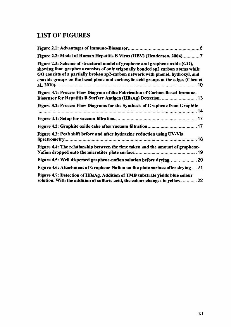
3.4.2 Reduction of Graphene Oxide (GO) to Graphene
The reduction was done by dispersing 0.1 grams of graphite oxide into a 50 ML DI
water. It was then be ultrasonicated for 30 minute. After that, the solution was
centrifuged to remove the unexfoliated materials. The supernatant, which is the top
layer, is the graphene oxide (GO). The supernatant graphene oxide was poured into a
round bottom flask and the pH of the solution was adjusted to 10 using SM of potassium
hydroxide (KOH). Later, 0.025 ML of hydrazine was added into the solution and reflux
in an oil bath at 95°C with stirring for 24 hours. The solution was filtered using 0.45-
micron PTFE filter paper and also washed with large amount of DI water. The filtrate
was having a final wash using acetone instead of DI water. The filtrate was then being
dried using a vacuum desiccator. The final products were the graphene, which will be
used in the fabrication process.
LGRAPHITEJ - ---.--
I GO I METHOD
C:G®R-A-PHITt-'--OXIDE __
I GRAPHENE
EXFOUATION
bI&IIL L OXIDE
REDUCE
GRAPHENE
Figure 3.2: Process Flow Diagrams for the Synthesis of Graphene from Graphite
14

3.4.3 Fabrication of Biosensor
The graphene was used to grow the graphene-Nafion nanosheet by ultrasonicating the
graphene in 0.2 5% Nafion-water. Subsequently, graphene-Nafion nanosheet was
dropped onto the 96-well microtiter plate and allowed to dry. After the graphene-nafion
layer dried, it was then being soaked in Thionine solution. The biosensor fabrication
process is as described in Su et al. (2009). The amount of graphene-Nafion dropped
onto the microtiter plate was examined to obtain the appropriate amount to coat the
surface completely.
3.4.4 Immobilization of Hepatitis B Surface Antigen Antibody (Anti-HIBsAg)
Immobilization of anti-HBsAg on the fabricated biosensor is based on carbodiimide-
assisted amidation reaction. A solution of polyclonal antibodies against hepatitis B
surface antigen (anti-HBsAg IgG) was incubated on the biosensor. After 12 hours,
Bovine Serum Albumin (BSA) was applied to block the remaining active groups and
eliminate non-specific binding sites; this was followed by washing step. The final
biosensor can be stored at 4°C when not in use.
3.4.5 Identification of Graphene Oxide and Graphene
The graphene oxide and graphene produced was examined using UV-Vis spectroscopy.
The peak shift was observed and results were compared with the literature.
3.5 Summary of Chapter
This chapter covers the materials and instrument used in the study. It explains in detail
regarding to the procedure for the synthesis of graphene. It also discussed the process
flow from sample preparation, fabrication of biosensor to immobilization of antibodies.
Beside, this chapter also discuss on the methods in collecting data for the analysis.

4 RESULT AND DISCUSSION
4.1 Overview This chapter will discuss on the result obtained. The result obtained focuses on the
synthesis of graphene, the coating of graphene onto the microtjter plate, and lastly on
the detection of HBsAg.
4.2 Synthesis of Graphene
There is one minor step in synthesizing graphite oxide known as the pre-oxidation step
in which graphite powder was mixed with a solution of sulphuric acid, phosphorus
pentoxide and potassium persulfate. The graphite supposed to change to dark blue
mixture after the mixing. However, the colour does not seem to change much during the
synthesis. Anyway, it was believed that this pre-oxidation step would not affect the
quality of the graphite oxide as this step was done to speed up the whole experiment.
During solid-liquid separation process, several methods such as vacuum filtration and
centrifugation using bench top centrifuge had been carried out to determine the best
method in separating the graphite oxide from its solution. It was found that
centrifugation was not a feasible method in separating the graphite oxide. The graphite
oxide would not settle at the bottom of the centrifuge tube. The possible reason for this
is that the centrifugal force applied was not sufficient. The maximum limit of rotation
for bench top centrifuge is 10000 rpm.
The problem was then be solved by using vacuum filtration. It was clearly shown that
the separation is more efficient using vacuum filtration. The filter cake was the product
needed. The setup for vacuum filtration and its end product was shown in the figure below.
16

Figure 4.1: Setup for vaccum filtration.
Figure 4.2: Graphite oxide cake after vacuum filtration
17