Embed Size (px)
Citation preview
Facial Nerve PalsyFacial Nerve Palsy
Dr. Saud ALROMAIHDr. Saud ALROMAIH

AnatomyAnatomy
Facial nerve is a mixed nerve, having Facial nerve is a mixed nerve, having a motor root and a sensory root.a motor root and a sensory root.
Motor root supplies all the mimetic Motor root supplies all the mimetic muscles of the face which develop muscles of the face which develop from the 2from the 2ndnd brachial arch. brachial arch.
AnatomyAnatomy
Sensory root “nerve of Wrisberg” Sensory root “nerve of Wrisberg” carries taste fibers from the anterior carries taste fibers from the anterior 2/3 of the tongue and general 2/3 of the tongue and general sensation from the concha and sensation from the concha and retroauricular skin.retroauricular skin.
AnatomyAnatomy
Also it carries secretomotor fibers to Also it carries secretomotor fibers to the lacrimal, submandibular and the lacrimal, submandibular and sublingual glands as well as those in sublingual glands as well as those in the nose and palate.the nose and palate.
Anatomy: NucleusAnatomy: Nucleus
Pons.Pons.
precentral gyrus.precentral gyrus.
Upper part of the nucleus:Upper part of the nucleus:– Upper faceUpper face– Involuntary emotional movements Involuntary emotional movements
Anatomy: CourseAnatomy: CourseMotor fibers originate…Motor fibers originate…Hooks around…Hooks around…Joined by…Joined by…Facial n. leaves the brainstem…Facial n. leaves the brainstem…Travels through…Travels through…Enters the IAC.Enters the IAC.Then traverse the temporal bone through Then traverse the temporal bone through facial n. canalfacial n. canalLeaves the temporal bone throughLeaves the temporal bone throughFinally divides into terminal branches.Finally divides into terminal branches.
Anatomy: PartsAnatomy: Parts
Intracranial partIntracranial part
Intratemporal partIntratemporal part
Extracranial partExtracranial part
Anatomy: Intratemporal segmentsAnatomy: Intratemporal segments
MeatalMeatal
LabyrinthineLabyrinthine
Tympanic, horizontalTympanic, horizontal
Mastoid, verticalMastoid, vertical
Anatomy: BranchesAnatomy: Branches
Greater superficial petrosal nerve:Greater superficial petrosal nerve:
Nerve to stapedius:Nerve to stapedius:
Chorda tympani:Chorda tympani:
Comunicating branch:Comunicating branch:
Posterior auricular nerve:Posterior auricular nerve:
Muscular branches:Muscular branches:
Peripheral branches: “Pes anserinus”Peripheral branches: “Pes anserinus”
Anatomy: Surgical landmarksAnatomy: Surgical landmarks
Middle Ear and Mastoid Surgery:Middle Ear and Mastoid Surgery:– Processus chocleariformisProcessus chocleariformis– Oval window and horizontal canalOval window and horizontal canal– Short process of the incusShort process of the incus– PyramidPyramid
Anatomy: Surgical landmarksAnatomy: Surgical landmarks
Parotid Surgery:Parotid Surgery:– Cartilaginous pointer:Cartilaginous pointer:– Styloid processStyloid process– Posterior belly pf digastric musclePosterior belly pf digastric muscle
Anatomy: Structure of the nerveAnatomy: Structure of the nerve
From inside outward:From inside outward:– AxonAxon– Myelin sheathMyelin sheath– NeurolimmaNeurolimma– EndoneuriumEndoneurium– PerineuriumPerineurium– EpineuriumEpineurium
Anatomy: Severity of injuryAnatomy: Severity of injury
Saunderland classification:Saunderland classification:– 1°: Partial block: Neuropraxia1°: Partial block: Neuropraxia– 2°: Loss of axons: axonotemesis2°: Loss of axons: axonotemesis– 3°: Injury to the endoneurium: 3°: Injury to the endoneurium:
neurotemesisneurotemesis– 4°: Injury to the perineurium: partial 4°: Injury to the perineurium: partial
transectiontransection– 5°: Injury to the epineurium: complete 5°: Injury to the epineurium: complete
transectiontransection

History:History:
Onset: Sudden vs. GradualOnset: Sudden vs. Gradual
Duration:Duration:
Rate of progression:Rate of progression:
Recuurent or familialRecuurent or familial
Associated symptomsAssociated symptoms
Medical historyMedical history
Previous surgeriesPrevious surgeries
Physical exam:Physical exam:
Complete vs. incompleteComplete vs. incomplete
Segmental vs. uniform involvementSegmental vs. uniform involvement
Unilateral vs. bilateralUnilateral vs. bilateral
Cranial nerves assessmentCranial nerves assessment
Neurologic evaluationNeurologic evaluation
Cerebellar signsCerebellar signs
Physical exam:Physical exam:
Microscopic otoscopyMicroscopic otoscopy
Complete head and neck examComplete head and neck exam

Physical exam:Physical exam:
Localization of facial nerve lesion:Localization of facial nerve lesion:
Central vs. Peripheral. Central vs. Peripheral.
Physical exam:Physical exam:
Localization of facial nerve lesion:Localization of facial nerve lesion:
Peripheral:Peripheral:– Level of nucleusLevel of nucleus– CPA level:CPA level:– Bony canal level: TopodiagnosticsBony canal level: Topodiagnostics– Outside the Temporal boneOutside the Temporal bone
Physical exam:Physical exam:
Topodiagnostics:Topodiagnostics:– Schirmer’s test:Schirmer’s test:– Stapedial reflex:Stapedial reflex:– Taste test:Taste test:– Submandibular salivery flow test: Submandibular salivery flow test:
Warton’s ductsWarton’s ducts
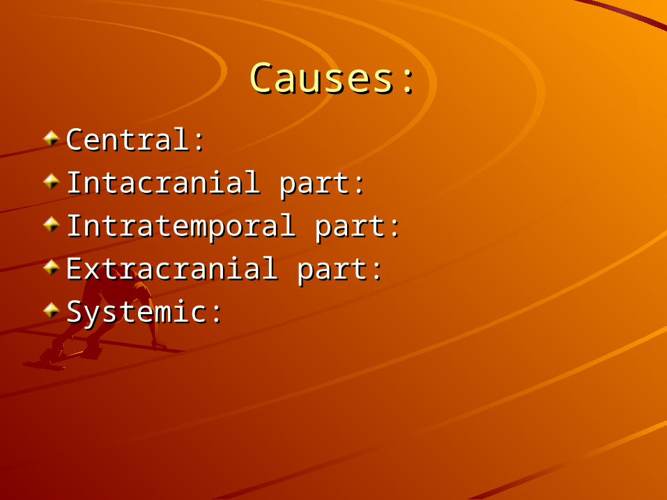
Causes:Causes:
Central:Central:
Intacranial part:Intacranial part:
Intratemporal part:Intratemporal part:
Extracranial part:Extracranial part:
Systemic:Systemic:
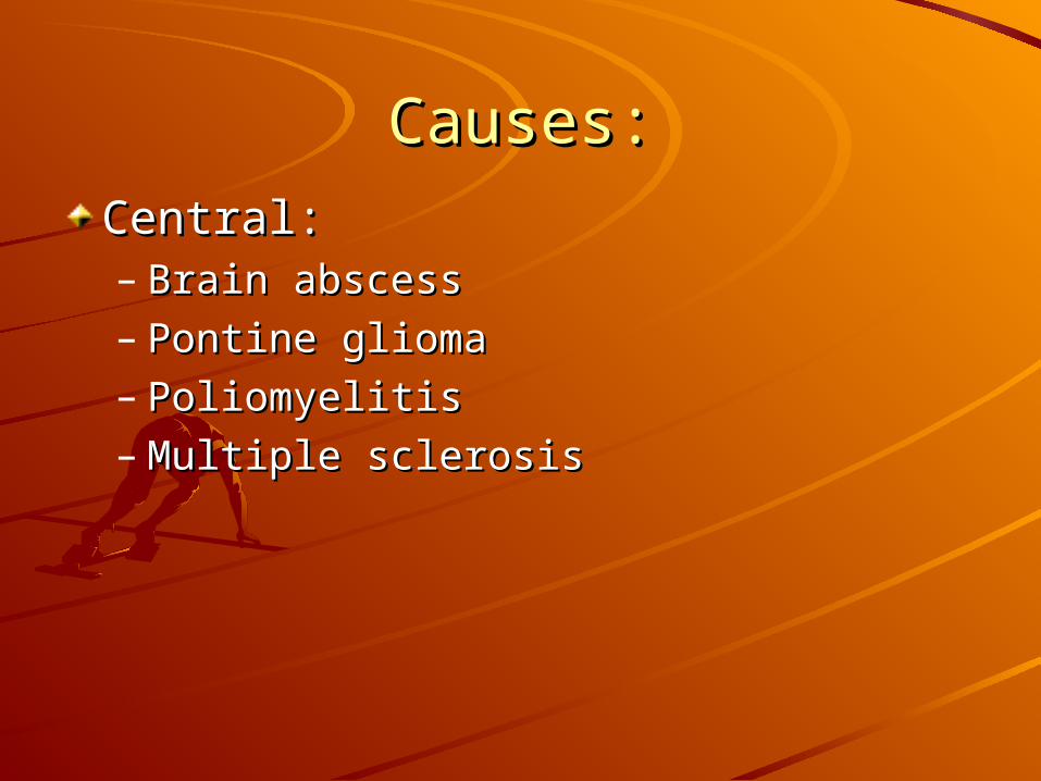
Causes:Causes:
Central:Central:– Brain abscessBrain abscess– Pontine gliomaPontine glioma– PoliomyelitisPoliomyelitis– Multiple sclerosisMultiple sclerosis
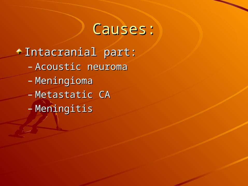
Causes:Causes:
Intacranial part:Intacranial part:– Acoustic neuromaAcoustic neuroma– MeningiomaMeningioma– Metastatic CAMetastatic CA– MeningitisMeningitis

Causes:Causes:
Intratemporal part:Intratemporal part:– Idiopathic:Idiopathic:
Bell’s palsyBell’s palsy
Melkersson’s syndromeMelkersson’s syndrome
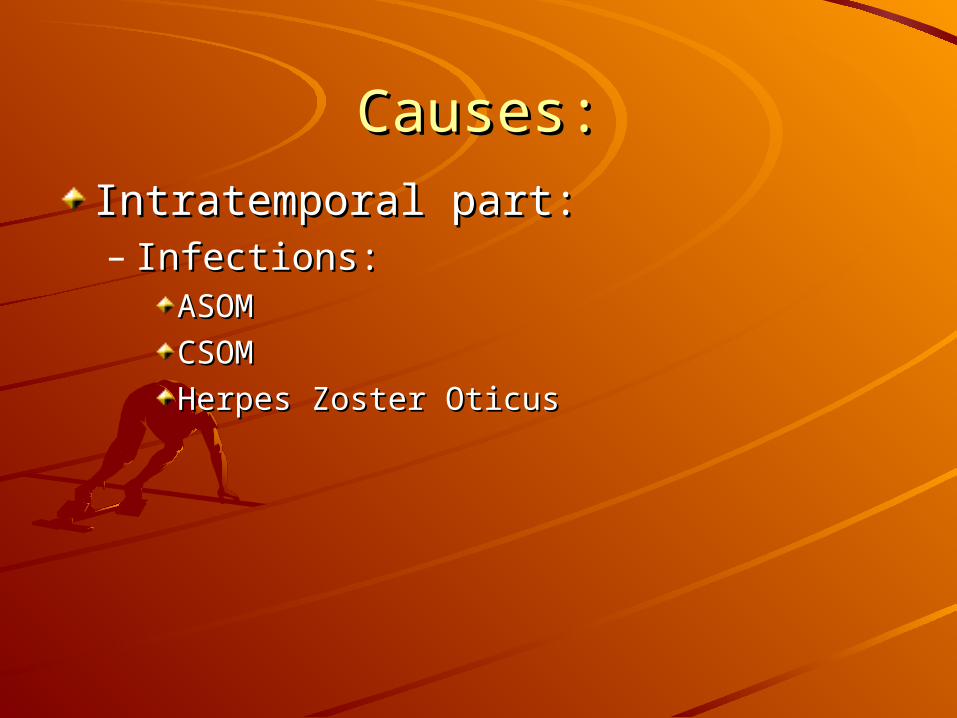
Causes:Causes:
Intratemporal part:Intratemporal part:– Infections:Infections:
ASOMASOM
CSOMCSOM
Herpes Zoster OticusHerpes Zoster Oticus
Causes:Causes:
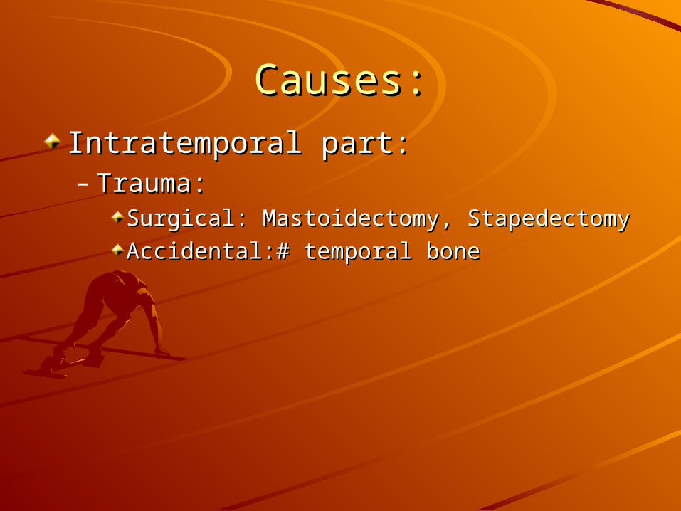
Intratemporal part:Intratemporal part:– Trauma:Trauma:
Surgical: Mastoidectomy, StapedectomySurgical: Mastoidectomy, Stapedectomy
Accidental:# temporal boneAccidental:# temporal bone
Causes:Causes:
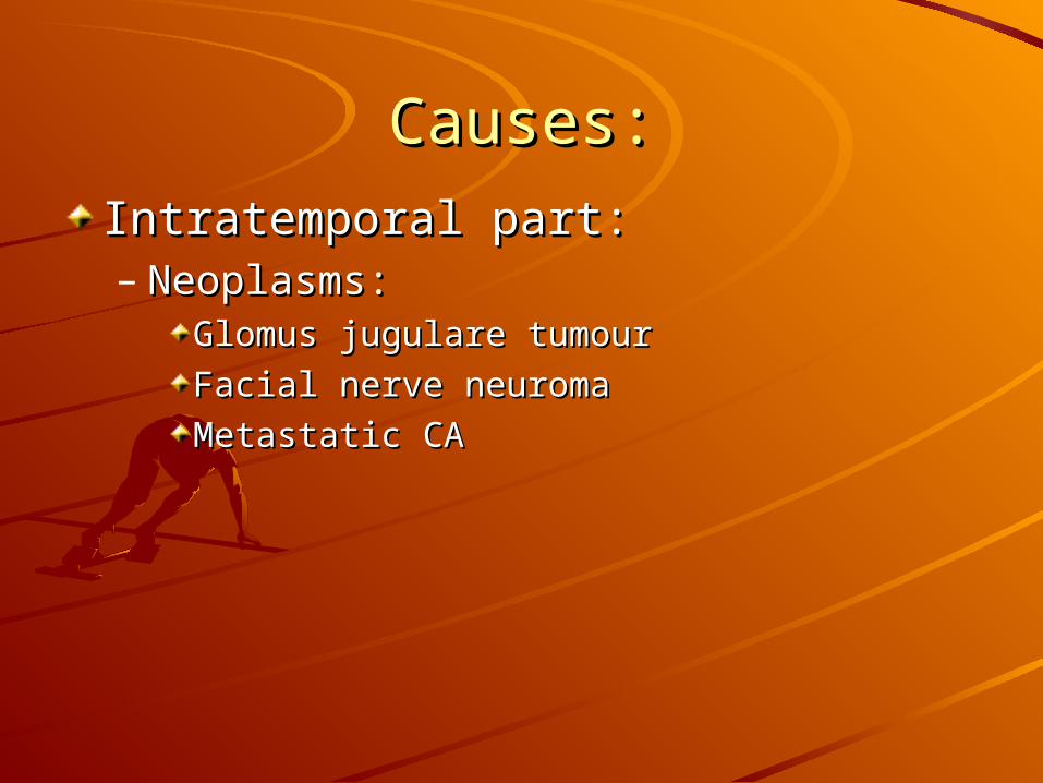
Intratemporal part:Intratemporal part:– Neoplasms:Neoplasms:
Glomus jugulare tumourGlomus jugulare tumour
Facial nerve neuromaFacial nerve neuroma
Metastatic CAMetastatic CA
Causes:Causes:
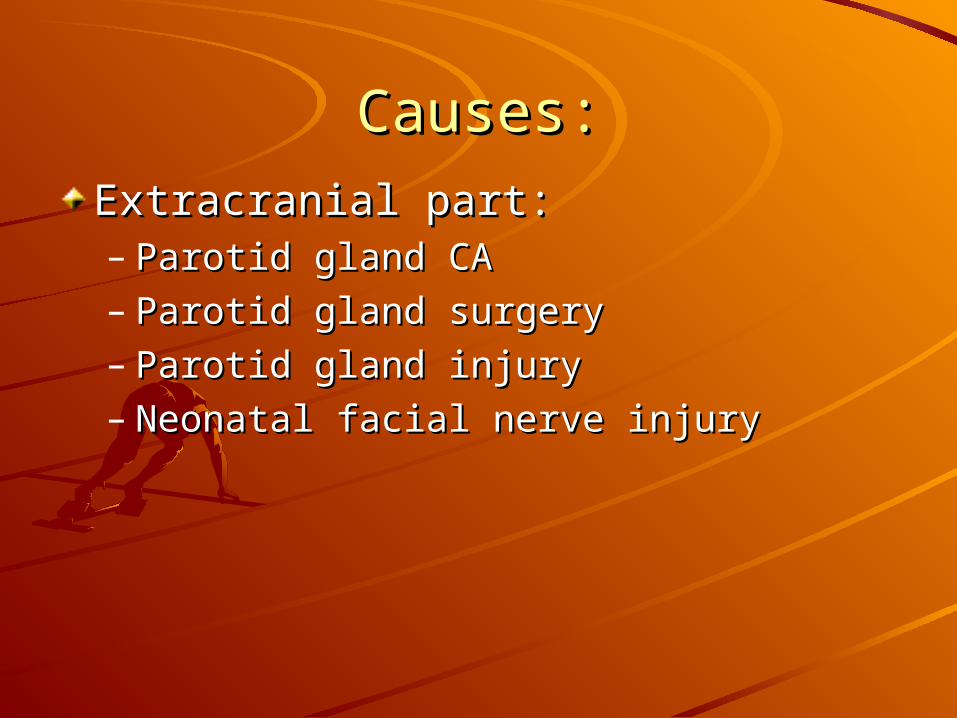
Extracranial part:Extracranial part:– Parotid gland CAParotid gland CA– Parotid gland surgeryParotid gland surgery– Parotid gland injuryParotid gland injury– Neonatal facial nerve injuryNeonatal facial nerve injury
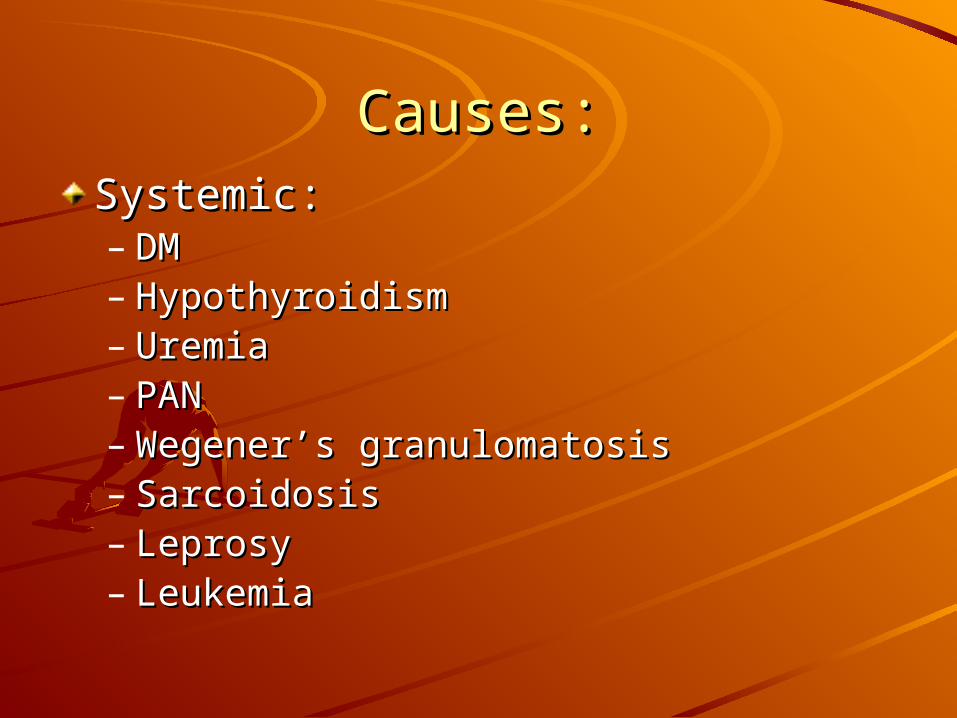
Causes:Causes:Systemic:Systemic:– DMDM– HypothyroidismHypothyroidism– UremiaUremia– PANPAN– Wegener’s granulomatosisWegener’s granulomatosis– SarcoidosisSarcoidosis– LeprosyLeprosy– LeukemiaLeukemia
Labs:Labs:
Pure-tune audiometryPure-tune audiometry
Electrophysiologic testsElectrophysiologic tests
Imaging testsImaging tests
OthersOthers

Labs:Labs:
Electrophysiologic tests:Electrophysiologic tests:– Nerve Excitability Test: NETNerve Excitability Test: NET– Maximum stimulation Test: MSTMaximum stimulation Test: MST– Electroneurography: ENoGElectroneurography: ENoG– Electromyography: EMGElectromyography: EMG
Complications:Complications:Incomplete recoveryIncomplete recoveryExposure keratitisExposure keratitisSynkinesisSynkinesisTics and spasmsTics and spasmsContracturesContracturesCrocodile tearsCrocodile tearsFrey’s syndrome “gustatory sweating”Frey’s syndrome “gustatory sweating”Psychological and social problemsPsychological and social problems

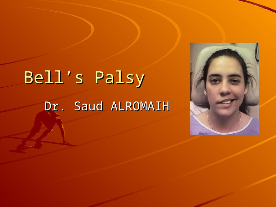
Bell’s PalsyBell’s Palsy
Dr. Saud ALROMAIHDr. Saud ALROMAIH
Background:Background:
one of the most common neurologic one of the most common neurologic disorders affecting the cranial disorders affecting the cranial nerves. nerves.
abrupt, unilateral, peripheral facial abrupt, unilateral, peripheral facial paresis or paralysis without a paresis or paralysis without a detectable cause.detectable cause.
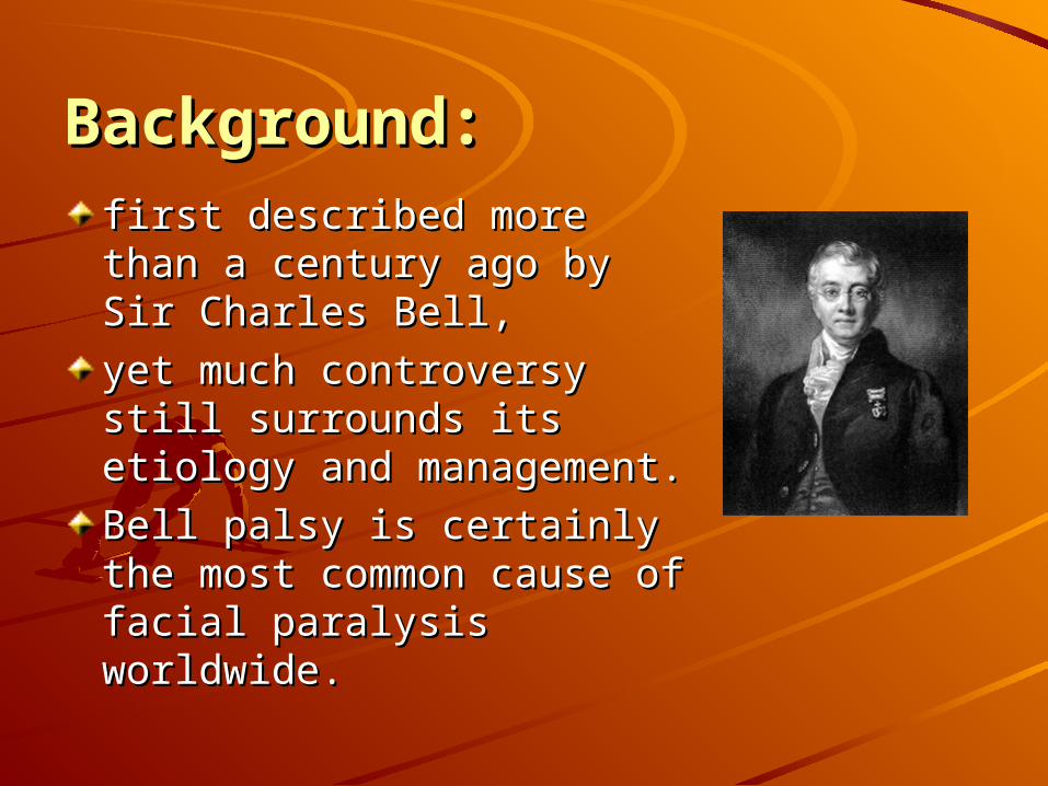
Background:Background:first described more than a first described more than a century ago by Sir Charles century ago by Sir Charles Bell,Bell,
yet much controversy still yet much controversy still surrounds its etiology and surrounds its etiology and management.management.
Bell palsy is certainly the Bell palsy is certainly the most common cause of most common cause of facial paralysis worldwide. facial paralysis worldwide.
Incidence:Incidence:
United StatesUnited States
Internationally Internationally
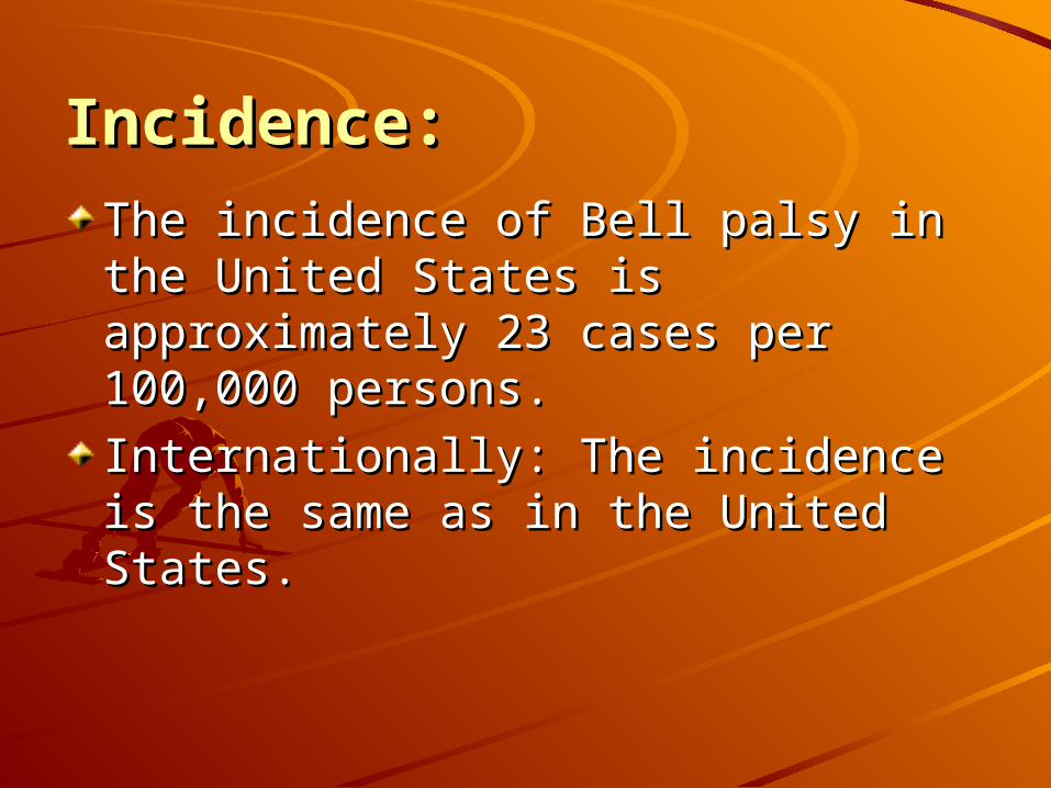
Incidence:Incidence:
The incidence of Bell palsy in the The incidence of Bell palsy in the United States is approximately 23 United States is approximately 23 cases per 100,000 persons.cases per 100,000 persons.
Internationally:Internationally: The incidence is the The incidence is the same as in the United States. same as in the United States.

Demographics:Demographics:
Race:Race:
Sex:Sex:
Age:Age:
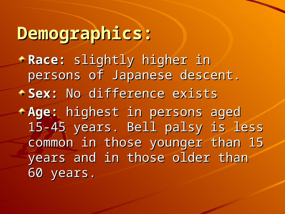
Demographics:Demographics:
Race: Race: slightly higher in persons of slightly higher in persons of Japanese descent. Japanese descent.
Sex: Sex: No difference exists No difference exists
Age: Age: highest in persons aged 15-45 highest in persons aged 15-45 years. Bell palsy is less common in years. Bell palsy is less common in those younger than 15 years and in those younger than 15 years and in those older than 60 years. those older than 60 years.
Pathophysiology:Pathophysiology:Main cause of Bell's palsy is latent Main cause of Bell's palsy is latent herpes viruses (herpes simplex virus herpes viruses (herpes simplex virus type 1 and herpes zoster virus), type 1 and herpes zoster virus), which are reactivated from cranial which are reactivated from cranial nerve ganglia.nerve ganglia.Polymerase chain reaction Polymerase chain reaction techniques have isolated herpes techniques have isolated herpes virus DNA from the facial nerve virus DNA from the facial nerve during acute palsy.during acute palsy.
Pathophysiology:Pathophysiology:
Inflammation of the nerve initially Inflammation of the nerve initially results in a reversible neurapraxia,results in a reversible neurapraxia,
Herpes zoster virus shows more Herpes zoster virus shows more aggressive biological behaviour than aggressive biological behaviour than herpes simplex virus type 1herpes simplex virus type 1
History:History:
The most alarming symptom of Bell's The most alarming symptom of Bell's palsy is paresispalsy is paresis
Up to three quarters of affected Up to three quarters of affected patients think they have had a stroke patients think they have had a stroke or have an intracranial tumour.or have an intracranial tumour.
History:History:
The palsy is often sudden in onset The palsy is often sudden in onset and evolves rapidly, with maximal and evolves rapidly, with maximal facial weakness developing within facial weakness developing within two days.two days.
Associated symptoms may be Associated symptoms may be hyperacusis, decreased production of hyperacusis, decreased production of tears, and altered taste.tears, and altered taste.
History:History:
Patients may also mention otalgia or Patients may also mention otalgia or aural fullness and facial or aural fullness and facial or retroauricular pain, which is typically retroauricular pain, which is typically mild and may precede the palsy.mild and may precede the palsy.
A slow onset progressive palsy with A slow onset progressive palsy with other cranial nerve deficits or other cranial nerve deficits or headache raises the possibility of a headache raises the possibility of a neoplasm neoplasm

Physical exam:Physical exam:
Bell's palsy causes a peripheral lower Bell's palsy causes a peripheral lower motor neurone palsy,motor neurone palsy,
which manifests as the unilateral which manifests as the unilateral impairment of movement in the impairment of movement in the facial and platysma muscles, facial and platysma muscles, drooping of the brow and corner of drooping of the brow and corner of the mouth, and impaired closure of the mouth, and impaired closure of the eye and mouth.the eye and mouth.
Physical exam:Physical exam:
Bell's phenomenon—upward Bell's phenomenon—upward diversion of the eye on attempted diversion of the eye on attempted closure of the lid—is seen when eye closure of the lid—is seen when eye closure is incomplete. closure is incomplete.
Physical exam:Physical exam:
Polyposis or granulations in the ear Polyposis or granulations in the ear canal may suggest cholesteatoma or canal may suggest cholesteatoma or malignant otitis externa.malignant otitis externa.
Vesicles in the conchal bowl, soft Vesicles in the conchal bowl, soft palate, or tongue suggest Ramsay palate, or tongue suggest Ramsay Hunt syndrome Hunt syndrome
Physical exam:Physical exam:
The examination should exclude The examination should exclude masses in the head and neck.masses in the head and neck.
A deep lobe parotid tumour may only A deep lobe parotid tumour may only be identified clinically by careful be identified clinically by careful examination of the oropharynx and examination of the oropharynx and ipsilateral tonsil to rule out ipsilateral tonsil to rule out asymmetry.asymmetry.
Investigations:Investigations:
Serum testing for rising antibody Serum testing for rising antibody titres to herpes virus is not a reliable titres to herpes virus is not a reliable diagnostic tool for Bell's palsy.diagnostic tool for Bell's palsy.
Salivary PCR for herpes simplex virus Salivary PCR for herpes simplex virus type 1 or herpes zoster virus is more type 1 or herpes zoster virus is more likely to confirm virus during the likely to confirm virus during the replicating phase, but these tests replicating phase, but these tests remain research tools.remain research tools.
Investigations:Investigations:
MRI has revolutionised the detection MRI has revolutionised the detection of tumours.of tumours.
Investigations:Investigations:
Topognostic tests and Topognostic tests and electroneurography may give useful electroneurography may give useful prognostic information but remain prognostic information but remain research tools. research tools.
Diagnosis:Diagnosis:
Bell palsy is a diagnosis of exclusion.Bell palsy is a diagnosis of exclusion.
Other disease states or conditions Other disease states or conditions that present with facial palsies are that present with facial palsies are often misdiagnosed as idiopathic. often misdiagnosed as idiopathic.

Management:Management:The main aims of treatment in the acute The main aims of treatment in the acute phase of Bell's palsy are to speed recovery phase of Bell's palsy are to speed recovery and to prevent corneal complications.and to prevent corneal complications.Treatment should begin immediately to Treatment should begin immediately to inhibit viral replication and the effect on inhibit viral replication and the effect on subsequent pathophysiological processes subsequent pathophysiological processes that affect the facial nerve.that affect the facial nerve.Psychological support is also essential, and Psychological support is also essential, and for this reason patients may require for this reason patients may require regular follow up. regular follow up.
Management, Eye careManagement, Eye care
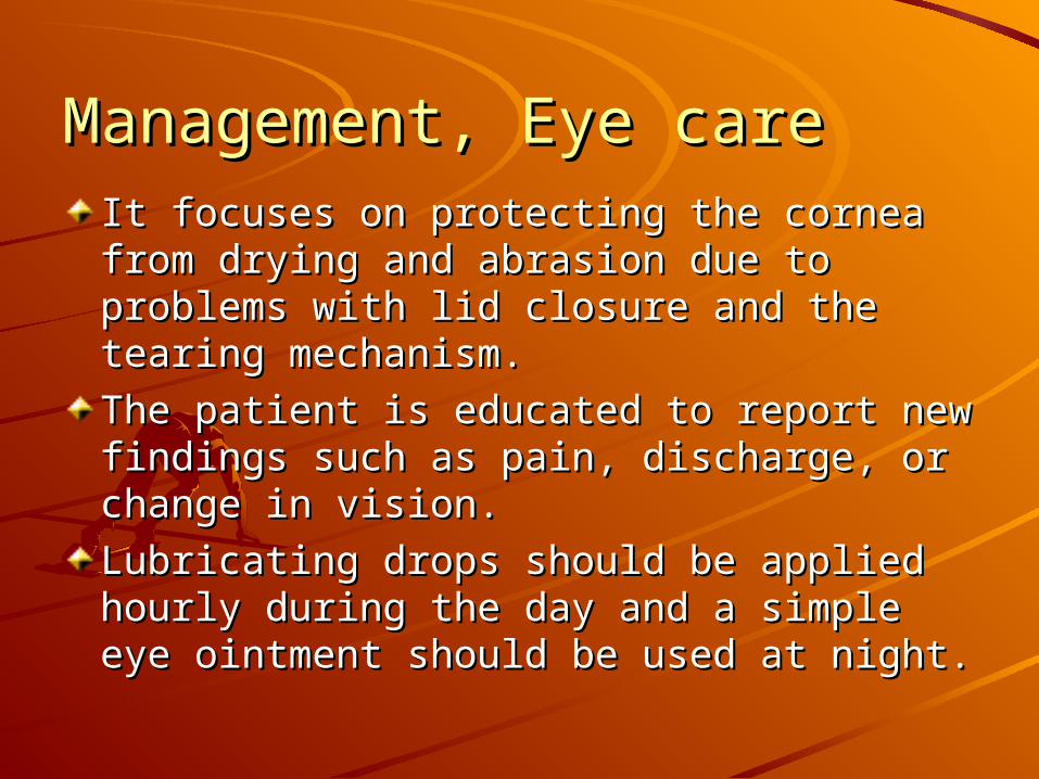
Management, Eye careManagement, Eye careIt focuses on protecting the cornea from It focuses on protecting the cornea from drying and abrasion due to problems with drying and abrasion due to problems with lid closure and the tearing mechanism.lid closure and the tearing mechanism.
The patient is educated to report new The patient is educated to report new findings such as pain, discharge, or findings such as pain, discharge, or change in vision.change in vision.
Lubricating drops should be applied hourly Lubricating drops should be applied hourly during the day and a simple eye ointment during the day and a simple eye ointment should be used at night. should be used at night.
Management, SteroidManagement, Steroid
Management, SteroidManagement, Steroid
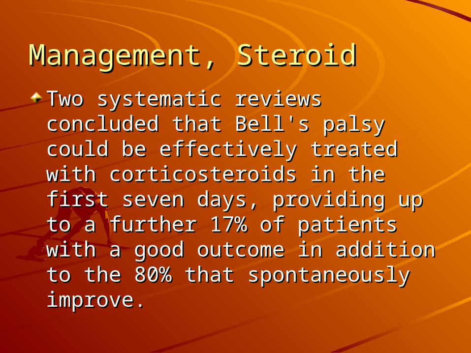
Two systematic reviews concluded Two systematic reviews concluded that Bell's palsy could be effectively that Bell's palsy could be effectively treated with corticosteroids in the treated with corticosteroids in the first seven days, providing up to a first seven days, providing up to a further 17% of patients with a good further 17% of patients with a good outcome in addition to the 80% that outcome in addition to the 80% that spontaneously improve.spontaneously improve.
Management, SteroidManagement, Steroid
Usual regimen is 1mg/kg/day for 1 Usual regimen is 1mg/kg/day for 1 week.week.
To be tapered in the 2To be tapered in the 2ndnd week. week.
Management, SteroidManagement, SteroidCochrane review*:Cochrane review*:
““There is insufficient evidence about the effects of There is insufficient evidence about the effects of corticosteroids for people with Bell's palsy, although corticosteroids for people with Bell's palsy, although their anti-inflammatory effect might prevent nerve their anti-inflammatory effect might prevent nerve damage.”damage.”
**Salinas RA, Alvarez G, Ferreira J. Corticosteroids for Bell's palsy (idiopathic Salinas RA, Alvarez G, Ferreira J. Corticosteroids for Bell's palsy (idiopathic facial paralysis). facial paralysis). Cochrane Database of Systematic ReviewsCochrane Database of Systematic Reviews 2004, Issue 4. 2004, Issue 4.
Art. No.: CD001942.Art. No.: CD001942.
Management, AntiviralsManagement, Antivirals
It seems logical in Bell's palsy It seems logical in Bell's palsy because of the probable involvement because of the probable involvement of herpes viruses.of herpes viruses.
Aciclovir, a nucleotide analogue, Aciclovir, a nucleotide analogue, interferes with herpes virus DNA interferes with herpes virus DNA polymerase and inhibits DNA polymerase and inhibits DNA replication.replication.
Management, AntiviralsManagement, Antivirals
Usual regimen is 4000mg/24hrs Usual regimen is 4000mg/24hrs divided into 5 doses for 7 to 10 daysdivided into 5 doses for 7 to 10 days
Bell’s palsy:Bell’s palsy:Antivirals:Antivirals:
Cochrane review*:Cochrane review*:““More evidence is needed to show whether the More evidence is needed to show whether the
antiviral drugs acyclovir or valacyclovir are effective antiviral drugs acyclovir or valacyclovir are effective in aiding recovery from Bell's palsy.”in aiding recovery from Bell's palsy.”
** Allen D, Dunn L. Acyclovir or valaciclovir for Bell's palsy (idiopathic facial Allen D, Dunn L. Acyclovir or valaciclovir for Bell's palsy (idiopathic facial paralysis). Cochrane Database of Systematic Reviews 2004, Issue 3. Art. paralysis). Cochrane Database of Systematic Reviews 2004, Issue 3. Art. No.: CD001869. No.: CD001869.
Outcomes:Outcomes:It has a fair prognosis without It has a fair prognosis without treatment, with almost three treatment, with almost three quarters of patients recovering quarters of patients recovering normal mimetical function and just normal mimetical function and just over a tenth having minor sequelae.over a tenth having minor sequelae.A sixth of patients are left with either A sixth of patients are left with either moderate to severe weakness, moderate to severe weakness, contracture, hemifacial spasm, or contracture, hemifacial spasm, or synkinesis.synkinesis.
Outcomes:Outcomes:
Patients with a partial palsy fair Patients with a partial palsy fair better, with 94% making a full better, with 94% making a full recovery.recovery.
The outcome is worse when herpes The outcome is worse when herpes zoster virus infection is involved in zoster virus infection is involved in partial palsy. partial palsy.

Outcomes:Outcomes:
In patients who recover without In patients who recover without treatment, major improvement treatment, major improvement occurs within three weeks in most.occurs within three weeks in most.
If recovery does not occur within this If recovery does not occur within this time, then it is unlikely to be seen time, then it is unlikely to be seen until four to six months, when nerve until four to six months, when nerve regrowth and reinnervation have regrowth and reinnervation have occurred. occurred.
Bad Prognostic Factor:Bad Prognostic Factor:Complete facial palsyComplete facial palsyNo recovery by three weeksNo recovery by three weeksAge over 60 yearsAge over 60 yearsSevere painSevere painRamsay Hunt syndrome (herpes zoster Ramsay Hunt syndrome (herpes zoster virus)virus)Associated conditions—hypertension, Associated conditions—hypertension, diabetes, pregnancydiabetes, pregnancySevere degeneration of the facial nerve Severe degeneration of the facial nerve shown by electrophysiological testingshown by electrophysiological testing
??

ThanksThanks
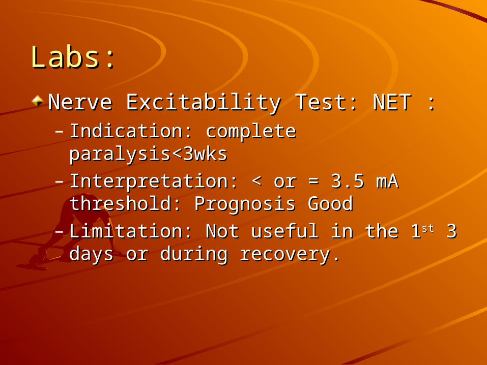
Labs:Labs:
Nerve Excitability Test: NET :Nerve Excitability Test: NET :– Indication: complete paralysis<3wksIndication: complete paralysis<3wks– Interpretation: < or = 3.5 mA threshold: Interpretation: < or = 3.5 mA threshold:
Prognosis GoodPrognosis Good– Limitation: Not useful in the 1Limitation: Not useful in the 1stst 3 days or 3 days or
during recovery.during recovery.
Labs:Labs:
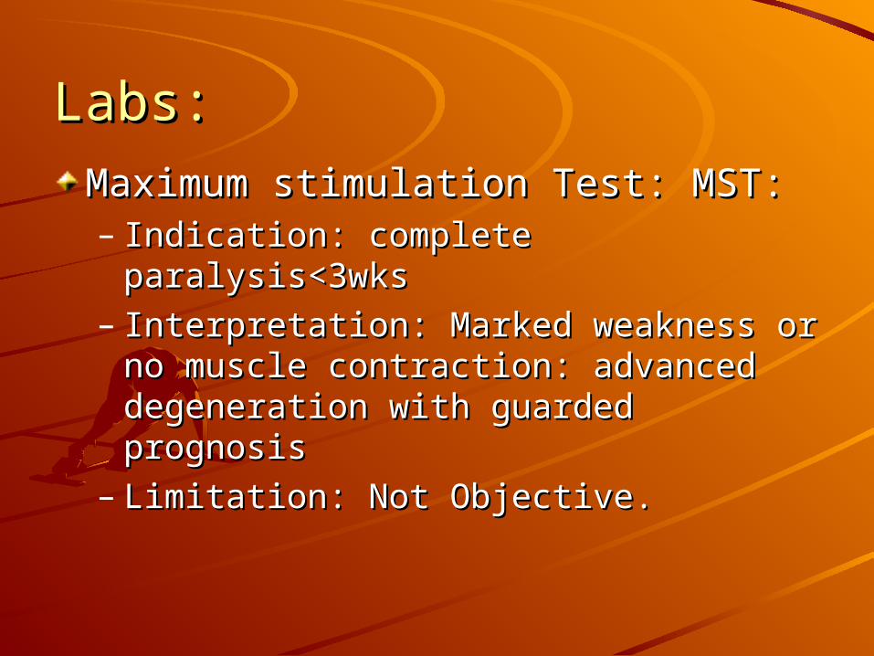
Maximum stimulation Test: MST:Maximum stimulation Test: MST:– Indication: complete paralysis<3wksIndication: complete paralysis<3wks– Interpretation: Marked weakness or no Interpretation: Marked weakness or no
muscle contraction: advanced muscle contraction: advanced degeneration with guarded prognosisdegeneration with guarded prognosis
– Limitation: Not Objective.Limitation: Not Objective.
Labs:Labs:
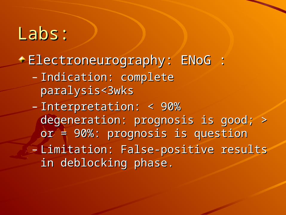
Electroneurography: ENoG :Electroneurography: ENoG :– Indication: complete paralysis<3wksIndication: complete paralysis<3wks– Interpretation: < 90% degeneration: Interpretation: < 90% degeneration:
prognosis is good; > or = 90%: prognosis is good; > or = 90%: prognosis is questionprognosis is question
– Limitation: False-positive results in Limitation: False-positive results in deblocking phase.deblocking phase.
Labs:Labs:Electromyography: EMGElectromyography: EMG– Indication: Acute paralysis less than 1 week or Indication: Acute paralysis less than 1 week or
chronic paralysis longer than 2 weekschronic paralysis longer than 2 weeks– Interpretation:Interpretation:
Active mu: intact motor axonsActive mu: intact motor axons
Mu + fibrillation potentials: partial degenerationMu + fibrillation potentials: partial degeneration
Polyphasic mu: regenerating nervePolyphasic mu: regenerating nerve
– Limitation: cannot assess degree of Limitation: cannot assess degree of degeneration or prognosis for recovery.degeneration or prognosis for recovery.