Embed Size (px)
Citation preview

Facilitating Mismatch Discrimination by Surface-Affixed PNA Probesvia Ionic RegulationSrabani Ghosh, Sourav Mishra, Trambaki Banerjee, and Rupa Mukhopadhyay*
Department of Biological Chemistry, Indian Association for the Cultivation of Science, Jadavpur, Kolkata-700 032, India
*S Supporting Information
ABSTRACT: There has been a search for alternative nucleicacids that can be more effectively used in nucleic acid detectiontechnologies compared to the DNA probes. Peptide nucleicacid (PNA), which contains a non-ionic peptidic backbone,offers such possibilities since it is nuclease-resistant, it binds toDNA with high affinity, and it can be readily self-assembledonto solid substrates, e.g., gold(111), with a molecularbackbone orientation away from the substrate. Althoughapplication of PNA as a sensor probe has been exemplified, sofar there is little or no account of the ionic modulation ofsingle base mismatch discrimination capacity of surface-tethered PNA probes. Herein, we report “on-surface” meltingtemperatures of PNA-DNA duplexes formed on gold(111) surface, as obtained from fluorescence measurements. We show thatsurface-tethered PNA forms a stabler duplex than DNA, and is more effective in single base mismatch discrimination than DNA.Importantly, although PNA backbone is non-ionic, variation in the ionic components in hybridization buffer, i.e., varyingconcentration of monovalent sodium ion, and the nature of anion and the cation, exhibits clear effects on the mismatchdiscrimination capacity of PNA probes. In general, with decreasing cation concentration, PNA-DNA duplexes are stabilized andmismatch discrimination capacity of the PNA probes is enhanced. The stabilizing/destabilizing effects of anions are found tofollow the Hofmeister series, emphasizing the importance of hydrophobic interaction between nucleobases for stability of thePNA-DNA duplexes. Interestingly, the nature of ionic dependence of “on-surface” mismatch detection ability of PNA probesdiffers significantly from the “solution” behavior of these probes.
■ INTRODUCTION
Peptide nucleic acid (PNA) is a DNA analogue, in which thenegatively charged phosphodiester units of DNA backbone arereplaced by 2-aminoethyl-glycine linkages (Figure 1).1 As aresult, in the absence of the strand−strand electrostaticrepulsions, unlike in the case of DNA, formation of closeassociation of ssPNA strands becomes relatively straightfor-ward, and compact self-assembled PNA films can be readilygenerated on solid substrates like gold(111) surface by a simpleimmersion method.2 In such films, the immobilized ssPNAstrands can be oriented away from the surface, as elicited fromreflection absorption infrared spectroscopy (RAIRS) experi-ments,2 and nonspecific interactions with the underlying goldsubstrate can be largely avoided, creating an ideal situation forthe target nucleic acid strands to access the immobilized sensorPNA probes. On the contrary, the DNA films comprising thenegatively charged ssDNA strands have been found to bemostly disordered/poorly ordered,3 where nonspecific DNA−surface interactions could occur through the relatively exposednucleobases, resulting in reduced bioactivity of the film.4
Further advantages of using PNA are that PNA probes canbind to DNA oligomers in a sequence-specific manner withhigher affinity compared to the DNA probes obeying Watson−Crick hydrogen bonding rule5−8 and that PNA is not
susceptible to hydrolytic (enzymatic) cleavage. For thesebenefits, PNA appears to be an attractive candidate as a sensorprobe in solid-state DNA detection technologies. So far, reportson solid-state DNA-DNA hybridization have been made.4,9−13
A number of reports on solid-state DNA detection by PNAhave also been made.14−22 Important information, such as howthe single base mismatch discrimination ability of surface-confined PNA probes can be controlled by ionic variations, ishowever lacking.In solution-phase studies, it is a standard observation that the
sequence specificity of a sensor PNA probe toward the targetoligonucleotide sequences is reflected in the melting temper-ature (Tm) values of the respective sensor-target duplexesformed.23−25 For example, the thermal stabilities of the PNA-DNA duplexes are considerably lowered by the presence ofmismatches in the target DNA oligomers.23,24,26,27 Assumingthat the sequence specificity would be reflected in the Tm valuesin the case of solid-state PNA-DNA hybridization too, weinvestigated the sequence specificity of 12-mer ssPNA sensorprobes toward the DNA target oligomer, in fully matched and
Received: July 27, 2012Revised: February 8, 2013Published: February 17, 2013
Article
pubs.acs.org/Langmuir
© 2013 American Chemical Society 3370 dx.doi.org/10.1021/la400125x | Langmuir 2013, 29, 3370−3379

singly mismatched combinations, in comparison to the 12-merssDNA sensor probes, on gold(111) surface, by fluorescence-based measurement of the respective “on-surface” Tm values.We varied the ionic environment of the surface-confined
duplexes by varying salt concentration and the nature of salt inthe hybridization buffer. The purpose was to understandwhether and how the ionic factors could control the mismatchdiscrimination ability of the surface-confined PNA probes.While our observations revealed an increase in the thermalstability of both PNA-DNA and DNA-DNA duplexes ongold(111) surface compared to solution, we also found that thesingle base mismatch discrimination was better achieved byPNA probes compared to DNA probes. Importantly, suchdiscrimination could be fine-tuned by controlling the ionicsettingsthe nature of “on-surface” ionic control differingnoticeably from “solution” behavior of the PNA probes.
■ MATERIALS AND METHODSPreparation of PNA Sensor Probe Solutions. The 12-mer
ssPNA sensor probes PNA 1, PNA 2, PNA 3, and Cy3-PNA 1 (Table1), all having a −(CH2)6SH group at N-ter position (Panagene,Korea), were dissolved in filtered autoclaved Milli-Q water (resistivity18.2 MΩcm)/sodium phosphate buffer (20 mM sodium phosphate, xmM sodium chloride, pH 7.00, where x could be 2/50/100/500/1000mM). PNA 1 and PNA 2 were the pairs of oligomers having a singlebase difference between each other, while the PNA 3 was the
completely non-complementary sequence used in control experiments.The exact concentrations of the PNA solutions were determined byUV−visible spectrophotometry, using absorbance value at 260 nm[(ε260 (L/(mol × cm)) for PNA-1, PNA-2, PNA-3, and Cy3-PNA 1taken as 116700, 123800, 116700, and 122200, respectively (all theε260 values presented here or later were obtained from themanufacturer-provided data sheets)].
Preparation of DNA Sensor Probe Solutions. The 12-merssDNA sensor probes DNA 1, DNA 2, and DNA 3 (Table 1), allhaving a −(CH2)6SH group at 5′ position (Alpha DNA, Canada),were dissolved in sodium phosphate buffer (20 mM sodiumphosphate, 100 mM sodium chloride, pH 7.00). DNA 1 and DNA 2were the pairs of oligomers having a single base difference betweeneach other, while DNA 3 was the completely non-complementarysequence that served for control experiments. The exact concen-trations of the DNA solutions were determined by UV−visiblespectrophotometry, using absorbance values at 260 nm [(ε260 (L/(mol× cm)) for DNA-1, DNA-2, and DNA-3 taken as 123020, 131350, and123020, respectively].
Preparation of Cy3 Labeled DNA Target Probe Solutions.The Cy3-labeled DNA target probe samples Cy3-DNA 1 and Cy3-DNAnc (IDT, Canada) (Table 1) were taken in sodium phosphatebuffer (20 mM sodium phosphate, x mM sodium chloride/sodiumsulfate/sodium nitrate/tetra methyl ammonium chloride as perexperimental design, pH 7.00, where x could be 2/50/100/500/1000 mM). The exact concentrations of the DNA solutions weredetermined by UV−visible spectrophotometry, using absorbance valueat 260 nm [(ε260 (L/(mol × cm)) for Cy3-DNA-1 and Cy3-DNAnctaken as 124400 and 116000, respectively].
Preparation of Label-Free DNA Target Probe Solution. TheDNA sequence T-DNA 1 (Table 1) (Alpha DNA, Canada) was usedas the unlabeled target DNA probe. It was dissolved in sodiumphosphate buffer (20 mM sodium phosphate, x mM sodium chloride,pH 7.00, where x could be 2/50/100/500/1000 mM). The exactconcentrations of the DNA solutions were determined by UV−visiblespectrophotometry, using absorbance value at 260 nm [(ε260 (L/(mol× cm)) taken as 133300].
Preparation of Gold(111) Surface. Gold on mica (Phasis,Switzerland) substrate was flame-annealed following a previouslyreported procedure2 and always immediately before the nucleic acidmodification step, since cleanliness of the substrate surface is anessential requirement for effective anchoring of nucleic acid sequenceson gold surface via gold−thiol bond formation.28
Melting Experiments in Solution. For obtaining meltingtemperatures of PNA-DNA and DNA-DNA duplexes, in fully matchedand singly mismatched combinations, equal volumes of equimolarsolutions of the target DNA and the PNA/DNA sensor probes weremixed and kept at room temperature (24 ± 1 °C) for 30 min. All thehybridization reactions were carried out in sodium phosphate buffer(20 mM sodium phosphate, x mM sodium chloride, pH 7.00, where xcould be 2/50/100/500/1000 mM). All the melting experiments wereperformed using a Peltier control Perkin-Elmer DTP1 UV−visspectrophotometer. The melting temperatures of the duplexes weredetermined by measuring the absorbance values at 260 nm over thetemperature range 25 to 90 °C at a heating rate of 1 °C/min. In orderto check the reversibility of melting transitions, cooling curves werecollected for fully matched and singly mismatched PNA-DNAduplexes for the salt concentration of 100 mM at an annealing rateof 1 °C/min.
In order to calculate the melting temperatures, the fraction ofmelted base pairs, θ, was calculated from the standard formula, θ = (A− AL)/(AU − AL), where A, AL, and AU are sample absorbance,absorbance of the lower baseline, and absorbance of the upperbaseline, respectively. Tm is defined as the temperature where θ =0.5.29 To calculate the melting temperature from the experimentaldata, a sigmoidal fit was carried out employing Boltzman functionusing the data evaluation software Origin 8 (OriginLab Cooperation,Northampton, MA, USA). The equation used for fitting was y = A2 +(A1 − A2)/(1 + exp((x − x0)/dx)), where A1 = initial y value, A2 =final y value, and x0 = center, i.e., the value of x at (A1 + A2)/2, dx =
Figure 1. Chemical structures of deoxyribonucleic acid (DNA) andpeptide nucleic acid (PNA).
Table 1. Nucleic Acid Sequences Applied in the PresentStudy
DNA/PNA sequence
DNA 1 5′-HS-C6-CTA-TGT-CAG-CAC-3′DNA 2 5′-HS-C6-CTA-TGT-AAG-CAC-3′DNA 3 5′-HS-C6-CGA-TCT-GCT-AAC-3′PNA 1 N-ter-HS-C6-CTA-TGT-CAG-CAC-CONH2-C-terPNA 2 N-ter-HS-C6-CTA-TGT-AAG-CAC-CONH2-C-terPNA 3 N-ter-HS-C6-CGA-TCT-GCT-AAC-CONH2-C-terCy3-PNA 1 N-ter-HS-C6-CTA-TGT-CAG-CAC-Lys(Cy3)Cy3-DNA 1 5′-Cy3-GTG-CTG-ACA-TAG-3′Cy3-DNAnc 5′-Cy3-CGA-TCT-GCT-AAC-3′T-DNA 1 5′-GTG-CTG-ACA-TAG-3′
Langmuir Article
dx.doi.org/10.1021/la400125x | Langmuir 2013, 29, 3370−33793371

time constant where the constraint is dx! = 0. The meltingtemperatures were calculated from the inflection point of the fitfunction as reported earlier.30 The standard error of meltingtemperature measurement was ±0.2 °C.Melting Experiments on Gold(111) Surface. To investigate the
melting behavior of the PNA-DNA and DNA-DNA duplexes ongold(111) surface, the thiolated sensor PNA oligomers PNA 1/PNA2/PNA 3 and the thiolated sensor DNA oligomers DNA 1/DNA 2/DNA 3 were first immobilized onto gold(111) surface by theimmersion method. For this, freshly annealed gold on mica pieceswere immersed in thiolated PNA/DNA sensor probe solutions of 0.5μM concentration and kept at room temperature (24 ± 1 °C) for 4 h.Then, the gold pieces were washed with 2 mL (500 μL × 4) of sodiumphosphate buffer (20 mM sodium phosphate, 100 mM sodiumchloride, pH 7.00) followed by deposition of a 20 μL droplet of theCy3-labeled DNA target probe solution on the modified gold surface,and incubation in a humidity chamber for 1 h at room temperature.The gold pieces were then washed with 4 mL (500 μL × 8) of therespective sodium phosphate buffer, i.e., 20 mM sodium phosphate, xmM sodium chloride/sodium sulfate/sodium nitrate/tetramethylammonium chloride as per requirement, pH 7.00, where x could be2/50/100/500/1000 mM, so that the washing buffer could be kept thesame as the hybridization buffer, i.e., the buffer in which the DNAtarget probes were suspended. The gold pieces were dried with softnitrogen jet and the fluorescence images were captured.For melting of the duplexes, the samples were placed in 600 μL of
sodium phosphate buffer of the same composition as that of thehybridization buffer and heated at desired temperatures for 15 min.Heating of a sample was performed in steps that could be as small as0.7 °C (near the anticipated melting temperature value) or as high as5.0 °C (away from the melting temperature value). The samples werethen taken out of the sample container for washing at a constanttemperature (in our case, it is room temperature), so that an“isothermal wash” could be given, and the “nonequilibrium thermaldissociation” of the duplexes could be avoided. Washing wasperformed with 2 mL (500 μL × 4) of sodium phosphate buffer(same composition as that of the hybridization buffer) at roomtemperature using an accupipette, followed by drying the samples withsoft nitrogen jet and the fluorescence images were captured. It wasexpected that the labeled target DNA strands would be separated fromthe surface-anchored sensor PNA probes primarily during the heating(i.e., melting) step.Since, in the present case, the melting reaction proceeds to achieve
the equilibrium between solution and gold surface probe concen-trations, further removal of target DNA strands from the PNA film,during washing, cannot be ruled out. The absolute Tm values that wereport are therefore unlikely to be an accurate representation ofthermal stability of the duplexes, and the accurate Tm values of thesurface-confined duplexes would be somewhat higher than the Tmvalues reported herein. However, since for oligonucleotides thedifference between the nonequilibrium Tm and equilibrium Tm couldbe small, as reasoned by Anshelevich et al.31 and Wartell et al.,32 it islikely that the reported Tm values would not deviate from theequilibrium Tm values to any significant extent. Also, the extent oftarget strand removal during washing step should nearly be the same ineach instance, since the same wash procedure is applied in each case,making the differences in the Tm values for different ionic conditionsdepending primarily on the heat-induced denaturation of duplexes,and therefore the thermal stability of the duplexes. Importantly, whena comparative view (i.e., the relative values of Tm for different saltconcentrations, or for different types of anions and cations) isconsidered, any such effect of wash should largely be canceled out. Themelting temperatures were calculated from the experimental dataemploying Boltzman function using the data evaluation softwareOrigin 8 (Origin Lab Cooperation, Northampton, MA, USA) in thesame manner as in the case of solution measurements (see previoussection).For assessing the ability of the PNA/DNA sensor probe modified
gold(111) surfaces to retain the hybridization efficiency after storagefor one day to one week after the first use, the PNA-DNA or DNA-
DNA duplexes were first formed on gold(111) surface in sodiumphosphate buffer (20 mM sodium phosphate, 100 mM sodiumchloride, pH 7.00) by usual procedure and then dehybridized byheating the respective sample in sodium phosphate buffer of the samecomposition as for hybridization. Rehybridization was performed afterkeeping the gold pieces for a few hours to few days. The fluorescenceimages of all the rehybridized samples were obtained at roomtemperature. The reusability of the sensor probe modified goldsurfaces was assessed for the physiologically relevant salt concentrationof 100 mM only.
Fluorescence Data Acquisition and Analysis. The fluorescenceimages were obtained with an Olympus BX61 fluorescence micro-scope. All the images were recorded considering λexc = ∼550 nm andλem = ∼570 nm. The exposure time was kept fixed for all theexperiments. All the fluorescence experiments were done in darkcondition. The fluorescence images were taken from twenty differentareas of two different samples and then averaged out. The fluorescenceintensity was measured by the Image-pro MC6.1 software (MediaCybernetics, Bethesda, MD), which is provided with the OlympusIX61 fluorescence microscope. Then, the reduction in fluorescenceintensity with increase in temperature was plotted to obtain themelting profile of the surface-confined duplexes and the meltingtemperature was determined.
Sample Preparation for AFM Experiments. Freshly flame-annealed gold on mica substrate was immersed in 150 μL of PNA/DNA solution of 0.5 μM and incubated at room temperature (24 ± 1°C) for 4 h. After incubation was complete, the substrate was washedwith 2 mL (4 × 500 μL) filtered autoclaved Milli-Q water, dried inambient condition, and imaged by AFM.
AFM Data Acquisition and Analysis. Images were recorded inambient condition at room temperature (24 ± 1 °C) using PicoLEAFM equipment of Agilent Corp. (USA). Imaging was carried out inthe intermittent contact mode to minimize sample damage and using a10 μm scanner. The cantilevers (μmasch, Estonia) having back sidecoated with Al, and frequencies within 208−232 kHz and forceconstant values 3.5−12.5 N/m, were used for the imaging experi-ments. The probe cleaning procedure, scan parameters, scanconditions, and image analysis procedure were applied as reportedpreviously.2 The AFM images were taken at least from five to sixdifferent areas of each sample to check for reproducibility of thefeatures observed.
Control Experiments. In order to test the effect of heat on thefluorescence capacity of the labeled DNA probes, 20 μL of 1.0 μMCy3-DNA 1 was heated at 70 °C for 15 min and then cooled down toroom temperature and deposited on the modified gold surface. Thissample was incubated in the humidity chamber for 1 h, washed with 2mL (500 μL × 4) sodium phosphate buffer (20 mM sodiumphosphate, 100 mM sodium chloride, pH 7.00), and dried under softnitrogen, and the fluorescence images were captured.
In order to test whether nonspecific adsorption of the DNA targetprobes occurs on the sensor probe modified gold(111) surfaces, theDNA 3 modified gold surface was incubated with 20 μL of 1.0 μMCy3-DNAnc (fully mismatched sequence) in the humidity chamber for1 h. The sample was then washed with 2 mL (500 μL × 4) phosphatebuffer (20 mM sodium phosphate, 100 mM sodium chloride, pH7.00), dried under soft nitrogen, and fluorescence images wereobtained.
For checking whether the thiol-PNA probes were desorbed fromthe gold surface upon heating, the labeled thiol-PNA probe modifiedgold(111) substrates were heated at 50 and 70 °C, and fluorescenceimages of the surface so obtained were taken. These images and therespective fluorescence intensities were compared with the image (andthe fluorescence intensity) obtained at room temperature.
For calculating the probe density onto the gold(111) surface, thelabeled thiol-PNA probe was immobilized onto the gold(111) surface,and then, the modified substrate was immersed in 12 mM 2-mercaptoethanol for 20 h for removal of the PNA probes from surface.The gold piece was removed and the fluorescence intensity of thesolution was measured using a Perkin-Elmer PTP Fluorescence Peltiersystem.
Langmuir Article
dx.doi.org/10.1021/la400125x | Langmuir 2013, 29, 3370−33793372

■ RESULTS AND DISCUSSION
In this study, the mismatch discrimination ability of surface-anchored PNA probes (and the DNA probes, which wereapplied for drawing a comparison with the PNA probeperformance) has been assessed in varied ionic conditions, byfluorescence-based measurement of the melting temperaturesof the PNA-DNA duplexes. The ionic differences wereintroduced by varying sodium chloride concentration, and thenature of anionic/cationic components of the salt in hybrid-ization buffer.Gold(111), which is widely used in biosensor applica-
tions,33,34 has been the substrate of choice, since the sensormolecules can be effectively anchored onto gold surface viagold−thiol bond formation.28 Thiolated 12-mer oligonucleo-tides and thiolated 12-mer ssPNA oligomers having a thiolgroup along with a hexyl spacer unit [−(CH2)6SH] at the 5′position (for oligonucleotides) and the N-terminal (for PNAoligomers), respectively, were employed as the sensor probes.The hexyl spacer [−(CH2)6−] is one of the standard spacers,which is widely used for keeping the nucleic acid part awayfrom the gold surface so that nonspecific adsorption vianucleobases can be avoided and the sequence can remainexposed for target binding in a biosensor experiment. Thetarget DNA oligomers were formulated considering hybrid-ization to the sensor probes in antiparallel fashion, since it wasshown earlier that the duplexes formed in antiparallelorientation are more stable than the duplexes formed inparallel orientation.35
The thiolated PNA and DNA oligomers were immobilizedonto gold(111) surface by incubating the gold pieces in 0.5 μM
nucleic acid solutions for 4 h in fully immersed condition. Theapplied condition for formation of the self-assembled PNA filmon gold(111) surface was optimized before to attain highcoverage of the two-dimensionally ordered molecular arrange-ment and an upright molecular orientation.2 The PNA andDNA films were characterized by AFM, a high-resolutionimaging method for studying surface features. The Tmmeasurements in solution phase were carried out by UV−visspectrophotometry. For Tm measurements on gold(111)surface, the fluorescence intensity of the fluorophore-labeled(5′-Cy3 modified) target oligonucleotides was monitored byfluorescence imaging.
“On-Surface” Melting Behavior of PNA-DNA Duplexeson Gold(111) Surface. The effective immobilization of thethiolated PNA probes (PNA 1, PNA 2, PNA 3) and thethiolated DNA probes (DNA 1, DNA 2, DNA 3) ontogold(111) surface was first ensured as per standardizedprocedures.2 The modified gold surfaces were then exposedto the labeled target DNA probes (Cy3-DNA 1, Cy3-DNAnc),and the fluorescence images were captured (the representativeimages for PNA-DNA duplexes are shown in Figures S1 and S2in Supporting Information). To determine the Tm values, thegold pieces were heated to desired temperatures. Since thesamples were thoroughly washed after each heating step, whichshould ensure total removal of the dehybridized Cy3-DNA 1strands from the surface, the fluorescence intensity obtainedafter each heating step should be directly proportional to theremaining portion of the duplexes on the surface. With increasein temperature, the fluorescence intensity was found to bereduced as the duplexes were increasingly dehybridized, andfinally, the fluorescence was non-detectable after reaching a
Figure 2. Melting behavior of the duplexes (A) DNA 1−Cy3-DNA 1 (fully matched), (B) DNA 2−Cy3-DNA 1 (singly mismatched), (C) PNA 1−Cy3-DNA 1 (fully matched), (D) PNA 2−Cy3-DNA 1 (singly mismatched) on gold(111) surface.
Langmuir Article
dx.doi.org/10.1021/la400125x | Langmuir 2013, 29, 3370−33793373

particular temperature, which was different for different PNA-DNA/DNA-DNA duplexes. The fluorescence intensity valueswere plotted against temperature in each case, and from thedenaturation profiles, the Tm values were determined (Figure2).It is revealed from the Tm values that an increase in Tm took
place in the case of the fully matched PNA-DNA/DNA-DNAduplexes, while the Tm of the singly mismatched PNA-DNA/DNA-DNA duplexes remained almost the same on gold(111)surface, compared to the solution Tm values (Table 2). A
theoretical investigation reported by Schmitt et al.36 revealsthat, in the case of DNA, hybridization between the fullymatched sequences could be enhanced on surface. This wasprimarily attributed to a greater sensor probe density (i.e., no.of probes within a unit volume on the surface) achievable onsurface due to anchoring of the probes on surface (here,gold(111)) compared to that achievable in solution phase,where the probes are free to diffuse, considering the same unitvolume and probe concentration as applicable in the case ofsurface modification. The difference between the PNA probedensity on surface, which is estimated to be ∼3.8 × 10¹³
strands/cm2 or 2.8 × 10²¹ strands/cm3 (considering a volumeas relevant for single molecular layer thickness of 7.59 nm,which is the length of a PNA strand in stretched configuration,and where the PNA strand is assumed to be in perfectly uprightcondition) and that in solution, which is ∼9.03 × 10¹⁴ strands/cm3, could result in a difference between the number of PNA-DNA duplexes formed on surface and in solution and,therefore, between the corresponding Tm values of the fullymatched duplexes. In the case of the singly mismatchedduplexes, since hybridization is largely inhibited anyway due tolack of complementarity, especially since the mismatch islocated at the central region of the sequence, the probe densityfactor became less influential, and led to almost similar “onsurface” and “solution” Tm values of the singly mismatchedduplexes. In effect, the single base mismatch discriminationcould be better performed on gold(111) surface than insolution phase, as reflected in the respective Tm values (Table2).The considerable increase in Tm values for the surface-
anchored fully matched PNA-DNA and DNA-DNA duplexes,compared to their respective solution Tm values, could partlyalso arise due to spatial confinement of the duplexes within afilm. Removal of the target strands from a film, upondehybridization, could not be a single step event, especially ifduplex density on the surface was too high, and therefore eachduplex was sterically jammed by the surrounding duplexes thatcould make travel of the dehybridized target strands to the bulk
solution difficult. The energy requirement to overcome thishindrance in target strand’s exit from the film could play a rolein elevation of Tm values of the surface-confined duplexes. Theother factors that may give rise to the altered “on-surface” Tmvalues could be a different duplex environment at the solid−liquid interface, compared to that in bulk solution, due todifferent dielectric constant of water near a solid surface37 andthe hydrophilic nature of gold surface.38,39
For control experiments, the thiolated PNA 3 and DNA 3sensor strands were immobilized on gold(111) surface keepingthe sample preparation condition the same as in the case ofimmobilization of PNA 1/PNA 2 or DNA 1/DNA 2 oligomers.The modified gold substrates were then treated with the fullymismatched Cy3-DNAnc strands. No fluorescence signal couldbe detected, meaning that nonspecific attachment of the DNAtarget probes onto the PNA/DNA sensor probe modifiedgold(111) surface was negligible, in either of the two cases(Figures S3a, S3b in Supporting Information). In order to testwhether heating of the samples could alter the fluorescencecapacity of the fluorophore-labeled DNA target probes andthereby interfere with the Tm measurements, the Cy3-DNA 1solution was heated and applied onto the PNA 1 modifiedgold(111) surface. The fluorescence image (Figure S3c inSupporting Information) and the intensities were found to besimilar to those of the PNA 1−Cy3-DNA 1 sample, which wasprepared by using unheated Cy3-DNA 1 solution. Nosignificant loss of fluorescence intensity could be detectedupon heating a labeled thiolated PNA probe modifiedgold(111) surface (see Figure S4 in Supporting Information)indicating that the PNA probes were not desorbed from thesurface due to heating.The effectiveness of the PNA/DNA sensor films retained
upon storage, after first use, was checked by assessing thehybridization efficiency for a second/third time detection thatwas carried out on the same day and after a week’s storageusing the same chip. The efficiency was generally reducedcompared to the original hybridization efficiency (Table S1 inSupporting Information). The PNA films appeared to be morerobust than the DNA films, since the reduction in hybridizationefficiency was less in the case of the PNA probes (Table S1 inSupporting Information). Apparently, the more sturdy natureof PNA film could be attributed to the nuclease-resistant PNAbackbone and the more ordered compact structure of the PNAfilm compared to the DNA film (see Figure 3).
Effects of Salt Concentration Variation on the MeltingBehavior of PNA-DNA Duplexes on Gold(111) Surface.
Table 2. Melting Temperatures of the Respective DuplexesFormed in Solution (20 mM sodium phosphate, 100 mMsodium chloride, pH 7.00) and on Gold(111) Surfacea
DNA/PNA Tm/°C (in solution) Tm/°C (on surface)
DNAfully matched 38.6 47.8DNAsingly mismatched 28.7 29.5ΔTm (DNA) 9.9 18.3PNAfully matched 52.2 61.8PNAsingly mismatched 39.1 39.0ΔTm (PNA) 13.1 22.8
aDifferences in melting temperature between fully matched and singlymismatched situations are shown as ΔTm.
Figure 3. AFM topographs of the (a) PNA and (b) DNA modifiedgold(111) surface prepared using 0.5 μM PNA/DNA concentrations,at room temperature and for 4 h incubation time. Scale bar for (a) and(b) 150 nm and Z range for (a) 0−1.03 nm, (b) 0−1.04 nm.
Langmuir Article
dx.doi.org/10.1021/la400125x | Langmuir 2013, 29, 3370−33793374
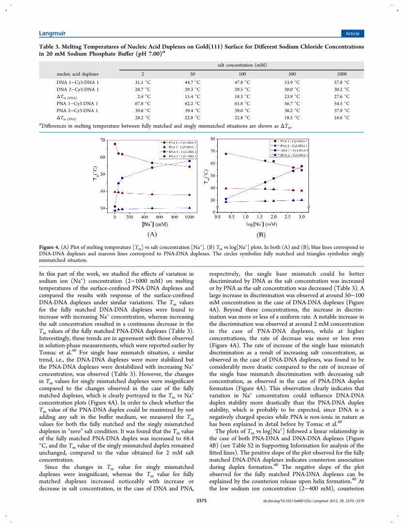
In this part of the work, we studied the effects of variation insodium ion (Na+) concentration (2−1000 mM) on meltingtemperatures of the surface-confined PNA-DNA duplexes andcompared the results with response of the surface-confinedDNA-DNA duplexes under similar variations. The Tm valuesfor the fully matched DNA-DNA duplexes were found toincrease with increasing Na+ concentration, whereas increasingthe salt concentration resulted in a continuous decrease in theTm values of the fully matched PNA-DNA duplexes (Table 3).Interestingly, these trends are in agreement with those observedin solution-phase measurements, which were reported earlier byTomac et al.40 For single base mismatch situation, a similartrend, i.e., the DNA-DNA duplexes were more stabilized butthe PNA-DNA duplexes were destabilized with increasing Na+
concentration, was observed (Table 3). However, the changesin Tm values for singly mismatched duplexes were insignificantcompared to the changes observed in the case of the fullymatched duplexes, which is clearly portrayed in the Tm vs Na+
concentration plots (Figure 4A). In order to check whether theTm value of the PNA-DNA duplex could be maximized by notadding any salt in the buffer medium, we measured the Tmvalues for both the fully matched and the singly mismatchedduplexes in “zero” salt condition. It was found that the Tm valueof the fully matched PNA-DNA duplex was increased to 68.4°C, and the Tm value of the singly mismatched duplex remainedunchanged, compared to the value obtained for 2 mM saltconcentration.Since the changes in Tm value for singly mismatched
duplexes were insignificant, whereas the Tm value for fullymatched duplexes increased noticeably with increase ordecrease in salt concentration, in the case of DNA and PNA,
respectively, the single base mismatch could be betterdiscriminated by DNA as the salt concentration was increasedor by PNA as the salt concentration was decreased (Table 3). Alarge increase in discrimination was observed at around 50−100mM concentration in the case of DNA-DNA duplexes (Figure4A). Beyond these concentrations, the increase in discrim-ination was more or less of a uniform rate. A notable increase inthe discrimination was observed at around 2 mM concentrationin the case of PNA-DNA duplexes, while at higherconcentrations, the rate of decrease was more or less even(Figure 4A). The rate of increase of the single base mismatchdiscrimination as a result of increasing salt concentration, asobserved in the case of DNA-DNA duplexes, was found to beconsiderably more drastic compared to the rate of increase ofthe single base mismatch discrimination with decreasing saltconcentration, as observed in the case of PNA-DNA duplexformation (Figure 4A). This observation clearly indicates thatvariation in Na+ concentration could influence DNA-DNAduplex stability more drastically than the PNA-DNA duplexstability, which is probably to be expected, since DNA is anegatively charged species while PNA is non-ionic in nature ashas been explained in detail before by Tomac et al.40
The plots of Tm vs log[Na+] followed a linear relationship inthe case of both PNA-DNA and DNA-DNA duplexes (Figure4B) (see Table S2 in Supporting Information for analysis of thefitted lines). The positive slope of the plot observed for the fullymatched DNA-DNA duplexes indicates counterion associationduring duplex formation.40 The negative slope of the plotobserved for the fully matched PNA-DNA duplexes can beexplained by the counterion release upon helix formation.40 Atthe low sodium ion concentration (2−400 mM), counterion
Table 3. Melting Temperatures of Nucleic Acid Duplexes on Gold(111) Surface for Different Sodium Chloride Concentrationsin 20 mM Sodium Phosphate Buffer (pH 7.00)a
salt concentration (mM)
nucleic acid duplexes 2 50 100 500 1000
DNA 1−Cy3-DNA 1 31.1 °C 44.7 °C 47.8 °C 53.9 °C 57.8 °CDNA 2−Cy3-DNA 1 28.7 °C 29.3 °C 29.5 °C 30.0 °C 30.2 °CΔTm (DNA) 2.4 °C 15.4 °C 18.3 °C 23.9 °C 27.6 °CPNA 1−Cy3-DNA 1 67.8 °C 62.2 °C 61.8 °C 56.7 °C 54.5 °CPNA 2−Cy3-DNA 1 39.6 °C 39.4 °C 39.0 °C 38.2 °C 37.9 °CΔTm (PNA) 28.2 °C 22.8 °C 22.8 °C 18.5 °C 16.6 °C
aDifferences in melting temperature between fully matched and singly mismatched situations are shown as ΔTm.
Figure 4. (A) Plot of melting temperature [Tm] vs salt concentration [Na+]. (B) Tm vs log[Na+] plots. In both (A) and (B), blue lines correspond to
DNA-DNA duplexes and maroon lines correspond to PNA-DNA duplexes. The circles symbolize fully matched and triangles symbolize singlymismatched situation.
Langmuir Article
dx.doi.org/10.1021/la400125x | Langmuir 2013, 29, 3370−33793375

release/uptake between the single-stranded and duplex statewas greater, but at the moderate or high salt concentration(>400 mM), a significantly small amount of counterion release/uptake occurred upon duplex formation (Figure 4A). This is inagreement with the “salting out” effect, where the watermolecules that are involved in interaction with the peptidiclinkages at low salt concentrations get increasingly moreinvolved in interacting with the salt ions as the saltconcentration increases, thereby assisting in increasing PNA-PNA association. This enhanced interaction between the PNAstrands of different PNA-DNA duplexes could reduce the basestacking interactions within a PNA-DNA duplex leading todestabilization of the duplex and therefore decrease the Tmvalue as the salt concentration was increased.The melted fractions ( f) of the single PNA/DNA strands
against temperature (T) were obtained by fitting the meltingprofile in two-state transition model that assumes that only twospecies (fully associated and fully dissociated) are present(Figure 5). This model is often accurate for short
oligonuceotides, i.e., less than 14mer oligonucleotides.41,42
From the plots, it is clearly observed that the melting transitionin the case of PNA-DNA duplexes was more abrupt, since ittook place within a much narrower range of temperature ofabout 6.5 °C, compared to the transition in the case of DNA-DNA duplexes that took place within the range of about 12 °C.The narrower melting transition observed in the case of
PNA-DNA duplexes could be due to a greater extent ofconformational homogeneity present in the PNA-DNA duplexpopulation compared to the DNA-DNA duplexes. Therelatively restricted conformational flexibility of PNA-DNAduplexes compared to that of the DNA-DNA duplexes could berelated to the fact that the PNA strands are less flexible thanDNA strands due to the presence of planar amide groups in its
polyamide backbone.14,43,44 In fact, in a study on moleculardynamics simulations of single-stranded PNA, DNA, and RNAin aqueous solvent, it has been revealed that fluctuations in theend-to-end distance of the oligomers are the least in the case ofPNA strand, followed by DNA strand, and the highest in thecase of RNA strand, making PNA globally less flexible thanDNA and RNA.43 Experimentally, it has been shown by single-wavelength anomalous diffraction experiments that the doublehelix flexibility of the PNA strand is rather restricted despite theabsence of cyclic moieties.44 The fact that the melting transitionin the case of PNA-DNA duplexes was sharper than thetransition for DNA-DNA duplexes means that the free energychange for the PNA-DNA duplex dehybridization is moretemperature dependent. A sharper transition also indicates thatthe affinity constant would be more strongly temperaturedependent. Consequently, at the physiologically relevanttemperature, which is 37 °C, the PNA-DNA duplexes areexpected to be more stable than the DNA-DNA duplexes.42
Effects of Salt Concentration Variation on “Solution”Melting Behavior of PNA-DNA Duplexes. The measure-ments carried out at the physiologically relevant saltconcentration of 100 mM revealed monophasic profiles,which is characteristic of melting of duplexes (see Figures S5and S6 in Supporting Information). It was also observed thatheating and cooling curves were reversible, indicating that thetransitions were at equilibrium (see Figure S6 in SupportingInformation). The PNA-DNA duplexes were found to beconsiderably more stable than the corresponding DNA-DNAduplexes (Table 2), which is consistent with the previousfindings.40 Single base mismatch discrimination was also foundto be better performed by the PNA sensor probes than theDNA sensor probes, as shown earlier.40
The Tm values of the PNA-DNA duplexes (fully matched aswell as singly mismatched) obtained for other salt concen-trations, i.e., 20/50/500/1000 mM, in sodium phosphate buffer(20 mM sodium phosphate, x mM sodium chloride, pH 7.00),clearly indicate a distinctly different conduct (Table 4)compared to the “on-surface” behavior (Table 3). While thevariation in Tm values of the singly mismatched PNA-DNAduplexes with changes in salt concentration (2 to 1000 mM)was found to be quite small (approximately 1.5 °C) in the caseof “on-surface” measurement, the variation in Tm for a similarchange in salt concentration was found to be approximately 5.0°C in the case of solution measurement. Interestingly, thevariation in Tm of the fully matched PNA-DNA duplexes wasfound to be more (approximately 13.0 °C) in the case of “on-surface” measurement compared to the variation observed(approximately 9.0 °C) in the case of solution measurement.Effectively, mismatch discrimination by the PNA probes couldappreciably be better performed on surface than in solution forall the salt concentrations applied (see Tables 3 and 4).
Figure 5. Typical melted fraction ( f) vs temperature curves, wherehybridization buffer is 20 mM sodium phosphate, 100 mM sodiumchloride, pH 7.00. Filled circles correspond to DNA-DNA duplexesand empty circles correspond to PNA-DNA duplexes.
Table 4. Melting Temperatures of PNA-DNA Duplexes in Solution for Different Sodium Chloride Concentrations in 20 mMSodium Phosphate Buffer (pH 7.0)a
NaCl conc. (mM)
nucleic acid duplexes 2 50 100 500 1000
PNA 1−T-DNA 1 55.6 °C 54.3 °C 52.2 °C 47.4 °C 46.2 °CPNA 2−T-DNA 1 40.4 °C 40.2 °C 39.1 °C 36.1 °C 35.5 °CΔTm 15.2 °C 14.1 °C 13.1 °C 11.3 °C 10.7 °C
aDifferences in melting temperature between fully matched and singly mismatched situations are shown as ΔTm.
Langmuir Article
dx.doi.org/10.1021/la400125x | Langmuir 2013, 29, 3370−33793376

Effects of Variation in Nature of Salt Components onthe Melting Behavior of PNA-DNA Duplexes onGold(111) Surface. Since the nature of the ions is knownto affect the thermal stability of specific conformations ofproteins/nucleic acids, we measured the Tm values of PNA-DNA duplexes for different types of salt components, which aremonovalent anions CH3COO
−, Cl−, NO3−, and divalent anion
SO42−, for the fixed countercation, i.e., Na+, and monovalent
cation NMe4+, having Cl− as the counteranion. An increasingly
destabilizing effect was observed in the case of the fullymatched duplexes as the anion was changed from CH3COO
−
to Cl− and NO3− (Table 5), in agreement with the solution
phase behavior as reported earlier by Tomac et al.40 No distinctdifferences could however be observed in the case of the singlymismatched duplexes (Table 5). Interestingly, the divalentanion SO4
2−, which is expected to exert the least destabilizingeffect due to its terminal position in the Hofmeister series(cation series: Mg2+ > Li+ > Na+ > K+ > NH4
+; anion series:SO4
2− > HPO42− > CH3COO
− > Cl− > Br− > NO3− > I− >
ClO4− > SCN−)40,45,46 induced almost similar destabilizing
effects as NO3− (Table 5). The destabilizing effect of SO4
2− onthe singly mismatched duplex was rather prominent, incomparison to the effects of the monovalent anions (Table 5).The cation NMe4
+ was selected on the basis of the very highdegree of nondenaturing (or stabilizing) effect it can exert onthe nucleic acid structures in the solution phase.45 In our study,NMe4
+ was found to stabilize the fully matched PNA-DNAduplexes better than Na+, at the minimum concentration, i.e., 2mM, and more prominently at the maximum concentration, i.e.,1000 mM (Tables 3 and 5). The singly mismatched duplexeswere found to be much less affected and the Tm values werealmost the same within the total concentration range of 2−1000 mM for both Na+ and NMe4
+ (Tables 3 and 5). Thestabilizing influence of NMe4
+ was also reflected in theobservations that the Tm value was reduced by only 2.5 °C asthe salt concentration was increased from 2 mM to 1000 mM(Table 5), while in the case of Na+, the reduction in Tm for asimilar increase in salt concentration was 13.3 °C (Table 3).The Hofmeister series, which is considered to be a predictive
guide for the stabilizing versus destabilizing effects of particularanions, is likely to be better followed at high salt concentrations(beyond 1000 mM), where the electrostatic contributionssaturate,47 since this series is based on lyotropic propensities ofa material which are most pronounced when there is a waterstructure rearrangement at the water−solute interface as aresult of high salt concentration.45,46 In the case of PNA-DNAduplex formation in solution phase, it has been observed thatthe Hofmeister series of anions is clearly reflected in the Tmvalues, whereas the cation series has little or no influence on theTm values.40 The present observation that the duplexesresponded to anion variations at a moderate salt concentrationof 100 mM could be due to the fact that, in the present study,we employed a close-packed PNA assembly formed at the
gold−water interface, where the water content and the waterarrangement around the duplexes would be different than thatin the solution phase. This argument would be especiallyrelevant considering the fact that the hydrophobic effects thatare exerted by PNA due to the presence of a non-ionic peptidicbackbone in its structure will be more prominent when thePNA strands are closely spaced, as in the case of a close-packedfilm.
■ CONCLUSIONIn conclusion, mismatch discrimination ability of surface-anchored PNA probes could be successfully enhanced via ionicadjustments, i.e., by varying salt concentration and the type ofcounterion. While the nature of ionic dependence of “on-surface” behavior of PNA probes deviates significantly from the“solution” behavior of these probes, e.g., in the case of thesingly mismatched duplexes, considerable similarities were alsoobserved, e.g., in the case of the fully mismatched duplexes. Wenot only provide an effective means for improving performanceof the PNA probes, but also discuss a strategy for measurementof “on-surface” melting temperature values of PNA-DNAduplexesthe latter contribution requiring further refinementthough to obtain accurate melting temperature values.Considering the need for developing more sensitive, target-specific, and robust high-throughput array technologies, PNA-based nucleic acid detection assays, as presented in this report,could offer practical inputs in achieving better control on theon-surface DNA detection capabilities.
■ ASSOCIATED CONTENT*S Supporting InformationFluorescence images of PNA-Cy3-DNA duplexes on gold(111)surface at different heating stages, results of control experi-ments, solution melting curves, and information on rehybrid-ization efficiency of the PNA film and the DNA film.Thismaterial is available free of charge via the Internet at http://pubs.acs.org.
■ AUTHOR INFORMATIONCorresponding Author*E-mail: [email protected] authors declare no competing financial interest.
■ ACKNOWLEDGMENTSWe gratefully acknowledge the financial support (Grant No.BT/PR-11765/MED/32/107/2009) from Department ofBiotechnology, Govt. of India, and (Grant No. SR/FTP/CS-66/2006) from SERC Fast Track scheme of Department ofScience and Technology, Govt. of India; and the researchfellowships of S.G., S.M., and T.B. from the Council ofScientific and Industrial Research, Govt. of India.
Table 5. Melting Temperatures of PNA-DNA Duplexes on Gold(111) Surface Varying the Anion (salt concentration 100 mM)and Cation in 20 mM Sodium Phosphate Buffer (pH 7.00)
salt
monovalent anion divalent anion monovalent cation
nucleic acid duplexes NaOCOCH3 NaCl NaNO3 Na2SO4 NMe4Cl (0 mM) NMe4Cl (2 mM) NMe4Cl (1000 mM)
PNA 1−Cy3-DNA 1 62.9 °C 61.8 °C 54.7 °C 55.3 °C 68.4 °C 68.4 °C 65.9 °CPNA 2−Cy3-DNA 1 39.3 °C 39.0 °C 39.6 °C 35.1 °C 39.6 °C 39.4 °C 38.9 °CΔTm 23.6 °C 22.8 °C 15.1 °C 20.2 °C 28.8 °C 29.0 °C 27.0 °C
Langmuir Article
dx.doi.org/10.1021/la400125x | Langmuir 2013, 29, 3370−33793377
■ REFERENCES(1) Nielsen, P. E.; Egholm, M.; Berg, R. H.; Buchardt, O. Sequenceselective recognition of DNA by strand displacement with a thyminesubstituted polyamide. Science 1991, 254, 1497−1500.(2) Ghosh, S.; Mukhopadhyay, R. An atomic force microscopyinvestigation on self-assembled peptide nucleic acid structures ongold(111) surface. J. Colloid Interface Sci. 2011, 360, 52−60.(3) Wang, H.; Tang, Z.; Li, Z.; Wang, E. Self-assembled monolayer ofssDNA on Au(111) substrate. Surf. Sci. 2011, 480, L389−L394.(4) Herne, T. M.; Tarlov, M. J. Characterization of DNA probesimmobilized on gold surfaces. J. Am. Chem. Soc. 1997, 119, 8916−8920.(5) Egholm, M.; Buchardt, O.; Christensen, L.; Behrens, C.; Freier, S.M.; Driver, D. A.; Berg, R. H.; Kim, S. K.; Norden, B.; Nielsen, P. E.PNA hybridizes to complementary oligonucleotides obeying theWatson-Crick hydrogen-bonding rules. Nature 1993, 365, 566−568.(6) Wittung, P.; Nielsen, P. E.; Buchardt, O.; Egholm, M.; Norden, B.DNA-like double helix formed by peptide nucleic acid. Nature 1994,368, 561−563.(7) Leijon, M.; Gras̈lund, A.; Nielsen, P. E.; Buchardt, O.; Norden,B.; Kristensen, S. M.; Eriksson, M. Structural characterization of PNA-DNA duplexes by NMR. Evidence for DNA in a B-like conformation.Biochemistry 1994, 33, 9820−9825.(8) Carlsson, C.; Jonsson, M.; Norden, B.; Dulay, M. T.; Zare, R. N.;Noolandi, J.; Nielsen, P. E.; Tsui, L. C.; Zielenski, J. Screening forgenetic mutations. Nature 1996, 380, 207.(9) Levicky, R.; Herne, T. M.; Tarlov, M. J.; Satija, S. K. Using self-assembly to control the structure of DNA monolayers on gold: aneutron reflectivity study. J. Am. Chem. Soc. 1998, 120, 9787−9792.(10) Peterson, A. W.; Heaton, R. J.; Georgiadis, R. Kinetic control ofhybridization in surface immobilized DNA monolayer films. J. Am.Chem. Soc. 2000, 122, 7837−7838.(11) Peterson, A. W.; Heaton, R. J.; Georgiadis, R. The effect ofsurface probe density on DNA hybridization. Nucleic Acids Res. 2001,29, 5163−5168.(12) Harris, N. C.; Kiang, C. H. Defects can increase the meltingtemperature of DNA−nanoparticle assemblies. J. Phys. Chem. B 2006,110, 16393−16396.(13) Nasef, H.; Ozalp, V. C.; Beni, V.; O’Sullivan, C. K. Meltingtemperature of surface tethered DNA. Anal. Biochem. 2006, 406, 34−40.(14) Briones, C.; Marti, E. M.; Navarrob, G. C.; Parroa, V.; Romanb,E.; Gago, J . A. M. Ordered self-assembled monolayers of peptidenucleic acids with DNA recognition capability. Phys. Rev. Lett. 2004,93, 2081031−2081034.(15) Marti, E. M.; Briones, C.; Pradier, C. M.; Gago, J. A. M. A DNAbiosensor based on peptide nucleic acids on gold surfaces. Biosens.Bioelectron. 2007, 22, 1926−1932.(16) Calabretta, A.; Wasserberg, D.; Trumpie, G. A. P.;Subramaniam, V.; Amerongen, A. V.; Corradini, R.; Tedeschi, R.;Sforza, S.; Reinhoudt, D. N.; Marchelli, R.; Huskens, J.; Jonkheijm, P.Patterning of peptide nucleic acids using reactive microcontactprinting. Langmuir 2011, 27, 1536−1542.(17) Wittung, P.; Rodahl, M.; Kasemo, B.; Nielsen, P. E.; Norden, B.Detection of point mutations in DNA by PNA-based quartz-crystalbiosensor. Colloids Surf., A: Physicochem. Eng. Aspects 2000, 174, 269−273.(18) Kambhampati, W.; Nielsen, P. E.; Knoll, W. Investigating thekinetics of DNA−DNA and PNA−DNA interactions using surfaceplasmon resonance-enhanced fluorescence spectroscopy. Biosens.Bioelectron. 2001, 16, 1109−1118.(19) Liu, J.; Tian, S.; Nielsen, P. E.; Knoll, W. In situ hybridization ofPNA/DNA studied label-free by electrochemical impedance spectros-copy. Chem. Commun. 2005, 2969−2971.(20) Xu, F.; Pellino, A. M.; Knoll, W. Electrostatic repulsion andsteric hindrance effects of surface probe density on deoxyribonucleicacid (DNA)/peptide nucleic acid (PNA) hybridization. Thin SolidFilms 2008, 516, 8634−8639.
(21) Park, H.; Germini, S.; Sforza, S.; Corradini, R.; Marchelli, R.;Knoll, W. Kinetic and affinity analyses of hybridization reactionsbetween peptide nucleic acid probes and DNA targets using surfacePlasmon field-enhanced fluorescence spectroscopy. Biointerphases2006, 1, 113−122.(22) Park, H.; Germini, A.; Sforza, S.; Corradini, R.; Marchelli, R.;Knoll, W. Effect of ionic strength on PNA-DNA hybridization onsurfaces and in solution. Biointerphases 2007, 2, 80−88.(23) Jensen, K. K.; Orum, H.; Nielsen, P. E.; Norden, B. Kinetics forhybridization of peptide nucleic acids (PNA) with DNA and RNAstudied with the BIAcore technique. Biochemistry 1997, 36, 5072−5077.(24) Ratilainen, T.; Holmen, A.; Tuite, E.; Haaima, G.; Christensen,L.; Nielsen, P. E.; Norden, B. Hybridization of Peptide Nucleic Acid.Biochemistry 1998, 37, 12331−12342.(25) Ratilainen, T.; Holmen, A.; Tuite, E.; Nielsen, P. E.; Norden, B.Thermodynamics of sequence-specific binding of PNA to DNA.Biochemistry 2000, 39, 7781−7791.(26) Wittung, P.; Nielsen, P. E.; Buchardt, O.; Egholm, M.; Norden,B. DNA-like double helix formed by peptide nucleic acid. Nature 1994,368, 561−563.(27) Ratilainen, T.; Holmen, A.; Tuite, E.; Nielsen, P. E.; Norden, B.Thermodynamics of sequence-specific binding of PNA to DNA.Biochemistry 2000, 39, 7781−7791.(28) Nuzzo, R. G.; Allara, D. L. Adsorption of bifunctional organicdisulfides on gold surfaces. J. Am. Chem. Soc. 1983, 105, 4481−4483.(29) Owczarzy, R. Melting temperatures of nucleic acids:Discrepancies in analysis. Biophys. Chem. 2005, 117, 207−215.(30) Proll, F.; Mohrle, B.; Kumpf, M.; Gauglitz, G. Label-freecharacterisation of oligonucleotide hybridisation using reflectometricinterference spectroscopy. Anal. Bioanal. Chem. 2005, 382, 1889−1894.(31) Anshelevich, V. V.; Vologodskii, A. V.; Lukashin, A. V.; Frank-Kamenetskii, M. D. Slow relaxational processes in the melting of linearbiopolymers: a theory and its application to nucleic acids. Biopolymers1984, 23, 39−58.(32) Wartell, R. M.; Benight, A. S. Thermal denaturation of DNAmolecules: A comparison of theory with experiment. Phys. Rep. 1985,126, 67−107.(33) Wang, J. Survey and summary: From DNA biosensors to genechips. Nucleic Acids Res. 2000, 28, 3011−3016.(34) Drummond, T. G.; Hill, M. G.; Barton, J. K. ElectrochemicalDNA sensors. Nat. Biotechnol. 2003, 21, 1192−1199.(35) Eriksson, M.; Nielsen, P. E. Solution structure of a peptidenucleic acid−DNA duplex. Nat. Struct. Biol. 1996, 3, 410−413.(36) Schmitt, T. J.; Knotts, T. A. Thermodynamics of DNAhybridization on surfaces. J. Chem. Phys. 2011, 134, 205105−205109.(37) Soares, D. M.; Tenan, M. A.; Gomide, A. B.; Gomes, W. E.Physical properties of water near a gold surface: a nanorheologicalanalysis. ChemPhysChem 2010, 11, 905−911.(38) Smith, T. The hydrophilic nature of a clean gold surface. J.Colloid Interface Sci. 1980, 75, 51−55.(39) Zheng, J.; Chin, W. C.; Khijniak, E.; Khijniak, E., Jr.; Pollack, G.H. Surfaces and interfacial water: Evidence that hydrophilic surfaceshave long-range impact. Adv. Colloid Interface Sci. 2006, 127, 19−27.(40) Tomac, S.; Sarkar, M.; Ratilainen, T.; Wittung, P.; Nielsen, P. E.;Norden, B.; Graslund, A. Ionic effects on the stability andconformation of peptide nucleic acid complexes. J. Am. Chem. Soc.1996, 118, 5544−5552.(41) Breslauer, K. J.; Sturtevant, J. M.; Tinoco, I. Calorimetric andspectroscopic investigation of the helix-to-coil transition of a ribo-oligonucleotide: rA7U7. J. Mol. Biol. 1975, 99, 549−565.(42) Mergny, J. L.; Lacroix, L. Analysis of thermal melting curves.Oligonucleotides 2003, 13, 515−537.(43) Sen, S.; Nilsson, L. MD simulations of homomorphous PNA,DNA, and RNA single strands: characterization and comparison ofconformations and dynamics. J. Am. Chem. Soc. 2001, 123, 7414−7422.
Langmuir Article
dx.doi.org/10.1021/la400125x | Langmuir 2013, 29, 3370−33793378

(44) Menchise, V.; Simone, G. D.; Tedeschi, T.; Corradini, R.;Sforza, S.; Marchelli, R.; Capasso, D.; Saviano, M.; Pedone, C. Insightsinto peptide nucleic acid (PNA) structural features: the crystalstructure of a D-lysine-based chiral PNA−DNA duplex. Proc. NatlAcad. Sci. U.S.A. 2003, 100, 12021−12026.(45) Hamaguchi, K.; Geiduschek, E. P. The effect of electrolytes onthe stability of the deoxyribonucleate helix. J. Am. Chem. Soc. 1962, 84,1329−1338.(46) Collins, K. D.; Washabaugh, M. D. The Hofmeister effect andthe behaviour of water at interfaces. Q. Rev. Biophys. 1985, 18, 323−422.(47) Arakawa, T.; Timasheff, S. N. Mechanism of protein salting inand salting out by divalent cation salts: balance between hydration andsalt binding. Biochemistry 1984, 23, 5912−5923.
Langmuir Article
dx.doi.org/10.1021/la400125x | Langmuir 2013, 29, 3370−33793379


![Rudder Design PNA[1]](https://img.pdfslide.net/doc/110x75/547ac40ab37959652b8b4af1/rudder-design-pna1.jpg)
