Embed Size (px)
Citation preview

1
Faculty of Medicine Ramathibodi Hospital,
Mahidol University
RACE 699 Dissertation
Doctor of Philosophy Programme in Clinical Epidemiology
(International Programme)
Title: Ramathibodi Appendicitis Score (RAMA-AS): A useful tool for
diagnosis of appendicitis
Name student Chumpon Wilasrusmee, MD
ID RACE/D 5336192

2
CONTENTS
CHAPTER 1 .............................................................................................................................. 811
BACKGROUND & RATIONALE .......................................................................................... 811
1.1 Research Questions ...................................................................................................... 912
1.2 Research Objectives ..................................................................................................... 912
CHAPTER 2 ............................................................................................................................ 1114
LITERATURE REVIEW ...................................................................................................... 1114
2.2 Definitions ................................................................................................................. 2528
2.2.1 Appendicitis . ........................................................................................................... 2528
2.2.2 Migration of pain ...................................................................................................... 2528
2.2.3 Pain aggravated by coughing .................................................................................. 2528
2.2.4 Rebound tenderness. ................................................................................................ 2528
2.2.5 Rigidity. ..................................................................................................................... 2528
2.2.6 Abdominal pain ........................................................................................................ 2528
2.2.7 Vomiting. ................................................................................................................... 2528
2.2.8 Polymorphonuclear leukocytosis ............................................................................ 2528
2.2.9 Rovsing sign .............................................................................................................. 2528
2.2.10 Appendectomy .......................................................................................................... 2528
2.2.11 Negative appendectomy ........................................................................................... 2629
2.3 Conceptual framework ................................................................................................. 2730
CHAPTER 3 ............................................................................................................................ 3033
METHODOLOGY ................................................................................................................. 3033
3.1 Study design and setting .......................................................................................... 3033
3.2 Study subjects ........................................................................................................... 3134
3.3 Variables and Measurement ................................................................................... 3134
3.4 Data Collection ......................................................................................................... 3538

3
3.5 Sample size estimation ............................................................................................. 3639
3.6 Cost effectiveness analysis of RAMA-AS ............................................................... 4144
3.7 Implementation of RAMA-AS and impact analysis .............................................. 4346
3.8 Ethics considerations ................................................................................................ 4447
3.9 Budget ........................................................................................................................ 4750
3.11 Time Frame ................................................................................................................ 4750
ACKNOWLEDGEMENT ...................................................................................................... 4851
TABLES ................................................................................................................................... 5558
A) Case record forms .................................................................................................... 7982
D) Dummy tables ....................................................................................................... 102106
B) Informal sheet & consent form ........................................................................... 108112
4
TABLE OF CONTENTS
Table 1. Characteristics of studies that had developed prediction scores for
appendicitis…………………………………………………………………………54
Table 2. Risk of bias assessment……………………………………………………61
Table 3. Describe performances of predictive models of appendicitis……………..64
Table4. Describe variables that were included in the derive phase of predictive scores
of appendicitis………………………………………………………………………67
Table 5 Data from pilot study in 60 patients who was consulted for management of
abdominal pain and suspected of appendicitis at surgical unit of Ramathibodi
Hospital………………………………………………………………………………69
Table 6 Risk of bias assessment for clinical prediction rule of appendicitis……..…70
Table 7 Sample size estimation…………………………………………….……….75

5
FIGURE OF CONTENTS
Figure 1: Identification of studies for inclusion……………………………………..77
Figure 2 Flow of patients and process of data collection……………………………78
Figure 3 Diagram of data analysis…………………………………………………..79
Figure 4 Decision tree of the RAMAAS model in diagnosis of appendicitis………80

6
TITLE RAMATHIBODI APPENDICITIS SCORE (RAMA-AS): A
USEFUL TOOL FOR DIAGNOSIS OF APPENDICITIS
Chumpon Wilasrusmee, MD
Section of Clinical Epidemiology and Biostatistic, Ramathibodi Hospital
Department of Surgery, Faculty of Medicine Ramathibodi Hospital, Mahidol
University
Email: [email protected]
Supervisors
Ammarin Thakkinstian, Ph.D.
Section for clinical Epidemiology and Biostatistics, Faculty of Medicine,
Ramathibodi Hospital, Mahidol University, Bangkok
Email: [email protected]
Assist. Professor Dr. Patarawan Woratanarat
Department of Orthopedics, Faculty of Medicine Ramathibodi Hospital,
Mahidol University
Section for clinical Epidemiology and Biostatistics, Faculty of Medicine,
Ramathibodi Hospital, Mahidol University, Bangkok
Email: [email protected]

7
Panuwat Lertsitthichai, MD
Department of Surgery, Faculty of Medicine Ramathibodi Hospital, Mahidol
University
Email: [email protected]
8
CHAPTER 1
BACKGROUND & RATIONALE
Appendicitis is one of the most important clinical causes among acute abdominal
pain, with an incidence of 110/100,0001. Although many attempts have been made to
improve the diagnostic accuracy, false positive and false negative rates remain
common with rates of negative appendectomy of 15% to 26%2 3 and perforated
appendectomy of 10% to 30%4.
Several scoring systems have been developed for diagnosis of appendicitis with
interesting results nevertheless these systems have been less routinely applied in
general practice. We therefore performed a systematically review how those scores
had been developed and validated, and how their performances were. This review of
14 relevant studies suggested that 5 to 14 variables were included in the models with
the common variables were migration of pain, nausea/vomiting, and duration of pain
for clinical signs; rebound tenderness, RLQ tenderness, and elevated temperature for
symptoms. Most studies (64.3%) created prediction scores based on univariate results
or non-statistical models. The discrimination coefficient C statistics were 0.79 (95%
CI: 0.67, 0.90) for the derive studies and 0.84(0.77, 0.92) for the internal validation
studies. In addition, the pooled C statistics were 0.74 (95%CI, 0.69, 0.79) for external
validation of Alvarado scores. Our review suggested that qualities of research
methods for scoring systems of appendicitis were varied.
Although there are several diagnostic scoring systems available, applying them to the
general population might be questionable due to improper methods used for creating
scores. The more appropriate scores with internal and external validations are still

9
required. We therefore conduct a cross-sectional study with aims at developing
Ramathibodi appendic score (RAMA-AS) using proper methods for creating and
validating a good clinical decision rule (CDR) in diagnosis of appendicitis.
1.1 Research Questions
Primary research question
1.1.1 What are variables contained in the RAMA-AS and how to calculate the
RAMA-AS?
1.1.2 Can the RAMA-AS perform well in classifying appendicitis from non-
appendicitis patients?
1.1.3 Will the RAMA-AS work well in internal and external settings?
Secondary research questions
1.1.4 Does our RAMA-AS work better than previous CDRs in diagnosis of
appendicitis?
1.1.5 Is our RAMA-AS cost-effective when compared with imaging diagnostic
including ultrasonography (US), computerized tomography (CT), and
magnetic resonance imaging (MRI) and diagnostic laparoscopy?
1.1.6 Will the RAMA-AS be used by physicians’ and help them to improve
outcomes in the diagnosis of appendicitis?
1.2 Research Objectives
Primary Objectives

10
1.2.1 To develop a RAMA-AS for diagnosis of appendicitis.
1.2.2 To internally and externally validate RAMA-AS
Secondary objectives
1.2.3 To compare the performance of the RAMA-AS with previous scoring systems
including RIPASA score, Appendicitis Inflammatory response score, Alvarado
score, Fenyö-Lindberg score, Ohmann score, Eskelinen score, Simple scoring
system, Practical score of Ramirez and Deus, and Teicher score.
1.2.4 To compare the cost effectiveness of the RAMA-AS to diagnostic imaging
including US, CT, and MRI and diagnostic laparoscopy in the diagnosis of
appendicitis.
1.2.5 To develop RAMA-AS software which can simply classify appendicitis and
non-appendicitis patients.
1.2.6 To assess whether use of the RAMA-AS will be able to reduce diagnosis
imaging including US, CT, and MRI in real clinical practice.

11
CHAPTER 2
LITERATURE REVIEW
2.1 A Systematic review of scoring systems for diagnosis of appendicitis
Appendicitis is one of the most important clinical causes among acute
abdominal pain, with an incidence of 110/100,0001. Although many attempts have
been made to improve the diagnostic accuracy, false positive and false negative rates
remain common with rates of negative appendectomy of 15% to 26%2 3 and
perforated appendectomy of 10% to 30%4. Several scoring systems have been
included computer-based models and algorithms that had been developed with good
performances at the initial evaluation, but fair when applied to general populations.
Nevertheless, these scoring systems have been occasionally applied in general routine
practice, because of a lack of accuracy in validation studies5. The drawback of the
negative appendectomy (i.e., false positive) was less life threatening than a false
negative which could be as worse as mortality from appendiceal perforation and
peritonitis from a perforated appendicitis. As a result, the aggressive surgical
approach was frequently applied when the situation was in doubt which resulted in
removal of normal appendices. In order to reduce the aggressive management,
diagnostic tests for appendectomy are required to improve performance in
discriminating those patients who require prompt surgical intervention from the
patients who need only observation without a risk of complication of appendicitis.
Imaging modalities have been used to improve diagnostic accuracy. However,
there are some disadvantages including cost, less accessible particularly in developing
countries, lack of radiologists, examiner-dependent efficacy (e.g., ultrasound),
potential harmful ionization (e.g., computerized tomography, CT), and low
performance in low or high prevalence of disease. Clinical scoring systems by

12
synthesizing clinical information have been developed and should be useful for those
countries where imaging is less accessible. The scores are derived by incorporating
physical examination, clinical signs and symptoms in a mathematical equation.
Currently, there are a number of diagnostic scores constructed by many camps using
various statistical methods1 6-40. Some scores have been validated either internally8 39
or externally7-9 11 15 16 21 27 30 37-39 41 whereas some scores have been applied without
validation18 33. Performances of those scores varied from fair to good in validation
phases, but some scores were still questionable. We therefore conducted a systematic
review which aimed at exploring score performances in both development and
validation phases. Strengths and limitations of previous diagnostic scores were
critically appraised. Lessons from this review will help to identify the most valid
model/s or lead to create the new model if required. This model can be later applied in
general settings in developing countries where resources are limited.
Methods
Search strategy
We searched Medline from 1949 and EMBASE from 1974 to March 2012 to
identify relevant articles published in English. Search terms were included as follows:
appendicitis, gangrenous appendicitis, phlegmon, perforated appendicitis, abdominal
pain, score, scoring system, prediction score, prediction model, diagnostic score,
assessment tool, ultrasonogram, ultrasonography, computer tomography, accuracy,
negative appendectomy, sensitivity, specificity, likelihood ratio, false positive, false
negative, true positive, true negative, ROC, AUC. The search strategies are described
in the appendix.
13
Study selection
Studies were reviewed based on titles and abstracts. If a decision could not be
made, full articles were retrieved. Observational studies (cohort, case-control, or
cross-sectional) published in English were selected if they met with the following
criteria: suspected adult appendicitis, considered more than one risk factor in the
prediction score, had the outcome as appendicitis versus non-appendicitis, applied any
equation (e.g., Logistic regression, Bayesian method, or non-mathematical-
investigator opinion based) to build up the prediction model, and reported each
model’s performance (i.e., calibration and discrimination parameters).
Data extraction
The general characteristics of studies (i.e., author, journal, publication year,
type of participants, ethnicity, study design, number of subjects, rate of negative
appendectomy, percent of complicated appendicitis, and specific objective/s (i.e.,
develop or validate score, or both)) were extracted. If the diagnostic model was firstly
developed, specific information about model building (i.e., type of statistical model,
predictive factors, creating scores using coefficients or exponential of coefficients)
were extracted. Calibration (a ratio of expected versus observed value (E/O ratio)),
and discrimination parameters (i.e., the concordance statistic (C-statistic)) along with
95% confidence interval (CI) were also extracted. These parameters were calculated if
the study did not directly report, but did provide summary data which allowed for
calculations. For studies which aimed at a validated model, the type of validations
(internal, external, or both) and results were also recorded. If authors had modified
the previous prediction models, the following aspects were recorded: whether any of

14
the original included variables were removed or modified; and whether new predictive
factors were added.
Risk of bias assessment
The risk of bias assessment tool was developed based on a user’s guide for
clinical prediction rule42, which considered both derivation and validation phases.
Four domains were considered for the derivative phase, i.e., selection bias
(representative of spectrum), information bias (ascertainment of outcome
measurements, blinding outcome assessment, number of predictors, assessment
predictors without knowledge of outcome, proportion of important predictors),
confounding bias (used multi-variate regression analysis, created score properly), and
other issues (sample size, clinically sensible). For the validation phase, only 3
domains were considered, i.e., selection bias (representative of spectrum), information
bias (ascertainment of outcome measurement, blinded assessment of outcome,
accurate interpretation), and other issue (i.e., follow up). Each item was classified as
yes (low risk of bias), no (high risk of bias), and unclear if there was insufficient
information to judge (Table 1).
Two reviewers (CW and TA) had independently extracted data and assessed
risk of bias for all included studies. Any disagreement was discussed with the third
party (AT) to resolve.

15
Statistical analysis
Model performances were described separately by derivative and validation
phases. Calibration (O/E ratio) and discrimination (C-statistic) coefficients along with
their 95% CIs were estimated for each study. A meta-analysis was applied to pool O/E
and C-statistic using the equations as described in the appendix. Heterogeneity was
assessed using Q statistic and a degree of heterogeneity I2 was estimated. If it was
present (p value <0.10 or I2 > 25%), a random-effect model was used to pool data,
otherwise a fixed-effect model was applied. All analyses were performed using
STATA version 12.0.
Results
Description of studies
We identified 440 studies of which 37 studies met our inclusion criteria and
thus were eligible for the review, see Figure 1. The characteristics of these studies
have been described in Table 2. Among those 37, 10 studies7 11 15 16 18 21 27 28 37 43 had
aimed only at derived prediction scores or modified the previous prediction models
(hereafter called derived studies), 4 studies8 33 38 39 had derived and internally and
externally validated in the same studies, whereas 23 studies had only aimed at
internal9 30 and external1 6 10 12-14 17 19 20 22-26 29 31 32 34-36 40 validations.
Among 14 derived studies7 8 11 15 16 18 21 27 28 33 37-39 43, all studies focused on
adult patients, and most studies included patients with suspected appendicitis who
received operation or their conditions were being observed whereas 3 studies33 37 39
included only patients who received operations. Ten models7 8 11 15 16 18 27 33 37-39 43
were developed in Caucasian populations while three models11 21 28 were in Asian

16
populations. The models were mostly constructed based on cohorts either
retrospective7 33 39 or prospective cohorts.8 15 16 18 21 27 28 38 43
Among 23 studies that aimed only for validation, 20 studies had validated
models on patients with suspected appendicitis whereas 3 studies had focused on
operated patients. Most study designs were prospective cohorts. Fifteen studies were
done in Caucasian while 8 studies were done in Asian populations.

17
Risk of bias assessment
Risk of bias assessments was performed and results have been described in
Table 3. Among 14 derivative studies, 8/14 (57.1%) studies had recruited consecutive
patients with chief complaints of abdominal pain, or randomly sampled patients from
a well defined population frame of abdominal pain; whereas the remaining studies had
recruited a specific group of patients who had at least a few clinical signs and
symptoms. Most studies (92.9%) had confirmed the diagnosis of appendicitis by
histology without mention of whether histology was performed without blinding
clinical information. The numbers of predictors used in the prediction models were
covered and appropriated (i.e., low risk of bias) if authors considered and used
predictors from all categories which were demographic, clinical signs, symptoms, lab,
and imaging data; otherwise this item was graded as high risk of bias. Ten out of
fourteen (71.4%) studies clearly listed all categories of predictors where the remaining
studies considered only a few categories. Only 5/14 (35.71%) studies stated clearly
how they measured or collected predictors in the way that assessors were blinded
from knowledge of the final diagnosis of appendicitis, lab, and imaging findings,
whereas 57.14% of studies used predictors which were not blinded or assessed with
knowledge of possible diagnosis of appendicitis.
Eleven out of fourteen studies (78.7%) had performed statistical estimations or
tests for all predictors, whereas 3/14 (21.3%) studies did not apply any statistical
method. However, only 5/14 (35.7%) studies had applied multivariate regression by
simultaneously including significant predictors in the models, and used coefficients or
relative risks suggested from regression models to create scores, whereas the
remaining studies created prediction scores based on univariate results or non-
statistical models.

18
Twelve (85.7%) studies had sufficient numbers of subjects for either appendicitis or
total subjects considered based on a rule of thumb (1 predictor per 10 appendicitis
subjects or 20-30 per total subjects). Some studies (71.4%) included predictors that
seemed to be clinically sensible, the scores were easy to apply and also had suggested
a course of clinical action.
For validation studies, 21/25 (84%) studies were less likely for selection bias.
An ascertainment of diagnosis of appendicitis was clearly defined in 24/25 (96%)
studies. All studies did not mention whether diagnosis of appendicitis was masked
from clinical data. Thirteen out of twenty five (52%) studies clearly described that
interpretation of the rule was not influenced by information of final diagnosis of
appendicitis, while 24% was influenced by diagnosis of appendicitis and 24% did not
mention it. Only 6 (24%) studies had followed up all included patients.
Score development
Among 14 derivative studies, 5 categories of predictive variables were
considered in the models including demographic data, clinical signs, clinical
symptoms, laboratory results, and imaging (Table 4). Among 2 demographic
variables, gender was the more commonly included in the model compared with age
(42.9% vs 14.3%). Ten symptom variables were considered in which nausea (9/14,
64.3 %) was the most commonly included in the model followed with migration of
pain, pain at presentation, or duration of pain (all were 46.2%). Nine clinical signs
were considered and the most common variables used were rebound tenderness
(76.9%), followed with right lower quadrant (RLQ) tenderness (61.5%), and RLQ
guarding (53.9%) or elevated temperature (53.9%). Among 10 clinical symptoms,
nausea/vomiting (53.9%) followed with migration and duration of pain (46.4%) were

19
the most commonly included in the predictive models. Most studies (84.6%)
considered at least one lab variable. Among these, rising white blood cell count
(76.9%) was the most commonly used followed with left shift of polymorphonuclear
cell (46.2%). Only a few studies used radiological data (e.g. ultrasonography and
abdominal radiograph) in creating scoring systems.
As described in Table 2, these prediction scores were developed using
statistical modeling in 5 studies8 15 27 38 39 whereas 9 studies7 11 16 18 21 28 33 37 43 did not
apply statistical modeling. Among 5 studies with statistical modeling, 4 studies8 15 27
38 applied multivariate logistic regression and 1 study39 used discriminant analysis.
Scoring schemes of these models were created based on regression coefficients of the
logit or discriminant regression models. Among 9 studies that did not apply statistical
models, a univariate analysis (e.g., Chi-square test, relative risk) and estimated
diagnostic parameters (e.g., likelihood ratio, sensitivity, specificity) were used for
assessing associations in 6 studies whereas 3 studies did not apply any statistical
analysis tests.
Model performances
The models’ performances using C-statistics and O/E calibration coefficients
were extracted from individual studies, if reported, otherwise they were estimated
using summary data reported in the articles, see Table 5. Among 10 studies where the
calibration coefficient O/Es were available, the O/Es were very similar across studies
with the overall pooled O/E of 1 (95% CI: 0.97, 1.03). Contrastingly, the
discrimination coefficient C statistics varied from poor (0.54) to excellent (0.97)
discrimination with the pooled C statistic of 0.79 (95% CI: 0.67, 0.90). The C

20
statistics were very varied among 2 studies15 38 (i.e., ranged from 0.59 to 0.97) with
appropriate statistical methods to derive prediction scores.
Six out of 14 prediction models had internally validated their prediction
scores, but only 5 had data available. The discrimination coefficient C statistics
ranged from 0.61 to 0.92 with the pooled C statistic of 0.84 (0.77, 0.92). Pooling
within subgroups according to appropriateness of derived predictive scores suggested
similar results with the C statistics of 0.81 (95%CI = 0.65, 0.97) and 0.88 (95%CI =
0.85, 0.91) for appropriate and inappropriate derived predictive scores, respectively.
Twenty-three studies had been conducted which aimed at external validation
of 14 prediction models. The Alvarado score7 was frequently validated in 14 studies6 8
10 12 13 19 20 23 26 29 31 32 35 38 followed by Fenyo model in 3 studies14 17 38. The study by
Tzanakis et al38 had externally validated 8 previous models, and thus was a major
contributor of data in poolings. Most studies created diagnostic scores using
predictive factors according to the original scores. Data used for validations were 15
Caucasian1 12-14 17 20 22 23 25 29-31 34 40 43 and 8 Asian6 10 19 24 26 32 35 36 population studies.
Fourteen studies had externally validated Alvarado scores. All eight variables
(i.e., migration of pain, anorexia, nausea/vomiting, elevated temperature, rebound
tenderness, RLQ tenderness, increase WBC, and PMN left shift) were included in the
external validated models with the pooled E/O and pooled C-statistic of 0.99 (95%CI,
0.91 to 1.09) and 0.74 (95%CI, 0.69, 0.79), respectively. The Alvarado score was also
modified by two subsequent studies which excluded the shift to left of PMN because
this data was unavailable in a routine laboratory 21 41, or replaced it with a few other
variables (i.e. cough test, Rovsing’s sign, rectal tenderness)41.This made the score
performance change from 0.80 (95%CI, 0.73, 0.86) to 0.76 (95%CI, 0.60, 0.92) with
PMN exclude, and even worst for replacing PMN with a few more variables with the

21
C-statistic of 0.54 (95%CI, 0.45, 0.63). External validation of other scoring systems
was performed in 9 models with the pooled statistic of 0.81 (95%CI: 0.77, 0.84), but
this was mainly contributed to by Tzanakis, et al38 which had validated 7 models. (In
the above paragraph you stated 8 models.) Pooling external validated studies
according to appropriate and inappropriate original model construction resulted in the
C statistics of 0.80 (95% CI: 0.65, 0.94) and 0.77 (95%CI: 0.74, 0.81), respectively.
Discussion
We have reviewed performances of diagnostic models for appendicitis. Most
models yielded relatively fair to good performances in discrimination with the pooled
C-statistic of 0.84 (95%CI 0.77, 0.91) in settings where the models were developed
and 0.78 (95%CI 0.74, 0.82) in settings where the models were applied. However,
only one third of scores were appropriately derived based on regression models.
For those models with good to excellence external performances (C-statistic
≥0.8), 10 variables were commonly included in the models, which were migration of
pain, anorexia, nausea/vomiting, duration of pain, elevated temperature, rebound
tenderness, right lower quadrant tenderness, guarding, increased white blood cells,
and left shift of PMN. These models were originally developed using proper
statistical modeling (i.e., logistic regression) in only 2/23 studies whereas the rest had
used results of diagnostic parameters or univariate analysis (i.e., Chi-square test)
without proper rationale for weighting in prediction scores.
The Alvarado score , developed by Alvarado et al7 since 1986, was the most
popular prediction model for diagnosis of appendicitis which aimed at identifing
patients who should go to operation or observe. The model was originally developed
using data from 277 Caucasians to assess the association between 8 predictive factors

22
and appendicitis. These predictive factors included localized tenderness at the right
lower quadrant (RLQ) of abdomen, migration of pain, elevation of temperature,
nausea/vomiting, anorexia/acetone in urine, rebound tenderness at RLQ of abdomen,
leukocytosis, and shift to the left of polymorphonuclear cell (PMN). The diagnostic
parameters (i.e., sensitivity, specificity and accuracy) were estimated for each
individual predictive factor and used for creating the predictive score. The
discrimination ability C-statistic was 0.80 (95%CI, 0.73, 0.86) in the derivative phase
which dropped to 0.74 (95%CI, 0.69, 0.79) after external validation.
Since the PMN is unavailable in a routine laboratory 21 41, it was excluded
from the model which yielded a bit lower performance in the derived phases (C-
statistics 0.80 vs 0.76), but it was far worst if it was replaced with a few clinical
variables (i.e. cough test, Rovsing’s sign, rectal tenderness; C-statistics 0.80 vs 0.54).
The E/O ratio is commonly used to measure the closeness of the predicted and
the observed values. The C-statistic is usually applied to measure how well the model
will assign a higher probability of having an event to an appendicitis group and a
lower probability to a non-appendicitis group44. The association between diagnostic
factors and appendicitis derived from the derived data may occur by chance. This
problem is prominent in situations in which there is a relatively small sample size
compared with the diagnostic factors included in the model. With a small sample size,
it is more likely to select unimportant variables, but omit some important variables
from the model34. Conversely, a very large sample size is more likely to include
statistically important variables without clinical importance. The number of subjects
with events should be at least 10 and safer with 20 or larger per one risk factor to
derive a valid model as for simulation studies35 36. For the results of our review, the
number of variables included in the model varied from 4 to 18 variables, so the

23
required number of appendicitis cases should therefore be at least 40 to 180 subjects,
and 80 to 360 subjects to be safer. Six out of 14 derived studies9 11 18 21 27 37 had their
number of appendicitis cases less than the required numbers by including 5 to 14
variables with total appendicitis cases of 43 to 261 patients.
Although the performances of predictive scores in the derived phase, internal,
and external phases were good (C-statistic = 0.79, 0.84, and 0.78, respectively),
applying these scores to a general population was less confident because most scores
were created inappropriately. Most studies (64.3%) derived predictive scores using
univariate analyses or estimated diagnostic parameters and used results from these
analyses to create scores. The rationale of choosing a method for creating the
predictive score was not clearly described. In addition, these scores were based on
univariate analyses and thus confounding bias might be present.
Differences in the distribution of risk factors across populations may also
affect the generalizability of the model to different populations. The C-statistics
derived from external validation usually tend to be lower than the C-statistics derived
from internal validation. Our results suggested that the pooled C-statistic from
external validations had slightly lower C-statistic than from internal validation.
However, pooling of external validations was dominated by the Tzanakis, et al38
study which had used the same subjects to validate 9 scoring systems. Excluding this
study from pooling resulted in the C-statistic of 0.75 (95%CI 0.74, 0.82).
Our review suggested that the research methods and reporting of research
findings in diagnostic scoring systems of appendicitis showed discrepancy. Some
research groups have advocated and developed research methods and reporting
recommendations for conducting research in this area.45 46 In addition, a user’s guide
for reading and use of evidence for this area has been also developed42 to improve

24
research methods, reporting, and use of evidence of prediction scores. We have
modified and used this tool for our study. The type of studied subjects, study design,
validity of measurements for outcome and diagnostic factors, and use of statistical
methods were mainly reported in most of the model developments. However, only
50% (7/14) of the derived studies had developed scores and were internally tested in
the same studies. Most studies (64.3%) had created scores without applying statistical
modeling. None of the studies reported calibration parameters and only 9 (39.1%)
studies in external validation performed discrimination analysis and reported the C-
statistic. The models seemed to be clinically sensible in 71.4% (10/14 studies) which
were simple and easily interpretable.
In conclusion, although there are several diagnostic scoring systems for
appendicitis, applying them to a general population might be questionable due to
improper methods used for creating scores and lack of external validations. The more
appropriate scores with internal and external validations are still required.

25
2.2 Definitions
2.2.1 Appendicitis is inflammation of the appendix, which is the small, finger-
shaped pouch attached to the beginning of the large intestine on the lower-
right side of the abdomen. Appendicitis is a medical emergency, and if left
untreated, the appendix may rupture and cause a potentially fatal infection.
2.2.2 Migration of pain to the right lower quadrant pain starting either centrally
in the epigastric area, or in the whole abdomen then eventually migrating
down to the right lower abdomen.
2.2.3 Pain aggravated by coughing patient was asked to cough and any
worsening of pain was recorded.
2.2.4 Rebound tenderness elicited in the right lower quadrant when a hand
pressing the abdomen for 10-15 seconds was suddenly withdrawn.
2.2.5 Rigidity involuntary contraction of the abdominal muscles in the absence
of diagnostic evidence from an attribute.
2.2.6 Abdominal pain abdominal pain (not only right lower quadrant)
2.2.7 Vomiting one or more episodes.
2.2.8 Polymorphonuclear leukocytosis study as a total count 10,000/mm3
with polymorphs 75%.
2.2.9 Rovsing sign named by the Danish surgeon, Niels Thorkild Rovsing, and it
is a sign of suspected appendicitis. If palpation on the left lower abdominal
quadrant results in more pain in the right lower quadrant, the patients have
a positive Rovsing sign and may have appendicitis.
2.2.10 Appendectomy a surgical procedure to remove the appendix.

26
2.2.11 Negative appendectomy histological of normal appendix was found from
appendectomy that was done for the purpose of treatment after the
diagnosis of appendicitis.

27
2.3 Conceptual framework
RAMA-AS
Validation: Evidence of reproducible accuracy Internal (narrow) Validation External (broad) Validation
- Bootstap - Thammasat University Hospital - Chonburi Hospital
Introduce the software to RCST Evidence that the RAMA-AS: - Change physician practice -Improves patients outcomes - Reduces costs from imagings
Important Parameter - Demographic data: age, sex. - Symptom: migration of pain, anorexia, nausea. - Sign: elevated temperature, guarding of RLQ, rebound tenderness. - Lab: Increase WBC, PMN left shift, CRP.
Derivative phase: Identification of risk factors with appropriated model and predictive power
Economic analysis - Cost effectiveness - Cost utility
RAMA-AS Software development
Impact analysis
Pilot implementation - Faculty of Medicine Ramathibodi Hospital - Thammasat University Hospital

28
Clinical decision in the diagnosis of appendicitis is based on clinician experience
which is the integration of patient history, physical examination, and investigation.
Scoring system in diagnosis of appendicitis is a clinical decision rule which quantifies
the individual contributions that various parameters of patient history, physical
examination, and basic investigation results make toward the diagnosis of
appendicitis. This will pave the way to use a formal test, simplify, and increase the
accuracy of clinicians in the assessment of appendicitis. Development and validation
of RAMA-AS will involve 3 steps as shown: creating the clinical decision rule or
scoring system, validating the rule, and assessing the impact of the rule on clinical
behavior.
Derivative phase
The creating of a scoring system will involve the identification of parameters
with predictive power. All predictive parameter will be included and clearly
identified. Assessment of appendicitis will be done by a pathologist who is blinded to
the assessing parameters as well as the assessor for the parameters will be blinded
from the pathological diagnosis of appendicitis.
Validation phase
This phase will test the reproducible accuracy of RAMA-AS which will be
divided into 2 studies. Internal validation is a narrow validation which will apply the
RAMA-AS in a similar clinical setting and population as the derivative phase.
External validation is a broad variation which will apply the RAMA-AS in multiple
clinical settings with varying prevalence of appendicitis.
Impact analysis
This phase aims to find the evidence that RAMA-AS changes clinicians’
behavior and improves outcomes regarding the diagnosis of appendicitis.

29
Variables and appendicitis
Important parameters in the diagnosis of appendicitis will be divided into 4
groups: demographic data, symptoms, clinical signs, and laboratory results. Two
demographic variables that were commonly used in previously developed appendicitis
scoring systems were age and gender. Ten symptom variables were considered in
which nausea/vomiting, followed with migration and duration of pain were the most
commonly included in the predictive models. Nine clinical signs were considered with
the most common variable used was rebound tenderness, followed with right lower
quadrant (RLQ) tenderness and RLQ guarding or elevated temperature. Most of the
scoring systems considered at least one lab variable. Among these, rising white blood
cell count was the most common used followed with left shift of polymorphonuclear
cell.
All important parameters from previous systematic reviews will be included
in the derivative phase. Included predictive parameters in RAMA-AS must be
clinically sensible, easy to use, and suggest a course of action.

30
CHAPTER 3
METHODOLOGY
3.1 Study design and setting
The study will consist of 3 parts as follows:
Part I: Development and validation of RAMA-AS
Part II: Cost effectiveness analysis
Part III: Implementation of RAMA-AS
Part I: Development and validation of RAMA-AS
3.1.1 Study design and setting
The study design will be a cross-sectional study of patients who present with
abdominal pain and are suspected of appendicitis at the emergency department
between January 2013 to May 2014 will be included in this study.
Setting:
Development and internal validation of RAMA-AS: A Single center study
prospective cohort study in the Faculty of Medicine Ramathibodi Hospital,
Mahidol University from January 2013-May 2014.
External validation: Patients who were suspected to have appendicitis at the
Faculty of Medicine, Thammasart University and surgical unit at Chonburi
Hospital will be tested by using the RAMA-AS from June 2014-Decmber 2015
31
3.2 Study subjects
Inclusion criteria:
‐ Patients age 18-60 years
‐ Present at Emergency unit or Surgical outpatient department, Faculty of
Medicine Ramathibodi Hospital with right-side abdominal pain within 7 days
‐ Are suspected of having appendicitis by have at least one of the following
symptoms and signs:
o Symptoms: right lower abdominal pain, migration of abdominal pain,
anorexia, nausea, vomiting
o Signs: fever, right lower quadrant tenderness, guarding, rebound
tenderness, and decrease bowel sound
‐ Agree to participate and give consent to the study
Exclusion criteria
- Patients who cannot give the history of illness by themselves
- Patients who have severe underlying diseases and moribund such as severe
myocardial infarction and terminal illness
- Patients who have palpable abdominal mass
- Patients who are diagnosed as tumor or malignancy of appendix
- Patients who have metastatic tumor to the appendix
3.3 Variables and Measurement
3.3.1 Outcome of interest
3.3.1.1 Appendicitis
Acute appendicitis is a histo-pathological diagnosis which is diagnosed according to
the following criteria48:
32
‐ Macroscopic finding: intravascular injection of serosa, fibrinous, purulent
film, edematous, hemorrhagic, necrotic changes of the wall, and blood or pus
on opening of the appendix.
‐ Microscopic finding: focal or expanded erosion, ulceration, abscess.
Histological criteria for acute appendicitis is an inflammatory reaction with
polymorphonuclear leucocytes in the mucous layer of the appendix and edema.
3.3.1.2 Complicated appendicitis will be defined by a surgeon and/or pathologists
follows:
- The surgeon clearly identifies the perforation.
- A peritoneal swab or fluid culture grows at least one definite bowel organism
- The histopathologist identifies a perforation in association with gangrene or
full thickness necrosis.
3.3.1.3 Negative (non) appendicitis
Negative appendicitis is a histological normal appendix that is found from
appendectomy. For non-surgical patient, a negative clinical findings will be assessed
by telephone follow up at 1 month. If the patient cannot be contacted by telephone, we
will attempt to contact via express mail.
3.3.2 Predictive Variables
There are 4 domains of predictive variables as follows:

33
3.3.2.1 Demographic variables
-Age: age will be recorded as years at diagnosis. Appendicitis is most frequently
found in the age more than 40 years old with the mean and median age of 31.3 and 22
years, respectively.
-Sex: sex will be recorded as males or females. Male are more slightly prominent than
female with the ratio of 1.2 to 1.3:1.
3.3.2.2 Clinical symptoms
All clinical data will be assessed by residents at ER clinic or surgical
outpatient department. These are included:
-Onset of pain will be recorded as insidious or sudden. Onset is a description of
speed/manner onset of pain. Sudden onset of pain is occurred abruptly and severe
while insidious onset of pain is occurred in mild degree and gradually.
- Duration of pain is time since pain occurs until patient arrives to the hospital.
-Right lower quadrant (RLQ) abdominal pain will be recorded as present or absent.
-Migration of pain will be recorded as present or absent. In appendicitis, pain starts
either in epigastric area, centrally, or in the whole abdomen then eventually migrating
down to the right lower abdomen.
-Anorexia will be recorded as present or absent. Anorexia is loss of appetite,
especially as a result of disease.
-Aggravation of pain with cough will be recorded as present or absent. Patient will be
asked to cough and any worsening of pain will be recorded.
-Nausea will be recorded as present or absent. Nausea is sensation of unease and
discomfort in the upper stomach with an involuntary urge to vomit.
-Vomiting will be recorded as present or absent. Vomiting is a forceful expulsion of
the contents of one's stomach through the mouth and sometimes the nose.

34
-Dysuria will be recorded as present or absent. Dysuria is painful or difficult
urination.
Diarrhea will be recorded as present or absent. Diarrhea is the condition of having
three or more loose or liquid bowel movements per day.
Clinical signs will be recorded as present or absent.
-Fever: temperature >37.8c by oral route
-Tenderness at RLQ: pain when a hand pressing at right lower quadrant part of
abdomen especially at McBurney's point (the point over the right side of the abdomen
that is one-third of the distance from the anterior superior iliac spine to the umbilicus
(the belly button). This point roughly corresponds to the most common location of the
base of the appendix where it is attached to the cecum.
-Rebound tenderness: elicited in the right lower quadrant when a hand pressing the
abdomen for 10-15 seconds was suddenly withdrawn.
-Abdominal guarding: the tensing of the abdominal wall muscles to guard inflamed
organs within the abdomen from the pain of pressure upon them. The tensing is
detected when the abdomen wall is pressed. Abdominal guarding is also known as
'défense musculaire'.
-Rovsing sign: palpation on the left lower abdominal quadrant results in more pain in
the right lower quadrant.
-Per rectal examination tenderness: Pain at suprapubic area or within rectum after
rectum examination and exerts pressure on the peritoneum of the cul-de-sac of
Douglas.
3.3.2.3 Laboratory results
-Increased white blood cell count: a total white blood cell count 10,000/mm3.
-Polymorphonuclear leukocytosis: a polymorphonuclear count 75%.

35
-Normal urinalysis will be recorded as normal or abnormal.It is useful to rule out the
urinary tract cause of abdominal pain.
All data will be measured as described above.
In order to standardize data collection process, the quality control process will
be started by training the first and second year surgical residents. The manual of data
collection will be clear with detailed explanations including definition of signs,
symptoms, and laboratory tests, methods of examination, and follow up at
appointment or telephone follow up. Double data entering froms the case record form
will be used.
3.4 Data Collection
Case record form (see appendix A)
Case record forms (CRF) will be consist of 6 parts, which are log sheet,
demographic data, clinical sign, clinical symptom, laboratory data, and outcome parts,
see appendix A. The CRFs will be prepared at Section for Clinical Epidemiology and
Biostatistics, Faculty of Medicine, Ramathibodi Hospital and distributed by them to
all research sites.
Training
Surgical residents will be trained about this research project, informed consent,
and assessment for all parameters that will be used in the diagnosis of appendicitis at
the beginning of this project and retrained every rotation at general the surgical unit.
Research assistants will be trained for data collection, queries, and check for all
variables that will be used in the diagnosis of appendicitis at the beginning of the
project and retrained every 6 months.
36
The manual of data collection is shown in the appendix B.
Data flow, queries, quality control, and project monitoring
Consecutive cases of suspected appendicitis (as described in inclusion criteria)
will be included at the emergency or out-patient surgical unit. The first or second year
surgical residents and a research assistant who have passed the protocol training will
be responsible for data collection.
The outcome of having appendicitis and variables including age, sex, onset of
pain, duration of pain, progression of pain, right lower quadrant (RLQ) abdominal
pain, migration of pain, anorexia, aggravation of pain with cough, nausea or vomiting,
dysuria, diarrhea, fever (temperature >37.8 c by oral route), tenderness at RLQ,
rebound tenderness, guarding, Rovsing sign, tenderness at RLQ during per rectal
examination, increased white blood cell count, polymorphonuclear leukocytosis,
normal urinalysis, and CRP will be collected prospectively in consecutive cases for
the development and validation of RAMAAS.
The flow of patients and process of data collection is shown in figure 3.
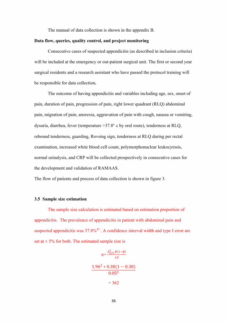
3.5 Sample size estimation
The sample size calculation is estimated based on estimation proportion of
appendicitis. The prevalence of appendicitis in patient with abdominal pain and
suspected appendicitis was 37.8%47 . A confidence interval width and type I error are
set at ± 5% for both. The estimated sample size is
n= /
1.96 ∗ 0.38 1 0.380.05
= 362

37
Taking into account for missing data of 5% yields 380 subjects that are required to
include in the study. As such, the expected numbers of appendicitis are 155 subjects.
A total of 8-10 variables are expected to use for generate the scores. Using the
rule of thumb, 10 subjects with appendicitis are required for 1 variable associated with
appendicitis in the analysis to generate the clinical prediction score. Approximately 80
and 100 subjects with appendicitis will be need for 8 and 10 parameters, respectively.
As such, a total sample size of 380 should be covered considering requirement of
patients according to the rule of thumb.
For external validation, we will include 114 (30% of derived subjects) for each of 2
hospitals.
Statistical analysis
3.5.1 Data management
Databases will be created based on the case record forms (CRFs) by using
EPIDATA program version 3.1. The principal investigator will check for the
completeness of CRFs before data entry. Double data entry will be done by
independent staff and typing errors will be validated in two data sets. Data checking
and cleaning will be done monthly with clarification of missing, unclear, or un-
sensible data. A safe area will be used to keep data. Only principle investigators,
supervisors, and the data manager will be able to access the data. Real time back up of
data will be done automatically at the DMU server to prevent data loss.
3.5.2 Derivative phase
Descriptive data

38
Mean and standard deviation (SD) or median and range will be used for
describing continuous data while frequency or percentage will be used for categorical
data. The baseline characteristics of patients will be compared between appendicitis
and non-appendicitis groups using independent t test (or Mann-Whitney test) and chi
square (or exact test) for continuous and categorical data, respectively.
Univariate analysis
The whole data sets collect at Ramathibodi Hospital will be used for the
derivative phase. Simple logistic regression analysis will be used to define the
explanatory variables that influence the diagnosis of appendicitis. Three groups of
variables will be analyzed as follows:
‐ Demographic parameters: age and sex
‐ Clinical symptom parameters: onset of pain, duration of pain, progression
of pain, right lower quadrant (RLQ) abdominal pain, migration of pain,
anorexia, aggravation of pain with cough, nausea or vomiting, dysuria,
diarrhea
‐ Clinical sign parameters: fever (temperature >37.8 c by oral route),
tenderness at RLQ, rebound tenderness, guarding, Rovsing sign,
tenderness at RLQ during per rectal examination
‐ Laboratory parameters: increased white blood cell count,
polymorphonuclear leukocytosis, and normal urinalysis
The maximum likelihood function will be used to estimate the parameter and
the simple with logit. Likelihood ratio (LR) test will be used to test the association
between variable and appendicitis.
Multivariate analysis

39
‐ Model selection
Parameters whose p values are less than 0.20 in the univariate analysis will be
simultaneous considered in the multivariate analysis. Multiple logistic regression
analysis will be used to estimate effect size and select the variable that should remain
in the model. The LR test will be applied to select variables in the model with
forward or back ward elimination to find the final parsimonious model.
‐ Goodness of fit
Exploration for goodness of fit will be done to define whether the model fits
well with data (predicted and observed values are close). Model goodness of fit will
be explored using chi square test. The Hosmer-Lemeshow test will be used to assess
the goodness fit of the models. In addition, a calibration coefficient oi-ei or oi/ ei
along with their 95% confidence intercal (CI) will be estimated. Pearson chi-square
residual, residual sum square, and deviance residual will be calculated if the model
does not fit well with data. Outliers will be identified and assessed affect on prediction
values and coefficients.
‐ Creation prediction score
The coefficients or odds ratio of the final parsimonious model will be used to
create prediction score (RAMA-AS). Each parameter will be weighted differently
according to its coefficient. The score will be calculated by “sum up” coefficients or
odds ratio. The receiver operating curve (ROC) analysis will be used to calibrate the
cutoff point of the scores. Diagnostic parameters (i.e., sensitivity, specificity,
likelihood ratio positive (LR+) and negative) will be estimated for each distinct value
of the scores.
Two cut-off points will be attempted to identify. The first cutoff will have
high sensitivity which will refer to complicated appendicitis and the second cutoff

40
will have high specificity for acute appendicitis. As a result, 3 diagnostic zones rule
out (high sensitivity) and rule in (high specificity), and non-appendicitis will be
generated. An area under curve or C statistic will be also estimated.
3.5.3 Validation phases
Internal validation
The bootstrap technique with 200 replications will be applied for internal
validation of the RAMA-AS 46 48. For each bootstrap sample, the RAMA-AS scores
will be created for each patient and then will be fitted in logistic model. The
discrimination parameter C statistic will be estimated. In addition, calibration
coefficient of observe(O)/predicted value (E) will be estimated. Further more, the
correlation between the O and E values of appendicitis will be assessed using the
Somer’D correlation, called Dboot. Calibration of the model will be then assessed by
subtracting the original Somer’D correlation from the mean Dboot. Discrimination of
the model will be assessed by comparing the original C statistic versus an average C
statistic from the bootstraps.
External validation
Data from a prospective cross sectional study at Department of Surgery,
Faculty of Medicine, Thammasart University and/or surgical unit at Chonburi
Hospital will be used to validate the generalized capability of RAMA-AS. The
RAMA-AS will be calculated for each patient. The ROC curve analysis will be
applied to estimate C-statistic. Then, the original C statistic (from boostrap internal
validation) and this C statistic (external validation) will be compared.

41
Comparison of RAMA-AS and previous scores
Fourteen scoring systems from the previous systematic review in chapter 2
will be compared with RAMA-AS. Calibration (O/E ratio), discrimination (C-
statistic) coefficients along with their 95% CIs, and AUC will be estimated for each
study. Comparison of O/E ratio, C-statistic, and AUC will be performed to evaluate
the performance of RAMA-AS.
The sensitivity, specificity, positive predictive value, negative predictive
value, and accuracy will be analyzed for each scoring system. To compare the
diagnostic performance of RAMA-AS and the previously proposed scoring system,
the area under the ROC curve (AUC) will be calculated for each scoring system. The
AUC of each scoring system will be compared using chi square method, with p value
< 0.05 considered statistically significant.
3.6 Cost effectiveness analysis of RAMA-AS
Society cost will be calculated from direct and indirect costs. Direct medical
costs (include medicine, medical supplies, laboratory and imaging tests, hospital
charges, and nursing care) and indirect non-medical cost (travel costs, food,
accommodation and opportunity lost from providing informal care and visits of
relatives and friends at hospital and home) will be collected. The incremental cost
effectiveness ratio (ICER) will be calculated by comparing a difference of cost
between outcomes predicted by RAMA-AS versus non RAMA-AS. RAMA-AS may
reduce the cost of investigation (U/S, CT scan). Utility will be estimated form EQ5D
as recommended by guideline for economic evaluation in Thailand49 ICER will be
calculated by dividing the difference of cost by the difference of utility between
RAMA-AS versus non RAMA-AS.

42
A decision tree will be used to model the important outcomes after patients are
suspected of having appendicitis. The decision tree starts from the patients who are
suspected of appendicitis, but the diagnostic strategy will differ based on whether the
RAMA-AS or routine clinical diagnosis with imaging is used for initial diagnosis. In
the pro RAMA-AS pathway the highly suspicious of appendicitis (say score more
than 7) group will be treated by appendectomy. The final outcome is either true
diagnosis of appendicitis or negative appendectomy. For the inconclusive patients
(score 4 to 7) further investigation by ultrasonography will be used for diagnosis.
Positive ultrasonographic patients will be treated by appendectomy while negative
group will receive further investigation with computer tomography (CT scan).
Positive CT scan patients will be treated with appendectomy while the negative
patients will be discharged (figure 4).
The direct and indirect costs occurred by health care providers and households
will be identified in two pathways: patient questionnaire interviews and data
extraction from hospital records. Ramathibodi, Thammasat and Chonburi Hospitals
will be used for the costing study. The prospective reviews of medical records of all
patients who are suspected of having appendicitis will be performed. The case records
review aims to measure hospital resources used such as clinician's time, type and
number of investigations, disposable equipments, and length of hospital admission.
The valuation of the hospital costs, including capital and overhead costs, for each
individual patient will be determined from each department involved in the services
for patients with suspected appendicitis. Daily event and cost questionnaires will be
used to accumulate patient-specific information and estimation of other household
expenses such as travel costs, food, accommodation and opportunity lost from
providing informal care and visits of relatives and friends at hospital and home

43
including the patient's recovery time to full activity after surgery. The face-to-face
interviews will be performed with patients at the post-operation visit (7-10 days after
surgery). For the patients who are lost to follow up or having longer follow-up period
than 4 weeks, telephone interview will be performed to collect that data. The costs
will be represented in Thai Baht at 2011 value (30 Baht = 1USD). All costs and
outcomes that occur beyond one year will be discounted using the same rate of 3.5%.
Uncertainty analysis
To determine if values within a plausible range for all input variables resulted
in a different conclusion, the probabilistic uncertainty analysis, assigning a beta
distribution for all probability and utility parameters and gamma distribution for all
cost parameters, and generating 1,000 rounds of simulations using Microsoft Excel®
with macro function on all estimated quantities will be carried out. The cost-
effectiveness acceptability curve based on the net benefit approach will also be
provided to present the relationship between the values of the ceiling ratio
(willingness to pay for one unit more of QALY) and probability favoring each
treatment strategy.
In addition, the scores will be converted to Thai utility using a conversion
index as suggested by Sakthong et al49 (appendix C). The calculation is done by
subtracting the relevant coefficients from 1.000. The constant term is used if there is
any dysfunction. The N3 term will be used if any dimension is at level 3.
3.7 Implementation of RAMA-AS and impact analysis
To encourage physician use our RAMA-AS in a routine practice, a computer
algorithm software will be developed. Once it’s developed, we will pilot it at
Ramathibodi and Thammasart hospitals. An accuracy of the RAMA-AS will be

44
collected. In addition, numbers of using it and reasons for using and not using will be
collected and discussed.
We will then introduce and demonstrate our software to the Royal College
Surgeon of Thailand. We will ask for collaboration from about 5-8 hospitals where
physicians are willing to try our software. The software will then be installed and
demonstrate. Data will be collect for assessing accuracy of the RAMA-AS. The
RAMA-AS performance and costs will then be compared with using imagings.
3.8 Ethics considerations
Informed consent if needed
The informed consent is shown in the appendix section.
Submit to the ethic committee for approval
This will be conducted according to the principles of the Helsinki Declaration
and in accordance with the Medical Research Involving Human Subjects Act. The
protocol will be submitted for approval from the ethics committee of Faculty of
Medicine Ramathibodi Hospital, Mahidol University. The principles of respect for
persons, beneficence, nonmaleficence, and justice will be applied in this research.

45
Respect for Persons
This principle of two ethical convictions will be considered. All participants
will be treated as autonomous and second, participants with diminished autonomy will
be protected. They will be treated with dignity and respect. The medical and surgical
care will not be disturbed by this study. This study will not used participants as a
means to an end or in a manner inconsistent with the person's interests or wishes. All
the treatment will be given to participants in the standard quality of care. The decision
of sending participants for investigations, giving medical treatment and surgery will
not be disturbed. The data will be collected to case record form by history taking,
physical examination, and reviewing medical records. Telephone follow up may
disturb participants, so the convenient time of participants will be recorded and used
for contact.
Beneficience
This study will prevent and remove harm as well as promote the good of the
person by minimizing the possible harms or risks and maximizing the potential
benefits. The nonmaleficence which prohibits the infliction of harm, injury, or death
upon others will be applied (the maxim Primum non nocere: “Above all do no harm”).
This study does not disturb the process of care. There is no new therapy or medicine
use in this study.
Justice
Each participant will be treated fairly and equitably, and be given his or her
due. The study will use available resources fairly and distribute them fairly and
equitably. All participants will be treated equally. They have the right to leave the
study at anytime without affecting their quality of care.

46
Protection
This study will assure that participation is voluntary and treated with equality
and fairness. The participants will be informed of all the procedures which will
happen related to patients.

47
3.9 Budget
3.11 Time Frame
Activities January 2013 – May 2015 June 2015 – May 2016 June 2016– May 2017 June 2017
Developing and internal
validation of RAMAAS
-Data collection
-Data analysis
External validation
-Data collection
-Data analysis
Economic analysis
Impact analysis
Data analysis
Presentations
Level ITEMS Cost/Unit Units COST Responsible unit
Study level Data collection
Data management
Research assistance
External monitoring
Software development
300
250
15,000
4,000
50,000
608
608
12
2
1
6,000
152,000
20,000
45,000
8,000
50,000
-Study sites
-Central DMU
-Central coordinating
center
Subject level Laboratory test 120 720 86,400 -Study sites
Total 367,400

48
ACKNOWLEDGEMENT
This study proposal is a part of the dissertation for (Student name’s) training in Ph.D. (Clinical Epidemiology), Faculty of Medicine Ramathibodi Hospital and Faculty of Graduates, Mahidol University.
Funding resource

49
REFERENCES
1. Tepel J, Sommerfeld A, Klomp HJ, Kapischke M, Eggert A, Kremer B. Prospective
evaluation of diagnostic modalities in suspected acute appendicitis.
Langenbecks Arch Surg 2004;389(3):219-24.
2. Addiss DG, Shaffer N, Fowler BS, Tauxe RV. The epidemiology of appendicitis
and appendectomy in the United States. Am J Epidemiol 1990;132(5):910-25.
3. Horntrich J, Schneider W. [Appendicitis from an epidemiological viewpoint].
Zentralbl Chir 1990;115(23):1521-9.
4. Temple CL, Huchcroft SA, Temple WJ. The natural history of appendicitis in
adults. A prospective study. Ann Surg 1995;221(3):278-81.
5. Andersson RE. Meta-analysis of the clinical and laboratory diagnosis of
appendicitis. Br J Surg 2004;91(1):28-37.
6. Al Qahtani HH, Muhammad AA. Alvarado score as an admission criterion for
suspected appendicitis in adults. Saudi J Gastroenterol 2004;10(2):86-91.
7. Alvarado A. A practical score for the early diagnosis of acute appendicitis. Ann
Emerg Med 1986;15(5):557-64.
8. Andersson M, Andersson RE. The appendicitis inflammatory response score: a tool
for the diagnosis of acute appendicitis that outperforms the Alvarado score.
World J Surg 2008;32(8):1843-9.
9. Chong CF, Adi MIW, Thien A, Suyoi A, Mackie AJ, Tin AS, et al. Development of
the RIPASA score: A new appendicitis scoring system for the diagnosis of
acute appendicitis. Singapore Medical Journal 2010;51(3):220-25.
10. Chong CF, Thien A, Mackie AJ, Tin AS, Tripathi S, Ahmad MA, et al.
Comparison of RIPASA and Alvarado scores for the diagnosis of acute
appendicitis. Singapore Med J 2011;52(5):340-5.

50
11. Christian F, Christian GP. A simple scoring system to reduce the negative
appendicectomy rate. Ann R Coll Surg Engl 1992;74(4):281-5.
12. de Castro SM, Unlu C, Steller EP, van Wagensveld BA, Vrouenraets BC.
Evaluation of the Appendicitis Inflammatory Response Score for Patients with
Acute Appendicitis. World J Surg 2012.
13. Denizbasi A, Unluer EE. The role of the emergency medicine resident using the
Alvarado score in the diagnosis of acute appendicitis compared with the
general surgery resident. Eur J Emerg Med 2003;10(4):296-301.
14. Enochsson L, Gudbjartsson T, Hellberg A, Rudberg C, Wenner J, Ringqvist I, et
al. The Fenyo-Lindberg scoring system for appendicitis increases positive
predictive value in fertile women--a prospective study in 455 patients
randomized to either laparoscopic or open appendectomy. Surg Endosc
2004;18(10):1509-13.
15. Eskelinen M, Ikonen J, Lipponen P. A computer-based diagnostic score to aid in
diagnosis of acute appendicitis. A prospective study of 1333 patients with
acute abdominal pain. Theoretical Surgery 1992;7(2):86-90.
16. Fenyo G. Routine use of a scoring system for decision-making in suspected acute
appendicitis in adults. Acta Chir Scand 1987;153(9):545-51.
17. Fenyo G, Lindberg G, Blind P, Enochsson L, Oberg A. Diagnostic decision
support in suspected acute appendicitis: validation of a simplified scoring
system. Eur J Surg 1997;163(11):831-8.
18. Galindo Gallego M, Fadrique B, Nieto MA, Calleja S, Fernandez-Acenero MJ,
Ais G, et al. Evaluation of ultrasonography and clinical diagnostic scoring in
suspected appendicitis. Br J Surg 1998;85(1):37-40.

51
19. Hsieh CH, Lu RH, Lee NH, Chiu WT, Hsu MH, Li YC. Novel solutions for an old
disease: diagnosis of acute appendicitis with random forest, support vector
machines, and artificial neural networks. Surgery 2011;149(1):87-93.
20. Inci E, Hocaoglu E, Aydin S, Palabiyik F, Cimilli T, Turhan AN, et al. Efficiency
of unenhanced MRI in the diagnosis of acute appendicitis: Comparison with
Alvarado scoring system and histopathological results. European Journal of
Radiology 2011;80(2):253-58.
21. Kalan M, Talbot D, Cunliffe WJ, Rich AJ. Evaluation of the modified Alvarado
score in the diagnosis of acute appendicitis: a prospective study. Ann R Coll
Surg Engl 1994;76(6):418-9.
22. Kanumba ES, Mabula JB, Rambau P, Chalya PL. Modified Alvarado Scoring
System as a diagnostic tool for Acute Appendicitis at Bugando Medical
Centre, Mwanza, Tanzania. BMC Surgery 2011;11.
23. Konan A, Hayran M, Kilic YA, Karakoc D, Kaynaroglu V. Scoring systems in the
diagnosis of acute appendicitis in the elderly. Ulusal Travma ve Acil Cerrahi
Dergisi 2011;17(5):396-400.
24. Kurane SB, Sangolli MS, Gogate AS. A one year prospective study to compare
and evaluate diagnostic accuracy of modified Alvarado score and
ultrasonography in acute appendicitis, in adults. Indian Journal of Surgery
2008;70(3):125-29.
25. Lamparelli MJ, Hoque HM, Pogson CJ, Ball AB. A prospective evaluation of the
combined use of the modified Alvarado score with selective laparoscopy in
adult females in the management of suspected appendicitis. Ann R Coll Surg
Engl 2000;82(3):192-5.

52
26. Limpawattanasiri C. Alvarado score for the acute appendicitis in a provincial
hospital. J Med Assoc Thai 2011;94(4):441-9.
27. Lintula H, Pesonen E, Kokki H, Vanamo K, Eskelinen M. A diagnostic score for
children with suspected appendicitis. Langenbecks Arch Surg
2005;390(2):164-70.
28. Malik AH, Wani RA, Saima BD, Wani MY. Small lateral access--an alternative
approach to appendicitis in paediatric patients: a randomised controlled trial.
Int J Surg 2007;5(4):234-8.
29. McKay R, Shepherd J. The use of the clinical scoring system by Alvarado in the
decision to perform computed tomography for acute appendicitis in the ED.
Am J Emerg Med 2007;25(5):489-93.
30. Ohmann C, Franke C, Yang Q. Clinical benefit of a diagnostic score for
appendicitis: results of a prospective interventional study. German Study
Group of Acute Abdominal Pain. Arch Surg 1999;134(9):993-6.
31. Pouget-Baudry Y, Mucci S, Eyssartier E, Guesdon-Portes A, Lada P, Casa C, et
al. The use of the Alvarado score in the management of right lower quadrant
abdominal pain in the adult. J Visc Surg 2010;147(2):e40-4.
32. Pruekprasert P, Geater A, Ksuntigij P, Maipang T, Apakupakul N. Accuracy in
diagnosis of acute appendicitis by comparing serum C-reactive protein
measurements, Alvarado score and clinical impression of surgeons. Journal of
the Medical Association of Thailand 2004;87(3):296-303.
33. Ramirez JM, Deus J. Practical score to aid decision making in doubtful cases of
appendicitis. Br J Surg 1994;81(5):680-3.

53
34. Sitter H, Hoffmann S, Hassan I, Zielke A. Diagnostic score in appendicitis.
Validation of a diagnostic score (Eskelinen score) in patients in whom acute
appendicitis is suspected. Langenbecks Arch Surg 2004;389(3):213-8.
35. Sun JS, Noh HW, Min YG, Lee JH, Kim JK, Park KJ, et al. Receiver operating
characteristic analysis of the diagnostic performance of a computed
tomographic examination and the Alvarado score for diagnosing acute
appendicitis: emphasis on age and sex of the patients. J Comput Assist Tomogr
2008;32(3):386-91.
36. Talukder D.B. SA. Modified Alvarado Scoring System in the Diagnosis of Acute
Appendicitis. JAFMC Bangladesh 2009;5(1):18-20.
37. Teicher I, Landa B, Cohen M. Scoring system to aid in diagnoses of appendicitis.
Annals of Surgery 1983;198(6):753-59.
38. Tzanakis NE, Efstathiou SP, Danulidis K, Rallis GE, Tsioulos DI, Chatzivasiliou
A, et al. A new approach to accurate diagnosis of acute appendicitis. World J
Surg 2005;29(9):1151-6, discussion 57.
39. Van Way CW, 3rd, Murphy JR, Dunn EL, Elerding SC. A feasibility study of
computer aided diagnosis in appendicitis. Surg Gynecol Obstet
1982;155(5):685-8.
40. Yoldas O, Karaca T, Tez M. External validation of Lintula score in Turkish acute
appendicitis patients. International Journal of Surgery 2012;10(1):25-27.
41. Malik AH, Wani RA, Saima BD, Wani MY. Small lateral access-an alternative
approach to appendicitis in paediatric patients: A randomised controlled trial.
International Journal of Surgery 2007;5(4):234-38.
42. Guyatt G RD. Users' Guides to The Medical Literature: A Menual for Evidence-
based Clinical Practice. 2 ed. Chicago: American Medical Association

54
Printed in the United States of America., 2002.
43. Lintula H, Kokki H, Pulkkinen J, Kettunen R, Gröhn O, Eskelinen M. Diagnostic
score in acute appendicitis. Validation of a diagnostic score (Lintula score) for
adults with suspected appendicitis. Langenbeck's Archives of Surgery
2010;395(5):495-500.
44. Normand SL. Meta-analysis: formulating, evaluating, combining, and reporting.
Stat Med 1999;18(3):321-59.
45. Altman DG, Royston P. What do we mean by validating a prognostic model? Stat
Med 2000;19(4):453-73.
46. Harrell FE, Jr., Lee KL, Mark DB. Multivariable prognostic models: issues in
developing models, evaluating assumptions and adequacy, and measuring and
reducing errors. Stat Med 1996;15(4):361-87.
47. van Randen A, Bipat S, Zwinderman AH, Ubbink DT, Stoker J, Boermeester MA.
Acute appendicitis: meta-analysis of diagnostic performance of CT and graded
compression US related to prevalence of disease. Radiology 2008;249(1):97-
106.
48. Schumacher M, Hollander N, Sauerbrei W. Resampling and cross-validation
techniques: a tool to reduce bias caused by model building? Statistics in
medicine 1997;16(24):2813-27.
49. Sakthong P. Measurement of clinical-effect: utility. J Med Assoc Thai 2008;91
Suppl 2:S43-52.

55
TABLES
Table 1 Risk of bias assessment for clinical prediction rule of appendicitis
i) Derivative phase Domain Item Low risk of
bias
Comments
Selection bias - Was the spectrum of patients representative of the patients who will receive the evaluation in practice? (representative spectrum)
Recruited all consecutive patients with chief complain of abdominal pain, or random samples patients from well define population frame of abdominal pain
Yes
Recruited a specific group of patients (e.g., having at least a few clinical sign and symptoms)
No
Information provided could not classify Y/N Unclear
No information to assess NA
Information bias
Ascertainment of outcome measurement
- Was appendicitis clearly defined and acceptable in this field?
Diagnosed by biopsy
Yes
Diagnose by Surgeon using operation finding
No
Information provided could not classify Y/N Unclear
No information to assess NA
- Were those assessing the outcome event blinded to the presence of the predictors?
Appendicitis was proven by biopsy and pathologist was masked to know about clinical information
Yes
Appendicitis was diagnosed by Surgeon without proven by biopsy & surgeon could access to clinical data
No
Information provided could not classify Y/N Unclear Did not mention NA

56
Ascertainment of predictors - Were all important predictors consider to include in the derivation process?
Clearly list all items of predictors and considered number of predictors for each item as demographic(1/2), clinical sign (4/9), symptom (5/10), lab(2/4), and imaging (1/2)
Yes
Did not consider all 4 items and/or numbers of predictors for each item were < a half of overall predictors
No
Information provided could not classify Y/N Unclear
Did not mention NA
Were assessing of presence of predictors blinded to the outcome event?
Stated clearly how to assess predictors and assessors were blind for knowledge of the final diagnosis of appendicitis, lab, and imaging findings when undertook physical examinations or vice versa
Yes
Physical examination, lab, or imaging was not blind or these predictors were assessed with knowledge of possible diagnosis of appendicitis
no
Information provided could not classify Y/N Unclear Did not mention NA
- Were all important predictors present in a significant proportion of the study population?
Statistic test was applied for all predictors and only statistical (or clinically with reasons) significant predictors were considered to include in the model
Yes
Did not apply statistical test, or rationale for include predictors was not provided
No
Information provided could not classify Y/N Unclear
Did not mention NA
Confounding bias
- Multivariate regression analysis was applied for derive model.
Applied muitivariate (logistic, Poisson, discriminant) regression using conventional or Bayesian statistical methods, or using recursive partitioning analysis by simultaneously included all predictors in the same model
Yes
Did not apply multivariate regression analysis No
Information provided could not classify Y/N Unclear Did not mention NA

57
- Was score or weight created properly? Score or weight for each predictor was created
based on suggestion from regression model (e.g., coefficient or relative risk)
Yes
Was not based on regression results or weight without proper rationale
No
Information provided could not classify Y/N Unclear
Did not mention NA
Other issue bias - Were the total sample size and number of events adequate or modeling?
Number of appendicitis and total subjects were sufficient base on a rule of thumb (1 predictor/10 appendicitis or 20-30 total subjects)
Yes
N < 10 appendicitis or n < 20-30 total patients per 1 predictor
No
Information provided could not classify Y/N Unclear
Did not mention NA
- Does the rule make clinical sensible? Included predictors seem clinically sensible,
easy to use, suggested a course of action
Yes
Included predictor did not make any sense, not easy to calculate, or provided only probability of appendicitis without course of action
No
Information provided could not classify Y/N Unclear Did not mention NA

58
ii) Validation phase
Domain Item Low risk of bias
Comments
Selection bias - Was the spectrum of patients representative of the patients who will receive the evaluation in practice? (representative spectrum)
Recruited all consecutive patients with chief complain of abdominal pain, or random samples patients from well define population frame of abdominal pain
Yes
Recruited a specific group of patients (e.g., having at least a few clinical sign and symptoms)
No
Information provided could not classify Y/N Unclear
No information to assess NA
Information bias Ascertainment of outcome measurement
- Was appendicitis clearly defined and acceptable in this field?
Diagnosed by biopsy
Yes
Diagnose by Surgeon using operation finding No
Information provided could not classify Y/N Unclear
No information to assess NA
- Were those assessing the outcome event blinded to the presence of the predictors?
Appendicitis was proven by biopsy and pathologist was masked to know about clinical information
Yes
Appendicitis was diagnosed by Surgeon without proven by biopsy & surgeon could access to clinical data
No
Information provided could not classify Y/N Unclear Did not mention NA
- Was there an explicit and accurate interpretation of the predictors and/or the actual rule without knowledge of the outcome.
Clearly described interpretation of the rule was not influenced by information of final diagnosis of appendicitis
Yes
Clinical rule classification was influenced by No

59
diagnose of appendicitis
Information provided could not classify Y/N Unclear
Did not mention NA
Other issue bias - Was there 100% follow-up of those enrolled? All included patients were included at the
end of study
Yes
Some patients were lost to follow up No
Information provided could not classify Y/N Unclear
Did not mention NA
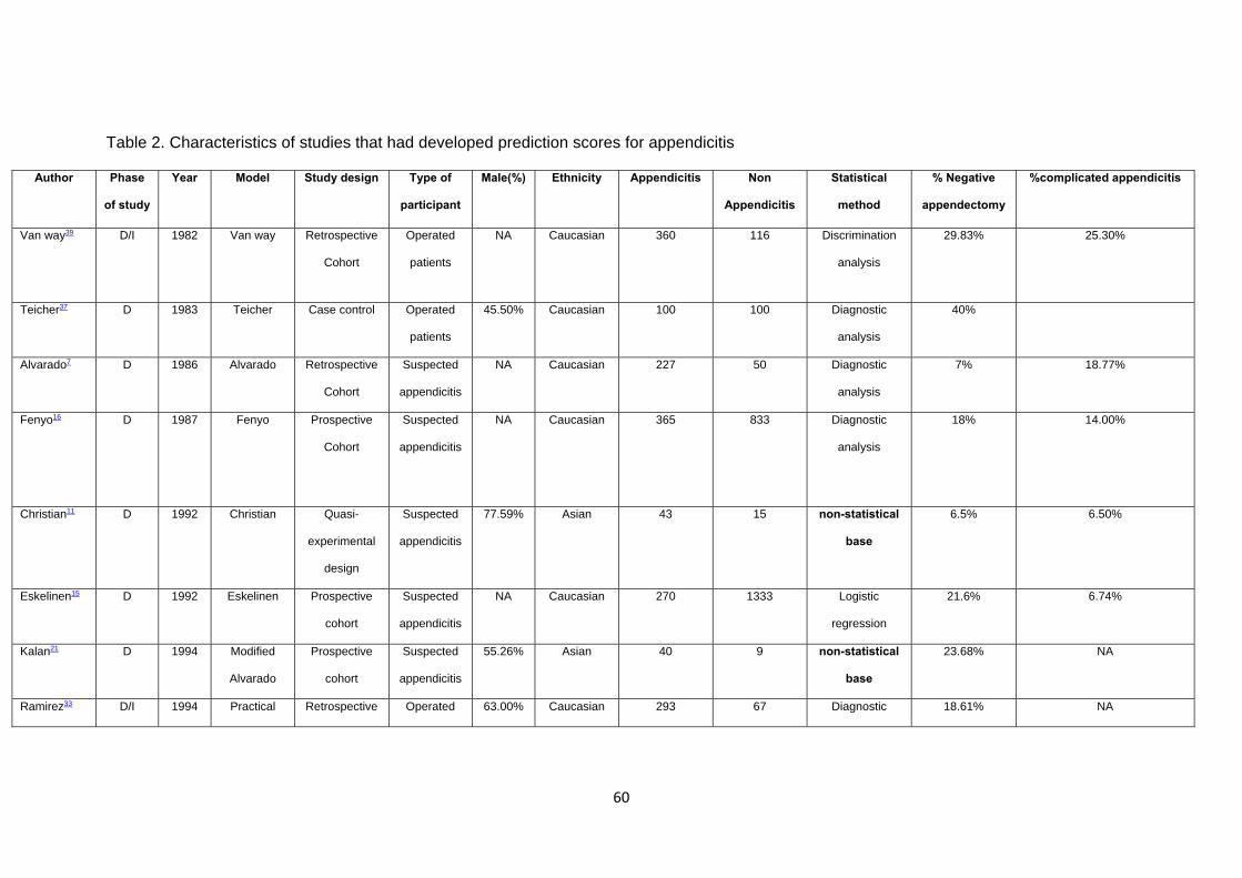
60
Table 2. Characteristics of studies that had developed prediction scores for appendicitis
Author Phase
of study
Year Model Study design Type of
participant
Male(%) Ethnicity Appendicitis Non
Appendicitis
Statistical
method
% Negative
appendectomy
%complicated appendicitis
Van way39 D/I 1982 Van way Retrospective
Cohort
Operated
patients
NA Caucasian 360 116 Discrimination
analysis
29.83% 25.30%
Teicher37 D 1983 Teicher Case control Operated
patients
45.50% Caucasian 100 100 Diagnostic
analysis
40%
Alvarado7 D 1986 Alvarado Retrospective
Cohort
Suspected
appendicitis
NA Caucasian 227 50 Diagnostic
analysis
7% 18.77%
Fenyo16 D
1987 Fenyo Prospective
Cohort
Suspected
appendicitis
NA Caucasian 365 833 Diagnostic
analysis
18% 14.00%
Christian11 D 1992 Christian Quasi-
experimental
design
Suspected
appendicitis
77.59% Asian 43 15
non-statistical
base
6.5% 6.50%
Eskelinen15 D 1992 Eskelinen Prospective
cohort
Suspected
appendicitis
NA Caucasian 270 1333 Logistic
regression
21.6% 6.74%
Kalan21 D 1994 Modified
Alvarado
Prospective
cohort
Suspected
appendicitis
55.26% Asian 40 9 non-statistical
base
23.68% NA
Ramirez33 D/I 1994 Practical Retrospective Operated 63.00% Caucasian 293 67 Diagnostic 18.61% NA

61
score of
Ramirez and
Deus
Cohort patients analysis
Gallego18 D 1998 Gallego Prospective
Cohort
Suspected
appendicitis
NA Caucasian 101 91 Diagnostic
analysis
8.85% 18.23%
Tzanakis38 D/I/E 2005 Tzanakis
Prospective
Cohort
Suspected
appendicitis
56.10 Caucasian 217 504 Logistic
regression
19.20% 10.23%
Lintula27 D 2005 Lintula Prospective
cohort
Suspected
appendicitis
100% Caucasian 43 84 Logistic
regression
13% NA
Malik28 D 2007 Modified
Alvarado
Prospective
cohort
Suspected
appendicitis
55.12% Asian 174 80 non-statistical
base
11.49% 12.07%
Andersson8
D/I/E 2008 Appendicitis
inflammatory
response
score
Prospective
Cohort
Suspected
appendicitis
46.00% Caucasian 191 254 Logistic
regression
11.00% 14.00%
Chong9 D 2010 RIPASA Retrospective
cohort
Operated
patients
57.69% Asian 261 51 Univariate
analysis
16.30% NA
External validation phase
Fenyo17 E 1997 Fenyo Prospective
Cohort
Suspected
appendicitis
11.2% Caucasian 392 775 Diagnostic
analysis
17.5% NA

62
Lamparelli25 E 2000 Mod.
alvarado by
Kalan
Prospective
Cohort
Suspected
appendicitis
NA Caucasian 56 26 Diagnostic
analysis
NA NA
Denizbasi13 E 2003 Alvarado Prospective
Cohort
Suspected
appendicitis
53.6% Caucasian 175 46 Diagnostic
analysis
NA NA
AlQahtani6 E 2004 Alvarado Prospective
Cohort
Suspected
appendicitis
59% Asian 121 30 Diagnostic
analysis
12.5% NA
Pruekprasert
32
E 2004 Alvarado Prospective
Cohort
Suspected
appendicitis
46% Asian 186 45 Diagnostic
analysis,
Discrimination
analysis
NA NA
Enochsson14 E 2004 Fenyo Prospective
Cohort
Suspected
appendicitis
12% Caucasian 330 96 Diagnostic
analysis
NA NA
Sitter34 E 2004 Eskelinen Prospective
Cohort
Suspected
appendicitis
NA Caucasian 662 1697 Diagnostic
analysis,
Discrimination
analysis
21.6% NA
Tepel1 E 2004 Ohmann Retrospective
Cohort
Suspected
appendicitis
37% Caucasian 113 287 Diagnostic
analysis
22% NA
McKay29 E 2007 Alvarado Retrospective
Cohort
suspected
appendicitis
NA Caucasian 48 96 Diagnostic
analysis
NA NA

63
Sun35 E 2008 Alvarado Retrospective
Cohort
Suspected
appendicitis
66.2% Asian 213 159 Diagnostic
analysis,
Discrimination
analysis
NA NA
Kurane24 E 2008 Mod.
Alvarado by
kalan
Prospective
Cohort
Suspected
appendicitis
48.33% Asian 23 37 Diagnostic
analysis
61.67% NA
Ohmann30 I 2008 ohmann Prospective
Cohort
Suspected
appendicitis
43.8% Caucasian 235 1019 Diagnostic
analysis
10.2% 13.4%
Talukder36 E 2009 Mod
Alvarado
malik
Prospective
Cohort
Suspected
appendicitis
58% Asian 84 16 Diagnostic
analysis
16% NA
Hsieh19 E 2010 Alvarado Retrospective
Cohort
Suspected
appendicitis
47% Asian 115 65 Diagnostic
analysis,
Discrimination
analysis
NA NA
Pouget-
Baudry31
E 2010 Alvarado Prospective
Cohort
Operated
patients
48.07% Caucasian 171 62 Diagnostic
analysis
NA NA
Lintula43 I 2010 Lintula RCT Suspected
appendicitis
50% Caucasian 52 44 Diagnostic
analysis
13% NA
Chong10 E 2011 Alvarado Prospective Suspected 47.9% Asian 101 91 Diagnostic 22.9% 6.1%

64
Chong
(2010)
Cohort appendicitis analysis,
Discrimination
analysis
Inci20 E 2011 Alvarado Prospective
Cohort
Suspected
appendicitis
NA Caucasian 57 9 Diagnostic
analysis
13.6% NA
Limpawattan
asiri26
E 2011 Alvarado Prospective
Cohort
Suspected
appendicitis
40.7% Asian 715 285 Diagnostic
analysis,
Discrimination
analysis
14.7% NA
Konan23 E 2011 Alvarado Retrospective
Cohort
Operated
patients
NA Caucasian 41 41 Diagnostic
analysis,
Discrimination
analysis
NA NA
Kanumba22 E 2011 Mod.
alvarado
kalan
Cross
sectional study
Suspected
appendicitis
29.1% Caucasian 85 42 Diagnostic
analysis
33.1% 62.9%
Yoldas40 E 2011 Lintula Retrospective
Cohort
Operated
patients
50.7% Caucasian 132 24 Diagnostic
analysis,
Discrimination
analysis
NA NA
Castro12 E 2012 Alvarado
Andersson
Retrospective
Cohort
Suspected
appendicitis
44% Caucasian 340 595 Diagnostic
analysis,
21% NA

65
Discrimination
analysis
NA: not applicable, D=Derivative, I=Internal validation, E=external validation, Diagnostic=100%, Discrimination analysis=39.13%

66
Table 3. Risk of bias assessment.
Study Phase
Selection
bias Information bias Confounding bias Other issue
Representativ
e spectrum
Ascertainment
of outcome
measurement
Blinded
assess
outcome
No
predic
tors
Predictor
s blinded
outcome
Significant
predictors
Accurate
interpreta
tion
Multivariate
regression
analysis
Created
score
properly
Sample
size
Clinical
sensible
Other
issue
Van way,198239 D N NA NA N N Y - Y Y Y Y -
Teicher,198337 D Y Y NA Y NA Y - N N Y Y -
Alvarado,19867 D Y Y NA Y NA Y - N N Y Y -
Fenyo,1987***16 D Y Y NA Y NA Y - N N Y N -
Christian,199211 D N Y NA N Y N - N N N Y -
Eskelinen,199215 D Y Y NA Y Y Y - Y Y Y Y -
Kalan,199421 D N Y NA N NA Y - N N N Y -
Ramirez,199433 D N Y NA Y NA Y - N N Y N -
Gallego,199818 D Y Y NA Y NA Y - N N Y N -
Tzanakis,200538 D Y Y NA Y Y Y - Y Y Y Y -
Lintula, 200527 D Y Y NA Y Y Y - Y Y Y Y -
Malik,200728 D N Y NA N NA Y - N N Y Y -
Andersson, 20088 D Y Y NA Y Y Y - Y Y Y Y -

67
Chong,20109 D N Y NA Y NA Y - N N Y N -
Van way,198239 I N NA NA - - - N - - - - N
Fenyo,1987***16 I Y Y NA - - - Y - - - - N
Ramirez,199433 I N Y NA - - - N - - - - N
Tzanakis,200538 I Y Y NA - - - Y - - - - Y
Andersson, 20088 I Y Y NA - - - Y - - - - Y
Lintul,201043 I Y Y NA - - - Y - - - - Y
Fenyo, 199717 E Y Y NA - - - N - - - - N
Denizbasi,200313 E Y Y NA - - - Y - - - - NA
AlQahtani,20046 E Y Y NA - - - Y - - - - Y
Pruekprasert,
200432
E Y Y NA - - - Y - - - - Y
Enochsson,
200414
E UN Y NA - - - Y - - - - NA
Sitter,200434 E Y Y NA - - - Y - - - - NA
Tzanakis,200538 E Y Y NA - - - NA - - - - Y
Mckay,200729 E Y Y NA - - - N - - - - NA
Andersson,20088 E Y Y NA - - - NA - - - - Y
Kurane,200824 E Y Y NA - - - Y - - - - NA
Sun, 200835 E Y Y NA - - - N - - - - NA
Talukder,200936 E Y Y NA - - - Y - - - - NA
Hsieh, 201019 E Y Y NA - - - N - - - - NA

68
Pouret-
Baudry,201031
E Y Y NA - - - Y - - - - Y
Chong,201110
E Y
Y
Y
Y
NA
NA
- - - Y
Y
- - - - NA
NA
Inci,201120 E Y Y NA - - - Y - - - - Y
Limpawattan,
201126
E Y Y NA - - - Y - - - - NA
Konan,201123 E N Y NA - - - N - - - - NA
Kanumba,201122 E Y Y NA - - - Y - - - - NA
Yoldas,201140 E N Y NA - - - N - - - - NA
Castro,201212 E Y
Y
Y
Y
NA
NA
- - - Y
Y
- - - - NA
NA

69
Table 4. Describe performances of predictive models of appendicitis
Author, Year
Derived model Internal validation External validation
No. Variable/
event/N O/E C-statistic
No. Variable/
event/N O/E C-statistic Author, Year
No. Variable
/event/N O/E C-statistic
Van way, 198239 4/NA/219 NA NA 4/169/257 1(0.92,1.08) 0.61(0.54,0.68) Tzanakis, 200538 4/87/201 1(0.76,1.24) 0.78(0.72,0.84)
Teicher, 198337 7/100/200 1(0.75,1.25) 0.78(0.72,0.83) - - - Tzanakis, 200538 4/87/201 1(0.69,1.31) 0.86(0.81,0.91)
Alvarado, 19867 8/227/277 1(0.93,1.07) 0.80(0.73,0.86) - - -
Denizbasi, 200313 8/175/221 0.99(0.91,1.09) 0.71(0.63,0.78)
AlQahtani, 20046 8/121/151 1(0.79,1.21) 0.84(0.76,0.93)
Pruekprasert, 200432 8/186/231 1(0.93,1.07) 0.74(0.67,0.81)
Tzanakis, 200538 4/87/201 1(0.69,1.31) 0.83(0.78,0.88)
McKay, 200729 8/48/144 1(0.70,1.30) 0.74(0.66,0.82)
Andersson, 20088 8/76/225 0.99(0.79,1.21) 0.63(0.58,0.68)
Sun, 200835 8/213/372 1(0.90,1.10) 0.65(0.61,0.70)
Hsieh, 201019 8/115/180 1(0.85,1.15) 0.77(0.70,0.83)
Pouget-
Baudry,201031 8/171/233 1(0.92,1.08) 0.68(0.62,0.73)
Chong, 201110 14/101/192 0.99(0.82,1.18) 0.78(0.72,0.84)
Inci, 201120 8/57/66 1(0.89,1.11) 0.75(0.58,0.92)

70
Limpawattanasiri,
201126 8/715/1000 1(0.94,1.06) 0.81(0.78,0.84)
Konan,201123 8/41/82 1(0.69,1.31) 0.84(0.76,0.91)
Castro, 201212 11/340/935 1(0.91,1.09) 0.62(0.60,0.65)
Fenyo, 198716 16/109/259 1(0.75,1.25) 0.91(0.89,0.93) - - -
Fenyo,199717 4/392/1167 1(0.88,1.12) 0.80(0.77,0.82)
Enochsson,200414 4/330/426 1(0.94,1.06) 0.73(0.68,0.78)
Tzanakis, 200538 4/87/201 1(0.65,1.35) 0.88(0.83,0.92)
Christian, 199211 5/43/58 1(0.56,1.44)* 0.87(0.77,0.98)* - - - Tzanakis, 200538 4/87/201 1(0.71,1.29) 0.85 (0.80,0.90)
Eskelinen, 199215 6/572/1333 1(0.93,1.07) 0.59(0.58,0.61) - - - Sitter, 200434 6/662/2359 1(0.90,1.10) 0.82(0.80,0.84)
Tzanakis, 2005 4/87/201 1(0.71,1.28) 0.87(0.83,0.92)
Kalan, 199421
(Modified Alvarado ) 7/40/49 1(0.74,1.26)* 0.76(0.60,0.92)* - - -
Lamparelli, 200025 7/56/84 1(0.80,1.20) 0.82(0.74,0.90)
Kurane, 200824 7/23/60 1(0.49,1.51) 0.81(0.71,0.92)
Kanumba, 201122 7/85/127 0.99(0.72,1.28) 0.92(0.87,0.97)
Ramirez, 199433 7/293/360 1(0.94,1.06) 0.72(0.60,0.84) NA NA NA - - - -
Gallego, 199818 6/101/192 1(0.78,1.23) 0.89(0.85,0.93) - - - - - - -
Ohmann, 199930 - - - 8/235/1254 1(0.44,1.56) 0.87(0.84,0.90) Tepel, 20041 4/113/400 1(0.78,1.20) 0.73(0.68-0.78)
Tzanakis, 200538 4/87/201 1(0.60,1.40) 0.88(0.84,0.93)
Tzanakis, 200538 10/130/303 1(0.45,1.55) 0.97(0.95,0.99) NA NA NA Tzanakis, 200538 10/87/201 - 0.96(0.93,0.99)
Malik, 200728
(Modified Alvarado ) 8/174/254 1(0.93,1.07) 0.54(0.45,0.63) - - - Talukder, 200936 8/84/100 1(0.91,1.09) 0.65(0.52,0.78)
Andersson, 20088 7/115/316 NA 0.87 7/115/316 1(0.43,1.57) 0.89(0.86,0.93) Castro, 201212 11/348/945 0.99(0.91,1.09) 0.55(0.54,0.57)

71
Chong, 20109 NA NA NA 14/101/192 1(0.45,1.55) 0.90(0.85,0.94) Tzanakis, 200538 14/101/192 0.99(0.82,1.18) 0.78(0.72,0.84)
Lintula, 200527 NA NA NA 11/52/96 1(0.64,1.36) 0.92(0.87,0.97) Yoldas, 201140 11/132/156 1(0.92,1.08) 0.79(0.73,0.86)
Pool effect - 1(0.97,1.03) 0.79(0.67,0.90) - 1(0.92,1.07) 0.84(0.77,0.91) - - 0.99(0.98,1.02) 0.78(0.74,0.82)

72
Table4. Describe variables that were included in the derive phase of predictive scores of appendicitis
Study, Year
Demographic data Clinical Symptoms
Age Sex Total Migration of pain
Anorexia Nausea/
vomiting
Duration of pain
Progression of pain
GU symptom
Aggravation with cough,
movement
Pain at presentation
Diarrhea Rigor Total
Van way, 1982 - 1 - - - - - - - 3
Teicher, 1983 2 - - - - - - - - 2
Alvarado, 1986 - - 0 - - - - - - - 3
Fenyo, 1987 - 1 - 9
Christian, 1992 - - 0 - - - - - - - - 2
Eskelinen, 1992 - - 0 - - - - - - - - 2
Kalan, 1994 - - 0 - - - - - - - 3
Ramirez, 1994 - 1 - - - - - - - - 2
Gallego, 1998 - - 0 - - - - - - - - - - 0
Lintula, 2005 - 1 - - - - - - - 3
Tzanakis, 2005 - - 0 - - - - - - - - 2
Malix, 2007 - - 0 - - - - - - 4
Andersson, 2008 - - 0 - - - - - - - - 2
Chong, 2010 2 - - - - - 5
Total 2 6 NA 7 6 9 6 2 1 2 6 2 1 NA

73
Study, Year
Signs Laboratory
Imaging Total
Elevated tempera
ture
Guarding
RLQ
Rebound
tenderness
Rigidity
RLQ tenderness
Rovsing’s sign
rectal tenderness
Pain outside
RLQ
Rectal Mass
R.
side
Total Increase WBC
PMN left shift
Negative UA
CRP
Total Abdominal
radiograph
Ultrasonographic
Total
Van way, 1982 - - - - - - - - - 0 - - - - 0 - - 0 4
Teicher, 1983 - - - - - - - 2 - - - 1 - - 0 7
Alvarado, 1986 - - - - - - 3 - - 2 - - 0 8
Fenyo, 1987 - - 7 - - - 1 - - 0 18
Christian, 1992 - - - - - - - 2 - - - 1 - - 0 5
Eskelinen, 1992 - - - - - - 3 - - - 1 - - 0 6
Kalan, 1994 - - - - - - 3 - - - 1 - - 0 7
Ramirez, 1994 - - - - - - - 2 - - 2 - - 0 7
Gallego, 1998 - - - - - - - 2 - - 2 2 6
Lintula, 2005 - - - - - 4 - - - - 0 - - 0 8
Tzanakis, 2005 - - - - - 4 - 3 - 1 10
Malix, 2007 - - - - 5 - - - 1 - - 0 10
Andersson, 2008 - - - - - - - 2 - 3 - - 0 7
Chong, 2010 - - - - 5 - - 2 - - 0 14

74
Total 9 7 11 2 9 2 2 1 1 NA 9 8 1 2 NA 1 2 NA NA

75
FIGURES
Figure 1: Identification of studies for inclusion

76
Figure 2 Flow of patients and process of data collection
Patient abdominal pain at ER or surgical OPD
Appendicitis
Acute abdomen and suspected appendicitis
Not suspected
appendicitis
Home
Not improve
ER or OPD
- Suspected for appendicitis - Consult surgical service for evaluation and
management
Not suspected
appendicitis
Home telephone follow
up at 1 month
Not improve
Observation or
InvestigationAppendicitis
Surgery
(Appendectomy)
Non‐appendix
Appendicitis Negative finding

77
Figure 3 Diagram of data analysis
Clean and check data
Simple logistic regression analysis ‐age, sex, onset of pain, duration of pain, progression of pain, right lower quadrant (RLQ) abdominal pain, migration of pain, anorexia, aggravation of pain
with cough, nausea or vomiting, dysuria, diarrhea, fever (temperature >37.8 c by oral route), tenderness at RLQ, rebound tenderness, guarding, Rovsing sign, tenderness at RLQ during per rectal examination, increase white blood cell count, polymorphonuclear leukocytosis, normal urinalysis, and CRP ‐Outcome: Appendicitis 0/1
Multiple logistic regression analysis ‐ Parsimonious model
‐ Goodness of fit, residuals, outliers ‐ Receiver operating curve ‐ Cutoff threshold ‐ Sensitivity, specificity, PPV, NPV ‐ Odd ratio, coefficience, generation of scoring system
Internal validation
External validation
Economic analysis ‐ Cost effectiveness ‐ Cost utility
Impact analysis:
‐ pilot study
‐ implement at RCST
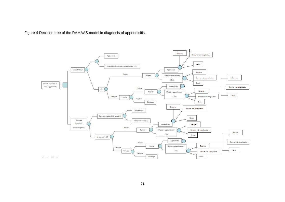
78
Figure 4 Decision tree of the RAMAAS model in diagnosis of appendicitis.

79
APPENDICES
A) Case record forms
โครงการศกษา ระบบคะแนนเพอชวยวนจฉยภาวะไสตงอกเสบ "รามาธบด อะเพนดไซทส สกอร"
Form 1: Eligible data and preoperative EQ5D
วน/เดอน/ป (พศ.) // HN
ผกรอกขอมล …………………………………….
สถานทเกบขอมล 1. รามาธบด 2. ธรรมศาสตร 3. ชลบร กรณาทาเครองหมายกากบาทหนาเกณฑการคดเลอกถาผ ปวยผานเกณฑคดเลอกขอนน ผปวยทเขารวมการวจยตองผานเกณฑเขารวมการวจยครบทกขอ ดงตอไปน
ปวดทองดานขวาเปนเวลา 7 วน
ไดรบการวนจฉย หรอสงสยวาเปนไสตงอกเสบ
มอายระหวาง 18 ถง 60 ป ไมไดตงครรภ
สามารถอาน และสอสารภาษาไทยได ไมไดรบประทานยาในกลมสเตอรอยด เชน prednisolone หรอยากดภมคมกน เชน methotrexate, cyclophosphamide,
azathioprine, mercaptopurine, anthracycline, mitomycin, Ciclosporin, Tacrolimus ไมเคยไดรบการวนจฉยวา
- เปนผปวยตดเชอ HIV ทมอาการ AIDS เชน cryptococcal meningitis, tuberculosis, pneumocystis carinii infection
- ไมเคยมประวตโรคไตวายระยะสดทาย - ไมเคยมประวตโรคตอตานภมคมกนตนเอง เชน โรค SLE - ไมเปนผปวยภาวะตบแขงทมทองมานรวมดวย - ไมมภาวะอวนผดปกต (BMI > 40 kg/m2) - ไมเปนผปวยทเปนมะเรงชองทองในระยะสดทาย
ในกรณทผ ปวยผานเกณฑเขารวมวจยครบทกขอ ใหขอมลผ ปวยเกยวเนองกบการวจย (Information process and consent form) ยนดทจะเขารวมการวจย และ เซน inform consent ยนด (participants) Participants ID - ไมยนด (non-participants) Non-participants ID -
ลงขอมลในแบบขอมลพนฐาน และคณภาพชวต
ถาผ ปวยยนด เขารวมโครงการ (Patient’s log sheet and preoperative EQ5D)

80
โครงการศกษา ระบบคะแนนเพอชวยวนจฉยภาวะไสตงอกเสบ "รามาธบด อะเพนดไซทส สกอร"
Form 1: Eligible data and preoperative EQ5D
ID -
แบบประเมนคณภาพชวตกอนผาตด (Preoperative EQ5D)
ภาวะการเจบปวยของทานในปจจบนนมผลตอทานในหวขอตอไปนอยางไรบาง
1. การเคลอนไหว 1. ขาพเจาไมมปญหาในการเดน
2. ขาพเจามปญหาในการเดนบาง 3. ขาพเจาไมสามารถไปไหนได และจาเปนตองอยบนเตยง
2. การดแลตนเอง 1.ขาพเจาไมมปญหาในการดแลตนเอง 2.ขาพเจามปญหาในการอาบนาหรอการแตงตวบาง
3.ขาพเจาไมสามารถอาบนาหรอแตงตวดวยตนเองได 3. กจกรรมททาเปนประจา (เชน การทางาน, การเรยนหนงสอ, การทางานบาน การทากจกรรมในครอบครว หรอการทากจกรรมยามวาง) 1.ขาพเจาไมมปญหาในการทากจกรรมททาเปนประจา 2.ขาพเจามปญหาในการทากจกรรมททาเปนประจาอยบาง 3.ขาพเจาไมสามารถทากจกรรมททาเปนประจาได 4. ความเจบปวด/ 1.ขาพเจาไมมอาการเจบปวดหรออาการไมสขสบาย ความไมสขสบาย 2.ขาพเจามอาการเจบปวดหรออาการไมสขสบายปานกลาง 3.ขาพเจามอาการเจบปวดหรออาการไมสขสบายมากทสด 5. ความวตกกงวล/ 1.ขาพเจาไมรสกวตกกงวลหรอซมเศรา ความซมเศรา 2.ขาพเจารสกวตกกงวลหรอซมเศราปานกลาง 3.ขาพเจารสกวตกกงวลหรอซมเศรามากทสด

81
โครงการศกษา ระบบคะแนนเพอชวยวนจฉยภาวะไสตงอกเสบ "รามาธบด อะเพนดไซทส สกอร"
Form 2: Patient’s log sheet
HN Participants ID -
ขอมลผ ปวย
ชอ ........................................ นามสกล ..................................................................................... วน/เดอน/ป เกด (พ.ศ.) // ทอย บานเลขท ............................... หม ............... ซอย .....................................................................
หมบาน ............................................. ถนน ....................................ตาบล/แขวง .................................... จงหวด ............................................. รหสไปรษณย ................................... เบอรโทรศพทบาน .................................................. เบอรโทรศพทเคลอนท ........................................... ญาตทสามารถตดตอไดชอ ...................................... เบอรโทรศพทเคลอนท ...........................................
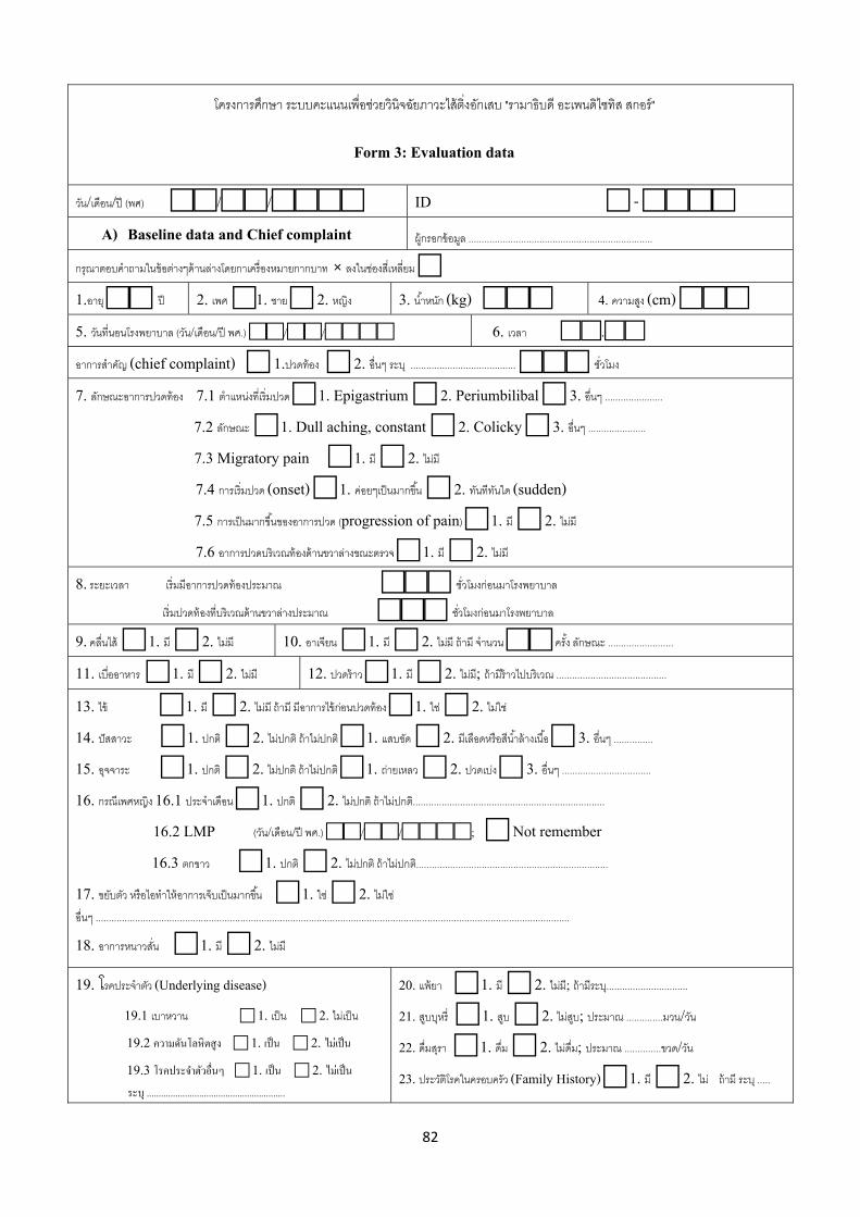
82
โครงการศกษา ระบบคะแนนเพอชวยวนจฉยภาวะไสตงอกเสบ "รามาธบด อะเพนดไซทส สกอร"
Form 3: Evaluation data
วน/เดอน/ป (พศ) // ID - A) Baseline data and Chief complaint ผกรอกขอมล ......................................................................
กรณาตอบคาถามในขอตางๆดานลางโดยกาเครองหมายกากบาท × ลงในชองสเหลยม 1.อาย กป 2. เพศ 1. ชาย 2. หญง 3. นาหนก (kg) 4. ความสง (cm)
5. วนทนอนโรงพยาบาล (วน/เดอน/ป พศ.) // 6. เวลา .
อาการสาคญ (chief complaint) 1.ปวดทอง 2. อนๆ ระบ ........................................ ชวโมง
7. ลกษณะอาการปวดทอง 7.1 ตาแหนงทเรมปวด 1. Epigastrium 2. Periumbilibal 3. อนๆ ......................
7.2 ลกษณะ 1. Dull aching, constant 2. Colicky 3. อนๆ ...................... 7.3 Migratory pain 1. ม 2. ไมม
7.4 การเรมปวด (onset) 1. คอยๆเปนมากขน 2. ทนททนใด (sudden) 7.5 การเปนมากขนของอาการปวด (progression of pain) 1. ม 2. ไมม 7.6 อาการปวดบรเวณทองดานขวาลางขณะตรวจ 1. ม 2. ไมม
8. ระยะเวลา เรมมอาการปวดทองประมาณ ชวโมงกอนมาโรงพยาบาล
เรมปวดทองทบรเวณดานขวาลางประมาณ ชวโมงกอนมาโรงพยาบาล
9. คลนไส 1. ม 2. ไมม 10. อาเจยน 1. ม 2. ไมม ถาม จานวน ครง ลกษณะ .........................
11. เบออาหาร 1. ม 2. ไมม 12. ปวดราว 1. ม 2. ไมม; ถามราวไปบรเวณ ..........................................
13. ไข 1. ม 2. ไมม ถาม มอาการไขกอนปวดทอง 1. ใช 2. ไมใช 14. ปสสาวะ 1. ปกต 2. ไมปกต ถาไมปกต 1. แสบขด 2. มเลอดหรอสนาลางเนอ 3. อนๆ ............... 15. อจจาระ 1. ปกต 2. ไมปกต ถาไมปกต 1. ถายเหลว 2. ปวดเบง 3. อนๆ .................................. 16. กรณเพศหญง 16.1 ประจาเดอน 1. ปกต 2. ไมปกต ถาไมปกต......................................................................... 16.2 LMP (วน/เดอน/ป พศ.) //; Not remember
16.3 ตกขาว 1. ปกต 2. ไมปกต ถาไมปกต......................................................................... 17. ขยบตว หรอไอทาใหอาการเจบเปนมากขน 1. ใช 2. ไมใช อนๆ .................................................................................................................................................................................... 18. อาการหนาวสน 1. ม 2. ไมม
19. โรคประจาตว (Underlying disease)
19.1 เบาหวาน 1. เปน 2. ไมเปน 19.2 ความดนโลหตสง 1. เปน 2. ไมเปน 19.3 โรคประจาตวอนๆ 1. เปน 2. ไมเปน ระบ ..........................................................
20. แพยา 1. ม 2. ไมม; ถามระบ...............................
21. สบบหร 1. สบ 2. ไมสบ; ประมาณ ..............มวน/วน 22. ดมสรา 1. ดม 2. ไมดม; ประมาณ ..............ขวด/วน
23. ประวตโรคในครอบครว (Family History) 1. ม 2. ไม ถาม ระบ .....

83
โครงการศกษา ระบบคะแนนเพอชวยวนจฉยภาวะไสตงอกเสบ "รามาธบด อะเพนดไซทส สกอร"
Form 3: Evaluation data
B) Physical examination ID - Vital sign: 1. BT . °c 2. PR /min 3. BP / mmHg 4. RR /min HEENT 5. Pale conjunctiva 1. Yes 2. No 6. Icteric sclera 1. Yes 2. No Respiratory tract 7. Breath sound 1. Normal 2. Abnormal; specify …………………………………. Cardiovascular 8. Heart sound 1. Normal 2. Abnormal; specify …………………………………. 9. Heart rate 1. Regular 2. Irregular; specify …………………………………. Abdomen 10. Inspection 1. Normal 2. Abnormal; specify …………………………………………. 11. Bowel sound 1. Normal 2. Decrease 3. Increase; specify site ……………………. 12. Tender 1. Yes 2. No; If yes, site of tender 1. RLQ 2. LLQ 3. RUQ 4. RLQ+LLQ 5. generalized 13. Rebound tenderness 1. Yes 2. No; If yes, site of tender 1. RLQ 2. LLQ 3. RUQ 4. RLQ+LLQ 5. Generalized 14. Guarding 1. Yes 2. No 15. Rigidity 1. Yes 2. No 16. Indirect rebound tenderness (Rovsing’s sign) 1. Positive 2. Negative 17. Psoas’s sign 1. Positive 2. Negative 18. Obturator’s sign 1. Positive 2. Negative Per rectal examination 19. Tender 1. Yes 2. No 3. Not perform; specify ….………………………… Per vaginal examination (Only female) 20. 1. Normal 2. Abnormal 3. Not perform 21. Specify if abnormal …………………………………………………………………. 22. Gynecologic suggestion 1.No Gynecologic condition 2. Gynecologic condition Others

84
โครงการศกษา ระบบคะแนนเพอชวยวนจฉยภาวะไสตงอกเสบ "รามาธบด อะเพนดไซทส สกอร"
Form 3: Evaluation data
C) Laboratory and radiologic results ID - Complete blood cell count 1. WBC (cell/mm3) 2. Hemoglobin (mg %) . 3. Hematocrit (%) 4. Neutrophil (%) 5. Lymphocyte (%) Urinalysis 1. White blood cell count (cells/HF) f- 2. Red blood cell count (cells/HF) f- 3. specific gravity . 4. epithelial cell f- BUN/Cr (mg %) / Blood sugar (mg %) ; not performed Anti HIV 1. Check 2. Don’t check; If check 1. Positive 2. Negative LFT: not performed; if performed, Albumin . g/dL, others .………………………………………… Ultrasound abdomen 1. Done 2. Not done Results 1. Appendicitis 2. Suspected appendicitis 3. Not appendicitis If done specify ………………………………………………………………………………. CT scans 1. Done 2. Not done; Results 1. Appendicitis 2. Suspected appendicitis 3. Not appendicitis If done specify ………………………………………………………………………………. Others Provisional diagnosis …………………………………………………….. Differential diagnosis 1. ………………………………………….. 2. ………………………………………….. 3. …………………………………………..

85
โครงการศกษา ระบบคะแนนเพอชวยวนจฉยภาวะไสตงอกเสบ "รามาธบด อะเพนดไซทส สกอร" Form 4: Operative data
ID - เวลาเรม . am/pm สนสด . am/pm ชอ นามสกล วน/เดอน/ป ทผาตด (พศ)//
แพทยผาตด นามสกล 1.แพทยใชทน 2.แพทยประจาบาน 3.ศลยแพทย
Anesthesia 1.GA 2.Spinal block 3. Epidural Scrub 1.Betadine 2.Chlorhexidine ASA classification I II III IV ; specify …………………………………………… Preoperative antibiotics 1.Yes 2.No; If yes please specify Postoperative diagnosis Procedure 1 Incision Type of incision 1.Mcburney’s 2.Lanz 3.Additional midline Incisional length . cm. Subcutaneous fat thickness . cm Findings : Appendix Location 1.Paracecal 2.Retrocecal 3.Pelvic type Erythematous or swelling of appendix 1.Yes 2.No (No = negative appendectomy) Severity of appendicitis : Suppurative : Suppurative appendicitis (No frank pus) 1.Yes 2.No Gangrenous : Gangrenous wall (dark, grayish color) 1.Yes 2.No Ruptured : 1.Yes 2.No Ruptured diagnosis criteria: Hole in appendix 1.Yes 2.No Intraoperative rupture 1.Yes 2.No Frank pus 1.Yes 2.No If frank pus is present; visible wound contamination 1.Yes 2.No Degree of contamination Exudative fluid 1.Yes 2.No; If yes, visible wound contamination 1.Yes 2.No Fecal material 1.Yes 2.No; If yes, visible wound contamination 1.Yes 2.No Phlegmon 1.Yes 2.No Fecalith 1.Yes 2.No Localized abscess formation 1.Yes 2.No Other findings ……………………………………………………………………………………………………….

86
โครงการศกษา ระบบคะแนนเพอชวยวนจฉยภาวะไสตงอกเสบ "รามาธบด อะเพนดไซทส สกอร" Form 4: Operative data (cont.)
ID -
Procedures related data Appendiceal stump was secured with 1. Silk 2#0 double ligation 2. Chromic ligation 3. Suture ligation with ………………… 4. Others ………………………… Purse string suture 1. Done 2. Not done Closed suction drain 1. Used 2. Did not use Culture of intra-abdominal fluid 1. Done 2. Not done Others ……………………………………………………………………………………………………………… ……………………………………………………………………………………………………………………… ……………………………………………………………………………………………………………………..

87
โครงการศกษา ระบบคะแนนเพอชวยวนจฉยภาวะไสตงอกเสบ "รามาธบด อะเพนดไซทส สกอร"
Form 5: Postoperative Inpatient data
Participants ID - A) Postoperative pain
- Postoperative day 1 date (dd/mm/yyyy) / /
- Postoperative day 3 date (dd/mm/yyyy) / /
- Use of opioids (morphine) as required yes No if yes,
Day1 dosage …………….mg/day, Day2 dosage……………./day
B) แบบประเมนคณภาพชวต (EQ5D) Postoperative day 3; date (dd/mm/yyyy) / / การผาตดททาใหมแผลผาตดในครงนมผลตอทานในหวขอตอไปนอยางไรบาง
1. การเคลอนไหว 1. ขาพเจาไมมปญหาในการเดน
2. ขาพเจามปญหาในการเดนบาง 3. ขาพเจาไมสามารถไปไหนได และจาเปนตองอยบนเตยง
2. การดแลตนเอง 1.ขาพเจาไมมปญหาในการดแลตนเอง 2.ขาพเจามปญหาในการอาบนาหรอการแตงตวบาง
3.ขาพเจาไมสามารถอาบนาหรอแตงตวดวยตนเองได
3. กจกรรมททาเปนประจา (เชน การทางาน, 1.ขาพเจาไมมปญหาในการทากจกรรมททาเปนประจา การเรยนหนงสอ, การทางานบาน, การทากจกรรม 2.ขาพเจามปญหาในการทากจกรรมททาเปนประจาอยบาง ในครอบครว หรอการทากจกรรมยามวาง) 3.ขาพเจาไมสามารถทากจกรรมททาเปนประจาได
4. ความเจบปวด/ 1.ขาพเจาไมมอาการเจบปวดหรออาการไมสขสบาย ความไมสขสบาย 2.ขาพเจามอาการเจบปวดหรออาการไมสขสบายปานกลาง 3.ขาพเจามอาการเจบปวดหรออาการไมสขสบายมากทสด
5. ความวตกกงวล/ 1.ขาพเจาไมรสกวตกกงวลหรอซมเศรา ความซมเศรา 2.ขาพเจารสกวตกกงวลหรอซมเศราปานกลาง 3.ขาพเจารสกวตกกงวลหรอซมเศรามากทสด

88
โครงการศกษา ระบบคะแนนเพอชวยวนจฉยภาวะไสตงอกเสบ "รามาธบด อะเพนดไซทส สกอร"
Form 5: Postoperative Inpatient data (Cont.)
Participants ID - Discharge date (dd/mm/yyyy) / / C) การประเมนภาวะแทรกซอนหลงผาตด Postoperative day ; date (dd/mm/yyyy) / / สรปผลการวนจฉย 1. ตดเชอ 2. ไมตดเชอ โดยพบเกณฑของการตดเชอทแผลผาตดดงตอไปน
Infection occur within 30 days after the operative procedure AND Involves only skin and subcutaneous tissue of the incision AND one of the following 1. Purulent drainage from the superficial incision 2. Culture of fluid or tissue from the superficial incision positive 3. At least one of below sign or symptoms : pain or tenderness, localized swelling, redness, or heat PLUS
The superficial incision is deliberately opened by surgeon Opened date / /
ในผปวยทพบวามการตดเชอทแผลผาตด (SSI) Temperature at diagnosis of SSI . °C; WBC (cell/mm3)
Use of antibiotics: 1. Intravenous 2. Oral 3. None 1. Ceftriazone + Metronidazole 2. Ciprofloxacin + Metronidazole 3. Augmentin 4. Others, specify …………………………………………………………………… ผ ปวยไดรบการเยบปดแผลภายหลงจากการวนจฉยวามการตดเชอทแผลผาตด (resuture) 1. Yes 2. No If yes, resuture date (dd/mm/yyyy) / /
ภาวะแทรกซอนอนๆ (Other complications)
Pneumonia 1. Yes 2. No
Intraabdominal infection 1. Yes 2. No
Wound hematoma 1. Yes 2. No
Urinary tract infection 1. Yes 2. No
Thrombophlebitis 1. Yes 2. No
อนๆ 1. Yes 2. No ถาม.......................................

89
โครงการศกษา ระบบคะแนนเพอชวยวนจฉยภาวะไสตงอกเสบ "รามาธบด อะเพนดไซทส สกอร"
Form 6: Follow up 1 (Day 7-10 after discharge)
Participants ID - Follow up 1. Yes 2. No; date / /
A) ภาวะแทรกซอนหลงผาตด สรปผลการวนจฉย 1. ตดเชอ 2. ไมตดเชอ โดยพบเกณฑของการตดเชอทแผลผาตดดงตอไปน
Infection occur within 30 days after the operative procedure AND Involves only skin and subcutaneous tissue of the incision AND With one of the following 1. Purulent drainage from the superficial incision 2. Culture of fluid or tissue from the superficial incision positive 3. At least one of below sign or symptoms : pain or tenderness, localized swelling, redness, or heat PLUS
The superficial incision is deliberately opened by surgeon Opened date / /
Use of antibiotics: 1. Intravenous 2. Oral 3. No; If yes: 1. Ceftriazone + Metronidazole 2. Ciprofloxacin + Metronidazole 3. Augmentin 4. Others, specify……………………………. ในผปวยทพบวามการตดเชอทแผลผาตด
Temperature at diagnosis of SSI . °C WBC (cell/mm3)
ผ ปวยไดรบการเยบปดแผลภายหลงจากการวนจฉยวามการตดเชอทแผลผาตด (resuture) 1. Yes 2. No If yes, resuture date (dd/mm/yyyy) / /
ภาวะแทรกซอนอนๆ (Other complications)
Pneumonia 1. Yes 2. No
Intraabdominal infection 1. Yes 2. No
Wound hematoma 1. Yes 2. No
Urinary tract infection 1. Yes 2. No
Thrombophlebitis 1. Yes 2. No
อนๆ 1. Yes 2. No ถาม.................................

90
ผลการประเมนทางโทรศพทในกรณทผปวยไมไดมาตรวจตดตาม 1. แผลไมมลกษณะบวมแดง 2. อาการเจบแผลลดลง 3. แผลแหงไมมนาเหลอง หรอหนองออกจากแผล 4. ไมไดไปพบแพทยทใด ถามเกณฑดงกลาวครบทกขอ ใหสรปวาผ ปวยไมมการตดเชอทแผลผาตด ถามเกณฑไมครบใหแนะนาผ ปวยใหมาตรวจตดตาม สรปผลการวนจฉยจากการประเมนทางโทรศพท 1. ตดเชอ 2. ไมตดเชอ 3. ไมชดเจน แนะนาใหมาตรวจ
สรปผลการวนจฉยในกรณทผ ปวยมาตรวจตามคาแนะนา 1. ตดเชอ 2. ไมตดเชอ
ภาวะแทรกซอนอนๆ (Other complications)
Pneumonia 1. Yes 2. No
Intraabdominal infection 1. Yes 2. No
Wound hematoma 1. Yes 2. No
Urinary tract infection 1. Yes 2. No Thrombophlebitis 1. Yes 2. No อนๆ 1. Yes 2. No ถาม......................................

91
โครงการศกษา ระบบคะแนนเพอชวยวนจฉยภาวะไสตงอกเสบ "รามาธบด อะเพนดไซทส สกอร" Form 6: Follow up 1 (Day 7-10 after discharge)
Participants ID - Follow up date (dd/mm/yyyy) / / B) ผลการตรวจชนเนอ (Pathological examination)
Appendicitis 1. Complicated 2. Not complicated If complicated; 1. Gangrenous 2. Ruptured
C) ตนทนทางออมทเกดกบผปวยและญาต (Indirect cost) ชวงนอนโรงพยาบาล พกฟนและการตรวจตามนดครงท 1 C1 ตนทนทางออมทเกด ชวงผปวยนอนโรงพยาบาล
คาเดนทางไปและกลบโรงพยาบาลของผปวย บาท มญาตมาดแลในระหวางทนอนรกษาในโรงพยาบาลครงนหรอไม ม ไมม
ถาผปวยมญาตดแลใหลงขอมลดานลาง
- คาเดนทางไปและกลบโรงพยาบาลของญาต บาท/ครง; จานวนครงทมาโรงพยาบาล ครง - มญาตดแลทงหมดจานวน คน/วน
ญาตตองดแลผปวยโดยรวมทงหมด วน ญาตใชเวลาในการดแลโดยเฉลยตอวน 1.มากกวาครงวน หรอเตมวน 2. ครงวน หรอนอยกวาครงวน
ญาตคนท 1 ทดแลผ ปวยในโรงพยาบาลครงน เพศ 1. ชาย 2. หญง อาย ป ประกอบอาชพหรอไม 1.ไม 2.ประกอบอาชพ โดยมรายไดเฉลยเดอนละ บาท
ญาตคนท 2 ทดแลผ ปวยในโรงพยาบาลครงน เพศ 1. ชาย 2. หญง อาย ป ประกอบอาชพหรอไม 1.ไม 2.ประกอบอาชพ โดยมรายไดเฉลยเดอนละ บาท
ญาตคนท 3 ทดแลผ ปวยในโรงพยาบาลครงน เพศ 1. ชาย 2. หญง อาย ป ประกอบอาชพหรอไม 1.ไม 2.ประกอบอาชพ โดยมรายไดเฉลยเดอนละ บาท
- คาอาหารสวนทเพมขนจากปกตของญาต ในการดแลผปวยระหวางนอนโรงพยาบาล จานวนมอทตองเสยเพม มอ /คน จานวน คน คาใชจายในการรบประทานอาหารทตองเสยเพมประมาณ บาท/มอ/คน
- คาทพกของญาต เสย ไมเสย เสยคาทพกทงหมดจานวน คน คนละประมาณ บาท
C2 ตนทนทางออมทเกด ชวงผปวยพกฟน
ผ ปวยสามารถกลบไปทากจวตรประจาวนไดตามปกตหลงไดรบการรกษา วน ผปวยสามารถกลบไปทางานไดตามปกตหลงไดรบการรกษา วน
มญาตมาดแลในระหวางท ผปวยพกฟน หรอไม ม ไมม ถาผปวยมญาตดแลใหลงขอมลดานลาง

92
โครงการศกษา ระบบคะแนนเพอชวยวนจฉยภาวะไสตงอกเสบ "รามาธบด อะเพนดไซทส สกอร"
Form 6: Follow up 1 (Day 7-10 after discharge)
Participants ID - Follow up date (dd/mm/yyyy) / / C2 ตนทนทางออมทเกด ชวงผปวยพกฟน (ตอ)
- มญาตดแลทงหมดจานวน คน/วน
ญาตตองดแลผปวยโดยรวมทงหมด วน
ญาตใชเวลาในการดแลโดยเฉลยตอวน 1.มากกวาครงวน หรอเตมวน 2. ครงวน หรอนอยกวาครงวน ญาตคนท 1 ทดแลผ ปวยในโรงพยาบาลครงน เพศ 1. ชาย 2. หญง อาย ป ประกอบอาชพหรอไม 1.ไม 2.ประกอบอาชพ โดยมรายไดเฉลยเดอนละ บาท
ญาตคนท 2 ทดแลผ ปวยในโรงพยาบาลครงน เพศ 1. ชาย 2. หญง อาย ป ประกอบอาชพหรอไม 1.ไม 2.ประกอบอาชพ โดยมรายไดเฉลยเดอนละ บาท
ญาตคนท 3 ทดแลผ ปวยในโรงพยาบาลครงน เพศ 1. ชาย 2. หญง อาย ป ประกอบอาชพหรอไม 1.ไม 2.ประกอบอาชพ โดยมรายไดเฉลยเดอนละ บาท
C3 ตนทนทางออมทเกดจากการมาตรวจตดตามครงน (follow up) การเดนทาง มา โรงพยาบาล คาเดนทางของผ ปวย บาท การเดนทาง กลบ คาเดนทางของผ ปวย บาท คาอาหารสวน ทเพมขนของผปวย จากชวตปกต ในการมาโรงพยาบาลครงน ตองรบประทานอาหารทงหมด มอ คาใชจายในการรบประทานอาหารของผปวยประมาณ บาทตอมอ โดยปกตผปวยมคาใชจายในการรบประทานอาหารประมาณ บาทตอมอ คาทพกในกรณทเดนทางมาตรวจจากทไกล คาทพกประมาณ บาท
- มญาตมาดวยทงหมดจานวน คน ญาตคนท 1 ทพาผ ปวยมาโรงพยาบาลครงน เพศ 1. ชาย 2. หญง อาย ป ประกอบอาชพหรอไม 1.ไม 2.ประกอบอาชพ โดยมรายไดเฉลยเดอนละ บาท
ญาตคนท 2 ทพาผ ปวยมาโรงพยาบาลครงน เพศ 1. ชาย 2. หญง อาย ป ประกอบอาชพหรอไม 1.ไม 2.ประกอบอาชพ โดยมรายไดเฉลยเดอนละ บาท
ญาตคนท 3 ทพาผ ปวยมาโรงพยาบาลครงน เพศ 1. ชาย 2. หญง อาย ป ประกอบอาชพหรอไม 1.ไม 2.ประกอบอาชพ โดยมรายไดเฉลยเดอนละ บาท
- คาเดนทางไปและกลบ ของญาตในกรณทไมไดมากบผปวยประมาณ บาท - คาอาหาร สวนทเพมขน ของญาตทกคน จากคาใชจายปกต ทงหมดประมาณ บาท - คาทพกของญาตในกรณทเดนทางมาตรวจจากทไกล คาทพกประมาณ บาท

93
โครงการศกษา ระบบคะแนนเพอชวยวนจฉยภาวะไสตงอกเสบ "รามาธบด อะเพนดไซทส สกอร"
Form 7: Follow up 2 (Day 30-35 after discharge)
Participants ID 1 - Follow up date (dd/mm/yyyy) / /
สาหรบผปวยทไมไดมาตรวจตดตามนด วนทสมภาษณ (dd/mm/yyyy) / ผลการประเมนทางโทรศพทในกรณทผปวยไมไดมาตรวจตดตาม 1. แผลไมมลกษณะบวมแดง 2. อาการเจบแผลลดลง 3. แผลแหงไมมนาเหลอง หรอหนองออกจากแผล 4. ไมไดไปพบแพทยทใด ถามเกณฑดงกลาวครบทกขอ ใหสรปวาผ ปวยไมมการตดเชอทแผลผาตด ถามเกณฑไมครบใหแนะนาผ ปวยใหมาตรวจตดตาม สรปผลการวนจฉยจากการประเมนทางโทรศพท 1. ตดเชอ 2. ไมตดเชอ 3. ไมชดเจน แนะนาใหมาตรวจ
สรปผลการวนจฉยในกรณทผ ปวยมาตรวจตามคาแนะนา 1. ตดเชอ 2. ไมตดเชอ
ภาวะแทรกซอนอนๆ (Other complications)
Pneumonia 1. Yes 2. No
Intraabdominal infection 1. Yes 2. No
Wound hematoma 1. Yes 2. No
Urinary tract infection 1. Yes 2. No Thrombophlebitis 1. Yes 2. No อนๆ 1. Yes 2. No D) ผลการตรวจเพาะเชอ Operative culture Abdominal fluid culture positive negative; colony count * 10˄), results…………………………… Subcutaneous layer culture positive negative; colony count * 10˄), results…………………………… Incision before closure (DPC): positive negative; colony count * 10˄), results…………………………… Incision of DPC at day 4: positive negative; colony count * 10˄), results……………………………. SSI: positive negative; colony count * 10˄), results……………………………..
E) ตนทนทางออมทเกดกบผปวยและญาต (Indirect cost) ในการตรวจตามนดครงท 2 (follow up) การเดนทาง มา โรงพยาบาล คาเดนทางของผ ปวย บาท การเดนทาง กลบ คาเดนทางของผ ปวย บาท คาอาหารสวน ทเพมขนของผปวย จากชวตปกต ในการมาโรงพยาบาลครงน ตองรบประทานอาหารทงหมด มอ คาใชจายในการรบประทานอาหารของผปวยประมาณ บาทตอมอ โดยปกตผปวยมคาใชจายในการรบประทานอาหารประมาณ บาทตอมอ

94
คาทพกในกรณทเดนทางมาตรวจจากทไกล คาทพกประมาณ บาท - มญาตมาดวยทงหมดจานวน คน
ญาตคนท 1 ทพาผ ปวยมาโรงพยาบาลครงน เพศ 1. ชาย 2. หญง อาย ป ประกอบอาชพหรอไม 1.ไม 2.ประกอบอาชพ โดยมรายไดเฉลยเดอนละ บาท
ญาตคนท 2 ทพาผ ปวยมาโรงพยาบาลครงน เพศ 1. ชาย 2. หญง อาย ป ประกอบอาชพหรอไม 1.ไม 2.ประกอบอาชพ โดยมรายไดเฉลยเดอนละ บาท

95
โครงการศกษา ระบบคะแนนเพอชวยวนจฉยภาวะไสตงอกเสบ "รามาธบด อะเพนดไซทส สกอร"
Form 7: Follow up 2 (Day 30-35 after discharge)
Participants ID 1 - Follow up date (dd/mm/yyyy) / / C) ตนทนทางออมทเกดกบผปวยและญาต (Indirect cost) ในการตรวจตามนดครงท 2 (follow up) (ตอ) ญาตคนท 3 ทพาผ ปวยมาโรงพยาบาลครงน เพศ 1. ชาย 2. หญง อาย ป ประกอบอาชพหรอไม 1.ไม 2.ประกอบอาชพ โดยมรายไดเฉลยเดอนละ บาท
- คาเดนทางไปและกลบ ของญาตในกรณทไมไดมากบผปวยประมาณ บาท - คาอาหาร สวนทเพมขน ของญาตทกคน จากคาใชจายปกต ทงหมดประมาณ บาท - คาทพกของญาตในกรณทเดนทางมาตรวจจากทไกล คาทพกประมาณ บาท
D) แบบประเมนคณภาพชวต (EQ5D) 1 month date (dd/mm/yyyy) / /
ภาวะการเจบปวยของทานในปจจบนนมผลตอทานในหวขอตอไปนอยางไรบาง
1. การเคลอนไหว 1. ขาพเจาไมมปญหาในการเดน
2. ขาพเจามปญหาในการเดนบาง 3. ขาพเจาไมสามารถไปไหนได และจาเปนตองอยบนเตยง
2. การดแลตนเอง 1.ขาพเจาไมมปญหาในการดแลตนเอง 2.ขาพเจามปญหาในการอาบนาหรอการแตงตวบาง
3.ขาพเจาไมสามารถอาบนาหรอแตงตวดวยตนเองได 3. กจกรรมททาเปนประจา (เชน การทางาน, การเรยนหนงสอ, การทางานบาน การทากจกรรมในครอบครว หรอการทากจกรรมยามวาง) 1.ขาพเจาไมมปญหาในการทากจกรรมททาเปนประจา 2.ขาพเจามปญหาในการทากจกรรมททาเปนประจาอยบาง
3.ขาพเจาไมสามารถทากจกรรมททาเปนประจาได
4. ความเจบปวด/ 1.ขาพเจาไมมอาการเจบปวดหรออาการไมสขสบาย ความไมสขสบาย 2.ขาพเจามอาการเจบปวดหรออาการไมสขสบายปานกลาง 3.ขาพเจามอาการเจบปวดหรออาการไมสขสบายมากทสด
5. ความวตกกงวล/ 1.ขาพเจาไมรสกวตกกงวลหรอซมเศรา ความซมเศรา 2.ขาพเจารสกวตกกงวลหรอซมเศราปานกลาง 3.ขาพเจารสกวตกกงวลหรอซมเศรามากทสด
96
โครงการศกษา ระบบคะแนนเพอชวยวนจฉยภาวะไสตงอกเสบ "รามาธบด อะเพนดไซทส สกอร"
Form 8: Additional follow up
Participants ID - Follow up date (dd/mm/yyyy) / /
สาเหตของการมาตรวจตดตามครงน 1. สงสยตดเชอ 2. Pain 3. Others; specify…………………………………..
ผลการตรวจ SSI no SSI ตนทนทางออมทเกดจากการมาตรวจตดตามครงน (follow up)
การเดนทาง มา โรงพยาบาล คาเดนทางของผ ปวย บาท
การเดนทาง กลบ คาเดนทางของผ ปวย บาท คาอาหารสวน ทเพมขนของผปวย จากชวตปกต ในการมาโรงพยาบาลครงน ตองรบประทานอาหารทงหมด มอ
คาใชจายในการรบประทานอาหารของผปวยประมาณ บาทตอมอ โดยปกตผปวยมคาใชจายในการรบประทานอาหารประมาณ บาทตอมอ คาทพกในกรณทเดนทางมาตรวจจากทไกล คาทพกประมาณ บาท
- มญาตมาดวยทงหมดจานวน คน ญาตคนท 1 ทพาผ ปวยมาโรงพยาบาลครงน เพศ 1. ชาย 2. หญง อาย ป ประกอบอาชพหรอไม 1.ไม 2.ประกอบอาชพ โดยมรายไดเฉลยเดอนละ บาท
ญาตคนท 2 ทพาผ ปวยมาโรงพยาบาลครงน เพศ 1. ชาย 2. หญง อาย ป ประกอบอาชพหรอไม 1.ไม 2.ประกอบอาชพ โดยมรายไดเฉลยเดอนละ บาท
ญาตคนท 3 ทพาผ ปวยมาโรงพยาบาลครงน เพศ 1. ชาย 2. หญง อาย ป ประกอบอาชพหรอไม 1.ไม 2.ประกอบอาชพ โดยมรายไดเฉลยเดอนละ บาท
- คาเดนทางไปและกลบ ของญาตในกรณทไมไดมากบผปวยประมาณ บาท - คาอาหาร สวนทเพมขน ของญาตทกคน จากคาใชจายปกต ทงหมดประมาณ บาท - คาทพกของญาตในกรณทเดนทางมาตรวจจากทไกล คาทพกประมาณ บาท

97
B) Manual of data collection คมอการเกบขอมล
การศกษาระบบคะแนนเพอชวยวนจฉยภาวะไสตงอกเสบ "รามาธบด อะเพนดไซทส สกอร"
สถานท ลกษณะผปวย รายละเอยดงานทตองทา ผรบผดชอบ หองตรวจผปวยทแผนกฉกเฉนหรอหนวยตรวจผปวยนอกศลยกรรม
- ผปวยทกรายทมอาการปวดทองดานขวาภาวะไสตงอกเสบ - ผปวยทสงสยภาวะไสตงอกเสบ
- เชคเกณฑการเขารวมการวจย (Form 1) โดยผปวยตองผานเกณฑทกขอ
- ชวนผปวยเขารวมโครงการ โดยใหขอมลผปวยทผานเกณฑตามรายละเอยดใน
ใบ Information sheet - ถาผปวยไมเขารวม ไมตองสมภาษณตอ ใหเกบ Form 1 ไวในแฟม - ถาผปวยยนดเขารวมโครงการใหสมภาษณคณภาพชวตของผปวยกอนการผาตด
Form 1 Page 2 (Preoperative EQ5D) โดยซกถาม ผปวยถง
ผลกระทบของอาการปวดทองตอภาวะสขภาวะของผปวย - ลงขอมลใน log sheet (Form 2) - ลงขอมลใน Evaluation data (Form 3 A,B,C)
แพทยผรบผดชอบ หรอพยาบาล
หองผาตด ผปวยทไดรบการผาตดเนองจากสงสยภาวะไสตงอกเสบ
- กรอกขอมลผาตด ใน Form 4: Operative data;Page1-2 ASA classification ใหใชขอมลของวสญญ และเกณฑ
การจดกลมจากใบบนทกของวสญญ ลกษณะแผลผาตดแบบ Mc Burney เปนการลงแผลตามแนว external
oblique aponeurotic sheath Lanz แผลตามแนวผวหนง (Langer line)
เชคความครบถวน ของเกณฑการวนจฉยภาวะไสตงอกเสบแบบมเนอตาย
หรอมการแตกทะล โดยถามทงเนอตายและการแตกทะลใหวนจฉยเปนแบบแตกทะล
Finding Procedure related data
- เกบ Form เขาแฟมงานวจย
แพทยผาตด และพยาบาลหองผาตด (ตอ)
DMU & site
organization
unit
- รวบรวมแฟมวจยผปวยทเขาโครงการใหมตอนเชาทกวน - Assign ID - ตรวจความครบถวนของขอมล Form 1-4 ตดตอประสานงานกบแพทยหรอ
พยาบาลทรบผดชอบ หากขอมลไมครบถวน - หากขอมลครบถวน ถายสาเนา และสงขอมลเขา DMU - เตรยมแฟมพรอม Forms การประเมนหลงผาตด
ผชวยวจย
หอผปวยใน ผปวยหลงผาตด - เกบขอมลหลงผาตดวนท 1
- ประเมนความเจบปวด Form 5 Part A โดยอธบายพรอมใหดรป วา 0 คอไมมอาการเจบปวดเลย และ 100 คออาการ
ผชวยวจย IPD

98
เจบปวดมากทสดในชวต และใหผปวยเลอก

99
สถานท ลกษณะผ ปวย รายละเอยดงานทตองทา ผ รบผดชอบ หอผ ปวยใน (ตอ) ผ ปวยหลงผาตด (ตอ) - เกบขอมลหลงผาตดวนท 3
- ประเมนความเจบปวด Form 5 Part A - ภาวะสขภาวะ Form 5 Part B (Postoperative EQ5D)
ผชวยวจย IPD (ตอ)
- ประเมนภาวะแทรกซอนหลงการผาตด วนท 3-7 Form 5: Part C
เกณฑการวนจฉยการตดเชอ
- สาหรบกลมเยบปดแผลทนท ใหประเมนวนทๆผปวยกลบบาน
(หรอกอน 1 วน)
- สาหรบกลมเยบปดแผลในภายหลง ใหประเมนวนวนทๆเยบปด
แผล
- ในกรณทมการตดเชอทแผลผาตด กรอกขอมลเพมในสวนการตดเชอ กรอกวนทๆ เปดและปดแผล ในกรณทมการเปด หรอเยบ
ปดแผล ถาไมมใหตอบ No
แพทยผ รกษา
DMU & site organization unit
- รวบรวมแฟมวจย และตรวจความครบถวนของขอมล Form 1-5
- ตดตอประสานงานกบแพทยหรอพยาบาลทรบผดชอบ หากขอมลไมครบถวน - หากขอมลครบถวน ถายสาเนา และสงขอมลเขา DMU
- เตรยมความพรอมการตดตามผปวย แฟมพรอม Form 6: การตดตามครงท 1 โทรแจงผปวยใหมาตรวจตามนด
ผชวยวจย
FU 1: หองตรวจผ ปวยนอก
ผ ปวยหลงผาตด วนท 7-10 ผชวยวจยกลางพาผ ปวยเขาพบแพทย ผชวยวจย
- ประเมนการตดเชอทแผลผาตด Form 6: Part A
- ในกรณทมการตดเชอทแผลผาตด
- กรอกขอมลเกณฑการตดเชอ - ลงขอมลผลการตรวจชนเนอ Form 6: Part B
แพทยผประเมน
- ผชวยวจยสมภาษณผปวย ตนทนทางออมทเกดกบผปวยชวงนอนโรงพยาบาล
ชวงพกฟน และการตรวจตดตามครงท 1 Form 6: Part C1,C2,C3
ผชวยวจย
- ถาหากผปวยไมมาตามนด ใหแจง PI ของ site เพอสมภาษณอาการทางโทรศพท และกรอก Form 6: Part A
ผชวยวจย

100
สถานท ลกษณะผ ปวย รายละเอยดงานทตองทา ผ รบผดชอบ
FU 2: หองตรวจผ ปวยนอก
ผ ปวยหลงผาตด วนท 30-35
- ประเมนการตดเชอทแผลผาตด Form 7
- ในกรณทมการตดเชอทแผลผาตด Part A
- กรอกขอมลเกณฑการตดเชอ - การเยบปดแผลในภายหลง
- ผลการเพาะเชอ Part B
- ตนทนทางออม Part C
- ภาวะสขภาวะ Part D
- ในกรณทผปวยไมไดมาตรวจตดตาม สมภาษณทางโทรศพท - อาการตดเชอทแผล และกรอก Form 7: Part A
- ภาวะสขภาวะผปวย Form 7: Part D
แพทยผประเมน
หองตรวจผ ปวยนอก
หรอใน
ผ ปวยทมาตรวจนอกเหนอจาก
protocol เนองจากมการตดเชอ หรออนๆ
- ใหผชวยวจยแจงแพทยทเกยวของ และ PI
- กรอกขอมล Form 8:
- ผลการตรวจ - ขอมลคาใชจายทางออมจากการมาตรวจครงน
ผชวยวจย
DMU & site organization unit
- ผชวยวจยรวบรวมขอมล - คาใชจายทางตรง (direct cost) ของผปวยตงแตเขา
โรงพยาบาลถงการมาตรวจครงสดทาย - ตรวจความเรยบรอยของขอมล Form 1-Form 7/8
- สงแฟมขอมลทงหมดให PI ตรวจกอนสง DMU
ผชวยวจย

101
C) Calculating utility from EQ-5D
Dimension Level UK Thai Constant - 0.081 0.202
Mobility Level 1 0 0 Level 2 0.069 0.121 Level 3 0.314 0.432
Self care Level 1 0 0 Level 2 0.104 0.121 Level 3 0.214 0.242
Usual activities Level 1 0 0 Level 2 0.036 0.059 Level 3 0.094 0.118
Pain/discomfort Level 1 0 0 Level 2 0.123 0.072 Level 3 0.386 0.209
Anxiety/depression Level 1 0 0 Level 2 0.071 0.032 Level 3 0.236 0.11
N3 - 0.269 0.139

102
D) Dummy tables 1. Patients’ characteristic in appendicitis and non-appendicitis group
Parameters Appendicitis Non-appendicitis P value
Age (years) <40 ≥40
Sex M F
Onset of pain Sudden insidious
Migration of pain Presence Absence
Duration of pain < 48 hours ≥ 48 hours
Anorexia present absence
Aggravation of pain with cough present absence
Nausea present absence
Vomiting present absence
Dysuria present absence
Diarrhea present absence
Fever (T>37.8oC) present absence
Tender at RLQ present

103
absence
Abdominal guarding present absence
Rovsing sign present absence
PR tenderness present absence
Increase WBC present absence
PMN leucocytosis present absence
RLQ=right lower quadrant, PR=per rectal examination, WBC=white blood cell,
PMN=polymorphonuclear

104
2. Factors associated with appendicitis: multiple logistic regression analysis
Parameters Coefficient SE P value OR (95%CI)
Age (years) <40 ≥40
Duration of pain < 48 hours ≥ 48 hours
Anorexia present absence
Migration of pain Presence Absence
Fever (T>37.8oC) present absence
Tender at RLQ present absence
Abdominal guarding present absence
Increase WBC present absence
PMN leucocytosis present absence
RLQ=right lower quadrant, PR=per rectal examination, WBC=white blood cell,
PMN=polymorphonuclear

105
3. Scoring scheme: steps used to calculate RAMA-AS
Parameters Scoring Score for individual
Age (years) <40 ≥40
Duration of pain < 48 hours ≥ 48 hours
Anorexia present absence
Migration of pain Presence Absence
Total score ..…….. Low risk
Moderate risk High risk
………….. <X
X to Y ≥Y
RLQ=right lower quadrant, PR=per rectal examination, WBC=white blood cell,
PMN=polymorphonuclear
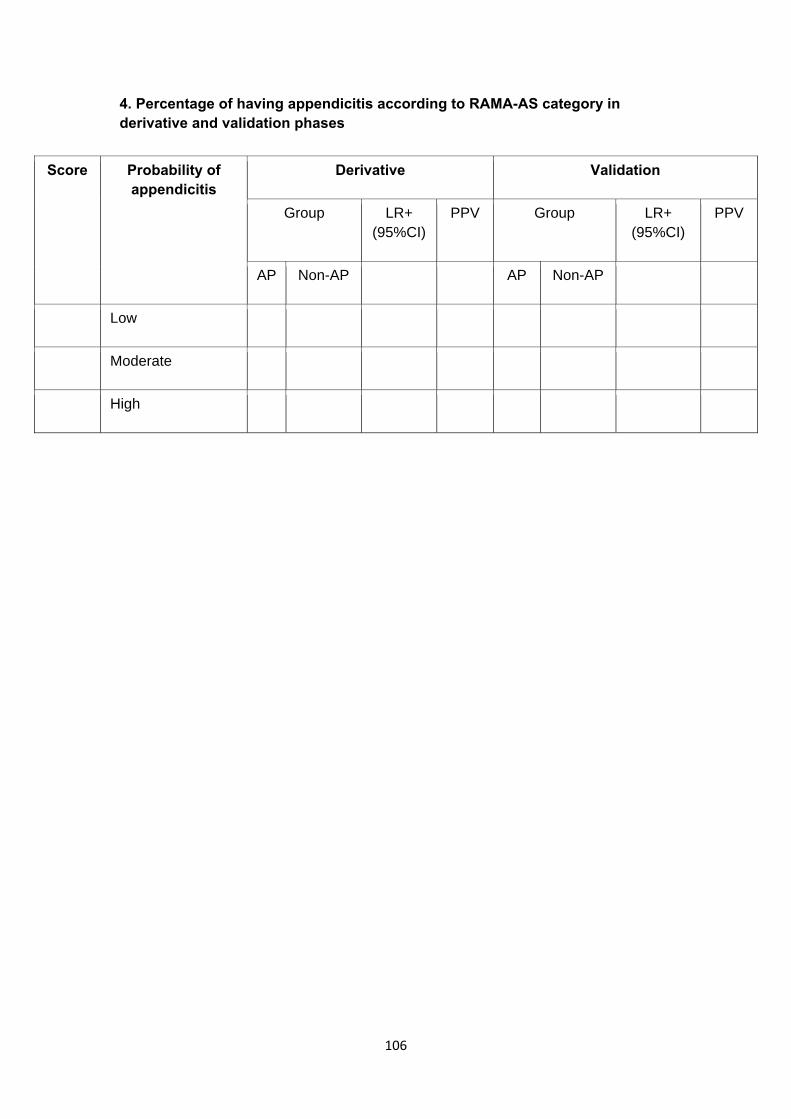
106
4. Percentage of having appendicitis according to RAMA-AS category in derivative and validation phases
Score Probability of appendicitis
Derivative Validation
Group LR+ (95%CI)
PPV Group LR+ (95%CI)
PPV
AP Non-AP AP Non-AP
Low
Moderate
High

107
5. ROC curve for predicting appendicitis
6. Comparison RAMA-AS with other scoring systems.
Scoring system No. variable/event/N O/E C-statistic
RAMA-AS
Alvarado
RIPSA
AIRS

108
B) Informal sheet & consent form
คาแนะนาผปวย (Information Sheet)
โครงการศกษาระบบคะแนนเพอชวยวนจฉยภาวะไสตงอกเสบ "รามาธบด อะเพนดไซทส สกอร"
ผรบผดชอบโครงการ ศ. จมพล วลาศรศม
สถานทตดตอ ภาควชาศลยศาสตร คณะแพทยศาสตรโรงพยาบาลรามาธบด
มหาวทยาลยมหดล เลขท 270 ถ.พระราม6 เขตราชเทว กทม. 10400 หมายเลข
โทรศพท 02-201-1527, 089-798-6337
การวจยนมวตถประสงคเพอศกษาศกษาระบบคะแนนเพอชวยวนจฉยภาวะไสตงอกเสบ "รามาธบด
อะเพนดไซทส สกอร"
ไสตงอกเสบเฉยบพลนเปนหนงในภาวะฉกเฉนทางศลยกรรมทพบมากทสดโดยมอตราความชก
ประมาณหนงในเจด อบตการณประมาณ 110 ตอ 100,0001 ซงหลายการศกษาไดทาการศกษาเกยวกบความ
ถกตองในการวนจฉย อตราผลลบบวก และผลลบลวง โดยทวไปอตราของการผาตด negative appendectomy
15% - 26%2,3 และอตรา perforate appendectomy 10% - 30%4
ในหลายระบบการใหคะแนนเพอวนจฉยโรคไสตงอกเสบไดมการพฒนาอยอยางตอเนองและมผลลพธ
ทนาสนใจ อยางไรกตามระบบการใหคะแนนยงนามาใชนอยในทางเวชปฏบตทวไป ดงนนผวจยจงดาเนนการ
พฒนาอยางเปนระบบ โดยพฒนาและตรวจสอบระบบคะแนนทใชในปจจบน จากการทบทวนวรรณกรรมท
เกยวของ 14 การศกษาไดแนะนาตวแปรทใชในการวนจฉยตงแต 5 – 14 ตวแปร โดยทวไปตวแปรตวแปรทใช
มากคอ migration of pain, nausea/vomiting, duration of pain, rebound tenderness, RLQ tenderness,
และ elevated temperature
การศกษาสวนใหญ (64.3%) เปนระบบคะแนนทใชจากผลจากการทาแบบจาลองแบบ univariate
หรอการแบบจาลองแบบไมใชสถต สาหรบคา coefficient C statistics เทากบ 0.79 (95% CI: 0.67, 0.90)
สาหรบการศกษาแบบ derive และ 0.84(0.77, 0.92) สาหรบการศกษาแบบ internal validation ในสวนของ
คา C statistics เทากบ 0.74 (95%CI, 0.69, 0.79) สาหรบการศกษาแบบ external validation ของระบบ
คะแนน Alvarado โดยการศกษาและคณภาพของระบบคะแนนแตละระบบจะคอนขางมความแตกตางกน
109
ถงแมจะพบวามการใชระบบคะแนนตางๆ มากมายในประชากรทวไป แตยงคงมความสงสยอยถง
ความถกตองของวธการทใชปฏบตวาเหมาะสมหรอไม ซงการตรวจสอบทาง internal และ external
validations ยงคงมความจาเปนเพอตรวจสอบความถกตองของระบบคะแนนตางๆ ในการวนจฉยโรคไสตง
อกเสบ ดงนนผวจยจงไดทาการศกษาแบบ cross-sectional เพอพฒนาระบบคะแนน Ramathibodi appendic
score (RAMA-AS) ทเหมาะสมและสรางระบบคะแนนทถกตองเพอการวนจฉยทมประสทธภาพในการตดสนใจทางคลนกทดทใชวนจฉยโรคไสตงอกเสบ
หากพบวามฉกเฉนสามารถตดตอนพ จทพล ไดตามเบอรโทรศพททใหไว
ผวจยขอรบรองวา ทานจะไดรบการดแลรกษา และปฏบตทเทาเทยมกน ขนตอนการผาตด อปกรณท
ใชสาหรบเยบแผล ยาปฏชวนะทใชหลงผาตด ตลอดการดแลรกษาอนๆ จะเทาเทยมกนไมวาทานจะถกสมให
ไดรบการปดแผลวธไหนกตาม ทานมอสระในการตดสนใจเขารวมโครงการ ทานมสทธทจะปฏเสธไมเขารวม
โครงการวจยได หรอเมอเขาโครงการแลวกสามารถทจะถอนตวจากโครงการตอมาไดทกเมอ โดยการปฏเสธ หรอ
การถอนตวน จะไมมผลตอการรกษาพยาบาลใดๆทงสน นอกจากน ผวจยขอยนยนวา ชอ-นามสกล ตลอดจน
ขอมลการรกษาทงหมดของทาน จะถกเกบเปนความลบ งานวจยนไดรบความเหนชอบจากคณะกรรมการ
จรยธรรมการวจยในมนษย จากสถาบนวจยหลก คณะแพทยศาสตรโรงพยาบาลรามธบด และ คณะแพทยศาสตรโรงพยาบาลธรรมศาสตร ตลอดจนสถาบนวจยรวมอนๆ และไดรบแหลงทนสนบสนนจาก
คณะกรรมการวจยแหงชาต ทานจะไดรบคาทดแทนการเสยเวลา สาหรบมาตรวจตามนดครงละ 200 บาท
จานวน 2 ครง
คณะผวจย ขอขอบคณในความรวมมอของทาน ทจะทาใหงานวจยครงนดาเนนลลวงไปไดดวยด

110
ใบยนยอมของอาสาสมคร (Consent form)
โครงการวจยเรอง ระบบคะแนนเพอชวยวนจฉยภาวะไสตงอกเสบ "รามาธบด อะเพนดไซทส สกอร"
วนทใหคายนยอม วนท ………………เดอน ……………………พ.ศ………………………
กอนทจะลงนามในใบยนยอมใหทาการวจยน ขาพเจาไดรบการอธบายจากผวจยถงวตถประสงคของการวจย วธการวจย อนตรายหรออาการทอาจเกดขนจากการวจย รวมทงประโยชนทจะเกดขนจากการวจยอยางละเอยด และมความเขาใจดแลว ซงผวจยไดตอบคาถามตางๆทขาพเจาสงสยดวยความเตมใจ ไมปดบง ซอนเรน จนขาพเจาพอใจ และเขารวมโครงการนโดยสมครใจ
ขาพเจามสทธทจะบอกเลกการเขารวมการวจยนเมอใดกได ถาขาพเจาปรารถนาโดยไมเสยสทธในการรกษาพยาบาลทจะเกดขนตามมาในโอกาสตอไป
ผวจยรบรองวาจะเกบขอมล เฉพาะเกยวกบตวขาพเจาเปนความลบและจะเปดเผยไดเฉพาะในรปทเปนสรปผลการวจย
การเปดเผยขอมลเกยวกบตวขาพเจาตอหนวยงานตางๆ ทเกยวของกระทาไดเฉพาะกรณจาเปนดวยเหตผลทางวชาการเทานนและจะตองไดรบคายนยอมจากขาพเจาเปนลายลกษณอกษร
ในการวจยครงน จะมการเพาะเชอจากแผลกอนทจะทาการเยบแผล 1 ครง โดยไมกอใหเกดอาการเจบปวดแตอยางใด
ผวจยรบรองวาหากเกดภาวะแทรกซอนใดๆ ทมสาเหตจากการวจยดงกลาว ขาพเจาจะไดรบการรกษาพยาบาลอยางไกลชด
ขาพเจายนยอมใหผ กากบดแลการวจย ผตรวจสอบ คณะกรรมการจรยธรรมการวจยในคน และคณะกรรมการทเกยวของกบการควบคมยา สามารถเขาไปตรวจสอบบนทกขอมลทางการแพทยของขาพเจา เพอเปนการยนยนถงขนตอนโครงการวจยทางคลนก โดยไมลวงละเมดเอกสทธ ในการปดบงขอมลของการสมครตามกรอบทกฎหมายและกฏระเบยบไดอนญาตไว

111
ขาพเจาไดอานขอความขางตนแลว และมความเขาใจดทกประการ และไดลงนามในใบยนยอมน ดวยความเตมใจ
ในกรณทขาพเจาไมสามารถอานหนงสอได ผวจยไดอานขอความในใบยนยอมนใหขาพเจาฟงจนเขาใจดแลว ขาพเจาจงลงนามในใบยนยอมนดวยความเตมใจ
ขาพเจาสามารถตดตอผวจยไดท ภาควชาศลยศาสตร คณะแพทยศาสตร มหาวทยาลยธรรมศาสตร
โรงพยาบาลธรรมศาสตรเฉลมพะเกยรต เลขท 95 หม 8 ต.คลองหนง อ.คลองหลวง จ.ปทมธาน 12120 โดย
บคคลทรบผดชอบเรองน คอ นพ. บญยง ศรบารงวงศ หมายเลขโทรศพท 02-926-9523 ในเวลาราชการ และ
โทรศพทเคลอนท 0819960365
ลงนาม…………………………………ผ ยนยอม
(...........................................)
ลงนาม…………………………………พยาน
(...........................................)
ลงนาม…………………………………พยาน
(..........................................)