Embed Size (px)
Citation preview

1
Hofte - Lyske
Henrik Aagaard
ortopædkirurg, idrætsmediciner
Amager Hospital
Artroskopisk Center Amager
Gildhøj Privathospital
Kgl. Ballet
MS ultralydsscanning Faggruppen for UL-scanning

2
UL (US) bruger - kliniker
• 15 års UL
• daglig amb. brug
– ort. kir.
– idrætmedicin/ballet
• kliniker
– bedsite UL
• klinikerens forlængede arm
– ≠ radiologisk specialist

Hofte - UL teknik, udstyr
• udfordring
– pos./neg.
• udstyr
– standard MS UL
– transducere
• lineær
– lavere frekvens, dybere
• curved
– dybde > 4-6 cm

4
MRI or/and US - hip/groin
• MRI has become the workhorse in the imaging
evaluation of the painful or clinically abnormal hip. It
provides an excellent anatomic overview and
demonstration of the bony structures, articular surfaces,
and surrounding soft tissues.
• Conversely, US can also demonstrate superficial
intraarticular structures and the periarticular soft tissues,
is quickly performed, allows dynamic evaluation of
tendons and muscles, and can guide percutaneous
procedures.
• These two modalities are complementary
Friedman, Miller. MRI, HSS. Clin N Am 2013

5
Indikation - formål med billeddiagnostik
(rtg, CT. MT UL m.fl.)
• diagnose
– assistent
– NB! klinisk us.
• forløb
– behandlingsstrategi
• (behandlingseffekt)
• dokumentation
• pt. forståelse/tilfredshed/kompliance

6
UL-scanning • adskil
– beskrivelse
– fortolkning
• beskriv
– form, struktur
– afvigelser fra normal
– uregelmæssigheder
– normalt
• fortolk
– mulig patologi
– ‘tegn på’, ‘forenelig med’
• anatomisk atlas
• Uv.-mål
– scanning strukturer - genkende
– eksempler patologi - kendskab

7
1a. Hofteled
MS ultralydsscanning
• caput (collum)
• ledkapsel, recess = omslagsfold
• labrum

Hofte artrose - rtg. 52 år, tidl. elite fodbold
• caput
– form
– osteofytter
• ledspalte
– afsmalning
– >50%, >2 mm -> a-skopi
• acetabulum
– form
– labrum, forkalkning
• ledkapsel (omslagsfold)
– ansamling
– bløddelshævelse
– mus

9
Hofte artrose - UL scan • caput
– form
– uregelmæssigheder
• ledflader/-spalte
– brusk
• acetabulum
– form
– labrum
• ledkapsel
– ansamling
– bløddelshævelse
– mus
Normal
mus
osteofyt
Artrose

10
Hofte artrose - UL scan
• caput
– form
– brusk
• bløddele
– fortykkelse
– hypoeccocitet
– ansamling
Tilspidsning - osteofyt
Uregelmæssig overflade
mus

FAI - CAM
• Femuro Acetabular
Impingement (FAI)
• form
• evt. artrose
– grad
– 50% ledspalte

FAI - CAM
• standard
• systematisk udmåling
• MR/røntgen
• golden standard
Buck Eur Rad 2011
MR
UL
UL
MR

FAI - CAM
• variation
– ‘bump’
– hyppig
• artrose grad?
– røntgen
Buck, Eur Rad 2011
MR
UL
Bump: ostofyt/exostose?
Omslagsfold, ansamling

Case - hip FAI
• 29 y. female b. dancer
• hip/groin pain 2 y.
– intermittent
– progressing
• FAI: X-ray, MRI
– cortisone inj.
– rehab.
• surgery?
– return to ballet?
15
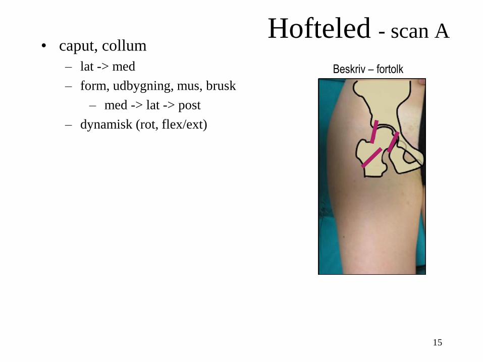
Hofteled - scan A • caput, collum
– lat -> med
– form, udbygning, mus, brusk
– med -> lat -> post
– dynamisk (rot, flex/ext)
Beskriv – fortolk
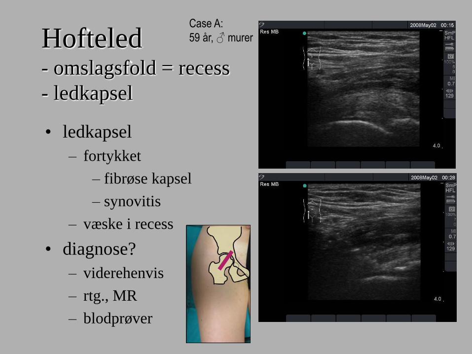
Hofteled - omslagsfold = recess
- ledkapsel
• ledkapsel
– fortykket
– fibrøse kapsel
– synovitis
– væske i recess
• diagnose?
– viderehenvis
– rtg., MR
– blodprøver
Case A:
59 år, ♂ murer

Hofteled
• led, knogle, sene/mm
– MR
– caput/collum
nekrose?
Case A:
59 år, ♂ murer

Hofteled - ledansamling
- i recess
• smerter, mobilitet
• UL: ansamling
– artritis
– synovitis
– årsag?
• diagnose?
– rtg., MR
– blodprøver
• UL vejledt blokade Case B:
66 år, ♀
hø.
coll. fem.
ve.
coll. fem.

Hofteled - ledansamling
- i recess
• diagnose
– MR
– AVasculær Nekrose (AVN)
Case B:
66 år, ♀

20
Doppler - hofteled
Artritis - infektion
Ansamling, synovitis – Ewing sarcom
Artritis – TB infektion

21
UL vejledt injektion - hofteled
US-guided hip injection. Needle (arrows) obliquely toward the femoral head neck
junction. (B) As the injection progresses, the capsule is distended by the hypoechoic
injectate (asterisk). A, acetabulum; H, femoral head; N, femoral neck.
• blokade: lokal anæsteticum (LA) + steroid
• diagnostisk/terapeutisk
• vs. MR
Curved transducer

22
Hofteled - scan B • ledkapsel, recess, omslagsfold
– følg lat -> med, dist -> prox
– dynamisk, adskil kapsel fra psoas
– hypoeccoicitet (væske)
– doppler v. basis
Beskriv – fortolk

FAI - pincer
(intro: labrum)
• Form
– form
– udbygning
– tag
• UL mindre egnet
osteofyt
pincer? normal

24
Labrum læsion
Acetabular labral tear. (A) MR image shows a high-signal-intensity tear (arrow)
through the low-signal-intensity labrum. (B) Longitudinal US image in the same patient
shows the hypoechoic tear (arrow) through the echogenic labrum.

25
Perilabral cyste
obs. labrum læsion
Perilabral cyst. (A) MR image shows a bilobed cyst (arrow). (B) Longitudinal US image of the
same patient shows the anechoic bilobed cyst (thin arrows). The labral tear (thick arrow) is
better appreciated on this image than the MR image. H, femoral head.

26
Perilabral cyste
Perilabrale cyster

27
Hip - labral tears: Diagnostic challenge Reiman MP BJSM 2013, review

28
Hofteled - scan C • (acetabulum)
– form, udbygning
– med -> lat -> post
• labrum
– identificér
– form, læsioner, cyster
Beskriv – fortolk

29
Hofteled - scan • caput, collum
– lat -> med
– form, udbygning, mus, brusk
– med -> lat -> post
– dynamisk (rot, flex/ext)
• ledkapsel, recess = omslagsfold
– følg lat -> med, dist -> prox
– dynamisk, adskil kapsel fra psoas
– hypoeccoicitet (væske)
– doppler v. basis
• (acetabulum)
• labrum
– form, læsioner, cyster
Beskriv – fortolk
30
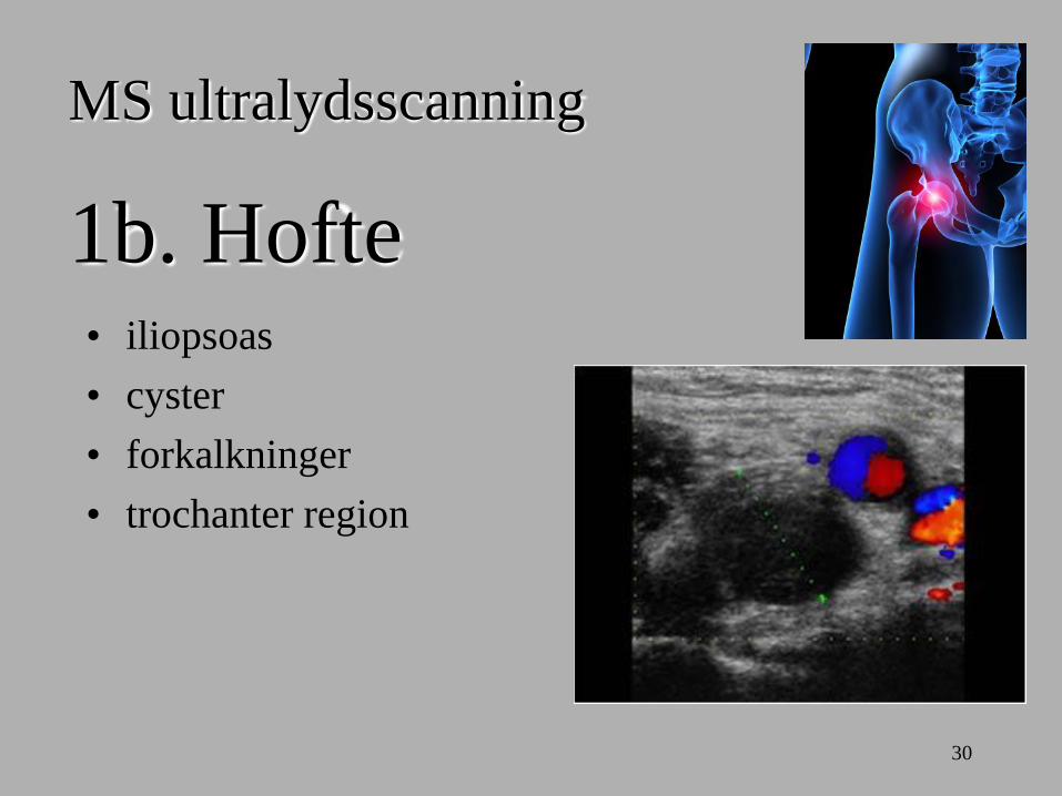
1b. Hofte
MS ultralydsscanning
• iliopsoas
• cyster
• forkalkninger
• trochanter region

31
Iliopsoas
iliopsoas tendon (arrows)

32
Cyste/bursa • iliopectinea/iliopsoas
• diff. diagnose
• DVT
• aneurisme
longitudinel
transverse
m. doppler A=arterie V=vene

33
Iliopsoas cyste
Iliopsoas bursitis. (A) MR image in a patient with a total hip arthroplasty shows a mildly
distended iliopsoas bursa (asterisk) adjacent to the iliopsoas tendon (arrow). The tendon is
difficult to see because of the susceptibility artifact from the arthroplasty components. (B)
Transverse US image of the same patient shows the bursa (asterisk) adjacent to the tendon
(arrow). There is no artifact from the arthroplasty.
THA

34
Iliopsoas cyste
35
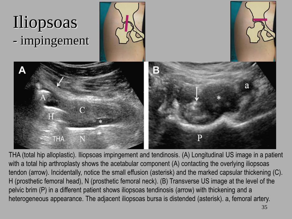
Iliopsoas - impingement
THA (total hip alloplastic). Iliopsoas impingement and tendinosis. (A) Longitudinal US image in a patient
with a total hip arthroplasty shows the acetabular component (A) contacting the overlying iliopsoas
tendon (arrow). Incidentally, notice the small effusion (asterisk) and the marked capsular thickening (C).
H (prosthetic femoral head), N (prosthetic femoral neck). (B) Transverse US image at the level of the
pelvic brim (P) in a different patient shows iliopsoas tendinosis (arrow) with thickening and a
heterogeneous appearance. The adjacent iliopsoas bursa is distended (asterisk). a, femoral artery.
THA

36
Iliopsoas - impingement
42 år, tidl. labrum repair. Smerter.
Prominerende acetabularkant. (A) Acetabulum, (Pile) psoas

37
Springhofte - intern
A. før klik - sene roteret
B. efter - normal, horizontalt
lateral medial

38
Iliopsoas
• 25 år ♂
• ikke motionsaktiv
• traume, nedspring, 1½-2 mdr.
• læsion
• intra-/peritendinøst flow
• doppler

39
Hofte - scan A • iliopsoas
– prox -> dist, langs/tværs
– struktur, fibre (sene mm.)
– relation til acetabulum, caput, kar
– dynamisk, adskil fra ledkapsel
Beskriv – fortolk

40
Rectus fem. - ruptur, forkalkning
• avusion
– posttraumatisk
– forkalkninger
– doppler
normal.
avulsion fra SIAI

41
Rectus fem. • læsion
– fiberstruktur
– omfang
– total/partiel ruptur
– dynamisk
– kontraktibilitet
22 år, fodbold, rectus fem.

42
Rectus fem. • mm. læsion
– ældre
– forkalkning
– væske
– (doppler, næppe)

43
Rectus fem.
• myositis ossificans
– post traumatisk
– hæmatom (ansamling)
– fiberlæsion
Vastus intermedius,
fluid + calcifications

44
Hofte - scan B • (rectus femoris)
– identificer, adskil fra andre mm.
– følg sartorius dist (tværs)
– prox -> dist på femur langs/tværs Beskriv – fortolk

45
Trochanter
• bursit
• trochanter major
– GT = Greater Trochanter
Bursitis trochanterica
Bursitis GMe
cranial

46
Bursa
• trochanter major (GT)
• UL vejledt blokade
• longitudinel
• langs
• fiberretning
• transversel
• tværs
Nål - blokade
Bursitis trochanterica

47

48
Glut. med. (GMed)
• GMed
• ruptur
• bursa

49
Glut. min. (GMin)
GMin tendinosis. (A) MR image shows a thickened and heterogeneous GMin tendon (white arrow)
with a focal insertional tear (black arrow) and highsignalintensity shallow tearing along its deep
surface. (B) Longitudinal US image in the same patient shows the thickened tendon (arrows) with
loss of the normal echogenic fibrillar appearance. G, greater trochanter.
GMi

50
Hofte - scan C • trochanter
– bursae
– fascie latae
– GMin, GMed Beskriv – fortolk

51
Hofte - scan • iliopsoas
– dist -> prox, langs/tværs
– struktur, fibre (sene mm.)
– relation til acetabulum, caput, kar
– dynamisk, adskil fra ledkapsel
• (rectus femoris)
– identificer, adskil fra andre mm.
– følg sartorius dist (tværs)
– prox -> dist på femur langs/tværs
• trochanter
– bursae
– fascie latae
– GMin, GMed
Beskriv – fortolk
52
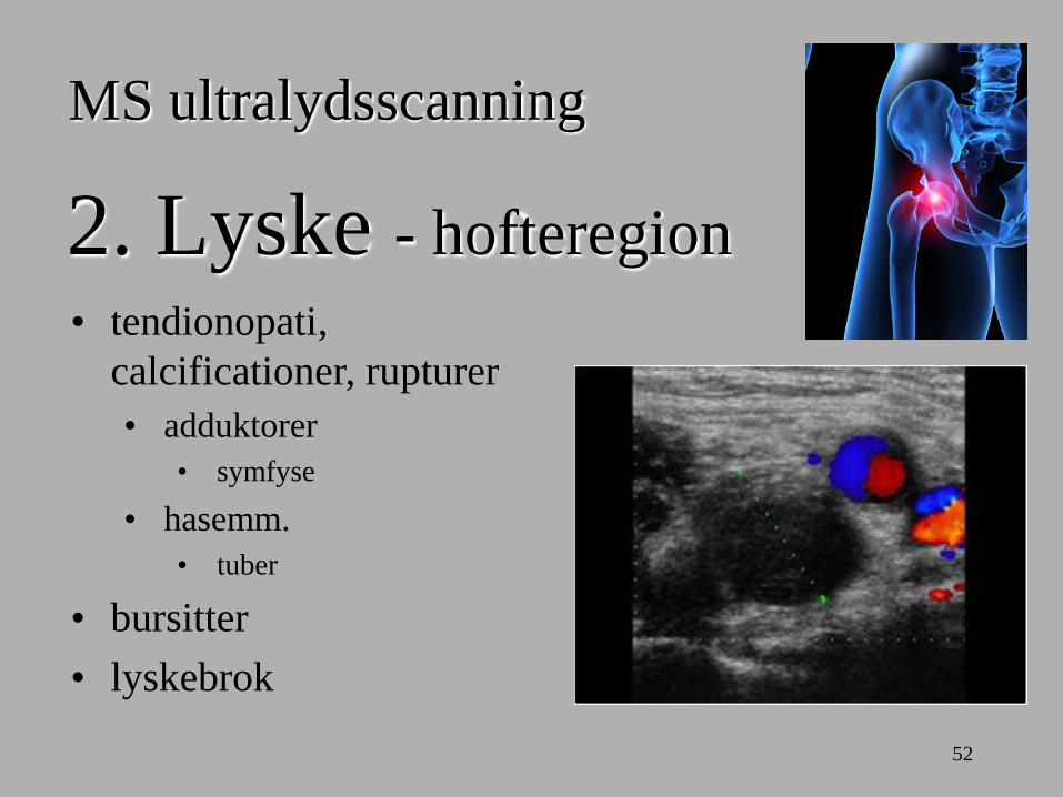
2. Lyske - hofteregion
MS ultralydsscanning
• tendionopati,
calcificationer, rupturer
• adduktorer
• symfyse
• hasemm.
• tuber
• bursitter
• lyskebrok

53

54
Adduktor
• mm./sene - udspring
• conjoint tendons
Fortykkelse, fortætning
Normal

55
Adduktor
• mm. - udspring
– med lille partiel læsion

Adduktor 32 år, ♂
volleyball
elite
• mm. - udspring
– doppler

57
Adduktor - intramuskulær (add.)
Adductor hematoma. (A) MR image shows a large hematoma (asterisk)
in the left adductor muscles. (B) Transverse US image in the same
patient shows the hypoechoic hematoma (asterisk).

58
Adduktor - ældre læsion
Adductor ruptur, forkalkning
Adductor ruptur, forkalkning
Adductor ruptur, væske + forkalkning

59
Symfysitis
Athletic pubalgia. (A) MR image shows a thickened and hyperintense rectus abdominis
aponeurosis (thin arrow) and extensive cortical erosion (thick arrow) of the anterior aspect of the
right superior pubic ramus. (B) Transverse sonographic image in a different patient during a
cortisone injection shows a thickened rectus abdominis aponeurosis (black arrow) with a
horizontal tear (secondary cleft sign) (thin solid white arrows). Note the cortical erosion (thick solid
white arrow) of the anterior aspect of the right superior pubic ramus and the cortical irregularity of
the anterior aspect of the left superior pubic ramus. The reverberation artifact from the needle is
present (hollow white arrow).
60
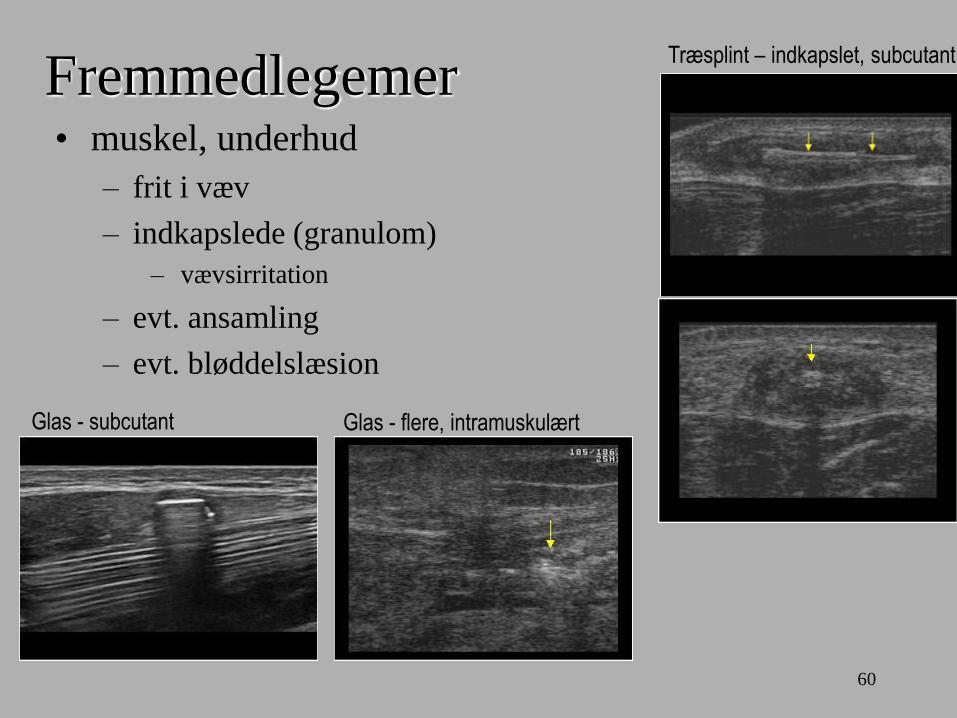
Fremmedlegemer • muskel, underhud
– frit i væv
– indkapslede (granulom)
– vævsirritation
– evt. ansamling
– evt. bløddelslæsion
Glas - subcutant Glas - flere, intramuskulært
Træsplint – indkapslet, subcutant

61
Lyske - scan A • adduktorer
– udspring, conjoint tendons, symfyse
– prox -> dist, langs/tværs
– struktur, fibre (sene mm.)
Beskriv – fortolk

62
Hasemm.
• hasemm.
• biceps F
• semiM
• semiT

63
Hasemm. - læsion

64
Tendino-/myopati
CT 3-D: Hamstring calcifications,
stress/overload induced
Hamstring calcification,
post traumatic (partiel lesion)

65
Lyske - scan C • hasemm.
– tuber iscii (GMax inf.)
– semiT, biceps F, semiM
– prox. -> dist, langs/tværs Beskriv – fortolk
66
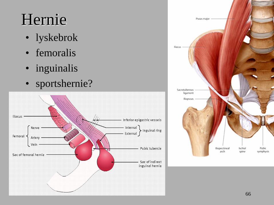
Hernie • lyskebrok
• femoralis
• inguinalis
• sportshernie?

Hernie
• symptomer
– pop ud
– smerter/ømhed
• intermitterende
– incarceration
• UL
– væske
– Lokalisation
– ligge/stå, bugpress

Hernie
• smerter
– intermitterende
– progression
• UL
– væske
– lokalisation
incarcereret hernie, væske, distenderet

69
Lymfeknuder – NB!
foreslå egen læge: henvisning til radiolog
metastase
blærecancer
Hodkin
lymfom
metastase
rectumcancer
metastase
blærecancer
lokal
infektion
lokal
infektion
De Gregorio Onc Gyn 2013

70
Lyske - scan C • (hernie)
– kar arterie/vene (doppler, bugpresse)
– inguinalkanal annulus int./ext.
– bugpresse, stående
Beskriv – fortolk

71
Lyske - scan • adduktorer
– udspring, conjoint tendons, symfyse
– prox -> dist, langs/tværs
– struktur, fibre (sene mm.)
• hasemm.
– tuber iscii (GMax inf.)
– semiT, biceps F, semiM
– prox. -> dist, langs/tværs
• (hernie)
– kar arterie/vene (doppler, bugpresse)
– inguinalkanal annulus int./ext.
– bugpresse, stående
Beskriv – fortolk
72
Litteratur • ESSR
– www.essr.org
• Textbook on Musculoskeletal US
– Bolvig L, Fredberg U, Rasmussen OS
– 2011
– ISBN: 9788762808249
• Cases
– www.ultrsoundcases.info
• Practical Musculoskeletal US
– McNally EG
– 2004
– ISBN-10: 0443073503
– ISBN-13: 978-0443073502