Embed Size (px)
Citation preview
Text
Important
Formulas
Numbers
Doctor notes
Notes and explanation
1
Lecture
No.3
“Failure Is Simply The Opportunity To
Begin Again, This Time More
Intelligently”
Physiology of the autonomic nervous system
Objectives:
1. The anatomy of somatic and autonomic nervous system.
2. Sympathetic and parasympathetic nerves.
3. Pre and post ganglionic neurons.
4. Functions of sympathetic and parasympathetic nerves in head & neck, chest, abdomen and pelvis.
5. Neurotransmitters release at pre and post ganglionic sympathetic / parasympathetic nerve endings.
6. Various responses due to stimulation of the sympathetic / parasympathetic nervous system.
2
Introduction
3
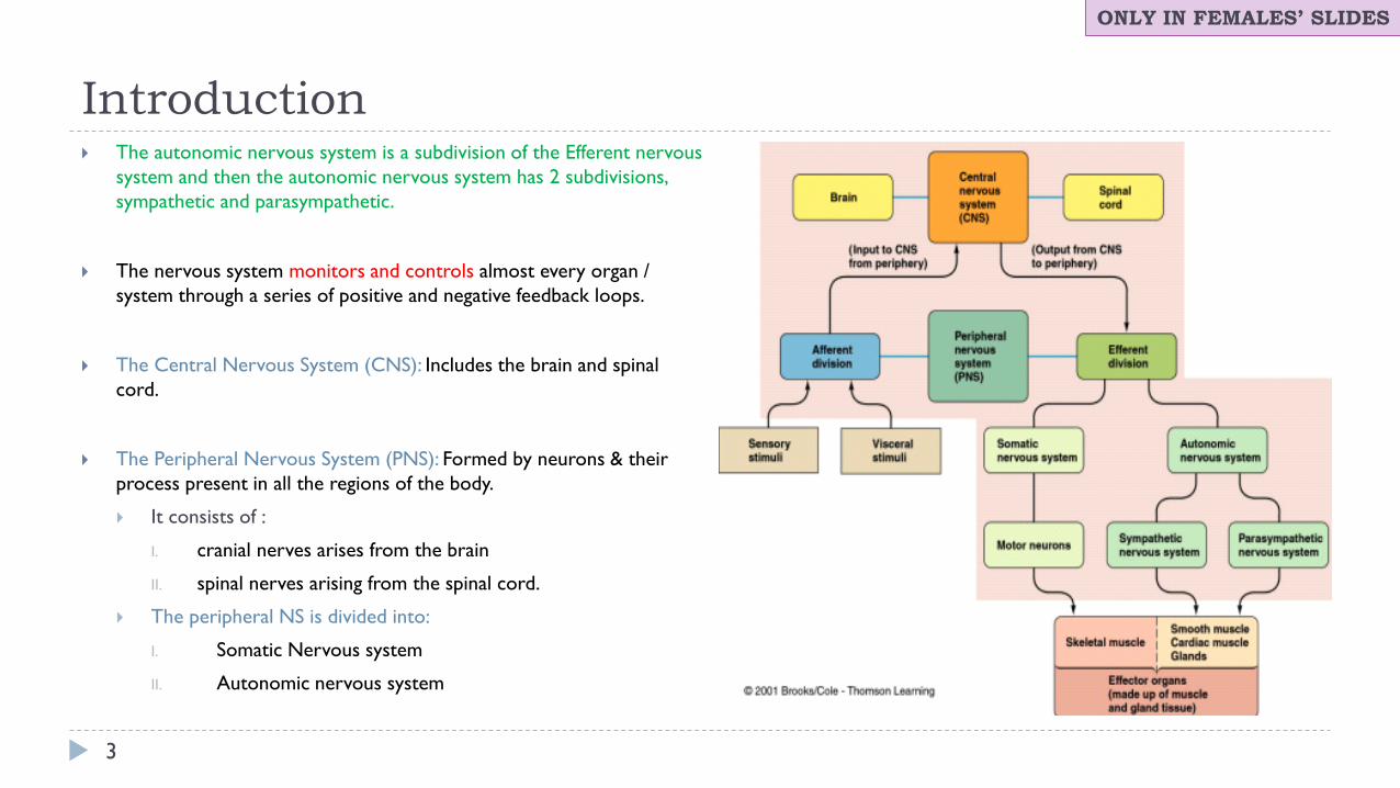
The autonomic nervous system is a subdivision of the Efferent nervous
system and then the autonomic nervous system has 2 subdivisions,
sympathetic and parasympathetic.
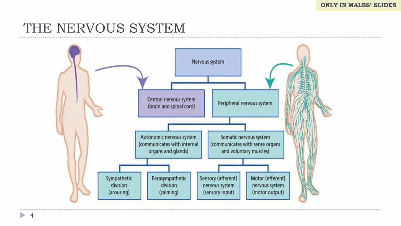
The nervous system monitors and controls almost every organ /
system through a series of positive and negative feedback loops.
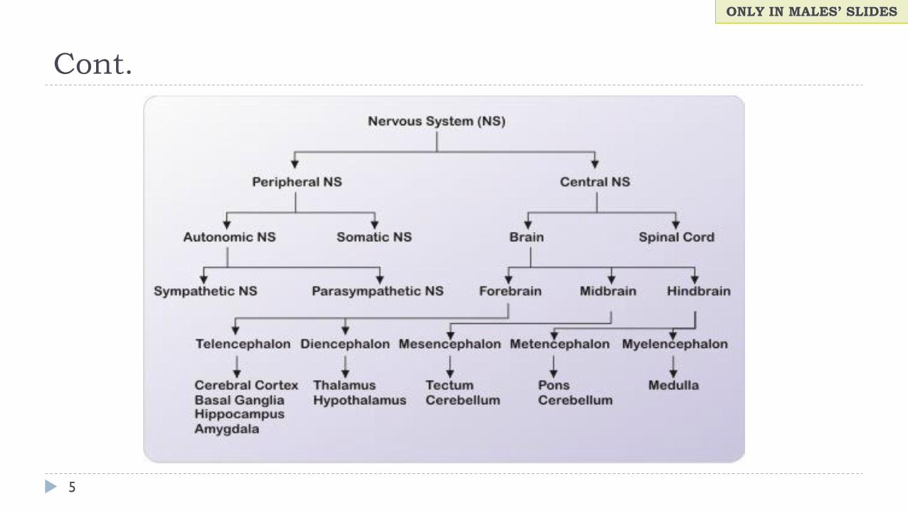
The Central Nervous System (CNS): Includes the brain and spinal
cord.
The Peripheral Nervous System (PNS): Formed by neurons & their
process present in all the regions of the body.
It consists of :
I. cranial nerves arises from the brain
II. spinal nerves arising from the spinal cord.
The peripheral NS is divided into:
I. Somatic Nervous system
II. Autonomic nervous system
ONLY IN FEMALES’ SLIDES
THE NERVOUS SYSTEM
4
ONLY IN MALES’ SLIDES
Cont.
5
ONLY IN MALES’ SLIDES
Anatomical Divisions of the Nervous System
6
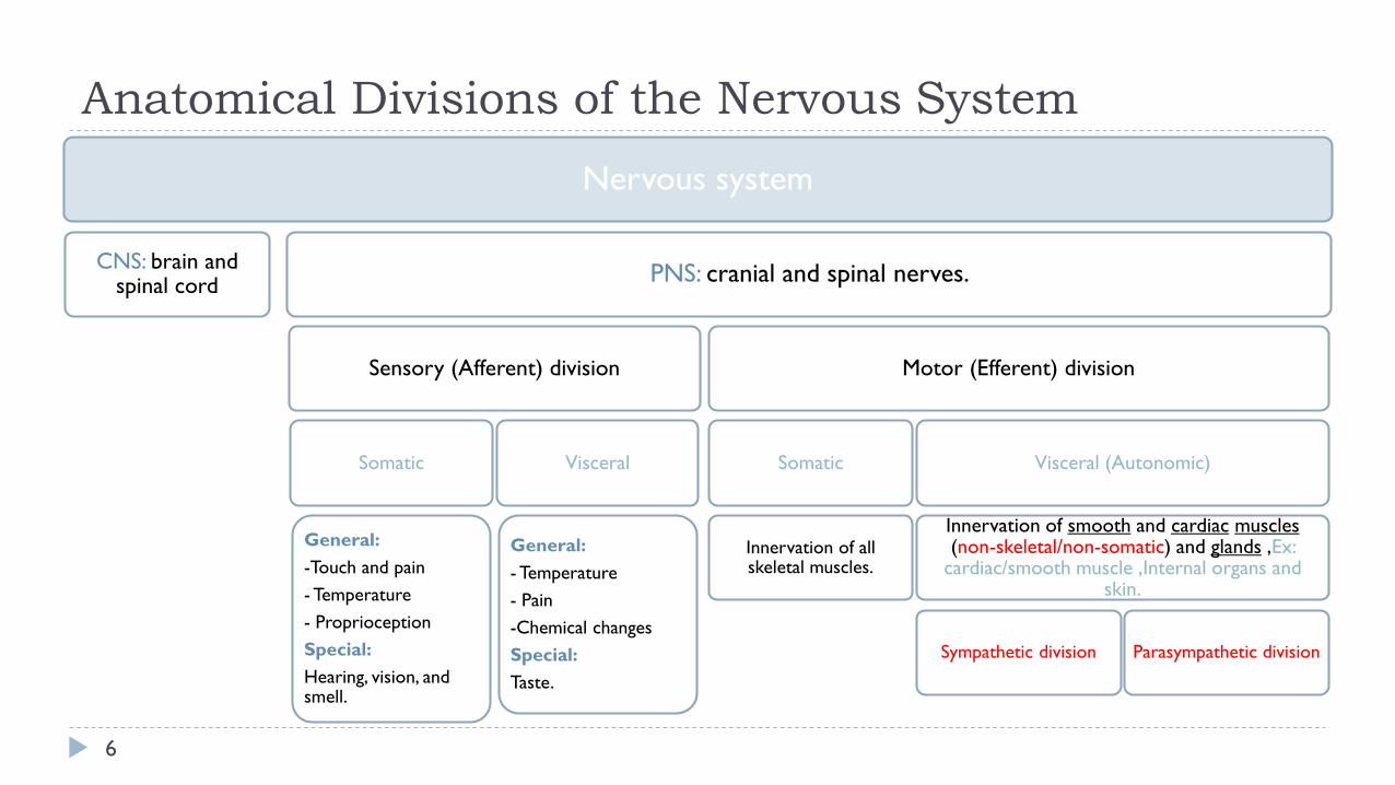
Nervous system
CNS: brain and spinal cord
PNS: cranial and spinal nerves.
Sensory (Afferent) division
Somatic
General:
-Touch and pain
- Temperature
- Proprioception
Special:
Hearing, vision, and smell.
Visceral
General:
- Temperature
- Pain
-Chemical changes
Special:
Taste.
Motor (Efferent) division
Somatic
Innervation of all skeletal muscles.
Visceral (Autonomic)
Innervation of smooth and cardiac muscles(non-skeletal/non-somatic) and glands ,Ex:
cardiac/smooth muscle ,Internal organs and skin.
Sympathetic division Parasympathetic division
What is Autonomic Nervous System?
7
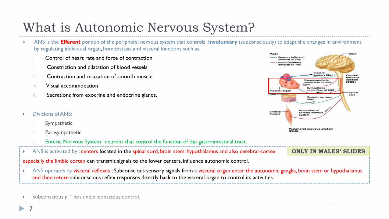
ANS is the Efferent portion of the peripheral nervous system that controls involuntary (subconsciously) to adapt the changes in environment
by regulating individual organ, homeostasis and visceral functions such as:
I. Control of heart rate and force of contraction
II. Constriction and dilatation of blood vessels
III. Contraction and relaxation of smooth muscle
IV. Visual accommodation
V. Secretions from exocrine and endocrine glands.
Divisions ofANS:
I. Sympathetic
II. Parasympathetic
III. Enteric Nervous System : neurons that control the function of the gastrointestinal tract.
ANS is activated by : centers located in the spinal cord, brain stem, hypothalamus and also cerebral cortex
especially the limbic cortex can transmit signals to the lower centers, influence autonomic control.
ANS operates by visceral reflexes ; Subconscious sensory signals from a visceral organ enter the autonomic ganglia, brain stem or hypothalamus
and then return subconscious reflex responses directly back to the visceral organ to control its activities.
Subconsciously = not under conscious control.
ONLY IN MALES’ SLIDES
THE AUTONOMIC NERVOUS SYSTEM
8
ONLY IN MALES’ SLIDES
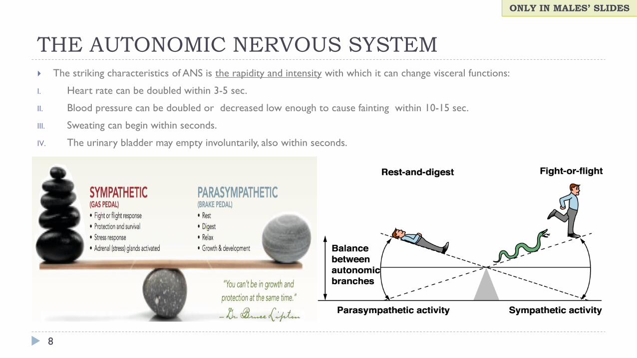
The striking characteristics of ANS is the rapidity and intensity with which it can change visceral functions:
I. Heart rate can be doubled within 3-5 sec.
II. Blood pressure can be doubled or decreased low enough to cause fainting within 10-15 sec.
III. Sweating can begin within seconds.
IV. The urinary bladder may empty involuntarily, also within seconds.
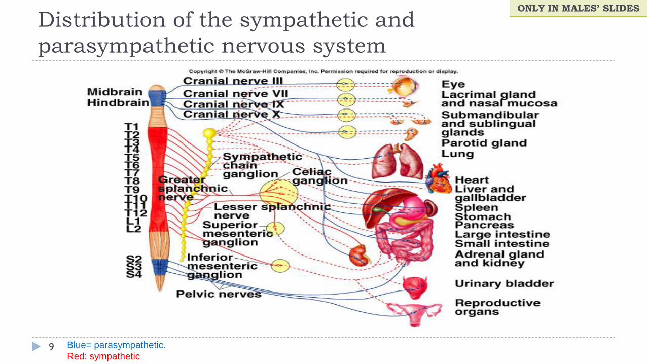
Distribution of the sympathetic and
parasympathetic nervous system
9
ONLY IN MALES’ SLIDES
Blue= parasympathetic.
Red: sympathetic

Comparison Between Autonomic and Somatic motor
systems
10
Basic anatomical difference between the motor pathways of the voluntary somatic nervous system (to skeletal muscles)
and those of the autonomic nervous system:
Somatic motor system Autonomic nervous system (Not under voluntary control)
• One motor neuron extends from CNS to skeletal muscle.
• Cell bodies of motor neurons reside in CNS (brain or spinal
cord).
• Their axons (sheathed in spinal nerves) extend all the way to
their skeletal muscles.
Chain of two motor neuron:
1st: Preganglionic neuron (in brain or cord).
2nd: Postganglionic neuron (Cell body in ganglion outside CNS).
Axons are thickly myelinated, conduct impulses rapidly. Conduction is slower due to lightly/thinly or unmyelinated axons.
No autonomic ganglion, only one neuron inside the Nervous
system.
Myelin sheath and node of Ranvier play a major rule is increasing the conduction of
impulses.
The conduction is slower because the preganglionic neuron is myelinated and
postganglionic neuron is unmyelinated.
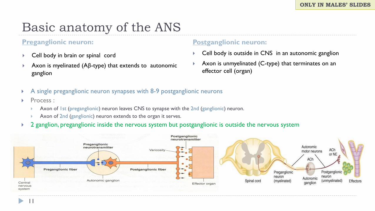
Basic anatomy of the ANSPreganglionic neuron: Postganglionic neuron:
11
Cell body in brain or spinal cord
Axon is myelinated (Aβ-type) that extends to autonomic
ganglion
Cell body is outside in CNS in an autonomic ganglion
Axon is unmyelinated (C-type) that terminates on an
effector cell (organ)
A single preganglionic neuron synapses with 8-9 postganglionic neurons
Process :
Axon of 1st (preganglionic) neuron leaves CNS to synapse with the 2nd (ganglionic) neuron.
Axon of 2nd (ganglionic) neuron extends to the organ it serves.
2 ganglion, preganglionic inside the nervous system but postganglionic is outside the nervous system
ONLY IN MALES’ SLIDES
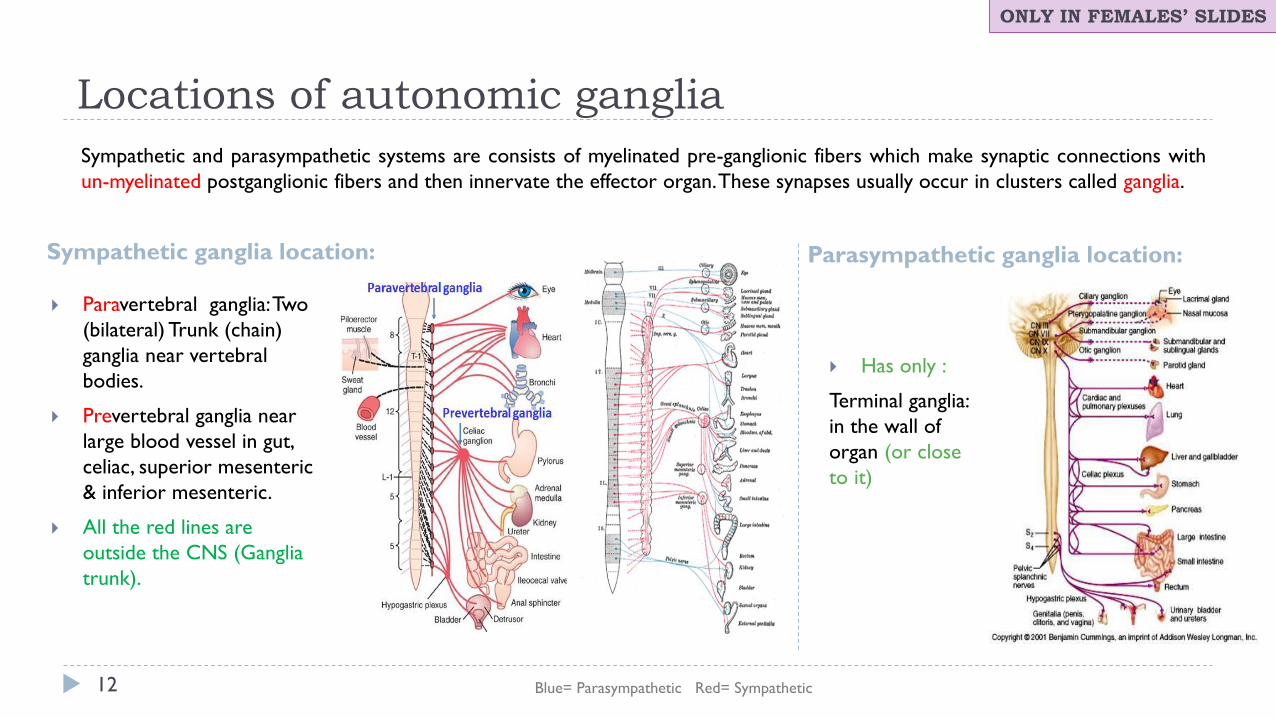
Locations of autonomic ganglia
Sympathetic ganglia location: Parasympathetic ganglia location:
12
Paravertebral ganglia: Two
(bilateral) Trunk (chain)
ganglia near vertebral
bodies.
Prevertebral ganglia near
large blood vessel in gut,
celiac, superior mesenteric
& inferior mesenteric.
All the red lines are
outside the CNS (Ganglia
trunk).
Has only :
Terminal ganglia:
in the wall of
organ (or close
to it)
Blue= Parasympathetic Red= Sympathetic
ONLY IN FEMALES’ SLIDES
Sympathetic and parasympathetic systems are consists of myelinated pre-ganglionic fibers which make synaptic connections with
un-myelinated postganglionic fibers and then innervate the effector organ. These synapses usually occur in clusters called ganglia.
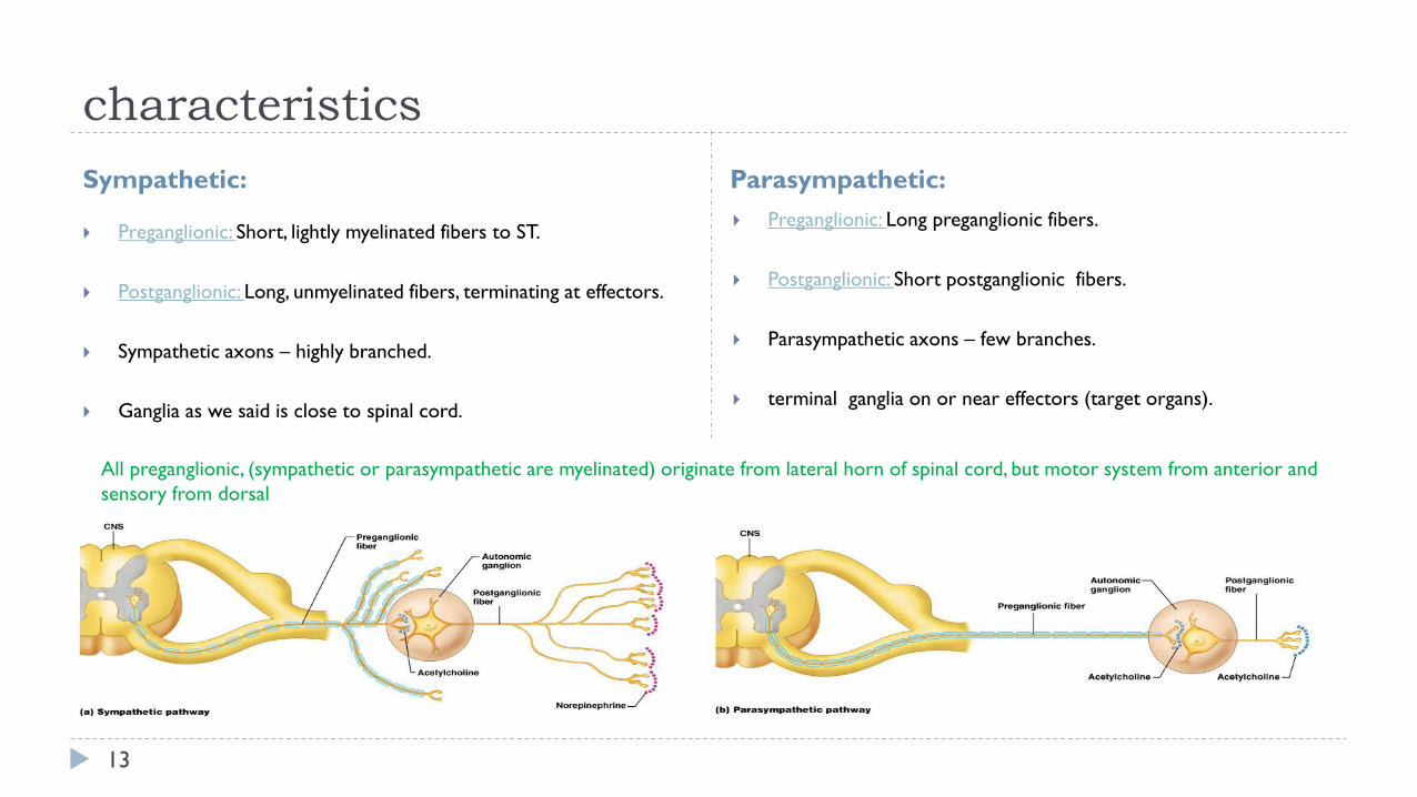
characteristics
Sympathetic: Parasympathetic:
13
Preganglionic: Short, lightly myelinated fibers to ST.
Postganglionic: Long, unmyelinated fibers, terminating at effectors.
Sympathetic axons – highly branched.
Ganglia as we said is close to spinal cord.
Preganglionic: Long preganglionic fibers.
Postganglionic: Short postganglionic fibers.
Parasympathetic axons – few branches.
terminal ganglia on or near effectors (target organs).
All preganglionic, (sympathetic or parasympathetic are myelinated) originate from lateral horn of spinal cord, but motor system from anterior and
sensory from dorsal
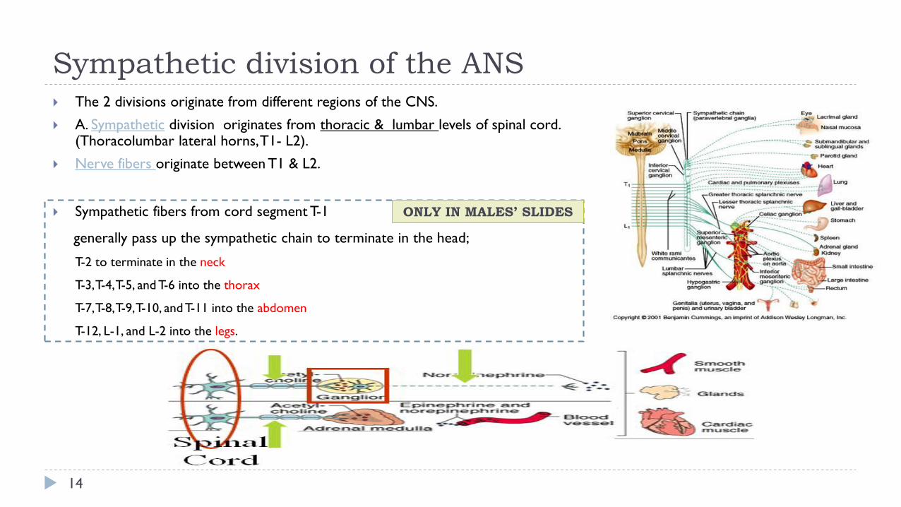
Sympathetic division of the ANS
14
The 2 divisions originate from different regions of the CNS.
A. Sympathetic division originates from thoracic & lumbar levels of spinal cord. (Thoracolumbar lateral horns, T1- L2).
Nerve fibers originate between T1 & L2.
Sympathetic fibers from cord segment T-1
generally pass up the sympathetic chain to terminate in the head;
T-2 to terminate in the neck
T-3,T-4,T-5, and T-6 into the thorax
T-7,T-8,T-9,T-10, and T-11 into the abdomen
T-12, L-1, and L-2 into the legs.
ONLY IN MALES’ SLIDES
Function of Sympathetic nervous system
15
The sympathetic system enables the body to be prepared for fear,
flight or fight.
Under stress condition
Sympathetic responses include an increase in heart rate, blood
pressure and cardiac output.
Diversion of blood flow from the skin and splanchnic vessels to
those supplying skeletal muscle. (No need for a lot of blood in the
skin under stress so there will be vasoconstriction, so the blood
will go to the brain, decrease blood supply to skin and GIT)
Bronchioles dilate, which allows for greater alveolar oxygen
exchange.
Blood flow to skeletal muscles, lungs is not only maintained, but
enhanced (by as much as 1200%), in case of skeletal muscles.
Decrease saliva, في حالة الخوف يكون الريق ناشف
Increased (Far vision) pupil size, contraction of sphincters( No time to go the bathroom even if you feel full ) and metabolic changes such as the mobilization of fat andglycogen
increases heart rate and the contractility of cardiac cells (myocytes), thereby providing a mechanism for the enhanced blood flow to skeletal muscles.
Sympathetic nerves dilate the pupil and relax the lens, allowing more light to enter the eye.
Also known as the “E” division:
Exercise.
Excitement.
Emergency.
Embarrassment
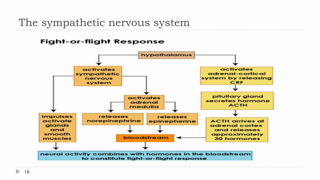
The sympathetic nervous system
16
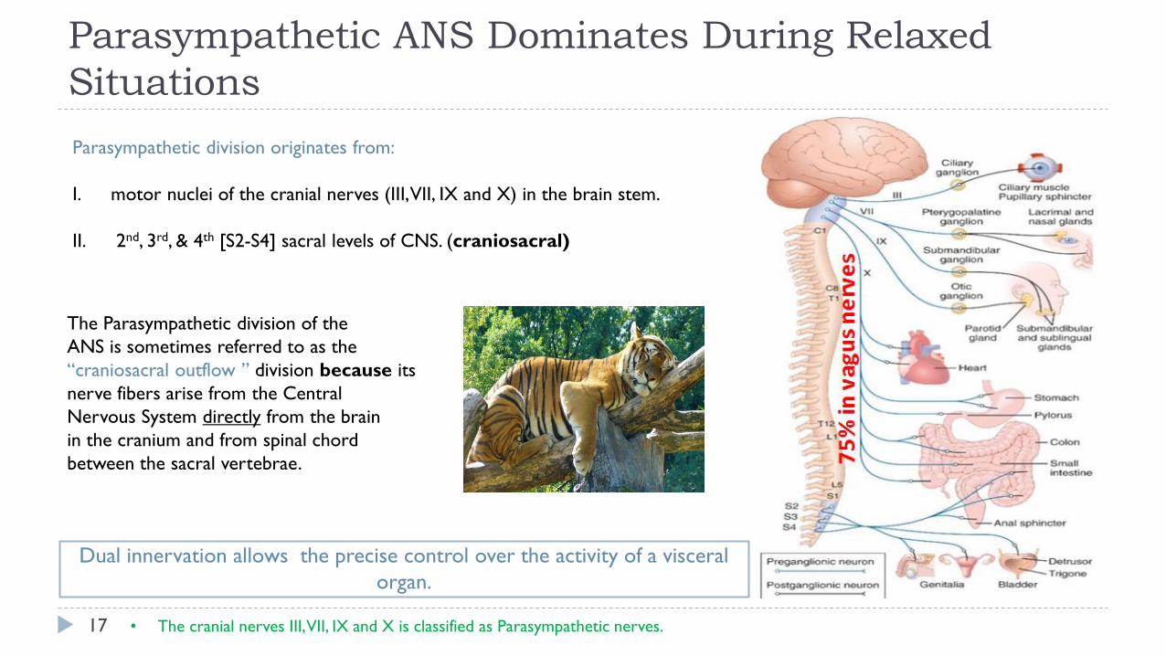
Parasympathetic ANS Dominates During Relaxed
Situations
17
Dual innervation allows the precise control over the activity of a visceral
organ.
• The cranial nerves III, VII, IX and X is classified as Parasympathetic nerves.
Parasympathetic division originates from:
I. motor nuclei of the cranial nerves (III, VII, IX and X) in the brain stem.
II. 2nd, 3rd, & 4th [S2-S4] sacral levels of CNS. (craniosacral)
The Parasympathetic division of the
ANS is sometimes referred to as the
“craniosacral outflow ” division because its
nerve fibers arise from the Central
Nervous System directly from the brain
in the cranium and from spinal chord
between the sacral vertebrae.
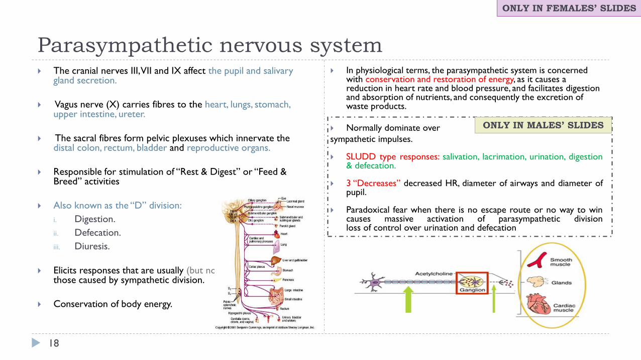
Parasympathetic nervous system
18
The cranial nerves III, VII and IX affect the pupil and salivary gland secretion.
Vagus nerve (X) carries fibres to the heart, lungs, stomach, upper intestine, ureter.
The sacral fibres form pelvic plexuses which innervate the distal colon, rectum, bladder and reproductive organs.
Responsible for stimulation of “Rest & Digest” or “Feed & Breed” activities
Also known as the “D” division:
i. Digestion.
ii. Defecation.
iii. Diuresis.
Elicits responses that are usually (but not always) opposite to those caused by sympathetic division.
Conservation of body energy.
In physiological terms, the parasympathetic system is concerned with conservation and restoration of energy, as it causes a reduction in heart rate and blood pressure, and facilitates digestion and absorption of nutrients, and consequently the excretion of waste products.
Normally dominate over
sympathetic impulses.
SLUDD type responses: salivation, lacrimation, urination, digestion & defecation.
3 “Decreases” decreased HR, diameter of airways and diameter of pupil.
Paradoxical fear when there is no escape route or no way to win causes massive activation of parasympathetic division loss of control over urination and defecation
ONLY IN FEMALES’ SLIDES
ONLY IN MALES’ SLIDES
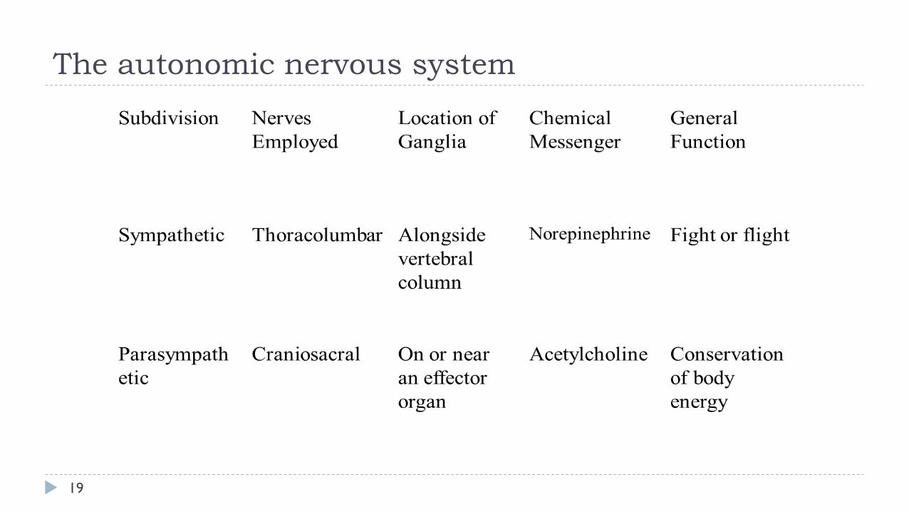
The autonomic nervous system
19
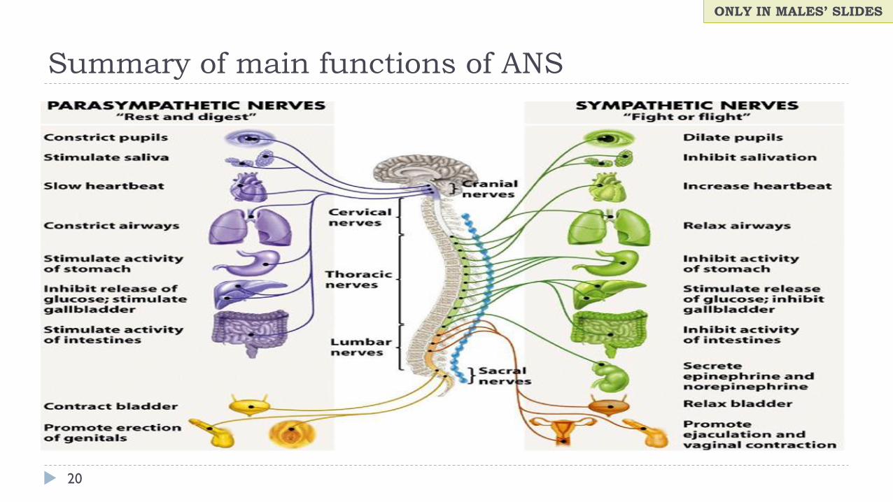
20
Summary of main functions of ANS
ONLY IN MALES’ SLIDES
Neurotransmitters & Receptors of the ANS
21
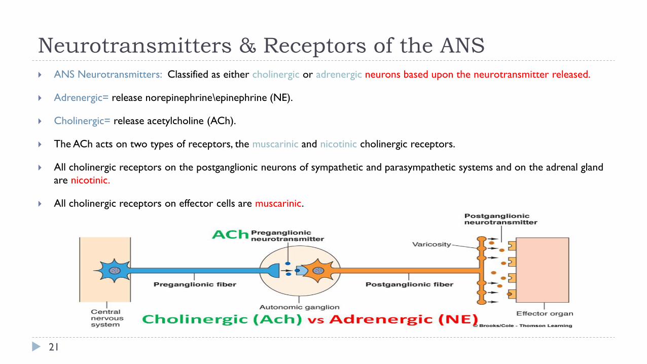
ANS Neurotransmitters: Classified as either cholinergic or adrenergic neurons based upon the neurotransmitter released.
Adrenergic= release norepinephrine\epinephrine (NE).
Cholinergic= release acetylcholine (ACh).
The ACh acts on two types of receptors, the muscarinic and nicotinic cholinergic receptors.
All cholinergic receptors on the postganglionic neurons of sympathetic and parasympathetic systems and on the adrenal gland
are nicotinic.
All cholinergic receptors on effector cells are muscarinic.

Cholinergic & Adrenergic Receptors
22
Cholinergic & adrenergic receptors
Sympathetic Parasympathetic
All preganglionic fibers of ANS release acetylcholine (ach) (cholinergic).
MOST postganglionic sympathetic fibers release norepinephrine
(adrenergic), except at sweat glands and some blood vessels in skeletal
muscles where they release ach.
All parasympathetic postganglionic fiber release acetylcholine (ach)
(cholinergic).
Neurotransmitters of autonomic nervous system
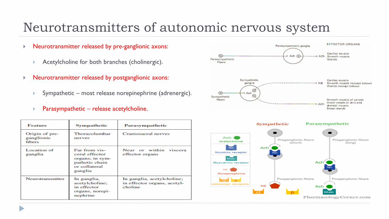
Neurotransmitter released by pre-ganglionic axons:
Acetylcholine for both branches (cholinergic).
Neurotransmitter released by postganglionic axons:
Sympathetic – most release norepinephrine (adrenergic).
Parasympathetic – release acetylcholine.
The autonomic nervous system
24
Acetylcholine activates mainly two types of receptors.They are called muscarinic and nicotinic receptors.
Muscarine activates only muscarinic receptors whereas nicotine activates only nicotinic receptors; acetylcholine activates both
of them.
Muscarinic receptors are found on all effector cells that are stimulated by the postganglionic cholinergic neurons of either the
parasympathetic nervous system or the sympathetic system.
Nicotinic receptors are found in the autonomic ganglia at the synapses between the preganglionic and postganglionic neurons
of both the sympathetic and parasympathetic systems.
Receptors
Cholinergic receptors: Adrenergic receptors:
25
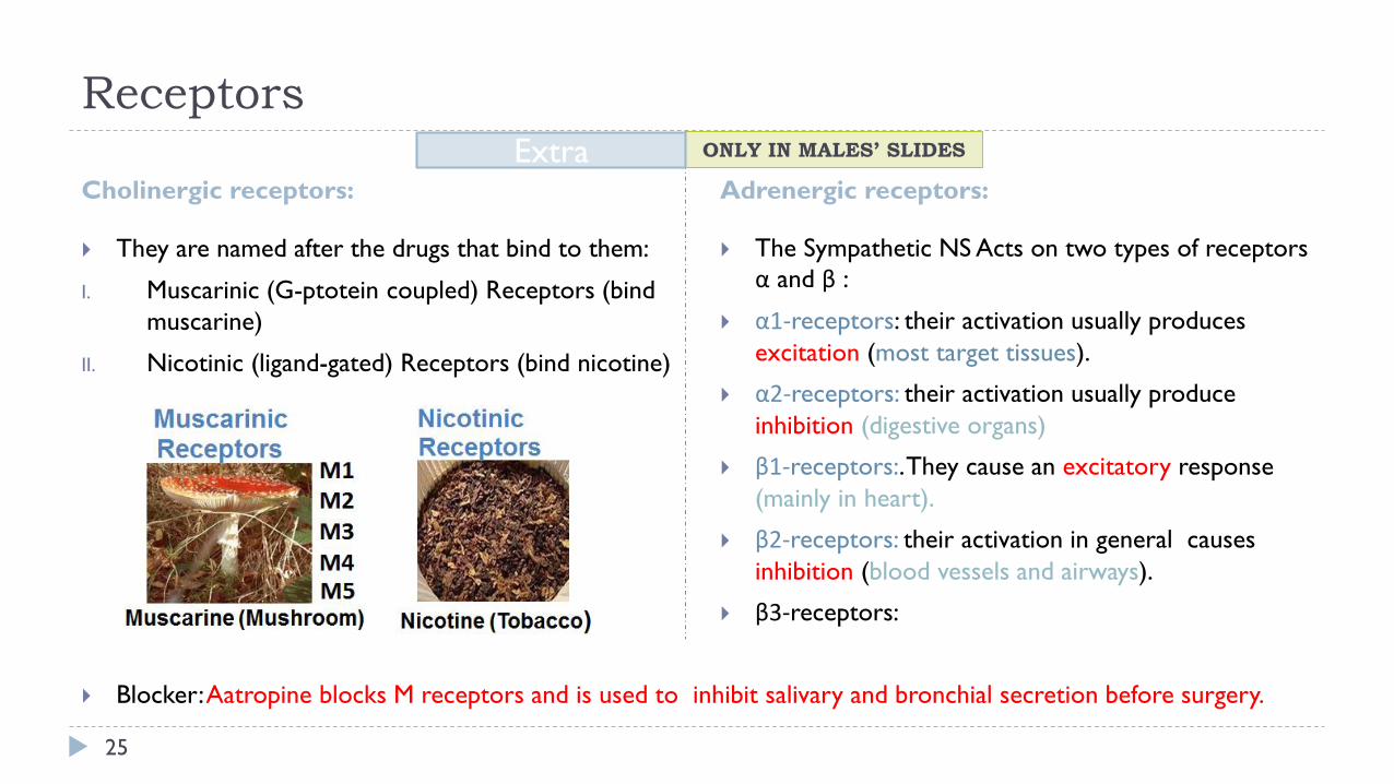
They are named after the drugs that bind to them:
I. Muscarinic (G-ptotein coupled) Receptors (bind
muscarine)
II. Nicotinic (ligand-gated) Receptors (bind nicotine)
The Sympathetic NS Acts on two types of receptors
α and β :
α1-receptors: their activation usually produces
excitation (most target tissues).
α2-receptors: their activation usually produce
inhibition (digestive organs)
β1-receptors:. They cause an excitatory response
(mainly in heart).
β2-receptors: their activation in general causes
inhibition (blood vessels and airways).
β3-receptors:
Blocker: Aatropine blocks M receptors and is used to inhibit salivary and bronchial secretion before surgery.
ONLY IN MALES’ SLIDES Extra
Receptors
26
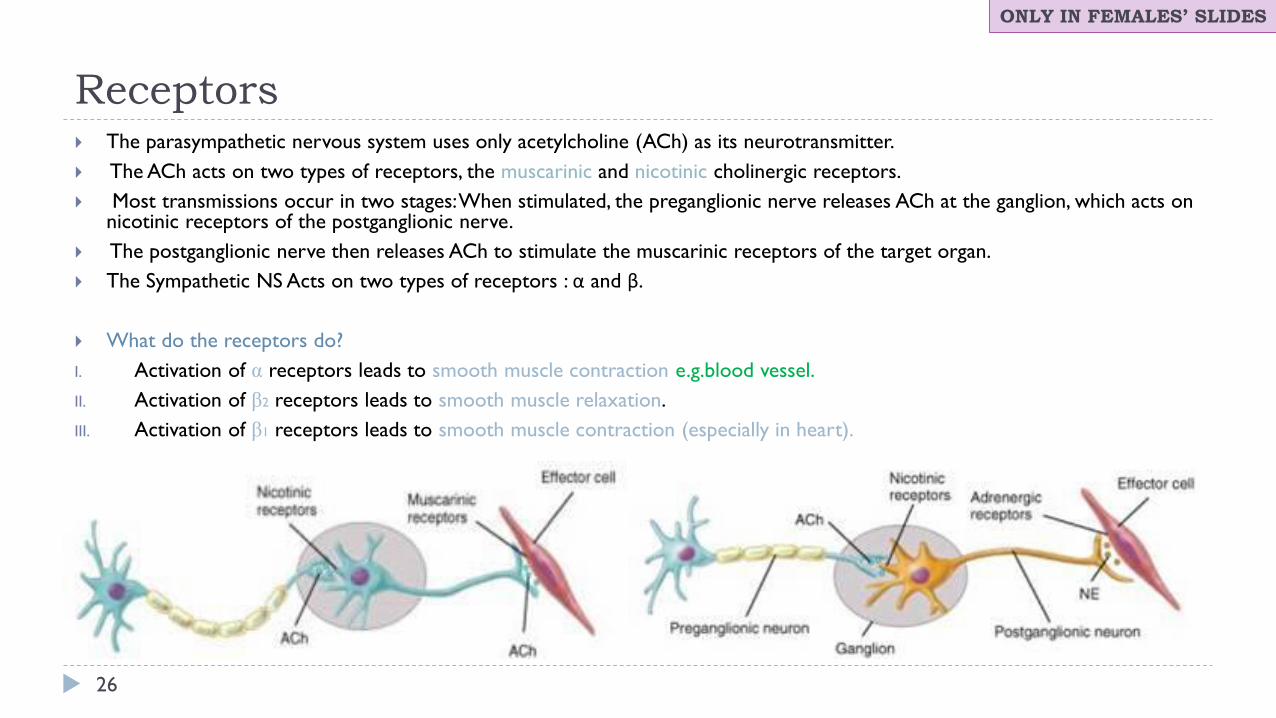
The parasympathetic nervous system uses only acetylcholine (ACh) as its neurotransmitter.
The ACh acts on two types of receptors, the muscarinic and nicotinic cholinergic receptors.
Most transmissions occur in two stages: When stimulated, the preganglionic nerve releases ACh at the ganglion, which acts on nicotinic receptors of the postganglionic nerve.
The postganglionic nerve then releases ACh to stimulate the muscarinic receptors of the target organ.
The Sympathetic NS Acts on two types of receptors : α and β.
What do the receptors do?
I. Activation of α receptors leads to smooth muscle contraction e.g.blood vessel.
II. Activation of β2 receptors leads to smooth muscle relaxation.
III. Activation of β1 receptors leads to smooth muscle contraction (especially in heart).
ONLY IN FEMALES’ SLIDES

Cont.
27
ONLY IN MALES’ SLIDES

Physiological functions of the autonomic
nervous system
28
Physiological functions of the autonomic nervous system
StructureSympathetic (adrenergic)
Parasympathetic
(muscarinic)
StructureSympathetic (adrenergic)
Parasympathetic
(muscarinic)
Endocrine - - circulatory system - -
Pancreas (islets) Α2: decreases secretion - cardiac output increases M2: decreases
Adrenal medulla N: secretes epinephrine - SA node: heart rate (chronotropic) β, β2: increases M2: decreases
Urinary system - -cardiac muscle: contractility
(inotropic)β, β2: increases
M2: decreases
(atria only)
Bladder wall Β2: relaxes Contracts conduction at AV node β1: increases M2: decreases
Ureter Α1: contracts Relaxes vascular smooth muscle
M3: contracts
Α= contracts
β2 = relaxes
-
Sphincter Α1: contracts; β2 relaxes Relaxes platelets α2: aggregates -
Sweat gland
secretions
M: stimulates
(major contribution)
α1: stimulates
(minor contribution)
- mast cells - histamine β2: inhibits -
Arrector pili Α1: stimulates - circulatory system - -
ONLY IN MALES’ SLIDES
Physiological functions of the autonomic
nervous system
29
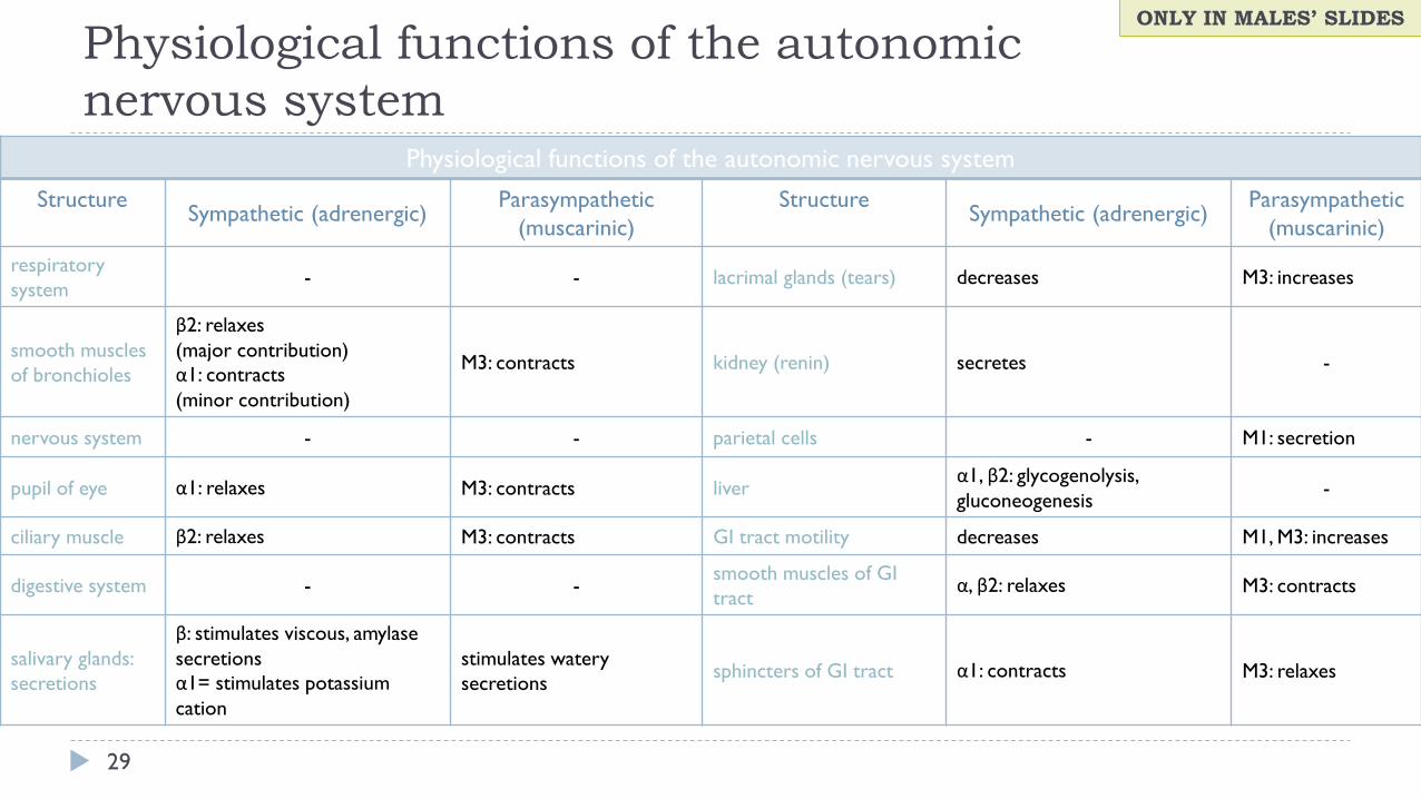
Physiological functions of the autonomic nervous system
StructureSympathetic (adrenergic)
Parasympathetic
(muscarinic)
StructureSympathetic (adrenergic)
Parasympathetic
(muscarinic)
respiratory
system- - lacrimal glands (tears) decreases M3: increases
smooth muscles
of bronchioles
β2: relaxes
(major contribution)
α1: contracts
(minor contribution)
M3: contracts kidney (renin) secretes -
nervous system - - parietal cells - M1: secretion
pupil of eye α1: relaxes M3: contracts liverα1, β2: glycogenolysis,
gluconeogenesis-
ciliary muscle β2: relaxes M3: contracts GI tract motility decreases M1, M3: increases
digestive system - -smooth muscles of GI
tractα, β2: relaxes M3: contracts
salivary glands:
secretions
β: stimulates viscous, amylase
secretions
α1= stimulates potassium
cation
stimulates watery
secretionssphincters of GI tract α1: contracts M3: relaxes
ONLY IN MALES’ SLIDES
The stress reaction
30
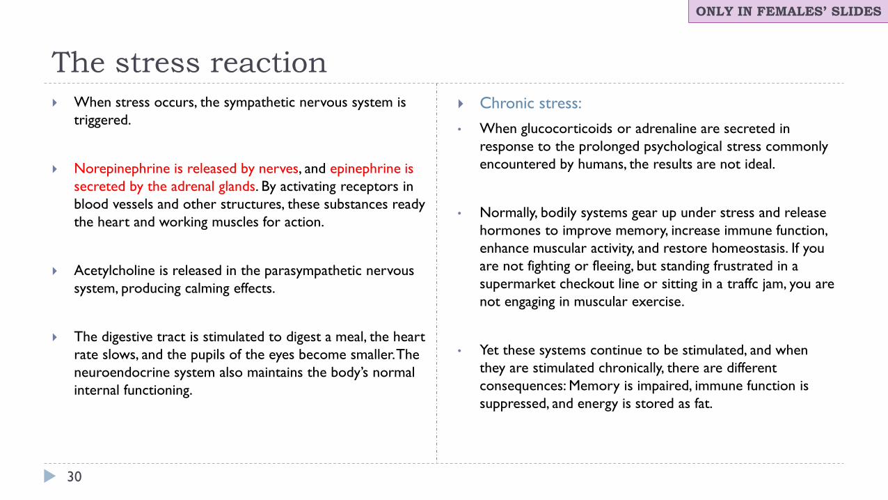
When stress occurs, the sympathetic nervous system is
triggered.
Norepinephrine is released by nerves, and epinephrine is
secreted by the adrenal glands. By activating receptors in
blood vessels and other structures, these substances ready
the heart and working muscles for action.
Acetylcholine is released in the parasympathetic nervous
system, producing calming effects.
The digestive tract is stimulated to digest a meal, the heart
rate slows, and the pupils of the eyes become smaller. The
neuroendocrine system also maintains the body’s normal
internal functioning.
Chronic stress:
• When glucocorticoids or adrenaline are secreted in
response to the prolonged psychological stress commonly
encountered by humans, the results are not ideal.
• Normally, bodily systems gear up under stress and release
hormones to improve memory, increase immune function,
enhance muscular activity, and restore homeostasis. If you
are not fighting or fleeing, but standing frustrated in a
supermarket checkout line or sitting in a traffc jam, you are
not engaging in muscular exercise.
• Yet these systems continue to be stimulated, and when
they are stimulated chronically, there are different
consequences: Memory is impaired, immune function is
suppressed, and energy is stored as fat.
ONLY IN FEMALES’ SLIDES

Response to stress
31
ONLY IN FEMALES’ SLIDES
Summary
32
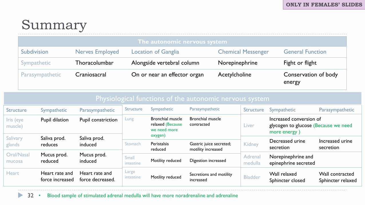
The autonomic nervous system
Subdivision Nerves Employed Location of Ganglia Chemical Messenger General Function
Sympathetic Thoracolumbar Alongside vertebral column Norepinephrine Fight or flight
Parasympathetic Craniosacral On or near an effector organ Acetylcholine Conservation of body
energy
Structure Sympathetic Parasympathetic
Iris (eye
muscle)
Pupil dilation Pupil constriction
Salivary
glands
Saliva prod.
reduces
Saliva prod.
induced
Oral/Nasal
mucosa
Mucus prod.
reduced
Mucus prod.
induced
Heart Heart rate and
force increased
Heart rate and
force decreased.
Structure Sympathetic Parasympathetic
Lung Bronchial muscle
relaxed (Because
we need more
oxygen)
Bronchial muscle
contracted
Stomach Peristalsis
reduced
Gastric juice secreted;
motility increased
Small
intestineMotility reduced Digestion increased
Large
intestine Motility reducedSecretions and motility
increased
Structure Sympathetic Parasympathetic
Liver
Increased conversion of
glycogen to glucose (Because we need
more energy )
KidneyDecreased urine
secretion
Increased urine
secretion
Adrenal
medulla
Norepinephrine and
epinephrine secreted
BladderWall relaxed
Sphincter closed
Wall contracted
Sphincter relaxed
Physiological functions of the autonomic nervous system
ONLY IN FEMALES’ SLIDES
• Blood sample of stimulated adrenal medulla will have more noradrenaline and adrenaline
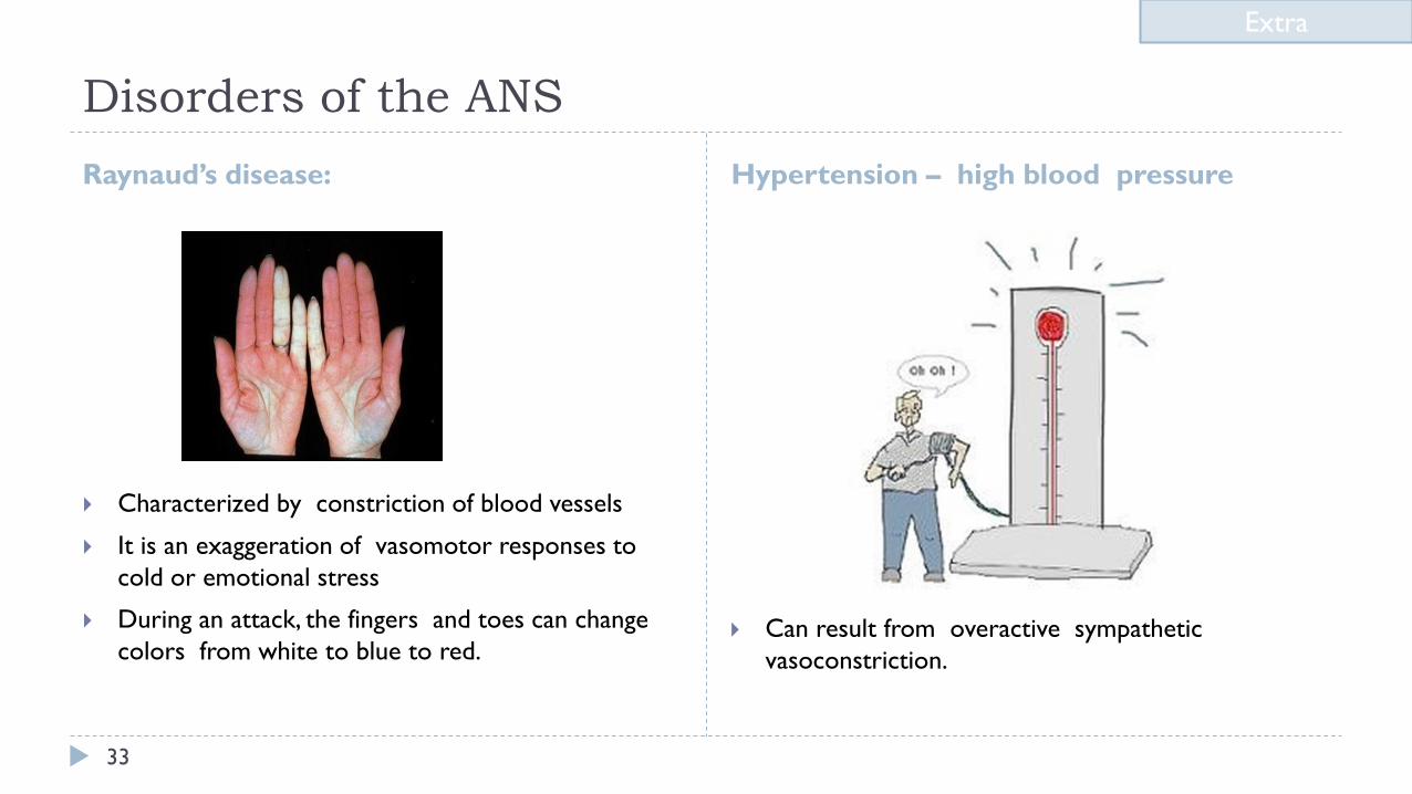
Disorders of the ANS
Raynaud’s disease: Hypertension – high blood pressure
33
Characterized by constriction of blood vessels
It is an exaggeration of vasomotor responses to
cold or emotional stress
During an attack, the fingers and toes can change
colors from white to blue to red. Can result from overactive sympathetic
vasoconstriction.
Extra
Thank you!
The Physiology 436 Team: Team Leaders: Lulwah Alshiha
Laila Mathkour
Mohammad Alayed
34
.اعمل لترسم بسمة، اعمل لتمسح دمعة، اعمل و أنت تعلم أن هللا ال يضيع أجر من أحسن عمال
Females Members:
Aseel Alsulimani
Lina alwakeel
Hayfaa Alshaalan
Contact us:
Males Members:
Hassan Alshammari
References: • Females and Males slides.
• Guyton and Hall Textbook of Medical Physiology (Thirteenth Edition.)