Embed Size (px)
Citation preview

Indiana University School of Optometry | eGrand Rounds Fall 2013 1
Fall 2013
Normal Tension Glaucoma Treatment-Not so fast!
VictorMalinovsky
Edited by Victor Malinovsky, O.D. All photos are property of Indiana University School of Optometry. © Copyright 2013 by the School of Optometry, Indiana University. All rights reserved. Cases presented in this issue are from
Cases from the Advanced Ocular Care Service.
Jane Ann Grogg
BrettKing

63 year old female patient initially diagnosed with normal tension glaucoma OD and has been followed for the last 7 years with no obvious progression. Entering IOPs were OD 18 & OS 16 mmHg with no significant diurnal variation. Central corneal thickness was OD 530 and OS 550 mm. History of migraine head-aches for several years, but no medications or reported drug allergies. No family history of eye disease. Slit lamp was WNL OU and gonioscopy revealed open angles. Dilated fundus exam was WNL except for a suspicious inferior temporal rim notching of the OD nerve with C/D .8/.6. The OS was WNL with a C/D .5/.5 & no Drance hemorrhages noted. Optic disc photo OD (Fig. 1) reveals inferior temporal rim thinner than superior. Humphrey visual fields (HVF) showed dense superior paracen-tral scotoma OD (Fig. 2); OS WNL.
Figure 1
Figure 2 Figure 3
Indiana University School of Optometry | eGrand Rounds Fall 2013 2
CASE 1

Initial OCT confirmed inferior temporal (I/T) rim damage with I/T NFL loss in only this quadrant. The patient was educated regarding diagnosis of NTG OD. After a discussion of the pros and cons of treatment, the patient decided on treatment: Alphagan P q 12 h OD only. Target pressures were 10-12. Follow up IOPs were 13 mmHg OD and have remained in the 13-15 range over the years. Glaucoma progression analysis of the HFA (Fig. 3) showed little progression. Heidelberg SD-OCT shows the loss of the inferior temporal NFL in the OD with no progression. Note that an infe-rior temporal nerve fiber wedge defect is seen on the confocal fundus image OD (Fig. 4). Recent posterior pole retinal thickness measurements to document the ganglion cell/RNF loss that can occur in glaucoma patients, shows significant loss of the retinal thickness in an arcuate shaped area inferior temporally that extends to the mac-ula (Fig. 5). Whether these retinal thickness changes may be an earlier marker of glauco-matous damage prior to optic nerve NFL loss is unknown. Is the stability of this NTG due to IOP lowering, neuroprotective properties of Alphagn P or a non-progressing NTG?
Figure 4
Figure 5 Heidelberg Posterior Pole Retinal Thickness Measurement
Indiana University School of Optometry | eGrand Rounds Fall 2013 3
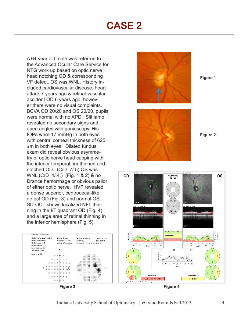
A 64 year old male was referred to the Advanced Ocular Care Service for NTG work up based on optic nerve head notching OD & corresponding VF defect. OS was WNL. History in-cluded cardiovascular disease, heart attack 7 years ago & retinal-vascular accident OD 6 years ago, howev-er there were no visual complaints. BCVA OD 20/20 and OS 20/20, pupils were normal with no APD. Slit lamp revealed no secondary signs and open angles with gonioscopy. His IOPs were 17 mmHg in both eyes with central corneal thickness of 625 mm in both eyes. Dilated fundus exam did reveal obvious asymme-try of optic nerve head cupping with the inferior temporal rim thinned and notched OD. (C/D .7/.5) OS was WNL (C/D .4/.4.) (Fig. 1 & 2) & no Drance hemorrhage or obvious pallor of either optic nerve. HVF revealed a dense superior, centrocecal-like defect OD (Fig. 3) and normal OS. SD-OCT shows localized NFL thin-ning in the I/T quadrant OD (Fig. 4) and a large area of retinal thinning in the inferior hemisphere (Fig. 5).
Figure 4Figure 3
Figure 2
Figure 1
Indiana University School of Optometry | eGrand Rounds Fall 2013 4
CASE 2

These findings can be seen with optic nerve damage in NTG pa-tients but if one performs a line scan over this area of retinal thin-ning we must rethink the etiology of damage. The line scan reveals loss /differentiation of NFL, GC, & Inner plexiform layers with what appears to be residual glial tis-sue. There is preservation of the outer nuclear, photoreceptors and RPE layers (Fig. 6), consistent with the loss of retinal tissue seen in BRAOs (see ref 7) with outer layers intact due to a normal cho-roidal supply. Revised diagnosis: previous BRAO with secondary retrograde optic neuropathy and NFL defect. Management at this time is no treatment with follow-up of HVFs and SD-OCT imaging to monitor progression. No progres-sion would confirm revised diag-nosis.
Figure 5 Large area of inferior retinal thinning-OD
Figure 6 Line Scan over Retinal Thinning Area
Indiana University School of Optometry | eGrand Rounds Fall 2013 5

Figure 1 Figure 2
Indiana University School of Optometry | eGrand Rounds Fall 2013 6
CASE 3
25 year old Asian female presents for a second opinion regarding treatment of NTG with Lumigan in each eye for the last year. No family history of glaucoma. Medical history is signifi-cant for a diagnosis of Myasthenia Gravis with no treatment at this time. Patient reports hav-ing several MRIs, but no abnormal findings found. BCVA OD 20/20 & OS 20/20. Slit lamp was WNL with no secondary signs and open angles. There was a mild but obvious afferent pupil-lary defect OS. Gonioscopy was WNL. IOPs were OD 15 & OS 14. Pachymetry was OD 579 and OS 598. The dilated fundus exam revealed very suspicious optic nerve heads especially in the left eye. 78 D exam OS revealed a large C/D with suspect superior and nasal rim thin-ning. The OD nerve also had a large C/D but the rim appeared to be very symmetric. There were no Drance hemorrhages, disc pallor, peripapillary atrophy, pits or partial coloboma in either optic nerve. Heidelberg retinal tomography (HRT) for disc size and rim tissue revealed superior and nasal rim flagged as abnormal in the OS and the OD with suspicious rim nasally. The parapapillary NFL was also thinner superiorly (Fig. 1). SD-OCT OS revealed nasal NFL thinning with suspect thinning inferiorly (Fig. 2). Retinal thickness OS with SD-OCT revealed an atypical pattern of thinning extending from the ONH superiorly and inferiorly and the hemi-sphere asymmetry plot revealing greater deviation superiorly & OD reveals questionable su-perior retinal thinning (Fig. 3). 24-2 HVFs showed OD WNL but OS revealed an inferior nasal depression. At first this looked like a trial lens artifact but the patient has no trial lens. (Fig. 4) The VF defect was confirmed with 30-2. Tentative diagnosis was an atypical optic neuropathy

Figure 3 Heidelberg Asymmetry Analysis
Figure 4
Indiana University School of Optometry | eGrand Rounds Fall 2013 7
not consistent with NTG. Previous records were requested, indicating that IOPs were 18 mmHg in each eye prior to treatment, the HVFS were very similar with no real progression and CAT scans & MRI were normal. Referral to a glaucoma special-ist indicated visual field defect in OS as somewhat variable over the years but relatively consistent and stable with no evidence of a progressive optic neuropa-thy. Optic nerve asymmetry thought to be a congenital anomaly. Recommendation was to discontinue Lumigan with an IOP check in 1 month. If pressures are stable, follow patient with visual fields every 6-12 months. With the absence of any neurolog-ical symptoms and normal MRI results, the atypical optic neuropathy was more likely the result of a congenital optic nerve defect that should not be progressive, sparing the need for this 25 year old to be using glau-coma drops for the rest of her life.

Indiana University School of Optometry | eGrand Rounds Fall 2013 8
Normal tension glaucoma (NTG) has been of great interest to the optometrist for years. NTG & POAG are not thought of as two different diseases, but a continuum of one disease with dif-ferent risk factors. There is really no magic cut-off of 22mmHg. It is better to think in terms of pressure dependent and non-pressure dependent damage or combinations. We all recognize that there are often risk factors other than IOP. A few of the presumed factors for development are chronic loss of retinal ganglion cells (RGC) due to a genetic hypersensitivity to IOP, ab-normal vascular factors including vasospasm (migraine, Raynaud’s), ischemia and hypoten-sion and autoimmune diseases. Also, NTG is more likely to occur in a female, Asian patient, migraine sufferer, and a patient with obstructive sleep apnea (OSA). Several other conditions must be considered in your differential diagnosis of NTG including: very thin CCT, large diur-nal IOPs, oral Beta blockers, intermittent angle closure, and a number of conditions where the IOPs were previously elevated and now have returned to normal (steroids, angle recession, pigmentary, exfoliation, glaucomatocyclitic crisis). Moreover, optic nerve problems including congenital anomalies (optic nerve colobomas, pits, buried drusens), past bouts of demyelinat-ing neuropathy, AIONs, arteriolar vascular occlusions, retrobulbar mass, and chiasmal/visual pathway lesions, i.e., one of the most important differentials is to determine if your patient’s optic neuropathy is neurological or truly NTG. Some of the key points to consider when questioning a possible neurological problem versus NTG are: is there is markedly asymmet-ric optic nerve disease, optic disc pallor greater than cupping, visual fields defects that don’t correspond to optic nerve head appearance, more advanced or respect the vertical meridian in VFs, a rapid progression of VFs or optic neuropathy, decreased visual acuity, color vision defects and APD. Does your patient present with any other neurological symptoms or signs? In most cases, routine CAT scan or MRI are not recommended but with any of the above fac-tors, a scan may be necessary.
One of the most perplexing issues with NTG is treatment. Does lowering IOP in these pa-tients have any real benefit? The Collaborative Normal-Tension Glaucoma Study (CNTGS) published in the early 2000’s, indicated that lowering IOP in these patients does help in some cases. The CNTGS found that with 30% of IOP reduction, treated patients had a 12% risk of progression versus 35% in the untreated patients over 5 years. Even though these results are somewhat convincing, a large number of untreated patients did not progress and/or were very slow in progressing. So which patients are to be treated versus careful observation? A fixation threatening VF defect, a documented progression of VFs or optic neuropathy and/or a presence of Drance hemorrhages are factors that would favor treatment. Treatment goals are a 30% IOP reduction with a target IOP of around 10-12. Prostaglandins are the most common glaucoma medication initially used. Nevertheless, it is often very difficult to achieve IOPs in a 10-12 range in NTG patients, and they often require more than one drug. Neuropro-tection treatment for NTG is an ongoing discussion. Neuroprotection may offer the potential of preventing non-pressure related factors that may also be contributing to the ON damage. Although we have read for years about laboratory evidence for glaucoma neuroprotection by several drugs, the evidence from randomized clinical trials has been lacking. A recent study, the Low-pressure Glaucoma Treatment Study (LoGTS), which for some reason has not made a tremendous impact in our present treatment of NTG, has revealed some very interesting
Discussion: Normal Tension Glaucoma Treatment-Not so fast!
UPCOMING CE COURSES
To learn more about CE events vist our website at: www.opt.indiana.edu/ce/ or email Cheryl Oldfield at: [email protected]
Indiana University School of Optometry | eGrand Rounds Fall 2013 9
results. The study was a randomized multicenter clinical trial of efficacy of treatment with 0.2% brimonidine versus 0.5% timolol eye drops twice a day to alter the course of low pressure glaucoma as measured by the rate of visual field progression. LoGTS revealed that there were statistically fewer brimonidine-treated patients (9, 9.1%) that had visual field progression than timolol-treated patients (31, 39.2 %). Even though we may still not really understand the rea-sons for these findings, a possible implication is that either Timolol drops may do something bad in these patients, or Brimonidine may have some true neuroprotective-like properties that are beneficial in the treatment of NTG. For the time being we treat most of our NTG patients with a PGA and/or Alphagan P.
REFERENCES
1. Collaborative Normal-Tension Glaucoma Study Group “Natural history of normal-tension glaucoma” Ophthalmology, 108 (2001), pp. 247–2532. Collaborative Normal-Tension Glaucoma Study Group “Comparison of glaucomatous progression between untreated patients with normal-tension glaucoma and patients with therapeutically reduced intraocular pressures” Am J Ophthalmol, 126 (1998), pp. 487–4973. Collaborative Normal-Tension Glaucoma Study Group “The effectiveness of intraocular pressure reduction in the treatment of normal-tension glaucoma” Am J Ophthalmol, 126 (1998), pp. 498–5054. Anderson DR “Collaborative normal tension glaucoma study.” Curr Opin Ophthalmol. 2003 Apr;14(2):86-90.5. Theodore Krupin, Jeffrey M. Liebmann, David S. Greenfield, Lisa F. Rosenberg, Robert Ritch, John W. Yang, “The Low-pressure Glaucoma Treatment Study (LoGTS) : Study design and baseline characteristics of enrolled patients” Ophthalmology Volume 112, Issue 3, March 2005, Pages 376–3856. Krupin T, Liebmann JM, Greenfield DS, Ritch R, Gardiner S; Low-Pressure Glaucoma Study Group “A randomized trial of brimonidine versus timolol in preserving visual function: results from the Low-Pressure Glaucoma Treatment Study.” Am J Ophthalmol. 2011 Apr; 151(4):671-81.7. Ritter, Sacu, Deak et al “In vivo identification of alteration of inner neurosensory layers in branch retinal artery occlusion” Br J Ophthalmol 2012 96: 201-2078. Apple DJ, Rabb MF & Walsh PM “Congenital anomalies of the optic disc.” Surv Ophthalmol. 1982 Jul-Aug; 27(1):3-41.
November 10, 2013
December 8, 2013
Louisville Seminar
Special CE Seminar Celebrating the Career of Dr. Malinovsky
February 23, 2014
March 29-30, 2014
Glaucoma and Coding/Compliance Seminar
2nd Annual Borish Symposium