Embed Size (px)
Citation preview
Integrative Systems
Familiarity Detection is an Intrinsic Property ofCortical Microcircuits with Bidirectional SynapticPlasticity
Xiaoyu Zhang,1 Han Ju,2 Trevor B. Penney,3 and Antonius M.J. VanDongen2
DOI:http://dx.doi.org/10.1523/ENEURO.0361-16.2017
1NUS Graduate School for Integrative Sciences and Engineering, National University of Singapore, 117456 Singapore,2Program for Neuroscience and Behavioral Disorders, Duke-NUS Medical School, 169857 Singapore, and3Department of Psychology, National University of Singapore, 117570 Singapore
AbstractHumans instantly recognize a previously seen face as “familiar.” To deepen our understanding of familiarity-noveltydetection, we simulated biologically plausible neural network models of generic cortical microcircuits consisting of spikingneurons with random recurrent synaptic connections. NMDA receptor (NMDAR)-dependent synaptic plasticity was imple-mented to allow for unsupervised learning and bidirectional modifications. Network spiking activity evoked by sensory inputsconsisting of face images altered synaptic efficacy, which resulted in the network responding more strongly to a previouslyseen face than a novel face. Network size determined how many faces could be accurately recognized as familiar. Whenthe simulated model became sufficiently complex in structure, multiple familiarity traces could be retained in the samenetwork by forming partially-overlapping subnetworks that differ slightly from each other, thereby resulting in a high storagecapacity. Fisher’s discriminant analysis was applied to identify critical neurons whose spiking activity predicted familiar inputpatterns. Intriguingly, as sensory exposure was prolonged, the selected critical neurons tended to appear at deeper layersof the network model, suggesting recruitment of additional circuits in the network for incremental information storage. Weconclude that generic cortical microcircuits with bidirectional synaptic plasticity have an intrinsic ability to detect familiarinputs. This ability does not require a specialized wiring diagram or supervision and can therefore be expected to emergenaturally in developing cortical circuits.
Key words: familiarity; learning; NMDA receptor; plasticity; recognition memory
IntroductionRecognition memory refers to the ability to recognize
previously experienced sensory inputs. Prior studies
(Nickerson, 1965; Shepard, 1967) found that immediatelyfollowing a single exposure to 612 pictures, subjectscould select the previously-seen picture in two-alternative
Received December 6, 2016; accepted April 27, 2017; First published May 8,2017.The authors declare no competing financial interests.
Author contributions: X.Z., H.J., T.B.P., and A.M.J.V. designed research; X.Z.and H.J. performed research; X.Z., T.B.P., and A.M.J.V. analyzed data; X.Z.,T.B.P., and A.M.J.V. wrote the paper.
Significance Statement
Humans recognize familiar faces instantly. The cellular mechanisms underlying this recognition memory arestill poorly understood. Simulations presented here demonstrate that bidirectional synaptic plasticity issufficient to endow recurrent spiking neuronal network models with the ability to detect familiar sensoryinputs through unsupervised learning. Network spiking activity evoked by a face image results in changesin synaptic connectivity and the formation of a unique strengthened subnetwork. Networks can recognizemultiple previously seen faces with high accuracy by forming partially overlapping subnetworks. Wetherefore propose that familiarity detection is an intrinsic property of generic cortical microcircuits withbidirectional synaptic plasticity.
New Research
May/June 2017, 4(3) e0361-16.2017 1–21

recognition tests with 98% accuracy. Later, Standing(1973) reported that the number of pictures correctly rec-ognized increases with the number of pictures presented(up to 10,000), suggesting a limitless capacity of recogni-tion memory. These experimental observations can beaccounted for by familiarity, a form of unsupervised learn-ing. Familiarity and recollection are two major processesthat underlie recognition memory (Yonelinas, 2002; Squireet al., 2007). While recollection demands accurate recallof the object’s features, familiarity merely requires a signalindicating that the object has been previously encoun-tered (Wixted, 2007). Evidence from functional imagingstudies shows that cortical regions surrounding the hip-pocampus become active when a human subject sensesa familiar input, whereas activation of the hippocampus isrequired for recollection (Hölscher et al., 2003; Dianaet al., 2007).
While learning and memory have been extensively stud-ied at the molecular/cellular (Wu et al., 2006; Baudry et al.,2015) and behavioral (Gale et al., 2014; Rapanelli et al.,2015) levels, it has been difficult to causally connect thesetwo levels of desciption (Morgado-Bernal, 2011). Memo-ries are believed to be encoded by and stored in a subsetof neurons (the “engram”), which are connected by syn-apses whose weights were altered by the learning experi-ence (Takeuchi et al., 2014). Given the current knowledge onhow learning induces changes in synaptic efficacy throughlong-term potentiation (LTP; Bliss and Lomo, 1973), long-term depression (LTD; Ito and Kano, 1982; Massey andBashir, 2007), and spike-timing-dependent plasticity (STDP;Gerstner et al., 1996; Markram et al., 1997; Bi and Poo,1998), it is meaningful to simulate the synaptic changes inbiologically plausible neural networks, to foster understand-ing from a systems perspective.
The liquid state machine (LSM) is a biologically-inspiredspiking neural network model, which closely emulates thecomplexity of a generic cortical microcircuit. It is designedto perform biologically realistic real-time computing ontime-varying inputs, providing an alternative to the widely-used attractor neural networks which require conver-gence to stable internal states (Maass et al., 2002). Theconstruction of an LSM network, which involves generat-ing random connections with random synaptic weights, istask independent. An important property of these net-works is that sensory inputs are expanded into high-dimensional feature space, allowing linear separation ofcomplex properties (Buonomano and Maass, 2009). Tem-poral and spatial input information is transiently preserved
in the form of fading memory, through multiple recurrentloops and short-term synaptic plasticity (depression andfacilitation). We have introduced activity-dependent long-term synaptic plasticity into the network model by incorpo-rating NMDA receptor (NMDAR) functionality (Shouval et al.,2002) in the excitatory synapses.
NMDARs are critically important for synaptic plasticity-dependent learning. When post-synaptic depolarizationcoincides with glutamate and glycine binding, NMDARsopen and allow Ca2� influx (Blanke and VanDongen,2009). Depending on the amount of Ca2� influx, it willselectively activate phosphatases (low calcium influx) orthe kinase CaMKII (high calcium influx), and trigger down-stream signaling for synaptic depression or potentiation,respectively (Salter et al., 2009; Malleret et al., 2010;Luscher and Malenka, 2012), a theory known as the cal-cium control hypothesis (Lisman, 1989). LTP and LTD canbe induced by tetanic synaptic inputs that regulate theCa2� influx. They are rate-based: low-frequency stimula-tion causes LTD, while a high-frequency tetanus inducesLTP. STDP is another form of synaptic plasticity regulatedby the temporal correlation of pre- and post-synaptic firing.By implementing back-propagating action potentials(BPAPs), NMDAR functionality can support STDP throughcalcium control (Waters et al., 2005; Paradiso and Wu,2009). We have implemented both rate-based (LTP/LTD)and spike-timing-based (STDP) plasticity, by modelingNMDAR functionality in the excitatory synapses using thecalcium control hypothesis.
The NMDAR-containing neural network is a model ofgeneric cortical microcircuits with the capability of unsu-pervised learning. We have used it here to study howfamiliarity could develop in the cortex. On a large scale,brain regions are wired into relatively deterministic neuralcircuits (Coutlee and Huettel, 2012; Fornito et al., 2012),yet randomness and flexibility prevails within local corticalregions, with functional connections being optimized byactivity-dependent changes (Sporns and Zwi, 2004; Fairet al., 2009). The main hypothesis underlying our simula-tions is that a sensory stimulus induces changes in syn-aptic weights in a neural microcircuit, altering the networkresponse such that it can distinguish familiar from novelinputs.
Materials and MethodsNeural network implementation
Neural networks were simulated using MATLAB and theCSIM package (a neural Circuit SIMulator, RRID: SCR_014256,available at http://www.lsm.tugraz.at/csim/), as describedpreviously (Natschläger et al., 2002; Ju et al., 2013).NMDAR-dependent synaptic plasticity was introduced intothe excitatory synapses following the model proposed byShouval et al. (2002). The neural networks consist of twoparts: an input layer, and the network reservoir. Input neu-rons send spikes to the network reservoir via static spikingsynapses, which have no plasticity. The network reservoirconsists of leaky integrate-and-fire (LIF) neurons recurrentlyconnected by NMDAR synapses. Seventy-five percent ofthe neurons are set as excitatory, the remaining being inhib-
This work was supported by grants MOE2012-T2-1-039 from the SingaporeMinistry of Education and OFIRG/0019/2016 from the National Medical Re-search Council (to A.M.J.V.) and by an award from the Singapore Ministry ofHealth and A�STAR, the Agency for Science, Technology and Research.
Correspondence should be addressed to Antonius M.J. VanDongen, Pro-gram for Neuroscience and Behavioral Disorders, Duke-NUS Medical School,Singapore 169857, E-mail: [email protected].
DOI:http://dx.doi.org/10.1523/ENEURO.0361-16.2017Copyright © 2017 Zhang et al.This is an open-access article distributed under the terms of the CreativeCommons Attribution 4.0 International license, which permits unrestricted use,distribution and reproduction in any medium provided that the original work isproperly attributed.
New Research 2 of 21
May/June 2017, 4(3) e0361-16.2017 eNeuro.org
itory. Each LIF neuron is modeled by a linear differentialequation:
�m
dVm
dt� ��Vm– Vresting� � Rm�Isyn � Iinject � Inoise� ,
(1)
where the parameters are: membrane time constant �m �30 ms, Vresting � 0 mV, membrane resistance Rm � 1 M�,input currents supplied by explicitly modeled synapsesIsyn, steady background current Iinject � 13.5 nA and forsome simulations random noise Inoise was added to thecurrent. For the first time step in the simulation, the mem-brane potential Vm was set to an initial random valuebetween �1 and 1 mV. When Vm increases to 15 mV (thefiring threshold), the neuron fires, and Vm is reset to arandom value between �1 and 1 mV after an absoluterefractory period of 3 ms for excitatory neurons and 2 msfor inhibitory neurons (Joshi, 2007).
In CSIM, the probability that two neurons are con-nected by a synapse is defined as:
P�D� � C·exp ��D2�a, b�
�2 � , (2)
where D(a,b) stands for the Euclidean distance betweenthe two neurons a and b. � and C are parameters used byCSIM that determine connectivity and synaptic strength,respectively. As � increases, both the connection proba-bility and the average connection length will increase. Thebase value of C depends on the type of connection: it isset to 0.3, 0.2, 0.4, and 0.1 for EE, EI IE, and II connec-tions, where E and I stand for excitatory and inhibitoryneurons. The values are based on recordings from rodentcortical brain areas (Gupta et al., 2000). The actual valueof C is modulated by a user-defined parameter, Cscale.Input layer neurons are all excitatory. Connections frominput neurons to the network reservoir and within thenetwork reservoir are randomly generated following theprobability P(D).
Once a connection is established, it is assigned aninitial synaptic weight, indicating synaptic efficacy. Initialsynaptic weights are drawn from the following gammadistribution:
y � f(x |a, b) �1
ba�(a)xa�1e
�x
b (3)
a �1
SH_W2, b � W·SH_W2 ,
where � (·) is the Gamma function. SH_W and W areparameters used by CSIM. SH_W (default 0.7) positivelycorrelates with the variance of the weight distribution andW correlates with the mean of the distribution. The basevalue of W is set to 3e�8 for EE, 6e�8 for EI, �1.9e�8 forIE and II. The actual value of W is modulated by a userdefined parameter, Wscale. The synaptic weight of anexcitatory NMDAR synapse is subject to strengthening(upper boundary � 6.5e�8) or weakening (lower boundary� 1.0e�9) by plasticity. Synapses from the inhibitory neu-rons have negative weights, and do not possess plastic-
ity. Synaptic weights of the static spiking synapses frominput neurons are fixed.
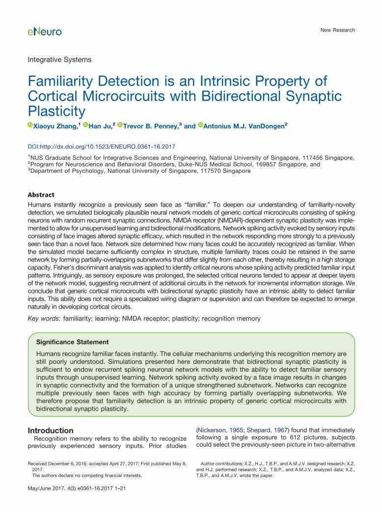
In CSIM, a network is generated by placing neurons ona 3-D grid. The networks described in Figure 1A had fivelayers with 10 � 10 neurons each (dimension, 10 � 10 �5). Input neurons formed synapses one-to-one with thefirst layer of the network reservoir, with fixed synapticweights of 2.7e�7. NMDAR synapses in the network res-ervoir were generated with � � 2.0 and Cscale � 1.0.Initial weights followed the gamma distribution withSH_W � 0.25 and Wscale � 0.5.
Networks described in Figure 1B consisted of five or sixlayers, with dimensions 20 � 20 � 5, 50 � 50 � 5, and50 � 50 � 6. In this case, input neurons formed synapsesrandomly with the network reservoir with Cscale � 0.04,0.004, and 0.005, respectively, and � was set to infinity inall cases to remove the limitation by distance. As a result,there was no topographical mapping of the input pattern.Input synaptic weights were still fixed but no longer uniform,following a gamma distribution (Wscale � 3, SH_W � 0.7 inall cases) instead. As for NMDAR synapses in the networkreservoir, � � 4.0 for the 20 � 20 � 5 networks; � � 3.0for 50 � 50 � 5 and 50 � 50 � 6 networks; Cscale � 1for all cases. The � values were chosen to make sure thateach neuron formed �100 synapses on average withothers in the network. Initial weights of NMDAR synapsesalso followed a gamma distribution with Wscale � 0.9 andSH_W � 0.25. By setting Wscale to 0.9, the initial weightswere set to intermediate values, leaving enough room forfuture potentiation and depression. By setting SH_W to0.25 for the network reservoir, we reduced the variation inthe initial weights, thereby reducing any preimposed net-work circuitry.
Synaptic plasticity implementationThe NMDAR-dependent plasticity we implement fol-
lows the model by Shouval et al. (2002). Synaptic plastic-ity (LTP/LTD and STDP) depends critically on theamplitude and timing of postsynaptic EPSPs and BPAPs.BPAPs were not implemented in the original CSIM, whileEPSPs were implemented using a single exponential de-cay function with a time constant of 3 ms. We introducedBPAPs and changed both BPAPs and EPSPs to followdouble-exponential decays, each with a fast and a slowcomponent. Decay time constants were scaled from thesuggested values (Shouval et al., 2002), with BPAP fastdecay time constant �f
bs � 1.2 ms and proportion Ifbs �0.75, slow decay time constant �s
bs � 10 ms and propor-tion Isbs � 0.25; EPSP fast decay time constant �f
ep � 2 ms,proportion Ifep � 0.5, slow decay time constant �s
ep � 20 msand proportion Ifep � 0.5.
BPAP�t� � BPAP_max·(Ifbse�t/�fbs
� Isbse�t/�sbs) (4)
EPSP�t� � EPSP_max·(Ifepe�t/�fep
� Isepe�t/�sep
) (5)
Using a double-exponential decay ensures that theBPAP has a sharp peak with a thin tail and that the EPSPhas a slower peak and more prominent tail, thereby pre-serving the difference in temporal signature between
New Research 3 of 21
May/June 2017, 4(3) e0361-16.2017 eNeuro.org
EPSPs and BPAPs. This contrast ensures that the changein intracellular Ca2� concentration induced by pre-postfiring exceeds the [Ca2�] change induced by post-prefiring, so that STDP will be induced properly (see Fig. 2 inShouval et al., 2002).
Activity-induced EPSPs generate the driving force forcalcium currents through the NMDAR:
H(V) ��0.42(V � Vr)
(1 � 0.6e�0.09V·[Mg2�]/3.57)(6)
where V � Vresting � EPSP � BPAP, and Vr is the reversalmembrane potential for calcium (130 mV). The Mg2� con-centration is set to 1 mM. The driving force function wasmodified from the function suggested by Shouval et al.(2002) which did not take into consideration the effect ofgrowing synaptic weights. In vitro recordings of ion cur-rents through NMDARs (Pattillo et al., 1999) show asteeper curve, and based on this, we re-parameterizedthe calcium driving force (Fig. 2A).
The calcium current through NMDARs (INMDA) is as-sumed to have this form:
INMDA�ti� � P0GNMDA[If�(t)e�t/�f � Is�(t)e�t/�s]H(V) (7)
where PO is the probability of opening, G is the conduc-tance of the NMDAR channel, If and Is are the fast andslow components of the NMDA currents, �f and �s are thetime constants for the fast and slow components.
Calcium control hypothesisThe activity-dependent change in synaptic calcium
concentration is modeled as a function of the NMDAcurrent INMDA:
dCa(t)dt
� INMDA�t� � (1/�Ca)[Ca(t)] (8)
where �Ca�t�� is the calcium concentration at the synapseat time t and �Ca is the decay time constant, which is
Figure 1. Network architecture and stimulus encoding. A, Diagram illustrating a 10 � 10 � 5 network with a 10 � 10 input layer,receiving a stimulus from a beard face image (10 � 10 pixels). Neurons in the network reservoir are located at positions with integercoordinates in a 3-D space. The input layer is located 1 unit away from the network reservoir. Input neurons form synapses one-to-onewith the first layer of the network reservoir. Each input neuron receives a spike train with a firing rate determined by the correspondingimage pixel intensity value. B, Diagram illustrating a 50 � 50 � 5 network (only a portion of size 15 � 15 � 5 is shown) with a 50 �50 input layer (only 15 � 15 portion is shown), receiving a stimulus from a human face image (50 � 50 pixels). Input neurons formsynapses with random neurons in the network reservoir. C, Examples of a 10 � 10-pixel beard face (top) and a 10 � 10-pixel no-beardface (bottom). D, Examples of 20 � 20-pixel images of car fronts, dog faces and human faces. E, Examples of 50 � 50-pixel humanfaces. F, A sample beard stimulus which contains spike trains (0–0.5 s), followed by a silent interval (0.5–1.0 s). Only 50 channels ofspike trains are shown for clarity.
New Research 4 of 21
May/June 2017, 4(3) e0361-16.2017 eNeuro.org
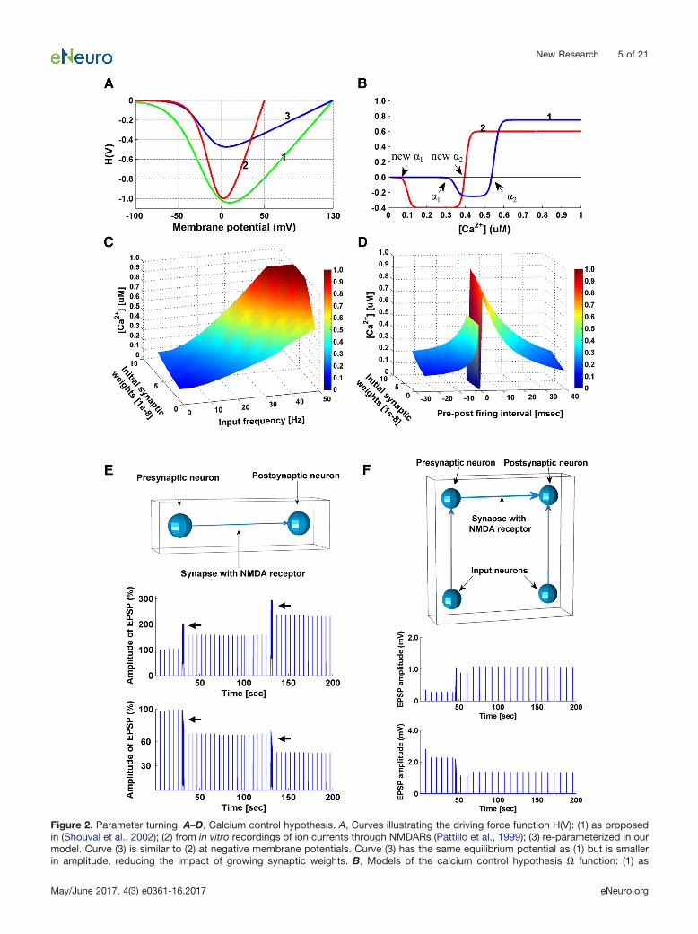
Figure 2. Parameter turning. A–D, Calcium control hypothesis. A, Curves illustrating the driving force function H(V): (1) as proposedin (Shouval et al., 2002); (2) from in vitro recordings of ion currents through NMDARs (Pattillo et al., 1999); (3) re-parameterized in ourmodel. Curve (3) is similar to (2) at negative membrane potentials. Curve (3) has the same equilibrium potential as (1) but is smallerin amplitude, reducing the impact of growing synaptic weights. B, Models of the calcium control hypothesis � function: (1) as
New Research 5 of 21
May/June 2017, 4(3) e0361-16.2017 eNeuro.org
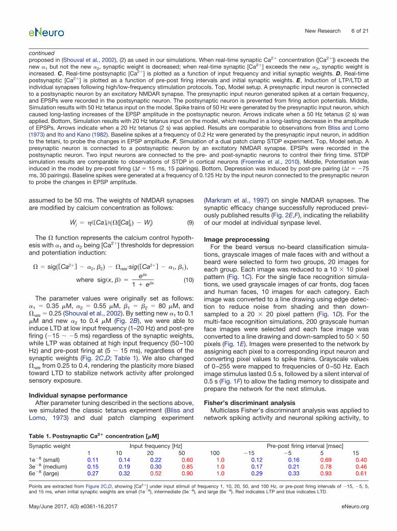
assumed to be 50 ms. The weights of NMDAR synapsesare modified by calcium concentration as follows:
Wj˙ � ��Ca�j��([Ca]j� � Wj) (9)
The � function represents the calcium control hypoth-esis with �1 and �2 being [Ca2�] thresholds for depressionand potentiation induction:
� sig��Ca2�� � �2, �2� � rate·sig��Ca2�� � �1, �1�,
where sig�x, �� �e�x
1 � e�x(10)
The parameter values were originally set as follows:�1 � 0.35 M, �2 � 0.55 M, �1 � �2 � 80 M, andrate � 0.25 (Shouval et al., 2002). By setting new �1 to 0.1 M and new �2 to 0.4 M (Fig. 2B), we were able toinduce LTD at low input frequency (1–20 Hz) and post-prefiring (�15 � �5 ms) regardless of the synaptic weights,while LTP was obtained at high input frequency (50–100Hz) and pre-post firing at (5 � 15 ms), regardless of thesynaptic weights (Fig. 2C,D; Table 1). We also changedrate from 0.25 to 0.4, rendering the plasticity more biasedtoward LTD to stabilize network activity after prolongedsensory exposure.
Individual synapse performanceAfter parameter tuning described in the sections above,
we simulated the classic tetanus experiment (Bliss andLomo, 1973) and dual patch clamping experiment
(Markram et al., 1997) on single NMDAR synapses. Thesynaptic efficacy change successfully reproduced previ-ously published results (Fig. 2E,F), indicating the reliabilityof our model at individual synpase level.
Image preprocessingFor the beard versus no-beard classification simula-
tions, grayscale images of male faces with and without abeard were selected to form two groups, 20 images foreach group. Each image was reduced to a 10 � 10 pixelpattern (Fig. 1C). For the single face recognition simula-tions, we used grayscale images of car fronts, dog facesand human faces, 10 images for each category. Eachimage was converted to a line drawing using edge detec-tion to reduce noise from shading and then down-sampled to a 20 � 20 pixel pattern (Fig. 1D). For themulti-face recognition simulations, 200 grayscale humanface images were selected and each face image wasconverted to a line drawing and down-sampled to 50 � 50pixels (Fig. 1E). Images were presented to the network byassigning each pixel to a corresponding input neuron andconverting pixel values to spike trains. Grayscale valuesof 0–255 were mapped to frequencies of 0–50 Hz. Eachimage stimulus lasted 0.5 s, followed by a silent interval of0.5 s (Fig. 1F) to allow the fading memory to dissipate andprepare the network for the next stimulus.
Fisher’s discriminant analysisMulticlass Fisher’s discriminant analysis was applied to
network spiking activity and neuronal spiking activity, to
continuedproposed in (Shouval et al., 2002), (2) as used in our simulations. When real-time synaptic Ca2� concentration ([Ca2�]) exceeds thenew �1 but not the new �2, synaptic weight is decreased; when real-time synaptic [Ca2�] exceeds the new �2, synaptic weight isincreased. C, Real-time postsynaptic [Ca2�] is plotted as a function of input frequency and initial synaptic weights. D, Real-timepostsynaptic [Ca2�] is plotted as a function of pre-post firing intervals and initial synaptic weights. E, Induction of LTP/LTD atindividual synapses following high/low-frequency stimulation protocols. Top, Model setup. A presynaptic input neuron is connectedto a postsynaptic neuron by an excitatory NMDAR synapse. The presynaptic input neuron generated spikes at a certain frequency,and EPSPs were recorded in the postsynaptic neuron. The postsynaptic neuron is prevented from firing action potentials. Middle,Simulation results with 50 Hz tetanus input on the model. Spike trains of 50 Hz were generated by the presynaptic input neuron, whichcaused long-lasting increases of the EPSP amplitude in the postsynaptic neuron. Arrows indicate when a 50 Hz tetanus (2 s) wasapplied. Bottom, Simulation results with 20 Hz tetanus input on the model, which resulted in a long-lasting decrease in the amplitudeof EPSPs. Arrows indicate when a 20 Hz tetanus (2 s) was applied. Results are comparable to observations from Bliss and Lomo(1973) and Ito and Kano (1982). Baseline spikes at a frequency of 0.2 Hz were generated by the presynaptic input neuron, in additionto the tetani, to probe the changes in EPSP amplitude. F, Simulation of a dual patch clamp STDP experiment. Top, Model setup. Apresynaptic neuron is connected to a postsynaptic neuron by an excitatory NMDAR synapse. EPSPs were recorded in thepostsynaptic neuron. Two input neurons are connected to the pre- and post-synaptic neurons to control their firing time. STDPsimulation results are comparable to observations of STDP in cortical neurons (Froemke et al., 2010). Middle, Potentiation wasinduced in the model by pre-post firing (t � 15 ms, 15 pairings). Bottom, Depression was induced by post-pre pairing (t � �75ms, 30 pairings). Baseline spikes were generated at a frequency of 0.125 Hz by the input neuron connected to the presynaptic neuronto probe the changes in EPSP amplitude.
Table 1. Postsynaptic Ca2� concentration [�M]
Synaptic weight Input frequency [Hz] Pre-post firing interval [msec]1 10 20 50 100 �15 �5 5 15
1e�8 (small) 0.11 0.14 0.22 0.60 1.0 0.12 0.16 0.69 0.403e�8 (medium) 0.15 0.19 0.30 0.85 1.0 0.17 0.21 0.78 0.466e�8 (large) 0.27 0.32 0.52 0.90 1.0 0.29 0.33 0.93 0.61
Points are extracted from Figure 2C,D, showing [Ca2�] under input stimuli of frequency 1, 10, 20, 50, and 100 Hz, or pre-post firing intervals of �15, �5, 5,and 15 ms, when initial synaptic weights are small (1e�8), intermediate (3e�8), and large (6e�8). Red indicates LTP and blue indicates LTD.
New Research 6 of 21
May/June 2017, 4(3) e0361-16.2017 eNeuro.org

obtain Fisher’s linear discriminant ratio (FDR). The FDR iscalculated as follows:
J�t� �� C( C�t� � �t�)2
� C � i�C(Si�t� � C�t�)2(11)
where Si�t� is the spike count at time bin t, in response tostimulus i; C�t� is the mean spike count at time t for classC stimuli, and �t� is the mean of the class means C�t�.The numerator and denominator of the discriminant ratioJ are known as the “between classes scatter” and “withinclass scatter.” A larger value of J indicates better discrim-ination. More information can be found in Ju et al. (2015).
Network FDR was obtained by summing up the FDRscalculated with network spiking activity in each time bin,serving as an indicator of network separability for inputstimuli. Neuronal FDR was calculated with the spikingactivity of individual neurons over the entire recordingperiod, serving as an indicator of how informative theneuron is for input discrimination.
ResultsWe performed simulations of cortical microcircuits
modeled with NMDAR-containing neural networks (seeMaterials and Methods), to investigate whether they candevelop recognition memory. Images were used as sen-sory inputs and plasticity was enabled during learning anddisabled during evaluation of responses to the input im-ages. Simulations typically consisted of three phases:baseline recording, learning and testing. First, to establisha baseline, network responses to all input stimuli wererecorded while NMDAR function was disabled. Networkfiring rate (spikes/second) during the stimulus presenta-tion was calculated and used as a measure of networkresponse. In the second phase, learning was switched onby enabling NMDAR plasticity, and networks were stim-ulated using only a subset of images, allowing stimulus-induced network spiking activity to alter synapticstrengths. These images should now be “familiar” to thenetworks. Finally, learning was switched off and networkresponses to all input stimuli were recorded again. Theeffect of the learning experience was evaluated for bothfamiliar images presented during the learning phase andthe “novel” images, by comparing testing responses withtheir corresponding baseline values. In all of our simula-tions, we found that the networks responded differentiallyto images presented during the learning phase.
Unsupervised classification: beard versus no-beardIn this first set of simulations, two classes of images
were used: human faces of males with and without abeard. In each simulation, only faces from a single class(beard or no-beard) were presented to a network duringthe learning phase. In each learning round, one face fromthe selected class was randomly chosen and presentedto the network for 1 s. NMDAR plasticity was switched offfor testing after each round, and network responses to allfaces from both classes were recorded. Figure 3A sum-marizes the performance of five randomly-generated 10 �10 � 5 networks for 40 rounds of image presentation.
Clearly, networks responded more strongly to faces fromthe trained class, and the firing rate discrepancy for thetwo classes increased as more faces were presented. Inother words, networks started to display a differentialresponse to the two classes as unsupervised learningtook place. This was true whether the beard or no-beardclass was used for training. The differential responseeventually reaches a steady state, in part because wehave set a maximum weight boundary (6.5e�8) for excit-atory synapses to prevent overtraining. We also investi-gated network performance by excluding faces that werepresented in the learning phase from the testing. Theresults showed a similar increase in discrepancy for theresponses to the two classes, only with a reduced mag-nitude (Fig. 3A). In this case, the networks were perform-ing a binary classification task, following unsupervisedlearning.
Single face recognitionNext, we performed single face recognition simulations,
for which we increased the dimension of the networks to20 � 20 � 5 and the input layer size to 20 � 20. A set of30 images was used as input stimuli, consisting of 10 carfronts, 10 dog faces, and 10 human faces. The simulationsagain consisted of three phases: baseline normalization,learning, and testing. NMDAR-dependent plasticity was onlyswitched on during the learning phase. Normalization wasperformed by recursively adjusting the mean pixel value foreach of the 30 images until they evoked comparable base-line responses. In the learning phase, a single human faceimage was selected from the set of 10, and presentedrepetitively for 15 s. NMDAR-dependent plasticity was thenswitched off for the testing phase, in which we recordednetwork responses to all images, to determine whether thenetwork responded differently to the selected (learned) face.
For this simulation, we used 30 randomly-generatednetworks. Instead of limiting the stimuli to human faces,we also conducted trials using images of car fronts anddog faces. For each network, three trials were conducted,each with a randomly selected car, dog, or human image.In total, 90 trials were performed on the 30 networks, andin 84 cases (93.3%), the networks exhibited the highestfiring rate to the image presented during the learningphase. Examples of network responses before and afterthe learning phase are shown in Figure 3B–D. Testingresponses are sorted by firing rate and shown as solidlines, while corresponding baseline responses are plottedas dashed lines. Network firing rate to the images se-lected for learning significantly increased and became thelargest on testing. Responses to novel inputs from thesame class as the image selected for learning tended tobe elevated from their baseline, although to a lesser ex-tent than the actual learned image. For instance, in thehuman face learning simulation (Fig. 3D), the networkresponses to 5 of the 10 human faces (excluding thelearned face) were higher than the responses to the dogand car images, suggesting that the network had alsogeneralized to distinguish human faces from dog facesand car fronts. Considering the degree of randomnessinvolved in network construction, and the similarity of the
New Research 7 of 21
May/June 2017, 4(3) e0361-16.2017 eNeuro.org

Figure 3. Unsupervised familiarity detection. A, Beard versus no-beard classification. Curves showing the discrepancy in network
New Research 8 of 21
May/June 2017, 4(3) e0361-16.2017 eNeuro.org
images within each class, we conclude that networks of20 � 20 � 5 neurons can learn to detect a familiar imagewith high specificity.
The network responses in Figure 3B–D are networkfiring rates averaged over the entire recording period. Infact, the dynamic network firing rates plotted as a functionof time underwent initial rising phases and declined tosteady state phases (Fig. 3E,F). By solely looking at firingrate difference, network response to the familiar inputseems to have increased over the entire recording periodafter learning (Fig. 3G). Whether the increase in individualtime bins is sufficient for familiarity detection is furtheranalyzed. If we assume the networks are able to detectfamiliarity in the time bins where networks exhibited thelargest firing rate to the familiar input, we can count thenumber of such time bins and compare it before and afterlearning. The results show that there is no specific timewindow that clearly separates the familiar input from thenovel inputs, but the separation gets better in the laterstage of stimulus presentation (Fig. 3H).
Multi-face recognitionNow that these relatively small neural networks have
demonstrated they can discriminate a single familiar facefrom many novel ones, we tested whether networks canperform familiarity detection to more than one face. Webegan with presenting two human face stimuli to a singlenetwork, using the same set of 30 images and 20 � 20 �5 networks. Unlike the high accuracy (93.3%) of the singleface recognition simulations, familiarity detection accu-racy for two face stimuli dropped to 70.0% (data notshown). This performance decline informs us of the lim-ited capacity of 20 � 20 � 5 networks.
We therefore increased dimensions of the networks to50 � 50 � 5 and input layers size to 50 � 50. In addition,the resolution of the 30 images was increased to 50 � 50pixels, so that their dimension matched that of the inputlayer. With these larger networks, the accuracy of famil-
iarity detection for two face stimuli increased to 96.7%.The performance improvement could be due to either theincrease in network size or the increase in resolution of theface images. To distinguish between these two options,we repeated the simulations on 50 � 50 � 5 networks, butwith the original low-resolution 20 � 20 pixel image stimulias inputs. Each pixel in the 20 � 20 images was replicatedonce to create images of 40 � 40. One column and onerow of void pixels were then added to the right andbottom of the matrices to make images of dimension 50 �50. When these low-resolution 50 � 50 stimuli were pre-sented to the networks, the accuracy of familiarity detec-tion remained at 96.7% (Fig. 4A), suggesting thatincreased network dimension was responsible for theaccuracy improvement.
Interclass learning of three image classes (car � dog �human) using 50 � 50 � 5 networks also produced stableand accurate results (data not shown). For multiple im-ages, we presented each image for 15 s and then movedto the next image. In contrast, we also tried loopingthrough all images 15 times. The first presentation proto-col resulted in slightly better accuracy, and was used forall the remaining simulations.
To further evaluate the limits of these neural networkson multi-face recognition, we converted a database of200 human face images to 50 � 50 pixel stimuli. In eachsimulation, a randomly-generated 50 � 50 � 5 networkwas presented with 10 human face images, each for 6 s,and testing was conducted with the 10 learned faces plus10 novel faces drawn randomly from the database. Weused 6 s instead of 15, to prevent overtraining of thenetwork. After sorting the network firing rate for all 20faces, a hypothetical threshold was drawn between theresponses to faces ranking 10th and 11th, to evaluate theability of the networks to separate familiar and novelfaces. This procedure is similar to the empirical rankingtheory of vision (Purves et al., 2011), which suggests thatsubjects perceive the relative color and brightness of
continuedresponses (firing rate [sec�1]) to beard and no-beard faces that develops with sensory exposure to one class. The blue curvesrepresent network response (beard-noBeard) after learning beard faces. The dark blue solid line shows response differences whentrained beard faces were used for testing. The light blue dashed line shows the results when untrained beard faces were used fortesting, i.e., networks generalizing familiarity to novel beard faces. The red curves represent the results from the same simulationparadigm with no-beard faces used for learning. The red solid line shows response differences when trained no-beard faces wereused for testing. The pink dashed line shows the results when untrained no-beard faces were used for testing, i.e., networksgeneralizing familiarity to novel no-beard faces. Error bars at each time point reflect the SEM of results from five randomly-generatednetworks. Each network underwent five trials of simulation with randomly-selected face inputs. Curves were normalized bysubtracting the discrepancy of network baseline response to the two classes at time 0. B–D, Single face recognition. B, The networktest responses (solid lines) and corresponding baseline responses (dashed lines) to all 30 images after learning car image 10. Testresponses to car images were sorted in descending order. As images of each class were indexed as 1–10, test responses to the othertwo classes are plotted following the sorted index order. C, Network responses to all 30 images after learning dog image 7. D, Networkresponses to all 30 images after learning human face 1. E, F, Firing rate dynamics during stimulus presentation of the network shownin D. E, Network baseline firing rate to all 30 stimuli. F, Network firing rate to all 30 stimuli after learning human face 1. The stimulusof human face 1 is referred as the familiar input. G, Firing rate difference between response to the familiar input and the average ofthe responses to the novel inputs. The average performance of 10 networks in 30 simulations has been shown. Blue represents thedifference in network baseline responses. Red represents the difference in network testing responses, after the single face recognitionsimulations. H, Histograms of time bins where the networks exhibited the highest firing rate to the familiar input. Network firing ratewas obtained as in E, F, binning was at 10 ms. Time bins where networks exhibited the highest firing rate to the familiar input wereidentified. The number of the identified time bins was summed for 30 simulations carried out 10 networks, and the percentage wascalculated with respect to 30 simulations, with chance level being 1/30 (3.3%).
New Research 9 of 21
May/June 2017, 4(3) e0361-16.2017 eNeuro.org

objects by internally ranking the empirical brain activitythey evoke. In our case, the percentage of learned facesappearing in the top 10 reflects familiarity detection ac-curacy. The 10 faces used for presentation to the net-
works were randomly selected from the database,indexed as 1–10 and fixed for all simulations. The 10 novelfaces were randomly drawn from the remaining databaseand were varied for each testing trial. Figure 4B shows the
Figure 4. Familiarity detection for multiple faces. A, Results of 50 � 50 � 5 networks learning two human faces. Each bar is locatedat the point whose coordinates represent the rank of network firing rate for the two learned faces. The height of each bar reflects thenumber of occurrences of such a combination (30 in total). Blue and magenta represent network baseline and testing response,respectively. Note that after learning, the two familiar faces ranked 1 and 2 in 29 of 30 trials and 1 and 3 in the remaining trial. B–E,Results of learning 10 faces and testing them against 10 novel faces randomly drawn from a database of 200 faces. B, Results of a50 � 50 � 5 network learning 10 faces. The curves show baseline and sorted testing response after learning faces 1–10. The networkranks nine familiar faces among the top 10, with only one outlier (face 62, highlighted in magenta). The accuracy of this networktherefore was 9/10 or 90%. C, The effect of network dimension and Cscale value was evaluated by performing 1000 trials for 10different networks (see Results). For each trial, the accuracy was calculated as in B. The curves illustrate the accuracy (averaged over1000 trials) for the 10 networks with dimensions and Cscale values indicated in the legend. D, Accuracy distribution of 1000 testingtrials conducted on the ten 50 � 50 � 6 networks with Cscale 0.005. E, Baseline and testing response of the best-performing 50 �50 � 6 network after learning face 1–10. Network firing rate differentiates familiar faces completely from novel faces. F, Networkaverage accuracy of the 50 � 50 � 6 networks after learning 10 faces (red) and the corresponding accuracy after learning 30 faces(green), 60 faces (blue), and chance level at 60 faces (gray, dotted line). Error bars are the SDs of 1000 trials. G, top, Baseline andtesting response of the best-performing 50 � 50 � 6 network to all 200 faces and their scrambled versions, after learning face 1–10.F, faces; SF, scrambled faces. Bottom, Pairwise network firing rate difference to faces and scrambled faces (F-SF).
New Research 10 of 21
May/June 2017, 4(3) e0361-16.2017 eNeuro.org
sorted responses and hypothetical threshold for a net-work that was able to detect 9 out of the 10 presentedfaces in one testing trial, and therefore was considered90% accurate for this trial. The average accuracy of anetwork was measured by averaging the results of 1000testing trials. Performance of ten randomly-generated 50� 50 � 5 networks for the above simulations is shown inFigure 4C, blue. The best network reached �85% accu-racy on average for 10-face familiarity detection.
In an attempt to further improve the performance, wegenerated a different set of ten randomly-generated net-works, with one more layer added to the network reservoir(50 � 50 � 6), and increased the connection probabilityfrom the input layer to the reservoir (Cscale � 0.004 toCscale � 0.005). The resulting network average accuracyis shown in Figure 4C, red. Not only did the overall accu-racy improve, but also a network with 95% accuracyemerged, indicating the network was able to detect all 10familiar faces accurately for at least half of the 1000testing trials. Figure 4D summarizes the accuracy distri-bution of 1000 testing trials for the ten 50 � 50 � 6networks. The distribution is significantly shifted fromwhat would be expected by chance alone, i.e., five familiarfaces falling in the top 10. Figure 4E plots the response ofthe best-performing 50 � 50 � 6 network for one of thetesting trials. Unsupervised learning has clearly modifiedthe network response and a hypothetical threshold can bedrawn which accurately differentiates familiar from novelfaces.
We also evaluated the performance of ten randomly-generated 50 � 50 � 5 networks with Cscale 0.005 andten randomly-generated 50 � 50 � 6 networks withCscale 0.004 (Fig. 4C, dotted lines). Neither achievedcomparable accuracy as 50 � 50 � 6 networks withCscale 0.005. From these results, it seems that both thenetwork size and the number of input connections areimportant. An increase of input connections together witha larger network results in larger available network spacefor better familiarity storage.
In fact, the network capacity of multi-face recognition isnot restricted to 10 faces. In Figure 4F, we plot theaccuracy of the ten 50 � 50 � 6 networks after learning10 faces (red), 30 faces (green), and 60 faces (blue), aswell as the chance level for 60 faces (gray). To our sur-prise, only two networks performed equivalently to orbelow their corresponding chance levels after learning 60faces. The remaining eight networks performed signifi-
cantly better than chance levels (Table 2, n � 1000, p �0.01). Accuracy after learning 30 and 60 faces was mea-sured by the percentage of familiar stimuli appearingabove the hypothetical threshold (set as below 50% of thepopulation) after pooling the same number of novel stimuliwith familiar stimuli. The chance level at 60 faces wascalculated by the percentage of familiar stimuli appearingabove the hypothetical threshold after sorting networkbaseline firing rates to the familiar and novel stimuli.
In addition, we generated scrambled versions of the 50 �50 face images from the same database by relocating allpixels to new random (x, y) positions, and recorded thenetwork responses to them after learning 10 human faces.The results show that networks not only discriminated thefamiliar faces from the novel faces, but also could discernnovel faces from their scrambled versions (Fig. 4G). Thetrained networks appeared to have acquired the conceptof a “face” and responded less to scrambled faces.
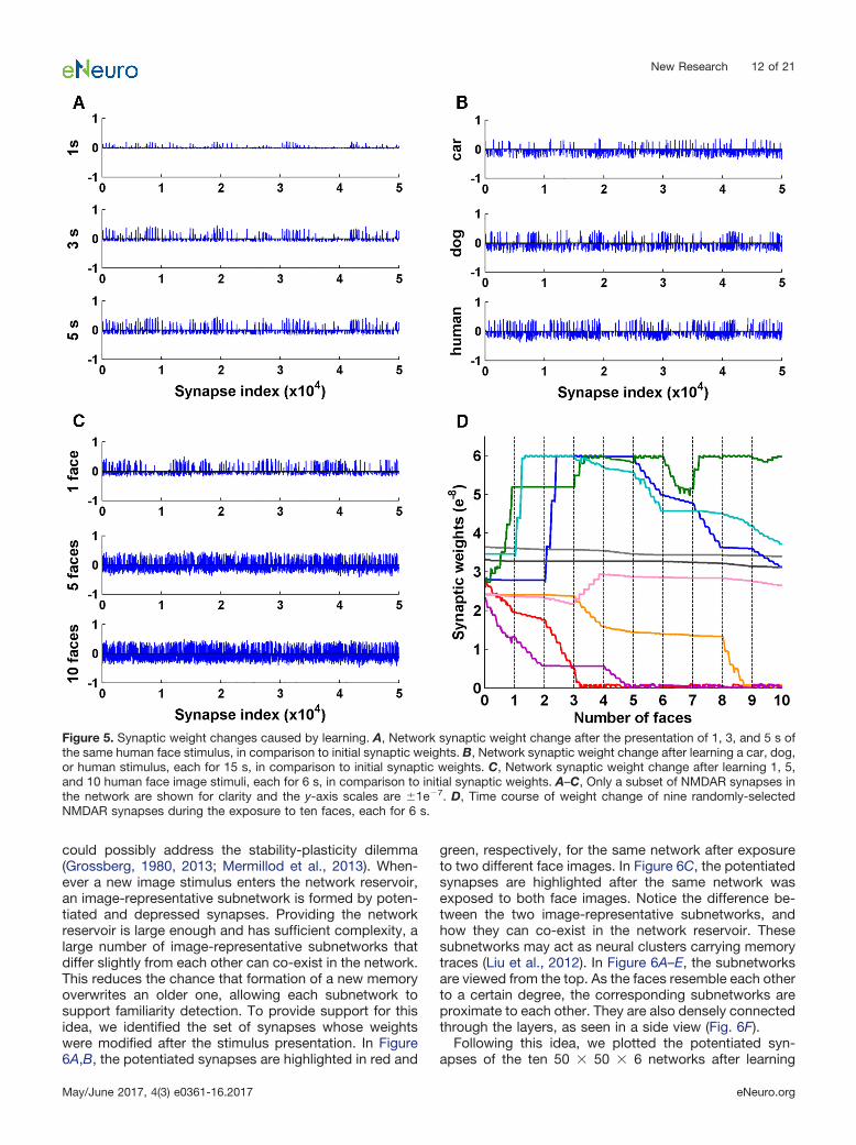
Synaptic weight changeGiven that synaptic weight changes underlie the ob-
served results described above, we plot in Figure 5 thenetwork weight changes in three scenarios: weightchange after exposure to a human face stimulus for 1, 3,and 5 s (Fig. 5A); weight change after exposure to a car,a dog or a human face image each for 15 s (Fig. 5B); andweight change after exposure to multiple face images(Fig. 5C). A common finding emerges for all three scenar-ios: unsupervised learning caused a subset of synapsesto be potentiated, and a different but larger subset ofsynapses to be depressed. The amplitude of potentiationis larger than the amplitude of depression, on average.This is in line with a proposed familiarization mechanismthat a small subset of neurons becomes strongly respon-sive after sensory exposure while depression occurs per-vasively among other neurons, sharpening the familiarityresponse (Freedman et al., 2006; Meyer et al., 2014).Figure 5D shows the time course of the change in synap-tic strength for nine representative synapses randomlypicked from a 50 � 50 � 6 network during the exposureto 10 faces. Synaptic weights changed in unique ways inresponse to each face stimulus.
Subnetwork formationIf we put together the phenomena of how synaptic
weights change after sensory exposure and how availablenetwork space affects accuracy, a theory emerges that
Table 2. Statistical table
Network Data structure Type of test p value1 Normality test: failed (p � 0.05) Mann-Whitney rank sum test (one-side) 5.29e�315
2 Normality test: failed (p � 0.05) Mann-Whitney rank sum test (one-side) 3.45e�322
3 Normality test: failed (p � 0.05) Mann-Whitney rank sum test (one-side) 0.004 Normality test: failed (p � 0.05) Mann-Whitney rank sum test (one-side) 0.005 Normality test: failed (p � 0.05) Mann-Whitney rank sum test (one-side) 0.036 Normality test: failed (p � 0.05) Mann-Whitney rank sum test (one-side) 5.16e�158
7 Normality test: failed (p � 0.05) Mann-Whitney rank sum test (one-side) 2.42e�322
8 Normality test: failed (p � 0.05) Mann-Whitney rank sum test (one-side) 3.71e�91
9 Normality test: failed (p � 0.05) Mann-Whitney rank sum test (one-side) 1.49e�140
10 Normality test: failed (p � 0.05) Mann-Whitney rank sum test (one-side) 1.00
New Research 11 of 21
May/June 2017, 4(3) e0361-16.2017 eNeuro.org
could possibly address the stability-plasticity dilemma(Grossberg, 1980, 2013; Mermillod et al., 2013). When-ever a new image stimulus enters the network reservoir,an image-representative subnetwork is formed by poten-tiated and depressed synapses. Providing the networkreservoir is large enough and has sufficient complexity, alarge number of image-representative subnetworks thatdiffer slightly from each other can co-exist in the network.This reduces the chance that formation of a new memoryoverwrites an older one, allowing each subnetwork tosupport familiarity detection. To provide support for thisidea, we identified the set of synapses whose weightswere modified after the stimulus presentation. In Figure6A,B, the potentiated synapses are highlighted in red and
green, respectively, for the same network after exposureto two different face images. In Figure 6C, the potentiatedsynapses are highlighted after the same network wasexposed to both face images. Notice the difference be-tween the two image-representative subnetworks, andhow they can co-exist in the network reservoir. Thesesubnetworks may act as neural clusters carrying memorytraces (Liu et al., 2012). In Figure 6A–E, the subnetworksare viewed from the top. As the faces resemble each otherto a certain degree, the corresponding subnetworks areproximate to each other. They are also densely connectedthrough the layers, as seen in a side view (Fig. 6F).
Following this idea, we plotted the potentiated syn-apses of the ten 50 � 50 � 6 networks after learning
Figure 5. Synaptic weight changes caused by learning. A, Network synaptic weight change after the presentation of 1, 3, and 5 s ofthe same human face stimulus, in comparison to initial synaptic weights. B, Network synaptic weight change after learning a car, dog,or human stimulus, each for 15 s, in comparison to initial synaptic weights. C, Network synaptic weight change after learning 1, 5,and 10 human face image stimuli, each for 6 s, in comparison to initial synaptic weights. A–C, Only a subset of NMDAR synapses inthe network are shown for clarity and the y-axis scales are �1e�7. D, Time course of weight change of nine randomly-selectedNMDAR synapses during the exposure to ten faces, each for 6 s.
New Research 12 of 21
May/June 2017, 4(3) e0361-16.2017 eNeuro.org
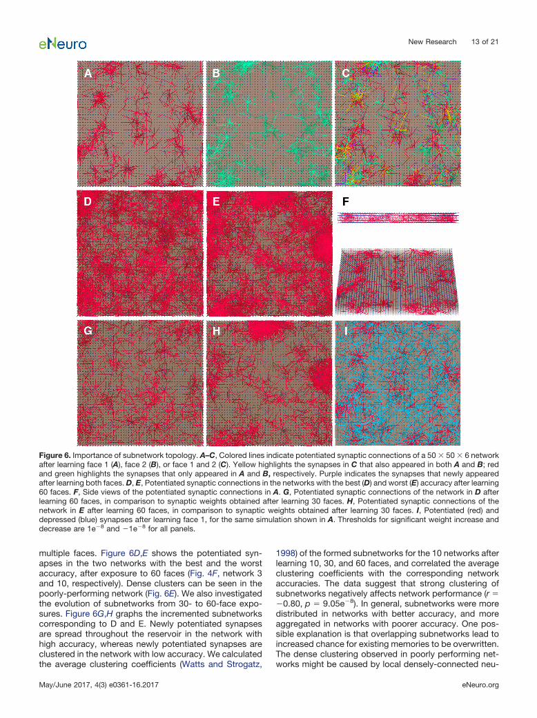
multiple faces. Figure 6D,E shows the potentiated syn-apses in the two networks with the best and the worstaccuracy, after exposure to 60 faces (Fig. 4F, network 3and 10, respectively). Dense clusters can be seen in thepoorly-performing network (Fig. 6E). We also investigatedthe evolution of subnetworks from 30- to 60-face expo-sures. Figure 6G,H graphs the incremented subnetworkscorresponding to D and E. Newly potentiated synapsesare spread throughout the reservoir in the network withhigh accuracy, whereas newly potentiated synapses areclustered in the network with low accuracy. We calculatedthe average clustering coefficients (Watts and Strogatz,
1998) of the formed subnetworks for the 10 networks afterlearning 10, 30, and 60 faces, and correlated the averageclustering coefficients with the corresponding networkaccuracies. The data suggest that strong clustering ofsubnetworks negatively affects network performance (r ��0.80, p � 9.05e�8). In general, subnetworks were moredistributed in networks with better accuracy, and moreaggregated in networks with poorer accuracy. One pos-sible explanation is that overlapping subnetworks lead toincreased chance for existing memories to be overwritten.The dense clustering observed in poorly performing net-works might be caused by local densely-connected neu-
Figure 6. Importance of subnetwork topology. A–C, Colored lines indicate potentiated synaptic connections of a 50 � 50 � 6 networkafter learning face 1 (A), face 2 (B), or face 1 and 2 (C). Yellow highlights the synapses in C that also appeared in both A and B; redand green highlights the synapses that only appeared in A and B, respectively. Purple indicates the synapses that newly appearedafter learning both faces. D, E, Potentiated synaptic connections in the networks with the best (D) and worst (E) accuracy after learning60 faces. F, Side views of the potentiated synaptic connections in A. G, Potentiated synaptic connections of the network in D afterlearning 60 faces, in comparison to synaptic weights obtained after learning 30 faces. H, Potentiated synaptic connections of thenetwork in E after learning 60 faces, in comparison to synaptic weights obtained after learning 30 faces. I, Potentiated (red) anddepressed (blue) synapses after learning face 1, for the same simulation shown in A. Thresholds for significant weight increase anddecrease are 1e�8 and �1e�8 for all panels.
New Research 13 of 21
May/June 2017, 4(3) e0361-16.2017 eNeuro.org
rons, which tend to self-potentiate excessively due torecurrent pathways, thereby reducing memory capacity.
For the network with the best accuracy, we attemptedto permute the initial weights of all NMDAR synapses ofthe network and repeated the same simulation of the60-face exposure. After permutation, the network accu-racy failed to remain the best. It seems that the preim-posed network circuitry, which is determined by the initialsynaptic connection weights, is another important factorfor network performance.
SeparabilityIn addition to using network firing rate as a readout for
familiarity detection, we applied Fisher’s discriminantanalysis to search for other features that may help definefamiliarity. Ten 20 � 20 � 5 networks and the single facerecognition simulations were used for the analysis. Net-work response to the familiar stimulus is referred to as thefamiliar response, and responses to the remaining 29stimuli are referred to as control responses. FDR is cal-culated between two classes (see Materials and Meth-ods). Specifically, familiar FDR is calculated by labelingthe familiar response as Class I and the control responsesas Class II. Control FDRs are calculated by labeling one ofthe control responses as Class I and the remaining controlresponses together with the familiar response as Class II.Baseline FDRs are calculated in the same way but withnetwork baseline responses to all 30 image stimuli. Base-line FDR values represent how well each image stimulus isseparated from others, before learning.
Compared with the corresponding baseline FDRs, wesee a general increase for both familiar FDR and controlFDRs after sensory exposure (Fig. 7A). In 26 out of 30cases, familiar FDRs show the largest magnitude of rela-tive increase from baseline compared with control FDRs;and in 22 out of 30 cases, familiar FDRs have the largestfinal values. Larger familiar FDRs indicates that the networkdiscriminated the familiar better from the control stimuli afterlearning, implying greater separability at the network level.Figure 7B shows the network FDRs calculated in each timebin. The larger familiar FDR can be explained by the largerFDR values in each time bin. Sensory exposure has alsoextended the network separability to beyond the stimuluspresentation window (0–0.5 s).
To further understand the changes occurring at theneuronal level that supported familiarity detection, weapplied the analysis to individual neurons. Similarly, famil-iar FDR is calculated by labeling the neuronal response tothe familiar stimulus as Class I and responses to controlstimuli as Class II; control FDRs are calculated by labelingthe neuronal response to one of the control stimuli asClass I and responses to the remaining control stimuli andthe familiar stimulus as Class II.
Compared with corresponding baseline FDRs, neuronsthat show increased FDRs to the familiar stimulus areconsidered as critical and potentially correlate with theemergence of familiarity. We noticed that the neurons withsignificantly increased FDRs tended to evolve more oftenfrom the neurons with negligible baseline FDRs (Fig. 7C).Neuronal baseline FDR was found to be negatively corre-
lated with FDR increase (r � �0.30, p � 4.46e�164). Thetop-ranking neurons showed little response at baselinebut increased firing rate after sensory exposure. The lowbaseline response implies that they did not receive muchinformation from the input layer initially, or equivalently,that they were not wired to the input-responding pathwaybefore the training. The increase in firing rate after learningimplies that their connections to the input-respondingpathway were strengthened. These critical neurons arerecruited by unsupervised learning to the subnetwork thatresponds to a specific stimulus. Once recruited, they mayalso fire to other nonlearned inputs, but their response tothe familiar input is stronger (Fig. 7D).
So far, we have looked at the formation of subnetworks(altered connections) and the emergence of critical neu-rons for familiarity detection. The intersection between thetwo intrigues us. Therefore, we investigated whether thecritical neurons belong to the subnetworks. The subnet-work in Figure 6A was analyzed. We selected 200 criticalneurons with top-ranking familiar FDR values and 118 ofthem belong to the subnetwork (Fig. 7E). If 200 neuronswere randomly selected from the network reservoir, wewould expect �28 neurons to overlap with the subnet-work by chance. For comparison, we also selected 200critical neurons with top-ranking control FDR values forcontrol faces 2–10 for the same network, after learningface 1. The intersections with the subnetwork dropped to43 � 8 (mean � SD) neurons (Fig. 7F). The observationheld when other faces were used for learning. Therefore,the critical neurons selected for the learned face and thesubnetwork formed after exposure are highly correlated.Their colocalization in space further supports the idea thatthe potentiated subnetwork is an important memory stor-age unit.
Effect of STDP and LTD/LTPAs the NMDAR-based plasticity we implemented sup-
ports both LTD/LTP and STDP mechanisms, we investi-gated how they contributed to familiarity detection.Learning in which LTP/LTD and STDP worked togetherstabilized network performance efficiently to the optimumaccuracy (Fig. 8A, STDP & LTP/LTD). Then we selectivelydeactivated STDP by removing BPAPs (Eq. 4) and re-peated the single face recognition simulations on 10 net-works. The results showed a severe reduction of accuracy(Fig. 8A, LTP/LTD). At later time points during learningwithout STDP, network accuracy reversed and networksresponded less to the familiar input than to novel inputson average. This is the result of an LTD-dominant �function (Eq. 10) tuned to account for the effect of BPAPs.To know what LTP/LTD alone is capable of, we need toreadjust the parameters of the � function to compensatefor the loss of BPAPs (Fig. 8A, LTP/LTD adjusted, �1 �0.3, �2 � 0.4 and rate � 0.3). Comparing the curves ofSTDP & LTP/LTD, and LTP/LTD adjusted (Fig. 8A), wepostulate that the effect of STDP is to increase learningspecificity.
Does this mean that STDP alone could be sufficient forfamiliarity detection? As STDP cannot be isolated to func-tion alone under the calcium control hypothesis, we
New Research 14 of 21
May/June 2017, 4(3) e0361-16.2017 eNeuro.org
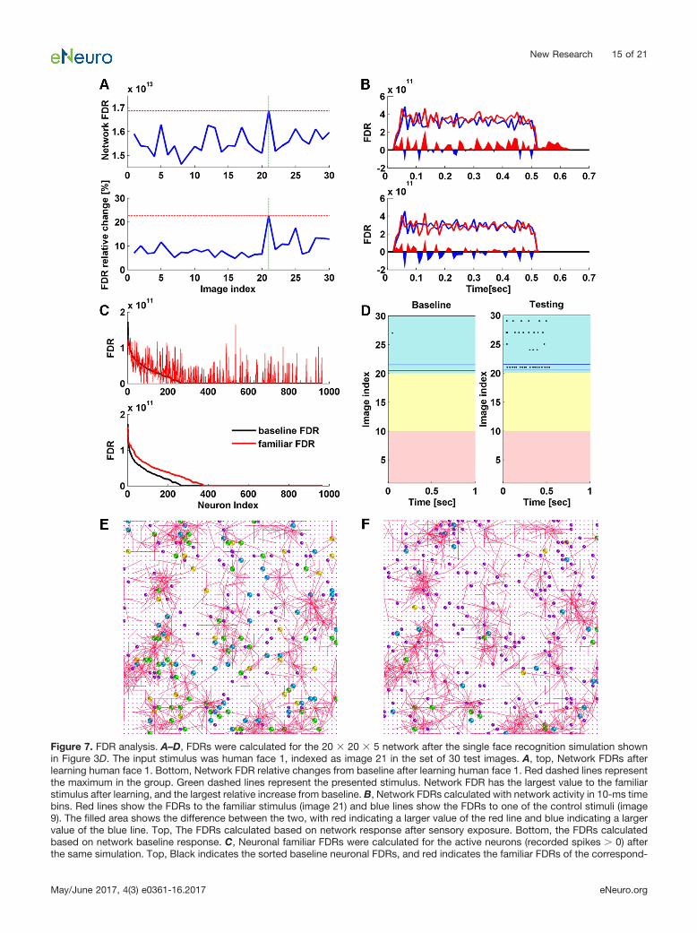
Figure 7. FDR analysis. A–D, FDRs were calculated for the 20 � 20 � 5 network after the single face recognition simulation shownin Figure 3D. The input stimulus was human face 1, indexed as image 21 in the set of 30 test images. A, top, Network FDRs afterlearning human face 1. Bottom, Network FDR relative changes from baseline after learning human face 1. Red dashed lines representthe maximum in the group. Green dashed lines represent the presented stimulus. Network FDR has the largest value to the familiarstimulus after learning, and the largest relative increase from baseline. B, Network FDRs calculated with network activity in 10-ms timebins. Red lines show the FDRs to the familiar stimulus (image 21) and blue lines show the FDRs to one of the control stimuli (image9). The filled area shows the difference between the two, with red indicating a larger value of the red line and blue indicating a largervalue of the blue line. Top, The FDRs calculated based on network response after sensory exposure. Bottom, the FDRs calculatedbased on network baseline response. C, Neuronal familiar FDRs were calculated for the active neurons (recorded spikes 0) afterthe same simulation. Top, Black indicates the sorted baseline neuronal FDRs, and red indicates the familiar FDRs of the correspond-
New Research 15 of 21
May/June 2017, 4(3) e0361-16.2017 eNeuro.org
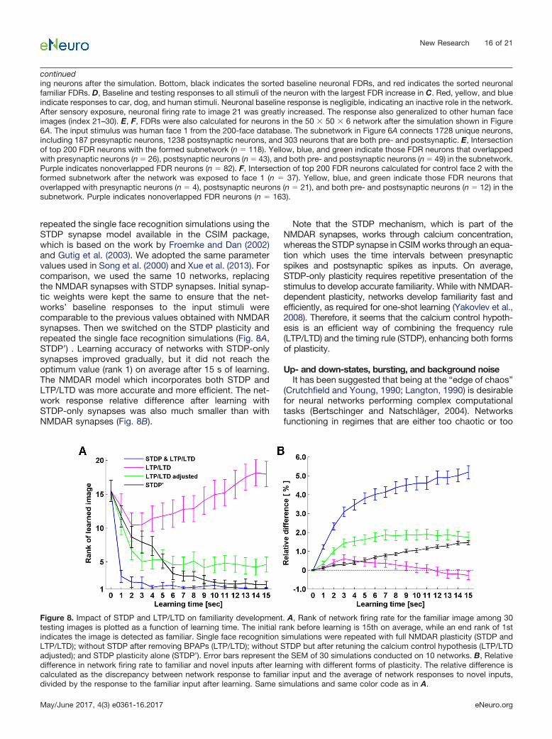
repeated the single face recognition simulations using theSTDP synapse model available in the CSIM package,which is based on the work by Froemke and Dan (2002)and Gutig et al. (2003). We adopted the same parametervalues used in Song et al. (2000) and Xue et al. (2013). Forcomparison, we used the same 10 networks, replacingthe NMDAR synapses with STDP synapses. Initial synap-tic weights were kept the same to ensure that the net-works’ baseline responses to the input stimuli werecomparable to the previous values obtained with NMDARsynapses. Then we switched on the STDP plasticity andrepeated the single face recognition simulations (Fig. 8A,STDP’) . Learning accuracy of networks with STDP-onlysynapses improved gradually, but it did not reach theoptimum value (rank 1) on average after 15 s of learning.The NMDAR model which incorporates both STDP andLTP/LTD was more accurate and more efficient. The net-work response relative difference after learning withSTDP-only synapses was also much smaller than withNMDAR synapses (Fig. 8B).
Note that the STDP mechanism, which is part of theNMDAR synapses, works through calcium concentration,whereas the STDP synapse in CSIM works through an equa-tion which uses the time intervals between presynapticspikes and postsynaptic spikes as inputs. On average,STDP-only plasticity requires repetitive presentation of thestimulus to develop accurate familiarity. While with NMDAR-dependent plasticity, networks develop familiarity fast andefficiently, as required for one-shot learning (Yakovlev et al.,2008). Therefore, it seems that the calcium control hypoth-esis is an efficient way of combining the frequency rule(LTP/LTD) and the timing rule (STDP), enhancing both formsof plasticity.
Up- and down-states, bursting, and background noiseIt has been suggested that being at the “edge of chaos”
(Crutchfield and Young, 1990; Langton, 1990) is desirablefor neural networks performing complex computationaltasks (Bertschinger and Natschläger, 2004). Networksfunctioning in regimes that are either too chaotic or too
continueding neurons after the simulation. Bottom, black indicates the sorted baseline neuronal FDRs, and red indicates the sorted neuronalfamiliar FDRs. D, Baseline and testing responses to all stimuli of the neuron with the largest FDR increase in C. Red, yellow, and blueindicate responses to car, dog, and human stimuli. Neuronal baseline response is negligible, indicating an inactive role in the network.After sensory exposure, neuronal firing rate to image 21 was greatly increased. The response also generalized to other human faceimages (index 21–30). E, F, FDRs were also calculated for neurons in the 50 � 50 � 6 network after the simulation shown in Figure6A. The input stimulus was human face 1 from the 200-face database. The subnetwork in Figure 6A connects 1728 unique neurons,including 187 presynaptic neurons, 1238 postsynaptic neurons, and 303 neurons that are both pre- and postsynaptic. E, Intersectionof top 200 FDR neurons with the formed subnetwork (n � 118). Yellow, blue, and green indicate those FDR neurons that overlappedwith presynaptic neurons (n � 26), postsynaptic neurons (n � 43), and both pre- and postsynaptic neurons (n � 49) in the subnetwork.Purple indicates nonoverlapped FDR neurons (n � 82). F, Intersection of top 200 FDR neurons calculated for control face 2 with theformed subnetwork after the network was exposed to face 1 (n � 37). Yellow, blue, and green indicate those FDR neurons thatoverlapped with presynaptic neurons (n � 4), postsynaptic neurons (n � 21), and both pre- and postsynaptic neurons (n � 12) in thesubnetwork. Purple indicates nonoverlapped FDR neurons (n � 163).
Figure 8. Impact of STDP and LTP/LTD on familiarity development. A, Rank of network firing rate for the familiar image among 30testing images is plotted as a function of learning time. The initial rank before learning is 15th on average, while an end rank of 1stindicates the image is detected as familiar. Single face recognition simulations were repeated with full NMDAR plasticity (STDP andLTP/LTD); without STDP after removing BPAPs (LTP/LTD); without STDP but after retuning the calcium control hypothesis (LTP/LTDadjusted); and STDP plasticity alone (STDP’). Error bars represent the SEM of 30 simulations conducted on 10 networks. B, Relativedifference in network firing rate to familiar and novel inputs after learning with different forms of plasticity. The relative difference iscalculated as the discrepancy between network response to familiar input and the average of network responses to novel inputs,divided by the response to the familiar input after learning. Same simulations and same color code as in A.
New Research 16 of 21
May/June 2017, 4(3) e0361-16.2017 eNeuro.org
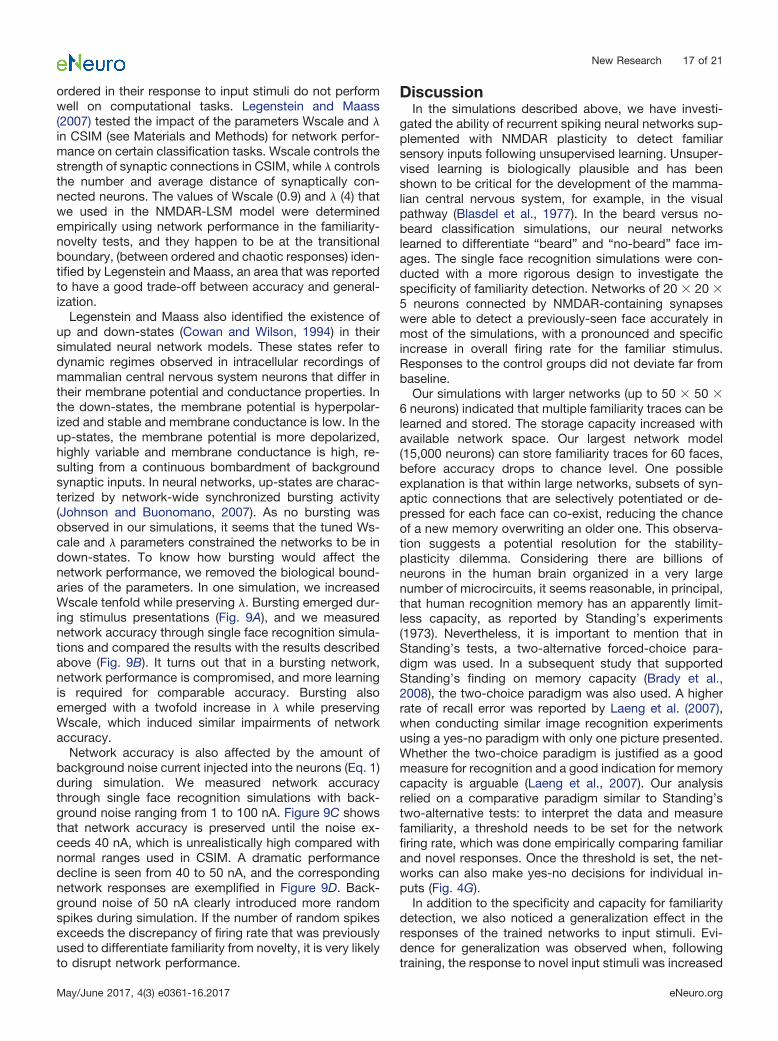
ordered in their response to input stimuli do not performwell on computational tasks. Legenstein and Maass(2007) tested the impact of the parameters Wscale and �in CSIM (see Materials and Methods) for network perfor-mance on certain classification tasks. Wscale controls thestrength of synaptic connections in CSIM, while � controlsthe number and average distance of synaptically con-nected neurons. The values of Wscale (0.9) and � (4) thatwe used in the NMDAR-LSM model were determinedempirically using network performance in the familiarity-novelty tests, and they happen to be at the transitionalboundary, (between ordered and chaotic responses) iden-tified by Legenstein and Maass, an area that was reportedto have a good trade-off between accuracy and general-ization.
Legenstein and Maass also identified the existence ofup and down-states (Cowan and Wilson, 1994) in theirsimulated neural network models. These states refer todynamic regimes observed in intracellular recordings ofmammalian central nervous system neurons that differ intheir membrane potential and conductance properties. Inthe down-states, the membrane potential is hyperpolar-ized and stable and membrane conductance is low. In theup-states, the membrane potential is more depolarized,highly variable and membrane conductance is high, re-sulting from a continuous bombardment of backgroundsynaptic inputs. In neural networks, up-states are charac-terized by network-wide synchronized bursting activity(Johnson and Buonomano, 2007). As no bursting wasobserved in our simulations, it seems that the tuned Ws-cale and � parameters constrained the networks to be indown-states. To know how bursting would affect thenetwork performance, we removed the biological bound-aries of the parameters. In one simulation, we increasedWscale tenfold while preserving �. Bursting emerged dur-ing stimulus presentations (Fig. 9A), and we measurednetwork accuracy through single face recognition simula-tions and compared the results with the results describedabove (Fig. 9B). It turns out that in a bursting network,network performance is compromised, and more learningis required for comparable accuracy. Bursting alsoemerged with a twofold increase in � while preservingWscale, which induced similar impairments of networkaccuracy.
Network accuracy is also affected by the amount ofbackground noise current injected into the neurons (Eq. 1)during simulation. We measured network accuracythrough single face recognition simulations with back-ground noise ranging from 1 to 100 nA. Figure 9C showsthat network accuracy is preserved until the noise ex-ceeds 40 nA, which is unrealistically high compared withnormal ranges used in CSIM. A dramatic performancedecline is seen from 40 to 50 nA, and the correspondingnetwork responses are exemplified in Figure 9D. Back-ground noise of 50 nA clearly introduced more randomspikes during simulation. If the number of random spikesexceeds the discrepancy of firing rate that was previouslyused to differentiate familiarity from novelty, it is very likelyto disrupt network performance.
DiscussionIn the simulations described above, we have investi-
gated the ability of recurrent spiking neural networks sup-plemented with NMDAR plasticity to detect familiarsensory inputs following unsupervised learning. Unsuper-vised learning is biologically plausible and has beenshown to be critical for the development of the mamma-lian central nervous system, for example, in the visualpathway (Blasdel et al., 1977). In the beard versus no-beard classification simulations, our neural networkslearned to differentiate “beard” and “no-beard” face im-ages. The single face recognition simulations were con-ducted with a more rigorous design to investigate thespecificity of familiarity detection. Networks of 20 � 20 �5 neurons connected by NMDAR-containing synapseswere able to detect a previously-seen face accurately inmost of the simulations, with a pronounced and specificincrease in overall firing rate for the familiar stimulus.Responses to the control groups did not deviate far frombaseline.
Our simulations with larger networks (up to 50 � 50 �6 neurons) indicated that multiple familiarity traces can belearned and stored. The storage capacity increased withavailable network space. Our largest network model(15,000 neurons) can store familiarity traces for 60 faces,before accuracy drops to chance level. One possibleexplanation is that within large networks, subsets of syn-aptic connections that are selectively potentiated or de-pressed for each face can co-exist, reducing the chanceof a new memory overwriting an older one. This observa-tion suggests a potential resolution for the stability-plasticity dilemma. Considering there are billions ofneurons in the human brain organized in a very largenumber of microcircuits, it seems reasonable, in principal,that human recognition memory has an apparently limit-less capacity, as reported by Standing’s experiments(1973). Nevertheless, it is important to mention that inStanding’s tests, a two-alternative forced-choice para-digm was used. In a subsequent study that supportedStanding’s finding on memory capacity (Brady et al.,2008), the two-choice paradigm was also used. A higherrate of recall error was reported by Laeng et al. (2007),when conducting similar image recognition experimentsusing a yes-no paradigm with only one picture presented.Whether the two-choice paradigm is justified as a goodmeasure for recognition and a good indication for memorycapacity is arguable (Laeng et al., 2007). Our analysisrelied on a comparative paradigm similar to Standing’stwo-alternative tests: to interpret the data and measurefamiliarity, a threshold needs to be set for the networkfiring rate, which was done empirically comparing familiarand novel responses. Once the threshold is set, the net-works can also make yes-no decisions for individual in-puts (Fig. 4G).
In addition to the specificity and capacity for familiaritydetection, we also noticed a generalization effect in theresponses of the trained networks to input stimuli. Evi-dence for generalization was observed when, followingtraining, the response to novel input stimuli was increased
New Research 17 of 21
May/June 2017, 4(3) e0361-16.2017 eNeuro.org
selectively for those that belonged to the same class asthe trained inputs. In the beard versus no-beard classifi-cation simulations, generalization was observed when thetrained networks were able to distinguish novel beardfrom novel no-beard images. In single face recognitionsimulations, the networks generalized familiarity responseto a subset of images from the same class as the pre-sented stimulus. In the multi-face recognition simulations,the networks apparently learned the concept face aftertraining on ten face images: they responded stronger tonovel faces than their scrambled versions. Recall that for10 � 10 � 5 networks, input neurons were set to formsynapses one-to-one with the first layer of the networkreservoir, whereas for networks of 20 � 20 � 5 or largerdimension, input neurons were set to form synapses ran-domly with the network reservoir by probability. Innerva-tion by probability is a better way to use the networkspace and prevents overtraining of the first layer of the
network reservoir. Meanwhile it disrupts the spatial pat-terns present in the face images, making the familiaritydetection a high-dimensional challenge. Yet the general-ization property remained with the randomly projectedinputs, which speaks to the robustness and computa-tional power of our networks, i.e., learning specificity waspreserved while generalization was acquired. When mul-tiple inputs from a class were learned, generalization al-lowed the networks to act as an unsupervised classifier,by automatically classifying input stimuli based on pastsensory experience. In contrast, conventional classifiersrequire properly labeled data of all classes to be trained.
Fisher’s discriminant analyses suggest that familiariza-tion occurred in high-dimensional feature space. Networkconnections were modified and neurons in hidden circuitswere recruited to respond to familiar inputs. Neuronshidden from the input layer are analogous to neurons fromdeep layers of a feed-forward network, or neurons from
Figure 9. Effect of bursting and noise. A, Response of a 20 � 20 � 5 up-state network after learning a human face image for 11 s.A network burst spontaneously emerged in the interval of 0.5–1.0 s when no input stimulus was given. B, Impact of bursting onnetwork performance. Rank of network firing rate to the familiar image among 30 testing images is plotted as a function of learningtime. Blue represents simulations with down-state networks, the same networks as we used in the single face recognition simulations.Red represents simulations with up-state networks (Wscale was set tenfold of the original value used in simulation). Error bars are theSEM of 90 simulations conducted on 30 networks. C, Impact of background noise on network performance. Average rank of networkfiring rate to the familiar image among 30 test images after learning for 15 s is plotted as a function of background noise. D, Networkspiking activity of a 20 � 20 � 5 network to the input stimulus, simulated with 40- and 50-nA background noise, respectively.
New Research 18 of 21
May/June 2017, 4(3) e0361-16.2017 eNeuro.org
the downstream circuits in higher-order brain regions.Their recruitment suggests how signals can be relayed tobrain regions other than the primary processing regionand cause differences in neuronal activity. As the deep-layer neurons showed unique firing patterns to the familiarstimulus, their activity can be used to identify the inputpattern, a function similar to the “grandmother-cells”identified by in vivo brain recordings (Quiroga et al., 2005).For decades neuroscientists have debated the possibleforms of information encoding in the brain, such as par-allel distributed processing versus single neuron firing.Results presented here show they are not mutually exclu-sive. While a familiar input is encoded in a network ofthousands of neurons, it may also selectively activatesingle neurons in deep layers.
Familiarity studies stemmed from experiments at the be-havioral level. Several groups have conducted experimentswith the familiarity/novelty paradigm in vivo. Studies usingfMRI measurements (Kosaka et al., 2003; Gobbini andHaxby, 2006) found an increase in the BOLD signal to famil-iar stimuli. Recordings in the inferior temporal lobe of behav-ing monkeys have demonstrated differential responses tonovel and familiar images (Anderson et al., 2008). Interest-ingly, familiar images evoked larger-amplitude local fieldpotentials, whereas multi-unit spiking responses weregreater for novel images. Finally, a phenomenon calledstimulus-selective response potentiation was identified inrodent visual cortex recordings (Frenkel et al., 2006; Cookeand Bear, 2010; Gavornik and Bear, 2014). It is a form ofexperience-dependent response enhancement during visualexperiments. We think it supports the existence of intrinsicfamiliarity in the visual cortex.
A few neural network models have been proposed tostudy network-level learning and memory. Several ofthem applied STDP to various network architectures. Forexample, a few groups (Lazar et al., 2007; Oliveri et al.,2007; Xue et al., 2013) investigated LSMs with STDP andthe results suggested an enhanced computational capa-bility. In these models, STDP was either applied to trainreadout neurons or to modulate neuron excitability, ratherthan allowing it to directly modify synaptic weights in thenetwork. Studies that used neural networks other thanLSMs (Clopath et al., 2010; Carlson et al., 2013; Klampfland Maass, 2013; Zheng et al., 2013; Srinivasa and Cho,2014) have reported emerging learning and memory afterapplying STDP to recurrent network architectures. Never-theless, the networks they used are either of a preim-posed wiring diagram (Klampfl and Maass, 2013) or highlysimplified (Srinivasa and Cho, 2014), and therefore poorlyreplicate cortical microcircuits. Furthermore, these stud-ies solely consider STDP for plasticity while we combinethe two major forms of plasticity, LTD/LTP (frequency rule)and STDP (timing rule), based on the calcium controlhypothesis, rather than phenomenological equations. Ev-idence for the interplay of LTD/LTP and STDP has beenfound in the literature, and separating them by firing rateor spike timing might lead to an artificial dichotomy(Sjöström et al., 2001). Our simulation results suggest amutual-enhancing effect by combining STDP with LTP/LTD, and this could potentially explain one-shot familiarity
memory (Yakovlev et al., 2008). Additionally, some of thereported models require homeostatic control (Clopathet al., 2010; Carlson et al., 2013; Zheng et al., 2013) orinhibitory-STDP (Srinivasa and Cho, 2014) to attain net-work stability, but in our NMDAR-LSM networks, we reliedon the intrinsic balance between LTD and LTP instanti-ated by the calcium control hypothesis for stability.
The plasticity model we implemented was created tomodel bidirectional synaptic plasticity through NMDARs(Shouval et al., 2002). Whether NMDARs are solely re-sponsible for this form of plasticity is still controversial, asvoltage-dependent Ca2� channels (Nevian and Sakmann,2006) and metabotropic glutamate receptors (Gladdinget al., 2009) have been reported to be capable of inducingsynaptic plasticity as well. Yet there is growing evidencethat bidirectional modifications can be induced throughNMDAR-dependent pathways alone (Hunt et al., 2013;Huang et al., 2014). The results presented here demon-strate that bidirectional synaptic plasticity is sufficient toendow neural network models of generic cortical micro-circuits with the ability to detect familiar sensory inputsthrough unsupervised learning. This has important con-sequences for mammalian brain development, since itsuggests that these universal building blocks of the cortexhave an inherent ability for familiarity detection.
In NMDAR-LSM networks, recurrent spiking neural net-works expand input stimuli into high-dimensional featurespace (Maass et al., 2002). Unsupervised learning alteredthe feature space to allow linear separation of familiarfrom novel faces, by formation of subnetworks specific foreach input stimulus. Learning multiple inputs belonging toa class (e.g., beard, face) resulted in generalization, allow-ing the network to classify novel input stimuli. This rela-tionship between familiarity detection, generalization, andclassification needs to be studied in more depth.
Note Added in Proof: The title of the article was incor-rectly listed in the Early Release version. The title has nowbeen corrected.
ReferencesAnderson B, Mruczek RE, Kawasaki K, Sheinberg D (2008) Effects of
familiarity on neural activity in monkey inferior temporal lobe.Cereb Cortex 18:2540–2552. CrossRef Medline
Baudry M, Zhu G, Liu Y, Wang Y, Briz V, Bi X (2015) Multiple cellularcascades participate in long-term potentiation and in hippocampus-dependent learning. Brain Res 1621:73–81. CrossRef Medline
Bertschinger N, Natschläger T (2004) Real-time computation at theedge of chaos in recurrent neural networks. Neural Comput 16:1413–1436. CrossRef Medline
Bi GQ, Poo MM (1998) Synaptic modifications in cultured hippocam-pal neurons: dependence on spike timing, synaptic strength, andpostsynaptic cell type. J Neurosci 18:10464–10472.
Blanke ML, VanDongen AMJ (2009) Activation mechanisms of theNMDA receptor. In: Biology of the NMDA receptor (Van DongenAM, ed). Boca Raton: CRC Press/Taylor & Francis.
Blasdel GG, Mitchell DE, Muir DW, Pettigrew JD (1977) A physiolog-ical and behavioural study in cats of the effect of early visualexperience with contours of a single orientation. J Physiol 265:615–636. Medline
Bliss TV, Lomo T (1973) Long-lasting potentiation of synaptic trans-mission in the dentate area of the anaesthetized rabbit followingstimulation of the perforant path. J Physiol 232:331–356. Medline
New Research 19 of 21
May/June 2017, 4(3) e0361-16.2017 eNeuro.org

Brady TF, Konkle T, Alvarez GA, Oliva A (2008) Visual long-termmemory has a massive storage capacity for object details. ProcNatl Acad Sci USA 105:14325–14329. CrossRef Medline
Buonomano DV, Maass W (2009) State-dependent computations:spatiotemporal processing in cortical networks. Nat Rev Neurosci10:113–125. CrossRef Medline
Carlson KD, Richert M, Dutt N, Krichmar JL (2013) Biologicallyplausible models of homeostasis and STDP: stability and learningin spiking neural networks. In: The 2013 International Joint Con-ference on Neural Networks (IJCNN), pp 1-8.
Clopath C, Büsing L, Vasilaki E, Gerstner W (2010) Connectivityreflects coding: a model of voltage-based STDP with homeostasis.Nat Neurosci 13:344–352. CrossRef Medline
Cooke SF, Bear MF (2010) Visual experience induces long-termpotentiation in the primary visual cortex. J Neurosci 30:16304–16313. CrossRef Medline
Coutlee CG, Huettel SA (2012) The functional neuroanatomy ofdecision making: prefrontal control of thought and action. BrainRes 1428:3–12. CrossRef Medline
Cowan RL, Wilson CJ (1994) Spontaneous firing patterns and axonalprojections of single corticostriatal neurons in the rat medialagranular cortex. J Neurophysiol 71:17–32. Medline
Crutchfield JP, Young K (1990) Computation at the onset of chaos.In: Entropy, complexity, and physics of information (Zurek W, ed),pp 223-269. Bouler: Addison-Wesley.
Diana RA, Yonelinas AP, Ranganath C (2007) Imaging recollectionand familiarity in the medial temporal lobe: a three-componentmodel. Trends Cogn Sci 11:379–386. CrossRef Medline
Fair DA, Cohen AL, Power JD, Dosenbach NU, Church JA, MiezinFM, Schlaggar BL, Petersen SE (2009) Functional brain networksdevelop from a local to distributed organization. PLoS ComputBiol 5:e1000381. CrossRef
Fornito A, Harrison BJ, Zalesky A, Simons JS (2012) Competitive andcooperative dynamics of large-scale brain functional networkssupporting recollection. Proc Natl Acad Sci USA 109:12788–12793. CrossRef Medline
Freedman DJ, Riesenhuber M, Poggio T, Miller EK (2006) Experience-dependent sharpening of visual shape selectivity in inferior tem-poral cortex. Cereb Cortex 16:1631–1644. CrossRef Medline
Frenkel MY, Sawtell NB, Diogo ACM, Yoon B, Neve RL, Bear MF(2006) Instructive effect of visual experience in mouse visual cor-tex. Neuron 51:339–349. CrossRef Medline
Froemke RC, Dan Y (2002) Spike-timing-dependent synaptic modi-fication induced by natural spike trains. Nature 416:433–438.CrossRef Medline
Froemke RC, Debanne D, Bi GQ (2010) Temporal modulation ofspike-timing-dependent plasticity. Frontiers in synaptic neurosci-ence 2:19.
Gale JT, Shields DC, Ishizawa Y, Eskandar EN (2014) Reward andreinforcement activity in the nucleus accumbens during learning.Front Behav Neurosci 8:114 CrossRef Medline
Gavornik JP, Bear MF (2014) Higher brain functions served by thelowly rodent primary visual cortex. Learn Mem 21:527–533. Cross-Ref Medline
Gerstner W, Kempter R, van Hemmen JL, Wagner H (1996) A neu-ronal learning rule for sub-millisecond temporal coding. Nature383:76–81. CrossRef Medline
Gladding CM, Fitzjohn SM, Molnár E (2009) Metabotropic glutamatereceptor-mediated long-term depression: molecular mechanisms.Pharmacol Rev 61:395–412. CrossRef Medline
Gobbini MI, Haxby JV (2006) Neural response to the visual familiarityof faces. Brain Res Bull 71:76–82. CrossRef Medline
Grossberg S (1980) How does a brain build a cognitive code?Psychol Rev 87:1–51. Medline
Grossberg S (2013) Adaptive resonance theory: how a brain learns toconsciously attend, learn, and recognize a changing world. NeuralNetw 37:1–47. CrossRef
Gupta A, Wang Y, Markram H (2000) Organizing principles for adiversity of GABAergic interneurons and synapses in the neocor-tex. Science 287:273–278. Medline
Gutig R, Aharonov R, Rotter S, Sompolinsky H (2003) Learning inputcorrelations through nonlinear temporally asymmetric Hebbianplasticity. J Neurosci 23:3697–3714.
Hölscher C, Rolls ET, Xiang J (2003) Perirhinal cortex neuronalactivity related to long-term familiarity memory in the macaque.Eur J Neurosci 18:2037–2046. Medline
Huang S, Rozas C, Treviño M, Contreras J, Yang S, Song L, Yosh-ioka T, Lee HK, Kirkwood A (2014) Associative Hebbian synapticplasticity in primate visual cortex. J Neurosci 34:7575–7579.CrossRef Medline
Hunt DL, Puente N, Grandes P, Castillo PE (2013) BidirectionalNMDA receptor plasticity controls CA3 output and heterosynapticmetaplasticity. Nat Neurosci 16:1049–1059. CrossRef Medline
Ito M, Kano M (1982) Long-lasting depression of parallel fiber-Purkinje cell transmission induced by conjunctive stimulation ofparallel fibers and climbing fibers in the cerebellar cortex. NeurosciLett 33:253–258. Medline
Johnson HA, Buonomano DV (2007) Development and plasticity ofspontaneous activity and up states in cortical organotypic slices. JNeurosci 27:5915–5925. CrossRef
Joshi P (2007) From memory-based decisions to decision-basedmovements: a model of interval discrimination followed by actionselection. Neural Netw 20:298–311. CrossRef
Ju H, Xu JX, Chong E, VanDongen AM (2013) Effects of synapticconnectivity on liquid state machine performance. Neural Netw38:39–51. CrossRef
Ju H, Dranias MR, Banumurthy G, VanDongen AM (2015) Spatiotemporalmemory is an intrinsic property of networks of dissociated corticalneurons. J Neurosci 35:4040–4051. CrossRef Medline
Klampfl S, Maass W (2013) Emergence of dynamic memory traces incortical microcircuit models through STDP. J Neurosci 33:11515–11529. CrossRef
Kosaka H, Omori M, Iidaka T, Murata T, Shimoyama T, Okada T,Sadato N, Yonekura Y, Wada Y (2003) Neural substrates partici-pating in acquisition of facial familiarity: an fMRI study. NeuroIm-age 20:1734–1742. Medline
Laeng B, Øvervoll M, Ole Steinsvik O (2007) Remembering 1500pictures: the right hemisphere remembers better than the left.Brain Cogn 63:136–144. CrossRef Medline
Langton CG (1990) Computation at the edge of chaos: phase tran-sitions and emergent computation. Physica D 42:12–37. CrossRef
Lazar A, Pipa G, Triesch J (2007) Fading memory and time seriesprediction in recurrent networks with different forms of plasticity.Neural Netw 20:312–322. CrossRef Medline
Legenstein R, Maass W (2007) Edge of chaos and prediction ofcomputational performance for neural circuit models. Neural Netw20:323–334. CrossRef
Lisman J (1989) A mechanism for the Hebb and the anti-Hebbprocesses underlying learning and memory. Proc Natl Acad SciUSA 86:9574–9578. Medline
Liu X, Ramirez S, Pang PT, Puryear CB, Govindarajan A, DeisserothK, Tonegawa S (2012) Optogenetic stimulation of a hippocampalengram activates fear memory recall. Nature 484:381–385. Cross-Ref Medline
Luscher C, Malenka RC (2012) NMDA receptor-dependent long-termpotentiation and long-term depression (LTP/LTD). Cold SpringHarb Perspect Biol 4. CrossRef
Maass W, Natschläger T, Markram H (2002) Real-time computingwithout stable states: a new framework for neural computationbased on perturbations. Neural Comput 14:2531–2560. CrossRef
Malleret G, Alarcon JM, Martel G, Takizawa S, Vronskaya S, Yin D,Chen IZ, Kandel ER, Shumyatsky GP (2010) Bidirectional regula-tion of hippocampal long-term synaptic plasticity and its influenceon opposing forms of memory. J Neurosci 30:3813–3825. Cross-Ref
Markram H, Lübke J, Frotscher M, Sakmann B (1997) Regulation ofsynaptic efficacy by coincidence of postsynaptic APs and EPSPs.Science 275:213–215. Medline
New Research 20 of 21
May/June 2017, 4(3) e0361-16.2017 eNeuro.org

Massey PV, Bashir ZI (2007) Long-term depression: multiple formsand implications for brain function. Trends Neurosci 30:176–184.CrossRef Medline
Mermillod M, Bugaiska A, Bonin P (2013) The stability-plasticitydilemma: investigating the continuum from catastrophic forgettingto age-limited learning effects. Front Psychol 4:504. CrossRefMedline
Meyer T, Walker C, Cho RY, Olson CR (2014) Image familiarizationsharpens response dynamics of neurons in inferotemporal cortex.Nat Neurosci 17:1388–1394. CrossRef Medline
Morgado-Bernal I (2011) Learning and memory consolidation: linkingmolecular and behavioral data. Neuroscience 176:12–19. Cross-Ref Medline
Natschläger T, Markram H, Maass W (2002) Computer models andanalysis tools for neural microcircuits. In: Neuroscience data-bases: a practical guide (Kötter R, ed). Boston: Kluver AcademicPublishers.
Nevian T, Sakmann B (2006) Spine Ca2� signaling in spike-timing-dependent plasticity. J Neurosci 26:11001–11013. CrossRef Med-line
Nickerson RS (1965) Short-term memory for complex meaningfulvisual configurations: a demonstration of capacity. Can J Psychol19:155–160. Medline
Oliveri A, Rizzo R, Chella A (2007) An application of spike-timing-dependent plasticity to readout circuit for liquid state machine. In:Neural networks, the 2007 International Joint Conference on Neu-ral Networks (IJCNN), pp 1441-1445.
Paradiso K, Wu LG (2009) Small voltage changes at nerve terminalstravel up axons to affect action potential initiation. Nat Neurosci12:541–543. CrossRef Medline
Pattillo JM, Artim DE, Simples JE Jr, Meriney SD (1999) Variations inonset of action potential broadening: effects on calcium currentstudied in chick ciliary ganglion neurones. J Physiol 514:719–728.CrossRef
Purves D, Wojtach WT, Lotto RB (2011) Understanding vision inwholly empirical terms. Proc Natl Acad Sci USA 108:15588–15595. CrossRef
Quiroga RQ, Reddy L, Kreiman G, Koch C, Fried I (2005) Invariantvisual representation by single neurons in the human brain. Nature435:1102–1107. CrossRef Medline
Rapanelli M, Frick LR, Miguelez Fernández AM, Zanutto BS (2015)Dopamine bioavailability in the mPFC modulates operant learningperformance in rats: an experimental study with a computationalinterpretation. Behav Brain Res 280:92–100. CrossRef Medline
Salter MW, Dong Y, Kalia LV, Liu XJ, Pitcher G (2009) Regulation ofNMDA receptors by kinases and phosphatases. In: Biology of theNMDA receptor (Van Dongen AM, ed). Boca Raton: CRC Press/Taylor & Francis.
Shepard RN (1967) Recognition memory for words, sentences, andpictures. J Verbal Learning Verbal Behav 6:156–163. CrossRef
Shouval HZ, Bear MF, Cooper LN (2002) A unified model of NMDAreceptor-dependent bidirectional synaptic plasticity. Proc NatlAcad Sci USA 99:10831–10836. CrossRef Medline
Sjöström PJ, Turrigiano GG, Nelson SB (2001) Rate, timing, andcooperativity jointly determine cortical synaptic plasticity. Neuron32:1149–1164. Medline
Song S, Miller KD, Abbott LF (2000) Competitive Hebbian learningthrough spike-timing-dependent synaptic plasticity. Nat Neurosci3:919–926. CrossRef Medline
Sporns O, Zwi JD (2004) The small world of the cerebral cortex.Neuroinformatics 2:145–162. CrossRef Medline
Squire LR, Wixted JT, Clark RE (2007) Recognition memory and themedial temporal lobe: a new perspective. Nat Rev Neurosci 8:872–883. CrossRef Medline
Srinivasa N, Cho Y (2014) Unsupervised discrimination of patterns inspiking neural networks with excitatory and inhibitory synapticplasticity. Front Comput Neurosci 8:159. CrossRef Medline
Standing L (1973) Learning 10,000 pictures. Q J Exp Psychol (Hove)25:207–222. CrossRef Medline
Takeuchi T, Duszkiewicz AJ, Morris RG (2014) The synaptic plasticityand memory hypothesis: encoding, storage and persistence. Phi-los Trans R Soc Lond B Biol Sci 369:20130288. CrossRef Medline
Waters J, Schaefer A, Sakmann B (2005) Backpropagating actionpotentials in neurones: measurement, mechanisms and potentialfunctions. Prog Biophys Mol Biol 87:145–170. CrossRef Medline
Watts DJ, Strogatz SH (1998) Collective dynamics of ’small-world’networks. Nature 393:440–442. CrossRef Medline
Wixted JT (2007) Dual-process theory and signal-detection theory ofrecognition memory. Psychol Rev 114:152–176. CrossRef Medline
Wu J, Rowan MJ, Anwyl R (2006) Long-term potentiation is mediatedby multiple kinase cascades involving CaMKII or either PKA orp42/44 MAPK in the adult rat dentate gyrus in vitro. J Neurophysiol95:3519–3527. CrossRef
Xue F, Hou Z, Li X (2013) Computational capability of liquid statemachines with spike-timing-dependent plasticity. Neurocomput-ing 122:324–329. CrossRef
Yakovlev V, Amit DJ, Romani S, Hochstein S (2008) Universal mem-ory mechanism for familiarity recognition and identification. J Neu-rosci 28:239–248. CrossRef Medline
Yonelinas AP (2002) The nature of recollection and familiarity: areview of 30 years of research. J Mem Lang 46:441–517. CrossRef
Zheng P, Dimitrakakis C, Triesch J (2013) Network self-organizationexplains the statistics and dynamics of synaptic connectionstrengths in cortex. PLoS Comput Biol 9:e1002848. CrossRefMedline
New Research 21 of 21
May/June 2017, 4(3) e0361-16.2017 eNeuro.org