Embed Size (px)
Citation preview
ORIGINAL RESEARCH ARTICLEpublished: 19 December 2014doi: 10.3389/fonc.2014.00368
Fanconi anemia repair pathway dysfunction, a potentialtherapeutic target in lung cancerWenrui Duan1,2*, Li Gao1, Brittany Aguila1, Arjun Kalvala1, Gregory A. Otterson1,2 andMiguel A. Villalona-Calero1,2,3*1 Comprehensive Cancer Center, The Ohio State University College of Medicine and Public Health, Columbus, OH, USA2 Division of Medical Oncology, Department of Internal Medicine, The Ohio State University College of Medicine and Public Health, Columbus, OH, USA3 Department of Pharmacology, The Ohio State University College of Medicine and Public Health, Columbus, OH, USA
Edited by:Masahiro Tsuboi, Kanagawa CancerCenter, Japan
Reviewed by:Shahab Babakoohi, Medstar GoodSamaritan Hospital, USAStephen V. Liu, GeorgetownUniversity, USA
*Correspondence:Wenrui Duan, Division of MedicalOncology, Department of Medicine,The Ohio State University,Comprehensive Cancer Center, 1230James CHRI, 300 West 10th Avenue,Columbus, OH 43210, USAe-mail: [email protected];Miguel A. Villalona-Calero, Division ofMedical Oncology, Department ofMedicine, The Ohio State University,Comprehensive Cancer Center, A455Starling-Loving Hall, 320 West10th Avenue, Columbus, OH 43210,USAe-mail: [email protected]
The Fanconi anemia (FA) pathway is a major mechanism of homologous recombination DNArepair. The functional readout of the pathway is activation through mono-ubiquitination ofFANCD2 leading to nuclear foci of repair. We have recently developed an FA triple-stainingimmunofluorescence based method (FATSI) to evaluate FANCD2 foci formation in formalinfixed paraffin-embedded (FFPE) tumor samples. DNA-repair deficiencies have been con-sidered of interest in lung cancer prevention, given the persistence of damage produced bycigarette smoke in this setting, as well as in treatment, given potential increased efficacy ofDNA-damaging drugs. We screened 139 non-small cell lung cancer (NSCLC) FFPE tumorsfor FANCD2 foci formation by FATSI analysis. Among 104 evaluable tumors, 23 (22%)were FANCD2 foci negative, thus repair deficient.To evaluate and compare novel-targetedagents in the background of FA deficiency, we utilized RNAi technology to render severallung cancer cell lines FANCD2 deficient. Successful FANCD2 knockdown was confirmedby reduction in the FANCD2 protein. Subsequently, we treated the FA defective H1299D2-down and A549D2-down NSCLC cells and their FA competent counterparts (empty vectorcontrols) with the PARP inhibitors veliparib (ABT-888) (5 µM) and BMN673 (0.5 µM), aswell as the CHK1 inhibitor Arry-575 at a dose of 0.5 µM. We also treated the FA defectivesmall cell lung cancer cell lines H719D2-down and H792D2-down and their controls withthe BCL-2/XL inhibitor ABT-263 at a dose of 2 µM. The treated cells were harvested at24, 48, and 72 h post treatment. MTT cell viability analysis showed that each agent wasmore cytotoxic to the FANCD2 knock-down cells. In all tests, the FA defective lung cancercells had less viable cells as comparing to controls 72 h post treatment. Both MTT andclonogenic analyses comparing the two PARP inhibitors, showed that BMN673 was morepotent compared to veliparib. Given that FA pathway plays essential roles in response toDNA damage, our results suggest that a subset of lung cancer patients are likely to bemore susceptible to DNA cross-link based therapy, or to treatments in which additionalrepair mechanisms are targeted. These subjects can be identified through FATSI analysis.Clinical trials to evaluate this therapeutic concept are needed.
Keywords: lung cancer, Fanconi anemia, pathway dysfunction, therapeutic target, FATSI
INTRODUCTIONWith more than 159,480 deaths estimated in 2013, lung can-cer is the number one cancer killer in the United States (1). Thestandard first-line treatment of advanced lung cancer is platinum-based chemotherapy. However, response rates to chemotherapyvary widely among patients with the most common type, non-small cell lung cancer (NSCLC), likely due to heterogeneity interms of platinum-sensitivity. Great efforts have been made to tryto identify molecular predictive markers of platinum resistance.Inability to repair platinum adducts by the lack of nucleotide exci-sion repair proteins (ERCC) has received considerable attention,as a potential predictor of the efficacy of adjuvant platinum-basedchemotherapy. Results for this strategy, however, are conflict-ing (2, 3), possibly due to poor discrimination by antibodies ofpertinent proteins isoforms.
Another major mechanism of DNA repair, related to homolo-gous recombination, is through the Fanconi anemia (FA) pathway.FA genes collaborate to form foci of DNA repair on chromatin fol-lowing DNA damage or during S phase of cell cycle (4). Cells withFA deficiency are hypersensitive to DNA damage agents such ascisplatin and mitomycin C (MMC) (4), and tumors from patientswith germ line deficiency in some of the genes of this pathway havebeen shown to be sensitive to DNA-damaging agents, as well asinhibitors of other repair pathways, such as PARP inhibitors (4–6).
Additional studies have shown disruption of the FA cascade insporadic cancers (7–9). These disruptions may involve epigeneticsilencing of the FA-core complex, or mutations of one of severalFA genes. The FA pathway contains 16 complementation groups,referred to as FA subtypes A, B, C, D1/BRCA2, D2, E, F, G, I, J, L, M,N, O, P, and Q. Eight of these proteins (A, B, C, E, F, G, L, and M)
www.frontiersin.org December 2014 | Volume 4 | Article 368 | 1
Duan et al. FA pathway dysfunction in lung cancer
are subunits of FA-core complex 1, a nuclear E3 ubiquitin ligase(10–18).
The FA complex I functions to activate FANCD2 and FANCI bymono-ubiquitinating the protein following response to DNA dam-age (12, 13). The activated FANCD2 and FANCI proteins are sub-sequently transported to subnuclear foci, which are thought to bethe sites of DNA repair and also contain BRCA1,FANCD1/BRCA2,proliferating cell nuclear antigen (PCNA) and Rad51 (12, 15, 19).
Given that the FA pathway plays an essential role in responseto therapy-induced DNA interstrand cross-links, it is very plausi-ble that cancers with defective FA pathway are more sensitive tocross-link based therapy. Since FANCD2 foci formation is crit-ical for cancer cells to resist MMC and cisplatin, the best wayto assess the functionality of this repair pathway as a whole isby evaluating FANCD2 foci formation. We have developed anFA triple-staining immunofluorescence based method (FATSI) toevaluate FANCD2 foci formation, and have generated preliminarydata showing somatic deficiency of this pathway in tumors acrossseveral organ sites (20).
Herein, we report our evaluation of FA deficiency in a series oftumors from patients with NSCLC and the response of lung can-cer cells with reduced FANCD2 expression (FANCD2 knock-downcell) to treatment with inhibitors of PARP, CHK1, and BCL-2/XL.
MATERIALS AND METHODSFA TRIPLE-STAINING IMMUNOFLUORESCENCE ANALYSISHuman NSCLC samples were obtained from The Tissue Pro-curement Shared Resource of the Ohio State University (OSU)Comprehensive Cancer Center and The Cooperative Human Tis-sue Network, Midwestern Division at OSU, after InstitutionalReview Board (IRB) approval. FFPE tumor tissue was cut at 4 µm,placed on positively charged slides and stained with hematoxylinand eosin. Additional sections for immunofluorescence stainingwere placed in a 60°C oven for 1 h, cooled, deparaffinized, andrehydrated through xylenes and graded ethanol solutions to waterin standard fashion. After antigen retrieval, the tissue sections wereincubated with a primary antibody cocktail of rabbit polyclonalFANCD2 antibody (Novus Biologicals, Littleton, CO, USA) at adilution of 1:1000 and a monoclonal anti-Ki67 mouse antibody(Dako, Carpenteria, CA, USA) at a dilution of 1:150, for 1 h atroom temperature. Sections were then co-incubated with a sec-ondary antibody (FITC conjugated to anti-rabbit IgG and Alexafluor 594 donkey anti-mouse IgG, Invitrogen, Carlsbad, CA, USA)at 1:1000 for 1 h at room temperature. All rinses were performedon the autostainer with TBS-T. The sections were mounted on glassslides using a 4′ 6-diamidino-2-phenylindole (DAPI)-containingembedding medium (Vysis Dapi 1, Abbott Laboratories, Down-ers Grove, IL, USA). Formalin fixed paraffin-embedded (FFPE)FANCD2 foci negative cells (PD20) and foci positive cells (MCF-7or FA corrected PD20) were used as controls on the sample slideduring the procedure. The slides were analyzed under a 100× oilobjective with a Nikon E-400 fluorescence microscope. See priorpublication (20).
GENERATION OF FANCD2 KNOCK-DOWN CELLSLung cancer cells A549, H1299 (NSCLC) H719, and H792 (small-cell) were plated 24 h before transduction. At 60% confluence, cells
were transduced with FANCD2-specific shRNA-expressing andpuromycin-resistant lentiviral particles or control shRNA lentivi-ral particles (Santa Cruz Biotechnology Inc.) according to themanufacturer’s protocol. One day after incubation in mediumcontaining polybrene agent, these transduced cells were trans-ferred to a dish that contains normal growth medium. The trans-duced cells were selected in 4 mg/ml puromycin. To create stablytransduced cells, 100–200 transduced cells were cultured in a100 mm dish, and medium was replaced with fresh puromycin-containing medium every 3 days, until resistant colonies wereidentified. Twenty colonies were picked for each cell line, and thenthe colonies were expanded. Successful FANCD2 knockdown wasconfirmed by western blot. Veliparib, ABT-263, and BMN673 wereobtained from Selleck Chemicals LLC; Arry-575 was provided byArray BioPharma.
CELL VIABILITY ANALYSISFive thousand FA defective and control lung cancer cellsfrom each line (H1299E/H1299D2-down, A549E/A549D2-down,H719E/H719D2-down, and H792E/H792D2-down) were seededin each well of a 96-well plate 24 h prior to treatment. Cells weretreated with the single agent at the designated dose (see Results).Dimethylthiazolyl-2-5-diphenyltetrazolium bromide (MTT) dyesolution (Sigma, St. Louis, MO, USA) was added into the 96-wellplate 20 h post treatment. The plate was incubated at 37°C for4 h, and the treatment terminated by adding stop solution (iso-propanol with 0.04 N HCl). MTT was cleaved by live cells to acolored formazan product. Absorbance at 560 nm wavelength wasrecorded using a Bio-Rad micro plate reader 680 (Bio-Rad Labo-ratories, Inc., Hercules, CA, USA). Each treatment was repeated inquadruplicate. An averaged absorbance of blank values (contain-ing all reagents except cells) was subtracted from all absorbance toyield corrected absorbance. The relative absorbance of each samplewas calculated by comparing the average of corrected absorbancewith an average of corrected untreated control.
WESTERN IMMUNOBLOT ANALYSIS AND ANTIBODIESWestern immunoblot analysis was performed as described pre-viously (21). Briefly, cells were digested with lysis buffer, whichcontained 250 mM NaCl; 5 mM EDTA; 1% Igepal; 5 mM dithio-threitol (DTT); 1 mM phenylmethylsulfonyl fluoride (PMSF); and1% protease inhibitor cocktail (Sigma, Saint Louis, MO, USA).Protein concentrations were evaluated using the Bradford reagent(Bio-Rad, Hercules, CA, USA). Hundred micrograms of total pro-tein was loaded onto NuPAGETM 4–12% Bis–Tris Gel (Invitrogen,Carlsbad, CA, USA). Protein on the gels was electro-transferredonto nitrocellulose membranes and blocked with blocking buffer(5% of non-fat milk, 500 mM of NaCl, 20 mM Tris, and 0.1%Tween-20). The membranes were incubated with primary anti-bodies at 4°C overnight. After washing with TBS-T (blockingbuffer without milk) five times, 10 min each, the membraneswere incubated with anti-mouse Ig or anti-rabbit Ig horserad-ish peroxidase linked to whole secondary antibodies (AmershamPharmacia Biotech, Piscataway, NJ, USA) at room temperature for1 h. After washing five times, 10 min each, a chemiluminescentdetection system (ECL western blotting detection reagents, GE)was used to detect the secondary antibody. Finally, the membranes
Frontiers in Oncology | Thoracic Oncology December 2014 | Volume 4 | Article 368 | 2
Duan et al. FA pathway dysfunction in lung cancer
were exposed to x-ray films. Antibodies used were: rabbit poly-clonal FANCD2 antibody (Novus Biologicals, Littleton, CO, USA),anti-tubulin monoclonal (Sigma, St. Louis, MI, USA).
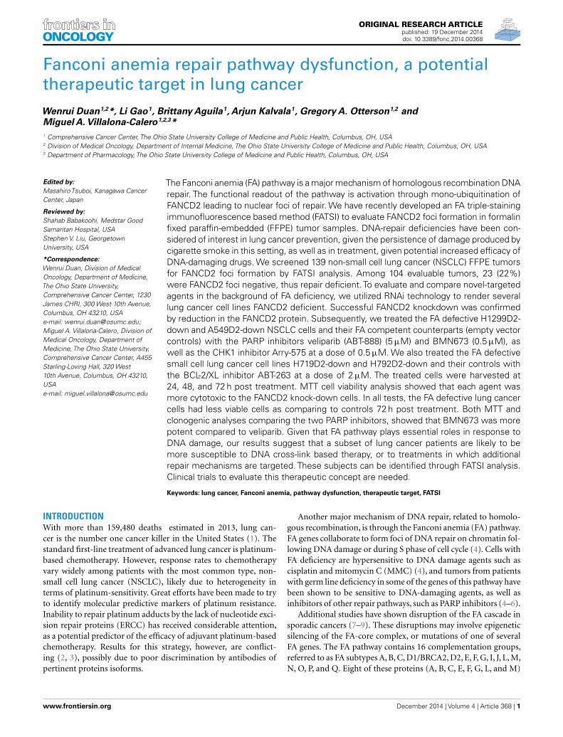
RESULTSFANCONI ANEMIA PATHWAY DEFICIENCY IN NON-SMALL CELL LUNGCANCER TUMOR SAMPLESWe used the FATSI method to evaluate FANCD2 foci formationor lack thereof in lung cancer samples. We screened a total of 139NSCLC FFPE tumors; 104 were evaluable for FANCD2 foci status(Figure 1). Eighty-one of the 104 (78%) evaluable tumors werefound FANCD2 foci positive and 23 (22%) were foci negative.
Forty-nine of the NSCLC samples were of adenocarcinomahistology by morphology examination, 46 were squamous, 5 largecell, and 4 of mixed histology. Thirteen (26.5%) adenocarcinomasand seven (15.2%) squamous cell were foci negative. Two of thefive large cell carcinoma and one of four mixed histology werefoci negative. The frequencies may suggest that adenocarcinomastumors have higher percentage of FANCD2 foci negative tumors ascomparing to squamous cell carcinoma tumors. This observationwill need corroboration with larger sample size and adjustmentwith other confirmatory tests, such as immunohistochemistry, notavailable to us for this dataset.
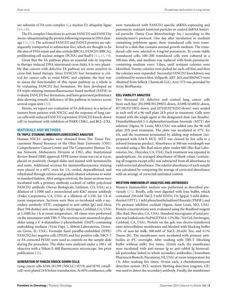
GENERATION OF FANCD2 KNOCK-DOWN CELLS AND EVALUATION OFSENSITIVITY TO PARP INHIBITORSNon-small cell lung cancer cell lines A549, H1299, and smallcell H719, H792 were transduced with FANCD2-specific shRNA-expressing and puromycin-resistant lentiviral particles, or controlshRNA lentiviral particles. To generate stably transduced cells, cellswere selected by puromycin. Successful FANCD2 knock-downcolonies were confirmed by western blot assessment of FANCD2protein. Figure 2A illustrates four lung cancer cell lines withreduced FANCD2 protein. We also evaluated the response of theH1299E (H1299 cell transduced with empty vector) and FANCD2knockdown (H1299D2-down) to treatment with cisplatin at a doseof 5 µg/ml, 72 h post treatment. We found that FANCD2 silencingresulted in sensitization of cells to cisplatin (Figure 2B).
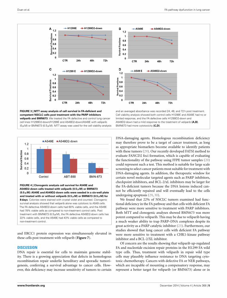
To evaluate the influence of defective FA pathway in regardsto cell viability following exposure to the PARP inhibitors veli-parib (ABT-888) and BMN673, we treated the FA defective NSCLCcell lines H1299D2-down and A549D2-down, as well as their FAcompetent counterparts (H1299E and A549E) (empty vectors)with veliparib at a dose of 5 µM or BMN673 at dose of 0.5 µM.MTT assay was used for the cell viability analysis and an aver-aged absorbance was recorded 24, 48, and 72 h post treatment.Cell viability analysis showed that the FA defective H1299D2-down cells had 80% of viable cells compared to non-treatmentcontrols 72 h post treatment with veliparib. In contrast, there wasno influence on viability of the H1299E cell with the same treat-ment (Figure 3A). Both the A549D2-down cell and A549E cellsresponded to some degree to the treatment with veliparib at 48and 72 h post treatment. The A549D2-down cells had 68% viablecells compared to 83% viable cells for the A549E cells, 72 h posttreatment (Figure 3B).
BMN673 is a new class of PARP inhibitor, which has in addi-tion strong PARP1-DNA complex trapping function (22, 23). Cell
FIGURE 1 | Detection of FANCD2 foci formation in human lung tumorsby the FATSI staining analysis. The paraffin-embedded lung tumor tissuessections were deparaffinized and rehydrated. The tissue sections wereincubated with a primary antibody cocktail of rabbit polyclonal FANCD2antibody (Novus Biologicals, Littleton, CO, USA) at a dilution of 1:1000 and amonoclonal anti-Ki67 mouse antibody (Dako, Carpenteria, CA, USA) at adilution of 1:150 for 1 h at room temperature. Sections then were incubatedwith a secondary antibody cocktail containing FITC conjugated anti-rabbitIgG and Alexafluor 594 donkey anti-mouse secondary for 1 h at roomtemperature. The sections were mounted on glass slides using a 4′
6-diamidino-2-phenylindole (DAPI)-containing embedding medium (VysisDapi 1, Abbott Laboratories, Downers Grove, IL, USA). The slides wereanalyzed under a fluorescence microscope. (A) FANCD2 foci positive NSCLtumor, and (B) FANCD2 foci negative NSCL tumor. Magnification: 1000×.
viability analysis showed that BMN673 was overall a more potentinhibitor (10-fold difference in active doses) compared to veli-parib. H1299 FANCD2 knock-down cancer cells were also moresensitive to BMN673 compared to empty vectors transfected con-trol cells (25 vs. 62% viable cells, respectively) 72 h post treatment(Figure 3C). A549D2-down cell had 29% viable cells and theA549E had 46% viable cells 72 h post treatment (Figure 3D). TheIC50 of BMN673 treated A549E cell was 0.64 µM and the IC50
www.frontiersin.org December 2014 | Volume 4 | Article 368 | 3
Duan et al. FA pathway dysfunction in lung cancer
FIGURE 2 | Creating FANCD2 knock-down cells and evaluatingresponse to cisplatin. (A) NSCLC cells H1299, A549, and small cell lungcancer cells H719, H792 were plated. At 60% confluence, cells weretransduced with FANCD2-specific shRNA-expressing andpuromycin-resistant lentiviral particles or control shRNA lentiviral particles(Santa Cruz Biotechnology Inc.) according to the manufacturer’s protocol.The transduced cells were selected in 4 mg/ml puromycin to create stablytransduced cells with reduced FANCD2 expression. Successful FANCD2knockdown was confirmed by western blot detection of the FANCD2protein. C is a control cell and D is FANCD2 knock-down cell. (B) TheH1299E (H1299 was transfected with empty vector) and FANCD2knock-down (H1299D2-down) lung cancer cells were treated with cisplatin(5 µg/ml) for 24, 48, and 72 h. The knock-down cell was more sensitive tothe treatment.
of A549D2-down was as low as 0.075 µM 72 h post treatment.The difference in the IC50 values between H1299E and H1299D2-down cells is smaller with 1.78 µM for the H1299E and 0.74 µMfor the H1299D2-down.
To further investigate differential response to treatment withPARP veliparib and BMN673 between FA defective and FA intactlung cancer cells, we conducted clonogenic survival analysis.A549D2-down/A549E cells were seeded in a six-well plate andtreated with veliparib (0.5 µM) or BMN673 (0.5 µM). Colonieswere stained with crystal violet and counted. Clonogenic sur-vival analysis showed that veliparib was cytotoxic to the FAdefective A549D2-down cells (60% viable cells as compared tonon-treatment control), and the A549E had 78% viable cells(Figure 4). Following treatment with BMN673 (0.5 µM), the FAdefective A549D2-down cells were 22% viable as compared tonon-treatment control. A549E cells were 43% viable (Figure 4).
EFFECT OF FANCONI ANEMIA REPAIR PATHWAY INTEGRITY ONRESPONSE TO CHECKPOINT INHIBITORSDNA-repair-deficient tumor cells have been shown to accumulatehigh levels of DNA damage. Therefore, the DNA-repair-deficientcells are dependent on other compensatory DNA-repair pathway,such as the CHK1-kinase pathway. FA defective cells are dependenton this G2/M checkpoint for viability, since the checkpoint activa-tion allows for the repair of damaged DNA prior to mitosis. CHK1is activated by the ATR kinase in response to DNA damage that
stalls replication fork progression (24, 25). Defects in FA pathwayhave been shown to be synthetic lethal with CHK1 inhibition orgenetic CHK1 depletion in human fibroblast and ovarian cancercells (24).
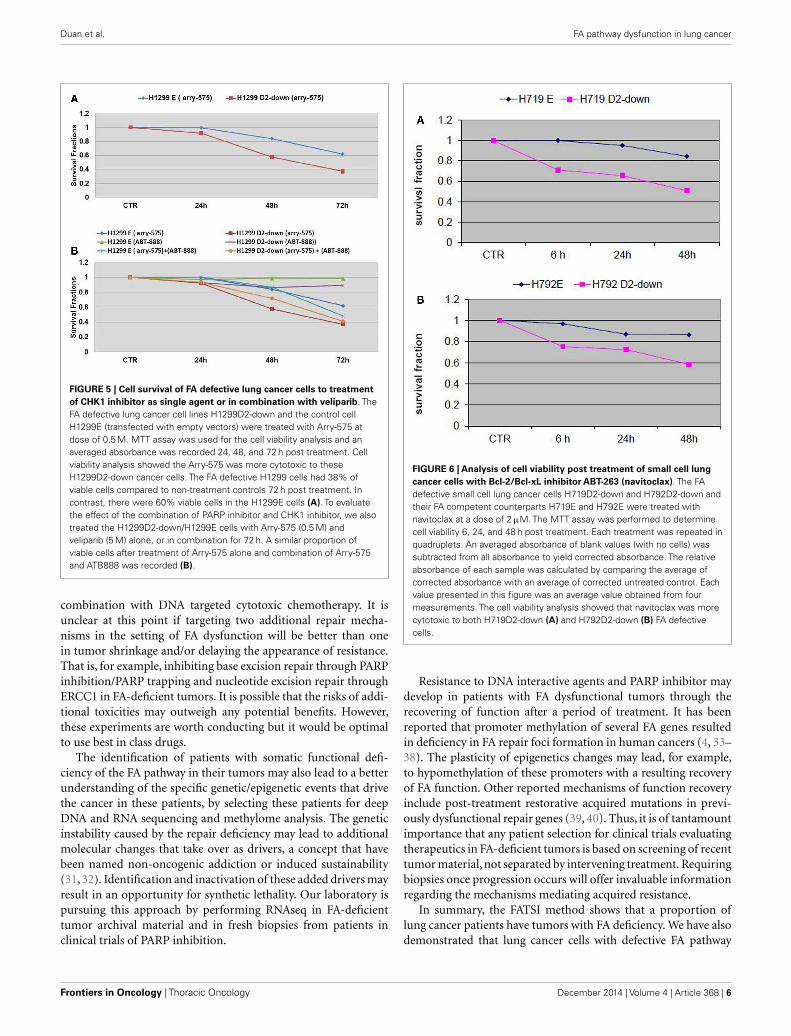
Arry-575 (GDC-0575) is a novel small molecule inhibitor ofCHK1, in FA-deficient lung cancer cells. We conducted a dosagetest on Arry-575 with H1299 cells, and found the IC50 values werearound 1 and 0.5 µM for the H1299E and the H1299D2-downcells 72 h post treatment. We treated the FA defective lung can-cer cell lines H1299D2-down and the control cell H1299E withArry-575 at a dose of 0.5 µM. MTT assay was used for cell viabil-ity analysis and an averaged absorbance was recorded 24, 48, and72 h post treatment. Cell viability analysis showed that Arry-575was more cytotoxic to the H1299D2-down cancer cells. The FAdefective H1299D2-down cells had 38% of viable cells comparedto non-treatment controls 72 h post treatment. In contrast, therewere about 60% viable cells in the control cell line H1299E cells(Figure 5A).
To evaluate potential synergy for the combination of PARPinhibition and CHK1 inhibition, we treated the H1299D2-downand the control cell H1299E with Arry-575 (0.5 µM) and veliparib(5 µM) alone, or in combination for 72 h. MTT assay analysisshowed a similar portion of viable cell between the treatment ofArry-575 alone and the combination (Figure 5B).
RESPONSE OF FANCD2 DEFECTIVE SMALL CELL LUNG CANCER CELLSTO Bcl-2/Bcl-xL INHIBITIONBcl-2 is a central apoptotic inhibitor, and overexpression is associ-ated with tumor progression and treatment resistance in cancers.Overexpression has been reported in up to 80% of small cell lungcancers (SCLC). ABT-263 (navitoclax) is a potent and selectiveinhibitor of Bcl-2 and Bcl-xL, disrupting their interactions withpro-death proteins leading to the initiation of apoptosis (26, 27).However, a recent phase II study of single-agent navitoclax showedlow rate of response to single-agent treatment in advanced andrecurrent SCLC (28). Thus, pre-selection of patients most likelyto derive benefit from BCL-2 inhibitors will be needed for furtherdevelopment of these agents in SCLC.
To evaluate the influence of the FA pathway to treatment withnavitoclax, the FA defective H719D2-down and H792D2-downcells as well as their FA competent counterparts (H719E andH792E) were treated with navitoclax at a dose of 2 µM. The treatedcells were then harvested at 6, 24, and 48 h post treatment. MTTcell viability analysis showed that navitoclax was more cytotoxic tothe FA-deficient H719D2-down compared to its control (51 and85% viable cells at 48 h, respectively) (Figure 6A). Similarly, theH792D2-down small cell lung cancer cells had 58% viable cellsand the H792E had 86% viable, 48 h post treatment (Figure 6B).
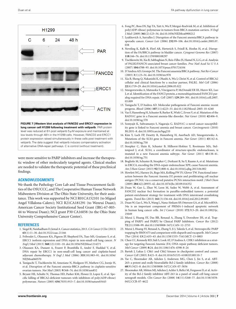
COMPENSATORY ACTIVATION OF ALTERNATIVE DNA-REPAIRPATHWAYS FOLLOWING EXPOSURE TO VELIPARIBWe performed Western immunoblot analysis to evaluate theexpression level of PAR, FancD2, and ERCC1, in the human cancercell lines H1299 following exposure to veliparib. PAR protein levelwas reduced at 6 h post veliparib exposure (5 µM) and maintainedat low levels through 48 h in the H1299 cells. However, FANCD2
Frontiers in Oncology | Thoracic Oncology December 2014 | Volume 4 | Article 368 | 4
Duan et al. FA pathway dysfunction in lung cancer
FIGURE 3 | MTT assay analysis of cell survival in FA-deficient andcompetent NSCLC cells post treatment with the PARP inhibitorsveliparib and BMN673. We treated the FA defective and control lung cancercell lines H1299D2-down/H1299E and A549D2-down/A549E with veliparib(5 µM) or BMN673 (0.5 µM). MTT assay was used for the cell viability analysis
and an averaged absorbance was recorded 24, 48, and 72 h post treatment.Cell viability analysis showed both control cells H1299E and A549E had no orlimited response, and the FA defective cells H1299D2-down andA549D2-down had a mild response to the treatment of veliparib (A,B).BMN673 had more cytotoxicity (C,D).
FIGURE 4 | Clonogenic analysis cell survival for A549E andA549D2-down cells treated with veliparib (0.5 µM) or BMN673(0.5 µM): A549E and A549D2-down cells were seeded in a six-well plateand treated with or without veliparib (0.5 µM) or BMN673 (0.5 µM) for8 days. Colonies were stained with crystal violet and counted. Clonogenicsurvival analysis showed that veliparib alone was cytotoxic to A549 cells.The FA defective A549D2-down cells had 60% viable cells, and the A549Ehad 78% viable cells as compared to non-treatment control cells. Posttreatment with BMN673 (0.5 µM), the FA defective A549D2-down cells has22% viable cells, and the A549E had 43% viable cells as compared tonon-treatment control.
and ERCC1 protein expression was simultaneously elevated inthese cells post treatment with veliparib (Figure 7).
DISCUSSIONDNA repair is essential for cells to maintain genome stabil-ity. There is a growing appreciation that defects in homologousrecombination repair underlie hereditary and sporadic tumori-genesis, conferring a survival advantage to cancer cells. How-ever, this deficiency may increase sensitivity of tumors to certain
DNA-damaging agents. Homologous recombination deficiencymay therefore prove to be a target of cancer treatment, as longas appropriate biomarkers become available to identify patientswith these tumors (29). Our recently developed FATSI method toevaluate FANCD2 foci formation, which is capable of evaluatingthe functionality of the pathway using FFPE tumor samples (20)could represent such a test. This method is suitable for large scalescreening to select cancer patients most suitable for treatment withDNA-damaging agents. In addition, the therapeutic window forcertain novel molecular targeted agents such as PARP inhibitors,checkpoint inhibitors, and BCL-2/xL inhibitors may be larger forthe FA-deficient tumors because the DNA lesions induced can-not be efficiently repaired and will eventually lead to the cellsundergoing apoptosis (29, 30).
We found that 22% of NSCLC tumors examined had func-tional deficiency in the FA pathway and that cells with deficient FApathway were more sensitive to treatment with PARP inhibitors.Both MTT and clonogenic analyses showed BMN673 was morepotent compared to veliparib. This may be due to veliparib havinga much weaker ability to trap PARP–DNA complexes despite itsgreat activity as a PARP catalytic inhibitor (23). Furthermore, ourstudies showed that lung cancer cells with deficient FA pathwaywere more sensitive to treatment with a CHK1-kinase pathwayinhibitor and a BCL-2/XL inhibitor.
Of concern are the results showing that veliparib up-regulatedFA and nucleotide excision repair proteins in the H1299 FA wildtype cells. Thus, treatment with veliparib in repair wild typecells may plausibly influence resistance to DNA targeting cyto-toxic chemotherapy. Cancers with defective FA or NER pathways,which are incapable of mounting a compensatory response, mayrepresent a better target for veliparib (or BMN673) alone or in
www.frontiersin.org December 2014 | Volume 4 | Article 368 | 5
Duan et al. FA pathway dysfunction in lung cancer
FIGURE 5 | Cell survival of FA defective lung cancer cells to treatmentof CHK1 inhibitor as single agent or in combination with veliparib. TheFA defective lung cancer cell lines H1299D2-down and the control cellH1299E (transfected with empty vectors) were treated with Arry-575 atdose of 0.5 M. MTT assay was used for the cell viability analysis and anaveraged absorbance was recorded 24, 48, and 72 h post treatment. Cellviability analysis showed the Arry-575 was more cytotoxic to theseH1299D2-down cancer cells. The FA defective H1299 cells had 38% ofviable cells compared to non-treatment controls 72 h post treatment. Incontrast, there were 60% viable cells in the H1299E cells (A). To evaluatethe effect of the combination of PARP inhibitor and CHK1 inhibitor, we alsotreated the H1299D2-down/H1299E cells with Arry-575 (0.5 M) andveliparib (5 M) alone, or in combination for 72 h. A similar proportion ofviable cells after treatment of Arry-575 alone and combination of Arry-575and ATB888 was recorded (B).
combination with DNA targeted cytotoxic chemotherapy. It isunclear at this point if targeting two additional repair mecha-nisms in the setting of FA dysfunction will be better than onein tumor shrinkage and/or delaying the appearance of resistance.That is, for example, inhibiting base excision repair through PARPinhibition/PARP trapping and nucleotide excision repair throughERCC1 in FA-deficient tumors. It is possible that the risks of addi-tional toxicities may outweigh any potential benefits. However,these experiments are worth conducting but it would be optimalto use best in class drugs.
The identification of patients with somatic functional defi-ciency of the FA pathway in their tumors may also lead to a betterunderstanding of the specific genetic/epigenetic events that drivethe cancer in these patients, by selecting these patients for deepDNA and RNA sequencing and methylome analysis. The geneticinstability caused by the repair deficiency may lead to additionalmolecular changes that take over as drivers, a concept that havebeen named non-oncogenic addiction or induced sustainability(31, 32). Identification and inactivation of these added drivers mayresult in an opportunity for synthetic lethality. Our laboratory ispursuing this approach by performing RNAseq in FA-deficienttumor archival material and in fresh biopsies from patients inclinical trials of PARP inhibition.
FIGURE 6 | Analysis of cell viability post treatment of small cell lungcancer cells with Bcl-2/Bcl-xL inhibitor ABT-263 (navitoclax). The FAdefective small cell lung cancer cells H719D2-down and H792D2-down andtheir FA competent counterparts H719E and H792E were treated withnavitoclax at a dose of 2 µM. The MTT assay was performed to determinecell viability 6, 24, and 48 h post treatment. Each treatment was repeated inquadruplets. An averaged absorbance of blank values (with no cells) wassubtracted from all absorbance to yield corrected absorbance. The relativeabsorbance of each sample was calculated by comparing the average ofcorrected absorbance with an average of corrected untreated control. Eachvalue presented in this figure was an average value obtained from fourmeasurements. The cell viability analysis showed that navitoclax was morecytotoxic to both H719D2-down (A) and H792D2-down (B) FA defectivecells.
Resistance to DNA interactive agents and PARP inhibitor maydevelop in patients with FA dysfunctional tumors through therecovering of function after a period of treatment. It has beenreported that promoter methylation of several FA genes resultedin deficiency in FA repair foci formation in human cancers (4, 33–38). The plasticity of epigenetics changes may lead, for example,to hypomethylation of these promoters with a resulting recoveryof FA function. Other reported mechanisms of function recoveryinclude post-treatment restorative acquired mutations in previ-ously dysfunctional repair genes (39, 40). Thus, it is of tantamountimportance that any patient selection for clinical trials evaluatingtherapeutics in FA-deficient tumors is based on screening of recenttumor material, not separated by intervening treatment. Requiringbiopsies once progression occurs will offer invaluable informationregarding the mechanisms mediating acquired resistance.
In summary, the FATSI method shows that a proportion oflung cancer patients have tumors with FA deficiency. We have alsodemonstrated that lung cancer cells with defective FA pathway
Frontiers in Oncology | Thoracic Oncology December 2014 | Volume 4 | Article 368 | 6
Duan et al. FA pathway dysfunction in lung cancer
FIGURE 7 | Western blot analysis of FANCD2 and ERCC1 expression inlung cancer cell H1299 following treatment with veliparib. PAR proteinlevel was reduced at 6 h post veliparib 5 µM exposure and maintained atlow levels through 48 h in the H1299 cells. However, FANCD2 and ERCC1protein expression raised simultaneously in these cells post treatment withveliparib. The data suggest that veliparib induces compensatory activationof alternative DNA-repair pathways. C is control (without treatment).
were more sensitive to PARP inhibitors and increase the therapeu-tic window of other molecularly targeted agents. Clinical studiesare needed to validate the therapeutic potential of these preclinicalfindings.
ACKNOWLEDGMENTSWe thank the Pathology Core Lab and Tissue Procurement facili-ties of the OSUCCC, and The Cooperative Human Tissue NetworkMidwestern Division at The Ohio State University, for their assis-tance. This work was supported by NCI R01CA152101 (to MiguelAngel Villalona-Calero); NCI R21CA181291 (to Wenrui Duan);American Cancer Society Institutional Seed Grant (IRG-67-003-44 to Wenrui Duan); NCI grant P30 CA16058 (to the Ohio StateUniversity Comprehensive Cancer Center).
REFERENCES1. Siegel R, Naishadham D, Jemal A. Cancer statistics, 2013. CA Cancer J Clin (2013)
63(1):11–30. doi:10.3322/caac.211662. Friboulet L, Olaussen KA, Pignon JP, Shepherd FA, Tsao MS, Graziano S, et al.
ERCC1 isoform expression and DNA repair in non-small-cell lung cancer. NEngl J Med (2013) 368(12):1101–10. doi:10.1056/NEJMoa1214271
3. Olaussen KA, Dunant A, Fouret P, Brambilla E, André F, Haddad V, et al.DNA repair by ERCC1 in non-small-cell lung cancer and cisplatin-basedadjuvant chemotherapy. N Engl J Med (2006) 355(10):983–91. doi:10.1056/NEJMoa060570
4. Taniguchi T, Tischkowitz M, Ameziane N, Hodgson SV, Mathew CG, Joenje H,et al. Disruption of the Fanconi anemia-BRCA pathway in cisplatin-sensitiveovarian tumors. Nat Med (2003) 9:568–74. doi:10.1038/nm852
5. Bryant HE, Schultz N, Thomas HD, Parker KM, Flower D, Lopez E, et al. Spe-cific killing of BRCA2-deficient tumours with inhibitors of poly(ADP-ribose)polymerase. Nature (2005) 434(7035):913–7. doi:10.1038/nature03443
6. Fong PC, Boss DS, Yap TA, Tutt A, Wu P, Mergui-Roelvink M, et al. Inhibition ofpoly(ADP-ribose) polymerase in tumors from BRCA mutation carriers. N EnglJ Med (2009) 361(2):123–34. doi:10.1056/NEJMoa0900212
7. Lyakhovich A, Surralles J. Disruption of the Fanconi anemia/BRCA pathway insporadic cancer. Cancer Lett (2006) 232:99–106. doi:10.1016/j.canlet.2005.07.038
8. Neveling K, Kalb R, Florl AR, Herterich S, Friedl R, Hoehn H, et al. Disrup-tion of the FA/BRCA pathway in bladder cancer. Cytogenet Genome Res (2007)118:166–76. doi:10.1159/000108297
9. Tischkowitz M, Xia B, Sabbaghian N, Reis-Filho JS, Hamel N, Li G, et al. Analysisof PALB2/FANCN-associated breast cancer families. Proc Natl Acad Sci U S A(2007) 104:6788–93. doi:10.1073/pnas.0701724104
10. D’Andrea AD, Grompe M. The Fanconi anaemia/BRCA pathway. Nat Rev Cancer(2003) 3(1):23–34. doi:10.1038/nrc970
11. Xia B, Sheng Q, Nakanishi K, Ohashi A, Wu J, Christ N, et al. Control of BRCA2cellular and clinical functions by a nuclear partner, PALB2. Mol Cell (2006)22(6):719–29. doi:10.1016/j.molcel.2006.05.022
12. Smogorzewska A, Matsuoka S, Vinciguerra P, McDonald ER III, Hurov KE, LuoJ, et al. Identification of the FANCI protein, a monoubiquitinated FANCD2 par-alog required for DNA repair. Cell (2007) 129:289–301. doi:10.1016/j.cell.2007.03.009
13. Taniguchi T, D’Andrea AD. Molecular pathogenesis of Fanconi anemia: recentprogress. Blood (2006) 107(11):4223–33. doi:10.1182/blood-2005-10-4240
14. Vaz F, Hanenberg H, Schuster B, Barker K, Wiek C, Erven V, et al. Mutation of theRAD51C gene in a Fanconi anemia-like disorder. Nat Genet (2010) 42:406–9.doi:10.1038/ng.570
15. Somyajit K, Subramanya S, Nagaraju G. RAD51C: a novel cancer susceptibil-ity gene is linked to Fanconi anemia and breast cancer. Carcinogenesis (2010)31:2031–8. doi:10.1093/carcin/bgq210
16. Kim Y, Lach FP, Desetty R, Hanenberg H, Auerbach AD, Smogorzewska A.Mutations of the SLX4 gene in Fanconi anemia. Nat Genet (2011) 43:142–6.doi:10.1038/ng.750
17. Stoepker C, Hain K, Schuster B, Hilhorst-Hofstee Y, Rooimans MA, Stel-tenpool J, et al. SLX4, a coordinator of structure-specific endonucleases, ismutated in a new Fanconi anemia subtype. Nat Genet (2011) 43:138–41.doi:10.1038/ng.751
18. Bogliolo M, Schuster B, Stoepker C, Derkunt B, Su Y, Raams A, et al. Mutationsin ERCC4, encoding the DNA-repair endonuclease XPF, cause Fanconi anemia.Am J Hum Genet (2013) 92(5):800–6. doi:10.1016/j.ajhg.2013.04.002
19. Howlett NG, Harney JA, Rego MA, Kolling FW IV, Glover TW. Functional inter-action between the Fanconi Anemia D2 protein and proliferating cell nuclearantigen (PCNA) via a conserved putative PCNA interaction motif. J Biol Chem(2009) 284(42):28935–42. doi:10.1074/jbc.M109.016352
20. Duan W, Gao L, Zhao W, Leon M, Sadee W, Webb A, et al. Assessment ofFANCD2 nuclear foci formation in paraffin-embedded tumors: a potentialpatient-enrichment strategy for treatment with DNA interstrand crosslinkingagents. Transl Res (2013) 161(3):156–64. doi:10.1016/j.trsl.2012.09.003
21. Duan W, Gao L,Wu X,Wang L, Nana-Sinkam SP, Otterson GA, et al. MicroRNA-34a is an important component of PRIMA-1-induced apoptotic networkin human lung cancer cells. Int J Cancer (2010) 172:313–20. doi:10.1002/ijc.25049
22. Murai J, Huang SY, Das BB, Renaud A, Zhang Y, Doroshow JH, et al. Trap-ping of PARP1 and PARP2 by Clinical PARP Inhibitors. Cancer Res (2012)72(21):5588–99. doi:10.1158/0008-5472.CAN-12-2753
23. Murai J, Huang SY, Renaud A, Zhang Y, Ji J, Takeda S, et al. Stereospecific PARPtrapping by BMN 673 and comparison with olaparib and rucaparib. Mol CancerTher (2014) 13(2):433–43. doi:10.1158/1535-7163.MCT-13-0803
24. Chen CC, Kennedy RD, Sidi S, Look AT, D’Andrea A. CHK1 inhibition as a strat-egy for targeting Fanconi Anemia (FA) DNA repair pathway deficient tumors.Mol Cancer (2009) 8:24. doi:10.1186/1476-4598-8-24
25. Bartek J, Lukas J. Chk1 and Chk2 kinases in checkpoint control and cancer.Cancer Cell (2003) 3:421–9. doi:10.1016/S1535-6108(03)00110-7
26. Tse C, Shoemaker AR, Adickes J, Anderson MG, Chen J, Jin S, et al. ABT-263: a potent and orally bioavailable Bcl-2 family inhibitor. Cancer Res (2008)68(9):3421–8. doi:10.1158/0008-5472.CAN-07-5836
27. Shoemaker AR, Mitten MJ, Adickes J, Ackler S, Refici M, Ferguson D, et al. Activ-ity of the Bcl-2 family inhibitor ABT-263 in a panel of small cell lung cancerxenograft models. Clin Cancer Res (2008) 14(11):3268–77. doi:10.1158/1078-0432.CCR-07-4622
www.frontiersin.org December 2014 | Volume 4 | Article 368 | 7
Duan et al. FA pathway dysfunction in lung cancer
28. Rudin CM, Hann CL, Garon EB, Ribeiro deOliveiraM, Bonomi PD, CamidgeDR, et al. Phase II study of single-agent navitoclax (ABT-263) and biomarkercorrelates in patients with relapsed small cell lung cancer. Clin Cancer Res (2012)18(11):3163–9. doi:10.1158/1078-0432.CCR-11-3090
29. Evers B, Helleday T, Jonkers J. Targeting homologous recombination repairdefects in cancer. Trends Pharmacol Sci (2010) 31(8):372–80. doi:10.1016/j.tips.2010.06.001
30. Helleday T, Petermann E, Lundin C, Hodgson B, Sharma RA. DNA repairpathways as targets for cancer therapy. Nat Rev Cancer (2008) 8(3):193–204.doi:10.1038/nrc2342
31. Tischler J, Lehner B, Frazer AG. Evolutionary plasticity of genetic interactionnetworks. Nat Genet (2008) 40:390–1. doi:10.1038/ng.114
32. Luo J, Somilini NL, Elledge SJ. Principles of cancer therapy: oncogene and non-oncogene addiction. Cell (2009) 136:823–37. doi:10.1016/j.cell.2009.02.024
33. Narayan G, Arias-Pulido H, Nandula SV, Basso K, Sugirtharaj DD, Var-gas H, et al. Promoter hypermethylation of FANCF: disruption of FanconiAnemia-BRCA pathway in cervical cancer. Cancer Res (2004) 64(9):2994–7.doi:10.1158/0008-5472.CAN-04-0245
34. Wang Z, Li M, Lu S, Zhang Y, Wang H. Promoter hypermethylation of FANCFplays an important role in the occurrence of ovarian cancer through dis-rupting Fanconi anemia-BRCA pathway. Cancer Biol Ther (2006) 5(3):256–60.doi:10.4161/cbt.5.3.2380
35. Wei M, Xu J, Dignam J, Nanda R, Sveen L, Fackenthal J, et al. Estrogen recep-tor alpha, BRCA1, and FANCF promoter methylation occur in distinct sub-sets of sporadic breast cancers. Breast Cancer Res Treat (2008) 111(1):113–20.doi:10.1007/s10549-007-9766-6
36. Potapova A, Hoffman AM, Godwin AK, Al-Saleem T, Cairns P. Promoter hyper-methylation of the PALB2 susceptibility gene in inherited and sporadic breastand ovarian cancer. Cancer Res (2008) 68(4):998–1002. doi:10.1158/0008-5472.CAN-07-2418
37. Hess CJ, Ameziane N, Schuurhuis GJ, Errami A, Denkers F, Kaspers GJ, et al.Hypermethylation of the FANCC and FANCL promoter regions in sporadic
acute leukaemia. Cell Oncol (2008) 30(4):299–306. doi:10.3233/CLO-2008-0426
38. Dhillon VS, Shahid M, Husain SA. CpG methylation of the FHIT, FANCF, cyclin-D2, BRCA2 and RUNX3 genes in Granulosa cell tumors (GCTs) of ovarianorigin. Mol Cancer (2004) 3:33. doi:10.1186/1476-4598-3-33
39. Swisher EM, Sakai W, Karlan BY, Wurz K, Urban N, Taniguchi T. Sec-ondary BRCA1 mutations in BRCA1-mutated ovarian carcinomas with plat-inum resistance. Cancer Res (2008) 68(8):2581–6. doi:10.1158/0008-5472.CAN-08-0088
40. Bouwman P, Aly A, Escandell JM, Pieterse M, Bartkova J, van der Gulden H,et al. 53BP1 loss rescues BRCA1 deficiency and is associated with triple-negativeand BRCA-mutated breast cancers. Nat Struct Mol Biol (2010) 17(6):688–95.doi:10.1038/nsmb.1831
Conflict of Interest Statement: The authors declare that the research was conductedin the absence of any commercial or financial relationships that could be construedas a potential conflict of interest.
Received: 22 July 2014; accepted: 04 December 2014; published online: 19 December2014.Citation: Duan W, Gao L, Aguila B, Kalvala A, Otterson GA and Villalona-CaleroMA (2014) Fanconi anemia repair pathway dysfunction, a potential therapeutic targetin lung cancer. Front. Oncol. 4:368. doi: 10.3389/fonc.2014.00368This article was submitted to Thoracic Oncology, a section of the journal Frontiers inOncology.Copyright © 2014 Duan, Gao, Aguila, Kalvala, Otterson and Villalona-Calero. This isan open-access article distributed under the terms of the Creative Commons AttributionLicense (CC BY). The use, distribution or reproduction in other forums is permitted,provided the original author(s) or licensor are credited and that the original publica-tion in this journal is cited, in accordance with accepted academic practice. No use,distribution or reproduction is permitted which does not comply with these terms.
Frontiers in Oncology | Thoracic Oncology December 2014 | Volume 4 | Article 368 | 8